the role of the bmi1-gsk3 pathway in glioblastoma · and other cancers, subpopulations of tumor...
TRANSCRIPT

The Role of the Bmi1-GSK3! pathway in
Glioblastoma
Inauguraldissertation
zur
Erlangung der Würde eines Doktors der Philosophie
vorgelegt der
Philosophish-Naturwissenschaftlichen
Fakultät der Universität Basel
Von
Serdar Korur
von Edirne, Türkei
Basel, 2010

Genehmigt von der Philosophisch-Naturwisseschaftlichen Fakultät
auf Antrag von Prof. Heinrich Reichert, Prof. Adrian Merlo, Prof. Ruth Chiquet-
Ehrismann und Dr. Brian Hemmings.
Basel, den 13. Oktober 2009
Prof. Dr. Eberhard Parlow
Dekan

Table of contents
SUMMARY ....................................................................................................................... 2
1. INTRODUCTION ........................................................................................................ 3
1.1. Cancer...................................................................................................................................................3
1.2. Cancer stem cells..................................................................................................................................5
1.3. General features of brain tumors ..........................................................................................................8
1.4. Glioblastoma ........................................................................................................................................9
1.5. Main pathways affected......................................................................................................................12
1.6. Glycogen synthase kinase 3 pathway.................................................................................................14
1.7. Bmi1 pathway ....................................................................................................................................15
1.8. Combinatorial therapies to overcome therapeutic resistance .............................................................16
2.RESULTS ..................................................................................................................... 18
Part 1: GSK3! regulates differentiation and growth arrest in glioblastoma .........................................18
Part 2: Combination of sublethal concentrations of epidermal growth factor receptor inhibitor and
microtubule stabilizer induces apoptosis of glioblastoma cells................................................................58
Part 3: Histone deacetylase inhibition and blockade of the glycolytic pathway synergistically induce
glioblastoma cell death.................................................................................................................................68
3. FUTURE PERSPECTIVES....................................................................................... 78
REFERENCES................................................................................................................ 86
ABBREVIATIONS....................................................................................................... 103
ACKNOWLEDGEMENTS ......................................................................................... 104

2
Summary
Malignant gliomas remain one of the deadliest of all cancers despite maximal
therapy. They present unique challenges to therapy with a median survival of 12 months.
Simultaneous activation of several growth promoting and anti-apoptotic pathways
represents the basis for the failure of monotherapies against this disease. In order to
efficiently block growth of glioblastoma (GBM) cells, we have applied several
combinatorial approaches. We have found that combination of histone deactylase
inhibitors along with the glycolytic inhibitor 2-deoxyglucose (2DG) efficiently induced
apoptosis in GBM cells. Furthermore, combination of the microtubule inhibitor
patupilone and AEE788 –an inhibitor of EGFR, which is frequently activated in gliomas,
induced apoptosis in GBM cells at doses that as single drugs were not effective. In GBM
and other cancers, subpopulations of tumor cells with stem cell properties that are
believed to constitute a tumor cell reservoir, have been identified. GBM cells frequently
express the progenitor cell markers Nestin and Sox2 and low levels of the differentiation
markers CNPase, GFAP and !-tubulin III. Bmi1 and Glycogen synthase kinase 3 (GSK3)
has been implicated in stem cell maintenance, but how Bmi1 regulates differentiation is
still unknown. We have identified a link between Bmi1 and GSK3 and showed that
blocking GSK3 may be instrumental to reduce the GBM cancer stem cell pool. We found
that the GSK3 inhibitors SB216763 as well as Lithium chloride depleted the cancer stem
cell population in GBM cells and induced tumor cell differentiation, irrespective of the
CD133 status. Cell proliferation and colony formation were markedly reduced in a dose-
dependent manner.
Future work giving a deeper insight into the regulatory mechanisms of the
receptor tyrosine kinases and downstream effectors will help us to identify more specific
targets. Understanding the mechanisms why some targeted therapies work and others fail
will finally bring us to the level that efficient long-term treatment strategies can be
envisaged.

3
1. Introduction
1.1. Cancer
Cancer is the main cause of death after circulatory diseases in western societies,
estimated to be the cause of death in one quarter of the population in the EU
(Niederlaender, 2006). In spite of the advances in the understanding of the molecular
biology of cancer and of the development of novel therapeutics, cancer remains one of
the deadliest of all diseases (Maher et al., 2001). Critical factors are to be identified prior
to the successful introduction of therapeutic interventions.
Cancer can be viewed as the backside of evolution, which maximizes the
probability of an organism to survive in a hostile environment (Maynard Smith and
Szathmáry, 1995). Life originated in an environment with dramatically changing
conditions, caused in part by exposure to toxic chemical compounds and by continuous
ultraviolet and gamma-radiation (Maynard Smith and Szathmáry, 1995; Ridley, 1993). In
order to maintain survival and stability, cells had to repair damage induced by external
forces, endogenous metabolic toxins and reactive oxygen species, formed during normal
metabolism. On the other hand, precise cellular repair systems would not allow genetic
variation of the gene pool and thus, will lead to lack of adaptability (Maynard Smith,
1989). Perfect organisms with a constant gene pool over their lifetime might extinct when
exposed to a different environmental parameter. Optimal organisms are formed by a
trade-off between genetic variability and stability, which includes the risk of acquiring
mutations that can give rise to cancer (Ridley, 1993). Those mutations may result in
formation of cells that ultimately break the most basic rules of the organism and exploit
every possibility of cellular regulatory pathways in order to proliferate indefinitely. The
huge research effort to understand and combat cancer has tremendously increased the
general knowledge in cell biology, as most of the cancer genes discovered play an
important role in pathways regulating DNA repair, cell signaling, cell cycle, programmed
4
cell death and tissue architecture (Alberts, 2002). Normal cells have to receive and
interpret an elaborate set of signals for the good of the organism, and damaged cells must
be sacrificed in order to maintain stability of the organism. Only a few cells that can
evade those protective mechanisms may constitute the candidate cancer initiating cells.
Those cells may further develop through a microevolutionary process governed by
several mutations, each conferring a growth advantage progressively leading to a
selection of ever more aggressive clones (Nowell, 1976). As a result, cancer cells gain the
ability to reproduce without restraint and colonize foreign tissues leading to death of the
organism by eventually causing malfunctioning of a vital organ (Knudson, 2001;
Knudson, 1971; Friend et al., 1986).
A fundamental feature of most cancer cells is that they are genetically unstable and have
high mutation rate caused by impaired DNA repair systems and increased replication
errors paving the way to the microevolutionary selection process. The fact that cancer is a
multistep process is reflected by the requirements needed by a cell to be capable of
cancerous growth (Alberts, 2002; Hanahan and Weinberg, 2000):
1. Insensitivity to extrinsic and intrinsic signals regulating cell proliferation
2. Evasion of apoptosis
3. Ability to overcome replicative senescense and avoid differentiation inducing signals
4. Genetic instability
5. Invasion
6. Survival in foreign sites.
Genetic alterations needed to push normal cells to a cancerous state can be
induced in different ways: i) direct environmental factors (e.g. radiation) ii) genetic
susceptibility to certain environmental factors (e.g. haploinsufficency of a gene involved
in DNA repair) iii) induction by genetic factors (e.g. presence of an oncogenic mutation
in the germline). Environmental factors might directly induce genetic alterations that
target genes involved in the regulation of the cell cycle, survival and genome integrity
(e.g. induction DNA adducts by cigarette smoking). Main environmental factors leading
to cancers are cigarette smoking (Witschi et al., 1995), UV-light (Fisher and Kripke,

5
2002) nuclear accidents, nuclear bombs (Little, 2000), and certain chemicals that
industrial workers are exposed to (e.g. asbestos, benzene, benzidine, vinyl chloride etc.)
(Jameson, 2000). On the other hand, alterations in genes involved in cancer might already
be present in germline causing inherited cancer syndromes. Genetic studies of families
with inherited cancer syndromes have led to the identification of many important genes.
Among those familial diseases, Li-Fraumeni syndrome (p53), Retinoblastoma (Rb),
neurofibromatosis (NF1 and NF2), Breast cancer (BRCA1), Colorectal cancer (APC),
Von-Hippel Lindau syndrome (VHL), Wilms tumor (WT1-4), Xeroderma pigmentosum
(XP genes), Ataxia-telengiectasia (ATM) and Bloom syndrome (BLM) (Fearon, 1997)
represent typical examples. Mutations can also occur in somatic cells, causing sporadic
forms of cancers, which constitutes the majority. Rb (Friend et al., 1986; He et al., 1995),
p16/p14 (Merlo et al., 1995; Labuhn et al., 2001) p27 (Alleyne et al., 1999) and HDM2
(Vogelstein and Kinzler, 2004) are examples of those genes that, when mutated, are able
to equip the cells with a growth advantage and induce the cancerous process.
1.2. Cancer stem cells
What are the normal cells of origin of cancer and why is this question so
important? The cancer-initiating cell could be a normal stem cell, a progenitor cell, or a
differentiated cell. This question was highly debated in recent years after the discovery of
cancer stem cells in leukemia (Lapidot et al., 1994) that was followed by the
identification of cancer stem cells in numerous solid tumors including glioblastoma
(Ignatova et al., 2002; Lochhead et al., 2001; Singh et al., 2003; Singh et al., 2004; Al-
Hajj et al., 2003; Gibbs et al., 2005; O'Brien et al., 2007; Ricci-Vitiani et al., 2007; Xin et
al., 2005; Burger et al., 2005). The failure to eradicate cancer may be as fundamental as a
misidentification of the target. Identification of a defined cell that could function as a
therapeutic target would facilitate development of successful treatment strategies (Figure
1). Conventional non-specific cancer treatments such as chemotherapy and radiotherapy,
which act on all dividing cells, usually fail, and the disease recurs.

6
One of the most typical and interesting features of stem cells is the self-renewal
characteristic that is also found in the cancer cells. Tumors might arise from the
transformation of normal stem cells into cancer cells since they share many genetic and
phenotypic features (Austin and Kimble, 1987; Bhardwaj et al., 2001; Chan et al., 1999;
Ellisen et al., 1991; Gailani and Bale, 1999; Henrique et al., 1997; Korinek et al., 1998;
Polakis, 2000; Varnum-Finney et al., 2000; Wechsler-Reya and Scott, 2001; Wechsler-
Reya and Scott, 1999; Zhang and Kalderon, 2001; Zhu and Watt, 1999; Figure 2). Those
cancer initiating cells are the driving force behind tumor propagation as well as the
critical mediators of both drug- and radiation resistance (Visvader and Lindeman, 2008)
and the reason behind the failure of conventional therapies.
Figure 1: Tumors are maintained and driven by a rare population of cancer cells termed –
cancer stem cells. Conventional therapies may kill tumor cells with limited proliferative
potential but if the cancer stem cells remain viable they will reform the tumor. On the other
hand, cancer stem cell specific therapies may lead to cures by extinguishing renewal potential
of the tumor (Reya et al., 2001).

7
Singh and colleagues were able to identify CD133 (also known as Prominin 1) as
a surface marker of cancer stem cells in brain tumors. As few as 100 of these CD133-
positive cells found to be able to induce tumors in transplantation experiments and
yielded phenocopies of the initial neoplasia (Singh et al., 2003; Singh et al., 2004). The
expression of multi drug resistance proteins (Dean et al., 2005) and efficient DNA repair
mechanisms (Bao et al., 2006) render CD133-positive cells highly resistant to chemo-
and radiotherapeutic regimens. However, CD133 may not be a reliable stem cell marker
for brain tumors as recent studies showed CD133-negative cells that are able to form
tumors in immunocompromised mice. The other hypothesis consists of the concept that
an adult astrocyte can dedifferentiate becoming a cancer cell as shown in an animal
model (Bachoo et al., 2002). If cancer originates from cancer stem cells then any
successful therapy will have to also eradicate this tumor promoting cell population to
prevent recurrence.
Figure 2: Several signaling pathways regulating normal stem cells found to
be deregulated in cancer (Reya et al., 2001)

8
1.3. General features of brain tumors
Any brain tumor having the histological, immunohistochemical and ultrastructural
proof of glial cell differentiation is defined as ”glioma”. Gliomas are classified into
different groups according to their degree of malignancy. The most widely accepted
classification sytem is based on World Health Organization (WHO), which classifies glial
tumors into four basic grades (I-IV astrocytoma) according to the degree of malignancy
defined by histopathological criteria. Grade I gliomas are usually benign, well
circumscribed and seldom progress into more advanced stages, whereas grades II to IV
are malignant and readily infiltrative into the brain parenychma. Survival ranges from 3
to 10 years in low-grade astrocytoma (grade II) from 2 to 5 years in Grade III anaplastic
astrocytomas, and about 1 year in grade IV tumors also known as glioblastomas (GBM)
(Maher et al., 2001). In Switzerland the incidence rate per 100,000 population/year, was
estimated as 3.32 in males and 2.24 in females (Ohgaki et al., 2004).
The blood-brain barrier (BBB) is an important cellular structure that prevents
toxic substances from entering the brain and allows passage of nutrients and small
compounds. On the other hand, it constitutes a major obstacle to the delivery of
pharmacological agents into the tumor tissue, an important problem in the treatment of
brain tumors (Sathornsumetee et al., 2007). The blood-brain barrier is formed by the tight
junctions made by endothelial cells, other vascular cells and astrocytic foot processes and
involves several active efflux transport systems including the prototype member P-
glycoprotein (P-gp) (Pardridge, 2003). Drugs that could have been invaluable for the
treatment of brain tumors either fail to pass the BBB or fail to pass blood-tumor barrier,
which is limited by the fact that tumors have a high interstitial pressure (Boucher et al.,
1997). 100% of large molecular and 98% of small molecular drugs do not cross the BBB.
Another approach to overcome BBB is a bur hole-based drug delivery via intracerebral
catheters (Merlo et al., 1999). Although those methods might efficiently supply drugs
into the tumor bed, they may not readily target metastatic cells as the diffusion of the
drugs to other areas of the brain than the tumor could be limited. A further development
is direct intra-tumoral injection of small peptides that are distributed in the tumor mass

9
prior to resection (Kneifel et al., 2006). Efficient BBB drug targeting strategies can be
built with the knowledge of the endogenous transporters within the brain capillary
endothelium. The development of novel carrier molecules such as conjugating a tumor-
targeting domain to a protein that can bind to molecules expressed on the BBB, and
mediating their entry into tissue are strongly needed.
1.4. Glioblastoma
Glioblastoma (GBM) is the most frequent and most aggressive type of primary
brain tumor in humans, accounting approximately for 50% of all tumors of glial origin
and 20% of all intracranial tumors (Louis et al., 2007). Any disease with prevalence of
less then 50 in 100.000 is classified as an orphan disease. Those diseases have not often
been adopted by the pharmaceutical industry, as the number of patients affected is too
low to make the drug-development cost-effective. Glioblastoma belongs to this class of
diseases with an occurence of about 5-10 per 100,000 persons (Rich et al., 2004).
Malignant gliomas present unique challenges to therapy and remain one of the
deadliest of all cancers with a median survival of 12 months. Even in the most favorable
cases patients die within two years (Deorah et al., 2006). The duration of survival
associated with malignant gliomas has improved only minimally despite tremendous
efforts of therapy and improvement in the understanding of the molecular biology of
cancer and in molecular medicine in the last decades (Rich and Bigner, 2004). Unique
challenges in combating GBM are associated with; i) high vulnerability of the tissue
where the tumor mass resides ii) diffuse invasiveness of tumor cells into the adjacent
brain parenchyma iii) recurrence of the disease by rapid growth of the infiltrating cells
(Merlo, 2003), resulting in very poor prognosis. GBM can manifest as de novo lesion
(primary GBM, >90%) or progress from less undifferentiated low-grade astrocytoma
(secondary GBM) (Ohgaki and Kleihues, 2007). Primary GBM usually develops in older
patients as a highly aggressive and invasive de novo lesion, without any clinical or
histological evidence of a less malignant precursor lesion. Secondary GBM manifest in

10
younger patients and develop through progression from low-grade to high-grade
astrocytoma in a time range of 5 to 10 years. Although there are common pathways
employed, primary and secondary GBMs develop through distinct molecular pathways
(Collins, 1998; Rasheed et al., 1999). Primary GBM manifests with loss of
heterozygosity at 10q (70% of cases), EGFR amplification (36%), p16INK4a deletion
(31%) and PTEN mutations (25%). On the other hand, in secondary GBM etiology TP53
mutations are the most frequent and earliest detectable genetic alterations, already present
in 60% of precursor low-grade astrocytomas. Additionally, primary and secondary
glioblastomas manifests significant differences in their pattern of promoter methylation
and in expression profiles at RNA and protein levels. (Ohgaki and Kleihues, 2007; Maher
et al., 2001; Wechsler-Reya and Scott, 2001; Zhu and Parada, 2002; Figure 3). Recently,
in a cancer genome-sequencing project, the IDH1 gene was identified as a gene, which is
somatically mutated predominantly in secondary glioblastomas (Parsons et al., 2008). It
was later found that IDH1 mutations are a strong predictor of better prognosis and a
highly selective molecular marker to distinguish primary glioblastomas from secondary
glioblastomas that complements clinical findings (Nobusawa et al., 2009). The IDH1
gene which encodes isocitrate dehydrogenase (IDH) 1 catalyzes the oxidative
carboxylation of isocitrate to "-ketoglutarate, resulting in the production of NADPH in
the Krebs cycle (Devlin, 2006). IDH1 mutations dominantly inhibit the function of the
enzyme through the production of catalytically inactive heterodimers (Zhao et al., 2009).
Further studies will provide molecular explanations for the role of IDH1 mutations in
GBM.
It is predictable that the mutations occurring in the precursor cancer cell in
primary GBM creates a much more unstable genetic background that facilitates further
mutations which accelerates tumor growth by selection of more malignant clones
(Nowell, 1976). In secondary GBM, specific founder mutations might cause milder
instability and may require longer time lapse in order to gain further mutations to
progress to a GBM (Ohgaki and Kleihues, 2007).

11
Standard therapy, including complete surgical resection is not successful because
of the infiltrative behavior of the tumor cells, which already invade multiple parts of the
brain during the progression of the disease even long before the time of the diagnosis.
Surgical intervention is usually followed by aggressive chemo- and radio-therapeutic
regimens, which has proven limited efficacy because of i) the expression of multi-drug
resistance proteins (Dean et al., 2005), ii) efficient DNA repair mechanisms of
glioblastoma cells (Bao et al., 2006), iii) serious side effects induced from the therapy.
Identifying novel molecular targets, and therapeutical strategies with improved efficacy
and reduced toxicity, are strongly demanded.
Figure 3: Genetic pathways to primary and secondary glioblastomas
(Ohgaki and Kleihues, 2007).

12
1.5. Main pathways affected
Key cellular pathways, controlling apoptosis, cell cycle arrest, proliferation,
survival and DNA repair are the most frequently disrupted pathways in GBM due to
alterations in TP53, p16/p14, RB, PTEN, EGFR and PDGFR genes (Figure 4).
EGFR/PTEN/PKB pathway
Epidermal growth receptor (EGFR) is a key protein involved in the development
of primary GBM (Kita et al., 2007), its overexpression occurs in about 60% of primary
GBMs but rarely in secondary GBM (ca. 10%) (Dropcho and Soong, 1996). The most
frequent mutant form is the constitutively active variant 3 (EGFRvIII) with the deletions
of exons 2 to 7 (Huang et al., 1997). Activation of the EGF receptor in turn promotes cell
proliferation in part through the suppression of the p27 gene via the PI3K/PKB pathway
and partly due to the activation of the Ras/MAPK pathway (Narita et al., 2002). PKB
activation due to the constitutive active EGFR results in increased cell proliferation and
cell survival. LOH at chromosome 10 is the most frequent genetic alteration in GBM.
PTEN is located at chromosome 10 and negatively regulates PI3K by
dephosphosphorylating phosphatidyl inositol triphosphate (PIP3). In PTEN mutant cells,
PKB is hyperphosphorylated by PI3K (Maehama and Dixon, 2000) and this leads to
increased proliferation and inhibition of apoptosis.
TP53/HDM2/p14ARF Pathway
The most frequent alteration found in diffuse astrocytoma is on the TP53 gene
(60%). TP53 mutations are also found in primary GBM but at a lower frequency (ca.
30%) and with a different distribution pattern through the gene. In the cases where p53 is
not mutated Hdm2 mutations have been detected (Maher et al., 2001) (less than 10% of
GBM). In addition p14/arf is frequently deleted (76%) in GBM. Disruption of the p53
pathway leads to evasion of apoptosis and allows proliferation of damaged cells.

13
p16INK4a/RB1 Pathway
A hallmark of astrocytomas is the high mitotic activity, a characteristic shared
also by primary and secondary GBM. Homozygous deletions of p16 is also frequently
detected, it is deleted in 31% in primary GBM and 19% secondary GBM. Promoter
methylation of RB1 gene occur 43% of secondary and in 14% of primary GBM (Ohgaki
and Kleihues, 2007; Labuhn et al., 2001). RB1 and p16 tumor suppressor proteins control
the progression through G1 to S phase of the cell cycle. Therefore inactivation of this
pathway allows G1/S phase progression leading to high mitotic activity.
Despite many efforts, median survival of GBM patients has not improved more
than a few months (Rich and Bigner, 2004). Thus, the development of specific bioactive
molecules that selectively target and inhibit tumor initiation and propagation capacity of
brain tumor stem cells might allow reduction or elimination of tumor establishment,
growth and recurrence (Reya et al., 2001). The pursuit of novel agents that fulfill these
criteria will allow a big leap towards successful treatment of brain tumor patients.

14
Figure 4: Main signaling pathways activated by growth factors. PI3K pathway and the MAP kinase
pathway are indicated. Designed by Emmanuel Traunecker ([email protected])
1.6. Glycogen synthase kinase 3 pathway
Glycogen synthase kinase 3 serine/threonine kinase was first identified as an
enzyme phosphorylating and inactivating glycogen synthase (Doble and Woodgett,
2003). Far behind its role in glycogen metabolism, further studies showed that GSK3 is a
key protein in the regulation of numerous signaling pathways. It was shown to be
inhibited in response to insulin signaling from PKB (Cross et al., 1995). It integrates
several signaling pathways and regulates many aspects of cell behavior such as cell cycle,
proliferation, differentiation and apoptosis (Cohen and Goedert, 2004; Doble and
Woodgett, 2003). Two mammalian GSK3 isoforms are known: GSK3" and GSK3!.

15
Knocking out the GSK3! isoform in mice is embryonically lethal due to massive liver
degeneration. The presence of the normal " isoform in the GSK3! #$ animals is not
able to rescue the phenotype (Hoeflich et al., 2000) indicating that at least some of the
functions of the two isoforms are not redundant. The two isoforms share 97% sequence
similarity within their kinase domains, but differ significantly outside this region, with
GSK3" containing an extended N-terminal glycine-rich tail (Frame and Cohen, 2001).
Controversial findings have been reported regarding the influence of GSK3 on the
induction of apoptosis. GSK3 has been shown to act as a pro-survival factor in pancreatic
cancer (Ougolkov et al., 2005) and as a proapoptotic factor in colorectal cancer (Tan et
al., 2005). These opposite findings indicate that the biological function of GSK3 depends
upon cellular context and microenvironment. Consequent to its key functions GSK3 is
involved in the etiology of several diseases such as Alzheimer’s disease (Ryder et al.,
2003), diabetes (Cline et al., 2002), bipolar disorder (Gould and Manji, 2002), and
recently cancer (Wang et al., 2008b).
Pathways regulating normal stem cell behavior are also utilized by cancer cells.
GSK3 is involved in the regulation of Wnt (Miller and Moon, 1996; Yost et al., 1996);
(Polakis, 2000), Shh (Jia et al., 2002) and Notch pathways (Foltz et al., 2002) which are
important for embryonic cell fate determination and normal stem cell maintenance.
Therefore we decided to investigate its role in brain tumor cell identity and maintenance
of the cancer stem cell pool.
1.7. Bmi1 pathway
In the recent years it became evident that cancer is not only a disease due to
genetic mutations but also epigenetic changes play a crucial role influencing malignant
transition (Jones and Baylin, 2002). Maintenance of chromatin structure is essential for
appropriate gene expression and every perturbation of the epigenetic regulations can lead
to inappropriate gene expression and genomic instability, driving normal cells into a
cancerous state. Polycomb group proteins are epigenetic gene silencers implicated in

16
neoplastic transformation. Bmi1, a member of the polycomb group (PcG) proteins is
involved in brain development (Leung et al., 2004). PcG proteins maintain embryonic
and adult stem cells by forming multiprotein complexes that function as transcriptional
repressors (Park et al., 2003; Zencak et al., 2005; Liu et al., 2006). Bmi1 was first
indentified as an oncogene due to its cooperation with Myc in lymphoma formation
(Jacobs et al., 1999b). It has been shown to block senescence in immortalized mouse
embryonic fibroblast through the repression of INK4A/Arf (Jacobs et al., 1999a) and it is
amplified and/or overexpressed in non small cell lung cancer (Vonlanthen et al., 2001),
colorectal carcinoma (Kim et al., 2006), medulloblastoma (Leung et al., 2004),
lymphoma (Haupt et al., 1993), multiple myeloma (Matsui et al., 2004) and primary
neuroblastoma (Nowak et al., 2006). Bmi1 regulates the Ink4a/Arf-locus that is a
frequent target for homozygous deletions in glioblastoma. Whether Bmi1 is expressed in
GBM is being debated (Leung et al., 2004) and its role in GBM is not well delineated. In
a mouse glioma model, Bmi1 had been implicated in brain tumorigenesis in an
Ink4a/Arf-independent manner (Bruggeman et al., 2007). In addition, it was recently
shown that inhibition of Bmi1 by micro RNA-128 attenuates glioma cell proliferation and
self renewal (Godlewski et al., 2008).
1.8. Combinatorial therapies to overcome therapeutic resistance
The glucose analog 2-deoxy glucose (2-DG) is a competitive inhibitor of glucose
uptake and metabolism. Once entering the cells 2-DG is metabolized by the hexokinase
to 2-deoxy glucose-6-phosphate (2-DG-6-P) which is not a substrate for glucose-6-
phosphate dehydrogenase or phosphohexoisomerase (Wick et al., 1957), therefore cannot
be further metabolized and accumulate in the cell until dephosphorylation by
phosphatases. GBM cells are highly proliferative they rely on high sources of energy. A
cardinal feature of glioblastoma cells is increased glucose uptake aided by high levels of
hexokinase and glucose transporters. Most glioblastoma cells maintains a huge part of
their energy supply from the glycolytic pathway as this pathways leads to faster
production of ATP when compared to oxidative phosphorylation and in order to maintain
their high growth and proliferation rates. Cancer cells can overcome drug effects by

17
turning on and off different genes to adapt changes in the environment, which requires
ATP. Thus, blocking cells’ energy production machinery in combination with another
cytotoxic drug might sensitize the effects of the therapeutics by not giving opportunity to
turn on and off redundant pathways. Tertiary structure of chromatin is a crucial factor in
determining whether a particular gene is expressed or not. The accessibility of DNA
wrapped around nucleosomes determines how efficiently a gene can be transcribed. This
is regulated in part by a series of chromatin modifying enzymes such as histone
acetlytransferases (HATs) and histone deactylases (HDAs). The alterations in chromatin
structure by mutations or aberrant transcription of genes involved in the control of
histone modifications are key events in cancer initiation. Changes in chromatin structure
might lead to silencing of tumor suppressor genes and activation of oncogenes leading to
carcinogenic progression. Various structurally diverse compounds (such as TSA, SAHA,
trapoxin A, Laq842, sodium butyrate) are available which can bind to histone deactylases
(HDACs) and induce histone acetylation and consequent reactivation of 2-10% of all
genes (Mariadason et al., 2000; Egler et al., 2008).
EGFR, activation is one of the most frequent alterations in primary GBM,
resulting in simultaneous activation of PKB and RAS pathways (Barker et al., 2001). The
failure to induce efficient cell death in GBM suggested additional crosstalk between
downstream pathways. Bioactive compounds such as PKI-166 or AEE788 with EGFR
protein kinase inhibitory (PKI) activity have been designed and found to have a cytostatic
effect in vitro on tumor cells that overexpress EGFR (Lane et al., 2001; Traxler et al.,
2004). In addition, the EGFR PKI imatinib (gefitinib) allowed tumor growth control in
10% of patients with non–small cell lung cancer who carried specific mutations in the
tyrosine kinase domain (Lynch et al., 2004). Several small molecular weight compounds,
of tyrosine kinase inhibitors such as Gleevec or erlotinib/gefinitib, when applied as
monotherapies, only resulted in limited efficacy in the treatment of GBM (Wen et al.,
2006). Highly mutator phenotype of GBM cells enabled them to gain alterations in
several growth promoting pathways and there is an obvious cross talk between several
signaling pathways. In conclusion, these findings supported the hypothesis that in order
to efficiently induce cell death in GBM cells, combination of two or more drugs is
required (Failly et al., 2007).

18
2.Results
Part 1: GSK3! regulates differentiation and growth arrest in
glioblastoma
Serdar Korur, Roland M. Huber, Balasubramanian Sivasankaran,
Michael Petrich, Pier Jr. Morin, Brian A. Hemmings, Adrian Merlo
and Maria Maddalena Lino.
PLoS One. 2009 Oct 13;4(10):e7443

19
GSK3! Regulates Differentiation and Growth Arrest in
Glioblastoma
Serdar Korur1, Roland M. Huber
1, Balasubramanian Sivasankaran
1, Michael Petrich
1,
Pier Morin Jr2, Brian A. Hemmings
2, Adrian Merlo
1* and Maria Maddalena Lino
1
1: Laboratory of Molecular Neuro-Oncology, University Hospital Basel, Hebelstrasse 20,
CH-4031 Basel, Switzerland
2: Friedrich Miescher Institute for Biomedical Research, Maulbeerstrasse 66, CH-4058
Basel, Switzerland
* Proofs should be addressed to Adrian Merlo
Tel.: +41 61 265 93 11; Fax: +41 61 265 90 60; E-mail: [email protected]
Running title: GSK3! regulates differentiation and growth arrest in glioblastoma
Key words: Glioblastoma, cancer stem cells, glycogen synthase kinase 3!, Bmi1,
differentiation therapy

!"#$%&'$((
!"#$%&'("&%()&*+%#(,-("(./.01"2*/#(/3($%11'(4*25(25%('2%6($%11(.&/.%&2*%'(/3('%137&%#%4"1(
"#)(0#1*6*2%)(8&/4259(:'("('0,./.01"2*/#(4*25*#(25%(206/&(6"'';(25%'%($%11'("&%(,%1*%+%)(
2/($/#'2*202%("(206/&($%11(&%'%&+/*&9(<"254"-'($/#2&/11*#8(25%(&%#%4"1(/3(#/&6"1('2%6($%11'(
"&%()%&%801"2%)(*#($"#$%&9(=5%(./1-$/6,(8&/0.(8%#%(>6*?;(45*$5(*'(&%@0*&%)(3/&(#%0&"1(
'2%6($%11('%137&%#%4"1("#)("1'/($/#2&/1'("#2*7/A*)"#2()%3%#'%(*#(#%0&/#';(*'(0.&%801"2%)(*#(
'%+%&"1($"#$%&';(*#$10)*#8(6%)011/,1"'2/6"9(B%(5"+%(3/0#)(25"2(>6*?(*'($/#'*'2%#21-("#)(
5*851-(%A.&%''%)(*#(C>D9(E/4#&%801"2*/#(/3(>6*?(,-('5FG:'(*#)0$%)("()*33%&%#2*"2*/#(
.5%#/2-.%("#)(&%)0$%)(%A.&%''*/#(/3(25%('2%6($%11(6"&H%&'(I/AJ("#)(G%'2*#9(
K#2%&%'2*#81-;(%A.&%''*/#(/3(81-$/8%#('-#25"'%(H*#"'%(L(,%2"(MCINL!O;(45*$5(4"'(3/0#)(
2/(,%($/#'*'2%#21-(%A.&%''%)(*#(.&*6"&-(C>D;("1'/()%$1*#%)9(=5*'('088%'2'("(30#$2*/#"1(
1*#H(,%24%%#(>6*?("#)(CINL!9(K#2%&3%&%#$%(4*25(CINL!("$2*+*2-(,-('*FG:;(25%('.%$*3*$(
*#5*,*2/&(I>J?PQPL;(/&(1*25*06($51/&*)%(MR*!1O(*#)0$%)(206/&($%11()*33%&%#2*"2*/#9(K#(
"))*2*/#;(206/&($%11("./.2/'*'(4"'(%#5"#$%);(25%(3/&6"2*/#(/3(#%0&/'.5%&%'(4"'(
*6."*&%);("#)($1/#/8%#*$*2-(&%)0$%)(*#("()/'%7)%.%#)%#2(6"##%&9(C>D($%11(1*#%'($/#'*'2(
6"*#1-(/3(!E?LL7#%8"2*+%(M!E?LL7O($%11'9(K#2%&%'2*#81-;(ex vivo($%11'(3&/6(.&*6"&-(
206/&(,*/.'*%'("11/4%)(25%(*)%#2*3*$"2*/#(/3("(!E?LL7('0,./.01"2*/#(/3($%11'(25"2(%A.&%''(
'2%6($%11(6"&H%&'("#)("&%()%.1%2%)(,-(*#"$2*+"2*/#(/3(CINL!9(E&08'(25"2(*#5*,*2(CINL;(
*#$10)*#8(25%(.'-$5*"2&*$()&08(R*!1;(6"-()%.1%2%(25%(C>D('2%6($%11(&%'%&+/*&(
*#)%.%#)%#21-(/3(!E?LL('2"20'9(

!
!"#$%&'(#)%"!
"#$#%&!'&()*#'!'(++#'&!&,-&!$-%$#.!'&#/!$#00'!-.#!&,#!).*1*%+!23.$#!4#,*%)!&(/3.*+#%#'*'!
5678!9:6;;!<-0'3!=%3>%!-'!?.3/*%*%!6@!>-'!*)#%&*2*#)!-'!-!'(.2-$#!/-.=#.!32!$-%$#.!'&#/!
$#00'!*%!4.-*%!&(/3.'!5A78!B'!2#>!-'!6CC!9:6;;DE3'*&*1#!<9:6;;F@!$#00'!>#.#!',3>%!&3!
*%)($#!&(/3.'!*%!&.-%'E0-%&-&*3%!#GE#.*/#%&'!+*1*%+!.*'#!&3!-!E,#%3$3EH!32!&,#!*%*&*-0!
%#3E0-'*-!5AI;78!9:6;;F!$#00'I!>,*$,!#GE.#''!/(0&*D).(+!.#'*'&-%$#!-%)!:JB!.#E-*.!
E.3&#*%'!5K7I!-.#!,*+,0H!.#'*'&-%&!&3!$,#/3D!-%)!.-)*-&*3%!&,#.-EH8!L3>#1#.I!'&#/%#''!*'!
%3&!.#'&.*$&#)!&3!&,#!#GE.#''*3%!32!&,#!9:6;;!/-.=#.I!'*%$#!9:6;;D%#+-&*1#!<9:6;;D@!
$#00!E3E(0-&*3%'!>#.#!-0'3!23(%)!&3!4#!&(/3.*+#%*$!5M78!9-%$#.!'&#/!$#00'!,-1#!-0'3!4##%!
)#&#$&#)!*%!+0*340-'&3/-!<NOP@I!&,#!/3'&!/-0*+%-%&!,(/-%!4.-*%!&(/3.I!>*&,!-%!-%%(-0!
*%$*)#%$#!32!;Q!E#.!/*00*3%!-%)!-!/#-%!'(.1*1-0!32!0#''!&,-%!6!H#-.!5QDR78!NOPI!-!,*+,0H!
*%1-'*1#!-%)!E.30*2#.-&*1#!&(/3.I!/-%*2#'&'!*&'#02!-'!-!de novo!0#'*3%!3.!E.3+.#''#'!2.3/!
0#''!(%)*22#.#%&*-&#)!03>D+.-)#!-'&.3$H&3/-8!!
O/*6!*'!-!/#/4#.!32!&,#!E30H$3/4!+.3(E!32!E.3&#*%'!*%1301#)!*%!4.-*%!)#1#03E/#%&!5S78!
?30H$3/4!+.3(E!E.3&#*%'!/-*%&-*%!#/4.H3%*$!-%)!-)(0&!'&#/!$#00'!4H!23./*%+!/(0&*D
E.3&#*%!$3/E0#G#'!&,-&!2(%$&*3%!-'!&.-%'$.*E&*3%!.#E.#''3.'!56CD6T78!O/*6!*'!-0'3!*%1301#)!
*%!$-%$#.!4H!$33E#.-&*3%!>*&,!PH$!*%!0H/E,3/-!23./-&*3%!56R7!-%)!403$=*%+!32!
'#%#'$#%$#!*%!*//3.&-0*U#)!/3('#!#/4.H3%*$!2*4.340-'&'!&,.3(+,!.#E.#''*3%!32!&,#!
V%=K-WB.2D03$('!56S78!V&!*'!-0'3!-/E0*2*#)!-%)W3.!31#.#GE.#''#)!*%!%3%D'/-00D$#00!0(%+!
$-%$#.I!$303.#$&-0!$-.$*%3/-I!%-'3E,-.H%+#-0!$-.$*%3/-I!/#)(00340-'&3/-I!0H/E,3/-I!
/(0&*E0#!/H#03/-!-%)!E.*/-.H!%#(.340-'&3/-!5SI6;I6SDAA78!X,#&,#.!O/*6!*'!#GE.#''#)!

!"#$%&#!'#()"*+),-+'!./#0123#4"#.#5)6'-#7/!)5.#5)8-/9#%5!:#;.'#!5</!(.*-8#!"#
*65)+!7-"-'!'#!"#."#4"=>.?@+AB!"8-<-"8-"*#5.""-+#0CD23#E6+*F-+5)+-9#!*#;.'#'F);"#
+-(-"*/G#*F.*#5!(+)HI@B:CJ#!"F!K!*'#<+)/!A-+.*!)"#."8#'-/AB+-"-;./#!"#7/!)5.#.*#/-.'*#
<.+*!.//G#KG#8);"+-76/.*!"7#%5!:#0C>23#
$/G()7-"#'G"*F.'-#=!".'-#D#L$MNDO9#.#'-+!"-?*F+-)"!"-#=!".'-9#+-76/.*-'#"65-+)6'#
'!7"./!"7#<.*F;.G'#!",)/,-8#!"#(-//#(G(/-#()"*+)/9#<+)/!A-+.*!)"9#8!AA-+-"*!.*!)"#."8#
.<)<*)'!'#0CP9CQ23#RF-#5.55./!."#!')A)+5'#$MND!#."8#$MND!#.+-#A6"(*!)".//G#
!"8-<-"8-"*#.'#$MND!#(."")*#+-'(6-#*F-#-5K+G)"!(.//G#/-*F./#<F-")*G<-#)A#$MND!#LB?BO#
5!(-#0CS23#$MND#F.'#K--"#8-'(+!K-8#.'#.#<+)B'6+,!,./#A.(*)+#!"#<."(+-.*!(#(."(-+#0CJ2#
."8#.'#.#<+)B.<)<*)*!(#A.(*)+#!"#()/)+-(*./#(."(-+#0C12#."8#!'#!"*-+()""-(*-8#;!*F#'-,-+./#
<.*F;.G'#."8#!5</!(.*-8#!"#@/TF-!5-+U'#8!'-.'-#0DV29#8!.K-*-'#0D:29#K!<)/.+#8!')+8-+#0DC29#
."8#5)+-#+-(-"*/G#(."(-+#0DD23##
W-#F.,-#."./GT-8#*F-#+)/-#)A#$MND#!"#5./!7"."*#7/!)5.'#."8#!*'#/!"='#*)#(+!*!(./#
'!7"./!"7#<+)*-!"'3#X);"+-76/.*!)"#)A#%5!:#+-86(-8#$MND!#/-,-/'#."8#!"86(-8#*F-#
8!AA-+-"*!.*!)"#)A#5./!7"."*#7/!./#(-//'3#X!+-(*#!"F!K!*!)"#)A#$MND!#KG#/!*F!65#(F/)+!8-#
LY!Z/O9#M%C:QSQD#."8#'!HI@#8-(+-.'-8#I-'*!"#."8#M)[C#/-,-/'#."8#!"86(-8#*F-#(-//#
8!AA-+-"*!.*!)"#5.+=-+'#ZI\.'-9#7/!./#A!K+!//.+G#.(!8!(#<+)*-!"#L$E@\O#."8#!B*6K6/!"#4443#
4"#.88!*!)"9#Y!Z/#."8#M%C:QSQD#8-</-*-8#(."(-+#'*-5#(-//'#7+);"#.'#F65."#$%&#ex
vivo#(-//#(6/*6+-'9#!"86(-8#8!AA-+-"*!.*!)"#."8#!"F!K!*-8#"-6+)'<F-+-#A)+5.*!)"3#RF6'9#
$MND#5.G#+-<+-'-"*#.#"),-/#*F-+.<-6*!(#*.+7-*#A)+#5./!7"."*#7/!)5.'3#

!"#$%&"'()"*+)!$#,-+(!
."#&$*#()
"#$%&!'($)*+'!%,-(./+0!1&%$!)(-.+/-'!0#&./2!(!/+#&%'#&2.3(*!)&%3+0#&+!4+&+!
.$$+0.(-+*5!1&%6+/!(/0!7+)-!(-!89:;<=!>**!)(-.+/-'!2(?+!-@+.&!4&.--+/!3%/'+/-!1%&!-@+!
/+#&%'#&2.3(*!)&%3+0#&+!(/0!1%&!(/%/5$%#'!'3.+/-.1.3!(/(*5'.'!%1!0.'+('+0!-.''#+!
(33%&0./2!-%!-@+!2#.0+*./+'!%1!-@[email protected]'!<%$$.--++!%1!B('+*C!D4.-6+&*(/0!EAFBBG=!!
)
/$'')01'#1%$)"*+)%$"2$*#()
HIJKLC!HIK9C!HIMKNC!OJPJC!HIMMLC!HIQ:KC!OJQJC!O9PC!BDKMN!(/0!R'S9J!2*.%$(!3+**!
*./+'!4.-@!0+1./+0!2+/+-.3!'-(-#'!%1!TP53, p16/p14!(/0!PTEN!TJQUC!V>WX!
$+0#**%,*('-%$(!(/0!BK:Q!/+#&%,*('-%$(!3+**!*./+'!4+&+!3#*-#&+0!./!A(2*+!$+0.#$!
'#))*+$+/-+0!4.-@!MN!$Y!2*#3%'+C!2*#-($./+C!'-(/0(&0!(/-.,.%-.3'C!(/0!K:Z![<D=!
BDM9P!3+**'!4+&+!3#*-#&+0!./!I+#&%,('(*!$+0.#$!E\/?.-&%2+/G!'#))*+$+/-+0!4.-@!,('.3!
1.,&%,*('-!2&%4-@!1(3-%&!EM:!/2]$*C!\/?.-&%2+/GC!+).0+&$(*!2&%4-@!1(3-%&!EM:!/2]$*C!^_V!
D5'-+$'GC!BMP!EK`G!(/0!IM!'#))*+$+/-!E:=N`G!E\/?.-&%2+/G=!>**!3+**'!4+&+!$(./-(./+0!(-!
JP;<!./!NZ!<WM=!"@+!3+**!*./+'!4+&+!'++0+0!./!S83$!)*(-+'!(-!Na:::8K:a:::!3+**']3$M!(/0!
2&%4/!1%&!MQ!@!)&.%&!-%!-&+(-$+/-=
!"!"#$"%&!"'()*(+(%,$'-./0'1&!',+%&()"2'3$,4'5,#*"6''
'
!"#"$%&'"()*+,"$&*--*%&
7,$'"&#*'#"88'8()"9':;;'#"88!'1"$"'<8&%"2'()'%$(<8(#&%"'()%,'=>?44'/"%$('2(!*"!'#,)%&()()@'
A;'48',3'#B8%B$"'4"2(B4'1(%*'A;C'7DE6'D"88!'1"$"'@$,1)'3,$'A>'2&F!'&%'GHID
'&)2':C'
DJK9'2B$()@'1*(#*'<"$(,2'%*"'4"2(B4'1&!'),%'#*&)@"26'D"88!'1"$"'%*")'3(L"2'1(%*'>C'
3,$4&82"*F2"'()'AL'/ME'&)2'!%&()"2'1(%*
'#$F!%&8'N(,8"%6'
'
./012&3ex vivo4&56##&#,$6&'"()*+,"$&*$7&$68("9*-*#&)67,8)&
7,88,1()@'()3,$4"2'#,)!")%9'&'%B4,$'!&4<8"'#8&!!(3("2'&!'OMP'+&!"2',)'%*"'QRJ'
#$(%"$(&'1&!',+%&()"2'3$,4'&'<&%(")%'B)2"$@,()@'!B$@(#&8'%$"&%4")%'&%'%*"'S)(N"$!(%F'
R,!<(%&89'M&!"89'E1(%T"$8&)26'Q(%*()'A?G'*'&3%"$'!B$@(#&8'$"4,N&89'%*"'!&4<8"'1&!'%$"&%"2'
1(%*'%*"'U"B$&8'0(!!B"'-(!!,#(&%(,)'V(%'WP(8%")F('M(,%"#'O4+RX'&##,$2()@'%,'%*"'
4&)B3&#%B$"$Y!'<$,%,#,86'0B4,$'#"88!'1"$"'#B8%B$"2'()'UMZ'4"2(&6'S)#,&%"2'<8&!%(#'
2(!*"!'1"$"'B!"2'3,$')"B$,!<*"$"'#B8%B$"',3'UMZ'#"88!6'
'
:#*-),7-;$+,<,(8-6-&*$7&+(*$-'65+,"$&
0*"'8")%(N($&8'N"#%,$!'<[VJ6A?<B$,?!#$&4+8"2?!*5U.'W.22@")"X'&)2'<[VJ6A?<B$,?
!*5U.'WE(@4&9'!*A;\A]'CCGGCCTAATACTTTCCAGATTGATCTCGAGATCAATCTGG
AAAGTATTAGGTTTTT9'!*\=G]'CCGGCCAGACCACTACTGAATATAACTCGAGTTATA
TTCAGTAGTGGTCTGGTTTTX'%&$@"%()@'M4(A'1"$"'%$&)!3"#%"2'()%,'RZVK=G'#"88!'
!"#$!%$&'()!%'*+,-.)/-'$01"/)0#'!%$'*,12,#)0#'3*4567/&879:;',0/'$0<$+"*$'*&"!$)0-'
3*5=>?6@6?A;'B-)0#'4,4+>'*&$1)*)!,!)"0C'D%$'1"01$0!&,!)"0'"E')0E$1!)"B-'*,&!)1+$-')0'
!%$'-B*$&0,!,0!'(,-'!)!&,!$/'B-)0#'F$G,'1$++-C'A+)".,'1$++-'($&$'!&,0-/B1$/'()!%'
)0E$1!)"B-'<)&,+'*,&!)1+$-C'@!,H+I'!&,0-E$1!$/'1+"0$-'($&$'-$+$1!$/'()!%'>'µ#J.+'
*B&".I1)0C'K.):'"<$&$L*&$--)"0'(,-'"H!,)0$/'()!%'*KMKN?*B&"',0/'*KMKN'*B&"?
K.):'B-)0#'4,4+>'*&$1)*)!,!)"0'E"&'8'%C'@!,H+I'!&,0-E$1!$/'1+"0$-'($&$'-$+$1!$/'()!%'>'
µ#J.+'*B&".I1)0C'-)OPM'!&,0-E$1!)"0'E"&'A@QR'(,-'*$&E"&.$/'B-)0#'!%$'A@Q?R!J"'
-)OPM'@)#0,+@)+$01$'Q)!'34$++'@)#0,+)0#'D$1%0"+"#I;',11"&/)0#'!"'!%$'.,0BE,1!B&$&S-'
)0-!&B1!)"0-C'4$++-'($&$'!&,0-E$1!$/'B-)0#'!%$'M.,L,'PB1+$"E$1!"&'/$<)1$'3G"0T,;C'4$++-'
!&,0-E$1!$/'()!%'0"0?-*$1)E)1'-)OPM'($&$'B-$/',-','1"0!&"+C'
'
!"#$%&'(()*+,"#-+.$)#%%#/%)
D&,0-($++'.)#&,!)"0',--,I-'($&$'*$&E"&.$/'B-)0#'."/)E)$/'K"I/$0'1%,.H$&'B0)!-'()!%'
*"+I1,&H"0,!$'E)+!$&-'"E'8?µ.'*"&"-)!I'34"-!,&;C'D%$'+"($&'-)/$'"E'!%$'E)+!$&'(,-'1",!$/'
()!%':U'.#J.+'E)H&"0$1!)0'E"&'>'%',!'RVW4C'D%$'H"!!".'1%,.H$&'(,-'E)++$/'()!%'=5N5'
1"0!,)0)0#':UX'Y4@C'4$++-'3:UZ'*$&'($++')0'-$&B.?E&$$'=5N5;'($&$'*+,!$/')0'!%$'B**$&'
1%,.H$&',0/')01BH,!$/'E"&'>Z'%'()!%'"&'()!%"B!'A@QR"')0%)H)!"&-C'ME!$&'&$."<,+'"E'!%$'
&$.,)0)0#'1$++-'E&".'!%$'B**$&'-B&E,1$'"E'!%$'E)+!$&['.)#&,!$/'1$++-',!'!%$'H"!!".'"E'!%$'
E)+!$&'($&$'E)L$/'()!%'RCVX'E"&.,+/$%I/$')0'\K@',0/'-!,)0$/'()!%'UC:X'1&I-!,+'<)"+$!C'
Y"&'$,1%'!&$,!.$0
!"##$%&"'"%&($)"*%&+,)%-.%/012%#3$"*%+4%5677"'%894,(+4+4:%;<%$9*+6=%*9*"83#%$6#7(,"%
>1?1@2%AB%=C%D'+$%EF%GHI2%BH-%C%*+,)+9,)'"+,9#%>?DD@2%59+#"*%(4*%6$"*%"+,)"'%
+=="*+(,"#3%9'%7'9J"4%(,%K;BL!H%/'9,"+4%#3$(,"$%&"'"%'"$9#M"*%94%*"4(,6'+4:%IK-;<%1?1K
E9#3(8'3#(=+*"%:"#$%(4*%,'(4$7"''"*%,9%4+,'98"##6#9$"%="=5'(4"$%>+0#9,%N"#%,'(4$7"'%
$,(8O$2%P4M+,'9:"4@H%D)"%79##9&+4:%E'+=('3%(4,+59*+"$%&"'"%6$"*Q%(4,+K0=+-%>RE$,(,"@2%
(4,+K!K8(,"4+4%(4*%(4,+KS"$,+4%>1(4,(%!'6J%0+9,"8)49#9:3@T%(4,+K!K,656#+4%PPP%(4*%(4,+K
NUV/%>1+:=(@T%(4,+K!S/($"%>!)"=+894@T%(4,+KN1WX!%>!"##%1+:4(#+4:@T%(4,+KS9,8);%
>?"M"#9E="4,(#%1,6*+"$%F35'+*9=(%0(4O@T%(4,+K19.;%>YZ?%$3$,"=$@T%(4,+K!?-XX%
>C+#,"43+%0+9,"8@T%(4,+KVO,%(4*%E)9$E)9KVO,%>1"'K[\X@%>C+##+E9'"2%0+##"'+8(%CV2%R1V@2%
(4,+KE-G]E-[2%(4,+K08#;2%(4,+K^'O%(4*%(4,+KE)9$E)9K^'O%>1(4,(%!'6J%0+9,"8)49#9:32%1(4,(%
!'6J%!V2%R1V@2%(4,+KV8,+4%>1+:=(KV#*'+8)2%1,H%_96+$2%R1V@H%?"89'(,"*%E'9,"+4$%&"'"%
'"M"(#"*%6$+4:%)9'$"'(*+$)%E"'9.+*($"K894`6:(,"*%(4,+K=96$"2%(4,+K'(55+,2%(4,+K'(,%>S"&%
^4:#(4*%0+9#(5$@%9'%(4,+K:9(,%>/+"'8"@%$"894*('3%(4,+59*+"$%(4*%M+$6(#+J"*%53%,)"%
8)"=9#6=+4"$8"48"%*","8,+94%$3$,"=%16E"'1+:4(#%a"$,%/+89%>D)"'=9%18+"4,+7+8@H%
/'9,"+4%5(4*$%&"'"%b6(4,+7+"*%&+,)%P=(:"c%$97,&('"%>),,EQ]]'$5H+479H4+)H:9M]+`]@H%Y"$6#,$%
&"'"%49'=(#+J"*%,9%(8,+4%#"M"#$H%
%
!"##$%&'()*+$,*-$.#&/$01(&2"('1$
!"##%?SV%894,"4,%(4*%(E9E,9$+$%&"'"%(4(#3J"*%53%7#9&%83,9=",'3%>!3V4%V?/%
V4(#3J"'2%0"8O=(4%!96#,"'@%(4*%,)"%'"$6#,$%$,(,+$,+8(##3%"M(#6(,"*%&+,)%16==+,%M[HX%
$97,&('"H%!"##$%&"'"%,'3E$+4+J"*2%7+."*%+4%+8"K89#*%\B<%",)(49#%79'%-%)%(4*%$,(+4"*%&+,)%
AB%d:]=#%E'9E+*+6=%+9*+*"%79'%UV!1%(4(#3$+$H%/"'8"4,%*"(*%8"##$%&($%*","'=+4"*%7'9=%

!"#$%&'%'&!(')$'*$+#,,-$()$-./012$%"3-#4$5#-.,!-$3&#$6(7#)$3-$8#3)$73,.#-$*&'8$!"&##$
()9#%#)9#)!$#:%#&(8#)!-4$;'&$<=2>>$3)3,?-(-@$(-',3!#9$+#,,-$A#&#$,3/#,#9$A(!"$3)!(0
<=2>>$3)!(/'9?$B2C2DE$*'&$2D$8()$3!$F°<@$A3-"#9$A(!"$GHI$3)9$-'&!#9$BJK;LMN$<#,,$
I'&!#&$/?$H=$H('-+(#)+#-E4$
;'&$H&9M$3)3,?-(-@$+#,,-$A#&#$%.,-#9$A(!"$2D$!O$/&'8'9#':?.&(9()#$*'&$P$"$3)9$
%&'+#--#9$A(!"$!"#$QG<0H&9M$R(!$3++'&9()6$!'$!"#$83).*3+!.&#&S-$()-!&.+!(')-$BH=$
G"3&8()6#)E4$;'&$*,.'&#-+#)!$,3/#,()6$A(!"$1;QG@$+#,,-$A#&#$*(:#9$3)9$%#&8#3/(,(T#9$
A(!"$!"#$<?!'*(:U<?!'%#&8$R(!$BH=$G"3&8()6#)E4$G#&8#3/(,(T#9$+#,,-$A#&#$()+./3!#9$
A(!"$3)!(01;QG$B2CPDDE$'&$83!+"()6$(-'!?%#$+')!&',$3)!(/'9?$*'&$>D$8()$')$(+#@$A3-"#9$
!A(+#$3)9$()+./3!#9$A(!"$!"#$+'&&#-%')9()6$-#+')93&?$3)!(/'9?$*'&$>D$8()$')$(+#$3)9$
3)3,?T#9$/?$*,'A$+?!'8#!&?$B<?Q)$Q=G$Q)3,?T#&@$H#+R83)$<'.,!#&E4$
$
!""#$%&'(%&)*"+,(-'.
<#,,-$A#&#$6&'A)$!'$VDW$+')*,.#)+?$3-$3$8')',3?#&$3-$9#-+&(/#9$3/'7#4$<#,,-$A#&#$!"#)$
*(:#9$()$FW$G;Q$()$GHI$*'&$2X$8()$3!$&''8$!#8%#&3!.&#@$A3-"#9$A(!"$GHI$3)9$
()+./3!#9$A(!"$!"#$%&(83&?$3)!(/'9?$'7#&)(6"!$3!$F°<$()$GHI$Y$2W$HIQ$Y$D42W$Z&(!')$
N2DD4$Q*!#&$!"'&'.6"$A3-"()6$A(!"$GHI@$!"#$-#+')93&?$3)!(/'9?$A3-$399#9$*'&$>$"$3!$
&''8$!#8%#&3!.$<#,,-$A#&#$(836#9$/?$+')*'+3,$8(+&'-+'%?4$Q,,$!.8'&$-38%,#-$
3)3,?T#9$A#&#$-!3()#9$A(!"$"#83!':?,()0#'-()4$
/+&-%0--0'.0$01',+,.%2.31+%"0..
HI$-#&(#-$3&#$%&(83&?$!.8'&$!(--.#-$'/!3()#9$*&'8$%3!(#)!-$9(36)'-#9$A(!"$%&(83&?$<KI$

!"#$%&'()*&&+,+-.'*(($%.+/0'!$'!1-'234'0%*.+/0'&5&!-#6'7$%#*)'8%*+/'!+&&"-'"&-.'*&'*'
!-#9)*!-',$%'#+(%$*%%*5':*&'$8!*+/-.',%$#'&*#9)-&'$,'8%*+/'&"%0-%5',$%'/$/;/-$9)*&!+('
.+&-*&-6'<$!*)'=7>',%$#'!:$'/$%#*)'8%*+/&?'@A'BCD'*/.'-+01!'*&!%$(5!$#*'&*#9)-&':*&'
*#9)+,+-.'*/.')*8-)-.'"&+/0'!1-'>,,5#-!%+E'A;(5()-'*#9)+,+(*!+$/'9%$!$($)'*(($%.+/0'!$'
!1-'#*/",*(!"%-%F&'+/&!%"(!+$/&'G>,,5#-!%+EH6'I*#9)-&':-%-'158%+.+J-.'!$'>,,5#-!%+E'
K@LLMA6N'B-/-O1+9&'*/.'&(*//-.'"&+/0'*/'>,,5#-!%+E'B-/-'O1+9'&(*//-%',$))$:+/0'!1-'
#*/",*(!"%-%F&'+/&!%"(!+$/&6'PE9%-&&+$/'M*)"-&':-%-'-&!+#*!-.'"&+/0'!1-'BO;=D>'
+#9)-#-/!*!+$/'+/'!1-'B-/-.*!*'=-,+/-%'Q6@'GB-/-.*!*?'C*&-)?'I:+!J-%)*/.H'9*(R*0-6'
S*!*;#+/+/0'*/.'M+&"*)+J*!+$/':*&'9-%,$%#-.'"&+/0'!1-'B-/-.*!*'>/*)5&!'Q6@'9*(R*0-6'
>))'&*#9)-&':-%-'T"*/!+)-'/$%#*)+J-.'*/.'#-.+*/'&(*)-.'!$'($%%-(!',$%'#+/$%'M*%+*!+$/'+/'
-E9%-&&+$/'.+&!%+8"!+$/6'>))'#+(%$*%%*5'.*!*'%-9$%!-.'+/'!1-'#*/"&(%+9!'*%-'+/'*(($%.*/(-'
:+!1'!1-'DU>DP'0"+.-)+/-&6'

!"#$%&#'
()*+'*#',-"."/0."##"1'*2'3(45',%*6,1"21.,6%*,)7'721'7#&.,89&,)7'
!"#$%&'()(*+)(,,#&-%./,%0((-%)(+&)1(2%#-%,('()/3%2#44()(-1%15"&)%16+(,%#-7352#-8%
"(2533&03/,1&"/%/-2%-(5)&03/,1&"/9%:-%/-%/-/36,#,%&4%!"#$%";<=%/-2%+)&1(#-%
(*+)(,,#&-%#-%>!?%7(33%3#-(,%/-2%+)#"/)6%0)/#-%15"&),@%/33%>!?%7(33%3#-(,%(*+)(,,(2%
.#8.%!"#$%3('(3,@%A#1.%1.(%B<C$D%3#-(%./'#-8%1.(%.#8.(,1%(*+)(,,#&-%7&"+/)/03(%1&%1.(%
)(4()(-7(%3#-(%E=FG%HDI%JK#85)(%$=%/-2%2/1/%-&1%,.&A-L9%:-%+)#"/)6%0)/#-%15"&)%
,/"+3(,@%!"#$%(*+)(,,#&-%A/,%"/)M(2%#-%$NO$D%JPQRL%&4%>!?@%SOT%JT$RL%&4%
&3#8&2(-2)&83#&"/%/-2%COT%JQURL%&4%/,1)&761&"/9%:-%7&-1)/,1@%45336%2#44()(-1#/1(2%-&)"/3%
0)/#-%1#,,5(%./2%-&%!"#$%+)&1(#-%JK#85)(%$!@%VL9%
%
#:!;<#'767*2#&'()*+'1,=2."6$%7&"#'3>?@!'
W&%,1526%1.(%)&3(%&4%!"#$%#-%>!?@%!"#$%(*+)(,,#&-%A/,%M-&7M(2%2&A-%5,#-8%3(-1#'#)/3X
"(2#/1(2%2(3#'()6%&4%,.;<=,9%!"#$%A/,%(44#7#(-136%2&A-)(853/1(2%#-%1.(%Y,NPC@%ZCTC@%
ZPT@%/-2%ZCQC%>!?%7(33%3#-(,%JK#85)(,%U=%/-2%[$=%/-2%2/1/%-&1%,.&A-L%5,#-8%2#44()(-1%
,.;<=%,(\5(-7(,%JK#85)(%[$!L9%[#-7(%1.(%]&367&"0%8)&5+%8(-(%!"#$%#,%#-'&3'(2%#-%1.(%
)(853/1#&-%&4%2('(3&+"(-1%/-2%15"&)#8(-(,#,@%1.(%(44(71%&4%!"#$%2&A-)(853/1#&-%#-%>!?%
7(33,%A/,%,7)((-(2%06%/-/36,#,%&4%+)&1(#-,%#-'&3'(2%#-%M(6%7(3353/)%+/1.A/6,%&4%1.(%7(33%
7673(@%2('(3&+"(-1@%"(1/0&3#,"@%/+&+1&,#,%/-2%8)&A1.@%#-7352#-8%^)M@%=M1@%>[_C!@%+$N%
/-2%+$Q@%!73XU@%7X?67@%<(,1#-%/-2%[&*U9%:-%7&-1)/,1%1&%-&-X-(&+3/,1#7%7(33,%#-%!"#$%
M-&7M&51%"#7(%H$DI@%!"#$%2&A-)(853/1#&-%#-%>!?%7(33,%2#2%-&1%/44(71%:-MQ/O=)4%+)&1(#-%
3('(3,%JK#85)(%U=L9%!"#$%2&A-)(853/1#&-%#-257(2%7(33%2#44()(-1#/1#&-%/,,&7#/1(2%A#1.%
"&)+.&3&8#7/3%7./-8(,%/-2%2(7)(/,(2%(*+)(,,#&-%&4%1.(%,1("%7(33X)(3/1(2%+)&1(#-,%<(,1#-%

!"#$%&'()$!**&+,!"-."/$."#0*1.&"$&2$!"$!314&*-1.*$2!15$."$6787$/9.&+!$*599$9."5)$
#5154+."5#$:-$."*45!35#$95;593$&2$1<5$!314&*-15=3,5*.2.*$+!4>54$?@AB)$!"#$#5*45!35#$
95;593$&2$&9./"#4&*-15=3,5*.2.*$+!4>54$CDB!35$E@./0453$(A=F$!"#$%(GHI$J"$*&"14!31)$
G+.K$&;545',4533.&"$!**&+,!".5#$#5#.22545"1.!1.&"$!3$3<&L"$:-$."*45!35#$D531."$
5',4533.&"$E@./045$%KCHI$J"154531."/9-)$?%M7!$95;593$L545$+!4>5#9-$45#0*5#$E@./0453$
(A$!"#$%KA=GHI$N<.3$4!.35#$1<5$O0531.&"$&2$L<51<54$?%M7!$+5#.!153$1<5$5225*13$
&:354;5#$&"$*599$#.22545"1.!1.&"I$N&$1<.3$5"#)$L5$035#$1<5$3+!99$+&95*0953$P.C9$!"#$
%G(KQ8Q7$!3$L599$!3$3.RDA$1&$."1542545$L.1<$?%M7!$!*1.;.1-$."$?GS$*5993I$
$
!"#$!%&'%()*+(''(,%&-%!./%
N<.41-=1L&$,4.+!4-$10+&4$1.33053$&:1!."5#$24&+$,!1.5"13$#.!/"&35#$L.1<$?GS$L545$
310#.5#$03."/$+.*4&!44!-$!"#$L53154"$:9&1$!"!9-3.3$1&$+5!3045$?%M7!$+RDA$!"#$
,4&15."$95;593I$?%M7!$+RDA$L545$<./<54$."$?GS$!"#$!314&*-1&+!$,!1.5"13$*&+,!45#$
L.1<$1<5$*&"14&9$E@./0453$(T)$@$!"#$%(H$!"#$,4&15."$L!3$2&0"#$1&$:5$5',45335#$."$1<5$
+!U&4.1-$&2$1<5$10+&43$!"!9-V5#I$N<5$<./<$?%M7!$5',4533.&"$."$!314&*-1&+!$L!3$
+&31$,4&:!:9-$#05$1&$/45!154$"5*4&3.3$."$1<5$?GS)$L.1<$."*45!35#$,4&15."$
#5/4!#!1.&"$E@./045$%(GHI$
$
'&0123%4-,%,+563&-,57(,%&-8&9&:&;-%;<%!"#$%&-7+(4'('%,&<<(+(-:&4:&;-%=4+>(+'%
N<5$5225*1$&2$1<5$."<.:.1.&"$&2$?%M7$&"$,4&15."$95;593$&2$,4&/5".1&4$ED531."$!"#$%&'(H$
!"#$#.22545"1.!1.&"$ECDB!35)$?@AB)$!"#$!=10:09."$JJJH$+!4>543$."$?GS$*599$9."53$6787)$
PD7KW)$G%K(X$!"#$PDKY$L!3$!"!9-V5#$03."/$1<5$#40/3$P.C9$!"#$%G(KQ8Q7$&4$3.RDA3I$
!=*!15"."$.3$1!4/515#$2&4$#5/4!#!1.&"$0,&"$,<&3,<&4-9!1.&"$:-$?%M7!"$Z(X[I$G9&*>."/$

!"#$!%&'()(*+)(%,(-./%&+%-00121,-&3+4%+*%!50-&(4346%7'30'%7-/%1/(.%-/%-%)(-.5+1&%*+)%&'(%
(**(0&/%+*%839,%-4.%":;<=>=$%+4%!"#$%-0&3?3&@%AB3C1)(%$:DE%F*&()%>;%'%&)(-&2(4&%73&'%
839,%-4.%":;<=>>$6%G(/&34%H)+&(34%,(?(,%.(0)(-/(.%34%&'(%G(/&345(IH)(//34C%J$>$6%
8G$<K%-4.%8G<L%0(,,%,34(/%AB3C1)(/%$F6%:D6%7'30'%7-/%0+4*3)2(.%M@%
32214+0@&+0'(23/&)@%AB3C1)(%N:DE%G(/&34%-4.%"+I;%7()(%4+&%(IH)(//(.%34%&'(%Oex vivoP%
:"<;Q%!:R%0(,,%,34(E%"+I;%,(?(,/%7()(%)(.10(.%34%8G<L6%J$>$%-4.%8G$<K%1H+4%!"#$%
34'3M3&3+4%AB3C1)(%$F6%:DE%S'(%+,3C+.(4.)+0@&(%/H(03*30%2-)T()%;U6%$U50@0,30%410,(+&3.(%$U5
H'+/H'+.3(/&()-/(%A9GV-/(D%-4.%&'(%4(1)+4-,%2-)T()%!5&1M1,34%WWW%340)(-/(.%34%:"<;Q6%
8G<L%-4.%8G$<K6%AB3C1)(%$F6%:DE%!"#$%34'3M3&3+4%-,/+%340)(-/(.%&'(%H)+&(34%,(?(,/%+*%
&'(%-/&)+0@&30%,34(-C(5/H(03*30%2-)T()%!BFV%34%8G$<K%-4.%:"<;Q%AB3C1)(%NFDE%
X+74)(C1,-&3+4%+*%!"#$%-0&3?3&@%34%8G<L%M@%/3YGF%)(.10(.%G(/&34%-4.%"+I;%H)+&(34%
,(?(,/%AB3C1)(%N9D6%0+4*3)234C%&'(%/H(03*303&@%+*%&'(%34'3M3&+)@%.)1C%":;<=>=$%34%
M,+0T34C%!"#$!E%S'(%(**(0&%+*%&'(%/H(03*30%!"#$%34'3M3&+)%":;<=>=$%7-/%2+)(%
H)+4+140(.%-4.%0+4/3/&(4&%&'-4%&'(%(**(0&%+*%839,6%7'30'%3/%T4+74%&+%&-)C(&%+&'()%
/3C4-,34C%2+,(01,(/%Z;=[E%S'1/%!"#$%34'3M3&3+4%/H(03*30-,,@%.(0)(-/(.%&'(%(IH)(//3+4%+*%
H)+C(43&+)%2-)T()/%AG(/&34%-4.%"+I;D%-4.%34.10(.%&'(%(IH)(//3+4%+*%.3**()(4&3-&3+4%
2-)T()/%A4(1)+4-,%2-)T()%!5&1M1,34%WWW6%+,3C+.(4.)+0@&(5/H(03*30%2-)T()%9GV-/(%-4.%&'(%
-/&)+0@&30%2-)T()%!BFVD%34%-%0(,,%,34(5.(H(4.(4&%2-44()E%
%
!"#$%$&$'"(')(*+,-(./01/&/2(*34(5/112(6$&#(7(2&/8(5/11(2$9"7&:;/(
S'(%H'(4+&@H30%/73&0'%&+7-)./%.3**()(4&3-&3+4%34%!:R%0(,,/%*+,,+734C%34'3M3&3+4%+*%
!"#$%)-3/(/%&'(%\1(/&3+4%+*%7'(&'()%!"#$%-0&3?3&@%)(C1,-&(/%0-40()%/&(2%0(,,%

!"!#$%&'"()*+,-.//0+%(1+,-.//2+3%(345+)&46+34$$)+7%84+944(+14)35'941+'(+:;<+
=>?/?/@2/AB*+C(+%(+%(%$D)')+"E+4'F7&+1'EE454(&+"5'F4('3+:;<+34$$+$'(4)+GHI.J?+HI>.@?+
HI/.K?+L/A/?+HI>>K?+L/M/?+;N.>@+%(1+O)PJ/Q+=/MB+E"5+&74+!54)4(34+"E+,-.//0+34$$)?+
"($D+HI/.K+3"(&%'(41+,-.//0+34$$)?+%&+%!!5"R'6%&4$D+.>S*+T"+&4)&+U74&745+&74+
,-.//0+!"!#$%&'"(+!"))4))41+%+3%(345+)&46+34$$2$'V4+37%5%3&45?+&74+4R!54))'"(+$484$)+"E+
)&46+34$$+6%5V45)+U454+%(%$DW41+'(+&74+,-.//24(5'3741+!"!#$%&'"(*+I4)&'(?+I"&37>+%(1+
;6'.+U454+7'F7$D+4R!54))41+54$%&'84+&"+&74+,-.//2+E5%3&'"(+GX'F#54+@,Q*+:NY/!+%(1+!2
3%&4('(+!5"&4'(+$484$)+U454+%$)"+7'F745+'(+&74+,-.//0+!"!#$%&'"(+&7%(+'(+&74+3"(&5"$+
GX'F#54+@,Q*+C(7'9'&'"(+"E+:NY/+'(+34$$+$'(4+HI/.K+U'&7+4'&745+H',$+"5+N;>.PAP/+
)7"U41+%+)4$43&'84+4EE43&+"(+&74+3%(345+)&46+34$$2$'V4+!"!#$%&'"(*+H',$+%&+.Z+6<+%(1+
N;>.PAP/+%&+>Z+µ<+'(1#341+%+@Z2PZS+14!$4&'"(+"E+,-.//0+34$$)+GX'F#54+@[Q*+T74+
4EE43&)+"E+:NY/+'(7'9'&"5D+15#F)+U454+E"#(1+&"+94+)!43'E'3+'(+&7%&+&74+4!'1456%$+F5"U&7+
E%3&"5+5434!&"5+'(7'9'&"5+G[\\AJJQ+%(1+&74+!2)4354&%)4+'(7'9'&"5+G-[]TQ+1'1+("&+
)'F('E'3%(&$D+%$&45+&74+,-.//0+!"!#$%&'"(+G1%&%+("&+)7"U(Q*+T"+E#5&745+3"()"$'1%&4+&7')+
"9)458%&'"(?+U4+4(5'3741+&74+HI/.K+,-.//0+!"!#$%&'"(+9D+X[,N+)"5&'(F?+"9&%'('(F+%+
,-.//0+34$$+!"!#$%&'"(+"E+%!!5"R'6%&4$D+JZS*+T7')+U%)+6%'(&%'(41+E"5+)4845%$+!%))%F4)+
%(1+&74(+)#9^43&41+&"+'(7'9'&'"(+"E+:NY/+9D+H',$+"5+N;>.PAP/?+U7'37+14!$4&41+&74+
,-.//0+E5%3&'"(+GX'F#54+@;Q*++
C&+7%)+944(+%5F#41+&7%&+:;<+34$$)+F5"U(+E"5+6%(D+!%))%F4)+'(+)&%(1%51+641'#6+1"+("&+
6'55"5+&74+)&46+34$$+3"6!%5&64(&+U'&7'(+&74+"5'F'(%$+"5+=/JB*+T"+4R%6'(4+&7')?+&74+_ex
vivo”+34$$+$'(4+;N>JA+U%)+%(%$DW41+U7'37+7%1+944(+')"$%&41+E5"6+%+E54)7+"5+9'"!)D+
%(1+1'543&$D+F5"U(+%)+(4#5")!7454)+'(+(4#5"9%)%$+641'#6+)#!!$464(&41+U'&7+9X:X+%(1+
\:X?+&7#)+E%8"5'(F+4R!%()'"(+"E+3%(345+)&46+34$$)*+C(&454)&'(F$D?+&74+!"!#$%&'"(+U'&7+%+

!"#$%&#''(')*#%!)+,-"./#%),%"0)!%&#''%'),#%1-!%/#2/#!#,"#3%45%"0#%67899(%-,3%,:"%45%"0#%
67899;%2:2.'-"):,%<=)+./#%>?(6@A%B,'5%"0#%67899(%2:2.'-"):,%#C2/#!!#3%#'#D-"#3%
'#D#'!%:E%F#!"),G%H:CI%-,3%J$)8%-,3%E:/$#3%,#./:!20#/#!%<=)+./#%>JG%6@A%K,0)4)"):,%:E%
LHM9%3#&/#-!#3%"0#%!"#$%&#''%')*#%<67899(@%2:2.'-"):,%<=)+./#%>7@%-,3%-'!:%-'"#/#3%
2/:"#),%'#D#'!%:E%!"#$%&#''%-,3%3)EE#/#,")-"):,%$-/*#/!G%$-),'5%3#&/#-!#3%H:CI%'#D#'!%
<=)+./#%H9@A%K,3.&"):,%:E%3)EE#/#,")-"):,%)$2-)/!%"0#%-4)')"5%:E%2/#&./!:/%&#''!%":%E:/$%
,#./:!20#/#!A%K,0)4)"):,%:E%LHM9%!)+,)E)&-,"'5%/#3.%"0#%,.$4#/%-,3%D:'.$#%:E%
,#./:!20#/#!%),%JHINO%&#''!%<=)+./#%>PG%=@A%Q0#%/#!.'"!%!0:1%"0-"%),0)4)"):,%:E%LHM9%
/#3.&#!%"0#%&-,&#/%!"#$%&#''%2::'%-,3%"0-"%67899%$-5%,:"%4#%-%/#')-4'#%&-,&#/%!"#$%&#''%
$-/*#/A%
%
!"#$%&'(&)&*&+'%,-./0-1%0+2+'3%4+,56*&+'%6'.%&'./0-1%67+7*+1&1%&'%!89%0-221%
Q0#%#EE#&"!%:E%LHM9%),0)4)"):,%:,%&#''%2/:')E#/-"):,%-,3%-2:2":!)!%),%"0#%LJR%&#''%'),#!%
SF8NG%T9O9G%SFI8U%-,3%SF98V%1#/#%-,-'5W#3%.!),+%&:':,5%E:/$-"):,G%/#'-")D#%&#''%
,.$4#/%-,3%&#''%3#-"0%-!%/#-3:."A%K,%-%&:':,5%E:/$-"):,%-!!-5G%4:"0%"0#%,.$4#/%-,3%!)W#%
:E%&:':,)#!%E:/$#3%-E"#/%8X%3-5!%:E%3/.+%"/#-"$#,"%1#/#%$#-!./#3A%LHM9%),0)4)":/!%
!)+,)E)&-,"'5%/#3.%&:':,5%E:/$-"):,%),%-''%LJR%&#''%'),#!%"#!"#3%&:$2-/#3%1)"0%"0#%
.,"/#-"#3%&:,"/:'%<=)+./#%O?@A%Q0#%LHM9%),0)4)":/%&:,&#,"/-"):,!%.!#3%1#/#%),%"0#%,:,(
":C)&%/-,+#Y%&#''%2/:')E#/-"):,%-,3%!./D)D-'%:E%"0#%0.$-,%-3)2:!#%")!!.#(3#/)D#3%2/:+#,)":/%
&#''!%<?888@%1#/#%,:"%,#+-")D#'5%-EE#&"#3%<=)+./#%O?@A%LHM9%),0)4)"):,%45%S)6'%:/%
HJI8>O>9%),3.%-%!')+0"%),&/#-!#%),%&#''%3#-"0%E:/%LJR%&#''%'),#!%SF98VG%SF8NG%T9O9%
-,3%JH8IU%-E"#/%OI%0%<=)+./#%OJ@A%K,3.&"):,%:E%&#''%3#-"0%1-!%!)+,)E)&-,"'5%#'#D-"#3%

!"#$%&'()%*$"*+*,-.%/*01%!23%4-5+*$#6%!*,"%,"#%3,2$62.6%&78%,"#.29#:,*4%
,#5-;-1-5*6#%*$%,"#%/<=>%4#11%1*$#%?@*A:.#%BCDE%0#11%6#2,"%!23%6-3#F6#9#$6#$,%2$6%
G2.*#6%H.-5%4#11%1*$#%,-%4#11%1*$#E%C*.#4,%4#11%4-:$,*$A%2H,#.%#I9-3:.#%-H%4#113%,-%/*01%-.%
'7J=KBK)%H-.%BJ%"%3"-!#6%*$"*+*,*-$%-H%,"#%9.-1*H#.2,*-$%-H%/<=>L%/<)=ML%N)B)%2$6%
/<J=O%4#113%?@*A:.#%B0DE%&JF8%244:5:12,*-$%!23%.#4-.6#6%*$%/<)=ML%/<=>L%N)B)%2$6%
&JF8%2..#3,%*$%/<J=O%?@*A:.#%'PDE%Q$%2%5*A.2,*-$%2332RL%/<)=M%3"-!#6%2%3*A$*H*42$,%
.#6:4,*-$%*$%4#11%5*A.2,*-$%H-11-!*$A%&'()%*$"*+*,*-$%?@*A:.#%BSDE%T"#3#%.#3:1,3%3"-!%
,"2,%&'()%3,.-$A1R%.#6:4#3%4-1-$R%H-.52,*-$%2$6%*$6:4#3%4#11%6#2,"%*$%&78%4#11F1*$#3E%
%
!"#$%##"&'((
75*=L%2%5#5+#.%-H%,"#%9-1R4-5+%A.-:9%9.-,#*$3L%*3%.#U:*.#6%H-.%3#1HF.#$#!21%-H%$#:.21%
3,#5%4#113%2$6%*3%:9.#A:12,#6%*$%3#G#.21%42$4#.3E%Q,%*3%213-%V$-!$%,-%.#9.#33%Q$VP2WX.H%
1-4:3%*$"*+*,*$A%9.-A#$*,-.%4#11%9.-1*H#.2,*-$%6:.*$A%$#:.21%6*HH#.#$,*2,*-$%Y)MZE%Q$%
6*HH#.#$,*2,#6%4#113L%75*=%1#G#13%6#4.#23#%!"*1#%Q$VP2WX.H%9.-,#*$%1#G#13%*$4.#23#%YP[ZE%X3%
,"#%Q$VP2WX.H%1-4:3%*3%H.#U:#$,1R%6#1#,#6%*$%+.2*$%,:5-.3%YP=ZL%,"#%.-1#%-H%75*=%
-G#.#I9.#33*-$%*$%&78%4#113%299#2.3%,-%+#%6*3,*$4,%H.-5%*,3%.#9.#33*-$%-H%,"#%Q$VP2WX.H%
1-4:3E%@-.%#I2591#L%6-!$.#A:12,*-$%-H%75*=%6*6%$-,%*$H1:#$4#%Q$VP2WX.H%9.-,#*$%1#G#13%*$%
,:5-.%4#113%,"2,%.#,2*$#6%,"#%Q$VP2WX.H%1-4:3E%T":3L%*$%&78%4#113L%75*=%,2.A#,3%2%
6*HH#.#$,%92,"!2RE%'4.##$*$A%-H%3#G#.21%V#R%9.-,#*$3%4-$,.-11*$A%4#11%4R41#L%6#G#1-95#$,L%
5#,2+-1*35L%29-9,-3*3%2$6%A.-!,"L%*$41:6*$A%S.VL%XV,L%&'()!%L%9=K%2$6%9=PL%741FJL%4F
8R4L%<#3,*$%2$6%'-IJL%3"-!#6%,"2,%6-!$.#A:12,*-$%-H%75*=%.#6:4#6%&'()!%9.-,#*$%
1#G#13%2$6%*$6:4#6%6*HH#.#$,*2,*-$%*$%42$4#.%4#113E%Q$%266*,*-$L%,:5-.%4#11%9.-1*H#.2,*-$L%

!"#$%$&'()*%+#&,%-.)&./)0'-.-+1.%0%,2)31#1)*)/'2)#1/"01/5))
6%!0-$1#1/)!-*1)78)21&#!)&+-)9:7;()<=>?)@&!)A11.)0-.!%/1#1/)-.'2)#101.,'2)&!)&)
,@1#&B1",%0),&#+1,)C-#)0&.01#)9??;5)D,)@&!)A11.)!@-3.),@&,),@%!)1.E2*1).1+&,%$1'2)#1+"'&,1!)
,@1)F.,()G1/+1@-+)&./)H-,0@)B&,@3&2!()3@%0@))&A1##&.,'2)&0,%$&,1/)%.)!1$1#&')
0&.01#!)9??(:?;5)I@%!)!"++1!,!),@&,)<=>?)%.@%A%,-#!)0-"'/)1J1#,)&),@1#&B1",%0&''2)
.1+&,%$1()B#-K!"#$%$&')1CC10,)-.),"*-#)01''!5)G-31$1#(),@1)'-.+K,1#*)*1/%0&')"!1)-C),@1)
<=>?)%.@%A%,-#)L%M')C-#),@1),#1&,*1.,)-C)B!20@%&,#%0)/%!-#/1#!)/%/).-,)'1&/),-)&.)%.0#1&!1)
%.)0&.01#)%.0%/1.01)9::;()&#+"%.+)&+&%.!,)&.)-.0-+1.%0)1CC10,)-C)<=>?)%.@%A%,-#!5)N.),@1)
0-.,#()M-@1.)et al.)/1*-.!,#&,1/),@&,)0&.01#)B#1$&'1.01)%.)B!20@%&,#%0)B&,%1.,!)-.)
'-.+K,1#*)L%M')*1/%0&,%-.)3&!)'-31#),@&.)%.),@1)+1.1#&')B-B"'&,%-.)9::;()!"++1!,%.+)
1$1.)&)B#-,10,%$1)1CC10,)-C)L%M'5)I@1)#1!"',!)B#1!1.,1/)@1#1)-CC1#)&)*-'10"'&#)1JB'&.&,%-.)
-C),@%!)1B%/1*%-'-+%0&')-A!1#$&,%-.O)&/*%.%!,#&,%-.)-C)L%M')%./"01!)/%CC1#1.,%&,%-.)&./)
%.@%A%,!)B#-'%C1#&,%-.)&./(),@1#1A2()*%+@,)1CC10,%$1'2)%.@%A%,),"*-#)C-#*&,%-.)&./)
B#-+#1!!%-.5)P"#,@1#*-#1(),@1)B'1,@-#&)-C)0'%.%0&')/&,&)-.)L%M')-CC1#)!-'%/)%.C-#*&,%-.)
&A-",)B-,1.,%&')!%/1K1CC10,!)&./)%,)&BB1&#!)!&C1),-)&!!"*1),@&,).-#*&')&/"',)!,1*)01''!))
.-,).1+&,%$1'2)&CC10,1/()1$1.)A2)'-.+K,1#*)"!1)-C),@1)/#"+5)I@1)$1#2)!%*%'&#)[email protected],2B%0)
&./)C".0,%-.&')&',1#&,%-.!)%./"01/)A2)1%,@1#)%.@%A%,%.+)<=>?)-#)A2)/-3.#1+"'&,%.+)Q*%R)
%.),@1)B#1!1.,)!,"/2)B-%.,!),-)&)C".0,%-.&')'%.4)A1,311.)Q*%R)&./)<=>?5)G-31$1#()
C"#,@1#)!,"/%1!)).11/1/),-)&.&'2E1)3@1,@1#),@1#1)%!)&)/%#10,)%.,1#&0,%-.)A1,311.)Q*%R)
&./)<=>?5)6-3.#1+"'&,%.+)<=>?)!B10%C%0&''2)/10#1&!1/),@1)!"AB-B"'&,%-.)-C)0&.01#)
01''!),@&,)0-.,&%.1/)&)0&.01#)!,1*)01''K'%41)!%+.&,"#1)A2)/#%$%.+),@1*)%.,-)/%CC1#1.,%&,%-.5))
=-J7)B#-,1%.)%!)3%/1'2)1JB#1!!1/)%.),@1)1&#'2).1"#&')B'&,1)&./)1&#'2).1"#&'),"A1)-C)!1$1#&')
!B10%1!)9:8;5)D.),@1)/1$1'-B%.+)01.,#&').1#$-"!)!2!,1*()=-J7)1JB#1!!%-.)A10-*1!)

!"#$!%&$"'($)($*"(+",!)"-%$*".%/.(&"..#()0($*"(1"+$!%&,./!(./2"!3(4*%&*(&)+$%+,"($)('%1%'"(
/+'("5*%6%$(/+(%77/$,!"(-*"+)$2-"8(9"..#($*/$(."/1"($*"(1"+$!%&,./!(./2"!(.)#"(:)5;(
"5-!"##%)+(<=>?8(@+$"!"#$%+A.23(:)5;(*/#(/.#)(6""+(%7-.%&/$"'(%+(BCD(<=E3=F?(/#(
')4+!"A,./$%)+()0(:)5;(!"',&"'(&"..(-!).%0"!/$%)+(/+'($,7)!%A"+%&%$2(%+(BCD(&"..#8(
G*"!"0)!"3(:)5;(4/#(-!)-)#"'(/#(/(+"4(BCD($*"!/-",$%&($/!A"$(<=E?8(H$(-!"#"+$3(
%+*%6%$)!#()0(:)5;(/!"(+)$(/1/%./6."(6,$($*"('/$/(-!"#"+$"'(*"!"(#*)4($*/$(%+*%6%$%)+()0(
B:IJ(#$!)+A.2(')4+!"A,./$"#(:)5;(%+(BCD(&"..#8(G*%#(!/%#"#($*"(-)##%6%.%$2($*/$(K%9.()!(
7)!"(#-"&%0%&(B:IJL%+*%6%$)!2('!,A#(&),.'(6"(,#"'($)('"&!"/#"($*"(:)5;L'"-"+'"+$(
$,7)!%A"+%&(-)$"+$%/.()0(BCD(&"..#8(
G4)(7/%+(#$!/$"A%"#(/!"(&,!!"+$.2(6"%+A("5-.)%$"'($)("!/'%&/$"($*"(&/+&"!(#$"7(&"..(M9:9N(
-)).O(i)(&*"7)$*"!/-",$%&(!"A%7"+#($*/$(#-"&%0%&/..2('!%1"(9:9(%+$)(/-)-$)#%#(/+'(
$*"!"62('"-."$"($*"(9:9(!"#"!1)%!()0($*"($,7)!3(/+'(ii)(#$!/$"A%"#(/%7%+A($)('!%1"(9:9(
%+$)('%00"!"+$%/$%)+(/+'($*"!"62(%+&!"/#"($*"%!(#,#&"-$%6%.%$2($)(-!)L/-)-$)$%&($!"/$7"+$#(
<P?8(B%1"+($*"(*%A*('"A!""()0('!,A(!"#%#$/+&"(/+'($*"(#*/!"'(&"..,./!(/+'(A"+"("5-!"##%)+(
-!)0%."#()0(/',.$(/+'(&/+&"!(#$"7(&"..#(<=Q?3($/!A"$%+A(9:9(*/#(-!)1"+($)(6"('%00%&,.$8(
R)4"1"!3(%+',&$%)+()0('%00"!"+$%/$%)+(!"7/%+#(/($*"!/-",$%&(#$!/$"A2(0)!(9:9(/#(S%&&%!%..)(
et al8(#*)4"'($*/$(6)+"(7)!-*)A"+"$%&(-!)$"%+#(&/+(%+',&"('%00"!"+$%/$%)+()0(9TPJJU(
BCD(&"..#3($*"!"62(!"',&%+A($*"%!($,7)!%A"+%&(-)$"+$%/.(<=V?8(R)4"1"!3($*"(,#"()0(
7)!-*)A"+#(6"/!#($*"(!%#W()0(%+$"!0"!%+A(4%$*($*"($%A*$.2(!"A,./$"'(/',.$(#$"7(&"..(+%&*"#8(
H+2(#$!/$"A2($)(%+',&"('%00"!"+$%/$%)+(%+(&/+&"!(#$"7(&"..#(7,#$(6"(&/!"0,..2(/##"##"'(0)!(
/+2(/'1"!#"("00"&$#()+($*"(/',.$(#$"7(&"..(-)-,./$%)+8(
X,!(!"#,.$#(#*)4(B:IJ(%+*%6%$%)+($)(6"(/+(/$$!/&$%1"(#$!/$"A2(0)!(#-"&%0%&/..2($/!A"$%+A(/(
#,6-)-,./$%)+()0(&/+&"!(&"..#(4%$*(#$"7(&"..L.%W"(&*/!/&$"!%#$%(Y5-!"##%)+()0(#$"7(&"..(

!"#$#%&&'('")%!)%*"$+!(,'(-$+*('$!../(!)'01$#'&%"'#$)2'$-/34*4/0!)%*"$*&$.'00-$5%)2%"$
678$.'00$0%"'-$!"#$ex vivo$)/+*($.'00-$)2!"$'94('--%*"$*&$)2'$:;<==$+!(,'(>$
?"!.)%@!)%*"$*&$3*)2$7+%<$!"#$6AB=$#'40')'#$4('./(-*($.'00-$('C/%('#$&*($)/+*($
+!%")'"!".'$!"#$4(*D('--%*">$E2'-'$#!)!$!##$!"*)2'($&!.')$)*$)2'$+!"1$'&&'.)-$*&$6AB=$
!-$!$('D/0!)*($*&$.!".'($.'00$%#'")%)1>$F'('G$6AB=$!.)%@!)%*"$%-$%#'")%&%'#$!-$!$,'1$'0'+'")$
%"$+!%")!%"%"D$-)'+$.'00H0%,'$.2!(!.)'(%-)%.-$%"$!$-/3-')$*&$.!".'($.'00-G$4(*@%#%"D$)2'-'$
.'00-$5%)2$!$2%D2'($-'0&H('"'5!0$.!4!.%)1>$I'.'")01G$#*5"('D/0!)%*"$*&$6AB=$5!-$-2*5"$
)*$%"#/.'$!4*4)*-%-$%"$D0%*+!$.'00-$!"#$)*$2!@'$!"$!")%H+%D(!)*(1$'&&'.)$%"$D0%*+!$
-42('*%#-$JKLGK<M>$E2'$(*0'$*&$6AB=$%"2%3%)*"$*"$#%&&'('")%!)%*"$5!-$"*)$!"!01N'#>$
O4)%+!0$)2'(!4%'-$&*($.!".'($!%+$)*$-4!('$"*(+!0$.'00-$5%)2$+%"%+!0$*($"*$D'"'(!0$
)*9%.%)1$52%0'$#'40')%"D$+!0%D"!")$.'00->$E2'$P")$4!)25!1$%-$%"@*0@'#$%"$('D/0!)%"D$.'00$
4(*.'--'-$!-$4(*0%&'(!)%*"G$!4*4)*-%-G$#%&&'('")%!)%*"G$+*3%0%)1$!"#$-)'+$.'00$-'0&H('"'5!0$
!"#$2!-$3''"$#'-.(%3'#$!0-*$!-$!$+!Q*($('D/0!)*($*&$!#/0)$"'/(*D'"'-%-$%"$)2'$
2%44*.!+4/-$JKRM>$?"$)2'$P")S!H.!)'"%"$4!)25!1$6AB=!$+'#%!)'-$!H.!)'"%"$
#'D(!#!)%*">$T-'$*&$6AB=$%"2%3%)*(-$0'!#-$)*$)2'$!../+/0!)%*"$*&$!H.!)'"%"G$52%.2$)2'"$
#(%@'-$.'00-$%")*$4(*0%&'(!)%*"$3/)$)2%-$'&&'.)$5!-$"*)$*3-'(@'#$%"$)2'$4('-'")$-)/#1>$E2%-$
+!1$3'$'940!%"'#$31$)2'$.*"-)%)/)%@'$!.)%@!)%*"$*&$-'@'(!0$D(*5)2H4(*+*)%"D$4!)25!1-G$
-/.2$!-$U6VI$!"#$W?=BG$.*++*"01$&*/"#$%"$678>$E2%-$.*/0#$0'!#$)*$+!9%+!0$P")$
-%D"!0%"D$)!(D')$!.)%@!)%*"$+!-,%"D$!##%)%*"!0$!.)%@!)%*">$:*"@'(-'01G$#%&&'('")%!)%*"H$!"#$
!4*4)*-%-H%"#/.%"D$4(*D(!+-G$52%.2$!('$0*5$%"$.!".'($.'00-G$.*/0#$3'$%"&0/'".'#$31$
6AB=$%"2%3%)%*"$!"#$!('G$)2'('&*('G$#%('.)01$#')'.)!30'>$O"$)2'$*44*-%)'$%"$"*(+!0$-1-)'+$
!-$%"$)2'$X<<<$.'00-$6AB=$%"2%3%)%*"$0'!#$)*$!"$%".('!-'#$.'00$4(*0%&'(!)%*"$%"$!..*(#!".'$
5%)2$)2'$4('@%*/-01$#'-.(%3'#$('-/0)-$%"$"'/(!0$4(*D'"%)*($.'00-$JK=M>$
!"#$%&'(')*!+&,-).&/01*23(45'+3/&*0+&0*56&789:&;4+&'5!0&,<5=>&$3!+)*>&80?=&'*/&*0+&
@.A&'*/&@.B&CD)(423&=%EF&G"3&-)<20'22'6&/'+'&!"013/&")("32&53H35!&0I&789:&3?@23!!)0*&
)*&;2')*&+4-02!&+"'*&)*&*02-'5&;2')*&+)!!43>&'*/&@20+3)*&1'!&I04*/&+0&;3&3?@23!!3/&)*&+"3&
-'J02)+6&0I&+"3&+4-02!&'*'56K3/&)*/)<'+)*(&'&2053&I02&789:&)*&7,L&CD)(423&=EF&789:&
<'*&+"4!&;3&23('2/3/&'!&'*&)-@02+'*+&23(45'+02&0I&+4-02&<355&)/3*+)+6&)*&7,LF&&
M*&<0*<54!)0*>&13&@20@0!3&789:&)*");)+026&/24(!>&3F(F&N)O5>&'!&@0!!);53&I)2!+P&'*/Q02&
!3<0*/P5)*3&+23'+-3*+!&<0-@53-3*+)*(&!+'*/'2/&<'*<32&+"32'@6F&&
G"3&'//)+)H3&3II3<+&0I&<0-;)*)*(&+"3&789:&)*");)+02&N)O5&'*/&+"3&!+'*/'2/&7,L&
+"32'@34+)<&+3-0K050-)/3&!4((3!+!&@0!!);53&!3*!)+)K'+)0*&/43&+0&+"3&)*/4<+)0*&0I&
/)II323*+)'+)0*&;6&)*+32I323*<3&1)+"&789:&'<+)H)+6F&M*&'//)+)0*>&+"3&H'!+&<5)*)<'5&
3?@32)3*<3&0I&+")!&/24(&1)+"&@!6<")'+2)<&@'+)3*+!&)*/)<'+3!&!'I3&'@@5)<'+)0*&'*/&+"3&50132&
<'*<32&@23H'53*<3&)*&N)O5P+23'+3/&@'+)3*+!&+"'*&)*&+"3&(3*32'5&@0@45'+)0*&!4((3!+!&'&
@20+3<+)H3&3II3<+&0I&+"3&/24(&RBBSF&O5)*)<'556>&N)O5&<045/&;3&+3!+3/&)*&@'+)3*+!&23<3)H)*(&
!+'*/'2/&+23'+-3*+!&)*&'*&'//)+)0*'5&+"32'@34+)<&'2->&+"3&<5)*)<'5&"6@0+"3!)!&;3)*(&+"'+&
50*(P+32-&N)O5&+"32'@6&)*&!+';)5)K3/&7,L&@'+)3*+!&-'6&/35'6&+4-02&23<4223*<3&I20-&+"3&
23!)/4'5&<'*<32&!+3-&<355&@005&;6&/2)H)*(&<'*<32&!+3-&<355!&)*+0&/)II323*+)'+)0*&'*/&
'@0@+0!)!F&&

!"#"$"%&"'(
!
"#!$%&'()*+!,-.!/%0)*1(0!2,!345567!8(09*+!&:*1!9*;;&!%0!&<;%)!:=1<=+&>!(99=1=;(:%0?!
*'%)*09*!(0)!=0+*&<;'*)!@=*&:%<0&#!A(:!B*'!8(09*+!6>!CDDECF6#!
4#!G%0?H!GI.!J(KL%0&!8.!8;(+L*!MN.!G@=%+*!,O.!P(Q(0%!,.!*:!(;#!3455R7!M)*0:%S%9(:%<0!<S!
H=1(0!T+(%0!:=1<=+!%0%:%(:%0?!9*;;&#!A(:=+*!RU4>!UVFER5"#!
U#!G%0?H!GI.!8;(+L*!MN.!W*+(&(L%!X.!P<00!$-.!J(KL%0&!8.!*:!(;#!3455U7!M)*0:%S%9(:%<0!<S!
(!9(09*+!&:*1!9*;;!%0!H=1(0!T+(%0!:=1<+&#!8(09*+!B*&!FU>!D64"ED646#!
R#!N*(0!X.!Y<Z<!W.!P(:*&!G!3455D7!W=1<=+!&:*1!9*;;&!(0)!)+=?!+*&%&:(09*#!A(:!B*'!
8(09*+!D>!4CDE46R#!
D#!,<<!IX.!I%1!G[.!,%0!\.!G<0?!G[.!I<0?!NG.!*:!(;#!345567!8;%0%9(;!(0)!T%<;<?%9(;!
%1];%9(:%<0&!<S!8N"UUE]<&%:%'*!(0)!8N"UUE0*?(:%'*!9*;;&!%0!?;%<T;(&:<1(&#!/(T!
M0'*&:!66>!656E6"D#!
F#!X*+;<!O!3455U7!2*0*&!(0)!](:HK(Q&!)+%'%0?!?;%<T;(&:<1(&!%0!H=1(0&!(0)!1=+%0*!
)%&*(&*!1<)*;&#!A*=+<&=+?!B*'!4F>!"RDE"D6#!
C#!A*K:<0!JP!3"VVR7!^+%1(+Q!T+(%0!:=1<+&>!+*'%*K!<S!*:%<;<?Q.!)%(?0<&%&!(0)!:+*(:1*0:#!
O1!Y(1!^HQ&%9%(0!RV>!C6CECVC#!
6#!A*K:<0!JP!3455R7!X<;*9=;(+!0*=+<E<09<;<?Q!(0)!:H*!)*'*;<]1*0:!<S!:(+?*:*)!
:H*+(]*=:%9!&:+(:*?%*&!S<+!T+(%0!:=1<+&#!^(+:!U>!T+(%0!:=1<+!%0'(&%'*0*&&#!-_]*+:!
B*'!O0:%9(09*+!WH*+!R>!65UE64"#!
V#!/*=0?!8.!/%0?T**L!X.!GH(LH<'(!`.!/%=!,.!W(0?*+!-.!*:!(;#!3455R7!P1%"!%&!*&&*0:%(;!S<+!
9*+*T*;;(+!)*'*;<]1*0:!(0)!%&!<'*+*_]+*&&*)!%0!H=1(0!1*)=;;<T;(&:<1(&#!A(:=+*!
R46>!UUCEUR"#!
"5#!`+;(0)<!$!3455U7!^<;Q9<1T.!*]%?*0<1*&.!(0)!9<0:+<;!<S!9*;;!%)*0:%:Q#!8*;;!""4>!DVVE
F5F#!
""#!A<K(L!I.!I*+;!I.!Y*H+!N.!I+(1]&!8.!2*&&0*+!8.!*:!(;#!3455F7!PXM"!%&!(!:(+?*:!?*0*!
<S!-4YE"!(0)!%&!&:+<0?;Q!*_]+*&&*)!%0!]+%1(+Q!0*=+<T;(&:<1(&#!A=9;*%9!O9%)&!B*&!
UR>!"CRDE"CDR#!
"4#!$(;LE/%0?T**L!X-.!P+=??*1(0!Ga.!'(0!/<H=%b*0!X!3455R7!G:*1!9*;;&!(0)!9(09*+c!
:H*!]<;Q9<1T!9<00*9:%<0#!8*;;!""6>!R5VER"6#!

!"#$%&'($)*+$,-''./-0$12+$34&'(5$,6$7899:;$<=.!+$/>5=$?544/+$&0@$/505/?50?5$'5AB4&>.-0#$
2$34.0$)0C5/>$!!"D$!EFG!EH#$
!:#$,-4-I/(J$KL+$%&'@&4$M+$)N&/O.>&$P+$%&'($)*+$34&'(5$,6+$5>$&4#$7899";$<=.G!$
@5Q50@50?5$@./>.0AB./O5/$05B'&4$/>5=$?544$/54IG'505N&4$I'-=$Q'-A50.>-'$
Q'-4.I5'&>.-0#$R&>B'5$:8FD$HS8GHSE#$
!F#$T50?&($U+$V.0AW55($,+$*-/>.?$3+$P5(&J&$,+$P&0A5'$X+$5>$&4#$7899F;$<=.!$4-//$
Q'-@B?5/$&0$.0?'5&/5$.0$&/>'-A4.&4$?544/$&0@$&$@5?'5&/5$.0$05B'&4$/>5=$?544$
Q-QB4&>.-0$&0@$Q'-4.I5'&>.-0#$2$R5B'-/?.$8FD$FEE:GFEY"#$
!S#$%&'($)*+$Z.&0$U+$*.54$,+$<5?(5'$,[+$%.O&4\&$,+$5>$&4#$7899";$<=.G!$./$'5]B.'5@$I-'$
=&.0>50&0?5$-I$&@B4>$/54IG'505N.0A$O&5=&>-Q-.5>.?$/>5=$?544/#$R&>B'5$:8"D$"98G
"9F#$
!E#$V.B$1+$U-0>B$^+$,&0>45$)U+$%&>54$1+$KO0$R1+$5>$&4#$7899S;$_5@A5O-A$/.A0&4.0A$&0@$
<=.G!$'5AB4&>5$/54IG'505N&4$-I$0-'=&4$&0@$=&4.A0&0>$OB=&0$=&==&'J$/>5=$
?544/#$3&0?5'$M5/$SSD$S9S"GS9E!#$
!Y#$2&?-W/$22+$1?O5.\50$<+$L-0?(50$2[+$*.5W--=$*+$<5'0/$K+$5>$&4#$7!HHH;$<=.G!$
?-44&W-'&>5/$N.>O$?G,J?$.0$>B=-'.A505/./$WJ$.0O.W.>.0A$?G,J?G.0@B?5@$&Q-Q>-/./$
C.&$)R*:&`KM6#$^505/$U5C$!"D$8SEYG8SH9#$
!H#$2&?-W/$22+$*.5W--=$*+$,&'.0-$1+$U5%.0O-$MK+$C&0$V-OB.a50$,$7!HHH;$PO5$
-0?-A505$&0@$%-4J?-=WGA'-BQ$A505$W=.G!$'5AB4&>5/$?544$Q'-4.I5'&>.-0$&0@$
/505/?50?5$>O'-BAO$>O5$.0(:&$4-?B/#$R&>B'5$"HED$!S:G!SY#$
89#$L-04&0>O50$1+$_5.AON&J$2+$K4>5'=&>>$_2+$^BAA5'$,+$*&QQ545'$K+$5>$&4#$7899!;$PO5$
W=.G!$-0?-Q'->5.0$./[email protected]'50>.&44J$5bQ'5//5@$.0$0-0G/=&44$?544$4B0A$?&0?5'$&0@$
?-''54&>5/$N.>O$)R*:KGKM6$4-?B/$5bQ'5//.-0#$<'$2$3&0?5'$Y:D$!"E8G!"ES#$
8!#$1O&(O-C&$c+$V5B0A$3+$,&'.0-$1$7899F;$<=.!$.0$@5C54-Q=50>$&0@$>B=-'.A505/./$-I$
>O5$?50>'&4$05'C-B/$/J/>5=#$2$,-4$,5@$Y"D$FHSGS99#$
88#$_&BQ>$d+$<&>O$,V+$_&''./$K[+$K@&=/$2,$7!HH";$W=.G!$>'&0/A505$.0@B?5/$
4J=QO-=&/$&0@$?-44&W-'&>5/$N.>O$=J?$.0$>B=-'.A505/./#$c0?-A505$YD$"!S!G"!S:#$
8"#$<'BAA5=&0$1[+$_B4/=&0$U+$P&0A5'$X+$<B?(45$P+$<4-=$,+$5>$&4#$7899E;$<=.!$
?-0>'-4/$>B=-'$@5C54-Q=50>$.0$&0$)0(:&`K'IG.0@5Q50@50>$=&005'$.0$&$=-B/5$
=-@54$I-'$A4.-=&#$3&0?5'$3544$!8D$"8YG":!#$

!"#$%&'()*+,-$./$0&*-1,-$23/$45&6-+7$8/$9-((-:;+$</$3=+>,-$8/$)=$:(#$?!@@AB$C:5D)=-6D$
&E$=F)$4;-GH$&61&D)6)I+=);$1)(($5)6)*:($E:1=&5$JK$;-15&L08GH!A$-6F-J-=+$
D(-&;:$M5&(-E)5:=-&6$:6'$+)(EG5)6)*:(#$N:61)5$L)+$OAP$QH!RGQHS@#$
!R#$T&J()$49/$9&&'D)==$.L$?!@@SB$%<UGSP$=5-1,+$&E$=F)$=5:')$E&5$:$;>(=-G=:+,-6D$,-6:+)#$
.$N)(($<1-$HHOP$HHVRGHHAO#$
!O#$N&F)6$W/$%&)')5=$2$?!@@"B$%<US$-6F-J-=&5+P$')X)(&M;)6=$:6'$=F)5:M)>=-1$M&=)6=-:(#$
0:=$L)X$T5>D$T-+1&X$SP$"VQG"AV#$
!V#$Y&)E(-1F$UW/$Z>&$./$L>J-)$[8/$C+:&$2</$.-6$3/$)=$:(#$?!@@@B$L)\>-5);)6=$E&5$
D(K1&D)6$+K6=F:+)$,-6:+)GSJ)=:$-6$1)(($+>5X-X:($:6'$0]G,:MM:4$:1=-X:=-&6#$0:=>5)$
"@OP$AOGQ@#$
!A#$3>D&(,&X$8^/$])56:6')7G_:M-1&$2[/$<:X&K$T0/$`55>=-:$L8/$4-((:'):>$TT$?!@@RB$
%(K1&D)6$+K6=F:+)$,-6:+)GSJ)=:$M:5=-1-M:=)+$-6$6>1():5$E:1=&5$,:MM:4G;)'-:=)'$
D)6)$=5:6+15-M=-&6$:6'$1)(($+>5X-X:($-6$M:615):=-1$1:61)5$1)((+#$N:61)5$L)+$ORP$
!@VOG!@AH#$
!Q#$C:6$./$_F>:6D$Z/$Z)&6D$Y</$aK)5$0%/$Z->$[C/$)=$:(#$?!@@RB$WF:5;:1&(&D-1$
;&'>(:=-&6$&E$D(K1&D)6$+K6=F:+)$,-6:+)GSJ)=:$M5&;&=)+$MRSG')M)6')6=$:M&M=&+-+$
=F5&>DF$:$'-5)1=$4:bG;)'-:=)'$;-=&1F&6'5-:($M:=F*:K$-6$1&(&5)1=:($1:61)5$1)((+#$
N:61)5$L)+$ORP$Q@H!GQ@!@#$
S@#$LK')5$./$<>$c/$Z->$]/$Z-$4/$_F&>$c/$)=$:(#$?!@@SB$T-X)5D)6=$5&()+$&E$%<US$:6'$NTUR$
-6$8WW$M5&1)++-6D#$4-&1F);$4-&MFK+$L)+$N&;;>6$SH!P$Q!!GQ!Q#$
SH#$N(-6)$%9/$.&F6+&6$U/$L)D-==6-D$9/$W)55)=$W/$C&77&$[/$)=$:(#$?!@@!B$[EE)1=+$&E$:$6&X)($
D(K1&D)6$+K6=F:+)$,-6:+)GS$-6F-J-=&5$&6$-6+>(-6G+=-;>(:=)'$D(>1&+)$;)=:J&(-+;$-6$
_>1,)5$'-:J)=-1$E:==K$?E:IE:B$5:=+#$T-:J)=)+$RHP$!Q@SG!QH@#$
S!#$%&>('$CT/$2:6d-$YU$?!@@!B$CF)$96=$+-D6:(-6D$M:=F*:K$-6$J-M&(:5$'-+&5')5#$
0)>5&+1-)6=-+=$AP$"QVGRHH#$
SS#$9:6D$_/$<;-=F$U</$2>5MFK$2/$W-(&=&$3/$<&;)5X:-(()$CN/$)=$:(#$?!@@AB$%(K1&D)6$
+K6=F:+)$,-6:+)$S$-6$2ZZ$()>,:);-:$;:-6=)6:61)$:6'$=:5D)=)'$=F)5:MK#$0:=>5)#$
S"#$a+F--$0/$2:-)5$T/$2)5(&$8/$C:':$2/$<:*:;>5:$c/$)=$:(#$?HQQQB$]5)\>)6=$1&G
:(=)5:=-&6+$&E$CWRS/$MHOINTU0!8/$MH"8L]/$WC[0$=>;&5$+>MM5)++&5$D)6)+$-6$
F>;:6$D(-&;:$1)(($(-6)+#$45:-6$W:=F&($QP$"OQG"VQ#$

!"#$%&'&($)*$+,-$.*$.(/&012/345$6*$7/28&'&($9*$:'0122-0&;$<*$&5$,3#$=>??@A$B)C!!=DA$
,;4$B)C!!=EA$F3'/G3,05/8,E4&('H&4$1,;1&($05&8$1&330$02/I$4'JJ&(&;5',3$F(/I52$
12,(,15&('05'10$,;4$8/3&1-3,($K(/J'3&0#$B,;1&($L&0$M@N$O?C?EO?C"#$
!M#$.J&;;';F&($BP*$L/012-KQ';,$R*$+&(5I'F$S*$T/55I'5U$)*$V;F3-;4$V*$&5$,3#$=>??@A$
B)C!!$'0$;/5$K(&0&;5$/;$;&-(/F&;'1$,05(/1W5&0$';$52&$,4-35$0-GH&;5('1-3,($U/;&*$
G-5$/;$&8G(W/;'1$;&-(,3$05&8$1&330*$&K&;4W8,3$1&330*$,;4$F3'/G3,05/8,$1&330#$
B,;1&($L&0$M@N$"@>@E"@!M#$
!@#$X,33'$L*$%';4,$V*$Y(J,;&33'$Z*$B'K&33&55'$%*$X('55'$9*$&5$,3#$=>??OA$[0/3,5'/;$,;4$
12,(,15&('U,5'/;$/J$5-8/('F&;'1*$05&8E3'Q&$;&-(,3$K(&1-(0/(0$J(/8$2-8,;$
F3'/G3,05/8,#$B,;1&($L&0$MON$@?CCE@?>C#$
!\#$7&&$<*$T/53',(/H,$]*$T/53',(/H$^*$7'$9*$]-$_*$&5$,3#$=>??MA$R-8/($05&8$1&330$4&('H&4$
J(/8$F3'/G3,05/8,0$1-35-(&4$';$GSXS$,;4$VXS$8/(&$13/0&3W$8'((/($52&$K2&;/5WK&$
,;4$F&;/5WK&$/J$K('8,(W$5-8/(0$52,;$4/$0&(-8E1-35-(&4$1&33$3';&0#$B,;1&($B&33$`N$
!`CEO?!#$
!`#$6/3/J0QW$9P*$+&$]*$%W4/;$6*$6/(('0/;$]<*$.,(4,3$L$=>??"A$%8'EC$K(/8/5&0$;&-(,3$
05&8$1&33$0&3JE(&;&I,3$,;4$;&-(,3$4&H&3/K8&;5$G-5$;/5$8/-0&$F(/I52$,;4$0-(H'H,3$
GW$(&K(&00';F$52&$KCM[;QO,$,;4$KC`9(J$0&;&01&;1&$K,52I,W0#$X&;&0$)&H$C`N$
CO!>ECO!@#$
O?#$6/3/J0QW$9P*$]3-50QW$]X*$</0&K2$a6*$+&$]*$.,(4,3$L*$&5$,3#$=>??MA$[;1(&,0';F$
KCM=[aTO,A$&bK(&00'/;$4&1(&,0&0$J/(&G(,';$K(/F&;'5/(0$,;4$;&-(/F&;&0'0$4-(';F$
,F&';F#$a,5-(&#$
OC#$7,G-2;$6*$</;&0$X*$]K&&3$V<*$6,'&($)*$cI&'J&3$B*$&5$,3#$=>??CA$_-,;5'5,5'H&$(&,3E
5'8&$.BL$4/&0$;/5$02/I$0&3&15'H&$5,(F&5';F$/J$KCO=9LSA$G-5$1/;1/8'5,;5$
';,15'H,5'/;$/J$G/52$KCM=[aTO9A$,;4$KCO=9LSA$';$C?"$2-8,;$K('8,(W$F3'/8,0#$
Y;1/F&;&$>?N$CC?!ECC?`#$
O>#$+&88';F0$%9*$^&33/I3&&0$)*$T&(;/2,;$<B*$B/2&;$.$=C`\CA$.-('J'1,5'/;$/J$
F3W1/F&;$0W;52,0&$Q';,0&$!$J(/8$(,GG'5$0Q&3&5,3$8-013&#$B/K-('J'1,5'/;$I'52$52&$
,15'H,5';F$J,15/($=S9A$/J$52&$=6FE9R.A$4&K&;4&;5$K(/5&';$K2/0K2,5,0&#$V-($<$
%'/12&8$CC`N$OO!EO"C#$
O!#$S/35U$)L*$],;5',F/$6B*$%&(&12'4$%V*$aW&$<]$=>??>A$X3W1/F&;$0W;52,0&$Q';,0&E
!G&5,$8/4-3,5&0$;/512$0'F;,3';F$,;4$05,G'3'5W#$B-(($%'/3$C>N$C??MEC?CC#$
!!"#$%&'(#)*#$&'+,-+#.*#/-,%+0#1*#2%30(#4#567789#$0(:',#;%,<-3-+=#-(#>?=:&-0+,-:#
>0+-'(+?@#-(ABC'(:'#%A#B-+&-C;#:0,<%(0+'#+,'0+;'(+"#2'3#D(:%B#6E@#FGHFI"#
!E"#J'K(',#2#567779#L,%;#&'03#+%#+%'?@#+&'#;CB+->B'#A0:'+?#%A#/%M#>,%+'-(?"#NC:B'-:#
.:-3?#O'?#GP@#6!Q7H6!GQ"#
!I"#R0(K';-#O2*#R,-AA',%#L*#20,C<<-#S*#1',',0#2*#$0>,0#2$*#'+#0B"#5GQQ89#/DTG#
/-B'(:-(K#-(#RB-%<B0?+%;0#UC;%,#V(-+-0+-(K#$'BB?#$0C?'?#/+%>#%A#1,%B-A',0+-%(#0(3#
W%??#%A#UC;%,-K'(-:-+="#/+';#$'BB?"#
!P"#L%(K#X*#X%&'(?+'-(#Y.*#S%(%Z0(#1[#5GQQ89#O'KCB0+-%(#%A#?'BAH,'('\0B#0(3#
>BC,->%+'(:=#<=#/%MG#-(#&C;0(#';<,=%(-:#?+';#:'BB?"#/+';#$'BB?#GI@#67F6H67F8"#
!8"#4'(H1%,0+&#V*#U&%;?%(#2J*#$0,'=#][*#R'#O*#4'BB#RJ*#'+#0B"#5GQQ89#.(#';<,=%(-:#
?+';#:'BBHB-^'#K'('#'M>,'??-%(#?-K(0+C,'#-(#>%%,B=#3-AA','(+-0+'3#0KK,'??-Z'#&C;0(#
+C;%,?"#N0+#R'('+#!Q@#!77HEQP"#
!7"#1-::-,-BB%#/R*#]'?:%Z-#.W#5GQQI9#4%('#;%,>&%K'('+-:#>,%+'-(?#,'KCB0+'#
+C;%,-K'(-:-+=#-(#&C;0(#KB-%<B0?+%;0#?+';#:'BB?"#_,(?+#/:&',-(K#L%C(3#/=;>#
1,%:@#E7H86"#
EQ"#N%\-:^-#2D*#S;-+,-'Z0#N*#/+'-(#.2*#$C++',#[W*#R%3B'\?^-#[*#'+#0B"#5GQQ89#W-+&-C;#
-(&-<-+?#-(Z0?-%(#%A#KB-%;0#:'BB?`#>%??-<B'#-(Z%BZ';'(+#%A#KB=:%K'(#?=(+&0?'#
^-(0?'HF"#N'C,%#D(:%B#6Q@#I7QHI77"#
E6"#Y%+B-0,%Z0#/*#10?+%,-(%#/*#Y%Z'BB#W$*#Y%+B-0,%Z#)*#/%(K#X*#'+#0B"#5GQQ89#RB=:%K'(#
?=(+&0?'#^-(0?'HF#-(&-<-+-%(#-(3C:'?#KB-%;0#:'BB#3'0+&#+&,%CK&#:H2)$*#(C:B'0,#
A0:+%,H^0>>04*#0(3#KBC:%?'#,'KCB0+-%("#$0(:',#O'?#I8@#II!FHIIE6"#
EG"#4=A-'B3#21*#2C,,0=#[U*#40:^',#[2#5GQQE9#&]>?F!#-?#0#(C+,-'(+H,'KCB0+'3#B->-3#^-(0?'#
,'aC-,'3#A%,#0:+-Z0+-%(#%A#>PQ#/I#^-(0?'"#[#4-%B#$&';#G8Q@#FFQPIHFFQ8G"#
EF"#NC??'#O*#LC',',#$*#$&-(K#J*#X0,(-?&#Y*#W%K0(#$*#'+#0B"#5GQQ89#J(+#?-K(0B-(K#0(3#
?+';#:'BB#:%(+,%B"#$%B3#/>,-(K#X0,<#/=;>#bC0(+#4-%B#PF@#E7HII"#
#

!
!"#$%&'(&#&)*+'
!"#$%&',-'./",'01+'2"#234'&56%&++&*'")'7.8-'"#$%!&'()&**$+,!$,!-./!0"1!2&33!3$,&*!
4,5!-"/!()$#4)6!7)4$,!89#+)*:!-;/!<48$+!+=!"#$%>(+*$8$?&!()$#4)6!89#+)*:!@"A!,+)#43!
7)4$,:!BCB@A!B(&,56#+#4!
!"#$%&'9-'7:;<!'01+'*=0)%&#$31>&*'?4'%&*$@>"=)'=A'./",'&56%&++"=)-!-./!0"1!2&33!
3$,&*!DEFE!4,5!G*HIE!8)4,*592&5!J$8K!*K<@.!4L4$,*8!"#$%!-*K%MH%/!+)!J$8K!*2)4#73&5!
*K<@.!-*2)/:!"#$%!5+J,)&L9348$+,!$,592&5!5+J,)&L9348$+,!+=!8K&!=+33+J$,L!()+8&$,*A!(>
.NOP!@&*8$,!-$,!DEFE/P!"23QP!4,5!0RNE!:!@+!&==&28*!4)&!&?$5&,8!+,!8K&!(%HS(%T.<UP!+)!
(>B<N!()+8&$,!3&?&3*:!-"/!R2)4#73&5!2+,8)+3!+)!DEFE!"#$%>5+J,)&L9348&5!2&33*!48!
*972+,=39&,26!-*97>2+,/!+)!2+,=39&,26!-2+,/:!"#$%>5+J,)&L9348&5!2&33*!*K+J!
5$==&)&,8$48&5!#+)(K+3+L6!-4))+JA!3+,LP!7)4,2K&5!()+2&**&*/:!-;/!OK&!L3$43!=$7)$334)6!
42$5$2!()+8&$,!-0U.C/!3&?&3*!$,!DEFE*2)!4,5!DEFE*K%MH%!2&33*!#&4*9)&5!76!=3+J!
268+#&8)6:!-V/!<&348$?&!;@C4*&!()+8&$,!3&?&3*!$,!DEFE*2)!4,5!DEFE*K%MH%!#&4*9)&5!76!
J&*8&),!73+88$,LP!W94,8$=$&5!9*$,L!X#4L&Y!*+=8J4)&!4,5!,+)#43$Z&5!8+!!>428$,!3&?&3*:!-B/!
0RNE!!#<@.!?439&*!=)+#!4!*&)$&*!+=!()$#4)6!7)4$,!89#+)*!-0"1!4,5!4*8)+268+#4/!4,5!
,+)#43!7)4$,:!-U/!0RNE!!()+8&$,!$*!&'()&**&5!$,!()$#4)6!0"1:![P\M:M]^!+,&>J46!
.@_`.!-@&J#4,>N&93*!1938$(3&!;+#(4)$*+,!O&*8/:!
!
!"#$%&'<-'7:;<'")2"?">=%+'+6&@"A"@1334'")*$@&*'*"AA&%&)>"1>"=)'")'7.8'@&33'3")&+-!
a&*8&),!73+8!4,436*$*!+=!@&*8$,P!R+'QP!;@C4*&P!!!>89793$,!XXX!4,5!!!>428$,!-3+45$,L!
2+,8)+3/!()+8&$,!3&?&3*!4=8&)!8)&48#&,8!+=!0"1!2&33!3$,&*!J$8K!0RNE!$,K$7$8+)*!-%M!#1!

!"#$%&'(&!)&*+',-.-/01&2345&46$$&$"76&859:6;&3&<=9>;"??6=67@"3@"97&=68<9786&@9&A*B/"&
"75"C"@9=&3<<$"43@"971&DE0&F=9@6"7&C37;8&GH37@"?"6;&:"@5&IJ3K6L&89?@:3=61&F=9@6"7&$6M6$8&
:6=6&79=J3$"N6;&@9&">34@"7&?9=&6345&46$$&$"761&D+0	==68<97;"7K&:68@6=7&C$9@81&
!"#$%&'()'*+,-!'./01%&#$234"/1'56'7"89:'4%&34;&14'"1.$<&.'."==&%&14"34"/1)'DE0&
O56&"7@678"@P&9?&AQEFR&37&38@=94P@6>8<64"?"4&J3=S6=R&:38&J638H=6;&CP&QE#*&373$P8"8&9?&
A+)&46$$&$"768&@=63@6;&:"@5&,(&J)&!"#$&?9=&.'&51&D+0&T68@"7&"JJH798@3"7"7K&9?&46$$&
$"768&U/./&37;&!T,V&3?@6=&A*B/"&"75"C"@"97&CP&!"#$&D!"0&9=&*+',-.-/&D*+0&?9=&.'&51&
D#0&A*B/"&8"WTE&=6;H46;&*9X'&37;&T68@"7&6X<=688"97&"7&@56&!T,V&46$$&$"761&YPZ(1([%&
976>:3P&ET\]E&DT6:J37>B6H$8&)H$@"<$6	J<3="897&O68@01&*43$6&C3=&"7&+&"8&/(&!J1&
!"#$%&'>)'*+,-!'"1?"5"4"/1'%&.$<&.'4?&'@AB--C'<&22'D/D$234"/1'/='3'*EF'<&22'2"1&1&
DE0&!T/,^&A+)&46$$&$"76&:38&@=63@6;&?9=&.'&5&:"@5&,(&9=&'(&J)&!"#$&9=&:"@5&*+',-.-/&
3@&,(&9=&'(&_)1&A*B/"&"75"C"@"97&=6;H46;&@56&#`,//><98"@"M6&D#`,//a0&<9<H$3@"971&D+0&
!T/,^&#`,//a&46$$8&:6=6&67="456;&@9&V(b&<H="@P&CP&46$$&89=@"7K1&O56&67="456;&
<9<H$3@"97&:38&@=63@6;&?9=&.'&5&:"@5&,(&9=&'(&J)&!"#$&9=&:"@5&*+',-.-/&3@&,(&9=&'(&
_)1&D#0&c68@6=7&C$9@&373$P8"8&9?&T9@45'R&T68@"7R&+J",R&">43@67"7R&A*B/"&37;&">34@"7&"7&
#`,//a&37;&#`,//>76K3@"M6&D#`,//>0&67="456;&<9<H$3@"978&49J<3=6;&:"@5&@56&497@=9$1&
YYPZ(1(,%&YYYPZ(1((,%&976>:3P&E79M3&DT6:J37>B6H$8&)H$@"<$6	J<3="897&O68@01&&
!"#$%&'G)'*+,-!'"1?"5"4"/1'%&.$<&.'74&;'<&22'<?3%3<4&%"74"<7'"1'31'Hex vivo”'*EF'
<&22'2"1&1&DE0&#`,//&89=@"7K&9?&@56&+*'V.&dex vivo”&A+)&46$$&$"761&D+0&A=9:@5&
453=34@6="8@"48&9?&@56&#`,//a&37;&#`,//>&<9<H$3@"978&"7&+*'V.1&D#0&c68@6=7&C$9@&
373$P8"8&9?&T68@"7R&*9X'R&+J",&37;&">34@"7&"7&+*'V.1&O56&#`,//>&<9<H$3@"97&859:6;&
8@6J&43746=>$"S6&453=34@6="8@"481&D`0$$8&"7&@56&#`,//a&<9<H$3@"97&:"@5&3&J9=6&

!"##$%$&'"('$!)*$&+,-.$&+'/-$)"&0%$(1$!)(#'$%)2345!)"&."6"'"+&7)89:);<)=&."6"'"+&)+#)
2345!)>$!)'+)()%$!?0'"+&)"&)'.$)&?@6$%)(&!)A+>?@$)+#)&$?%+1-.$%$1)"&)'.$)B3CDE)Fex
vivo”)0$>>)>"&$7)
!"#$%&'()'*+,-!'"./"0"1"2.'%&3$4&3'4252.6'72%891"2.'27'*:;'4&55<))8G<)G)0+>+&/)
#+%@('"+&)(11(/)H(1)-$%#+%@$!)+&)2BI)0$>>)>"&$1)'%$('$!)H"'.)JK)@I)L"M>)+%)CK)NI)
3BCJOEO5)#+%)JP)!(/17)8B:)M<)2BI)0$>>)>"&$1)'%$('$!)H"'.)L"M>)+%)3BCJOEO5)#+%)EC).7)
8B<)Q$%0$&')0$>>)!$('.)(&!)8M<)%$>('"A$)0$>>)&?@6$%)%$>('"A$)'+)'.$)"&"'"(>)1$$!"&*7)8R<)M$>>)
!$('.)!$'$%@"&$!)6/)Q=)1'("&"&*)+#)'.$)LSJD)0$>>)>"&$)(#'$%)'%$('@$&')H"'.)JK)@I)L"M>)
H"'.)+%)H"'.+?')TK)N*,@>)'$@+U+>+@"!$)8VIW<)#+%)EC).7)XPYK7KT:)#+%)'.$)0+@6"&('"+&)+#)
L"M>)(&!)VIW)0+@-(%$!)H"'.)$(0.)!%?*)(>+&$7)89<)2345!)"&."6"'"+&)1"*&"#"0(&'>/)
%$!?0$!)@"*%('"+&)+#)'.$)LS5JZ)0$>>)>"&$7)8B:)M)(&!)9<)V%$('$!)1(@->$1)H$%$)0+@-(%$!)
'+)'.$)0+%%$1-+&!"&*)0+&'%+>[)XPYK7KT\)XXPYK7KJ\)XXXPYK7KKJ\)+&$]H(/)GS^_G)
8S$H@(&]4$?>1)I?>'"->$)M+@-(%"1+&)V$1'<7)
=4>.2?5&3#&8&.1<@)V."1)H+%`)H(1)1?--+%'$!)6/)^&0+1?"11$)8^M3)])KJOJ5]JC]CKKP<)
(&!)'.$)a$*"+&(>)M(&0$%)L$(*?$)+#)B(1$>)3'(!')(&!)B(1$>)L(&!7)V.$)#?&!"&*)(*$&0"$1)
->(/$!)&+)%+>$)"&)1'?!/)!$1"*&:)!('()0+>>$0'"+&)(&!)(&(>/1"1:)!$0"1"+&)'+)-?6>"1.:)+%)
-%$-(%('"+&)+#)'.$)@(&?10%"-'7)b$)'.(&`)B$('%"0$)R+>!$%)(&!)R%7)9>"1(6$'.)V(/>+%)#+%)
A(>?(6>$)'$0.&"0(>)(11"1'(&0$:)9@@(&?$>)V%(?&$0`$%)(&!)_$%$&()cd**"&)#+%)0$>>)1+%'"&*:)
R%7)=A(&)I(%'"&)8e&"A$%1"'/)+#)B(1$><)#+%)(!"-+1$)'"11?$)!$%"A$!)-%+*$&"'+%)0$>>1:)R%7)
2+6$%!.(&)Q7)R"@%")8S+%'.H$1'$%&)e&"A$%1"'/:)9A(&1'+&:)e3G<)#+%)'.$)-B(6$]-?%+]
B@"J)A$0'+%:)(&!)I"0.(>)2%U@">)8;I=:)B(1$><)#+%)6"+"&#+%@('"017)b$)'.(&`)R%)Q('%"0`)
4"&*)8;I=<)#+%)$!"'"&*)'.$)@(&?10%"-'7)
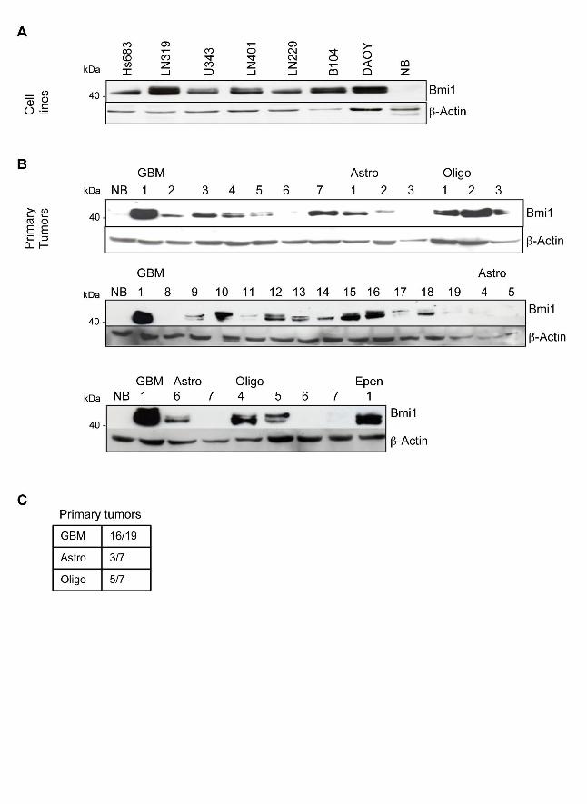


Supporting information Figure 1

Supporting information Figure 2
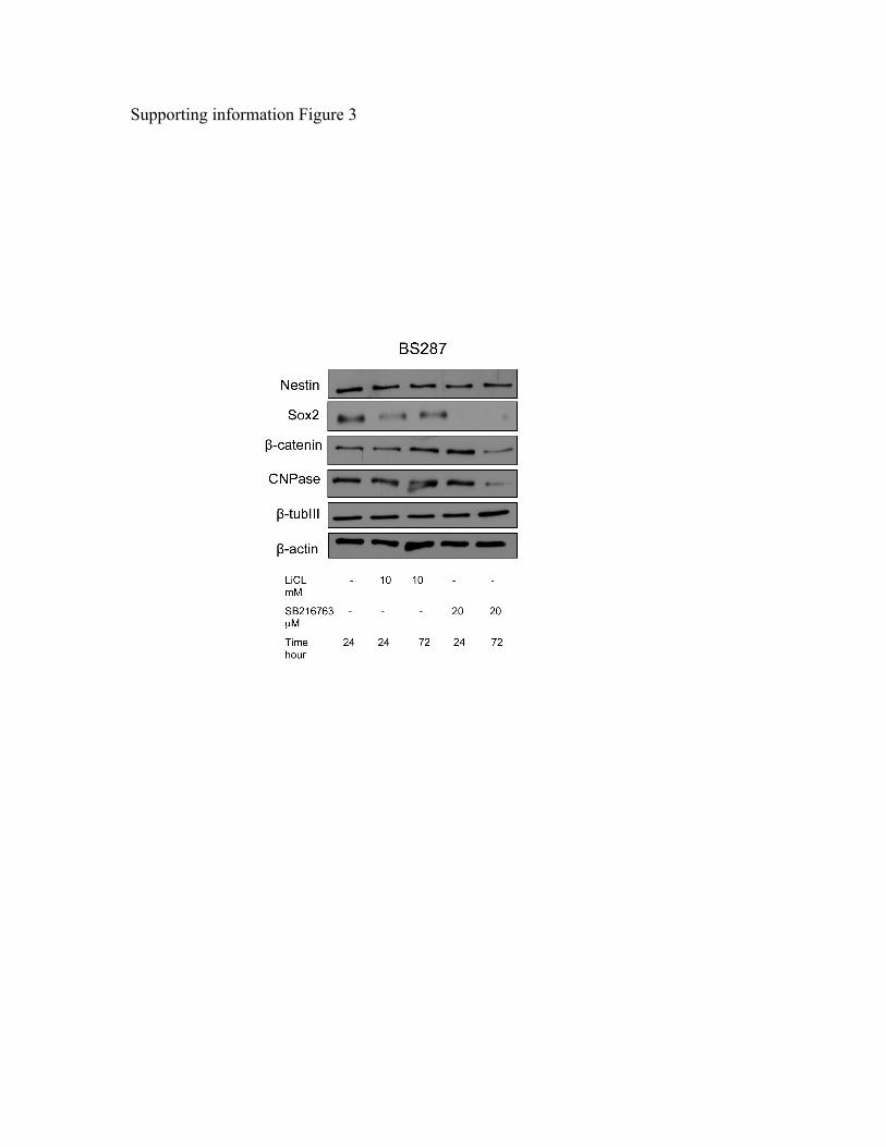
Supporting information Figure 3

Supporting information Figure 4

58
Part 2: Combination of sublethal concentrations of epidermal
growth factor receptor inhibitor and microtubule stabilizer
induces apoptosis of glioblastoma cells
Mike Failly, Serdar Korur*, Viviane Egler, Jean-Louis Boulay, Maria
Maddalena Lino, Roland Imber, and Adrian Merlo
Clin Cancer Res. 2008 May 15;14(10):3132-40
* Author contributed to: experimental design, performing experiments, data analysis and writing
the paper

Combination of sublethal concentrations of epidermalgrowth factor receptor inhibitor and microtubulestabilizer induces apoptosis of glioblastoma cells
Mike Failly, Serdar Korur, Viviane Egler,Jean-Louis Boulay, Maria Maddalena Lino,Roland Imber, and Adrian Merlo
Laboratory of Molecular Neurooncology, Departments ofResearch and Surgery, University Hospitals, Basel, Switzerland
AbstractThe oncogenic epidermal growth factor receptor (EGFR)pathway triggers downstream phosphatidylinositol 3-kinase (PI3K)/RAS-mediated signaling cascades. In trans-genic mice, glioblastoma cannot develop on single but onlyon simultaneous activation of the EGFR signaling media-tors RAS and AKT. However, complete blockade of EGFRactivation does not result in apoptosis in human glioblas-toma cells, suggesting additional cross-talk betweendownstream pathways. Based on these observations, weinvestigated combination therapies using protein kinaseinhibitors against EGFR, platelet-derived growth factorreceptor, and mammalian target of rapamycin, assessingglioblastoma cell survival. Clinically relevant doses ofAEE788, Gleevec (imatinib), and RAD001 (everolimus),alone or in combinations, did not induce glioblastoma cellapoptosis. In contrast, simultaneous inactivation of theEGFR downstream targets mitogen-activated protein/extracellular signal-regulated kinase (ERK) kinase andPI3K by U0126 and wortmannin triggered rapid tumorcell death. Blocking EGFR with AEE788 in combinationwith sublethal concentrations of the microtubule stabilizerpatupilone also induced apoptosis and reduced cellproliferation in glioblastoma cells, accompanied by re-duced AKT and ERK activity. These data underline thecritical role of the PI3K/AKT and the RAS/RAF/mitogen-activated protein/ERK kinase/ERK signaling cascades inthe cell-intrinsic survival program of sensitive glioblastoma
cell lines. We conclude that drug combinations, whichdown-regulate both ERK and protein kinase B/AKTactivity, may prove effective in overcoming cell resistancein a subgroup of glioblastoma. [Mol Cancer Ther 2007;6(2):773–81]
IntroductionGlioblastoma multiforme is the most frequent malignantneoplasm of the human central nervous system. Surgerycan only control the highly proliferative component of thedisease, whereas widespread tumor cell infiltration intonormal brain areas resists radiotherapy and chemotherapy(1, 2). Amplification and overexpression of the geneencoding the epidermal growth factor (EGF) receptor(EGFR) are detected in f50% of glioblastomas and aremainly associated with disease progression (3, 4). EGFR is amember of the ErbB family of receptor tyrosine kinases (5).Small molecular weight compounds with EGFR proteinkinase inhibitory (PKI) activity, such as PKI-166 or AEE788,have a cytostatic effect in vitro on tumor cells thatoverexpress EGFR (6, 7). In addition, treatment of non–small cell lung cancer with the EGFR PKI imatinib(gefitinib) resulted in tumor growth control in 10% ofpatients (8). In fact, tumors that responded to gefitinibspecifically carried specific mutations in the EGFR tyrosinekinase domain (8–10). Although responses to gefitinib havealso been observed in a limited number of glioblastomacases (11), a specific molecular profile is associated withresponse that differs from the lung signature (12–15). Todate, several small molecular weight inhibitors, such asGleevec or erlotinib/gefinitib, applied as monotherapiesfor the treatment of gliomas, only resulted in limitedeffectiveness (16). This has supported the hypothesis thatcombination of drugs would be a more appropriatetreatment for glioma.EGF mediates signaling via phosphatidylinositol 3-kinase
(PI3K)/AKT and RAS/mitogen-activated protein/extracel-lular signal-regulated kinase (ERK) kinase (MEK)/ERKpathways (17–19), which are redundantly stimulated byother activated growth factor receptors (e.g., the insulin-like growth factor receptor-I; ref. 20). In at least 50% ofglioblastoma, PI3K is activated either by loss of functionof the tumor suppressor PTEN (21) or by gain-of-functionmutations in the PIK3CA gene, which encodes the p110catalytic subunit of PI3K (22). Interestingly, activatedprotein kinase B (PKB)/AKT cooperated with RAS in theinduction of malignant gliomas in a murine brain tumormodel (23). Furthermore, activation of PKB/AKT has beendetected in several types of human cancers and found tobe associated with poor clinical outcome (24–26) andresistance to chemotherapy and radiotherapy (24, 27, 28).
Received 9/13/06; revised 11/22/06; accepted 12/21/06.
Grant support: Swiss National Foundation grant 31-67123.01,Oncosuisse grant 01338, Regional Cancer League of Basel, and NovartisOncology (Basel, Switzerland).
The costs of publication of this article were defrayed in part by thepayment of page charges. This article must therefore be hereby markedadvertisement in accordance with 18 U.S.C. Section 1734 solely toindicate this fact.
Requests for reprints: Adrian Merlo, Laboratory of MolecularNeurooncology, Departments of Research and Surgery, UniversityHospital Basel, Spitalstrasse 21, 4031 Basel, Switzerland.Phone: 41-61-265-71-82; Fax: 41-61-265-71-38.E-mail: [email protected]
Copyright C 2007 American Association for Cancer Research.
doi:10.1158/1535-7163.MCT-06-0566
773
Mol Cancer Ther 2007;6(2). February 2007

Although no RAS mutations have been detected inglioblastoma, high levels of RAS-GTP have been docu-mented in high-grade astrocytomas (29, 30). It has beensuggested that ERK plays a critical role in cellulartransformation and resistance to apoptosis (31). Evidencefor a cooperation between AKT and RAS has been provenby a transgenic mouse model in which both oncogenes areectopically expressed in normal astrocytes, giving rise toglioblastoma, whereas neither AKT nor RAS alone wassufficient for tumor induction (23).Several examples of combined therapies have recently
undergone advanced clinical trials in cancer treatment. Forexample, lapatinib with tamoxifen has been tested togetherin breast cancer (32), and in xenografted tumors (32), PKIAEE788 has been associated with the rapamycin-derivativeRAD001 for the treatment of glioblastoma (33–35).Successful drug combinations allow the use of lowerdoses, possibly reducing toxicity and limiting the degreeof acquired drug resistance. Given the limited efficacy ofPKI to induce cell death in rapidly proliferating tumor cells,these inhibitory drugs may depend on the coadministrationof a cytotoxic drug for the induction of cell death rather thanon the coadministration of a second cytostatic compound.The principle of cytotoxicity of patupilone (epothilone B,EPO906) relies on the inhibition of microtubule depolymer-ization, which is lethal for all dividing cells. The antitumoractivity of patupilone has been proven in vitro and in vivo inlung, breast, colon, and prostate cancers (36). Patupilonealso shows clinical activity in a range of solid tumors (37)and is now in phase III clinical development.In the present study, we examined the induction of cell
death on glioblastoma lines in vitro by targeting EGFR/ErbB2 by the receptor tyrosine kinase inhibitor AEE788alone or in combination with the cytotoxic compoundpatupilone. We further evaluated the activation status ofthe EGFR downstream signaling mediators PKB/AKT andERK following combined treatment with patupiloneand AEE788. Our study points to a critical role of bothPI3K and ERK in glioblastoma cell survival.
Materials andMethodsCell CultureAll cell lines were grown at 37jC in a 5% CO2 humidified
atmosphere. DMEM culture medium was supplementedwith 10% FCS and standard antibiotics. Cells weredetached with 1 mL trypsin-EDTA (1!) for 5 min at37jC. The genetic status of all cell lines at p53, p16, p14ARF ,and PTEN tumor suppressor genes has been previouslyreported (38, 39).Pharmacologic InhibitorsAEE788, RAD001, and patupilone (provided by Novartis
Pharma AG, Basel, Switzerland) and U0126 (Promega,Madison, WI) were dissolved in DMSO as 10 mmol/Lstocks and stored as aliquots at "20jC. Wortmannin(Sigma, St. Louis, MO) was prepared as a 1 mmol/L stocksolution in DMSO and stored at 4jC, and 12-O-tetradeca-noylphorbol-13-acetate (TPA; LC Laboratories, Woburn,
MA) was prepared as a 2 mmol/L stock solution in DMSO,aliquoted, and stored at "20jC. Treatment of the cells withTPA, U0126, or wortmannin consisted of daily additionswithout replacing the medium.Measurement of Cell Proliferation and Cell Cycle
ProfileIn experiments shown in Fig. 1B, cells were grown for
24 h in DMEM supplemented with 10% FCS and for anadditional 24 h in the presence of the drug. Bromodeoxyur-idine (BrdUrd) was added 1 h before cell harvesting to a10 Amol/L final concentration. Fluorescence-activated cellsorting analysis was done according to the manufacturer’sinstructions (Becton Dickinson, Franklin Lakes, NJ). The cellproliferation assays presented in Figs. 2C and 3B weredone with the Biotrak ELISA System version 2 (AmershamBiosciences Corp., Piscataway, NJ) according to the manu-facturer’s instructions. Cells (5 ! 103) were seeded in96-well plates and grown for 24 h, and drugs were appliedfor an additional 24 h. BrdUrd incorporation was allowedduring the last 2 h of treatment. In experiments shown inFig. 1D, cells were grown for 24 h in DMEM supplementedwith 10% FCS and for an additional 24 h in the presence ofthe drug. Cell DNA content and apoptosis were analyzed ona FACSCalibur. Cells were trypsinized and fixed in 70% ice-cold ethanol for 1 h and stained with 50 Ag/mL propidiumiodide for fluorescence-activated cell sorting analysis. Thepercentages of cells in the cell cycle phases are reported(results from three independent experiments).Measurement of ApoptosisExpanding cells were subjected to drug treatment for
24 h, briefly washed with PBS, and trypsinized. Cellsuspension was pelleted at 800 rpm for 5 min, fixed inice-cold 70% ethanol, and kept at 4jC for 30 min. Cells wereresuspended in a 1! PBS solution containing 50 ng/mLpropidium iodide and 50 Ag/mL RNase. Percentage of cellsin sub-G1 for apoptosis was determined by flow cytometry.Protein Extraction andWestern Blot AnalysisCells were washed with PBS, resuspended in 1! SDS
sample buffer (62.5mmol/LTris-HCl, 2%SDS, 10%glycerol,50 mmol/L DTT), and boiled at 95jC during 5 min; aliquotswere stored at "20jC. Proteins (30 Ag) were separated bysize on SDS-PAGE gels (10%) and transferred to nitrocellu-lose membranes (Hybond ECL, Amersham Biosciences).Western blot analysis was done using antibodies against thefollowing proteins: phosphorylated AKT (Upstate Biotech-nology, Lake Placid, NY), ERK1/2, phosphorylated ERK,phosphorylated EGFR (Tyr1173), and EGFR (Santa CruzBiotechnology, Santa Cruz, CA) and actin (Sigma). The anti-AKT antibody was a gift from Dr. Brian Hemmings(Friedrich Miescher Institute, Basel, Switzerland). Westernblots were developed with enhanced chemiluminescencereagents (Pierce, Rockford, IL).
ResultsSpecificTargeting of the EGF Signaling Pathway by
AEE788Nearly 50% of primary glioblastomas exhibit amplifica-
tion of the EGFR gene (3, 4), which is also associated with
Synergistic Induction of Glioblastoma Cell Apoptosis774
Mol Cancer Ther 2007;6(2). February 2007

poor prognosis. For this reason, the inhibitor of EGFR andof ErbB receptor protein kinase activities AEE788 has beenconsidered to be a potent drug to induce cell death inglioblastoma cell lines. AEE788 was tested for its ability toinduce cell growth arrest and apoptosis in various cell linesderived from glioblastoma, for which the genetic status ofestablished cancer genes had been previously defined(Fig. 1A; refs. 38, 39). For a preliminary determination ofthe minimal drug concentration needed to fully preventEGFR activation by EGF, increasing concentrations ofAEE788 were applied to the LN229 glioblastoma cell line,and EGFR/ErbB receptor activation status was monitoredby the presence of EGFR phosphorylated at Tyr1173. Tocompletely block receptor phosphorylation, 1 Amol/L wasneeded (data not shown). Further time course experimentdone with 1 Amol/L AEE788 showed a strong inhibition ofthe transient phosphorylation of Tyr1173 as well as of thedegradation of the receptor protein in all seven glioblasto-ma cell lines tested (Fig. 1B).Increasing concentrations of AEE788 were applied to
the glioblastoma cell lines, and proliferation was assayedby integration of BrdUrd after 1 day. At a 1 Amol/L
concentration, AEE788 gives up to 20% of proliferationreduction compared with the control cell population(Fig. 1C). Cell cycle analysis after 24 h of 1 and 10 Amol/Lof AEE788 treatment shows an increase of cell population inthe G1 phase (Fig. 1D).Cell viability was assayed by flow cytometry after 24, 48,
and 96 h. Very low or no apoptosis was induced at drugconcentrations <2 Amol/L as late as 96 h (Fig. 2A). Highlevels of apoptosis were observed at unphysiologicallyhigh concentration (10 Amol/L) in five of the seven celllines (SF767, LN215, U373, LN229, and LN71). Under thesame condition, LN401 and Hs683 did not show significantlevels of apoptosis. Because AEE788 was not capable ofinducing strong apoptosis in glioblastoma cells when usedas a single drug, we tested whether a specific double or atriple combination of compounds would significantlyincrease the induction rate of apoptosis. For this purpose,we combined suboptimal doses of AEE788 (0.5 Amol/L)with two additional biological drugs: Gleevec (imatinib,0.5 Amol/L) and RAD001 (everolimus, 20 nmol/L). Ingliomas, Gleevec is being explored as a PKI of platelet-derived growth factor receptor (34), which is activated in
Figure 1. Treatment of glioblasto-ma cell lines with EGFR inhibitor. A,genotyping of used glioma cell linesby Ishii et al. (39). B, cells werepreincubated in the presence orabsence of 1 Amol/L of the EGFRinhibitor AEE788 during 30 min andstimulated with 100 ng/mL EGF for10, 60, and 300 min. EGFR, phos-phorylated EGFR (Tyr1173), and actinwere detected by Western blot anal-ysis of cell lysates. C, cells wereincubated during 4 d with increasingconcentrations of AEE788 (0, 1, 2,5, and 10 Amol/L). Proliferation wasmeasured by BrdUrd and flow cytom-etry. Experiments were done in trip-licate. Points, average of threeindependent determinations; bars,SD. D, cell cycle phase analysis onglioblastoma cell lines on 1 and 10Amol/L of AEE788 treatment.
Molecular Cancer Therapeutics 775
Mol Cancer Ther 2007;6(2). February 2007

primary tumors (40, 41). Platelet-derived growth factorreceptor phosphorylation levels correlated with sensitivityto Gleevec in glioma primary cultures (42). RAD001, aderivative of rapamycin, inhibits mammalian target ofrapamycin, leading to inactivation of ribosomal S6K1 andinhibition of cap-dependent translation (43).The application of the combined drugs to 10 glioblastoma
cell lines for 4 days revealed that there was a combination-specific and a cell line–specific response with regard toinduction of cell death (Fig. 2B). The cell lines could bedivided into three categories. One group (SF767, LN18, andLN215; Fig. 2B, left) showed a modest additive inductionof apoptosis by all of the three drugs, reaching a maximumof 35% of cell death with the triple combination. A secondgroup of glioblastoma cell lines (LN401, LN71, LN229, andLN319; Fig. 2B, middle) was basically insensitive to all drugcombinations, showing the same rate of apoptosis asuntreated cells. The third group (Hs683, U373, and U343;Fig. 2B, right) exhibited a relatively high level of basalapoptosis (15–20%), which was, however, decreased by thethree drugs applied together. Especially, RAD001 exhibitedan antiapoptotic effect, alone or in combination. No differ-ences in apoptosis rate were evidenced between PTEN-mutant (LN215, LN401, LN71, and U373) and wild-type(Hs683, SF767, and LN229) cell lines on RAD001 applica-tion. Although, in theory, RAD001 was expected toovercome the constitutive AKT activation resulting fromloss of PTEN activity.Taken together, the results indicated that neither single
nor combined application of the three biological drugs, atphysiologic concentration, was able to strongly induce celldeath in glioblastoma cell lines.
Induction of Apoptosis in Glioblastoma Cell Lines byPatupiloneBecause neither the single nor the combined application
of the three biological drugs led to a consistent induction ofapoptosis, we tested whether the combination of AEE788with a cytotoxic drug could result in a more cooperativeinduction of apoptosis. Patupilone is a member of thegroup of epothilones, which represent a new class of lowmolecular weight compounds that target microtubules byinhibiting their depolymerization and therefore impairs celldivision (44). Patupilone prevents chromosome alignmentat metaphase and drives cells to undergo apoptosis (45). Ithas been shown to exhibit antitumor activity in vitro andin vivo (36, 37). As a result of inhibition of microtubuledepolymerization, after addition of patupilone, dramaticchanges in cell morphology occurred within 18 h (Fig. 3A,right). To investigate its ability to induce apoptosis,glioblastoma cells were treated with increasing concen-trations of patupilone in the picomolar and nanomolarconcentration range for 4 days (Fig. 3A, left). Although100% apoptosis was induced in all cell lines when thehighest drug concentration (35 nmol/L) was applied, levelsof apoptosis induced by 0.7 nmol/L patupilone stronglyvaried between the cell lines, from Hs683 and LN401, themost resistant, to LN229, the most sensitive one.Combined Treatment with AEE788 and Patupilone
Strongly Induced Apoptosis and Reduced Proliferationof Glioblastoma CellsAEE788 (1 Amol/L) was applied together with increasing
concentrations of patupilone on cell lines LN71, LN229,Hs683, and SF767, and cell survival was determined after4 days (Fig. 3B). The combined drugs induced apoptosis in
Figure 2. Survival of glioblastomacell lines on treatment with EGFRinhibitor and in combination withGleevec and RAD001. A, cells wereincubated during 4 d with increasingconcentrations of AEE788 (0, 1, 2,5, and 10 Amol/L). Apoptosis wasmeasured by flow cytometry. Experi-ments were done in triplicate. Col-umns, average of three independentdeterminations; bars, SD. B, combi-natorial study of biological drugsAEE788 (A), Gleevec (G), andRAD001 (R) on glioma cell survival.
Synergistic Induction of Glioblastoma Cell Apoptosis776
Mol Cancer Ther 2007;6(2). February 2007

a cooperative manner in LN71 and SF767 but not in Hs683and LN229 cell lines. In LN71 and SF767 lines, thepatupilone concentration required to induce apoptosis in50% of cells (AC50) revealed that AEE788 acted as asensitizer, reducing the patupilone concentration from ananomolar to a picomolar concentration range. Thus,addition of AEE788 shifted down the amount of patupilonerequired to induce apoptosis in 50% of cells, up to 2 ordersof magnitude in SF767 cells. It is noteworthy that thesynergistic effect of patupilone on those glioma cell linesseemed independent of their respective sensitivities toAEE788. Consistent with the absence of synergy ofboth compounds on LN229 cells, also proliferation wasnot affected either by AEE788 or patupilone or by thecombination of both drugs. In contrast, SF767 cells, on
which the AEE788/patupilone synergized the best, had asignificant reduction of proliferation up to 80% (P < 0.001)after each individual or combined drug application(Fig. 3C).Simultaneous Inhibition of Both ERK1/2 and AKT
KinaseActivities Parallels the Induction of ApoptosisBoth signaling pathways, PI3K/AKT and RAS/MEK/
ERK, are under the control of EGF-directed activation ofErbB receptors. To investigate the activation of these twopathways, phosphorylation status of the kinases AKT andERK was chosen as readouts. AEE788 (1 Amol/L) andpatupilone (0.7 nmol/L), alone and in combination, wereapplied for 24 and 72 h to the four glioblastoma cell linesthat differed remarkably in their sensitivities toward thosedrugs. In contrast to LN229 and Hs683, LN71 and SF767
Figure 3. Combined treatment ofglioblastoma cells stabilizing micro-tubules and blocking EGFR. A, in-creasing concentrations of patupilone(0.05, 0.1, 0.7, 5, and 35 nmol/L)were applied to seven cell lines for4 d and the percentage of viable cellswas determined using flow cytome-try. Points, average of three indepen-dent determinations; bars, SD.Confocal microscopy of LN229 cellsafter 18 h of patupilone treatment. B,cells were treated for 4 d either withincreasing concentrations of patupi-lone alone or in combination with1 Amol/L AEE788. Cell survival wasmeasured by flow cytometry. Points,average of three independent experi-ments; bars, SD. C, cells weretreated with 1 Amol/L AEE788 and/or 0.7 nmol/L patupilone and assayedfor proliferation with BrdUrd byELISA. D, top, cells were incubatedduring 24 h and 3 d with patupilone(0.7 nmol/L), AEE788 (1 Amol/L), orboth. Western blot analysis was doneon cell lysates. NT, no treatment;A, AEE788; P, patupilone. Bottom,poly(ADP-ribose) polymerase (PARP )cleavage after 24 h.
Molecular Cancer Therapeutics 777
Mol Cancer Ther 2007;6(2). February 2007

showed full down-regulation of phosphorylation ofboth protein kinases after 3 days of combined treatment(Fig. 3D), accompanied by widespread apoptosis between85% and 95% of cells. In contrast, LN229 and Hs683 stillretained strong phosphorylation of the two protein kinasesand showed much lower levels of apoptosis. These resultssuggested a link between the simultaneous inhibition of thetwo EGF-dependent signaling pathways and the inductionof apoptosis triggered by the combination of patupiloneand AEE788. To support this hypothesis, we analyzed thecleavage status of poly(ADP-ribose) polymerase, whichtriggers caspase-dependent apoptosis (46, 47). The cleavedform of poly(ADP-ribose) polymerase was indeed detectedin glioblastoma cells undergoing apoptosis (Fig. 3E).Simultaneous Inhibition of AKTand MEK Coopera-
tively Induces Apoptosis in a Subgroup ofGlioblastomaCell LinesTo test whether sensitivity of LN71 and SF767 cells to
the AAE788-patupilone combination is due to low phos-phorylation levels of AKT and/or ERK, inhibitors of PI3K(wortmannin) and MEK (U0126) were used to abrogateactivation of AKT and ERK pathways. The potential toinduce cell death was tested by applying single or
combined inhibitors to four glioblastoma cell lines andmeasuring the proportion of apoptotic cells (Fig. 4A). Whenused as a single compound, U0126 was able to induce asignificant level of apoptosis in LN215 and LN229 cells butto a much lesser extent in U373 and LN401 cells, whereaswortmannin alone had no or very little effect. However,strong induction of apoptosis was caused by the combina-tion of the two inhibitors. On U0126 application, prolifer-ation was significantly decreased up to 50% in LN229cell line (P < 0.001), whereas U373 cells showed a 20%reduction. Again, wortmannin alone had no effect onreduction of proliferation on both LN229 and U373 cells,and no synergistic effect was present after drug combina-tion (Fig. 4B).U373 and LN401 generally exhibited much less sensitiv-
ity toward the two inhibitors. To examine the long-termeffect of the inhibitors on phosphorylation of AKT andERK, the two most sensitive and the two much lesssensitive cell lines were treated for 24 h as well as for3 days by applying fresh doses of the compounds daily.The activation status of AKT and ERK was determinedas a readout for drug efficiency (Fig. 4C). In response tothe combination of U0126 and wortmannin, a decrease of
Figure 4. Combined treatment ofglioblastoma cells blocking PI3K andMEK. A, cell lines were treated withthe MEK inhibitor U0126 (20 Amol/L)and/or the PI3K inhibitor wortmannin(1 Amol/L) for 3 d, whereby theaddition of both drugs was repeatedevery day. Percentage of apoptoticcells was measured using flowcytometry. Columns, mean of threeindependent experiments; bars, SD.B, cells were treated with 20 Amol/LU0126 and/or 1 Amol/L wortmanninand assayed for proliferation withBrdUrd by ELISA. C, Western blotanalysis was done to determine therelative phosphorylation levels ofERK1/2 and AKT after 3 d of treat-ment. U, U0126; W, wortmannin.D, poly(ADP-ribose) polymerase(PARP) cleavage after 24 h.
Synergistic Induction of Glioblastoma Cell Apoptosis778
Mol Cancer Ther 2007;6(2). February 2007

phosphorylated ERK and phosphorylated AKT occurringin all glioblastoma lines confirmed the activity of thetwo drugs used. Unexpectedly, on treatment with theMEK inhibitor, U0126, U373, and LN229 cell lines showa decrease of phosphorylated AKT levels, suggesting across-talk between the RAS/MEK/ERK and PI3K/AKTpathways.In the sensitive cell lines LN215 and LN229, simultaneous
inactivation of protein kinases AKT and MEK wasassociated with poly(ADP-ribose) polymerase cleavageand apoptosis (Fig. 4D). However, in the resistant cell linesU373 and LN401, AKT and MEK inactivation was notsufficient to trigger apoptosis. There was no correlationbetween glioblastoma cell line sensitivity or resistance toapoptosis and a given genotype (Fig. 1A).Phorbol Ester TPA Restores Activation of MEK and
Inhibits Induction of ApoptosisPhorbol ester TPA activates protein kinase C and ERK1/2,
thereby modifying mitogenic signaling pathways (44, 48,49). When exposed to 20 or 100 nmol/L of TPA, LN215and LN229 cells showed a strong increase of phosphorylatedERK levels (Fig. 5A). Treatment of the cell lines withcombined U0126 and wortmannin for 4 days in the presenceof TPA (20 or 100 nmol/L) led to a substantial increase ofactivated ERK1/2, whereas phosphorylation was complete-ly down-regulated without TPA (Fig. 5A). In parallel, theextent of the induction of apoptosis was determined after4 days under the same conditions, revealing that theaddition of TPA to both glioblastoma cell lines substantiallydecreased the induction of apoptosis caused by the twoinhibitors (Fig. 5B). Taken together, the results furthersupported the model of a correlation between down-regulation of the two signaling pathways and induction ofapoptosis.
DiscussionWe report that the combination of the inhibitor of EGFRkinase AEE788 and of the microtubule depolymerizationinhibitor patupilone synergistically induced death of
glioblastoma cells. Of high interest is that this synergyoccurred at drug concentrations that were not effective toinduce cell death when each drug was applied alone.AEE788 alone at 1 Amol/L only induced low levels ofapoptosis in glioblastoma cells in vitro . Its main antitumoractivity consists of inhibition of cell growth and motility(50). Consistently, survival of animals bearing intracranialtumors had been extended by giving AEE788 at concen-tration below the maximally tolerated dose (34) andconfirmed a role of AEE788 as an antitumor agent.However, lasting responses cannot be obtained with acytostatic effect but require induction of tumor cell death,which may only be achieved by drug combinations.Therefore, we designed a strategy to trigger glioblastomacell apoptosis by combining the PKI AEE788 withpatupilone, a compound with strong cytotoxic potentialagainst various cancer types and also against multidrugresistance cancer cell lines (51). Glioblastoma cell deathcould be triggered in all tumor cell lines in vitro by35 nmol/L patupilone, a dose that can already be toxicin vivo . Interestingly, synergistic induction of apoptosis wasobserved in glioblastoma cells when using 1 Amol/LAEE788 in combination with only 0.1 nmol/L patupilone.Of interest, the genetic backgrounds of the cell linesthat responded the best to the combination therapy withAEE788 and patupilone were different with regard to thethree main glioma pathways: TP53, PTEN, and p14/p16(Fig. 1A). Whereas SF767 cells are wild-type at all three loci,LN71 cells are either mutated or null (39), suggesting thatsuch a combination could be applied to a wide spectrum ofgliomas. When analyzing the activation status of EGFR,PKB/AKT, and ERK as readouts for critical pathways inthe glioblastoma signaling network, we found that syner-gistic induction of apoptosis by combining patupilone andAEE788 was paralleled by inactivation of PKB/AKT andERK. Conversely, the two critical cell signaling mediatorsremained active in glioblastoma cells that did not show thisdrug synergism. Although AEE788, patupilone, and U0126,applied individually, reduced glioblastoma cell growth,AEE788/patupilone and U0126/wortmannin combinations
Figure 5. Phorbol ester TPA antagonizes induction ofapoptosis and maintains AKT and ERK1/2 activities. A,cells were treated with 20 and 100 nmol/L of TPA in thepresence or absence of wortmannin (W) and U0126 (U)for 3 d, whereby the additions were renewed every day.Cell extracts were prepared by the addition of SDSsample buffer, and Western blot analysis was done.U0126, 20 Amol/L; wortmannin, 1 Amol/L; TPA, 20 or100 nmol/L. B, cells were treated in the same way for4 d and the percentage of dead cells was determined byflow cytometry.
Molecular Cancer Therapeutics 779
Mol Cancer Ther 2007;6(2). February 2007

did not affect cell growth in a synergistic manner,suggesting that these processes are driven by distinctpathways. The importance of the two signaling pathwaysPI3K/PKB and RAS/RAF/MEK/ERK for glioblastomasignaling is further supported by the finding that PI3K isactivated in the majority of human glioblastoma either byloss of PTEN function (38, 39) or by activating mutations ofPI3KCA (22), whereas growth factor stimulation causesactivation of RAS in glioblastoma (52). Further evidencestems from a murine transgenic glioblastoma model, inwhich simultaneous activation of RAS and PKB gives riseto malignant gliomas (53, 54). Glioma animal models havefurther shown that the combination of ectopic expression ofactivated RAS and AKT in the glial lineage (23) led tospontaneous induction of murine gliomas, whereas anactivated allele of either RAS or AKT alone failed to inducetumor formation. In analogy to this animal model and theeffects on signaling induced by the described synergism,we hypothesized that direct inhibition of signaling medi-ators downstream of EGFR within the PI3K/PKB andRAS/RAF/MEK/ERK pathways will overcome glioblasto-ma cell resistance. Therefore, we specifically blockedactivity of both MEK and PI3K, which induced strongapoptosis in a subgroup of glioblastoma cell lines,indicating a critical role of these two pathways inglioblastoma signaling. Combining the MEK inhibitorU0126 and the PI3K inhibitor wortmannin led to synergisticinduction of apoptosis in LN215 and LN229 cell lines.Interestingly, although phosphorylation of ERK1/2 andPKB/AKT was decreased in all cell lines, it was notassociated with apoptosis in resistant lines, suggestingadditional defects in the proapoptotic machinery. Activa-tion of protein kinase C and ERK1/2 by TPA counteractedU0126- and wortmannin-dependent apoptosis, confirmingthe specificity of drug effect.An unresolved question is at which cellular level the
intervention has to take place. Our data argue thatcombined blocking of signaling mediators downstream ofgrowth factor receptors, interfering with the signalingcross-talk, may be more effective than inhibition of a singlecell surface receptor (e.g., EGFR; ref. 13). This view issupported by other findings in glioblastoma cell lines,where inhibition of PKB/AKT could be counteracted by astimulation of insulin-like growth factor receptor-I, result-ing in sustained activation of PI3K (19, 20).In conclusion, our results indicate that the induction of
apoptosis in glioblastoma cell lines requires drug combi-nation, which down-regulate distinct pathways. BlockingEGFR activation alone did not induce apoptosis unlesscomplemented with the microtubule stabilizer patupilone.If PKIs were targeted without the help of cytotoxic drugs,combined inhibition of MEK and PI3K was found to bethe most efficient combination to induce glioblastoma celldeath. Drug effects were independent of mutation statusesat the major glioblastoma pathways (Fig. 1A). Additionalstudies are needed to develop other target for treatment ofresistant glioblastoma cells to directly down-regulate keymembers of the critical pathways.
Acknowledgments
We thank Elisabeth Taylor for critical reading of the manuscript.
References
1. Shapiro WR, Green SB, Burger PC, et al. Randomized trial of threechemotherapy regimens and two radiotherapy regimens and two radio-therapy regimens in postoperative treatment of malignant glioma. BrainTumor Cooperative Group Trial 8001. J Neurosurg 1989;71:1–9.
2. Burger PC, Heinz ER, Shibata T, Kleihues P. Topographic anatomy andCT correlations in the untreated glioblastoma multiforme. J Neurosurg1988;68:698–704.
3. Libermann TA, Nusbaum HR, Razon N, et al. Amplification, enhancedexpression, and possible rearrangement of EGF receptor gene in primaryhuman brain tumours of glial origin. Nature 1985;313:144–7.
4. Wong AJ, Ruppert JM, Bigner SH, et al. Structural alterations of theepidermal growth factor receptor gene in human gliomas. Proc Natl AcadSci U S A 1992;89:2965–9.
5. Hynes NE, Lane HA. ERBB receptors and cancer: the complexity oftargeted inhibitors. Nat Rev Cancer 2005;5:341–54.
6. Lane HA, Motoyama AB, Beuvink I, Hynes NE. Modulation of p27/Cdk2complex formation through 4D5-mediated inhibition of HER2 receptorsignaling. Ann Oncol 2001;12 Suppl 1:S21–2.
7. Traxler P, Allegrini PR, Brandt R, et al. AEE788: a dual family epidermalgrowth factor receptor/ErbB2 and vascular endothelial growth factorreceptor tyrosine kinase inhibitor with antitumor and antiangiogenicactivity. Cancer Res 2004;64:4931–41.
8. Lynch TJ, Bell DW, Sordella R, et al. Activating mutations in theepidermal growth factor receptor underlying responsiveness of non-small-cell lung cancer to gefitinib. N Engl J Med 2004;350:2129–39.
9. Paez JG, Janne PA, Lee JC, et al. EGFR mutations in lung cancer:correlation with clinical response to gefitinib therapy. Science 2004;304:1497–500.
10. Pao W, Miller V, Zakowski M, et al. EGF receptor gene mutations arecommon in lung cancers from ‘‘never smokers’’ and are associated withsensitivity of tumors to gefitinib and erlotinib. Proc Natl Acad Sci U S A2004;101:13306–11.
11. Rich JN, Reardon DA, Peery T, et al. Phase II trial of gefitinib inrecurrent glioblastoma. J Clin Oncol 2004;22:133–42.
12. Barber TD, Vogelstein B, Kinzler KW, Velculescu VE. Somaticmutations of EGFR in colorectal cancers and glioblastomas. N Engl JMed 2004;351:2883.
13. Lassman AB, Rossi MR, Raizer JJ, et al. Molecular study of malignantgliomas treated with epidermal growth factor receptor inhibitors: tissueanalysis from North American Brain Tumor Consortium Trials 01-03 and00-01. Clin Cancer Res 2005;11:7841–50.
14. Marie Y, Carpentier AF, Omuro AM, Sanson M, Thillet J, Hoang-XuanK. EGFR tyrosine kinase domain mutations in human gliomas. Neurology2005;64:1444–5.
15. Mellinghoff IK, Wang MY, Vivanco I, et al. Molecular determinants ofthe response of glioblastomas to EGFR kinase inhibitors. N Engl J Med2005;353:2012–24.
16. Wen PY, Yung WK, Lamborn KR, et al. Phase I/II study of imatinibmesylate for recurrent malignant gliomas: North American Brain TumorConsortium Study 99-08. Clin Cancer Res 2006;12:4899–907.
17. Carpenter G. The EGF receptor: a nexus for trafficking and signaling.Bioessays 2000;22:697–707.
18. Graves LM, Guy HI, Kozlowski P, et al. Regulation of carbamoylphosphate synthetase by MAP kinase. Nature 2000;403:328–32.
19. Sibilia M, Fleischmann A, Behrens A, et al. The EGF receptor providesan essential survival signal for SOS-dependent skin tumor development.Cell 2000;102:211–20.
20. Chakravarti A, Loeffler JS, Dyson NJ. Insulin-like growth factorreceptor I mediates resistance to anti-epidermal growth factor receptortherapy in primary human glioblastoma cells through continued activationof phosphoinositide 3-kinase signaling. Cancer Res 2002;62:200–7.
21. Choe G, Horvath S, Cloughesy TF, et al. Analysis of the phosphati-dylinositol 3¶-kinase signaling pathway in glioblastoma patients in vivo .Cancer Res 2003;63:2742–6.
22. Samuels Y, Wang Z, Bardelli A, et al. High frequency of mutations ofthe PIK3CA gene in human cancers. Science 2004;304:554.
Synergistic Induction of Glioblastoma Cell Apoptosis780
Mol Cancer Ther 2007;6(2). February 2007

23. Holland EC, Celestino J, Dai C, Schaefer L, Sawaya RE, Fuller GN.Combined activation of Ras and Akt in neural progenitors inducesglioblastoma formation in mice. Nat Genet 2000;25:55–7.
24. Brognard J, Clark AS, Ni Y, Dennis PA. Akt/protein kinase B isconstitutively active in non-small cell lung cancer cells and promotescellular survival and resistance to chemotherapy and radiation. Cancer Res2001;61:3986–97.
25. Lee JI, Soria JC, Hassan KA, et al. Loss of PTEN expression as aprognostic marker for tongue cancer. Arch Otolaryngol Head Neck Surg2001;127:1441–5.
26. Perez-Tenorio G, Stal O. Activation of AKT/PKB in breast cancerpredicts a worse outcome among endocrine treated patients. Br J Cancer2002;86:540–5.
27. Ermoian RP, Furniss CS, Lamborn KR, et al. Dysregulation of PTENand protein kinase B is associated with glioma histology and patientsurvival. Clin Cancer Res 2002;8:1100–6.
28. Clark AS, West K, Streicher S, Dennis PA. Constitutive and inducibleAkt activity promotes resistance to chemotherapy, trastuzumab, ortamoxifen in breast cancer cells. Mol Cancer Ther 2002;1:707–17.
29. Guha A, Dashner K, Black PM, Wagner JA, Stiles CD. Expressionof PDGF and PDGF receptors in human astrocytoma operation speci-mens supports the existence of an autocrine loop. Int J Cancer 1995;60:168–73.
30. Rajasekhar VK, Viale A, Socci ND, Wiedmann M, Hu X, Holland EC.Oncogenic Ras and Akt signaling contribute to glioblastoma formation bydifferential recruitment of existing mRNAs to polysomes. Mol Cell 2003;12:889–901.
31. Wu CJ, Qian X, O’Rourke DM. Sustained mitogen-activated proteinkinase activation is induced by transforming erbB receptor complexes.DNA Cell Biol 1999;18:731–41.
32. Chu I, Blackwell K, Chen S, Slingerland J. The dual ErbB1/ErbB2inhibitor, lapatinib (GW572016), cooperates with tamoxifen to inhibit bothcell proliferation- and estrogen-dependent gene expression in antiestrogen-resistant breast cancer. Cancer Res 2005;65:18–25.
33. Doherty L, Gigas DC, Kesari S, et al. Pilot study of the combination ofEGFR and mTOR inhibitors in recurrent malignant gliomas. Neurology2006;67:156–8.
34. Goudar RK, Shi Q, Hjelmeland MD, et al. Combination therapy ofinhibitors of epidermal growth factor receptor/vascular endothelial growthfactor receptor 2 (AEE788) and the mammalian target of rapamycin(RAD001) offers improved glioblastoma tumor growth inhibition. MolCancer Ther 2005;4:101–12.
35. Nguyen TD, Lassmann AB, Lis E, et al. A pilot study to assess thetolerability and efficacy of RAD-001 (everolimus) with gefitinib in patientswith recurrent glioblastoma multiforme (GBM). Neuro-oncol 2006;8:447.
36. Altmann KH, Wartmann M, O’Reilly T. Epothilones and relatedstructures—a new class of microtubule inhibitors with potent in vivoantitumor activity. Biochim Biophys Acta 2000;1470:M79–91.
37. Goodin S, Kane MP, Rubin EH. Epothilones: mechanism of action andbiologic activity. J Clin Oncol 2004;22:2015–25.
38. Maier D, Zhang Z, Taylor E, et al. Somatic deletion mapping onchromosome 10 and sequence analysis of PTEN/MMAC1 point to the
10q25-26 region as the primary target in low-grade and high-gradegliomas. Oncogene 1998;16:3331–5.
39. Ishii N, Maier D, Merlo A, et al. Frequent co-alterations of TP53, p16/CDKN2A, p14ARF, PTEN tumor suppressor genes in human glioma celllines. Brain Pathol 1999;9:469–79.
40. Hermanson M, Funa K, Hartman M, et al. Platelet-derived growthfactor and its receptors in human glioma tissue: expression of messengerRNA and protein suggests the presence of autocrine and paracrine loops.Cancer Res 1992;52:3213–9.
41. Westermark B, Heldin CH, Nister M. Platelet-derived growth factor inhuman glioma. Glia 1995;15:257–63.
42. Hagerstrand D, Hesselager G, Achterberg S, et al. Characterization ofan imatinib-sensitive subset of high-grade human glioma cultures.Oncogene 2006;25:4913–22.
43. Huang S, Houghton PJ. Targeting mTOR signaling for cancer therapy.Curr Opin Pharmacol 2003;3:371–7.
44. Bollag DM, McQueney PA, Zhu J, et al. Epothilones, a new class ofmicrotubule-stabilizing agents with a taxol-like mechanism of action.Cancer Res 1995;55:2325–33.
45. Kamath K, Jordan MA. Suppression of microtubule dynamics byepothilone B is associated with mitotic arrest. Cancer Res 2003;63:6026–31.
46. Alano CC, Ying W, Swanson RA. Poly(ADP-ribose) polymerase-1-mediated cell death in astrocytes requires NAD+ depletion and mitochon-drial permeability transition. J Biol Chem 2004;279:18895–902.
47. Cregan SP, Fortin A, MacLaurin JG, et al. Apoptosis-inducing factor isinvolved in the regulation of caspase-independent neuronal cell death.J Cell Biol 2002;158:507–17.
48. Crews CM, Erikson RL. Purification of a murine protein-tyrosine/threonine kinase that phosphorylates and activates the Erk-1 geneproduct: relationship to the fission yeast byr1 gene product. Proc NatlAcad Sci U S A 1992;89:8205–9.
49. Lang FF, Miller DC, Koslow M, Newcomb EW. Pathways leading toglioblastoma multiforme: a molecular analysis of genetic alterations in 65astrocytic tumors. J Neurosurg 1994;81:427–36.
50. Maher EA, Furnari FB, Bachoo RM, et al. Malignant glioma: geneticsand biology of a grave matter. Genes Dev 2001;15:1311–33.
51. Chou TC, Zhang XG, Balog A, et al. Desoxyepothilone B:an efficacious microtubule-targeted antitumor agent with a promisingin vivo profile relative to epothilone B. Proc Natl Acad Sci U S A 1998;95:9642–7.
52. Guha A, Feldkamp MM, Lau N, Boss G, Pawson A. Proliferation ofhuman malignant astrocytomas is dependent on Ras activation. Oncogene1997;15:2755–65.
53. Ding H, Roncari L, Shannon P, et al. Astrocyte-specific expression ofactivated p21-ras results in malignant astrocytoma formation in atransgenic mouse model of human gliomas. Cancer Res 2001;61:3826–36.
54. Lassmann AB, Holland EC. Molecular biology and genetic models ofgliomas and medulloblastomas. In: MCLendon RE, Bigner DD, RosenblumMR, Bruner JM, editors. Russel and Rubinstein’s pathology of tumors ofthe nervous system. London: Arnold Health Sciences; 2006. p. 1039–91.
Molecular Cancer Therapeutics 781
Mol Cancer Ther 2007;6(2). February 2007

68
Part 3: Histone deacetylase inhibition and blockade of the
glycolytic pathway synergistically induce glioblastoma cell death
Viviane Egler, Serdar Korur*, Mike Failly, Jean-Louis Boulay,
Roland Imber, Maria Maddelena Lino, and Adrian Merlo
Mol Cancer Ther. 2007 Feb;6(2):773-8
* Author contributed to: experimental design, performing experiments, data analysis and writing
the paper.

Histone Deacetylase Inhibition and Blockade of the GlycolyticPathway Synergistically Induce Glioblastoma Cell DeathVivian Egler, Serdar Korur, Mike Failly, Jean-Louis Boulay, Roland Imber,Maria M. Lino, and AdrianMerlo
Abstract Purpose: High-grade gliomas are difficult to treat due to their location behind the blood-brainbarrier and to inherent radioresistance and chemoresistance.Experimental Design: Because tumorigenesis is considered a multistep process of accumu-latingmutations affecting distinct signaling pathways, combinations of compounds, which inhibitnonoverlapping pathways, are being explored to improve treatment of gliomas. Histone deacety-lase inhibitors (HDI) have proven antitumor activity by blocking cell proliferation, promoting dif-ferentiation, and inducing tumor cell apoptosis.Results: In this report, we show that the HDIs trichostatin A, sodium butyrate, and low nano-molar doses of LAQ824 combinedwith the glycolysis inhibitor 2-deoxy-D-glucose induce strongapoptosis in cancer cell lines of brain, breast, and cervix in a p53-independent manner. HDIs up-regulate p21, which is blocked by concomitant administration of 2-deoxy-D-glucose.Conclusions:We propose simultaneous blockade of histone deacetylation and glycolysis as anovel therapeutic strategy for several major cancers.
Glioblastoma multiforme (GBM) are the most frequenthuman brain tumors. These aggressive, highly invasive, andneurologically destructive tumors are among the deadliest ofhuman cancers, with a median survival ranging from 9 to 12months (1). Despite treatment efforts including new techno-logical advances in neurosurgery, radiation therapy, and clinicaltrials with novel therapeutic agents (2), median survival has notchanged significantly over the past two decades. Meanwhile,cancer drug development has been moving from conventionalcytotoxic chemotherapeutics to more sophisticated drugsexploiting biological mechanism of tumorigenesis (3).In high-grade gliomas, several genes and pathways are altered
due to a severe mutator phenotype that leads to theaccumulation of mutations in critical regulatory genes. Muta-tions of PTEN, RB, p16/p14, p53 (4), and receptor tyrosinekinase (5–7) result in up-regulation of pathways that promotetumor growth, invasion, and resistance to apoptotic stimuli (8).Besides classic mutations, epigenetic silencing of tumor
suppressor genes frequently leads to dysregulation of signalingpathways promoting tumorigenesis. Histone acetyltransferaseand histone deacetylase (HDAC) catalyze the acetylation and
deacetylation of lysine residues in the tails of histone proteins(e.g., lysine residue in histones 3 and 4), regulating the affinityof the protein transcriptional complexes to the DNA (9). Thus,the recruitment of histone acetyltransferase and HDAC isconsidered as a key element in the dynamic regulation of genesinvolved in cellular proliferation and differentiation duringnormal development and carcinogenesis (10). HDAC inhib-itors (HDI) induce re-expression of silenced tumor suppressorgenes (11, 12). HDIs induce differentiation and promoteapoptosis in transformed and cancerous but not in normal cells(11, 13). HDIs have been classified in different classes: short-chain fatty acid (as sodium butirrate, valproic acid, etc.),Epoxides (as depudecin and trapoxin), cyclic peptides, Benza-mides, and Hydroxamic acids [as trichostatin A (TSA), SAHA,and LAQ824]. Besides the chemical TSA, sodium butyrate(NaB), and SAHA (Vorinostat; Food and Drug Administrationapproved in 2006), a new potent cinnamic hydroxamic acidderivate, LAQ824 (13), is currently used in clinical trials forthe treatment of leukemia (14–16). Because this drug passesthe blood-brain barrier, it may potentially be useful for thetreatment of malignant gliomas. In vitro treatment of tumor celllines with the LAQ824 caused hyperacetylation of histones,restoring the expression of <2% of human genes, including thecell cycle kinase inhibitor p21WAF1/Cip1 (17). High concen-trations of LAQ824 induce accumulation in G2 phase of the cellcycle and are selectively toxic for transformed and cancer cells,whereas lower concentrations induce cell cycle arrest in G1,increase p21 expression, and only weakly induce apoptosis(18). The presence or the absence of p21 seems to play a criticalrole for the commitment of the targeted cell to either undergogrowth arrest or apoptosis in response to LAQ824.Maintenance of growth and proliferation of tumor cells
requires high cellular energy levels. Tumor cells have abnormalmitochondrial functions and essentially rely on glycolysis toprovide ATP for their metabolic requirements even under
Cancer Therapy: Preclinical
Authors’Affiliations: Laboratory of Molecular Neuro-oncology, Departments ofResearch and Surgery, University Hospitals, Basel, SwitzerlandReceived 9/12/07; revised12/10/07; accepted1/3/08.Grant support: Swiss National Foundation (Grant 31-67123.01), by Oncosuisse(01338; OCS-01613-12-2004), Regional Cancer League of Basel-Stadt andBaselland (No. 7-2004), and by Novartis Oncology, Basel, Switzerland.The costs of publication of this article were defrayed in part by the payment of pagecharges.This article must therefore be hereby marked advertisement in accordancewith18 U.S.C. Section1734 solely to indicate this fact.Requests for reprints: Adrian Merlo, Departments of Research and Surgery,University Hospital, Spitastrasse 21, CH-4031Basel, Switzerland. Phone: 41-61-265-74-56; Fax: 41-61-265-71-38; E-mail: [email protected].
F2008 American Association for Cancer Research.doi:10.1158/1078-0432.CCR-07-4182
www.aacrjournals.orgClin Cancer Res 2008;14(10) May15, 2008 3132

aerobic condition (Warburg effect). As a consequence, tumorcells have relatively low oxygen needs and can survive in ahypoxic environment that is not suitable for normal cells.Human malignant gliomas were shown by 18 F-fluoro-2-deoxyglucose positron emission tomography studies to bemuch more avid of glucose than the normal cortex (19). Thishigh energy demand of cancer cells is the rational basis to blockglycolysis in cancer cells (20). Glucose metabolism and ATPproduction are inhibited by the glucose analogue 2-deoxy-D-glucose (2-DG). Upon phosphorylation by hexokinase, 2-DGselectively accumulates in cancer cells by increased glucosetransporter expression, high hexokinase activity, and lowphosphorylase activity (21, 22). 2-DG strongly reduced growth
of tumor cells, when used as single agent (23). Moreover, 2-DGimpairs repair of radiation-induced DNA damage in tumor cellsand promotes tumor cell apoptosis by lowering intracellularenergy levels (24). In the treatment of human GBM, 2-DGis used as a radiosensitizer in combination with radiotherapy(24, 25). Moreover, at least two clinical trials using 2-DG forsolid tumor and intracranial metastases are ongoing.
Because HDIs induce re-expression of tumor suppressor genesin several distinct pathways and cancer cells critically dependupon continuous energy supply, we explored the therapeuticpotential of a combination of two compounds that simulta-neously target the epigenetic status and the energy demand ofcancer cells. Synergistic effects may even allow to reduce the drug
Fig. 1. Synergistic induction of apoptosis by 2-DG and HDAC inhibitor LAQ824 in glioma cell lines. Glioma cell lines Hs683, LN401, SF767, U343 and U373 (A), nontumorlines HEK and rat aorta smooth muscle cell, and normal rat astrocytes (B) were precultured for 48 h in standard medium and were exposed to various concentrations ofLAQ824 in the presence of 2-DG for 72 h. Apoptosis was determined by FACS analysis.The CI calculated for the combination of 25 mmol/L 2-DG and 60 nmol/L LAQ824were 0.085 (Hs683), 0.066 (LN401), 0.018 (SF767), 0.053 (U343), and 0.005 (U373), respectively. CIs lower than1indicate synergy in all cases (see the Materials andMethods section for cutoffs).
Cell Apoptosis by HDACand Glycolisis Inhibition
www.aacrjournals.org Clin Cancer Res 2008;14(10) May15, 20083133

dosage of potentially toxic HDIs (26). We found that thecombination of 2-DG andHDIs lead to a strong synergistic effectresulting in widespread apoptosis of tumor cells of the brain,breast, and cervix, while sparing normal rat astrocytes.
Materials andMethods
Cell lines and reagents. Hs683, LN401, SF767, U373, U343 gliomacell lines, for which the genetic status of established cancer genesTP53, p16/p14 , and PTEN has been defined (4), were cultured in Eaglemedium supplemented with 25 mmol/L glucose, glutamine, standardantibiotics, and 10% FCS. HCT116, HeLa, MCF-7, HBL, and SKBR-3cells were cultured in RPMI containing 25 mmol/L glucose, glutamine,standard antibiotics, and 5% FCS. Parental p21+/+ and engineered p21-/-
HCT116 (27) were a generous gift from Prof. Bert Vogelstein (JohnsHopkins University, BaltimoreMD). Fresh rat astrocytes were cultured asdescribed (28). All cells were maintained at 37jC in 5% CO2. All celllines were seeded in 3-cm plates at 35% density and grown for 48h before treatment with the indicated drugs. LAQ824, RAD001, andImatinib (Gleevec) were kindly provided by Novartis-Pharma AG. 2-DG,TSA, and NaB were purchased from Sigma (Saint-Louis). Drugconcentrations described are indicated in the figures.
Western analysis and antibodies. Cells were washed with 1! PBS;lysed in a buffer containing 2% SDS, 50 mmol/L Tris (pH 6.8), and0.1 mol/L DTT; boiled; and either used immediately or frozen at -20jC.
Protein lysates were resolved on denaturing SDS-polyacrylamide gelsranging from 8% to 13% and transferred to nitrocellulose membranes(Hybond; enhanced chemiluminescence; Amersham Biosciences).Membranes were probed with the following primary antibodies: anti-Akt (kindly provided by Dr. Brian Hemmings, Friedrich MiescherInstitute Basel, CH); anti-S6 (kindly provided by Dr. George Thomas,Friedrich Miescher Institute Basel, CH); antibodies against phosphor-ylated S6 protein (Ser-240/244), S6K and phospho-S6K (Ser-389),phospho-Akt (Ser-473), and poly(ADP)ribose polymerase (PARP) werepurchased from Cell Signaling; and antibodies against p21, Cyclin A,extracellular signal-regulated kinase (p42 and p44), and Phospho–extracellular signal-regulated kinase (Tyr-204 of p42 and p44) werepurchased from Santa Cruz Biotechnology. Decorated proteins wererevealed using horseradish peroxidase–conjugated anti-mouse or anti-rabbit immunoglobulins (New England Biolabs) and visualized byenhanced chemoluminescence (Amersham Biosciences).
Cell viability assay. Cell DNA content and apoptosis were analyzedwith a Flow cytometer (FACS Calibur; BD Biosciences), and statisticswere determined with Cell Quest software. Cells were trypsinized andfixed in ice-cold 70% ethanol for 1 h, stained with 50 Ag/mL propidiumiodide for fluorescence-activated cell sorting (FACS) analysis. Thepercentage of dead cells was determined by the proportion of cells inpro-G1 phase. Percentages reported result from three independentexperiments.
Combinatorial index calculation. Apoptosis resulted from thecombination of two drugs was analyzed for each cell line using
Fig. 2. Synergistic induction of apoptosis of 2-DG andHDAC inhibitor LAQ824 in nonglioma cell lines. HeLa(cervix) and HBL, MCF-7, and SKBR-3 (breast) cancercell lines were precultured for 48 h in standard mediumandwere exposed to drugs at various concentrations for72 h. Apoptosis was determined by FACS analysis.TheCI calculated for the combination of 25 mmol/L 2-DGand 60 nmol/L LAQ824 were 0.05 (HeLa), 0.4 (HBL),0.1 (MCF-7), and 0.6 (SKBR-3), respectively, indicatingsynergy in all cases.
Cancer Therapy: Preclinical
www.aacrjournals.orgClin Cancer Res 2008;14(10) May15, 2008 3134

CalcuSyn Software (Biosoft) to generate a combination index (CI). ACI greater than one indicates antagonism. A CI of one indicates anadditive effect. A CI less than one indicates synergism. Specifically, avalue between 0.85 and 0.90 indicate slight synergism, between 0.70and 0.85 indicates a moderate synergism, between 0.30 and 0.70indicates synergism, between 0.10 and 0.30 indicates strong synergism,and <0.1 indicates a very strong synergism.
Cell proliferation assay. Cells (5 ! 103) were seeded in 96-welldishes and grown for 8 h before treatment. Cells were then exposed to60 nmol/L LAQ824 and 25 mmol/L 2-DG for 24 h. Bromodeoxy-uridine incorporation was allowed for the last 2 h. Cell proliferationBiotrak Elisa system (Amersham Biosciences) was done according tomanufacturer’s instructions. Experiments were done in triplicate. Datawere analyzed with GraphPad prism4 software (GraphPad SoftwareCorporation).
ATP measurement. Cells cultured in 6-well dishes were treated with25 mmol/L 2-DG and 60 nmol/L LAQ824, lysed, and incubated at
room temperature for 5 min. ATP levels were measured using aluciferase-based assay kit (ATP Bioluminescence Assay kit CLS II;Roche). For each well, ATP levels were normalized by comparison withthe amount of total protein by the Bio-Rad protein assay reagent. AllATP level measurements and protein assays were done in triplicate.
Results
Blocking glycolysis strongly enhances apoptosis induced byHDAC inhibition. In malignant glioma cells, we targeted thehistone acetylation pathway with the HDI LAQ824, a knowninducer of cell death in tumor and immortalized cell lines, andblocked the energy pathway with the glucose analogue 2-DG.Increasing amounts of LAQ824 and 25 mmol/L 2-DG wereapplied to 5 glioma cell lines during 72 hours of treatment, andapoptosis was monitored by FACS analysis (Fig. 1A). The use of
Fig. 3. HDITSA andNaB combinedwith 2-DG synergistically induce apoptosis.A andB, combinationof 25mmol/L 2-DGwith 0.1to10mmol/LNaB (A) or10 to100 nmol/LTSA (B) on Hs683 and HeLa cells.C, time course of apoptosis. Hs683 cells precultured for 48 h in standard mediumwere treated with 25 mmol/L 2-DG and 60 nmol/LLAQ824 for various times. Apoptosis was determined by FACS analysis.D, time course of PARP cleavage. Hs683 were treated as described in 2C. PARP cleavage wasassessed by immunoblotting with an anti-PARP antibody that recognizes the native (116 kDa) and the cleaved forms (86 kDa).
Cell Apoptosis by HDACand Glycolisis Inhibition
www.aacrjournals.org Clin Cancer Res 2008;14(10) May15, 20083135

LAQ824 alone induced relative low levels of apoptosis in aconcentration-dependent manner in all glioma cell lines tested,whereas 2-DG alone triggered cell death just above baseline.However, the combination of LAQ824 and 2-DG significantlyenhanced cell death in all lines tested. The extent of synergismwas dependent on the concentration of both drugs butpredominantly on the concentration of 2-DG. The observedsynergism was found to be independent of the p53 statusbecause p53–wild-type SF767 and U343 and p53-mutantHs683, LN401, U373 tumor cells (4) responded in a similarway (Fig. 1A). The noncancer cell lines HEK293, rat aortasmooth muscle cells, and rat astrocytes do not show anenhanced cell death upon combining 2-DG with LAQ824(Fig. 1B).To test whether this synergism is also observed in other
tumor types, we tested the 2-DG/LAQ824 combination inbreast and cervix cancer cells MCF-7, HBL, SKBR-3, and HeLa.We found that these epithelial cancer cells exposed to 2-DGtogether with increasing doses of LAQ824 exhibited the samesynergistic induction of cell death as we had consistentlyobserved in a panel of glioma cells (Fig. 2).To establish whether the synergistic induction of apoptosis
triggered by the association of LAQ824 and 2-DG represents ageneral mechanisms not limited to the drug LAQ824, we usedother HDIs such as TSA, used as a antifungal antibiotic, and thechemical compound NaB, currently used in clinical trials, incombination with 2-DG. Consistent with the results obtainedwith LAQ824, the association of 2-DG with TSA or NaBstrongly induced apoptosis in glioma and cervical cancer lines(Fig. 3A and B).
Titration curves established that concentrations of 25 mmol/L2-DG and 60 nmol/L LAQ824 on Hs683 cells were optimal forsynergy (Fig. 1A). Using these conditions, apoptosis started asearly as 16 hours after treatment and constantly increased until72 hours, although application of each drug alone inducedlittle or no cell death at these concentrations (Fig. 3C). Theappearance of the cleaved 86 kDa form of PARP at 16 hoursindicated the onset of apoptosis induction resulting from thecombined treatment. Only low levels of cleaved PARP appearedafter 36 hours of 25 mmol/L 2-DG treatment alone, and noevidence for cleavage was present upon LAQ824 applicationalone (Fig. 3D).2-DG and LAQ824 affect cell cycle regulation. Next we
studied the effect of LAQ824 on Hs683 and HeLa cell cycle assingle agent or combined with 2-DG. After 24 hours oftreatment, both 2-DG and LAQ824 reduced Hs683 and HeLaproliferation rate (Fig. 4A). We also analyzed the effect of 2-DGand LAQ824 on G1 and G2 phases of cell cycle. 2-DG andLAQ824 as single agents induced G1 and G2 accumulation,respectively, whereas the combination had minimal effect of G1
and G2 distribution but strongly inhibited the S phase of thecell cycle (Fig. 4B).ATP levels and S6 phosphorylation are affected by 2-DG.
Decreased ATP levels lead to the activation of the energysensoring kinase AMP-protein kinase. Its activation blocks thehigh energy consuming mechanisms by reducing the activationof S6K and phosphorylation of the ribosomal protein S6 atserine residues 240 and 244, thereby blocking global proteintranslation. To establish whether synergistic induction ofapoptosis by 2-DG and/or LAQ824 involves the energy
Fig. 4. Combination of 2-DG and LAQ824 impairs tumor cell proliferation. Cellular responses to 2-DG/LAQ824 combined treatment for 24 h.A, Hs683 and HeLa cellproliferation.B, Hs683 cell cycle. C, ATP levels in Hs683 cells. ATP level of untreated cells at time t = 0 was set to100%.D, S6 phosphorylation in Hs683 cells.
Cancer Therapy: Preclinical
www.aacrjournals.orgClin Cancer Res 2008;14(10) May15, 2008 3136

regulatory pathway, ATP levels and the S6 protein phospho-rylation status were studied. Upon application of 25 mmol/L2-DG, a dramatic decrease in ATP levels was measured whilethe presence of LAQ824 did not alter ATP levels (Fig. 4C). Acomparable decrease was observed by combining both drugs(Fig. 4C). We observed a decrease in intracellular phosphory-lated S6 protein in cells treated with 2-DG, whereas theapplication of LAQ824 did not alter the phosphorylation of S6protein (Fig. 4D). Reduction of phosphorylated S6 proteinlevels were also observed in HeLa cells line after 2-DGtreatment (data not shown). These results show that 2-DGdramatically inhibits the energy regulatory pathway and mayconsiderably block translation by reducing the amounts ofphosphorylated S6 protein.2-DG abrogates p21 expression induced by LAQ824. We then
investigated the role of p21 in the synergistic induction ofapoptosis by following levels of p21 protein in the glioma cellline Hs683 after addition of the HDI LAQ824. Starting 4 hoursafter LAQ824 treatment, p21 levels showed a transientprominent increase dropping again to undetectable proteinlevels at 48 hours (Fig. 5A). When 2-DG and LAQ824 wereapplied together, the p21 increase disappeared (Fig. 5B). To testwhether lowering 2-DG concentrations would reduce p21protein levels, intermediate doses of 2-DG were combinedwith 60 nmol/L of LAQ824. After 8 hours of treatment, p21levels were significantly augmented in presence of LAQ824 andreduced with increasing doses of 2-DG (Fig. 5B). Consistentincrease of p21 levels upon addition of LAQ824 alone and itsdisappearance after 2-DG treatment was observed in HeLa cells(Fig. 5B) and all other glioma cell lines tested (data not shown).Moreover, as with LAQ824, the HDIs TSA and NaB caused anincrease in p21 protein levels that was strongly reduced in thepresence of 2-DG both on the glioma line Hs683 and the cervixcancer line HeLa (Fig. 5C).
Lack of p21 protein sensitizes cancer cells to apoptosis. To testwhether the 2-DG effect on p21 protein is a critical factor in thesynergistic induction of apoptosis, we tested the effect of theHDI LAQ824 on the parental colon cancer cell line HCT116,which is wild-type for p21 (p21+/+), and its p21-/- derivate (27).HCT116 p21+/+ cells were very sensitive to LAQ824, undergoingrapid apoptosis. Nevertheless, HCT116 p21-/- cell line showed a15% to 20% increase of apoptosis compared with the wild-typeconstellation HCT116 p21+/+, both after 24 hours and after72 hours of application of the HDI LAQ824 (Fig. 6A). Combi-nation of LAQ824 and 2-DG on HCT116 p21+/+ and p21-/- cellsdid not change the different apoptosis rates observed (Fig. 6B).These data show that the reduction of p21 protein levels mayincrease apoptosis rate upon treatment with the HDI LAQ824.
Synergistic induction of apoptosis in fresh ex vivo GBM cellcultures after the combined treatment of 2-DG with LAQ824.Because commonly used cell lines undergo many cycledivisions with a high probability to increase their geneticmutator phenotype, we tested LAQ824 and 2-DG in combina-tion on an ex vivo GBM-derived cell line BS153. BS153 cell lineshowed a synergistic response upon LAQ824/2-DG combina-tion when compared with the single drug application (Fig. 6C).Thus, targeting the energy and the epigenetic pathways may bean efficient treatment also in ex vivo GBM cell lines.
LAQ824 specifically synergizes with 2-DG but not with therapamycin derivative RAD001 or Imatinib. We tested thespecificity of the synergy between LAQ824 and 2-DG bymeasuring apoptosis of glioma cells Hs683, U87, and U373 inresponse to LAQ824 in combination with other compoundsthat target alternative nonoverlapping glioma pathways such asthe mammalian target of rapamycin inhibitor RAD001 or theplatelet-derived growth factor inhibitor Imatinib. Neithercombination increased the apoptosis rate in GBM cell lines(Fig. 6D), showing the specificity of synergy between combined
Fig. 5. p21levels are increased in thecourse of the synergistic induction ofapoptosis. A, time course of Hs683 p21levels in response to 60 nmol/L LAQ824(A). B and C, p21status of Hs683 andHeLa cells after 8 h of 2-DG/LAQ824(B) and 2-DG/TSA or 2-DG/NaB(C) combined treatment.
Cell Apoptosis by HDACand Glycolisis Inhibition
www.aacrjournals.org Clin Cancer Res 2008;14(10) May15, 20083137

inhibition of histone deacetylation and of the glycolytic path-way in glioma cells.
Discussion
Combination of drugs that inhibit nonoverlapping cancerpathways is a rational strategy to control tumor growth. Asynergistic effect after the use of two drugs can lower thesingle dosage and increase the patient tolerance, reducingcollateral effects. We have shown that 2-DG, an inhibitor ofthe glycolytic pathway, sensitizes glioma cells treated withHDI to undergo apoptosis. Epigenetic silencing of tumorsuppressor genes leads to dysregulation of cellular processespromoting tumorigenesis. It is well-established that 2-DG andHDIs such as Vorinostat (butyrates) are able to pass theblood-brain barrier, a prerequisite for glioma treatment, andare also well-tolerated by patients. We found that HDIs suchas TSA or NaB strongly synergize with 2-DG in the inductionof tumor cell death. We then tested LAQ824, an HDI that is
currently in clinical trial against leukemia and displays goodblood-brain barrier penetration, for its potential as a cancerdrug against malignant glioma. In response to higher nano-molar doses of LAQ824, glioma cell lines underwentapoptosis, whereas lower nanomolar doses only had acytostatic effect with markedly reduced cell death. However,a combination of 2-DG and low nanomolar concentrations ofLAQ824 showed a marked synergism in the induction oftumor cell apoptosis, not only in GBM cells, but also in breastand cervix cancer cells, regardless of their p53 status. Theobserved synergism seems to be specific because combinationwith other drugs that target glioma pathways such as RAD001and Imatinib did not lead to any increased apoptosis ratewhen combined with LAQ824.The glycolysis demand of many tumors represents the
rationale to block glucose metabolism in addition to histonedeacetylation inhibition. We first tested the effect of 2-DG perse on human GBM cell line survival, which induced apoptosisin GBM cells only at high concentrations (25 mmol/L) but
Fig. 6. HCT116 p21-/- cells show increased apoptosis rate upon LAQ824 treatment.A, apoptotic responses of p21-/- and p21+/+ HCT116 colon cancer cells treated with60 nmol/L LAQ824 for 24 or 72 h. B, apoptotic responses of p21-/- and p21+/+ HCT116 cells treated with 60 nmol/L LAQ824 alone or in combination with 25 mmol/L2-DG for 72 h. Apoptosis was determined by FACS analysis.C, apoptotic responses of fresh ex vivo derived glioblastoma cell line BS153 to 60 nmol/L LAQ824 and/or25 mmol/L 2-DG treatment for 72 h.D, RAD001or Imatinib do not synergize with LAQ824 to induce apoptosis in glioma cell lines. Apoptotic responses of glioma linesHs683, U87, and U373 in the presence of 60 nmol/L LAQ824 combined with either 20 Amol/LRAD001or10 mmol/L Imatinib.
Cancer Therapy: Preclinical
www.aacrjournals.orgClin Cancer Res 2008;14(10) May15, 2008 3138

efficiently inhibits proliferation of all glioma cell lines.Glycosylation of essential proteins such as transcription factorscan be profoundly affected by the absence of glucose (29) or byblocking glycolysis with 2-DG (30). Under conditions of energystarvation, the decreased ATP to AMP ratio activates the firstcellular energy sensor, the tumor suppressor LKB1 (31), and theTuberous Sclerosis Complex TSC1/2 (32) activate 5¶AMP-protein kinase. Activation of AMP-protein kinase leads to areduction of global translation and cell size by inhibitingphosphorylation of the downstream effectors such as themammalian target of rapamycin (33), the S6 40S ribosomalkinase protein kinase, and the eukaryotic initiation factor 4Ebinding protein. 2-DG attenuates the transcription factoractivity of Sp1 by influencing its O-GlcNAcylation levels (30).Because many transcription factors are modified by O-GlcNAc,it is likely that O-GlcNAcylation of other transcription factorscould regulate gene expression in response to glucose use.HDIs are able to restore the expression of 2% of the total
number of human genes (17), e.g., the cyclin-dependent kinaseinhibitor p21 whose up-regulation is considered to beresponsible for the antiproliferative effect of the drug (17, 34).A number of studies have shown that p21 expression causes G1
cell cycle arrest and protects cells against chemotherapeuticagents (18, 35, 36). However, among cells that have HDI-induced p21 expression, only cells in late G1 and S phaseproceed through an aberrant mitosis and rapid apoptosis,whereas cells in G1 phase accumulate p21 and undergo cellcycle arrest. Our results show that LAQ824, TSA, and NaBapplied alone induced a transient increase of p21 protein levels
that is completely abolished upon addition of 2-DG, whichthen drives the cells into apoptosis.
Other drug combinations (37), e.g., cisplatin and therapamycin derivative RAD001 (38), the HSP90 antagonist 17-AAG, and NaB (39, 40), can induce similar synergism regardingthe implication of p21 proteins in the regulation of cellsurvival. We therefore conclude that the p21 response to somedrug combinations including HDIs is not the causative factor ofinduction of tumor cell apoptosis because marked decrease ofp21 levels also accompanied induction of widespread tumorcell death in the combination of 2-DG with HDI. Nonetheless,p21 background slightly influences cell death response upondrug application, as found when exposing p21–wild-type andp21-knock out cancer cells.
In conclusion, we propose to develop a clinical protocol thatexplores the therapeutic potential of a combination of 2-DGand LAQ824 for glioblastoma that can be extended to othermalignancies such as breast and cervix cancer.
Disclosure of Potential Conflicts of Interest
Novartis covered 25% of all expenses and 75% of noncommercial expenses ofthe research.
Acknowledgments
We thank Prof. Bert Vogelstein for the HCT 116 p21+/+ and p21-/- cell lines,Dr Brian Hemmings for the anti-Akt antibodies, Dr GeorgeThomas for the anti-S6antibodies, Novartis Pharma AG, Switzerland for providing the LAQ824 compound,and Dr. KennethTakeda for supervising the thesis work ofViviane Egler.
Cell Apoptosis by HDACand Glycolisis Inhibition
www.aacrjournals.org Clin Cancer Res 2008;14(10) May15, 20083139
References1.Wechsler-Reya R, Scott MP. The developmental biol-ogy of brain tumors. Annu Rev Neurosci 2001;24:385^428.
2. ZhuY, Parada LF. The molecular and genetic basisof neurological tumours. Nat Rev Cancer 2002;2:616^26.
3. KondoY, Hollingsworth EF, Kondo S. Molecular tar-geting for malignant gliomas (Review). Int J Oncol2004;24:1101^9.
4. Ishii N, Maier D, Merlo A, et al. Frequent co-alterations of TP53, p16/CDKN2A, p14ARF, PTENtumor suppressor genes in human glioma cell lines.Brain Pathol 1999;9:469^79.
5. Mellinghoff IK, CloughesyTF, Mischel PS. PTEN-mediated resistance to epidermal growth factor recep-tor kinase inhibitors.ClinCancerRes2007;13:378^81.
6. Nicholas MK, Lukas RV, Jafri NF, Faoro L, Salgia R.Epidermal growth factor receptor - mediated signaltransduction in the development and therapy of glio-mas. Clin Cancer Res 2006;12:7261^70.
7. Hynes NE, Lane HA. ERBB receptors and cancer: thecomplexity of targeted inhibitors. Nat Rev Cancer2005;5:341^54.
8. Evan GI, Vousden KH. Proliferation, cell cycle andapoptosis in cancer. Nature 2001;411:342^8.
9. de Ruijter AJ, van Gennip AH, Caron HN, Kemp S,van Kuilenburg AB. Histone deacetylases (HDACs):characterization of the classical HDAC family. Bio-chemJ 2003;370:737^49.
10. CressWD, Seto E. Histone deacetylases, transcrip-tional control, and cancer. J Cell Physiol 2000;184:1^16.
11. Johnstone RW. Histone-deacetylase inhibitors:novel drugs for the treatment of cancer. Nat Rev DrugDiscov 2002;1:287^99.
12. MacLachlan TK, Sang N, Giordano A. Cyclins,cyclin-dependent kinases and cdk inhibitors: implica-tions in cell cycle control and cancer. Crit RevEukaryotGene Expr1995;5:127^56.
13. Atadja P, Gao L, Kwon P, et al. Selective growthinhibition of tumor cells by a novel histone deacety-lase inhibitor, NVP-LAQ824. Cancer Res 2004;64:689^95.
14.Weisberg E, Catley L, KujawaJ, et al. Histone deace-tylase inhibitor NVP-LAQ824 has significant activityagainst myeloid leukemia cells in vitro and in vivo.Leukemia 2004;18:1951^63.
15. Nimmanapalli R, Fuino L, Bali P, et al. Histonedeacetylase inhibitor LAQ824 both lowers expressionand promotes proteasomal degradation of Bcr-Abland induces apoptosis of imatinib mesylate-sensitiveor -refractory chronic myelogenous leukemia-blastcrisis cells. Cancer Res 2003;63:5126^35.
16. Nimmanapalli R, Fuino L, Stobaugh C, Richon V,Bhalla K. Cotreatment with the histone deacetylaseinhibitor suberoylanilide hydroxamic acid (SAHA)enhances imatinib-induced apoptosis of Bcr-Abl-positive human acute leukemia cells. Blood 2003;101:3236^9.
17. Richon VM, Sandhoff TW, Rifkind RA, Marks PA.Histone deacetylase inhibitor selectively inducesp21WAF1 expression and gene-associated histoneacetylation. Proc Natl Acad Sci U S A 2000;97:10014^9.
18. Burgess AJ, Pavey S,Warrener R, et al. Up-regula-tion of p21(WAF1/CIP1) by histone deacetylase inhib-itors reduces their cytotoxicity. Mol Pharmacol 2001;60:828^37.
19. Novak L, Molnar P, Lengyel Z, Tron L. Does in-creased 18FDG uptake reflect malignant transforma-
tion of a low-grade glioma? A diagnostic dilemma.Neurol India 2005;53:112^4.
20. Gatenby RA, Gillies RJ.Why do cancers have highaerobic glycolysis? Nat Rev Cancer 2004;4:891^9.
21. Kaplan O, Navon G, Lyon RC, Faustino PJ, StrakaEJ, Cohen JS. Effects of 2-deoxyglucose on drug-sensitive and drug-resistant human breast cancercells: toxicity and magnetic resonance spectroscopystudies of metabolism. Cancer Res 1990;50:544^51.
22.Gallagher BM, FowlerJS, Gutterson NI, MacGregorRR,WanCN,Wolf AP.Metabolic trapping as a principleof oradiopharmaceutical design: some factors respo-sible for the biodistribution of [18F] 2-deoxy-2-fluoro-D-glucose. JNucl Med1978;19:1154^61.
23. Cay O, Radnell M, Jeppsson B, Ahren B, BengmarkS. Inhibitory effect of 2-deoxy-D-glucose on livertumor growth in rats. Cancer Res1992;52:5794^6.
24. Dwarkanath BS, Zolzer F, Chandana S, et al.Heterogeneity in 2-deoxy-D-glucose-induced modifi-cations in energetics and radiation responses ofhuman tumor cell lines. Int J Radiat Oncol Biol Phys2001;50:1051^61.
25. Mohanti BK, Rath GK, Anantha N, et al. Improvingcancer radiotherapy with 2-deoxy-D-glucose: phaseI/II clinical trials on human cerebral gliomas. Int JRadiat Oncol Biol Phys1996;35:103^11.
26. Failly M, Korur S, Egler V, et al. Combination ofsublethal concentrations of epidermal growth factorreceptor inhibitor and microtubule stabilizer inducesapoptosis of glioblastoma cells. Mol Cancer Ther2007;6:773^81.
27.WaldmanT, Kinzler KW,Vogelstein B. p21 is neces-sary for the p53-mediated G1arrest in human cancercells. Cancer Res1995;55:5187^90.

Cancer Therapy: Preclinical
www.aacrjournals.orgClin Cancer Res 2008;14(10) May15, 2008 3140
28. FischerM, Kaech S, Knutti D,Matus A. Rapid actin-based plasticity in dendritic spines. Neuron 1998;20:847^54.
29.Han I, KudlowJE. ReducedOglycosylationof Sp1isassociated with increased proteasome susceptibility.Mol Cell Biol 1997;17:2550^8.
30. Kang HT, Ju JW, Cho JW, Hwang ES. Down-regulation of Sp1activity through modulation of O-glycosylationby treatmentwitha lowglucosemimetic,2-deoxyglucose. JBiol Chem2003;278:51223^31.
31. Shaw RJ, Bardeesy N, Manning BD, et al.The LKB1tumor suppressor negatively regulates mTOR signal-ing. Cancer Cell 2004;6:91^9.
32. Inoki K, Zhu T, Guan KL. TSC2 mediates cellularenergy response to control cell growth and survival.Cell 2003;115:577^90.
33.Dennis PB, JaeschkeA, SaitohM, Fowler B, Kozma
SC,Thomas G. MammalianTOR: a homeostatic ATPsensor. Science 2001;294:1102^5.
34. Blagosklonny MV, Robey R, Sackett DL, et al.Histone deacetylase inhibitors all induce p21 butdifferentially cause tubulin acetylation, mitoticarrest, and cytotoxicity. Mol Cancer Ther 2002;1:937^41.
35. Fueyo J, Gomez-Manzano C, Puduvalli VK, et al.Adenovirus-mediated p16 transfer to glioma cellsinduces G1 arrest and protects from paclitaxel andtopotecan: implications for therapy. Int J Oncol 1998;12:665^9.
36. SchmidtM,FanZ.Protectionagainstchemotherapy-induced cytotoxicity by cyclin-dependent kinaseinhibitors (CKI) in CKI-responsive cells comparedwith CKI-unresponsive cells. Oncogene 2001;20:6164^71.
37. Kim MS, Blake M, Baek JH, Kohlhagen G, PommierY, Carrier F. Inhibition of histone deacetylase increasescytotoxicity to anticancer drugs targeting DNA.Cancer Res 2003;63:7291^300.
38. Beuvink I, BoulayA, Fumagalli S, et al. The mTORinhibitor RAD001 sensitizes tumor cells to DNA-damaged induced apoptosis through inhibition of p21translation. Cell 2005;120:747^59.
39. Rahmani M,Yu C, Dai Y, et al. Coadministration ofthe heat shock protein 90 antagonist 17-allylamino-17-demethoxygeldanamycin with suberoylanilidehydroxamic acid or sodium butyrate synergisticallyinduces apoptosis in human leukemia cells. CancerRes 2003;63:8420^7.
40. Burgess A, Ruefli A, Beamish H, et al. Histonedeacetylase inhibitors specifically kill nonproliferatingtumour cells. Oncogene 2004;23:6693^701.

78
3. Future Perspectives
Malignant gliomas remain one of the deadliest of all cancers despite most aggressive
therapy. Traditional treatments are based on surgical resection and on nonspecific,
cytotoxic regimens mediating DNA damage. The combat against brain tumors
experiences particular challenges such as the highly infiltrative, chemo- and radio-
resistant nature of the cancer cells in addition to the vulnerability of normal brain tissue
to any intervention (Merlo, 2003). The probability of 2-year survival for a GBM patient is
not more than 10% (Stupp et al., 2005) and recurrence occurs in 80% of patients within 2
cm from the margins of the surgical resection reflecting the highly infiltrative behavior of
glioma cells (Wallner et al., 1989). Advances in our understanding of the molecular
biology and characterization of the genetic lesions of brain tumors led the opportunity for
specific targeting of those tumors with novel agents. However, monotherapies against
GBM did not show promising effects (Sathornsumetee et al., 2007; Wallner et al., 1989).
No subtype of GBM has been found to depend on a single tumor suppressor or oncogene
pathway. The frequent alterations in several non-redundant signaling pathways in GBM
pathogenesis gave those cells their unique malignant characteristics. It seems unlikely
that therapeutics towards a single target will help to achieve tumor control in GBM
patients. The clinical evaluation of several single agents showed only modest effects in a
minority of the GBM patients (Sathornsumetee et al., 2007). In order to efficiently induce
cell death combinatorial therapies targeting non-overlapping pathways will be a
prequisite. In addition, cancer cells may compensate for a single specific molecular
inhibition by activating upstream feedback loops from the primary target and thus, dual
targeting in a pathway upstream and downstream might be more successful.
Another attractive strategy would be targeting the same signaling pathway at two
different levels to maximize the level of inhibition. Novel preclinical studies showed the
success of that strategy, for example one study showed that combination of inhibitors of
translation initiation; MNK (CGP57380) and mTOR (Rapamycin) both acting on the
same pathway but at different levels had an additive effect on growth inhibition in

79
glioblastoma cell lines. MNK inhibitor reduced the light ribosomal fractions (40/60/80S)
and the latter inhibitor reduced the global ribosome abundance explaining the mode of
additive effect (Grzmil et al., 2009). In another report two drugs targeting EGFR;
cetuximab (blocking ligand binding) and gefinitib (inhibitor of tyrosine kinase activity)
offered combinatorial benefit in head and neck cancers by showing more pronounced
down regulation of downstream pathways (Huang et al., 2004). Two different
monoclonal antibodies against ERBB2: trastuzumab and pertuzumab showed synergistic
effects on breast cancer cells (Nahta et al., 2004) and the reason behind this phenomenon
was shown that ERBB receptors can utilize two different activation modes for relaying
downstream signals and combination of those antibodies inhibit both modes by binding to
different critical domains on ERBB2. Trastuzumab inhibits ligand independent
dimerization of ERBB2 with ERBB3, whereas pertuzumab inhibits ligand dependent
ERBB2 dimerization with other growth factor receptors including ERBB3. Thus,
inhibiting different modes of activation of a single receptor may enhance the success of
the therapy by limiting compensating mechanisms. On the other hand, it was found that
ERBB2 overexpressing cells with PI3K mutations were insensitive to trastuzumab
because PI3K resides downstream of ERBB2 and trastuzumab could not prevent
activation of PKB (Junttila et al., 2009). Thus, it is required to inhibit PI3K to block
growth of those kinds of cancer cells. Future work giving a deeper insight into the
regulatory mechanisms used by receptor tyrosine kinases and downstream effectors, and
analysis of efficiency of targeted therapies and identification of the mechanisms behind
the failure and/or resistance will help us to develop more efficient treatments.
Using combinatorial approaches in our lab, we discovered a specific strong
synergistic induction of cell death upon targeting energy metabolism together with
disruption of epigenetic regulation with histone deactylase inhibitors. We speculate that
re-expression of the silenced genes by HDIs lowered the apoptotic threshold of glioma
cells which becomes even more pronounced under energy deprivation leading to a
synergistic induction of cell death. The other synergistic effect we found by combining a
cytotoxic drug with a targeted drug (AEE788 and patupilone) proves the potential of
those kinds of combinations as well. A strategy is needed to determine the most
promising combinations because the possible combinations are unlimited. The successful
80
therapeutic agent for GBM must be highly selective for cancer cells relative to the normal
cells. In addition, efficient delivery across blood brain barrier into the brain parenchyma
is required. Furthermore, the synergistic effects shown by combining two drugs might
allow lowering the doses of the single agents and increase the patient tolerance.
Growing evidence indicates that many types of tumors are maintained by a rare
population of stem cell like cancer cells which are thought to be the reason behind the
chemo- and radio- resistant behavior of the tumor (Bao et al., 2006; Dean et al., 2005;
Visvader and Lindeman, 2008). Many pathways involved in the control of normal stem
cells are often found to be deregulated in cancer. Tumor development involves properties
such as self-renewal and unlimited growth potential. Elucidating regulatory mechanisms
shared between normal and cancer stem cells might facilitate the identification of novel
therapeutic targets. To this end, it is necessary to further identify better markers for
cancer initiating cells.
Recently, it was shown that cells derived from glioblastoma tumor samples grown
in the neurobasal medium with bFGF and EGF more closely mirror primary tumors than
serum cultured cell lines. This fact is represented by the phenotypic and genotypic
similarities of those cells with the original tumor, unlike the highly discrepant genomic
profile with the corresponding serum grown cell lines (Lee et al., 2006). Thus, it is
crucial to expand findings with serum-cultured cells to neurobasal medium-grown ex vivo
cell lines. Growing evidence challenged the initial judgment about CD133 positive cells
as the universal cancer stem cell marker in brain tumors. Our finding that the cancer stem
cells in a primary glioblastoma sample are contained in the CD133-negative population in
BS287 ex vivo cell line, also contradicted the initial reports. Indeed, CD133-negative
cells were also found to have stem cell characteristics, which differ, from the CD133+
cells. Beier et al proved that the CD133+ CSCs represent only a subset of primary GBM.
CD133- tumor cells also possess stem cell like characteristics but with distinct genomic
profiles and growth characteristics (Beier et al., 2007). Interestingly, Wang et al showed
that CD133- cells are tumorigenic and CD133+ cells can be obtained from the tumors
formed by the CD133- cells. CD133+ cells start to appear coinciding with angiogenesis
and shorter survival suggesting that they are not required to initiate the tumor, but are

81
involved during brain tumor progression (Wang et al., 2008a). Griguer et al showed that
CD133 expression is elevated under hypoxic conditions and with mitochondrial
dysfunction, suggesting that CD133 is a marker of bio-energetic stress in GBM (Griguer
et al., 2008). Additionally, the fact that xenotransplantation assays are used in order to
identify the tumor initiating cells may lead to incorrect conclusions as these assays may
select for cells that are able to survive in a foreign host but not represent cells with
superior abilities (Read et al., 2009). Read et al also have shown that in a mouse model of
medulloblastoma CD133- cells were the cells capable of initiating tumors. For all of the
above-mentioned reasons the concept that CD133 is a CSC marker needs to be revised.
Cancer cells usually upregulate several pathways in order to increase their
apoptotic threshold, and the efficacy of cell death inducing drugs by causing cell damage
has proven limited efficacy (Hanahan and Weinberg, 2000). Another attractive approach
for the treatment of GBM is differentiation therapy that is based on the concept that
neoplastic cells are normal cells that have been arrested in an immature or less
differentiated state. Differentiation therapy aims to force neoplastic cells to resume their
maturation process (Pierce, 1983). This approach results in tumor reprogramming and a
concomitant loss in proliferative capacity and induction of terminal differentiation or cell
death (Leszczyniecka et al., 2001). Although differentiation therapy does not necessarily
induce cell death, it restrains cell growth and allows the application of the standard
therapies under more favorable conditions. The first such agent found to be effective was
all-trans-retinoic acid (ATRA) which is used in the treatment of acute promyelocytic
leukemia (APL), causing premyelocytes to mature and stop proliferating (Huang et al.,
1988). An agent that showed promising effects in GBM was BMP4. It has an instructive
role in the development of normal brain. However, it was also found to induce
differentiation of brain tumor initiating cells and prolonged survival in the mice
transplanted with GBM (Piccirillo and Vescovi, 2006). It is well plausible that the future
work utilizing differentiation inducing therapeutic approaches will aid us to make bigger
steps in the race for more efficient treatment options.
Bmi1, is a member of the polycomb group proteins, which are involved in the
regulation of chromatin structure to fine-tune the expression of specific sets of genes

82
required for a certain developmental stage (Orlando, 2003). One major function of Bmi1
is to repress Ink4a/Arf locus, which is frequently deleted in brain tumors (Labuhn et al.,
2001). These data commended that high Bmi1 protein levels could influence brain tumor
development by repressing the Ink4a/Arf locus. Nonetheless, Bmi1 down-regulation did
not affect the Ink4a/Arf expression in the tumor cell lines analyzed. Hence, in GBM, we
proposed that Bmi1 is possibly influencing distinct pathways. We have shown that down-
regulation of Bmi1 reduced GSK3! protein, and in turn, induced differentiation in cancer
cells, and markedly reduced tumor cell proliferation, survival, migration and
clonogenicity without affecting Ink4a/Arf expression. Clonogenic potential of cancer
cells is reflected on their self-renewal capacity. One important role of Bmi1 is to regulate
stem cell self-renewal, which is also a cardinal feature of cancer cells. Thus, our results
identified a critical function of Bmi1-GSK3 route in GBM cancer cell identity. Inhibition
of GSK3 induced tumor cell reprogramming with a concomitant loss of stem cell related
markers and a stronger expression of differentiation markers. This led to a decrease in the
stem cell like population and a reduction in the colony-forming abilities. The efficacy of
differentiation therapy has not yet been broadly analyzed in cancers. Our findings show
that inhibiting pathways controlling normal stem cell self-renewal, also affects growth
and renewal of cancer cells and might lead to novel therapeutic approaches against GBM.
Although discovered three decades ago (Hemmings et al., 1981), glycogen
synthase kinase 3 has only recently been considered as a therapeutic target for cancer
therapies (Ougolkov et al., 2005; Wang et al., 2008b). GSK3 inhibits Wnt, Hedgehog and
Notch signaling, which are activated in several cancers (Foltz et al., 2002; Polakis, 2000)
This suggests a therapeutically negative pro-survival effect of GSK3 inhibitors in cancer
cells. This, however, may not be true since long-term medical use of the GSK3 inhibitor
lithium chloride in psychiatric patients did not result in increased cancer incidence
(Cohen et al., 1998) and also normal cells might well be equipped with preventive
mechanisms to balance activation of oncogenic pathways by GSK3 inhibitors. Likewise,
Cohen et al. demonstrated that psychiatric patients on long-term Lithium chloride
medication have a lower cancer prevalence compared to the general population (Cohen et
al., 1998), Surprisingly, this would suggest a protective effect of Lithium chloride. Our
results represent a molecular explanation for this epidemiological observation:

83
administration of Lithium chloride drives differentiation, inhibits proliferation and
thereby might effectively inhibit tumor formation and progression. Additionally,
excessive clinical data on Lithium chloride offer solid information on potential side
effects, and it appears safe to assume that normal adult stem cells are not negatively
affected even by long-term use of Lithium chloride. The complementary phenotypic and
functional alterations induced by either inhibiting GSK3 or by down-regulating Bmi1 in
this study points to a functional link between Bmi1 and GSK3. Nonetheless, further
studies are needed to clarify the nature of the bond between Bmi1 and GSK3. In essence,
as inhibitors of Bmi1 activity do not exist, GSK3 down-regulation can be viewed as a
novel approach to efficiently block the subpopulation of cancer cells that contain a cancer
stem cell like signature, inducing differentiation.
Sox2 protein is important for self-renewal of embryonic stem cells and has also
been implicated to play a role in GBM (Fong et al., 2008; Gangemi et al., 2008). Down-
regulation of Sox2 reduced cell proliferation and tumorigenicity in GBM. Sox2 was
therefore proposed as a new therapeutic target for GBM (Gangemi et al., 2008). At
present, inhibitors of Sox2 are not available. Our data show, however, that inhibition of
GSK3 strongly down-regulates Sox2 in GBM. Thus, by applying Lithium chloride or
SB216763 we can interfere with Sox2-dependent tumorigenic potential in GBM tumors.
Two main approaches are currently being investigated to eradicate the cancer
stem cell (CSC) pool. One method is based on chemotherapeutic regimens, which
specifically drive CSCs into apoptosis and thereby deplete the CSC reservoir of the
tumor. The other method, aims to drive CSC into differentiation along with lowering
their apoptotic threshold, thereby increasing their susceptibility to pro-apoptotic stimuli
(Visvader and Lindeman, 2008). Two important facts -high degree of drug resistance and
the shared cellular and gene expression profile between adult and cancer stem cells (Ben-
Porath et al., 2008)- made specific targeting of CSCs difficult. Induction of
differentiation represents a therapeutic strategy for CSC as Piccirillo et al. showed that
bone morphogenetic proteins (BMP) can induce differentiation of CD133-positive GBM
cells, thereby reducing their tumorigenic potential (Piccirillo and Vescovi, 2006). One
concern with the use of morphogens is the risk of interfering with the tightly regulated

84
adult stem cell niches. Therefore, a strategy to induce differentiation in cancer stem cells
has to be carefully assessed for any adverse effect on the adult stem cell population.
Our findings proposes GSK3 inhibition as an attractive strategy to specifically
target a subpopulation of cancer cells with stem cell like characteristics. Expression of
stem cell and differentiation markers define more accurately the subpopulation of cells
within GBM cell lines and the ex vivo tumor cells than expression of the CD133 marker.
Inactivation of either Bmi1 or GSK3 depleted precursor cells required for tumor
maintenance and progression. Our data add another facet to the diverse effects of GSK3
as a regulator of cancer cell identity. Here, we identified GSK3 activation as a key
element in maintaining stem cell like characteristics in a subset of cancer cells with
higher self-renewal capacity. Recent reports corroborated our findings on the importance
of GSK3 in GBM, as down-regulation of GSK3 was shown to induce apoptosis and to
have an anti-migratory effect in glioma spheroids (Nowicki et al., 2008; Kotliarova et al.,
2008), however, its effect on cell identity had not been analyzed.
GSK3 inhibitors consistently lead to accumulation of !-catenin protein, which
then might drive cell proliferation. However, we did not observe increased proliferation.
This may be explained by the constitutive activation of several growth promoting
pathways such as EGFR and PI3K commonly activated in GBM, which could lead to
saturation of Wnt signaling and masking additional activation. Conversely, pro-
differentiation and pro-apoptotic programs, which have to be kept low in cancer cells,
could be influenced by shRNAs against Bmi1, downregulating not only GSK3, but also
Bcl2, Nestin, Sox2. Our microarray and protein data showed increased levels of GSK3!
expression in brain tumors compared to normal brain tissue indicating a role of GSK3! in
GBM. Down-regulation of GSK3 depleted the cancer stem cell population. GSK3 can
therefore be regarded as an important regulator of tumor cell identity in GBM. Therefore,
we propose GSK3 inhibition (e.g. Lithium chloride) as a complementary approach to
standard therapy. Following control of the nodular tumor component, second line therapy
with GSK3 inhibition may prevent tumor recurrence by the residual cancer stem cells that
readily resist standard therapy.

85
Although analysis of glioma cells in vitro and ex vivo represents a fast and reliable
model to assess therapeutic agents, future studies using an orthotopic rodent GBM model
are required. An animal model would confirm the effects of our differentiation therapy
with GSK3 inhibition and combinatorial approaches with histone deacetylase inhibitors
and 2-DG. These animal studies will also enable us to better evaluate potential side
effects. In the combat against GBM, a better identification of the tumor initiating cell
along with an in depth study of the pathways regulating brain tumor initiating cells,
would open new windows for development of novel drugs. In addition, recent studies
showed that cancer stem cells as normal stem cells require a niche, specific extracellular
signals in order to maintain their growth and renewal potential (Sneddon and Werb,
2007). Inhibition of niche-specific signals and of mechanisms regulating migration of
normal stem cells might be applicable to limit invasive behavior of brain tumor cells.
The cancer stem cell field and our knowledge on cancer biology is expanding
rapidly which will ultimately enable us to interfere and destroy the devastating potential
of tumor cells in the near future.

86
References
Al-Hajj, M., Wicha, M.S., Benito-Hernandez, A., Morrison, S.J., and Clarke, M.F.
(2003). Prospective identification of tumorigenic breast cancer cells. Proceedings of the
National Academy of Sciences of the United States of America 100, 3983-3988.
Alberts, B. (2002). Cancer. In Molecular Biology of The Cell, B. Alberts, ed. (New York,
Garland Science), pp. xxxiv, 1464 p.
Alleyne, C.H., Jr., He, J., Yang, J., Hunter, S.B., Cotsonis, G., James, C.D., and Olson,
J.J. (1999). Analysis of cyclin dependent kinase inhibitors in malignant astrocytomas.
International journal of oncology 14, 1111-1116.
Austin, J., and Kimble, J. (1987). glp-1 is required in the germ line for regulation of the
decision between mitosis and meiosis in C. elegans. Cell 51, 589-599.
Bachoo, R.M., Maher, E.A., Ligon, K.L., Sharpless, N.E., Chan, S.S., You, M.J., Tang,
Y., DeFrances, J., Stover, E., Weissleder, R., et al. (2002). Epidermal growth factor
receptor and Ink4a/Arf: convergent mechanisms governing terminal differentiation and
transformation along the neural stem cell to astrocyte axis. Cancer cell 1, 269-277.
Bao, S., Wu, Q., McLendon, R.E., Hao, Y., Shi, Q., Hjelmeland, A.B., Dewhirst, M.W.,
Bigner, D.D., and Rich, J.N. (2006). Glioma stem cells promote radioresistance by
preferential activation of the DNA damage response. Nature 444, 756-760.
Barker, F.G., 2nd, Simmons, M.L., Chang, S.M., Prados, M.D., Larson, D.A., Sneed,
P.K., Wara, W.M., Berger, M.S., Chen, P., Israel, M.A., et al. (2001). EGFR
overexpression and radiation response in glioblastoma multiforme. International journal
of radiation oncology, biology, physics 51, 410-418.
87
Beier, D., Hau, P., Proescholdt, M., Lohmeier, A., Wischhusen, J., Oefner, P.J., Aigner,
L., Brawanski, A., Bogdahn, U., and Beier, C.P. (2007). CD133(+) and CD133(-)
glioblastoma-derived cancer stem cells show differential growth characteristics and
molecular profiles. Cancer Res 67, 4010-4015.
Ben-Porath, I., Thomson, M.W., Carey, V.J., Ge, R., Bell, G.W., Regev, A., and
Weinberg, R.A. (2008). An embryonic stem cell-like gene expression signature in poorly
differentiated aggressive human tumors. Nat Genet 40, 499-507.
Bhardwaj, G., Murdoch, B., Wu, D., Baker, D.P., Williams, K.P., Chadwick, K., Ling,
L.E., Karanu, F.N., and Bhatia, M. (2001). Sonic hedgehog induces the proliferation of
primitive human hematopoietic cells via BMP regulation. Nature immunology 2, 172-
180.
Boucher, Y., Salehi, H., Witwer, B., Harsh, G.R.t., and Jain, R.K. (1997). Interstitial fluid
pressure in intracranial tumours in patients and in rodents. British journal of cancer 75,
829-836.
Bruggeman, S.W., Hulsman, D., Tanger, E., Buckle, T., Blom, M., Zevenhoven, J., van
Tellingen, O., and van Lohuizen, M. (2007). Bmi1 controls tumor development in an
Ink4a/Arf-independent manner in a mouse model for glioma. Cancer Cell 12, 328-341.
Burger, P.E., Xiong, X., Coetzee, S., Salm, S.N., Moscatelli, D., Goto, K., and Wilson,
E.L. (2005). Sca-1 expression identifies stem cells in the proximal region of prostatic
ducts with high capacity to reconstitute prostatic tissue. Proceedings of the National
Academy of Sciences of the United States of America 102, 7180-7185.
Chan, E.F., Gat, U., McNiff, J.M., and Fuchs, E. (1999). A common human skin tumour
is caused by activating mutations in beta-catenin. Nature genetics 21, 410-413.
Cline, G.W., Johnson, K., Regittnig, W., Perret, P., Tozzo, E., Xiao, L., Damico, C., and
Shulman, G.I. (2002). Effects of a novel glycogen synthase kinase-3 inhibitor on insulin-

88
stimulated glucose metabolism in Zucker diabetic fatty (fa/fa) rats. Diabetes 51, 2903-
2910.
Cohen, P., and Goedert, M. (2004). GSK3 inhibitors: development and therapeutic
potential. Nat Rev Drug Discov 3, 479-487.
Cohen, Y., Chetrit, A., Sirota, P., and Modan, B. (1998). Cancer morbidity in psychiatric
patients: influence of lithium carbonate treatment. Med Oncol 15, 32-36.
Collins, V.P. (1998). Gliomas. Cancer surveys 32, 37-51.
Cross, D.A., Alessi, D.R., Cohen, P., Andjelkovich, M., and Hemmings, B.A. (1995).
Inhibition of glycogen synthase kinase-3 by insulin mediated by protein kinase B. Nature
378, 785-789.
Dean, M., Fojo, T., and Bates, S. (2005). Tumour stem cells and drug resistance. Nat Rev
Cancer 5, 275-284.
Deorah, S., Lynch, C.F., Sibenaller, Z.A., and Ryken, T.C. (2006). Trends in brain cancer
incidence and survival in the United States: Surveillance, Epidemiology, and End Results
Program, 1973 to 2001. Neurosurgical focus 20, E1.
Devlin, T.M. (2006). Textbook of biochemistry : with clinical correlations, 6th edn
(Hoboken, N.J., Wiley-Liss).
Doble, B.W., and Woodgett, J.R. (2003). GSK-3: tricks of the trade for a multi-tasking
kinase. J Cell Sci 116, 1175-1186.
Dropcho, E.J., and Soong, S.J. (1996). The prognostic impact of prior low grade
histology in patients with anaplastic gliomas: a case-control study. Neurology 47, 684-
690.

89
Egler, V., Korur, S., Failly, M., Boulay, J.L., Imber, R., Lino, M.M., and Merlo, A.
(2008). Histone deacetylase inhibition and blockade of the glycolytic pathway
synergistically induce glioblastoma cell death. Clin Cancer Res 14, 3132-3140.
Ellisen, L.W., Bird, J., West, D.C., Soreng, A.L., Reynolds, T.C., Smith, S.D., and Sklar,
J. (1991). TAN-1, the human homolog of the Drosophila notch gene, is broken by
chromosomal translocations in T lymphoblastic neoplasms. Cell 66, 649-661.
Failly, M., Korur, S., Egler, V., Boulay, J.L., Lino, M.M., Imber, R., and Merlo, A.
(2007). Combination of sublethal concentrations of epidermal growth factor receptor
inhibitor and microtubule stabilizer induces apoptosis of glioblastoma cells. Mol Cancer
Ther 6, 773-781.
Fearon, E.R. (1997). Human cancer syndromes: clues to the origin and nature of cancer.
Science (New York, NY 278, 1043-1050.
Fisher, M.S., and Kripke, M.L. (2002). Systemic alteration induced in mice by ultraviolet
light irradiation and its relationship to ultraviolet carcinogenesis. 1977. Bulletin of the
World Health Organization 80, 908-912.
Foltz, D.R., Santiago, M.C., Berechid, B.E., and Nye, J.S. (2002). Glycogen synthase
kinase-3beta modulates notch signaling and stability. Curr Biol 12, 1006-1011.
Fong, H., Hohenstein, K.A., and Donovan, P.J. (2008). Regulation of self-renewal and
pluripotency by Sox2 in human embryonic stem cells. Stem Cells 26, 1931-1938.
Frame, S., and Cohen, P. (2001). GSK3 takes centre stage more than 20 years after its
discovery. Biochem J 359, 1-16.
Friend, S.H., Bernards, R., Rogelj, S., Weinberg, R.A., Rapaport, J.M., Albert, D.M., and
Dryja, T.P. (1986). A human DNA segment with properties of the gene that predisposes
to retinoblastoma and osteosarcoma. Nature 323, 643-646.

90
Gailani, M.R., and Bale, A.E. (1999). Acquired and inherited basal cell carcinomas and
the patched gene. Advances in dermatology 14, 261-283; discussion 284.
Gangemi, R.M., Griffero, F., Marubbi, D., Perera, M., Capra, M.C., Malatesta, P.,
Ravetti, G.L., Zona, G.L., Daga, A., and Corte, G. (2008). SOX2 Silencing in
Glioblastoma Tumor Initiating Cells Causes Stop of Proliferation and Loss of
Tumorigenicity. Stem Cells.
Gibbs, C.P., Kukekov, V.G., Reith, J.D., Tchigrinova, O., Suslov, O.N., Scott, E.W.,
Ghivizzani, S.C., Ignatova, T.N., and Steindler, D.A. (2005). Stem-like cells in bone
sarcomas: implications for tumorigenesis. Neoplasia (New York, NY 7, 967-976.
Godlewski, J., Nowicki, M.O., Bronisz, A., Williams, S., Otsuki, A., Nuovo, G.,
Raychaudhury, A., Newton, H.B., Chiocca, E.A., and Lawler, S. (2008). Targeting of the
Bmi-1 oncogene/stem cell renewal factor by microRNA-128 inhibits glioma proliferation
and self-renewal. Cancer research 68, 9125-9130.
Gould, T.D., and Manji, H.K. (2002). The Wnt signaling pathway in bipolar disorder.
Neuroscientist 8, 497-511.
Griguer, C.E., Oliva, C.R., Gobin, E., Marcorelles, P., Benos, D.J., Lancaster, J.R., Jr.,
and Gillespie, G.Y. (2008). CD133 is a marker of bioenergetic stress in human glioma.
PloS one 3, e3655.
Grzmil, M., P., M.J., Lino, M.M., Wang, Y., Merlo, A., and Hemmings, B.A. (2009).
Targeting overexpressed MNK1 sensitizes glioblastoma cells to rapamycin.
Hanahan, D., and Weinberg, R.A. (2000). The hallmarks of cancer. Cell 100, 57-70.
Haupt, Y., Bath, M.L., Harris, A.W., and Adams, J.M. (1993). bmi-1 transgene induces
lymphomas and collaborates with myc in tumorigenesis. Oncogene 8, 3161-3164.

91
He, J., Olson, J.J., and James, C.D. (1995). Lack of p16INK4 or retinoblastoma protein
(pRb), or amplification-associated overexpression of cdk4 is observed in distinct subsets
of malignant glial tumors and cell lines. Cancer research 55, 4833-4836.
Hemmings, B.A., Yellowlees, D., Kernohan, J.C., and Cohen, P. (1981). Purification of
glycogen synthase kinase 3 from rabbit skeletal muscle. Copurification with the
activating factor (FA) of the (Mg-ATP) dependent protein phosphatase. Eur J Biochem
119, 443-451.
Henrique, D., Hirsinger, E., Adam, J., Le Roux, I., Pourquie, O., Ish-Horowicz, D., and
Lewis, J. (1997). Maintenance of neuroepithelial progenitor cells by Delta-Notch
signalling in the embryonic chick retina. Curr Biol 7, 661-670.
Hoeflich, K.P., Luo, J., Rubie, E.A., Tsao, M.S., Jin, O., and Woodgett, J.R. (2000).
Requirement for glycogen synthase kinase-3beta in cell survival and NF-kappaB
activation. Nature 406, 86-90.
Huang, H.S., Nagane, M., Klingbeil, C.K., Lin, H., Nishikawa, R., Ji, X.D., Huang, C.M.,
Gill, G.N., Wiley, H.S., and Cavenee, W.K. (1997). The enhanced tumorigenic activity of
a mutant epidermal growth factor receptor common in human cancers is mediated by
threshold levels of constitutive tyrosine phosphorylation and unattenuated signaling. The
Journal of biological chemistry 272, 2927-2935.
Huang, M.E., Ye, Y.C., Chen, S.R., Chai, J.R., Lu, J.X., Zhoa, L., Gu, L.J., and Wang,
Z.Y. (1988). Use of all-trans retinoic acid in the treatment of acute promyelocytic
leukemia. Blood 72, 567-572.
Huang, S., Armstrong, E.A., Benavente, S., Chinnaiyan, P., and Harari, P.M. (2004).
Dual-agent molecular targeting of the epidermal growth factor receptor (EGFR):
combining anti-EGFR antibody with tyrosine kinase inhibitor. Cancer research 64, 5355-
5362.

92
Ignatova, T.N., Kukekov, V.G., Laywell, E.D., Suslov, O.N., Vrionis, F.D., and
Steindler, D.A. (2002). Human cortical glial tumors contain neural stem-like cells
expressing astroglial and neuronal markers in vitro. Glia 39, 193-206.
Jacobs, J.J., Kieboom, K., Marino, S., DePinho, R.A., and van Lohuizen, M. (1999a). The
oncogene and Polycomb-group gene bmi-1 regulates cell proliferation and senescence
through the ink4a locus. Nature 397, 164-168.
Jacobs, J.J., Scheijen, B., Voncken, J.W., Kieboom, K., Berns, A., and van Lohuizen, M.
(1999b). Bmi-1 collaborates with c-Myc in tumorigenesis by inhibiting c-Myc-induced
apoptosis via INK4a/ARF. Genes Dev 13, 2678-2690.
Jameson, C.W. (2000). Environmental Carcinogens. In Environmental health secrets,
R.L.L. Luanne Kemp Williams, ed. (Elsevier Health Sciences).
Jia, J., Amanai, K., Wang, G., Tang, J., Wang, B., and Jiang, J. (2002). Shaggy/GSK3
antagonizes Hedgehog signalling by regulating Cubitus interruptus. Nature 416, 548-552.
Jones, P.A., and Baylin, S.B. (2002). The fundamental role of epigenetic events in cancer.
Nat Rev Genet 3, 415-428.
Junttila, T.T., Akita, R.W., Parsons, K., Fields, C., Lewis Phillips, G.D., Friedman, L.S.,
Sampath, D., and Sliwkowski, M.X. (2009). Ligand-independent HER2/HER3/PI3K
complex is disrupted by trastuzumab and is effectively inhibited by the PI3K inhibitor
GDC-0941. Cancer cell 15, 429-440.
Kim, T.Y., Zhong, S., Fields, C.R., Kim, J.H., and Robertson, K.D. (2006). Epigenomic
profiling reveals novel and frequent targets of aberrant DNA methylation-mediated
silencing in malignant glioma. Cancer Res 66, 7490-7501.
Kita, D., Yonekawa, Y., Weller, M., and Ohgaki, H. (2007). PIK3CA alterations in
primary (de novo) and secondary glioblastomas. Acta neuropathologica 113, 295-302.
93
Kneifel, S., Cordier, D., Good, S., Ionescu, M.C., Ghaffari, A., Hofer, S., Kretzschmar,
M., Tolnay, M., Apostolidis, C., Waser, B., et al. (2006). Local targeting of malignant
gliomas by the diffusible peptidic vector 1,4,7,10-tetraazacyclododecane-1-glutaric acid-
4,7,10-triacetic acid-substance p. Clin Cancer Res 12, 3843-3850.
Knudson, A.G. (2001). Two genetic hits (more or less) to cancer. Nat Rev Cancer 1, 157-
162.
Knudson, A.G., Jr. (1971). Mutation and cancer: statistical study of retinoblastoma.
Proceedings of the National Academy of Sciences of the United States of America 68,
820-823.
Korinek, V., Barker, N., Moerer, P., van Donselaar, E., Huls, G., Peters, P.J., and
Clevers, H. (1998). Depletion of epithelial stem-cell compartments in the small intestine
of mice lacking Tcf-4. Nature genetics 19, 379-383.
Kotliarova, S., Pastorino, S., Kovell, L.C., Kotliarov, Y., Song, H., Zhang, W., Bailey,
R., Maric, D., Zenklusen, J.C., Lee, J., et al. (2008). Glycogen synthase kinase-3
inhibition induces glioma cell death through c-MYC, nuclear factor-kappaB, and glucose
regulation. Cancer Res 68, 6643-6651.
Labuhn, M., Jones, G., Speel, E.J., Maier, D., Zweifel, C., Gratzl, O., Van Meir, E.G.,
Hegi, M.E., and Merlo, A. (2001). Quantitative real-time PCR does not show selective
targeting of p14(ARF) but concomitant inactivation of both p16(INK4A) and p14(ARF)
in 105 human primary gliomas. Oncogene 20, 1103-1109.
Lane, H.A., Motoyama, A.B., Beuvink, I., and Hynes, N.E. (2001). Modulation of
p27/Cdk2 complex formation through 4D5-mediated inhibition of HER2 receptor
signaling. Ann Oncol 12 Suppl 1, S21-22.

94
Lapidot, T., Sirard, C., Vormoor, J., Murdoch, B., Hoang, T., Caceres-Cortes, J., Minden,
M., Paterson, B., Caligiuri, M.A., and Dick, J.E. (1994). A cell initiating human acute
myeloid leukaemia after transplantation into SCID mice. Nature 367, 645-648.
Lee, J., Kotliarova, S., Kotliarov, Y., Li, A., Su, Q., Donin, N.M., Pastorino, S., Purow,
B.W., Christopher, N., Zhang, W., et al. (2006). Tumor stem cells derived from
glioblastomas cultured in bFGF and EGF more closely mirror the phenotype and
genotype of primary tumors than do serum-cultured cell lines. Cancer cell 9, 391-403.
Leszczyniecka, M., Roberts, T., Dent, P., Grant, S., and Fisher, P.B. (2001).
Differentiation therapy of human cancer: basic science and clinical applications.
Pharmacology & therapeutics 90, 105-156.
Leung, C., Lingbeek, M., Shakhova, O., Liu, J., Tanger, E., Saremaslani, P., Van
Lohuizen, M., and Marino, S. (2004). Bmi1 is essential for cerebellar development and is
overexpressed in human medulloblastomas. Nature 428, 337-341.
Little, J.B. (2000). Radiation carcinogenesis. Carcinogenesis 21, 397-404.
Liu, S., Dontu, G., Mantle, I.D., Patel, S., Ahn, N.S., Jackson, K.W., Suri, P., and Wicha,
M.S. (2006). Hedgehog signaling and Bmi-1 regulate self-renewal of normal and
malignant human mammary stem cells. Cancer Res 66, 6063-6071.
Lochhead, P.A., Coghlan, M., Rice, S.Q., and Sutherland, C. (2001). Inhibition of GSK-3
selectively reduces glucose-6-phosphatase and phosphatase and phosphoenolypyruvate
carboxykinase gene expression. Diabetes 50, 937-946.
Louis, D.N., Ohgaki, H., Wiestler, O.D., Cavenee, W.K., Burger, P.C., Jouvet, A.,
Scheithauer, B.W., and Kleihues, P. (2007). The 2007 WHO classification of tumours of
the central nervous system. Acta neuropathologica 114, 97-109.

95
Lynch, T.J., Bell, D.W., Sordella, R., Gurubhagavatula, S., Okimoto, R.A., Brannigan,
B.W., Harris, P.L., Haserlat, S.M., Supko, J.G., Haluska, F.G., et al. (2004). Activating
mutations in the epidermal growth factor receptor underlying responsiveness of non-
small-cell lung cancer to gefitinib. N Engl J Med 350, 2129-2139.
Maehama, T., and Dixon, J.E. (2000). [PTEN tumor suppressor: functions as a lipid
phosphatase]. Tanpakushitsu kakusan koso 45, 2405-2414.
Maher, E.A., Furnari, F.B., Bachoo, R.M., Rowitch, D.H., Louis, D.N., Cavenee, W.K.,
and DePinho, R.A. (2001). Malignant glioma: genetics and biology of a grave matter.
Genes Dev 15, 1311-1333.
Mariadason, J.M., Corner, G.A., and Augenlicht, L.H. (2000). Genetic reprogramming in
pathways of colonic cell maturation induced by short chain fatty acids: comparison with
trichostatin A, sulindac, and curcumin and implications for chemoprevention of colon
cancer. Cancer research 60, 4561-4572.
Matsui, W., Huff, C.A., Wang, Q., Malehorn, M.T., Barber, J., Tanhehco, Y., Smith,
B.D., Civin, C.I., and Jones, R.J. (2004). Characterization of clonogenic multiple
myeloma cells. Blood 103, 2332-2336.
Maynard Smith, J. (1989). Evolutionary genetics (Oxford [Oxfordshire] ; New York,
Oxford University Press).
Maynard Smith, J., and Szathmáry, E. (1995). The major transitions in evolution (Oxford
; New York, W.H. Freeman Spektrum).
Merlo, A. (2003). Genes and pathways driving glioblastomas in humans and murine
disease models. Neurosurg Rev 26, 145-158.
Merlo, A., Hausmann, O., Wasner, M., Steiner, P., Otte, A., Jermann, E., Freitag, P.,
Reubi, J.C., Muller-Brand, J., Gratzl, O., et al. (1999). Locoregional regulatory peptide

96
receptor targeting with the diffusible somatostatin analogue 90Y-labeled DOTA0-D-
Phe1-Tyr3-octreotide (DOTATOC): a pilot study in human gliomas. Clin Cancer Res 5,
1025-1033.
Merlo, A., Herman, J.G., Mao, L., Lee, D.J., Gabrielson, E., Burger, P.C., Baylin, S.B.,
and Sidransky, D. (1995). 5' CpG island methylation is associated with transcriptional
silencing of the tumour suppressor p16/CDKN2/MTS1 in human cancers. Nature
medicine 1, 686-692.
Miller, J.R., and Moon, R.T. (1996). Signal transduction through beta-catenin and
specification of cell fate during embryogenesis. Genes & development 10, 2527-2539.
Nahta, R., Hung, M.C., and Esteva, F.J. (2004). The HER-2-targeting antibodies
trastuzumab and pertuzumab synergistically inhibit the survival of breast cancer cells.
Cancer research 64, 2343-2346.
Narita, Y., Nagane, M., Mishima, K., Huang, H.J., Furnari, F.B., and Cavenee, W.K.
(2002). Mutant epidermal growth factor receptor signaling down-regulates p27 through
activation of the phosphatidylinositol 3-kinase/Akt pathway in glioblastomas. Cancer
research 62, 6764-6769.
Niederlaender, E. (2006). Atlas on mortality in the EU. In European Commission
Eurostat
Nobusawa, S., Watanabe, T., Kleihues, P., and Ohgaki, H. (2009). IDH1 Mutations as
Molecular Signature and Predictive Factor of Secondary Glioblastomas. Clin Cancer Res.
Nowak, K., Kerl, K., Fehr, D., Kramps, C., Gessner, C., Killmer, K., Samans, B.,
Berwanger, B., Christiansen, H., and Lutz, W. (2006). BMI1 is a target gene of E2F-1
and is strongly expressed in primary neuroblastomas. Nucleic Acids Res 34, 1745-1754.

97
Nowell, P.C. (1976). The clonal evolution of tumor cell populations. Science (New York,
NY 194, 23-28.
Nowicki, M.O., Dmitrieva, N., Stein, A.M., Cutter, J.L., Godlewski, J., Saeki, Y., Nita,
M., Berens, M.E., Sander, L.M., Newton, H.B., et al. (2008). Lithium inhibits invasion of
glioma cells; possible involvement of glycogen synthase kinase-3. Neuro Oncol 10, 690-
699.
O'Brien, C.A., Pollett, A., Gallinger, S., and Dick, J.E. (2007). A human colon cancer cell
capable of initiating tumour growth in immunodeficient mice. Nature 445, 106-110.
Ohgaki, H., and Kleihues, P. (2007). Genetic pathways to primary and secondary
glioblastoma. The American journal of pathology 170, 1445-1453.
Orlando, V. (2003). Polycomb, epigenomes, and control of cell identity. Cell 112, 599-
606.
Ougolkov, A.V., Fernandez-Zapico, M.E., Savoy, D.N., Urrutia, R.A., and Billadeau,
D.D. (2005). Glycogen synthase kinase-3beta participates in nuclear factor kappaB-
mediated gene transcription and cell survival in pancreatic cancer cells. Cancer Res 65,
2076-2081.
Pardridge, W.M. (2003). Molecular biology of the blood-brain barrier. Methods in
molecular medicine 89, 385-399.
Park, I.K., Qian, D., Kiel, M., Becker, M.W., Pihalja, M., Weissman, I.L., Morrison, S.J.,
and Clarke, M.F. (2003). Bmi-1 is required for maintenance of adult self-renewing
haematopoietic stem cells. Nature 423, 302-305.
Parsons, D.W., Jones, S., Zhang, X., Lin, J.C., Leary, R.J., Angenendt, P., Mankoo, P.,
Carter, H., Siu, I.M., Gallia, G.L., et al. (2008). An integrated genomic analysis of human
glioblastoma multiforme. Science 321, 1807-1812.
98
Piccirillo, S.G., and Vescovi, A.L. (2006). Bone morphogenetic proteins regulate
tumorigenicity in human glioblastoma stem cells. Ernst Schering Found Symp Proc, 59-
81.
Pierce, G.B. (1983). The cancer cell and its control by the embryo. Rous-Whipple Award
lecture. The American journal of pathology 113, 117-124.
Polakis, P. (2000). Wnt signaling and cancer. Genes Dev 14, 1837-1851.
Rasheed, B.K., Wiltshire, R.N., Bigner, S.H., and Bigner, D.D. (1999). Molecular
pathogenesis of malignant gliomas. Current opinion in oncology 11, 162-167.
Read, T.A., Fogarty, M.P., Markant, S.L., McLendon, R.E., Wei, Z., Ellison, D.W.,
Febbo, P.G., and Wechsler-Reya, R.J. (2009). Identification of CD15 as a marker for
tumor-propagating cells in a mouse model of medulloblastoma. Cancer cell 15, 135-147.
Reya, T., Morrison, S.J., Clarke, M.F., and Weissman, I.L. (2001). Stem cells, cancer,
and cancer stem cells. Nature 414, 105-111.
Ricci-Vitiani, L., Lombardi, D.G., Pilozzi, E., Biffoni, M., Todaro, M., Peschle, C., and
De Maria, R. (2007). Identification and expansion of human colon-cancer-initiating cells.
Nature 445, 111-115.
Rich, J.N., and Bigner, D.D. (2004). Development of novel targeted therapies in the
treatment of malignant glioma. Nature reviews 3, 430-446.
Rich, J.N., Reardon, D.A., Peery, T., Dowell, J.M., Quinn, J.A., Penne, K.L., Wikstrand,
C.J., Van Duyn, L.B., Dancey, J.E., McLendon, R.E., et al. (2004). Phase II trial of
gefitinib in recurrent glioblastoma. J Clin Oncol 22, 133-142.
Ridley, M. (1993). Evolution (Boston, Blackwell Scientific).

99
Ryder, J., Su, Y., Liu, F., Li, B., Zhou, Y., and Ni, B. (2003). Divergent roles of GSK3
and CDK5 in APP processing. Biochem Biophys Res Commun 312, 922-929.
Sathornsumetee, S., Reardon, D.A., Desjardins, A., Quinn, J.A., Vredenburgh, J.J., and
Rich, J.N. (2007). Molecularly targeted therapy for malignant glioma. Cancer 110, 13-24.
Singh, S.K., Clarke, I.D., Terasaki, M., Bonn, V.E., Hawkins, C., Squire, J., and Dirks,
P.B. (2003). Identification of a cancer stem cell in human brain tumors. Cancer Res 63,
5821-5828.
Singh, S.K., Hawkins, C., Clarke, I.D., Squire, J.A., Bayani, J., Hide, T., Henkelman,
R.M., Cusimano, M.D., and Dirks, P.B. (2004). Identification of human brain tumour
initiating cells. Nature 432, 396-401.
Sneddon, J.B., and Werb, Z. (2007). Location, location, location: the cancer stem cell
niche. Cell stem cell 1, 607-611.
Stupp, R., Mason, W.P., van den Bent, M.J., Weller, M., Fisher, B., Taphoorn, M.J.,
Belanger, K., Brandes, A.A., Marosi, C., Bogdahn, U., et al. (2005). Radiotherapy plus
concomitant and adjuvant temozolomide for glioblastoma. The New England journal of
medicine 352, 987-996.
Tan, J., Zhuang, L., Leong, H.S., Iyer, N.G., Liu, E.T., and Yu, Q. (2005). Pharmacologic
modulation of glycogen synthase kinase-3beta promotes p53-dependent apoptosis
through a direct Bax-mediated mitochondrial pathway in colorectal cancer cells. Cancer
Res 65, 9012-9020.
Traxler, P., Allegrini, P.R., Brandt, R., Brueggen, J., Cozens, R., Fabbro, D., Grosios, K.,
Lane, H.A., McSheehy, P., Mestan, J., et al. (2004). AEE788: a dual family epidermal
growth factor receptor/ErbB2 and vascular endothelial growth factor receptor tyrosine
kinase inhibitor with antitumor and antiangiogenic activity. Cancer research 64, 4931-
4941.

100
Varnum-Finney, B., Xu, L., Brashem-Stein, C., Nourigat, C., Flowers, D., Bakkour, S.,
Pear, W.S., and Bernstein, I.D. (2000). Pluripotent, cytokine-dependent, hematopoietic
stem cells are immortalized by constitutive Notch1 signaling. Nature medicine 6, 1278-
1281.
Visvader, J.E., and Lindeman, G.J. (2008). Cancer stem cells in solid tumours:
accumulating evidence and unresolved questions. Nat Rev Cancer 8, 755-768.
Vogelstein, B., and Kinzler, K.W. (2004). Cancer genes and the pathways they control.
Nature medicine 10, 789-799.
Vonlanthen, S., Heighway, J., Altermatt, H.J., Gugger, M., Kappeler, A., Borner, M.M.,
van Lohuizen, M., and Betticher, D.C. (2001). The bmi-1 oncoprotein is differentially
expressed in non-small cell lung cancer and correlates with INK4A-ARF locus
expression. Br J Cancer 84, 1372-1376.
Wallner, K.E., Galicich, J.H., Krol, G., Arbit, E., and Malkin, M.G. (1989). Patterns of
failure following treatment for glioblastoma multiforme and anaplastic astrocytoma.
International journal of radiation oncology, biology, physics 16, 1405-1409.
Wang, J., Sakariassen, P.O., Tsinkalovsky, O., Immervoll, H., Boe, S.O., Svendsen, A.,
Prestegarden, L., Rosland, G., Thorsen, F., Stuhr, L., et al. (2008a). CD133 negative
glioma cells form tumors in nude rats and give rise to CD133 positive cells. International
journal of cancer 122, 761-768.
Wang, Z., Smith, K.S., Murphy, M., Piloto, O., Somervaille, T.C., and Cleary, M.L.
(2008b). Glycogen synthase kinase 3 in MLL leukaemia maintenance and targeted
therapy. Nature.
Wechsler-Reya, R., and Scott, M.P. (2001). The developmental biology of brain tumors.
Annual review of neuroscience 24, 385-428.

101
Wechsler-Reya, R.J., and Scott, M.P. (1999). Control of neuronal precursor proliferation
in the cerebellum by Sonic Hedgehog. Neuron 22, 103-114.
Wen, P.Y., Yung, W.K., Lamborn, K.R., Dahia, P.L., Wang, Y., Peng, B., Abrey, L.E.,
Raizer, J., Cloughesy, T.F., Fink, K., et al. (2006). Phase I/II study of imatinib mesylate
for recurrent malignant gliomas: North American Brain Tumor Consortium Study 99-08.
Clin Cancer Res 12, 4899-4907.
Wick, A.N., Drury, D.R., Nakada, H.I., and Wolfe, J.B. (1957). Localization of the
primary metabolic block produced by 2-deoxyglucose. The Journal of biological
chemistry 224, 963-969.
Witschi, H., Pinkerton, K.E., Coggins, C.R., Penn, A., and Gori, G.B. (1995).
Environmental tobacco smoke: experimental facts and societal issues. Fundam Appl
Toxicol 24, 3-12.
Xin, L., Lawson, D.A., and Witte, O.N. (2005). The Sca-1 cell surface marker enriches
for a prostate-regenerating cell subpopulation that can initiate prostate tumorigenesis.
Proceedings of the National Academy of Sciences of the United States of America 102,
6942-6947.
Yost, C., Torres, M., Miller, J.R., Huang, E., Kimelman, D., and Moon, R.T. (1996). The
axis-inducing activity, stability, and subcellular distribution of beta-catenin is regulated in
Xenopus embryos by glycogen synthase kinase 3. Genes & development 10, 1443-1454.
Zencak, D., Lingbeek, M., Kostic, C., Tekaya, M., Tanger, E., Hornfeld, D., Jaquet, M.,
Munier, F.L., Schorderet, D.F., van Lohuizen, M., et al. (2005). Bmi1 loss produces an
increase in astroglial cells and a decrease in neural stem cell population and proliferation.
J Neurosci 25, 5774-5783.
Zhang, Y., and Kalderon, D. (2001). Hedgehog acts as a somatic stem cell factor in the
Drosophila ovary. Nature 410, 599-604.

102
Zhao, S., Lin, Y., Xu, W., Jiang, W., Zha, Z., Wang, P., Yu, W., Li, Z., Gong, L., Peng,
Y., et al. (2009). Glioma-derived mutations in IDH1 dominantly inhibit IDH1 catalytic
activity and induce HIF-1alpha. Science (New York, NY 324, 261-265.
Zhu, A.J., and Watt, F.M. (1999). beta-catenin signalling modulates proliferative
potential of human epidermal keratinocytes independently of intercellular adhesion.
Development (Cambridge, England) 126, 2285-2298.
Zhu, Y., and Parada, L.F. (2002). The molecular and genetic basis of neurological
tumours. Nat Rev Cancer 2, 616-626.

103
Abbreviations
2DG: 2-deoxyglucose
ATP: adenosine triphosphate
BBB: Blood-brain barrier
CSC: Cancer stem cell
DNA: Deoxyribonucleic acid
EGFR: Epidermal growth factor receptor
GBM: Glioblastoma
GSK3: Glycogen synthase kinase 3
HDM2: Human double minute 2
IDH: Isocitrate dehydrogenase
KO: Knockout
LOH: Loss of heterozygosity
MAPK: Mitogen activated protein kinase
NF1-2: Neurofibromatosis 1-2
PcG: Polycomb group genes
PKB: Protein kinase B
PKI: Protein kinase inhibitory
Rb: Retinoblastoma
UV: ultra-violet
WHO: World health organization

104
Acknowledgements
I would like to express my gratitude to my thesis supervisor, Prof. Adrian Merlo for his
continuous guidance and helpful advices. This study could never have been achieved
without his supreme experience that he never refrained from sharing with me. His
valuable criticisms helped me in every step of my study.
I would like to gratefully thank Dr. Maria Maddalena Lino for her advices in my
experimental work and subsequent manuscripts, as well as her supervision during my
study.
I owe grateful thanks to the other members of the thesis evaluation committee, Prof.
Heinrich Reichert, Prof. Ruth Chiquet-Ehrismann, and Dr. Brian A. Hemmings, who
gave their valuable time to read, examine and evaluate this thesis.
I would like to express my thanks to Beatrice Dolder and Elisabeth Taylor sharing their
knowledge and experience all throughout my studies. They were always giving a hand at
all the steps of this study.
I would like to express my thanks to Emmanuel Traunecker and Mustafa Sirin. They
were also great friends who were always with me during the hard times of my laboratory
work.
Last but not the least, I express my sincere thanks to all my colleagues, Family and
friends for their constant support and encouragement during my Ph.D career.
105
S E R D A R K O R U R
E-mail: [email protected]
Address: Klybeckstrasse 40, 4057 Basel, SWITZERLAND
Phone: +41 76 262 89 62
PERSONAL INFORMATION
Date and Place of Birth: November 08, 1979 – Merkez, Edirne, TURKEY
Nationality: Turkish
Marital Status: Single
EDUCATION
2004 – 2009
! PhD. in Department of Molecular Neuro-oncology, Basel University, Basel, SWITZERLAND
2002 – 2004
! M.S. in Department of Molecular Biology & Genetics, Bogaziçi University, Istanbul, TURKEY
1999 – 2002
! B.S. in Department of Molecular Biology & Genetics, Bogaziçi University, Istanbul, TURKEY
1997 – 1999
! Department of Chemistry, Bogaziçi University, Istanbul, TURKEY
LABORATOY EXPERIENCE
August, 2004 – Present PhD. Student
Prof. Adrian Merlo (Basel, Switzerland)
! The role of Bmi1-GSK3! in brain tumors
December, 2002 – 2004 Masters project
Prof.Aslı Tolun, Supervisor

106
" Analysis of Seckel Syndrome: Linkage Analysis, Gene localization, Mutation screening.
" Mutation screening of a new candidate gene leading to Azoospermia
August, 2002 – December, 2002 Research Scientist
Biotechnology company
" Biological kit design ( Iontek, TURKEY)
Summer 2002, Visiting Laboratory student
Prof. Adrian Merlo (Basel, Switzerland)
" Role of PTP epsilon in glioblastoma
Summer 2001, Visiting Laboratory student
Prof. Adrian Merlo (Basel, Switzerland)
" Role of focal adhesion kinase in brain tumors
Fall 2000 – Spring 2001 Project student
Prof. A.Nazlı Basak (Istanbul, Turkey)
" Mutation analysis in Frontotemporal Dementia
PUBLICATIONS football, gardening, computers.
1. Korur S, Huber R, Sivasankaran B, et al. GSK3! Regulates Differentiation and Growth Arrest in Glioblastoma. PLoS One. 2009 Oct 13;4(10):e7443. 2. Egler V, Korur S, Failly M, et al. Histone deacetylase inhibition and blockade of the glycolytic pathway synergistically induce glioblastoma cell death. Clin Cancer Res 2008;14(10):3132-40. 3. Failly M, Korur S, Egler V, et al. Combination of sublethal concentrations of epidermal growth factor receptor inhibitor and microtubule stabilizer induces apoptosis of glioblastoma cells. Mol Cancer Ther 2007;6(2):773-81.