the role of tryptophan 1072 in human pde3b inhibitor binding
TRANSCRIPT
Biochemical and Biophysical Research Communications 307 (2003) 1045–1050
www.elsevier.com/locate/ybbrc
BBRC
The role of tryptophan 1072 in human PDE3B inhibitor bindingq
Christine Chung,a Jeffrey P. Varnerin,a Nancy R. Morin,a Douglas J. MacNeil,a
Suresh B. Singh,b Sangita Patel,c Giovanna Scapin,c Lex H.T. Van der Ploeg,d
and Michael R. Totaa,*
a Department of Metabolic Disorders, Merck Research Laboratories, P.O. Box 2000, Mailstop: RY80M-213, Rahway, NJ 07065, USAb Department of Molecular Systems, Merck Research Laboratories, Rahway, NJ 07065, USAc Department of Medicinal Chemistry, Merck Research Laboratories, Rahway, NJ 07065, USA
d MRL San Diego Neuroscience Center, San Diego, CA, USA
Received 19 June 2003
Abstract
The catalytic domain of recombinant human PDE3B was expressed in Escherichia coli as inclusion bodies and refolded to form
active enzyme. A mutation at tryptophan 1072 in PDE3B disrupts inhibitor binding, but has minimal effect on cAMP hydrolysis.
The W1072A mutation caused a 158-fold decrease in affinity for cilostamide, a 740-fold decrease for cGMP, and a 15-fold decrease
in affinity for IBMX. The corresponding tyrosine mutation had a smaller effect. However, the Km of cAMP for the W1072A
mutation was only increased by about 7-fold. The data indicate that the inhibitor binding region is not completely coincident with
the substrate binding region. The homologous residue in PDE4B is located on helix 16 within 7�AA of the predicted bound substrate.
A model of PDE3B was constructed based on the X-ray crystal structure of PDE4B.
� 2003 Elsevier Inc. All rights reserved.
The PDE3 family of phosphodiesterases consists of
two closely related subtypes, PDE3A and PDE3B. These
two phosphodiesterases have different tissue distribu-
tions and abundances [1], offering an opportunity forselective PDE3B inhibitors to have unique pharmacol-
ogy utility. cGMP can serve as a substrate for PDE3,
but may also function as an inhibitor of its cAMP
phosphodiesterase activity. The cAMP and cGMP
binding sites are believed to have significant overlap, but
were shown to have distinct interaction points [2]. The
crystal structures of PDE4 [3,4] have provided insight
into the structure of the PDE family and have enhancedinterpretation of PDE mutagenesis studies. It is antici-
pated that these insights may improve the ability to
design specific PDE inhibitors.
Full-length PDE3B is a membrane-associated pro-
tein. However, the catalytic portion can be expressed as
qAbbreviations: PDE, phosphodiesterase; IBMX, 3-isobutylmeth-
ylxanthine; IPTG, 3-isopropylthio-b-DD-galactoside.* Corresponding author. Fax: 1-732-594-3337.
E-mail address: [email protected] (M.R. Tota).
0006-291X/03/$ - see front matter � 2003 Elsevier Inc. All rights reserved.
doi:10.1016/S0006-291X(03)01299-3
a soluble enzyme [5–7]. We have expressed the catalytic
portion of PDE3B in Escherichia coli as inclusion bo-
dies. Refolding of PDE4 from inclusion bodies has been
described [8] and we describe a refolding protocol toyield active PDE3B enzyme.
We mutated Trp1072 and found the mutated enzyme
to be active, but to have altered inhibitor binding. The
degree of perturbation was dependent on the inhibitor,
with cGMP being affected the most. The homologous
residue in PDE4B (Tyr480) appears to be adjacent to
residues important for substrate and inhibitor binding
[3,4,9].
Methods
Expression of PDE3B in E. coli. The recombinant human PDE3B
catalytic domain (residues 654–1073) was amplified by PCR and in-
serted in pET30a (Novagen) between the Nde1 and Xho1 sites. The
constructed protein had an additional N-terminal methionine. Muta-
tions at position 1072 were introduced by appropriate modifications of
the 30-end primer. The proteins were expressed in the Stratagene
CodonPlus (RIL) E. coli strain. E. coli were grown at 37 �C until the

Table 1
Inhibitor profiles for Trp1072 mutations in PDE3B catalytic domain expressed in E. coli. IC50�SE, nM
Compound Control N W1072Y N W1072A N 654–1071 N 654–1072 N
Cilostamide 14� 2 4 53� 11 4 2217� 270 6 3133� 385 3 47� 7.5 3
cGMP 58� 6 4 688� 185 4 43,000� 4480 6 50,667� 3480 3 227� 8.7 3
IBMX 198� 32 4 640� 183 4 2983� 881 6 4467� 203 3 667� 53 3
Milrinone 135� 5 2 1130� 175 4 28,333� 2390 6 ND ND
cAMP 75� 3 2 98� 6 4 480� 42 6 ND ND
Control is construct amino acids 654–1073, W1072Y is 654–1073 with Tyr substitution at Trp1072, and W1072A is 654–1073 with Ala
substitution at Trp1072. These constructs were prepared as refolded inclusion bodies from E. coli and cAMP PDE activities were performed as
described in “Methods.” Inhibitor assays were evaluated in an 11-point inhibitor titration in duplicate. IC50 values were determined by non-linear
regression (Fig. 1). The number of determinations (N ) is indicated. ND, Not determined.
Table 2
Kinetics analyses for various mutants created in PDE3B catalytic
domain expressed in E. coli. Vmax and Km for cAMP
PDE3B
construct
Km (nM)� SE Vmax (nmol/min/mg)� SE
N
Control 71� 11 226� 71 3
654–1072 225� 36 236� 23 3
654–1071 337� 82 115� 10 5
W1072Y 296� 86 511� 38 4
W1072A 527� 133 399� 111 5
cAMP PDE activity was measured as described under “Methods”
at eight cAMP concentrations between 10 and 1500 nM. Km and Vmaxvalues were taken from linear regression of 1=v vs 1=S plots. N is the
number of determinations.
1046 C. Chung et al. / Biochemical and Biophysical Research Communications 307 (2003) 1045–1050
OD 600nm was �1.0, then induced with 1mM IPTG. Inclusion bodies
were harvested after 2 h similar to Richter et al. [8]. Bacterial pellets
were resuspended in lysis buffer (50mM Tris, pH 7.5, 300mM NaCl,
and 1:1000 protease inhibitors (Sigma P-8849)), lysed with 0.1mg/ml
lysozyme/freeze–thaw, and treated with DNase1. The inclusion bodies
were isolated by centrifugation and washed with lysis buffer with 0.5%
Triton X-100, followed by lysis buffer with 20mM EDTA, and solu-
bilized in 50mM Tris, pH 8.0, 7M guanidine HCl, and 0.1M DTT for
2 h at 4 �C.Inclusion body refolding. The protein was refolded by dilution into
630mM Tris, pH 7.5, 30% glycerol, 20mM MgCl2, 40 lM ZnSO4,
0.02% NaN3, 0.1mM PMSF, 1:1000 protease inhibitors (Sigma P-
8849), 10mM DTT, 0.5M arginine, 5mM MnCl2, 10mM NaCl, and
0.5mM KCl for 48–72 h at 4 �C with no agitation.
Phosphodiesterase activity. PDE3 enzyme activity was determined
using the Phosphodiesterase [3H]cAMP SPA enzyme assay (Amersham
Life Science). The assay buffer was 50mM Tris, pH 7.5, 8.3mM
MgCl2, 1.7mM EGTA, and 10 nM [3H]cAMP (�40Ci/mmol). Thereactions were conducted in a total volume of 100ll at room tem-
perature for 2–15min. Assay data were analyzed in GraphPad Prism
software version 3.0.
Expression of human PDE3B in COS cells. Human PDE3B cDNA
was cloned by PCR from a human lung cDNA library (Clontech, Palo
Alto, CA) into the NheI and XhoI sites of expression vector pcI-Neo
(Promega, Madison, WI) utilizing an optimized Kozac sequence
(GCCGCCACCATGÞ. Mutations in human PDE3B were made usingthe GeneEditor in vitro mutagenesis kit (Promega, Madison, WI).
Clones were confirmed by DNA sequencing and electroporated into
COS-7 cells [10]. COS-7 cells transiently expressing recombinant hu-
man PDE3B were grown in Dulbecco’s modified Eagle’s medium
supplemented with 10% FBS, penicillin-G (100 IU/ml), and strepto-
mycin (100lg/ml). These cells were grown in 95% air and 5% CO2
humidified atmosphere at 37 �C. Membranes were prepared 72 h post-transfection as described previously [10].
Homology modeling. The alignment of PDE’s from Xu et al. [3] was
used to generate the homology models. All homology models were
generated in the following manner: 10 intermediate models of the
target sequence were generated based on the X-ray coordinates of the
PDE4B structure. The best structure of these 10 intermediate homol-
ogy models was created and subjected to steepest descent minimization
to obtain a nearest approximation to the starting model. Homology
modeling was performed using MOE version 2000.02 (Chemical
Computing Group, Montreal, CA).
Results and discussion
The catalytic region (residues 654–1073) of re-
combinant human PDE3B has been expressed in E. coli,
induced with IPTG to produce inclusion bodies, and
refolded to form an active enzyme (Tables 1 and 2). The
refolded catalytic domain interacted with inhibitors in a
manner similar to the catalytic domain and full-lengthprotein expressed in COS cells. The IC50 values for the
inhibitors, cilostamide, cGMP, IBMX, and milrinone
were determined and were similar to the IC50 values of
the truncated soluble form and full-length recombinant
human PDE3B protein expressed in COS cells (Tables 1
and 3). In an attempt to more accurately define the C-
terminus of the catalytic region, a series of deletion
mutations were constructed. The PDE3B proteins cor-responding to amino acids 654–1072, 654–1071, and
654–1070 were generated, expressed in E. coli as inclu-
sion bodies, and refolded. These shorter constructs
yielded proteins with varying levels of PDE3B enzyme
activities. Whereas, protein from construct 654–1072
had similar PDE3B enzyme activities when compared to
control (654–1073) enzyme, deletion mutant 654–1071
had about half of the activity (Table 2 and Fig. 2), anddeletion mutant 654–1070 had greatly reduced PDE3B
enzyme activity when compared to control 654–1073
(6% when assayed at 10 nM cAMP, data not shown).
Substrate affinities and inhibitor sensitivities of enzymes
from constructs 654–1072 and 654–1071 were measured
and compared to those values of 654–1073. The ability
of both mutants 654–1072 and 654–1071 to hydrolyze
cAMP was affected only slightly with a 3- to 5-fold in-crease in Km values (Table 2 and Fig. 2), whereas 654–
1072 mutant showed a slight increase (3- to 4-fold) in

Table 3
Inhibitor profiles for PDE3B expressed in COS cells IC50� SE, nM
Compound Soluble 3B N Full-length 3B N W1072Y N
Cilostamide 17� 13 2 17� 4 6 20� 3 2
cGMP 77� 33 2 84� 23 6 455� 65 2
IBMX 420� 30 2 1185� 615 2 1221� 6 2
Milrinone 119� 22 2 138� 31 4 400� 60 2
PDE3B was cloned into pcI-Neo and expressed in COS cells. Long form full-length PDE3B was membrane bound. The short form (soluble)
began at amino acid 387 and did not associate with the membrane fraction. Assays were performed in the presence of 3lM rolipram. IC50 values
were determined by non-linear regression. The number of determinations (N ) is indicated.
Fig. 1. Inhibition of phosphodiesterase activity of PDE3B catalytic
domain and mutants. PDE3B expressed in E. coli and refolded was
assayed in the presence of various inhibitors. PDE activity was pre-
formed as described in “Methods” and typical titration curves are
shown. CPM of [3H]AMP is plotted as a function of inhibitor con-
centration. The data are summarized in Table 1. The control is re-
folded PDE3B catalytic domain residues 654–1073.
C. Chung et al. / Biochemical and Biophysical Research Communications 307 (2003) 1045–1050 1047
IC50 for cilostamide, cGMP, and IBMX. The inhibitor
sensitivity for 654–1071 mutant shifted dramatically,
with 220-fold increase in IC50 for cilostamide, 880-fold
for cGMP, and 22-fold for IBMX (Table 1). Thus, the
enzyme ending in amino acid 1070 had a dramatic lossof activity, while the protein ending in amino acid 1071
had retained the ability to hydrolyze cAMP, but has
altered inhibitor binding. The deletion studies impli-
cated Trp1072 as important for inhibitor binding.
The role of tryptophan 1072 in inhibitor binding and
substrate catalysis was further examined by introducing
a conserved mutation at amino acid 1072 (tyrosine) or a
more divergent substitution (alanine). The mutantW1072A (654–1073, Trp1072 replaced with Ala), ex-
pressed in E. coli as inclusion bodies and refolded, had
reduced affinities for inhibitors, but maintained the
ability to catalyze cAMP hydrolysis (Tables 1 and 2).
The Km of the W1072A mutant for cAMP was altered by
<10-fold and the changes in Vmax were modest (Table 2).The sensitivity of the W1072A mutant for inhibitors
such as cilostamide, cGMP, IBMX, and milrinone wasgreatly reduced with the most dramatic effect seen for
cGMP (over 700-fold reduction), followed by milrinone
with a 200-fold reduction. The W1072A mutant protein
also displayed an alteration in the rank order of potency
for the inhibitors (Table 1 and Fig. 1). The more con-
served mutation W1072Y (654–1073, tryptophan 1072
replaced with tyrosine) resulted in less of a reduction in
inhibitor binding than the W1072A mutation (Table 1).However, the IC50 value for cGMP was altered the most,
as was observed for the W1072A mutant.
When the W1072Y mutation was introduced into
the full-length PDE3B and expressed in COS-7 cells,
the mutant had a reduced affinity for cGMP (5-fold),
although the enzyme activity was 14% that of control
(when assayed at 10 nM cAMP, data not shown). The
full-length W1072A mutant was also expressed inCOS-7 cells and low PDE3B enzyme activity (<1% of
control, data not shown) was detected. The low ex-
pression of the Trp1072 mutations in COS-7 cells is
unclear.
The data indicate that Trp1072 is important for in-
hibitor binding, but not essential for catalyzing cAMP
hydrolysis. The 1072 position may coincide with a re-
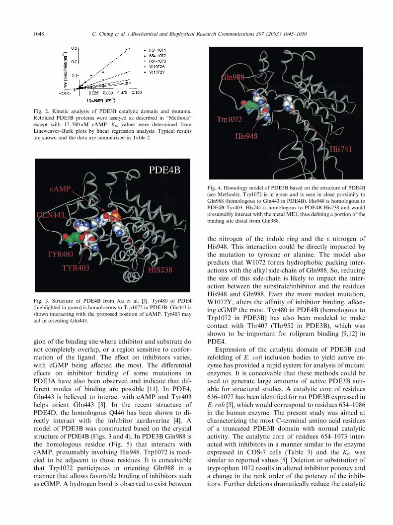
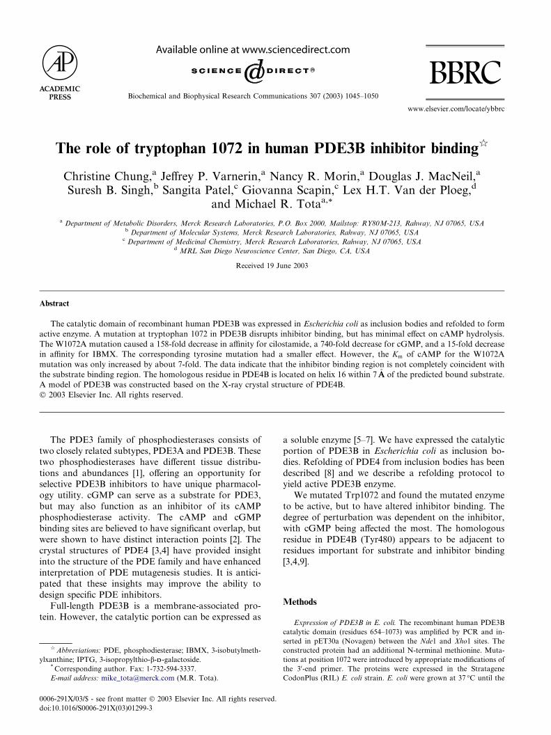
Fig. 4. Homology model of PDE3B based on the structure of PDE4B
(see Methods). Trp1072 is in green and is seen in close proximity to
Gln988 (homologous to Gln443 in PDE4B). His948 is homologous to
PDE4B Tyr403. His741 is homologous to PDE4B His238 and would
presumably interact with the metal ME1, thus defining a portion of the
binding site distal from Gln988.
Fig. 2. Kinetic analysis of PDE3B catalytic domain and mutants.
Refolded PDE3B proteins were assayed as described in “Methods”
except with 12–500 nM cAMP. Km values were determined from
Lineweaver–Burk plots by linear regression analysis. Typical results
are shown and the data are summarized in Table 2.
Fig. 3. Structure of PDE4B from Xu et al. [3]. Tyr480 of PDE4
(highlighted in green) is homologous to Trp1072 in PDE3B. Gln443 is
shown interacting with the proposed position of cAMP. Tyr403 may
aid in orienting Gln443.
1048 C. Chung et al. / Biochemical and Biophysical Research Communications 307 (2003) 1045–1050
gion of the binding site where inhibitor and substrate do
not completely overlap, or a region sensitive to confor-
mation of the ligand. The effect on inhibitors varies,
with cGMP being affected the most. The differential
effects on inhibitor binding of some mutations in
PDE3A have also been observed and indicate that dif-ferent modes of binding are possible [11]. In PDE4,
Gln443 is believed to interact with cAMP and Tyr403
helps orient Gln443 [3]. In the recent structure of
PDE4D, the homologous Q446 has been shown to di-
rectly interact with the inhibitor zardaverine [4]. A
model of PDE3B was constructed based on the crystal
structure of PDE4B (Figs. 3 and 4). In PDE3B Gln988 is
the homologous residue (Fig. 5) that interacts withcAMP, presumably involving His948. Trp1072 is mod-
eled to be adjacent to those residues. It is conceivable
that Trp1072 participates in orienting Gln988 in a
manner that allows favorable binding of inhibitors such
as cGMP. A hydrogen bond is observed to exist between
the nitrogen of the indole ring and the e nitrogen of
His948. This interaction could be directly impacted bythe mutation to tyrosine or alanine. The model also
predicts that W1072 forms hydrophobic packing inter-
actions with the alkyl side-chain of Gln988. So, reducing
the size of this side-chain is likely to impact the inter-
action between the substrate/inhibitor and the residues
His948 and Gln988. Even the more modest mutation,
W1072Y, alters the affinity of inhibitor binding, affect-
ing cGMP the most. Tyr480 in PDE4B (homologous toTrp1072 in PDE3B) has also been modeled to make
contact with Thr407 (Thr952 in PDE3B), which was
shown to be important for rolipram binding [9,12] in
PDE4.
Expression of the catalytic domain of PDE3B and
refolding of E. coli inclusion bodies to yield active en-
zyme has provided a rapid system for analysis of mutant
enzymes. It is conceivable that these methods could beused to generate large amounts of active PDE3B suit-
able for structural studies. A catalytic core of residues
636–1077 has been identified for rat PDE3B expressed in
E. coli [5], which would correspond to residues 654–1086
in the human enzyme. The present study was aimed at
characterizing the most C-terminal amino acid residues
of a truncated PDE3B domain with normal catalytic
activity. The catalytic core of residues 654–1073 inter-acted with inhibitors in a manner similar to the enzyme
expressed in COS-7 cells (Table 3) and the Km was
similar to reported values [5]. Deletion or substitution of
tryptophan 1072 results in altered inhibitor potency and
a change in the rank order of the potency of the inhib-
itors. Further deletions dramatically reduce the catalytic
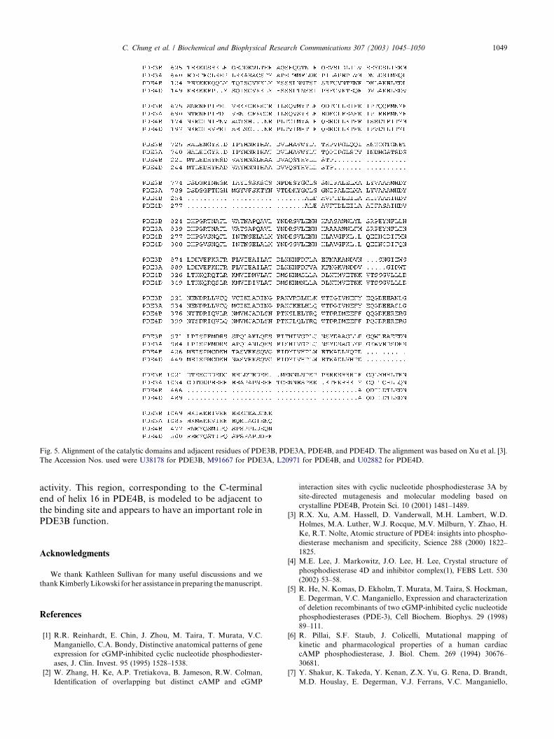
Fig. 5. Alignment of the catalytic domains and adjacent residues of PDE3B, PDE3A, PDE4B, and PDE4D. The alignment was based on Xu et al. [3].
The Accession Nos. used were U38178 for PDE3B, M91667 for PDE3A, L20971 for PDE4B, and U02882 for PDE4D.
C. Chung et al. / Biochemical and Biophysical Research Communications 307 (2003) 1045–1050 1049
activity. This region, corresponding to the C-terminal
end of helix 16 in PDE4B, is modeled to be adjacent to
the binding site and appears to have an important role in
PDE3B function.
Acknowledgments
We thank Kathleen Sullivan for many useful discussions and we
thankKimberlyLikowski forher assistance in preparing themanuscript.
References
[1] R.R. Reinhardt, E. Chin, J. Zhou, M. Taira, T. Murata, V.C.
Manganiello, C.A. Bondy, Distinctive anatomical patterns of gene
expression for cGMP-inhibited cyclic nucleotide phosphodiester-
ases, J. Clin. Invest. 95 (1995) 1528–1538.
[2] W. Zhang, H. Ke, A.P. Tretiakova, B. Jameson, R.W. Colman,
Identification of overlapping but distinct cAMP and cGMP
interaction sites with cyclic nucleotide phosphodiesterase 3A by
site-directed mutagenesis and molecular modeling based on
crystalline PDE4B, Protein Sci. 10 (2001) 1481–1489.
[3] R.X. Xu, A.M. Hassell, D. Vanderwall, M.H. Lambert, W.D.
Holmes, M.A. Luther, W.J. Rocque, M.V. Milburn, Y. Zhao, H.
Ke, R.T. Nolte, Atomic structure of PDE4: insights into phospho-
diesterase mechanism and specificity, Science 288 (2000) 1822–
1825.
[4] M.E. Lee, J. Markowitz, J.O. Lee, H. Lee, Crystal structure of
phosphodiesterase 4D and inhibitor complex(1), FEBS Lett. 530
(2002) 53–58.
[5] R. He, N. Komas, D. Ekholm, T. Murata, M. Taira, S. Hockman,
E. Degerman, V.C. Manganiello, Expression and characterization
of deletion recombinants of two cGMP-inhibited cyclic nucleotide
phosphodiesterases (PDE-3), Cell Biochem. Biophys. 29 (1998)
89–111.
[6] R. Pillai, S.F. Staub, J. Colicelli, Mutational mapping of
kinetic and pharmacological properties of a human cardiac
cAMP phosphodiesterase, J. Biol. Chem. 269 (1994) 30676–
30681.
[7] Y. Shakur, K. Takeda, Y. Kenan, Z.X. Yu, G. Rena, D. Brandt,
M.D. Houslay, E. Degerman, V.J. Ferrans, V.C. Manganiello,

1050 C. Chung et al. / Biochemical and Biophysical Research Communications 307 (2003) 1045–1050
Membrane localization of cyclic nucleotide phosphodiesterase 3
(PDE3). Two N-terminal domains are required for the efficient
targeting to, and association of, PDE3 with endoplasmic reticu-
lum, J. Biol. Chem. 275 (2000) 38749–38761.
[8] W. Richter, T. Hermsdorf, H. Lilie, U. Egerland, R. Rudolph, T.
Kronbach, D. Dettmer, Refolding, purification, and character-
ization of human recombinant PDE4A constructs expressed in
Escherichia coli, Protein Expr. Purif. 19 (2000) 375–383.
[9] O. Dym, I. Xenarios, H. Ke, J. Colicelli, Molecular docking of
competitive phosphodiesterase inhibitors, Mol. Pharmacol. 61
(2002) 20–25.
[10] D.J. MacNeil, J.L. Occi, P.J. Hey, C.D. Strader, M.P. Graziano,
Cloning and expression of a human glucagon receptor, Biochem.
Biophys. Res. Commun. 198 (1994) 328–334.
[11] W. Zhang, H. Ke, R.W. Colman, Identification of interaction sites
of cyclic nucleotide phosphodiesterase type 3A with milrinone and
cilostazol using molecular modeling and site-directed mutagenesis,
Mol. Pharmacol. 62 (2002) 514–520.
[12] R. Pillai, K. Kytle, A. Reyes, J. Colicelli, Use of a yeast expression
system for the isolation and analysis of drug-resistant mutants of a
mammalian phosphodiesterase, Proc. Natl. Acad. Sci. USA 90
(1993) 11970–11974.