the rostock university and rīga stradiņš university
TRANSCRIPT

14th Joint Symposium of
the Rostock University and
Rīga Stradiņš University
Maxillofacial Trauma Treatment
ABSTRACTS
24-26 May, 2018

2
14th Joint Symposium of the Rostock University and Rīga Stradiņš University Maxillofacial Trauma Treatment
24-26 May, 2018
Oral Presentation
ARMIN ANDRÄ AND ROSTOCK – RIGA IN MAXILLOFACIAL SURGERY
Andrejs Skagers, Department of Oral and Maxillofacial Surgery, Riga Stradins University, Latvia, [email protected]
The first joint symposium was organized by Wilhelm - Pieck - Universität Rostock, Faculty of Medicine, Section Stomatology, Regional Children Dental Clinic Rostock and Riga Medical Institute and held in Rostock 10 - 11 October 1989. There were 45 presentations, 6 from Riga. In appreciation of long - time successful work in Rostock - Riga cooperation Armin Andrä was awarded Doctor honoris causa in year 1990 as the first with such degree in Riga
Medical institute, now Riga Stradins University.
Afterwards Rostock–Riga symposiums becomes more international as 10th Joint symposium, Riga 2009 with participation of Rostock – Riga pioneers under leadership by professor A. Andrä, followed by professor’s Karsten Gundlach team. There were Colleagues from Austria, Estonia, Norway, Lithuania, Sweden, Russia, and Ukraine. Main topic was "Biomaterials and Bioengineering in Oral and Maxillofacial Surgery” A. Andrä did introduction lecture. Today we are together with third generation of Rostock Colleagues conducted by professor Bernhard Frerich. In the last letter (Rostock, dem 18.03.2018.) Professor A. Andrä wrote “I have a very good presentiment on my first visit in Riga. I never thought that 2018 will be the 14th Joint Sympsium Riga – Rostock.” But it is reality and may be explained by active work of A. Andrä particularly in first decades of cooperation when large part of Rostock staff
members were involved and visited Riga not only one time. We are grateful to Joachim Härtel, Ingrid and Michail Sonnenburg, Volker Bienengräber, H.J. Neumann, K.-O. Henkel and another colleague from Rostock University for our introduction into more modern for us West technologies in diagnostic and treatment methods during soviet period. Through Rostock, also as part of DDR, we had window to West Europe and world. We express our gratitude to professor Armin Andrä , professor Karsten Gundlach and professor Bernhard Frerich, all Rostock and international Colleagues in past, at the time being and I hope in future to keep spirit of Hanseatic Union into our specialties and clinics. Plenty of appreciations to the organizers and all participants of 14.Joint symposium Rostock – Riga.
Armin Andrä ,RSU Dr.h.c. ( 1990 )

3
14th Joint Symposium of the Rostock University and Rīga Stradiņš University Maxillofacial Trauma Treatment
24-26 May, 2018
Oral Presentation
Invited speaker
NAVIGATION IN CMF SURGERY
Maximilian Wagner

4
14th Joint Symposium of the Rostock University and Rīga Stradiņš University Maxillofacial Trauma Treatment
24-26 May, 2018
Oral Presentation
Invited speaker
THE NEW COMPREHENSIVE AO CMF TRAUMA CLASSIFICATION FOR ADULTS –
SCOPE, FEATURES AND CLINICAL CASE APPLICATIONS
Peter Cornelius
New imaging technologies reaching clinical practice and generating innovative treatment concepts have become particularly apparent in fractures of the mandibular condylar process and the internal orbit. The new comprehensive AO CMF trauma classification for adults has become a reality , i.e. the 3rd generation and is intended to account for the progress in the delineation of fracture patterns using CT-imaging. Three precision-levels, from elementary over basic to focused, provide the opportunity to document the location and morphology of fracture lines and patterns in hitherto unprecedented detail and clarity. The CMF partition of the AOCOIAC (AO COmprehensive Injury Automatic Classifer) Software Program for the entire human skeleton is used for this documentation. This is organized in a user friendly format visualizing the regions and subregions within each anatomical module. The topographical extent of a fracture is conveniently marked by locating and klicking the mouse cursor on the illustrations. The properties of the fracture are selected in pull-down menues or by ticking checkboxes in a side table data sheet. In Level 2 the mandible is divided into 9 subregions: Symphysis (S) + Bilaterally (2x): Body (B) , Angle / Ascending Ramus (A), Coronoid Process (C), Condylar Process (P). Midface Level 2 distinguishes between lateral (=Zygoma/Zygomatic Arch Ensemble) and central compartments, the latter being a a stack of three horizontal partitions: Upper Central Midface (UCM), Intermediate Central Midface (ICM) und Lower Central Midface (LCM). In Level 3 the topography of the subregions is further subdivided to display even more refined areas for fracture delineation. Moreover individual patient features, namely the pre-injury dental state and the degree of alveolar atrophy in edentulism can be recorded. Based on the refined structural layout it is easy to plot common fracture entities, i.e. Mandibular Condylar Process fractures, Nasal and Naso-Orbito-Ethmoid (NOE) Fractures, Zagoma / Zygomatic arch fractures and fractures of the internal orbits in their enormous range of variability and with ultimate sophistication as well as to assess tooth injuries, periodontal trauma and fracture involvement of the alveolar process. As a key attribute the focused Level 3 approach permits to document the fracture morphology in terms of fragmentation, displacement and bone loss. The initiators wish to express their hope that the present AO CMF Trauma Classification will provoke a flagship initiative for a departure to future innovations and amendments in the standadized description and diagnosis of CMF trauma in order to give an identical meaning to seemingly common terminology and contribute to more profound insights and understanding.

5
14th Joint Symposium of the Rostock University and Rīga Stradiņš University Maxillofacial Trauma Treatment
24-26 May, 2018
Oral Presentation
Invited speaker
CONDYLAR FRACTURES
Andreas Neff
Since the turn of the century a paradigm shift has challenged the centuries old tradition of non-surgical treatment for condylar fractures. Although first wire-based osteosyntheses had been performed already in 1924 by PERTHES, it was the advent of functionally stable osteosynthesis introduced by PAPE et al. into condylar traumatology in 1980, which step by step widened out the indications for surgical treatment, paralleled also by improvement in CT diagnostics and later continuous refinement of indication-specific osteosynthesis materials, with 3D-plates for the condylar base and neck and resorbable materials for the condylar head being the latest developments. Over the last decades, a series of new approaches was introduced, too, to cope with the specific demands for ORIF in the different fracture locations, which according to a recent consensus based proposal (AOCOIAC) are now classified as fractures of the condylar base, condylar neck and condylar head. By now even fractures of the condylar head, treated surgically by RASSE already in 1992, have moved into the focus of surgical interest and ORIF by now is far from being “experimental”, as dubbed only a decade ago. According to recent consensus (IBRA 2012) and based on improved evidence, ORIF may now be considered as the gold standard for both displaced or dislocated condylar base and neck fractures in adults, endoscopic approaches should be considered for base fractures with lateral displacement. Meanwhile, there is a growing tendency to perform ORIF in condylar head fractures and severely displaced or dislocated fractures in younger children with mixed dentition.

6
14th Joint Symposium of the Rostock University and Rīga Stradiņš University Maxillofacial Trauma Treatment
24-26 May, 2018
Oral Presentation
Invited Speaker
ORBITAL RECONSTRUCTION
Prof. Dr. Dr. Alexander Schramm, MD DDS Professor and Chairman, Department of Oral and Maxillofacial Surgery
University Hospital Ulm, Germany Clinical Director, Oral-, Maxillo and Plastic Facial Surgery
Military Hospital Ulm, Germany Email: [email protected]
Advances in imaging techniques pre- and intraoperatively and associated technologies have led within the past 20 years to improve preoperative and intraoperative guidance for the surgeon. Virtual reconstructions in complex trauma have become clinical routine in our patients care. Intraoperative guidance by navigational surgery or CAD-CAM templates are used. New software developments for automatic bone segmentation of standard CT data sets dramatically reduce the time period for preoperative planning in virtual orbital and periorbital reconstruction. The resulting virtual model is used for intraoperative navigation, guiding the reconstructive procedure to the desired result and controlled by intraoperative Ct scanning. Computer-assisted preoperative planning and surgery techniques have improved operators confidence in maxillofacial surgery. Using this technique all kinds of modifications of the shape of the plates can be virtually or mechanically preformed to allow patient specific prefabrication for primary and secondary reconstructions of. In this presentation the routine use of computer-assisted maxillofacial surgery is presented in orbital trauma repair.
7
14th Joint Symposium of the Rostock University and Rīga Stradiņš University
Maxillofacial Trauma Treatment 24-26 May, 2018
Oral Presentation
Invited speaker
INJURED FACES – LOST FACES
Peter Sieg, Lübeck
This presentation is about the peculiarity of our face region as in no other region do the aesthetic aspects play such a great role apart from the functional considerations. The aim of our daily surgical work in almost all situations deals with (re)-establishment of
function and form of different subunits of our body.
The set of tools we have to carry out our craft – the armamentarium we have – is first
of all our biological understanding of the underlying problem and secondly the methods of
reconstructive surgery and the fast growing amount of technological aid. Especially in
management of complex facial trauma new technical developments have changed the pathways
of the surgical treatment and have dramatically improved our options for reestablishing
function and form. Nevertheless in cases of complex trauma especially after parts of the face
have been destroyed or got lost our surgical options are still limited and followed by physical
and social impairment of our patients. To exemplify options and limits clinical examples will
be presented and discussed in detail with regard to different possible pathways of surgical
treatment and prospective surgical options along the road into the future.

8
14th Joint Symposium of the Rostock University and Rīga Stradiņš University
Maxillofacial Trauma Treatment 24-26 May, 2018
Oral Presentation
Invited speaker
TERROR IN NORWAY, JULY 22, 2011
Per Skjelbred Prof. MD,DDS,PhD, Dr.h.c.,University of Oslo
On July 22, 2011 two terror attacks hit Norway. They were both performed by the same Norwegian, right wing terrorist.
Oslo university hospital, Ullevål is the level one trauma unit for 2.7 million in the south of Norway. Annually 1500 patients are admitted, 40% with an Injury Severity Score (ISS) more than 15.
The terrorist detonated a car bomb (950 kg fertilizer/diesel) in the government area, killing 8 and injuring 98. Pre hospital triage resulted in 10 patients being admitted to the trauma unit.
The terrorist then drove 40 km to the port of the ferry to the island of Utøya, where a political summer camp for more than 600 social democratic youths took place.
Disguised as a policeman he entered the ferry and arrived at the island shortly after. For the next hour he went around, undisturbed, executing and wounding the children.
The police failed to protect and received massive criticism afterwards. 68 were killed and 61 wounded. 12 patients were transported by helicopter to the trauma unit directly, the rest to local hospitals. Later, 9 patients were admitted from these hospitals to the trauma unit.
The trauma unit received a total of 31 patients. One was severely injured by a headshot and died. A total of 163 specific injuries were noted. The total lengths of stay for the 31 patients were 503 days. This presentation includes the logistics, the injuries and special attention regarding 11 patients with facial injuries.

9
14th Joint Symposium of the Rostock University and Rīga Stradiņš University Maxillofacial Trauma Treatment
24-26 May, 2018
Oral Presentation
COMPARISON OF LOCKING AND NONLOCKING PLATES IN THE TREATMENT OF
MANDIBULAR ANGEL AND CONDYLE FRACTURES
B.Frerich, R.Seemann, S.Muller, O.Ploder, K.Schicho, A.Wagner, J.Piffko Rostock / Vienna / Leipzig / Munster / Feldkirch
Aim To adress the potential clinical advantage of an angular stable locking system in the treatment of standard fractures of the mandible.
Methods A prospective, randomized clinical multicenter trial was conducted comparing Modus Trilock 2.0 miniplates with standard 2.0 miniplates in mandibular angle fractures and mandibular condyle fractures. Patients with more than three fracture sites, comminuted fractures or additional central midface or alveolar process fracture and non-conscious or polytraumatized patients were excluded. Clinical and radiographic findings were recorded perioperatively, 6-10 weeks and 5-7 months postoperatively.
Results 160 patients were included, 50 patients with angle fractures and 129 patients with 146 condyle fractures. In mandibular angle fractures, no complications (0%) occurred in the locking group, but 3 osteosynthesis failures in the non-locking group (11%, p=0,236). In condyle fractures, osteosynthesis failures were distributed equally in both locking and non-locking group (2,8% vs. 2,7%, p=1). There was, however am marked difference by surgical approach (transoral: 4 failures, extraoral: 0 failures). The difference however, was not significant (p=0,3).
Conclusions No significant improvement was found by use of locking systems in the treatment of mandibular fractures, although there was tendency to an advantage in mandibular angle fractures. Correct anatomical reduction of high condyle fractures is better achieved by an extraoral surgical approach and seems to prevent osteosynthesis failure.

10
14th Joint Symposium of the Rostock University and Rīga Stradiņš University Maxillofacial Trauma Treatment
24-26 May, 2018
Oral Presentation
MAXILLOFACIAL TRAUMAS IN ESTONIA IN YEARS 2013-2017
Marianne Soots1; Riina Lutter1; Peeter Viidebaum2
1Department of Maxillofacial Surgery, Tartu University Hospital, Tartu, Estonia; [email protected]
2Face and Jaw Surgery Unit, North Estonia Medical Centre, Tallinn, Estonia
Aim The purpose of this study was to establish the type, quantity, etiology, treatment and
cost of maxillofacial traumas treated in two estonian maxillofacial surgery departments in Tallinn and Tartu in years 2013-2017.
Methods The current study was a retrospective overview. The data was collected from patients
treated in two estonian maxillofacial surgery departments in years 2013-2017. The statistics was collected and based by the diagnostic codes from The International Classification of Diseases, 10th Revision. It was assessed, which fractures were the most frequent by years, what was the etiology of the fractures, if the patients were treated operatively and what was the medium cost of a case treatment.
Results The most frequent on all years were mandibular fractures. These were followed by
maxillary, multiple and orbital floor fractures in descending order. Results didn`t include zygomatic fractures, because ICD-10 doesn`t have specific code for them, so these could be contained either with maxillary or multiple fractures. Nasal fractures were also excluded, because these are mostly treated by otorhinolaryngologists. The most common etiologies included fallings and assaults.
Conclusion The most commonly treated fractures by estonian maxillofacial surgeons were
mandibular, maxillary, multiple and orbital floor fractures. Etiology was mostly falling or assault. Patsients were treated either conservatively or operatively.

11
14th Joint Symposium of the Rostock University and Rīga Stradiņš University Maxillofacial Trauma Treatment
24-26 May, 2018
Oral Presentation
COMPLEX TREATMENT PLANNING FOR TRAUMATIC
MANDIBULAR NERVE INJURY
Kristina Vilkitskaya MD, PhD, Prof. Irina Pohodenko-Chudakova MD, PhD, DMSci Department of Oral surgery, Belarusian State Medical University, Minsk, Belarus
Introduction Because of the anatomical features of the mandibular nerve its sensitive branches can
be easily injured during dental manipulations. According to studies spontaneous recovery of nerve function after trauma may take up to six months, so patients are often not treated, that results in the development of chronic persistent neurosensory disturbances.
Aim To determine the factors influencing the treatment planning for patients with traumatic
mandibular nerve injury.
Methods Were enrolled 25 patients with inferior alveolar nerve injury and 5 – with lingual nerve
lesion. Physical examination included the determination of grade of sensory deficits and electropuncture diagnostics using “Bioreper” method. Complex treatment consisted of reflexology and medication.
Results The severity of sensory disturbances in 1 patient was mild, in 22 – moderate and in 7
cases – severe. Hyperfunction of nervous system was revealed by electropuncture testing in all patients indicating compensatory process. More positive treatment outcomes were observed when patients complained of partial or complete loss of sensation before therapy (without symptoms of sensory hypersensitivity or radiation of pain). The appearance of “pins and needles” feeling during treatment was a good prognostic sign. With early treatment (up to one month after nerve injury) 83% of patients achieved full recovery of sensitivity. The longer nerve was injured, the less was the positive outcome of therapy.
Conclusion The main factors affecting effectiveness of treatment of traumatic lesion of mandibular
nerve that must be taken into consideration when planning timelines and intensity of therapy are a timeframe following injury and severity of sensory disturbances.

12
14th Joint Symposium of the Rostock University and Rīga Stradiņš University Maxillofacial Trauma Treatment
24-26 May, 2018
Oral Presentation
NEW STRATEGY OF ORBITAL RECONSTRUCTION,
BASED ON CAD/CAM TECHNOLOGIES
Yurii Chepurnyi, Andrii Kopchak, Denis Chernogorskyi Department of Stomatology
O.O. Bogomolets National Medical University, Center of Maxillo-facial surgery and Stomatology in Kiev regional hospital
Kiev, Ukraine Contacts: [email protected]
The recent developments of CAD/CAM technologies and evidence of their effective clinical application in management of facial bone defects and deformities cause an increased interest to their usage in reconstructive surgery of the orbit.
Purpose The aim of this study was to evaluate the efficacy of patient specific implants at patients
with post-traumatic orbital defects and deformities.
Material and methods 31 patient with posttraumatic orbital defects and deformities with subsequent
reconstructive procedures using patient specific implants were included to the study. All patients were examined according to the standardized algorithm including local status examination, vision assessment and computer tomography before and after surgery. Implants positioning and its conformity with preoperative planning, as well as their clinical efficacy were evaluated.
Results In all the patients no postoperative inflammatory complications, decreased visual acuity
or loss of visual fields were found. The average period of implant design and manufacturing was 5.9±2.5 days. The average duration of the surgical interventions was 54.1±11.5 minutes. Elimination of functional disorders 1 month later surgical intervention was observed in 65.2% of cases, followed by 86.96% in 3 month term. Positive aesthetic results were obtained in 95.7% of patients.
Conclusion Orbital reconstruction using patient specific implants is an effective procedure that
allows to restore the complex anatomy of the orbit and to improve the functional outcomes in patients with post-traumatic orbital defects and deformities.

13
14th Joint Symposium of the Rostock University and Rīga Stradiņš University Maxillofacial Trauma Treatment
24-26 May, 2018
Oral Presentation
APPLICATION OF 3D PROTOTYPING FOR COMPLICATED ORBITAL WALL
FRACTURE RECONSTRUCTION
Martins Lauskis1; Girts Salms2
1Riga Stradins University, Riga, Latvia 2 Riga Stradins University, Institute of Stomatology, Riga, Latvia
Aim To present first two cases treated with pre-bent titanium mesh using 3D anatomical models.
Methods Two patients were included in this case series both of whom had complaints of double vision and changed eye position after blunt trauma to the eye. Two different 3D anatomical models were made for each patient, one with reconstructed orbit and one with the actual damaged orbit. Titanium mesh implants were formed for each patients specific anatomy.
Results In one patient, complete absence of diplopia was achieved after surgery. Second patient had major improvement of symptoms with some residual diplopia in extreme upgaze.
Conclusion The unique anatomy of the orbit and the limited surgical view during operation make the process of fitting and aligning implants difficult, time consuming and largely dependent from surgeon skills. 3D printing technology allows us to make relatively inexpensive anatomical models that can be used to pre-bend titanium mesh implants to patients specific anatomy. The use of 3D anatomical models to prepare pre-bent titanium mesh implants is a financially and clinically viable method to treat complicated orbital wall fractures.

14
14th Joint Symposium of the Rostock University and Rīga Stradiņš University Maxillofacial Trauma Treatment
24-26 May, 2018
Oral Presentation
USE OF β-Ti-Zr-Nb ALLOY WITH MODIFIED ELASTICITY MODULUS IN
TREATMENT OF PATIENTS WITH TRAUMATIC FACIAL BONES FRACTURES
Anna Romanova1 , Igor Skiba2 , Аndrey Kopchak1 1 Bogomolets National Medical University, Institute of postgraduate education,
Department of Dentistry, Kyiv, Ukraine 2 G.V. Kudyumov Institute For Metal Physics, Kyiv, Ukraine
e-mail: [email protected]
Aim To study the biomechanical properties of the "fixator-bone" system after open reduction and internal fixation with miniplates from a modified β-Ti-Zr-Nb alloy in patients with traumatic fractures of the facial bones.
Methods Clinical and instrumental studies included analysis of the immediate and long-term results of the osteosynthesis with β-Ti-Zr-Nb miniplates compared to the traditional titanium miniplates in patients with facial bones fractures, estimation of radiological data, intraoperative measurement of torques when fixing screws were installed, electron microscopy of titanium plates and fixators from β-Ti-Zr-Nb alloy, removed in remote terms after operation, computer simulation of the stress and strain state using the finite element (FE) method.
Results The installation of plates from modified β-Ti-Zr-Nb alloy with a low elasticity modulus in fractures of the facial bones provided reliable fixation stability and reduced the stress shielding effect. The value of torque in the installation of screws from this alloy consisted 10-50 Ncm with no significant differences with titanium screws. It determined the sufficient initial stability of the fixation system in most of the patients. The results obtained were in accordance with FE simulation. The potential risks of system failure were associated with a posttraumatic decrease in the bone "quality" in the area of the surgical intervention.
Conclusions Low modulus of elasticity, high value of the reversible deformation, high fatigue strength, low magnetic susceptibility and high level of X-ray contrast determine the clinical benefits of the β-Ti-Zr-Nb miniplates usage in patients with facial bone fractures.

15
14th Joint Symposium of the Rostock University and Rīga Stradiņš University Maxillofacial Trauma Treatment
24-26 May, 2018
Oral Presentation
ALLOPLASTIC RECONSTRUCTION OF THE TEMPOROMANDIBULAR JOINT FOR
REHABILITATION OF FUNCTION AND PAIN REDUCTION
Birte Julia Siegmund1, Karsten Winter
2, Jan Rustemeyer
1
1. Department of Craniomaxillofacial and Facial Plastic Surgery, Bremen Central Medical Center, Bremen, Germany
2. Institute of Anatomy, University of Leipzig, Faculty of Medicine, Leipzig, Germany
Aim The implantation of an alloplastic total endoprosthesis of the temporomandibular joint
is an innovative approach for treatment of diseases with severe internal derangement (Wilkes IV and V). Two types of replacements are existing: custom made cad/cam and stock prothesis. Effectiveness and stability of both designs are subjects of a clinical study in a six months follow up.
Methods The retrospective study includes 28 patients, devided in two groups: stock-prosthesis
and custom-made-cad/cam-prothesis, both from Biomet Microfixation (Jacksonville, USA). Clinical examination was done at five different time points up to six months. Examined parameters were mouth opening (mm) and a visual analogue scale (0-10) regarding pain. Patient-specific diet was documented (liquid, soft or normal).
Results Both groups showed significant improvement in mouth opening and pain reduction
with statistically significance after six months (p < 0.001). No patient had a liquid diet at the end of treatment. 64% in group 1 and 100% of patients in group 2 showed an improvement of well-being. Complications were observed in 33% of all patients including temporary paralysis of the facial nerve, permanent paralysis and a salivary fistula of the glandula parotidea. Infections, mal occlusion or lost of joint prosthesis were not observed within the study.
Conclusions The results of the presented study showed that alloplastic tmj reconstruction is a
promising approach for treatment of severe tmj disorders. Within the presented study, patients showed a high significant improvement of complaints with a stability of six months.

16
14th Joint Symposium of the Rostock University and Rīga Stradiņš University Maxillofacial Trauma Treatment
24-26 May, 2018
Oral Presentation
INTRAOPERATIVE DETECTION OF FACIAL NERVE BRANCHES USING
STIMULATION WITH FOCEPS-TYPE ELECTRODES
Janis Dumpis1, Gunars Lauskis2, Mario Alkhamisi3, Karlis Mackevics4 1, 2 Department of Oral and Maxillofacial Surgery, Riga Stradiņš University
3 Student, Faculty of Dentistry, Riga Stradiņš University 4 Student, Faculty of Mechanical Engineering, Transport and Aeronautics, Riga
Technical University [email protected]
Aim Making surgical operations has a risk in the scar to damage the motor nerves, which
results in permanent muscle paresis postoperatively. Such situations should be avoided.
Methods If a naked nerve is touched directly with electricity, the impulse causes visible
contractions of innervated muscles. This method was used for identification of nerve in the surgical scar. After the identification operator can choose other routes for cuts, without to damage the nerve.
Experiments were realized in dissected specimens of frogs back with the hinder limb. The sciatic nerve was stimulated with direct current impulses of different voltage, causing frogs leg muscles contractions.
The forceps-type electrodes were elaborated by experiments, and electrical device for nerve control was designed.
Device was used for detection of facial nerve branches in operations for patients with parotid adenoma.
Results Function of patient’s musculature remained in a good condition postoperatively.
Conclusion The electrodes were convenient, and device was effective for use in the detection of
nerves.

17
14th Joint Symposium of the Rostock University and Rīga Stradiņš University Maxillofacial Trauma Treatment
24-26 May, 2018
Oral Presentation
METABOLIC PROFILING REVEALS ITACONIC ACID
AS A MARKER OF OSTEOPOROSIS
Nadja Engel1, Jan Lisec2, Dagmar-Christiane Fischer3, Bernhard Frerich1
1 Department of Oral and Maxillofacial Surgery, Facial Plastic Surgery, Rostock University Medical Center, Rostock, Germany Email: [email protected]
2 Department of Analytical Chemistry, BAM Federal Institute for Materials Research and Testing, Richard-Willstätter-Str. 11, 12489 Berlin, Germany
3 Department of Pediatrics, Rostock University Medical Center, Rostock, Germany
Aim
This study was conducted to identify potent biomarkers that are indicative of osteoporosis induction. If possible, the biomarkers should be detected in the blood or urine in order to detect early osteoporotic alterations and to monitor treatment successes. Therefore, metabolomics alterations of urine and blood of osteoporotic vs. control mice and rats were analyzed.
Methods
By liquid chromatography combined with mass spectrometry (LC-MS) based metabolomics a set of 700 metabolites were analyzed. All statistical tests on metabolite profiles have been conducted in R (www.r-project.org). All data were log10-transformed to improve normality.
Results
Subsequently to sample preparation, 225 metabolites in the mice urine were identified based on retention times, characteristic ions and mass spectra, comprising mainly amino acids, organic acids and mono- and disaccharides. A ranking of altered metabolites between young, old, female and male mice with or without osteoporosis revealed that the metabolite itaconic acid (Kegg no. C00490) is significantly increased in in osteoporotic female mice. No alterations of the itaconic acid concentrations were detected in all control groups. Furthermore, a panel of amino acids for example alanine and cysteine as well metabolites of the folate metabolism showed reduced concentrations in osteoporotic mice.
Conclusion
Initial results of this study highlighted raised concentrations of itaconic acid as a potential marker of osteoporosis. Currently, blood and urine samples of ovariectomized Sprague Dawley rats as another osteoporosis model, treated with the Pro Bone Morphogenetic protein-2, are under metabolomic examinations.

18
14th Joint Symposium of the Rostock University and Rīga Stradiņš University Maxillofacial Trauma Treatment
24-26 May, 2018
Oral Presentation
PATTERNS OF MAXILLOFACIAL FRACTURES IN A UNIVERSITY MEDICAL CENTRE
DURING FIVE YEARS – A CHANGE IN ETIOLOGY
M. Goedecke1, D. Thiem2, D. Schneider3, M Dau2, B. Saka2, B. Frerich2, P.W.Kämmerer2
1Department of Oral and Maxillofacial Surgery, Charité Universitätsmedizin Berlin, Augustenburger Platz 1, 13353 Berlin, Germany, Dr. med. Dr. med. dent.
Maximilian Goedecke, [email protected] 2Department of Oral and Maxillofacial Surgery, University Medical Centre Rostock, Rostock, Germany, Dr. med. Daniel Thiem, Dr. med. Dr. med. dent. Michael Dau, PD
Dr. med. Dr. med. dent. Bassam Saka, Prof. Dr. med. Dr. med. dent. Bernhard Frerich, PD Dr. med. Dr. med. dent. Peer-Wolfgang Kämmerer
3Department of Oral and Maxillofacial Surgery, Helios Kliniken Schwerin, Schwerin, Germany, Dr. med. Dr. med. dent. Daniel Schneider
Aim The purpose of the present study was to illuminate the patterns of maxillofacial fractures
in a University Medical Centre and to analyze potential changes in patient population and treatment during the period of five years.
Methods In a retrospective analysis, 573 patients with maxillofacial fractures treated in a level
one Trauma Center (Rostock University) from January 2010 to December 2014.
Results The most common causes for fractures were assaults (28.8%; n = 165), followed by falls (23.9%; n = 137). Nevertheless, fall became the main reason for maxillofacial fractures in 2013 and 2014. The mean age for patients suffering from facial fractures due to falls was significantly higher compared to those injured by other reason. Most commonly, fractures of the zygomaticomaxillary complex with or without orbital floor involvement were seen (31.6%; n=291) followed by fractures of the mandible (20.6%; n=190) and fractures of the nose (15.2%; n=140). The majority of patients was treated surgically (89.5%; n=513) even though an increase of non-surgical treatment was found over the examination period, especially in older patients.
Conclusion Maxillofacial fractures are mostly seen in younger male patients. Though, there is a
change in etiology; while assault was the main pattern of injury during the whole examination period, falls in older patients have become the main cause within the last 2-years. Most likely there will be an increase in facial fractures due to falls.

19
14th Joint Symposium of the Rostock University and Rīga Stradiņš University Maxillofacial Trauma Treatment
24-26 May, 2018
Oral Presentation
Invited speaker
GUNSHOT TRAUMAS
Oleg Kovtunyak, Ukraine

20
14th Joint Symposium of the Rostock University and Rīga Stradiņš University Maxillofacial Trauma Treatment
24-26 May, 2018
Oral Presentation
FIREWORK CAUSED FACIAL TRAUMA
CASE REPORT
Girts Salms, Ina Freimane Riga Stradins University Pauls Stradins University Hospital
Department of Maxillofacial surgery Latvia e mail: [email protected]
Fireworks are frequently used in celebrations on public events. Careless use of fireworks may cause severe tissue damage.
Aim To report a case of the patient suffered from firework blow in the face, problems, treatment and complications.
Method Accident happened with male patient on 2014 New Year Eve when patient was 32 years old. Bottle rocket did not act and when patient checked it suddenly blasted in his face. As result of trauma patient lost vision on both eyes achieved NOE, maxillary segmented and orbital fractures, cerebral hematoma and contusion. Patient had emergency operations for his eye and maxillary fracture. Neurosurgical operation was not performed. One month after treatment in the University hospital patient was discharged from there. 6 months after trauma patient was looking for reconstruction of nose and closing of persisting fistula on radix of nose. Unfortunately, Latvian specialists did not find will and resources to do nasal reconstruction. Patient find the opportunity to do reconstruction in the UK. 2016 July his operation was performed in the Northwestern hospital in London.
Results Frontal sinus fistula was successfully treated and nose reconstruction was done by calvaria bone graft covered with pericranial flap. 3 weeks after surgery gradual exposure of flap was found. On the following 6 months several revisions were necessary to debride bone graft. Further graft exposure was prevented however large piece of transplanted bone was lost.
Conclusion Frontal sinus infection was eliminated however good aesthetic result was not achieved. Total reconstruction of nose is highly demanding surgery asking careful planning and experience. Further nose reconstruction is needed.

21
14th Joint Symposium of the Rostock University and Rīga Stradiņš University Maxillofacial Trauma Treatment
24-26 May, 2018
Oral Presentation
Invited speaker
ORTHOGHNATHIC SURGERY AFTER MAXILLOFACIAL TRAUMA
Simonas Grybauskas, Lithuania
Orthognathic surgery is a well established tool for correction of congenital and acquired dentofacial deformities. Maxillofacial trauma and/or the consequences of its treatment are a part of acquired deformities. The specificity of planning and treatment of posttraumatic deformities lies in the fact that trauma and/or inadequate repair leads to positional and morphological changes of the facial bones, thus orthognathic surgery alone may not guarantee a full aesthetic satisfying outcome if bone morphology is not corrected simultaneously. Therefore, orthognathic surgery should be usually preceded with a meticulous 3D planning of new position of bones, and reshaping/grafting or application of patient specific implants.
A series of cases with posttraumatic mandibular and midface deformities will be analyzed and current orthognathic surgery techniques will be demonstrated during the lecture.

22
14th Joint Symposium of the Rostock University and Rīga Stradiņš University Maxillofacial Trauma Treatment
24-26 May, 2018
Oral Presentation
TREATMENT OF INFANTS WITH SYNDROMIC ROBIN SEQUENCE (SRS) WITH
THE PRE-EPIGLOTTIC BATON PLATE (PEBP),
A MINIMALLY INVASIVE TREATMENT OPTION
Silvia Muller-Hagedorn (1, 2, 3, 4), Wolfgang Buchenau (1, 2) Jorg Arand (1, 2) Margit Bacher (5), Christian Poets (1, 2)
(1) Interdisciplinary Center for Craniofacial Malformations, Tuebingen University Hospital, Tuebingen, Germany
(2) Department of Neonatology, Tuebingen University Hospital, Tuebingen, Germany (3) Department of Orthodontics, Tuebingen University Hospital, Tuebingen, Germany
(4) Department of Orthodontics, University of Rostock, Rostock, Germany (5) BIP Orthodontic Practice, Tuebingen, Germany
Aim Infants with SRS suffer from more severe upper airway obstruction (UAO) and feeding
problems than those with isolated RS. We investigated whether our treatment concept for isolated RS, consisting of a palatal plate with a velar extension (PEBP) shifting the tongue into a more anterior position and functional treatment, is also effective in SRS.
Methods A retrospective chart review of all children admitted to our department with syndromic
RS over a 7-year period was carried out. UAO was quantified by cardiorespiratory sleep studies performed before and during PEBP treatment. Obstructive sleep apnea was defined as a mixed obstructive sleep apnea index (MOAI) >3/h. Feeding modalities and weight were also evaluated.
Results Of 68 children meeting inclusion criteria, 56 completed treatment. Underlying
diagnoses included craniofacial dysostosis (N=13) and synostosis syndromes (N=5), unspecified dysmorphic syndromes (N=23) and miscellaneous rare conditions (N=27). Median MOAI decreased from 8.5 (range 0.3-76.0) at admission to 1.1 (0.0-5.2) at discharge (p<0.001). 51 children received only a TPP and 5 additional respiratory support (e.g., CPAP) during sleep for mild residual OSA. The number of exclusively gavage fed infants was reduced from 23 to 7. Median SDS for weight improved from -1.6 (-3.5-1.7) to -1.3 (-4.1-2.5). Of the 12 infants not completing treatment, 3 ultimately underwent tracheostomy.
Conclusion Treatment of UAO and feeding problems in children with syndromic RS by a PEBP
was shown to be effective and safe. If confirmed in prospective studies, it may help to avoid more invasive interventions.

23
14th Joint Symposium of the Rostock University and Rīga Stradiņš University Maxillofacial Trauma Treatment
24-26 May, 2018
Oral Presentation
SPEECH OUTCOMES IN PATIENTS WITH UNILATERAL CLEFT LIP AND PALATE
AFTER ONE- OR TWO-STAGE PALATOPLASTY
Ann Dieckmann1, Susanne Josko1, Ija Bauska2, Andzela Steinberga2, Asta Lipnickiene3, Marika Padrik4, Karsten Gundlach1 , Franka Stahl 5, Jan-Hendrik Lenz 1
1 Department of Oral and Maxillofacial and Facial Plastic Surgery, University of Rostock, Rostock, Germany
2 Department of Logopaedics, Stradins University, Riga, Latvia 3 Department of Logopaedics, University of Vilnius, Vilnius, Lithuania 4 Department of Speech Pathology, University of Tartu, Tartu, Estland
5 Department of Orthodontics, University of Rostock, Rostock, Germany
Aim To evaluate speech in patients with unilateral cleft, lip, alveolous and palate after one-
stage or two-stage palatoplasty and compare speech with dynamic and static orofacial dysfunctions
Material 92 patients with unilateral total clefts of lip, alveolus and palate (Q.37.5) from 4
different European cleft palate centers at the age of 3 and 5 years with a self developed protocol for minimal documentation. The main difference was, that in centers A and C palates were closed in two-stage whilst in centers B and D palatoplasty was performed in just one operation.
Methods Evaluating primary orofacial functions (like breathing, sucking, chewing, biting,
swallowing and tongue rest position) as well as secondary functions (like speech and voice) with standardized objective tests: payne-technique according to Garliner, functional tonometer according to Thiele, articulation test according to Dieckmann/Dieckmann and A-I-check according to Gutzmann. Pearson’s chi square test was used for statistical analysis and p≤0,05 was defined as significant.
Results The open mouth posture (40%), unphysiological tongue rest position (37%) and visceral
swallowing pattern (79%) were the most frequently diagnosed primary orofacial dysfunctions with all test persons. There were no significant differences between the participating cleft palate centers. Malarticulation and hypernasality (being secondary orofacial dysfunctions) were diagnosed in 35% and 82% of all subjects respectively with significant differences between the four cleft palate centers. This can be explained by the differences in treatment plans. Non significant differences in velopharyngeal function were found among these four groups at 3 and 5 years of age.
Conclusions The two-stage palatoplasty in patients with unilateral clefts of lip, alveolus and palate
in combination with an early treatment of primary and secondary orofacial dysfunctions is still a valuable treatment protocol; especially when comparing results with single-stage palatoplasty outcomes.

24
14th Joint Symposium of the Rostock University and Rīga Stradiņš University Maxillofacial Trauma Treatment
24-26 May, 2018
Oral Presentation
RESULTS FROM A PROSPECTIVE INTERDISCIPLINARY STUDY (BALTIC CLEFT
NEWTWORK) – COMPARISON OF LOGOPEDIC AND ORTHODONTIC RESULTS IN
5-YEAR OLD PATIENTS HAVING UCLAP
J-H. Lenz1 , Ann Dieckmann1, Susanne Josko1, M. Strosinski2, Juliane Neubert2, Ija Bauska4, Inese Maulina3, Inta Zepa3, Dace Pride3, Ilze Akota4,
K.Gundlach1, Franka Stahl2 1 Dept. of Oral-Maxillofacial-Surgery, Universitatsmedizin Rostock, Germany
2 Dept. of Orthodontics, Universitatsmedizin Rostock, Germany 3 Dept. of Orthodontics, Riga Stradins University, Latvia
4 Dept. of Oral-Maxillofacial-Surgery, Riga Stradins University, Latvia
Aim To compare logopedic and orthodontic results from two European Cleft Centers with
different treatment protocols (NAM: pre- vs. postoperatively; Cheiloplasty: Pfeifer vs. Millard; Veloplasty (Kriens): 6 months vs. 9-11 months; Uranoplasty (v.Langenbeck): 2-3 years vs. 3-4 years; Speech therapy: start with 1 year vs. 3 years of age). In the Cleft Centers prospective records were taken using one single interdisciplinary protocol.
Patients and methods In 50 caucasian non-syndromic 5-year old patients with UCLAP from the two Cleft
Centers 3-D scanning of orthodontic casts (T1-birth, T2-before cheiloplasty, T3-before veloplasty, T4-before uranoplasty T5- 5 years old) have been performed. Orthodontic analysis oft casts had followed regarding maxillary anterior and posterior dental arch dimensions, palatal volumetry and analysis of intermaxillary relationsships (overjet, overbite, lateral cross-bites). Speech therapists have compared data from primary (breathing, sucking, biting, chewing, swallowing, tongue rest position) and secondary dysfunctions (articulation, phonation). Finally interdisciplinary comparison of results had followed. Statistics: Chi-square-test p≤0.05.
Results Measurment of anterior and posterior width of maxillary dental arch dimensions have
shown 29.2mm and 39.8mm in average. The Goslon Yard Stick Index has been 2.64 in mean. Comparison of orthodontic measurements regarding the transversal plane haven´t shown significant differences between the centers. In the sagittal plane Angle Class II occlusal relationsships were found most frequently on the cleft side in both centers; on non-cleft side Angle Class I relationsships were found predominantly. In both centers primary dysfunctions were found frequently: mouth breating (40%), tongue rest position (37%), visceral swallowing (79%). Analysis of records have even shown speech dysfunctions (35%) und hypernasality (82%) in both centers. Significant differences were found when comparing primary and secondary dysfunctions in the centers.
Conclusions 3D-analysis of dental casts in a 5-year treatment period can be used to get valuable
additional information in interdiscipliary treatment comparisons of patients having UCLAP. The technique allows a concise analysis regarding relationsships of form and function. The

25
effect of presurgical NAM and early myofunctional therapy will be reflected comparing 5-year old patients with UCLAP in both centers.
26
14th Joint Symposium of the Rostock University and Rīga Stradiņš University Maxillofacial Trauma Treatment
24-26 May, 2018
Oral Presentation
NASALOLABIAL ANTHROPOMETRY IN PATIENTS WITH UNILATERAL
COMPLETE CLEFT LIP, ALVEOLUS AND PALATE AND CONTROL GROUP
Ieva Bagante1, Ilze Akota2
1,2Department of Maxillofacial Surgery, Riga Stradiņš University Institute of Stomatology, Latvia
Aim Aim of the study was to evaluate and compare the nasolabial appearance in patients
with unilateral cleft lip, alveolus and palate (UCLP) and control group.
Methods All consecutive 35 patients born between 1994 and 2004 with nonsyndromic complete
UCLP were included. Of 35 patients, 30 responded for recall with mean age 14 years (range 10-18). In the control group, 35 non-cleft participants at 10 years of age were included. The nasolabial anthropometry was performed in 3D images with 25 anthropometric landmarks and 18 distances. Symmetry index was calculated as: distance in cleft side versus distance in non-cleft side x 100.
Results In UCLP group, statistically significant difference between cleft and non-cleft side was
found in alar wing length. The difference in control group between left and right side was not significant. In UCLP group the symmetry index showed a statistically significant difference in length of red lip from crista philtrum to midline, nose lateral length and alar wing length. In control group, the symmetry index showed statistically significant asymmetry of nostril height. Comparing symmetry index between UCLP and control group, statistically significant difference was found in the length of white lip, nose lateral length and alar wing length.
Conclusion The nasolabial appearance with acceptable symmetry after reconstructive surgery was
achieved. Symmetry of the nasolabial appearance in patients with UCLP differed from control group. Symmetry index was informative for nasolabial symmetry comparison of the UCLP group and the control group as there was variation in age and stature, excluding direct comparison of anthropometric distances.
27
14th Joint Symposium of the Rostock University and Rīga Stradiņš University Maxillofacial Trauma Treatment
24-26 May, 2018
Oral Presentation
QUALITY OF LIFE OF PATIENTS WITH ADVANCED ORAL CANCER AFTER
SURGERY REQUIRING FREE FLAP RECONSTRUCTION
Kalvis Pastars1, Janis Zarins1, Juris Tars2, Anna Ivanova 3, prof. Andrejs Skagers3
1. Microsurgery Centre of Latvia, Riga, Latvia
2. Latvian Oncology Centre, Riga, Latvia 3. Riga Stradins University Institute of Stomatology
Background Oral squamous cell carcinoma (OSCC) is a common type of cancer affecting people
worldwide, with still large proportion of patients diagnosed with the disease in the advanced stage. Numerous studies have been published evaluating and measuring the Quality of Life (QOL) of the treated patients affected by oral cancer at any stage. Though not so many studies deal with evaluation of the impact on QOL of patients with OSCC in advanced stages. The goal of this study is to assess the quality of life of the advanced-stage oral cancer patients after ablative surgery and reconstruction of oral cavity with free flap.
Methods 36 patients were enrolled in this study, but only 20 patients completed the second and
third survey. 18 were males (90 %) and 2 were females (10%). Average age at the time of surgery was 53.66 years (from 34 till 76 years). Patients completed University of Washington Quality of Life Questionnaire (UW-QOL v4); it was completed before surgery, 3 months after surgery and 12 months after surgery.
Results Having compared the data before surgery and 12 month after surgery, there is
improvement in all three global questions. The mean and SE of patients scores illustrate improvement after surgery in four domains. There are no changes before surgery and after surgery in two domains, while deterioration is observable in six domains.
Conclusion While the global QOL is improved significantly, the data of domain scores show
variable results. Despite microsurgical reconstruction technique advancement, this study shows that general trend is deterioration in patients' disease-specific QOL after surgery.

28
14th Joint Symposium of the Rostock University and Rīga Stradiņš University Maxillofacial Trauma Treatment
24-26 May, 2018
Oral Presentation
CHARACTERIZATION OF HIGH RISK BASAL CELL CARCINOMA OF HEAD AND
NECK REGION
Egils Kornevs 1, Ingus Arnolds Apse 2, 3, Ulrika Ulla Andersone 3, Gunars Lauskis 4
1 Riga Stradins University, Department of Oral and Maxillofacial Surgery Assoc.Prof 2 Riga Stradins University, Faculty of Dentistry, Latvia 3Riga Stradins University, Faculty of Medicine, Latvia
4Riga Stradins University, Department of Oral and Maxillofacial Surgery [email protected]
Aim To characterize basal cell carcinoma (BCC) from histomorphometric characteristics,
marginal involvement and localization and to analyze in a terms of recurrence rates.
Methods Three hundred ninety-two (392) patients with BCC localized in neck, nose and ear
regions who were surgically treated in Latvian Oncology Centre from 2006 till 2011 were analyzed retrospectively. Classification of excised BCC was introduced based on localization, histomorphometrics and marginal involvement. Surgical outcomes and recurrence rates were assessed based on this classification.
Results Total of 225 cases presented with BCC in the nose region, 85- neck region and 82 in
the ear region. As for the post-operative histopathology 25 cases presented with morpheaform and metatypical BCC (Group I), but 256 cases presented with nodular, pigmented, adenoid, keratotic or cystic BCC (Group II). Superficial BCC (Group III) presented in 21 cases, but mixed BCC (Group IV) in 94 cases. With clear marginal excision borders (370), 6 (1,53% from all 392 cases) recurrence cases were reported, from which either superficial or mixed BCC was present (4 cases of Group III and 2 cases of Group IV histological subtype). None of the recurrent BCC from clean marginal excision borders was present in the neck region. With involved marginal excision borders (22), 13 (3,32% from all 392 cases) recurrence cases were reported, from which 3 (23%) patients had BCC localized in the neck region, 7 (54%) patients - in the nose region and 3 (23%) patients - in the ear region. As for the histological recurrence analyzations from all of the 19 recurrence cases 15 cases presented in either Group III (6 cases or 32%) or IV (9 cases or 47%), but recurrence rates were much lower for the histological subtypes in Group II (1 case or 5%) and Group I (3 cases or 16%).
Conclusion As for the localization, high and low risk groups may be divided into two groups: Neck
region as “Low risk” localization but ear and nose region - as “High risk” localization. Histological results show that Group III and IV cases or superficial and mixed BCC may be considered as high risk histopathological subtypes.

29
14th Joint Symposium of the Rostock University and Rīga Stradiņš University Maxillofacial Trauma Treatment
24-26 May, 2018
Oral Presentation
ROLE OF MICROENVIRONMENT FACTORS AND SONIC HEDGEHOG
SIGNALING PATHWAY EXPRESSION IN PRIMARY AND RECURRENT
BASAL CELL CARCINOMA
Jelena Moisejenko-Golubovica1; Julianna Muceniece 1; Oleg Volkov1; Kristine Zabludovska1; Anna Ivanova2; Valeria Groma3
1Riga Stradins University, Riga, Latvia 2 Riga Stradins University, Institute of Stomatology, Riga, Latvia
3 Riga Stradins University, Institute of Anatomy and Anthropology, Riga, Latvia
Aim This study aimed to evaluate peculiarities of laminin, collagen type IV, actin expression
and SonicHedgehog (Shh) signaling implicated in development of primary and recurrent BCC in head and neck region assessed using immunohistochemistry.
Methods During studying 31 preparations were analysed, each with 20 fields of vision. Tissues
were sectioned and stained immunohistochemically with anti-laminin, anti-collagen type IV, anti-actin and anti-Shh antibodies. Semiquantitative estimation of samples in 20 randomly selected microscopic fields was applied. Expression of antibodies was evaluated by express expression as 0.1.2, where 0 – 0%, 1- < 50%, and 2 - > 90%, respectively.Tumor and stromal expressions were estimated separately. Statistical analysis was preformed using SPSS 22.0 programme and tissue samples were analysed using Leica microscope (x 400).
Results Differences in laminin, collagen IV, actin expression in the stroma and tumor, itself
were found, depending on the type: primary or recurrent tumor. Laminin expression was slightly higher (69.7%) compared with collagen (60.3%). The actin expression of the two groups was very similar - 47.9% (relapses) and 45% (primary), respectively. Statistically higher stromal expression was evident in recurrent BCC, where Shh immunopositivity was up to 58.3%, but in primary BCC was only 19.3%.
Conclusions The presence of a high-level stromal expression suggests on possible paracrine
communication and involvement of it in the development of relapse via the Shh pathway. Also, the involvement of epithelial cells is clearly expressed, but their function in the pathogenesis and process of tissue damage by tumor cells have not been fully revealed.
30
14th Joint Symposium of the Rostock University and Rīga Stradiņš University Maxillofacial Trauma Treatment
24-26 May, 2018
Poster Presentation
CHANGES IN BREASTFEEDING AFTER FRENOTOMY AMONG NEWBORNS WITH
I AND II TYPE ANKYLOGLOSSIA
Ruta Rasteniene, Indre Zykute Bruziene, Dalius Matkevicius Vilnius University Faculty of Medicine Institute of Odontology
Vilnius: 2018 [email protected]
Aim A significant improvement in breastfeeding after frenotomy is reported in foreign
literature, however there were no such type of research conducted in Lithuania. The aim was to evaluate any breastfeeding changes in newborns with type I or II ankyloglossia after frenotomy.
Materials and methods This study was aproved by Vilnius Region Biomedical Research Ethics Commitee.
35 newborns of up to 3 months of age, with breastfeeding problems were included in to the study as a test group. Type of ankyloglossia was evaluated by oral surgeon and only I and II type included in to the study. Mothers were asked to complete a breastfeeding complaint questionnare, and if there were indications - frenotomy was performed. After four weeks’ second assessment was performed, the same questionaire was used. The control group consisted of 13 healthy newborns of up to 3 months with no signs of ankyloglossia. The same questionnares were given to mothers in control group. IBM SPSS Statistics 17.0 program was used for data analysis.
Results Type I ankyloglossia was found in 51% of cases and the remaining 49% of the patients had type II ankyloglossia. In 91,4% of cases, there was a breastfeeding improvement after frenotomy. All other aspects had positive changes. Based on eight assessment criteria, the quality of breastfeeding was found to be higher in the test group after a frenotomy, compared to the control group. When only 6 criteria were taken into consideration, the quality of breastfeeding in the test group remained worse compared to the control group, even after a performed frenotomy. There was a minor positive correlation between the newborn’s age and the change in breastfeeding after frenotomy (k=0,б285б p=0,114).
Conclusions There was an improvement in breastfeeding after frenotomy for newborns with type I or II ankylogosia. Further studies are nescessary.

31
14th Joint Symposium of the Rostock University and Rīga Stradiņš University Maxillofacial Trauma Treatment
24-26 May, 2018
Poster Presentation
MADELUNG’S DISEASE: A CASE REPORT
Evelina Gineviciute, Vilnius University, Institute of Odontology, Vilnius, Lithuania Otilija Kutanovaite, National Cancer Institute, Vilnius, Lithuania Jolita Gibaviciene, National Cancer Institute, Vilnius, Lithuania
Madelung’s disease is a rare fat metabolism disorder with an incidence of 1:25,000, with strong men predominance with a history of alcoholism. Curently, over 200 cases have been published in the literature. The disorder is defined as the presence of multiple and symmetrical fatty deposits, usually involving the upper trunk, head and neck. The masses are nonencapsulated and can eventually reach a very large size causing pain and diminishing the range of motion of the neck and upper extremities or even obstruction of the upper aerodigestive tract. Multiple comorbidities are reported, such as hepatic disease (60%), metabolic syndrome (40%), chronic obstructive pulmonary disease (23%), hypothyroidism (10%), malignant tumors of the upper airways (rarely), and chronic alcohol consumption (> 95%). Surgery proven to be most effective treatment available for the disease.
Patient and methods One patient diagnosed with Madelung’s disease was referred for treatment to National Cancer Institute. Patients’ health status, co-morbidity, clinical presentation, management and outcomes are considered in this paper.
Results Considering the extension and the infiltrating nature of the head and neck lipomatous masses, surgery was the treatment of choice with a successful result.
Conclussion Madelung’s disease can lead to severe clinical complications, such as mediastinal syndrome, tracheobronchial obstruction, dysphagia, dysphonia, limited mobility of the neck, and somatic and autonomic neuropathies. We hope that this rare case report will help in avoiding potential misdiagnosis and contribute to timely assigning correct treatment.

32
14th Joint Symposium of the Rostock University and Rīga Stradiņš University Maxillofacial Trauma Treatment
24-26 May, 2018
Poster Presentation
GORLIN – GOLTZ SYNDROME : CLINICAL CASE
Kulpaviciute Monika, Vaiciunas Mindaugas, Kapusinskas Gediminas
Department of Oral Maxillofacial Surgery Vilnius University hospital Zalgiris Clinic
Aim Gorlin-Goltz syndrome is an autosomal dominant disorder with a high degree of penetrance and variable expressivity. It is characterized by basal cell carcinomas, odontogenic keratocysts, palmar or plantar pits, and ectopic calcifications of the falx cerebri. It involves multiple organs, such as the skin, skeleton and jaws. Aim of this presentation is to describe marsupialization and enucleation of keratocystic odontogenic tumors as preserving and minimal invasive technique.
Methods We report clinical case of 34-year old female affected by Gorlin-Goltz Syndrome. Patient underwent mandible keratocyst decompression bilaterally and after signicant decrease, enucleation with teeth and inferior alveolar nerve preservation followed. The ostectomy and osteoplasty were made in order to remove damaged bone, epithelial remnants and satellite cysts, and to possibly minimize the recurrence risk. All surgical samples were subjected to histopathological examination and the patient were followed-up.
Results Keratocystic Odontogenic Tumors were removed successfully with healing of all the surgical wounds without complications and without reccurence after 4 months.
Conclusion Marsupialization converts the cystic lesion into a pouch, leading to cystic decompression. It is followed by enucleation, thus allowing partial KCOTs decrease in size and preservation of vital structures such as teeth or the inferior alveolar nerve. It also prevents from aggressive procedures and complications such as mandible fractures, if KCOTs are large as in our case. This treatment method is minimal invasive and preserving vital structures, but it takes time and care, so patient has to be very motivated in order to achieve good outcome.

33
14th Joint Symposium of the Rostock University and Rīga Stradiņš University Maxillofacial Trauma Treatment
24-26 May, 2018
Poster Presentation
GENERAL REACTOGENICITY OF BIPHASIC CALCIUM PHOSPHATE
BIOCERAMICS AFTER IMPLANTATION IN
EXPERIMENTAL OSTEOPOROTIC BONE
Aleksandrs Grisulonoks1, Ilze Salma1, Girts Salms1, Laura Neimane2, Andrejs Skagers1, Janis Locs3
1Riga Stradins University, Department of Oral and Maxillofacial Surgery, Latvia 2Riga Stradins University, Department of Diagnostic Radiology, Latvia
3Riga Technical University, Rudolfs Cimdins Centre for Innovations and Development of Biomaterials, Latvia
E-mail: [email protected]
Aim Aim of this study is to measure bone mineral density ( BMD) in lower jaw, far away from bioceramic's implantation site in rabbits with experimental osteoporosis.
Methods Experimental osteoporosis was induced on twenty 8 months old female rabbits by ovarioectomy and methylprednisolone course i/m 6 weeks 1mg/kg. Bone defect was made in trochanter major region and filled with biphasic calcium phosphate ( BCP, Ca/P 30/70) granules saturated with Stroncium 5% (Sr). In control group were 6 rabbits. After 12 weeks animals were euthanased and samples from different bone parts were taken. Bone mineral density was measured using HU statistics programm in 3DCT iCAT unit in different parts of lower jaw.
Results Average HU value of rabbits jaw premolar region in controle group - 712,66, statistically significant in comparison with biphasic calcium phosphate group – 0,008 (P<.05) and statistically significant in comparison with Stroncium group – 0,001 (P<.05), average HU value in group only of biphasic calcium phosphate granules - 437,14, but average HU value in group, were granules were saturated with Stroncium - 446,14. Difference between them was not statistically significan t– 0,920 (P>.05).
Conclusion 1) Bone mineral density (BMD) in premolar region of lower jaw of rabbits with experimental osteoporosis is significantly lower as in healthy animals. 2) Bone mineral density in rabbits lower jaw premolar region, far away from bioceramic's implantation site on rabbits with experimental osteoporosis in comparison with implantation of biphasic calcium phosphate and granules with Stroncium are statistically without significant difference.

34
14th Joint Symposium of the Rostock University and Rīga Stradiņš University Maxillofacial Trauma Treatment
24-26 May, 2018
Poster Presentation
CALCIUM PHOSPHATE BIOCERAMIC MATERIAL LOCAL INFLUENCE ON
THE OSTEOPOROTIC BONE BIOMECHANICAL PROPERTIES
(EXPERIMETAL RESEARCH)
Vladislavs Ananjevs1, Janis Vetra2, Andrejs Skagers1, Ilze Salma1, Janis Locs3, Vladimir Kasyanov4
1Department of Oral and Maxillofacial Surgery, Riga Stradins University, Riga, Latvia 2Institute of Anatomy and Athropology, Riga Stradins University, Riga, Latvia
3Rudolfs Cimdins Riga Biomaterials Innovations and Development Centre, Institute of General Chemical Engineering, Faculty of Material Science and Applied Chemistry,
Riga Technical University, Riga, Latvia 4Laboratory of Biomechanics, Riga Stradins University, Riga, Latvia
[email protected] Our research objective was to define whether local use of Calcium phosphate bioceramic material in the femur greater trochanter region influences on the biomechanical properties of the femur at animals with experimental osteoporosis.
Material and Methods Experimental osteoporosis was induced in 19 eight month female rabbits by ovariectomy and following injections of methylprednisolone 1 mg/kg daily for 6 weeks. The control group consists of 4 female rabbits. On 15 animals defects in the greater trochanter region were created, 8 of which have filled with granules of a hydroxyapatite and tricalcium phosphate (HAP/TCP 70/30); 7 defects have filled with HAP/TCP 70/30 together with 5% strontium. After 3 month animals were euthanazed and squared samples (15 mm length) have been cut out from the femur body. Samples were smoothed. For investigation of biomechanical properties of the bone tissue a 3-point bending test were used.
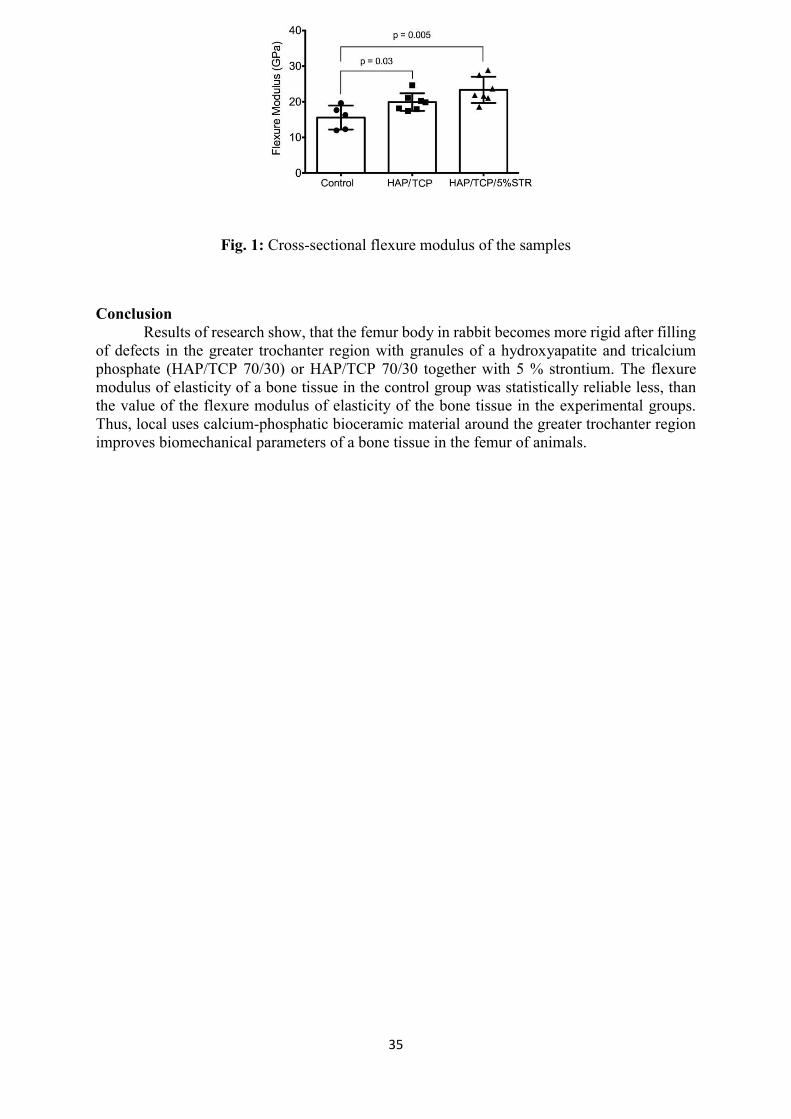
Results The flexure modulus of elasticity characterizes rigidity of material, and if the value of the flexure modulus of elasticity is higher, the material is more rigid. The analysis of the received results has shown that the flexure modulus of elasticity of a bone tissue in the control group was 16.31 GPa (18.66 – 12.15), that is statistically reliable less (p = 0.03), than the value of the flexure modulus of elasticity of the bone tissue in the 2nd group (Md = 19.87 GPa (21.10 – 17.94)) and in the 3rd group (Md = 21.88 GPa (27.53 – 21.09)), p = 0.005, respectively. Flexure modulus of samples in the 2nd and 3rd group statistically did not differ (p = 0.053) (Fig. 1).
35
Fig. 1: Cross-sectional flexure modulus of the samples
Conclusion Results of research show, that the femur body in rabbit becomes more rigid after filling of defects in the greater trochanter region with granules of a hydroxyapatite and tricalcium phosphate (HAP/TCP 70/30) or HAP/TCP 70/30 together with 5 % strontium. The flexure modulus of elasticity of a bone tissue in the control group was statistically reliable less, than the value of the flexure modulus of elasticity of the bone tissue in the experimental groups. Thus, local uses calcium-phosphatic bioceramic material around the greater trochanter region improves biomechanical parameters of a bone tissue in the femur of animals.

36
14th Joint Symposium of the Rostock University and Rīga Stradiņš University Maxillofacial Trauma Treatment
24-26 May, 2018
Poster Presentation
CLOSURE OF ORO-ANTRAL COMMUNICATION WITH VASCULARISED
BUCCAL FAT PAD FLAP
Viktors Andrjusenko1, Lana Micko1 1. Riga Stradins University, Riga, Latvia
2. Daugavpils Regional Hospital, Daugavpils, Latvia
Aim To find out effectiveness of closure oro-antral communications (OAC) with vascularised buccal fat pad (BFP) flap.
Materials and Methods Retrospectively collect and analyse data of 23 patients in age from 30 to 50 admitted to the Daugavpils Regional Hospital Maxillofacial surgery department with diagnosis “Oro-antral communication” treated using vascularised BFP flap. The origin of OACs was posterior maxillary teeth extractions. Patients with severe concomitant illnesses were excluded. All patients were observed and results of treatment and their satisfaction were evaluated.
Results In 23 patients surgery using vascularised BFP flap for OACs closure were done and data of all patients were analysed. The most frequent cause was the extraction of the first upper molar. The average size of communication was 0.6 cm. In 3 cases OAC was presented 1-2 days after tooth extractions and surgery was done under local anesthesia. Other 20 patients had extractions 2-3 month before and already had “Chronic sinusitis”, so they underwent antrotomy with OAC closure under general anesthesia supplemented with local anesthesia. In 1 case additional surgery was required due to relapse.
Conclusion Closure of OAC using vascularized BFP flap was proved to be appropriate method suitable for posterior region. This surgical technique could be used under general and local anesthesia, is less traumatic and shows acceptable treatment results. The size limitation of the BFP must be considered in order to obtain the best results. This surgical technique should be taken into consideration in order to select the best approach.

37
14th Joint Symposium of the Rostock University and Rīga Stradiņš University Maxillofacial Trauma Treatment
24-26 May, 2018
Poster Presentation
THE OUTCOME OF 1 MONTH OLD CRANIOFACIAL TRAUMA. A CASE REPORT
Agnija Taluma, Girts Salms, Riga Stradins University Clinical Hospital, Department of Oral and Maxillofacial Surgery, Rīga, Latvia
Frontal sinus and naso –orbital – ethmoid fractures are between the most challenging injuries of maxillofacial trauma. A 46 – years – old male came to the hospital 1 month after criminal trauma. He had complains of frontal and nasal deformity, lacrimation and nasal obstruction. Computed tomography revealed fracture of anterior and posterior table of frontal sinus and naso – orbito – ethmoidal fractures. Patient had no signs and symptoms of neurological deficites. The outer table and nasal bones were reconstructed and fixed by coronal approach. Postoperative computed tomography scans showed adequate reduction of displaced bone fragments. Cosmetic deformities were effectively restored. Esthetic results were satisfactory and no complications were observed.

38
14th Joint Symposium of the Rostock University and Rīga Stradiņš University Maxillofacial Trauma Treatment
24-26 May, 2018
Poster Presentation
NEW CLINICAL SYMPTOM OF MANDIBULAR RAMUS FRACTURE
Sergei Shuvalov, Olesia Kulitskaia Department of Surgical Stomatology and Maxillofacial Surgery, National Pirogov
Memorial Medical University, Vinnytsya, Ukraine [email protected]
Vincent symptom is well known in the practice of maxillofacial surgeons. We detected another special zone of numbness, which is not directly related to the damage of the inferior alveolar nerve. It is also located on the chin, but slightly below the known zone of numbness during Vincent symptom.
Aim Of the study was to describe the new symptom of chin numbness.
Methods Our clinical observations, as well as anatomical studies in humans and animals.
Results Suggest that the mylohyoid nerve includes sensitive fibers that innervate certain areas of the skin in the chin area. In the clinic a specific zone of innervation of the skin with the mylohyoid nerve is observed. We constantly observe this condition in osteotomies and fractures of the mandibular ramus. The traumatization of the mylohyoid nerve separately from the inferior alveolar nerve can be explained by its location in the mylohyoid groove on the internal surface of the mandible and it is traumatized by the fractured jaw fragment, as well as when the sharp edge of the split fragment is displaced during BSSO.
Conclusion The zone of skin numbness during the mylohyoid nerve damage may be an additional criterion for assessing the features of isolated traumatic fractures of the mandibular ramus, as well as the quality of the mandibular osteotomy performed during orthognathic surgery. The described clinical symptom is the new one in maxillofacial surgery and complements the possibilities of evaluating the control and prognosis of treatment, and also significantly supplements the Vincent symptom.

39
14th Joint Symposium of the Rostock University and Rīga Stradiņš University Maxillofacial Trauma Treatment
24-26 May, 2018
Poster Presentation
PENETRATING FACIAL INJURY WITH KNIFE: CASE REPORT
Kulpaviciute Monika 1, Purliene Ineta 1, Senkus Linas 2 Department of Oral Maxillofacial Surgery Vilnius University hospital Zalgiris Clinic 1
National Cancer Institute 2
Aim Penetrating knife injuries in the maxillofacial region are relatively uncommon. Those with impacted knife blades are even less commonly reported. These injuries can be life-threatening, especially where the major blood vessels of the face, facial nerve, parotid gland, parotic duct may are involved. The approach to treatment should be multidisciplinary, beginning with the trauma unit to provide airway maintenance and haemodynamic stabilization.
Methods We report clinical case of 24-year old male with impacted knife in his right side of face, which penetrates through the right maxilla , extending into the right maxillary sinus and oral cavity, through the tongue. Knife was removed surgically under general anesthesia using gentle, controlled force without bleeding or complications following removal.
Results Knife with blade was removed without any bleeding or complications. Stitches were removed after 7 days, no complications of knife injury or removal was seen.
Conclusion Foreign bodies following penetrating injury to the maxillofacial region by a knife blade are fairly uncommon and rarely reported. Patient with penetrating knife injury to the maxillofacial region management requires a multidisciplinary approach. Initial airway and hemodynamic stabilization of the patient should be performed, as well as assessment of damage to vital structures. Once the patient is stable, appropriate laboratory and radiographic evaluations may then be performed. The ideal method of removing the retained knife blade is careful extraction through the initial entrance wound, in a controlled setting under general anesthesia.

40
14th Joint Symposium of the Rostock University and Rīga Stradiņš University Maxillofacial Trauma Treatment
24-26 May, 2018
Poster Presentation
BILATERAL CONDYLECTOMY ON PSYCHYATRIC PATIENT
WITH LONG TIME LOWER JAW DISLOCATION
AND FIBROUS ANKYLOSIS – CASE REPORT
Martins Lauskis*, Viktors Andrjusenko**, Andrejs Skagers* *Riga Stradins University, Department of Oral and Maxillofacial Surgery
** Daugavpils Regional Hospital, Department of Maxillofacial Surgery E-mail – [email protected]
Variance in the duration of dislocation and anatomical considerations make the treatment for long-standing dislocation complex and controversial (1). It is particularly difficult to treat the condition as it worsens over time due to progressive fibrosis, adhesions in and around the joint. A range of invasive surgical procedures such as eminectomy, cindylectomy, menisectomy, and various osteotomies have been performed (2). On rare situations, mandibular dislocation may not be percieved by the patient and remain undiagnosed or misdiagnosed for a long period (3).
Patient and method A 60-year-old female suffering from schizophrenia and dementia was referred to Daugavpils Regional hospital from Psycho-neurological hospital. Three months ago, the staff of hospital noticed that the patient is n’t able to close mouth cinsidered as symptom of dementia. Bilateral anterior mandibular dislocation was clinically diagnosed and confirmed by CT. Both condyles were located 30 mm anterior from glenoid fossa. Repeated a attempts of manual reposition under general anesthesia were unsuccessful. In 19.09.2017. bilateral condylectomy was performed using Blair’s preauricular approach. Two screws were put in each jaw and rigid fixation was achieved for 2 weeks. Mouth opening was more than 30 mm and the patient was able to eat again.
Conclusion The more radical treatment method as condylectomy and rigid fixation for 2 weeks was chosen for our patient to minimize the risk of recurrence because options for post-surgical physical therapy were limited. References 1. Baur DA et al./Int J Oral Maxillofac Surg. 2013; 42:1030-3. 2. Holmund A et al.Br J Oral Maxillofac Surg. 2013; 51:206-10. 3. Sanders B et al.. J Oral Surg 1979: 37:346-364

41
14th Joint Symposium of the Rostock University and Rīga Stradiņš University Maxillofacial Trauma Treatment
24-26 May, 2018
Poster Presentation
LATE JATROGENIC POSTOPERATIVE PAIN AFTER OSTEOSYNTHESIS OF
CONDYLAR FRACTURE. CASE REPORT
Julianna Muceniece, Marina Sevastjanova, Department of oral and maxillofacial surgery, Riga Stradins University Institute of Stomatology
Aim Mandibular condylar fractures is reported as those with controversies to its conduct. Osteosynthesis is common used, although third trigeminal nerve brunch (TTNB) damage in different degrees still exists. We are aimed to report the case of persistent unilateral facial pain in function and limited movement of jaw due to time developed after repaired condylar and parasymphyseal fractures. Attempts to present diferential diagnosis and successful treatment have been made.
Methods Retrospective and prospective analysis of patients history (female, 40 years), clinical examination, chairside qualitative sensory examination of trigeminal nerve and radiological data obtained in period from May 2013 till April 2018.
Results Patient developed persistent pain in function in two year period after repair of fracture. Conservative pain reducer medication was unsuccessful. Dynamic and static mechanical allodynia of one side TTNB was revealed. The clinical context of complains and results obtained indications to perform CB3DCT. Radiological findings approved identifiable traumatic event – contact of osteosynthesis titanium plate screw with mandibular canal. Removal of titanium plate was recommended. After performing surgical treatment and prescribing intensive physiotherapy, patient undergo fast rehabilitation. No pain in jaw function was reported. In one month period patient reported 2,2 cm mouth opening, 3,0 cm in 6 month check-up and 3,4 cm one year post-operative.
Conclusion Performing osteosynthesis of condylar fracture, anatomical considerations of mandibular canal must been always examinated individualy. In case of persistent postoperative pain – clinical examination, evaluation of somatosensory system and CB3DCT is recommended.

42
14th Joint Symposium of the Rostock University and Rīga Stradiņš University Maxillofacial Trauma Treatment
24-26 May, 2018
Poster Presentation
A STRUCTURAL AND FUNCTIONAL CHARACTERISTIC OF EXPERIMENTAL
LOWER JAWS FRACTURES AFTER OSTEOSYNTHESIS WITH
MINI-PLATES OF NANO-STRUCTURED TITANIUM
Matchin A.A.1, Stadnikov A.A2, Klevtsov G.V.3, Merson E.D.3, Nosov E.V.1 1 Department of Stomatology and Maxillofacial Surgery, Orenburg State Medical University, Orenburg, Russia 2 Department of Histology, Cytology and Embriology,
Orenburg State Medical University 3Department of Nanotechnology, Material Science and Mechanics,
Togliatti State University, Togliatti, Russia E-mail - [email protected]
Introduction of new structured materials for medical devices for use in cranial and maxillofacial surgery demands the investigation of biological response of bone and soft tissue.
Aim of study Structural histochemical characterization of lower jaw consolidation after experimental fractures on rabbits and osteosynthesis using nano - structured mini-plates and screws.
Material and methods On rabbits osteotomy and osteosynthesis of lower jaw with mini-plates and screws
produced out of nanostructured titanium were done. Animals were eliminated out of
experiment 1,5 month after osteosynthesis. Rearing of animals in conformity with principles
of Convention Strasbourg (1986) and rules of Russian Federation Ministry of Health (Nr.
199n from 1.04.2016) was provided. Samples of lower jaw evaluated using confocal scaning
laser microscope LEXT OLS X 400, standart light microscoppy histochemical staining.
Including in sample of confocal scaning laser microscoty, done in Toliati University, fracture
line and surrounding healthy bone tissue did assessment of microstructural response to
nanostructured titanium implants
Results On radiovisiogramms pathology of bone tissue was not observed. On LCFSM dense connection of bone and titanium structures were obtained. Structure of new formed bone on threads of minisrews was clearly seen on 3D scans. On light microphotogramms in border line neoosteogenesis with forming of trabecular bone, including mineralized osteomucoid, octeocytes and microvascular structures were observed. Foci of bone resorbtion and osteoclasts were seen in border zone filled with fibroblast type connective tissue, fibres, granulocytes and lymphocytes. Some islets of new hyalin cartilage were in sites of bone resorbtion. Histochemical evaluation using monoclonal antibodies for identification of proteins p53, bcl 2, Ki-67 alowed analysis of expression of pro- and antiapoptic , also proliferative genes in osteoblasts, endothelyocytes and macrophages. Results acknowledged the optimisation of reparative osteogenesis as response to nanostructured titanium material for osteosynthesis mini-plates and screws.
Conclusion
43
Results of experimental morphological investigation of bone repair in border to
nanotitainic mini-plates and mini-screws give evidence of high integrative potential of implants
manufactured from materials of this class and confirm the recommendation of nanostructural
titanium for manufacture of medical products for cranio-maxillo-facial surgery.

44
14th Joint Symposium of the Rostock University and Rīga Stradiņš University Maxillofacial Trauma Treatment
24-26 May, 2018
Poster Presentation
3 STEPS FACE SURGERY AFTER CAR ACCIDENT. CASE REPORT
V. Klimecs1,2. T. Bogdanova2. E. Jursevics2. A. Keirans2,4. K. Jursevics3. A. Skagers1.
1. Riga Stradins University. Maxillofacial department 2. E. Jursevics aesthetic private clinic
3. Riga Stradins University. Medical student 4. Microsurgery centre. Latvia
Maxillofacial injuries are commonly encountered in the practice of emergency medicine. More than 50% of patients with these injuries have multisystem trauma that requires coordinated management between emergency physicians and surgical specialists in oral and maxillofacial surgery, otolaryngology, plastic surgery, ophthalmology, and trauma surgery. [1, 2, 3]. Etiology In developing countries, motor vehicle accident (MVA) is the most common cause of jaw fracture, whereas, in developed countries, assault is the most common cause and MVA is the second most common. [4]. Pathophysiology The kinetic energy present in a moving object is a function of the mass multiplied by the square of its velocity. The dispersion of this kinetic energy during deceleration produces the force that results in injury. High-impact and low-impact forces are defined as greater or lesser than 50 times the force of gravity. These parameters impact on the resultant injury because the amount of force required to cause damage to facial bones differs regionally. The supraorbital rim, the maxilla and the mandible (symphysis and angle), and frontal bones require a high-impact force to be damaged. A low-impact force is all that is required to damage the zygoma and nasal bone. The face is made up of vertical and horizontal buttresses where bone is thicker to neutralize forces applied to it. Reduction and fixation of these key areas are the basis of maxillofacial reconstruction. [5]. Case report. The patient (C.I.) 42 years old female, has addressed to us in clinic with complaints to appearance. not satisfied with -
asymmetry of the face. deformation of the nose, breathing was disturbed. deformation of the lips. a disfiguring scar on the cheek and lip.
The patient would like to improve the appearance, improve breathing, reduce the scar on the lip and cheek, and also improve the appearance of the lower right eyelid. From an anamnesis it is known that in 2015 the patient got into an auto accident, was taken to a hospital where operations were performed to restore the integrity of the facial skeleton (osteosynthesis with mini plates). The primary treatment of wounds was carried out, foreign bodies (glass) were removed. (First stage). At the second stage, an operation was performed to restore the nose (septorhinoplasty) using the costal graft, as well as plastic surgery of the upper lip, excision of scars on the cheek and neck (after tracheostomy)

45
14th Joint Symposium of the Rostock University and Rīga Stradiņš University Maxillofacial Trauma Treatment
24-26 May, 2018
Poster Presentation
CASE REPORT: EPISTAXIS TREATMENT BY EMBOLIZATION:
RELATED COMPLICATIONS
DD Jurgita Vaicaityte, DD Dalius Matkevicius, MD PhD Assoc.prof. Linas Zaleckas Centre of Oral and Maxillofacial Surgery
Institute of Odontology Faculty of Medicine Vilnius University
Objectives Maxillofacial and ENT surgeon must deal with nasal and oral bleeding almost daily. Severe bleeding demands extensive measures such as vascular embolization. The purpose of this presentation is to analyse the indications of embolization use, present our clinical case and complications related to embolization procedure.
Methods 51-year- old male arrived in the hospital because of nasal bleeding. The patient came conscious, with irregular heartbeat, unstable hemodynamic. Persistent bleeding from left nostril was observed. Anterior and posterior nasal packing, silver nitrate cauterization was tried without success. Angiography showed acute bleeding source – left maxillary artery. It was decided to apply embolization procedure using disconnecting platinum coils. Three arteries were embolized – a. maxillaris interna, a. sphenopalatine and a. facialis. Embolization was successful, bleeding stopped. The fourth day post-operation patient started complaining about limited mouth opening and difficult mastication. Trismus has arisen from embolization procedure. There was severe trismus remaining after 20 days post-operation. The limited opening was observed – less than 1 centimetre between lower and upper incisors. Individual mechanotherapy was suggested. Regular pain killers prescribed. Simple mechanotherapy resulted in increase of wider opening - up to 3 centimetres in three weeks.
Results Successful vascular embolization procedure was performed in Vilnius University Hospital Santaros Klinikos. Bleeding was controlled within first hour after embolization. Some embolization related complications were noticed: a patient developed trismus. No permanent complications related with embolization procedure was noticed. Because of post haemorrhagic anaemia blood transfusion was done.
Conclusion Vascular embolization is useful and safe minimal invasive method controlling severe nasal bleeding. No permanent side effects were observed using this method. The case showed that mechanotherapy can effectively treat embolization related trismus.

46
14th Joint Symposium of the Rostock University and Rīga Stradiņš University Maxillofacial Trauma Treatment
24-26 May, 2018
Poster Presentation
COMPLEX SOFT TISSUE AND BONY INJURY OF THE FACE OF
A 13 YEAR OLD BOY
Lena Cohrs, Peter Sieg Department of Maxillofacial Surgery, University Hospital of Lubeck, Germany
Aim The poster depicts the case of a 13-year-old boy who suffered severe facial injuries due to a car accident in his home country Afganistan. Primary care was provided in a hospital in Kabul. Supported by the organisation "Robin Aid" he was presented in our department for reconstruction of the facial soft tissues and the bony structures.
Methods The initial findings, the individual reconstructive steps and results are explained in a case description.
Results In the area of the upper and lower lip and the right cheek extensive substance defects were noticeable. Due to a fracture of the mandible, healed in a malposition, there was a frontally open bite. Osteomyelitis of the mandible in the area of the right ramus and corpus was present. The skin of the right side of the face contained keloids extending over the neck to the clavicular region causing an incipient sternomental contracture. The reconstruction was performed according to a multi-step surgical concept with the help of microsurgically anastomosed grafts to compensate the large soft tissue deficit.
Conclusion Microsurgical grafts were necessary to eliminate the extensive substance defects and the resulting functional deficits. Although the boy's situation has improved significantly, a stigmatized face remains.