the shape of the human language-ready brainlithornis.nmsu.edu/~phoude/shape of the language-ready...
TRANSCRIPT

HYPOTHESIS AND THEORY ARTICLEpublished: 04 April 2014
doi: 10.3389/fpsyg.2014.00282
The shape of the human language-ready brainCedric Boeckx 1,2* and Antonio Benítez-Burraco 3
1 Catalan Institute for Advanced Studies and Research (ICREA), Barcelona, Spain2 Department of Linguistics, Universitat de Barcelona, Barcelona, Spain3 Department of Spanish Philology and its Didactics, University of Huelva, Huelva, Spain
Edited by:
Simon E. Fisher, Max Planck Institutefor Psycholinguistics, Netherlands
Reviewed by:
W. Tecumseh Fitch, University ofVienna, AustriaErich Jarvis, Duke University MedicalCenter, USA
*Correspondence:
Cedric Boeckx, Department ofLinguistics, Universitat de Barcelona,Gran Via de les Corts Catalanes, 585,08007 Barcelona, Spaine-mail: [email protected]
Our core hypothesis is that the emergence of our species-specific language-ready brainought to be understood in light of the developmental changes expressed at the levels ofbrain morphology and neural connectivity that occurred in our species after the split fromNeanderthals–Denisovans and that gave us a more globular braincase configuration. Inaddition to changes at the cortical level, we hypothesize that the anatomical shift that ledto globularity also entailed significant changes at the subcortical level. We claim that thefunctional consequences of such changes must also be taken into account to gain a fullerunderstanding of our linguistic capacity. Here we focus on the thalamus, which we argueis central to language and human cognition, as it modulates fronto-parietal activity. Withthis new neurobiological perspective in place, we examine its possible molecular basis.We construct a candidate gene set whose members are involved in the development andconnectivity of the thalamus, in the evolution of the human head, and are known to giverise to language-associated cognitive disorders. We submit that the new gene candidateset opens up new windows into our understanding of the genetic basis of our linguisticcapacity. Thus, our hypothesis aims at generating new testing grounds concerning coreaspects of language ontogeny and phylogeny.
Keywords: language-ready brain, cognitive biology, evolution of language, comparative neuroscience, human
evolution, globularity, biolinguistics
HYPOTHESIS AND OVERVIEWThe aim of this paper is to contribute to the field of biolinguis-tics, here understood as an umbrella term encompassing all theinterdisciplinary attempts to identify the biological foundationsof our species’ ability to spontaneously develop mental rule sys-tems that are put to use in thought and communication. Such rulesystems, known as natural languages, have well-defined propertiesthat decades of linguistic research have revealed and that, takentogether, make these systems different from what other speciesare mentally and behaviorally capable of (Chomsky, 1965; Pinker,1994; Boeckx, 2010). We endorse the conclusion that it is aspectsof our biology, specifically of our brain, that endow us with thismental ability.
In the generative linguistics tradition, this biological endow-ment is referred to as “Universal Grammar” or the “LanguageOrgan” (Chomsky, 1965, 1975). Because these terms have cometo be seen as too ideologically loaded, we prefer to speak here ofthe “language-ready brain.” This term has been adopted by severalresearchers of very different theoretical persuasions (Kegl, 2004;Arbib, 2012), and it has several advantages over its competitors.First, the term draws attention to the brain as the focus of inquiry.Second, it enables us to keep clearly separate two entities: one, thelanguage-ready brain, understood as the cluster of brain proper-ties that sets the stage for language ontogeny and phylogeny, andthe other, language, understood as the collection of propertiesthat humans eventually acquire as a result of social interactions.As Deacon (2010) points out, building on differences between twosongbirds, the White-backed Munia and its domesticated cousin,the Bengalese finch, documented by Okanoya (2004), behavioral
complexity is likely to have important consequences at the levelof brain organization. In the case of songbirds, the domesticatedstrain of the wild White-rumped Munia, the Bengalese finch, isknown to have a distinct song pattern with a more complicatedsyntax than the wild strain. Interestingly, Wada et al. (2013) notonly identified differential androgen receptor (AR) expression inbasal ganglia nucleus Area X GABAergic neurons between thetwo strains, they also revealed an epigenetic modification: DNAmethylation state in regions upstream of AR in Area X.
A similar state of affairs is likely to hold when we compare thelanguage-ready brain and the fully linguistic brain. In the case ofthe latter, we expect epigenetic changes, as areas are recruited toenable vocalization of complex signals, reading, writing, and so on(what Dehaene, 2009 calls “neuronal recycling”).
Whereas the linguistic systems that the modern human braininternalizes depend, of course, on the brain being language-ready,it is clear that many properties of languages are also the products ofcultural evolution (Deacon, 1997; Arbib, 2012; Okanoya, 2012). Inothers words, in order to eventually characterize modern linguisticsystems completely, it will be necessary to appeal to a broad rangeof evolutionary mechanisms. In particular, it will be necessary tocharacterize adequately the emergence of the socio-cultural con-texts that can support, enhance, and perhaps even select for theuse of our linguistic capacity. Offering such a complete character-ization of language evolution is not our goal here. It is a far toodemanding task for any single paper. Our aim is more modest. Weseek to shed light on the emergence of the language-ready brainunderstood as but one aspect of the fully fledged linguistic brainof modern humans.
www.frontiersin.org April 2014 | Volume 5 | Article 282 | 1

Boeckx and Benítez-Burraco The shape of the language-ready brain
Such a fully fledged linguistic brain crucially requires, in addi-tion to those aspects we focus on below, a proper description ofthe externalization component necessary for cultural transmis-sion, which has at its core the sensorimotor systems dedicated tospeech for spoken languages and to signing for sign languages.This is the topic of much work, and rapid progress in currentbiolinguistics, which we will not review here. We refer interestedreaders to Jarvis (2004), Fitch (2010), Arbib (2012), and Morrillet al. (2012) for comprehensive treatments.
A complete understanding of the modern linguistic brain alsorequires hypotheses concerning the social conditions that facili-tate the learning of cultural variants (Tomasello, 1999, 2008, 2009;Kirby, 2013). Covering all of these aspects would obviously take ustoo far afield. We focus on properties of the language-ready brainthat we feel have so far been neglected, and which we hypothe-size are central to language ontogeny and phylogeny. Thus, we askreaders to view our hypothesis as the identification of an additionalpiece of a larger puzzle, to be complemented with the existing lit-erature on externalization and communication. To be perfectlyexplicit: although we do not address details of certain propertiessuch as vocal learning, we do not mean to diminish the impor-tance of these in characterizing our linguistic brain. We take thehuman language faculty to be similar to many other traits: a mosaicmade up of various components of distinct evolutionary origins(see Boeckx, 2013a). The hypothesis we develop in this paper isintended to address a facet of this mosaic for which substantialgaps in our understanding remain to be filled, with few leadingcandidate hypotheses on offer.
The facet we focus on pertains to the syntax–semantics inter-face: the characteristic syntactic complexity of human languagethat gives rise to compositional meaning. While we recognize thepossibility of an evolutionary continuum regarding syntactic abil-ities, we want to ask which aspect of our brain is responsible forthe more advanced form of combinatorial syntax attributed to ourspecies.
Building on Broca’s writings (see Harrington, 1987), it has oftenbeen hypothesized that lateralization patterns are central to char-acterize the language-ready brain (Crow, 2008). As reviewed inToga and Thompson (2003), prominent asymmetries are indeedfound in the gross anatomy of the two brain hemispheres inanatomically modern humans (AMHs). Noticeable protrusionsof the hemispheres, anteriorly and posteriorly, are observed, aswell as differences in the widths of the frontal and occipital lobes.These protrusions produce imprints on the inner skull surface,known as petalia. A twisting effect is also observed, known asYakovlevian torque, in which structures surrounding the rightSylvian fissure are “torqued forward” relative to their counter-parts on the left. The left occipital lobe is also splayed acrossthe midline and skews the interhemispheric fissure in a rightwarddirection. A related shape asymmetry is also commonly observedin the occipital horns of the lateral ventricles: these tend to projectmore deeply into the occipital lobes on the left than on theright.
Although we believe that hemispheric asymmetries certainlyplay a role in characterizing linguistic competence at the brainlevel, at least two considerations convinced us that laterality can-not be as central as it is often taken to be. First, the distinctive
pattern of lateralization observed in human adults appears to beacquired through linguistic interaction (Minagawa-Kawai et al.,2011). Second, brain laterality is an aspect of many species.It is salient, for example, in non-human vocal learners likebirds (Moorman et al., 2012). Thus, to the extent that lateral-ity bears on the linguistic brain, we think that it is likely tobe tied to the communicative function of language, or what wehave referred to above as the “externalization” component. Wetake the evidence coming from birdsong studies to be partic-ularly suggestive in this regard. As reviewed in Berwick et al.(2011, 2012), birdsongs and human languages diverge mostlyat the levels of syntax and semantics. Although songs displaysome syntactic rules and are not devoid of meaning, “there isno compelling evidence to date that birdsong matches the char-acteristic syntactic complexity of human language, arising fromthe composition of smaller forms like words and phrases intolarger ones” (Berwick et al., 2012, p. 1), the type of syntax that lin-guists claim give rise to semantic compositionality. The similaritiesbetween birdsongs and human languages pertain to external-ization. Given that we find lateralization patterns for the songcircuit in birds, we think it reasonable to conclude that the asym-metries found in the human brain are not responsible for thesyntax–semantics interface that we will focus on in what fol-lows. This conclusion is in fact what Broca (1861) appears tohave had in mind, since he clearly distinguished between thefaculty of language and the faculty of articulate language. ForBroca, only the latter was associated with lateralization patterns.Our conclusion is also in line with more recent studies cast-ing doubt on a direct link between laterality and language asa whole (see, among others, Benítez-Burraco and Longa, 2012;Bishop, 2013; Cochet and Byrne, 2013; Fitch and Braccini, 2013;Gómez-Robles et al., 2013; Greve et al., 2013; Hancock and Bever,2013).
Rather than laterality, we hypothesize that the relevant autapo-morphy is one that has so far received no attention in thecontext of biolinguistics, and that is most visibly expressed inthe globular aspect of the human endocranial morphology, par-ticularly salient in early postnatal development (Vannucci et al.,2013). We will refer to this trait as “globularity” in what fol-lows. As we will show in the next two sections, we have reasonsto claim that the neuroanatomical and physiological propertiesgiving rise to globularity contributed significantly to making ourbrain language-ready. Once we have made this clear, we willuse the information to generate some testable predictions of ourhypothesis. In particular, in Section “Molecular Basis,” we willput forward a set of candidate genes that contribute to the reli-able emergence of a globular, language-ready brain and that couldbe used in future studies in the genetic basis of our linguisticability.
GLOBULARITYA detailed examination of endocasts from fossil specimens ofthe genus Homo some 10 years ago (Bruner et al., 2003; Bruner,2004) has revealed that modern humans, in contrast to the other-wise heavily encephalized Neanderthals, “show a species-specificneomorphic hypertrophy of the parietal volumes, leading to adorsal growth and ventral flexion (convolution) and consequent
Frontiers in Psychology | Language Sciences April 2014 | Volume 5 | Article 282 | 2

Boeckx and Benítez-Burraco The shape of the language-ready brain
globularity of the whole structure” (Bruner, 2004, p. 279). Sub-sequent research (Gunz et al., 2010, 2012; Neubauer et al., 2010;Lieberman, 2011) has established that globularity is the resultof a unique developmental trajectory in modern humans, takingplace at a stage of growth where the brain is the primary deter-minant of skull shape. (Incidentally, this very difference betweenNeanderthals and us argues against the idea, still popular in neuro-science, that globularity is merely a side-effect of upright walkingin animals, given that Neanderthals and us had quite the samemode of locomotion).
Comparing endocranial shape changes during ontogeny inhumans and chimpanzees, Neubauer et al. (2010) have shown that“while some aspects of the pattern of endocranial shape changeare shared between humans and chimpanzees, the shape trajec-tories differ substantially directly after birth until the eruption ofthe deciduous dentition: in humans but not in chimpanzees, theparietal and cerebellar regions expand relatively (contributing toneurocranial globularity) and the cranial base flexes within thefirst postnatal year when brain growth rates are high.” (p. 555).Neubauer et al. (2010) refer to this early developmental stage asthe “globularization phase,” but we will continue to use the term“globularity” to refer to both the developmental process and to theend product of this process.
Neubauer et al. (2010) stress that the shape changes giving riseto globularity are unique to humans and do not occur in chim-panzees before or after birth. Nor do they occur in Neanderthals(Gunz et al., 2010, 2012). Although Neanderthals had brain sizescomparable to modern humans, their brain cases were elongatedand not globular. Comparing shapes of virtual endocasts extractedfrom computed-tomographic scans of crania of modern humansand virtual reconstructions of fossil humans, including the Nean-derthal neonate Le Moustier 2 and Mezmaiskaya, Gunz et al. (2010,2012) conclude that the globularization phase seen in the neuro-cranial development of modern humans after birth is absent fromNeanderthals, confirming Bruner et al.’s (2003) claim that modernhumans and Neanderthals reached large brain sizes along differentevolutionary pathways.
In sum, modern paleoneurology tells us that compared to ourclosest living and extinct relatives, humans have a large, specialized,and complex brain embedded in a uniquely shaped braincase.Specifically, the research we draw from in this section associatesthe emergence of this novel morphological trait with a distinctivedevelopmental trajectory at the level of the brain.
As is well-known, brains do not fossilize, and only indirectevidence from fossil endocasts, combined with evidence frommodern humans and our closest living relatives, the great apes,is what one has to rely on. But we are confident about the infer-ences about brains drawn in the literature we have mentioned inthis section, for all the reasons reviewed in Zollikofer and Poncede León (2013).
Along with the authors of the works just reviewed, we take itto be reasonable to think that the morphological changes givingrise to globularity are the products of factors that have impor-tant neurofunctional consequences. In other words, globularityis not just a superficial property of braincases. It crucially entailsmodifications of neural connections, for it is brain growth thatinfluences the formation and shape of the braincase, especially in
the first year of life. As we will see in Section “Molecular Basis,”all the genes that we have been able to link to globularity con-tribute significantly to neurogenesis, arealization of the neocortex,synaptic plasticity, and the like. In other words, they are notconfined to bone formation. Indeed, the very signals they sendto build the brain case are those that have been independentlyargued to contribute to brain organization. Thus, a crucial com-ponent of our hypothesis is that if the brain grows differently,it wires differently. Obviously, the differences are to be under-stood amidst the many commonalities that we expect to find inthe context of encephalization. But, as we review in more detailbelow, even subtle changes can have wide-ranging implicationsfor cognition. What we find particularly intriguing is that cer-tain cognitive disorders known to result from deviations in neuralconnectivity also lead to deviations from the norm in the contextof head shape, suggesting that there is indeed a link to explorebetween how the brain grows and how the head develops as awhole (see, e.g., Cheung et al., 2011 in the context of autism). Inaddition, differential growth is likely to lead to a reallocation ofbrain resources, or rewiring that may give rise to distinct cognitivephenotypes.
In the context of globularity, the results reported so far lead toa change of perspective in thinking about what makes the mod-ern human brain special. In particular, it suggests a possible linkbetween a special head shape and special aspects of our cogni-tion. This is the link we want to explore. More precisely, we wantto examine the possibility that globularity is what underlies ourspecies’ language-readiness.
We thus assume, along with many authors, that Neanderthals’brains were not language-ready, at least not in the way or to theextent in which sapiens’ brains are. This, of course, does not meanthat Neanderthals did not engage in symbolic activities, or wereincapable of vocal learning, or had no syntactic abilities at all.We certainly appreciate the range of anatomical evidence sug-gesting that Neanderthals had complex auditory and articulatorycapacities not unlike ours (Martínez et al., 2004; D’Anastasio et al.,2013), and engaged in complex, symbolic, cultural practices (Zil-hão et al., 2010; Rendu et al., 2014), some of which indeed used tobe claimed to be unique to us. It is true that, while these abilitiesand practices were thought to be attested only in modern humanpopulations, they were claimed to be closely linked to language,but such links were poor (Balari et al., 2011). As impressive as theNeanderthal achievements may be, we think it fair to conclude thatas of now, “no data or analytical tools currently available” indicatethat Neanderthals were “capable of the critical thought and syn-tactical ability necessary for complex language” (D’Anastasio et al.,2013, p. 6). Attempts to show otherwise (e.g., Dediu and Levin-son, 2013) are inconclusive (Benítez-Burraco and Barceló-Coblijn,2013; Berwick et al., 2013b), and a range of considerations con-tinue to provide evidence for key cognitive differences betweenNeanderthals and AMHs (Wynn and Coolidge, 2011; Longa,2013), differences that we will associate with the syntax–semanticsinterface in Section “Globularity and the Language-ReadyBrain.”
In concluding this section, we would like to make two moreremarks concerning globularity in connections with issues thathave been frequently discussed in the neurolinguistic literature. In
www.frontiersin.org April 2014 | Volume 5 | Article 282 | 3

Boeckx and Benítez-Burraco The shape of the language-ready brain
addition to moving us away from laterality, globularity suggeststhat not only brain size, but also shape matters. The size factor,understood as body/brain ratio, cannot, of course, be ignored. Asreviewed in Deacon (1997), the brain of modern humans is anevolutionary and developmental outlier. At birth, it has the sizeof an adult chimpanzee brain and expands by a factor of 2 duringthe first postnatal year. Large neonatal brain size and rapid initialgrowth contrast with slow maturation, which extends well intoadolescence. These aspects of the human brain undoubtedly playan important role in the emergence of modern human cognition.But we believe that they are not the whole story. Consistent withthis stance, we expect cognitive innovations linked to brain sizealone to be present in other hominins. That is to say, to understandtraits uniquely associated with AMHs, we hypothesize that it isnecessary to look beyond brain size.
In addition, globularity de-emphasizes the role of the frontallobes in giving rise to modern human cognition. One of the mostpervasive assumptions about human brain evolution has indeedbeen that it involved relative enlargement of the frontal lobes. Theliterature on globularity indicates that at the very least parietalvolumes are equally important. As Bruner (2010) observes, “asbrain size increases, the parietal lobes undergo relative flatteningin non-modern humans. This pattern is stressed in Neanderthals,which show, however, a certain widening of the parietal volumes.Only Homo sapiens shows a generalized enlargement of the entireparietal surface.” (p. S77). It is indeed reasonable to think thatthe morphological changes in the parietal region are to be relatedto important neurofunctional consequences, complementing thefunctions of the frontal lobes.
In this context, it is worth taking seriously studies like Bartonand Venditti (2013) or Smaers and Soligo (2013) showing thatthe size of human frontal lobes, and of specific frontal regions,is as expected relative to the size of other brain structures. Thus,although Barton and Venditti (2013) confirmed that absolute andproportional frontal region size increased rapidly in humans, thischange was tightly correlated with corresponding size increases inother areas and whole brain size, and with decreases in frontalneuron densities. Barton and Venditti (2013) conclude that “thesearch for the neural basis of human cognitive uniqueness shouldtherefore focus less on the frontal lobes in isolation and moreon distributed neural networks” (p. 9001) Recent work on cogni-tive impairments essentially reaches the same conclusion (Turkenand Dronkers, 2011; Dick and Tremblay, 2012). As will becomeevident in the next section, our position agrees with this per-spective, which we think is gradually becoming the norm inneurolinguistics.
Having described the nature and origin of globularity, as wellas the limits of hypotheses based on laterality and brain size, weare now in a position to formulate our hypothesis, which is to linkglobularity with the language-ready brain.
GLOBULARITY AND THE LANGUAGE-READY BRAINAs we saw in the previous section, we take it that globularity isnot just a superficial property of braincases. It crucially entailsmodifications of neural connections. We wish to put forward theidea that the developmental trajectory giving rise to globularityis critical to the formation of a network of neural connections
capable of supporting the most distinctive mode of cognition thatnumerous scholars have associated with language and that currentevidence suggests is absent in Neanderthals. Put succinctly, theglobular brain gives rise to the language-ready brain. Spelling outthis hypothesis is the purpose of this section.
To be testable, our hypothesis requires us to articulate anexplicit linking hypothesis between mind and brain, that is,between the properties we as linguists associate with language-readiness and the neural connections that could support suchmental properties. Once this is done, we must show howthese neural connections become available in the context ofglobularity.
Our hypothesis is that the species-specific anatomical compo-nent we have highlighted in the previous section is responsiblefor what is computationally unique about our species’ linguisticabilities. Thus, in order to link globularity to computational oper-ations, we must first be clear about what is computationally uniqueabout our mental life. In line with the recommendations formu-lated in Fitch (2009) and Poeppel (2005, 2011, 2012), we seekto formulate these computational properties “at a fine enoughgrain that one can discuss algorithmic and implementationalapproaches to [them]” (Fitch, 2009, p. 298). These computationalproperties should be, “ideally, elemental and generic. . .. Genericformal operations at this level of abstraction can form the basisfor more complex linguistic representation and computation.”(Poeppel, 2005, p. 11).
Comparative psychology has established that unlike otherspecies, modern humans excel at unifying and combining con-ceptual units that belong to distinct “core knowledge systems”(Spelke, 1994, 2000, 2004; Boeckx, 2010). Core knowledge sys-tems roughly correspond to the well-known Fodorian “modules”(Fodor, 1983). They are the building blocks that enable animalsto make sense of the world around them. As reviewed in Kinzlerand Spelke (2007), we have very robust evidence for four or fivecore knowledge systems in many species: one system specializingin objects and their mechanical interactions, another specializingin agents (animate things) and their goal-directed actions, a thirdconcerned with sets and numbers (number sense), a fourth deal-ing with places and geometric relationships (natural geometry),and a fifth core knowledge system dealing with social partners,groups, and relations, and the way we understand other minds(theory of mind). Core knowledge systems are at the root of ourcapacity to form rudimentary theories of the world around us.These theories are the foundations of physics (object mechan-ics), mathematics (number sense), biology (animate vs. inanimatebeings), navigation (natural geometry), and psychology/social sci-ence (theory of mind). These core knowledge systems give us andother animals an intuitive grasp of what is going on in each of thesedomains.
There is a lot of evidence from a range of fields that humans areunique – or, to put it in the context of an evolutionary continuum,far better than other species – in transcending the signature lim-its of core knowledge systems, going beyond modular boundaries(Mithen, 1996; Carruthers, 2002, 2006; Spelke, 2003; Wynn andCoolidge, 2004; Pietroski, 2007; Hauser, 2009; Boeckx, 2011a,b).This ability, which has all the characteristics of a phase transition,is at the heart of cognitive novelty, and subsequently, material and
Frontiers in Psychology | Language Sciences April 2014 | Volume 5 | Article 282 | 4

Boeckx and Benítez-Burraco The shape of the language-ready brain
cultural innovation, leading to the establishment of a new cogni-tive phenotype (Balari and Lorenzo, 2013; Boeckx, 2013a). Thisability is what Hauser (2009) dubbed “humaniqueness.” Hauser(2009) defines the latter as follows: the ability to “create and easilyunderstand symbolic representations of computation and sensoryinput,” to “apply the same rule or solution to one problem to adifferent and new situation,” and to “combine and recombine dif-ferent types of information and knowledge in order to gain newunderstanding.”
Several of the authors just cited have put forth the idea thatthis distinctively human mode of thought is likely to be intimatelyrelated to language. We propose to capture this in the followingway.
The core combinatorial operation in natural language thatcombines elementary linguistic units is called “Merge” in the ter-minology of Chomsky (1995), and it is the best candidate we knowof to account for the combinatorial property at issue. Accordingto Berwick et al.’s (2012) careful comparison between humans andsong birds, the unrestricted combinatorial operator that Chom-sky called Merge is absent in birds. Its absence means that birdsongs are devoid of the compositional, freely combining, sys-tematic, cross-modular semantics that is manifest in all humanlanguages.
To be useful at all in thought and action, such a freely combin-ing Merge must be regulated. As reviewed in Boeckx (2013a,b),we have linguistic reasons to believe that this regulation takes theform of integration/embedding: Merge is constrained in virtue ofits interfacing with and being embedded inside cognitive systemsresponsible for interpretation and externalization. This regulationis what the formal linguistics literature refers to as “Spell Out” or“Unify” (Jackendoff, 2002; Hagoort, 2005). We suggest that thisembedding takes the form of a generic coding mechanism that isalready well established in neuroscience (Lisman, 2005; Buzsaki,2008): internally generated oscillations at a high frequency such asthe gamma range are embedded inside an oscillation operating ata lower frequency such as the alpha range. Such lower-frequencyoscillations, characteristic of the thalamus, are known to be partic-ularly well-suited to synchronize distant cortical areas (Whitmanet al., 2013). Building on Boeckx (2013a,b), we hypothesize thatthis distant synchronization allows for the binding of featuresdistributed across core knowledge systems.
The mechanism of achieving interareal communication via anadaptive coupling of rhythms synchronizing spatially distributedoscillations is a generic strategy of the brain, neither specific tohumans nor to language. But we put forth the hypothesis thatthis mechanism gained its linguistic specificity and characteristiccomplexity when it found itself in a new anatomical context in ourlineage: globularity.
As should be obvious from our discussion of what globularity isin Section “Globularity,” the new anatomical context that gave riseto the language-readiness does not refer to a specific brain area.Rather, it refers to a set of areas brought into connection with oneanother, a situation we may refer to as one of “dynamic connec-tivity.” Certainly, the prefrontal and parietal areas are involved,as these gained special prominence in a globular context, but webelieve that in addition to these, there is at least a third anatomicalstructure that is traditionally ignored, but that we think is equally
relevant to link globularity to language-readiness: the thalamus.This is the reason why we focus mainly on this brain structurehere, returning to the contribution of the frontal lobe and theparietal lobe toward the end of the section, in the context of afronto-parieto-thalamic network.
We have several reasons to adduce in support of our hypothesisconcerning the relevance of the thalamus in the context of theglobular and the language-ready brain.
First, the thalamus is central in more than one way. In a globularcontext, it sits right in the middle of the brain, and as such appearsstrategically placed to connect distant areas. As a matter of fact,it has been suggested that the globular brain shape of modernhumans might have a positive effect on the wiring efficiency of thebrain’s neural network (Hofman, 1989; Chklovskii and Stevens,2000; McCarthy, 2001; Chklovskii et al., 2002). Developmentally,the thalamus forms from the diencephalon, and the cerebrumforms from the telencephalon. The telencephalon corresponds tothe most bulbous part of the rostral end of the ballooning neuraltube during development, and the diencephalon corresponds tothe swelling just caudal to that. As the brain develops the cerebrumand cerebellum come to surround the thalamus. The thalamus hassignificant connections to them, so it’s sensible that it occupies acentral position.
Second, Bishop et al. (2000), Price et al. (2006), and Chou et al.(2013) show that input from the thalamus, the main switching sta-tion in the brain for sensory information, is crucially required tocomplement the action of the genes in determining how the cere-bral cortex grows into separate functional areas and subsequentlydedicates itself to higher-order cognitive functions.
Third, the thalamus acts as a necessary relay center to con-nect many brain structures that have already been implicated inresearch on language (Lieberman, 2002; Murdoch, 2010): inter-actions between cortical areas and the basal ganglia or betweencortical areas and with the cerebellum cannot take place inthe absence of the thalamus (the same holds of the amygdalaand other limbic structures that have been implicated in cer-tain aspects of human “distinctness”). In fact, the literature onFOXP2 and its interactome has often mentioned the thalamus as animportant expression site of the genes involved (Vargha-Khademet al., 2005; Reimers-Kipping et al., 2011), a point to which wereturn in the context of molecular considerations in Section“Molecular Basis.”
Fourth, despite the cortical focus of many imaging studies andthe technical difficulties in getting recordings from the thalamus,this brain structure’s role has been highlighted in some neurolin-guistic studies, especially those pertaining to the syntax–semanticsinterface, the language component that is missing in non-humanvocal learners (Wahl et al., 2008; David et al., 2011).
Fifth, there is rapidly accumulating evidence that cognitivedisorders that are routinely associated with language and the dis-tinctive mode of thought it entails such as schizophrenia, autism,dementia, major depression, verbal working memory impair-ments, etc. crucially involve thalamic disorders, especially as theyaffect the mediodorsal nucleus and the pulvinar. This is a complextopic which we hope to return to in future work. For now, letus just refer to important studies such as Parnaudeau et al. (2013)and works along similar lines (Popken et al., 2000; Byne et al., 2001;
www.frontiersin.org April 2014 | Volume 5 | Article 282 | 5

Boeckx and Benítez-Burraco The shape of the language-ready brain
Dagenbach et al., 2001; Young et al., 2004; Alelu-Paz and Giménez-Amaya, 2008; Kovacs et al., 2013; Nair et al., 2013; Uhlhaas et al.,2013).
Finally, and perhaps most importantly, outside of languageproper, the thalamus has routinely been assigned a key role in con-trolling attention, regulating oscillations generated in the cortex,etc. (Saalmann et al., 2012) – functions that, though not specificto language, must surely also be part of a comprehensive neuralcharacterization of the language-ready brain.
Many neuroscientists continue to think of the thalamus simplyas a relay station, where sensory information from the periph-ery converges and is then passed on to the cortex. The cortexis thought to be the site of perception and cognition, withdifferent cortical areas specialized to subserve different func-tions. Communication between cortical areas can be mediatedby axonal tracts running in the white matter of the cortex. Thisleads readily to the view that once information reaches the cor-tex it is processed and integrated with other information aboutthe external world and internal states entirely within the cortex,resulting in conscious perception or some kind of motor or emo-tional output. But Theyel et al. (2009) demonstrate unequivocallythat cortical areas can also pass information indirectly via thethalamus.
It has been known for some time that communication betweenthalamus and cortex is bidirectional. According to Theyel et al.(2009) the thalamus receives, in fact, far more inputs from thecortex than it does from the periphery. As they note, the cir-cuits between thalamus and cortex can be broken down intotwo main types: those that drive the activity of their target neu-rons (whether in thalamus or cortex) and those that act moreto modulate the activity of their targets, especially their tempo-ral responsiveness. These pathways can be distinguished basedon their neurochemical profiles, the types of synapses that theyform and, in the case of projections from thalamus to cortex,the layers which they innervate. Driving connections from tha-lamus project with quite precise topography to layers 4 and 6,while modulatory connections project more diffusely within lay-ers 1 and 5. These modulatory connections from the thalamus areessential mediators of communication between cortical areas, dueto their crucial role in the synchronization of ongoing neuronaloscillations.
As Theyel et al. (2009) note, this frequency tuning can bemediated by corticothalamocortical loops, where the corticotha-lamic connection is driving and the thalamocortical connectionis modulatory. In this context, however, the information itselfis transferred via direct cortical connections. Theyel et al. (2009)show that even if these cortical connections are severed, informa-tion can still be transferred from one cortical area to another ifcorticothalamocortical circuits remain intact. In this case both thecorticothalamic and the thalamocortical connections are driving.This finding reinforces the important point that the function ofthe cortex cannot be divorced from that of the thalamus. It empha-sizes that perception is not simply a matter of passing informationalong a hierarchy of processing stations. Rather, it is a process ofreiterative comparison of top-down predictions with bottom-upinformation, much of which may be mediated by reverberatingactivity in corticothalamocortical circuits.
In his recent review on cortical dynamics, Singer (2013)strengthens our claim regarding the relevance of the thalamus, ashe notes that thalamic input crucially allows for an enrichment ofthe range of oscillatory activity in different frequency bands (seealso Cannon et al., 2014; Parnaudeau et al., 2013; Uhlhaas et al.,2013).
The modulatory or regulatory role of the thalamus is fur-ther enhanced when the thalamic reticular nucleus is taken intoaccount. The thalamic reticular nucleus consists of a thin layer ofGABAergic cells adjacent to the relay nuclei of the dorsal thala-mus. It occupies a striking control position in the brain, sendinginhibitory axons back to the thalamus, roughly to the same regionwhere they receive afferents, and has been hypothesized to play apivotal role in dynamic attention by controlling thalamocorticalsynchronization (Crick, 1984; Min, 2010).
Addressing the issue of the evolution of intelligence, Kircherand Glendenning (2002) point out that in addition to the sizeof the neocortex, the amount of neural inhibition to which thecortex is subjected may play a major role. As we have argued inthe context of Merge, where we noted that a completely unre-stricted Merge operation is cognitively unhelpful, and thereforerequires embedding, Kircher and Glendenning (2002) observe thatan expanded brain that is out of control is not helpful. There mustbe modulation of this enhanced cortex. Kircher and Glenden-ning (2002) show that a primary source of this modulation comesfrom the enhanced inhibitory capabilities of the thalamus, andthe increased number of neurons sensitive to the most commoninhibitory neurotransmitter found, GABA. By its influence on ourneocortex, the thalamus provides greater control of neural pro-cessing. Kircher and Glendenning (2002) propose that it may beour ability to inhibit our cortex that has resulted in our increased“intelligence,” which many authors have linked to language fordecades.
The range of evidence reviewed so far suggests to us that aproper characterization of the language-ready brain that does notrecognize a central role to the thalamus is unlikely to be correct, forit would miss the critical engagement of the thalamus in regulatingcortical activity. By providing low-frequency oscillations capableof embedding higher-frequency oscillations across distant brainregions, the thalamus provides the crucial regulation needed toform the sort of meaningful cross-modular conceptual structuresthat are characteristic of language.
In hindsight, it is somewhat surprising that the role of thethalamus is not yet well established in the neurolinguistic liter-ature, despite the fact that the thalamus has been implicated inthe context of many human-specific traits like intelligence or con-sciousness, which Darwin (1871) already suggested depend onthe exercise of the language faculty. This is true even in mod-els that go beyond the standard cortico-centric perspective onhigher-order cognition (Lieberman, 2002). That globularity offersus independent reasons to focus on the thalamus suggests to usthat our initial hypothesis can lead to some productive rethink-ing in this area. Hopefully, our hypothesis will help redirectattention to cases of thalamic aphasia, which have been knownfor a while even if their significance has tended to remain atthe periphery of neurolinguistic models. Significantly, Crosson(2013), Hebb and Ojemann (2013), and Klostermann et al. (2013)
Frontiers in Psychology | Language Sciences April 2014 | Volume 5 | Article 282 | 6
Boeckx and Benítez-Burraco The shape of the language-ready brain
review and re-assess the significance of thalamic aphasia andreach conclusions that go in the direction of our hypothesis. Ourhypothesis may also help us re-assess the role of the thalamusin other aspects of our language-faculty, such as vocal learning,where the relevance of the thalamus has long been recognized(Jarvis,2004; Person and Perkel,2005), and recently re-emphasized(Goldberg and Fee, 2011, 2012).
Still, for all our emphasis on the thalamus, we do not wantto leave the reader with the impression that this is the only rele-vant brain structure to link globularity and language-readiness.As should be clear, the thalamus gains its significance in thecontext of a network that involves the frontal and the parietallobes.
Bruner (2004, 2010) already drew attention to these two lobesin the context of globularity, although he did not make theconnection with language hypothesized here. Other works onfronto-parietal connections clearly converge with aspects of ourhypotheses, even if they do not always recognize the role ofthe thalamus, or link them to language. For example, the func-tion of the fronto-parieto-thalamic network envisaged here shareproperties with a family of models of higher-order human cog-nition such as the models formulated by Dehaene et al. (1998)and Tononi and Edelman (1998) in the domain of conscious-ness, the multiple-demand system of Duncan (2010, 2013), the“connective core” model of Shanahan (2012), or the integrativearchitecture for general intelligence and executive function in Bar-bey et al. (2012). These models recognize a crucial role for thefronto-parietal regions in achieving what we have referred to ascross-modular concept formation above, which we take to be thecentral aspect of language-readiness.
Thus, Dehaene et al.’s (1998) neuronal workspace modelemphasizes the role of distributed neurons with long-distance con-nections, particularly dense in prefrontal, cingulate, and parietalregions, interconnecting multiple specialized, modular processorsand “broadcasting” signals at the brain scale in a spontaneous andsudden manner, forming a “global neuronal workspace.” Throughthis workspace, Dehaene et al. (1998) claim that modular pro-cessors can exchange information very flexibly, that informationcan be accumulated across time and across different proces-sors, that incoming information arising from analog statisticalinputs can be discretized, and that chains of operations can beperformed.
Already a century ago Ramón y Cajal (1909) had underlinedthe special morphology of the pyramidal cells from the cerebralcortex and suggested they might be the “substratum of the highestnervous activities.” Building on this insight, Dehaene et al. (1998)view as key building blocks of the workspace “a distributed set ofcortical neurons characterized by their ability to receive from andsend back to homologous neurons in other cortical areas, horizon-tal projections through long-range excitatory axons.” (p. 14529).As they point out, “long-range corticocortical tangential connec-tions, including callosal connections, mostly originate from thepyramidal cells of layers 2 and 3” (p. 14529), and propose that“the extent to which a given brain area contributes to the globalworkspace would be simply related to the fraction of its pyrami-dal neurons contributing to layers 2 and 3, which is particularlyelevated in [. . .] dorsolateral prefrontal and [. . .] inferior parietal
cortical structures.” (p. 14529). These are, of course, particularlyrelevant regions in the context of globularity.
As Dehaene et al. (1998) note, the pyramidal neurons fromlayers 2 and 3 “establish, in addition, vertical and reciprocal con-nections with layer 5 neurons and thus corresponding thalamicnuclei. These connections contribute to both the stability and thedynamics of workspace activity, via, for instance, self-sustainedcircuits, but also mediate the direct access to and from the process-ing networks.” It is these connections with the thalamus that webelieve are crucial to regulate the activity of long-distance corticalconnections, leading to cross-modularity.
It is also worth pointing out that the fronto-parieto-thalamicnetwork that we take to emerge in the context of globularity andto underlie the human brain’s language-readiness shares featuresof the top-down, fronto-parietal attentional regulation network(Miller and Buschman, 2013). It is a circuit that has been claimedto have evolved from the foraging network of primates and even-tually came to be used in the context of foresight (Genovesio et al.,2014). The network we envisage also bears a family resemblancewith the default mode network that Gruberger et al. (2011) claimis responsible for mind-wandering and inner speech, a functionthat Chomsky (2012) describes as more central to language thanits communicative use. The network we envisage comes closestto what Vincent et al. (2008) call the “frontoparietal control sys-tem,” a network that is anatomically interposed between the dorsalattention system and the hippocampal–cortical memory system.The frontoparietal control system is said to be “uniquely posi-tioned to integrate information coming from the other two systemsand to adjudicate between potentially competing inner- vs. outer-directed processes” (p. 3334). The only missing component ofthese existing models is the thalamus. (An important exception isBohlken et al., 2013, where the thalamus receives the attention thatwe think it deserves).
There may have been other benefits of an improved fronto-parietal network, regulated by the thalamus. According to a DTIanalysis by Hecht et al. (2013), there is an increase in the ratiofronto-parietal vs. fronto-temporal connectivity from monkeys toapes to modern humans, which is a possible substrate for the evo-lutionary shift from emulation to imitation. Emulation here refersto the ability to copy the final product of an action, while imi-tation refers to the ability to copy a process. It is imitation thatis likely to underlie the possibility of cultural innovation that isso characteristic of modern humans, as compared to our clos-est living relatives or even Neanderthals, to judge from the fossilrecord.
A recent study by Pearce et al. (2013) may give us some clueas to how the fronto-parieto-thalamic network invoked here mayhave achieved its degree of robustness in modern humans. Focus-ing on the fact that Neanderthals had larger eyes than our species,Pearce et al. (2013) suggest that more of their brain was devotedto seeing in the long, dark nights in Europe, at the expense ofhigh-level processing. This is so because larger eyes entail a muchlarger visual processing area at the back of their brains. In otherwords, more of the Neanderthal brain would have been dedi-cated to vision and body control. A reduction of the visual areain modern humans has been independently supported by Sher-wood et al. (2008), and it may have led to an expansion of the
www.frontiersin.org April 2014 | Volume 5 | Article 282 | 7

Boeckx and Benítez-Burraco The shape of the language-ready brain
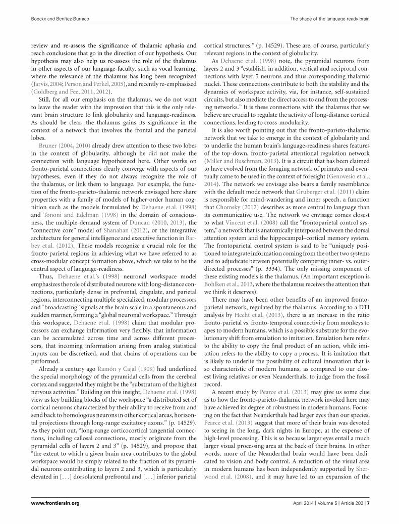
FIGURE 1 | Illustration of the hypothesis. (A) Observable skull differencesbetween anatomically modern human (left) and Neanderthal (right). (B)
Identification of the strategic position of the thalamus in a modern human
brain. (C) Representation of the hypothesis concerning the global connectiverole of the thalamus in an evolutionary perspective (image adapted fromBruner and Manzi, 2008).
parietal region, and a re-allocation of the computational power ofthe pulvinar, the part of the dorsal thalamus that modulates cor-tical visual processing (Saalmann et al., 2012), in service of othercognitive domains, such as language. A recent study on ultra-fast speech comprehension in blind subjects (Dietrich et al., 2013)and another on language processing in congenitally blind adults(Bedny et al., 2011) also indicate a significant recruitment of thepulvinar.
In this respect, it is worth mentioning that Streidter (2005)reports that the pulvinar is disproportionally large in humans,compared to other nuclei that lack prefrontal connections. (Thisis true also of the mediodorsal nucleus.) Streidter (2005) goes on(p. 331f) to note that “the human pulvinar is especially intrigu-ing because its enlargement is causally related to a major changein its embryogenesis. Only in humans does the pulvinar containneurons that migrated into the thalamus from the telencephalon[. . .] The other fascinating aspect of human pulvinar hypertro-phy is that it involves mainly the dorsal pulvinar, which hasstrong reciprocal connections with the lateral prefrontal, pari-etal, and temporal cortices (refs. omitted). This dorsal pulvinaris probably unique to primates, and separate from the ventralpulvinar, whose major function is to convey visual informationfrom the midbrain to the telencephalon. Collectively, these data
indicate that what enlarged in humans is not a motley group ofareas and nuclei, but an entire circuit that includes the lateralprefrontal cortex and several “associates” in both the neocortexand the thalamus.” In the same context, it is worth pointing outthat Bruner et al. (2010) found a positive correlation betweenthe parietal expansion that contributed to globularity and themorphology of posterior subcortical landmarks, including thethalamus.
Based on the evidence we have obtained from the literature,we hypothesize that the dorsal thalamus, specifically the pulvinarand the mediodorsal nucleus, played a significant role, but werecognize that only future progress in neurolinguistics will enableus to draw a more precise map of which parts of the thalamus arecritical for language-readiness.
To sum up this section (see Figure 1), our perspective onthe emergence of the language-ready brain converges with muchrecent work in neuroscience concerning cognitive specialization,well captured in the following passage from Barton and Venditti(2013): “coordinated expansion of functionally and anatomicallyconnected areas, potentially including both cortical and non-cortical regions.” As they note, and as we have just discussed,“neocortex, cerebellum, and intermediate nuclei, for example,show closely correlated evolution in terms of both volume and
Frontiers in Psychology | Language Sciences April 2014 | Volume 5 | Article 282 | 8
Boeckx and Benítez-Burraco The shape of the language-ready brain
neuron numbers, after controlling for variability in the size orneuron numbers of other brain regions.” For Barton and Venditti(2013), “the evolution of frontal regions such as PFC [prefrontalcortex] may be best understood in terms of their participation inmore distributed networks”“natural selection selectively enlargedsuch distributed networks and that these – rather than more local-ized size change of frontal cortical regions – are likely to form thebasis of human cognitive specialization” (p. 9005).
MOLECULAR BASISOne of the major aims of biolinguistics is to arrive at a geneticcharacterization of language. If our hypothesis in Section “Globu-larity and the Language-Ready Brain” is on the right track, insightinto the molecular basis of globularity is central to any ultimategenetic description of our linguistic competence. The goal of thissection is to use our hypothesis to generate a set of candidate genesthat will complement what can already be found in the literatureon the genetics of language.
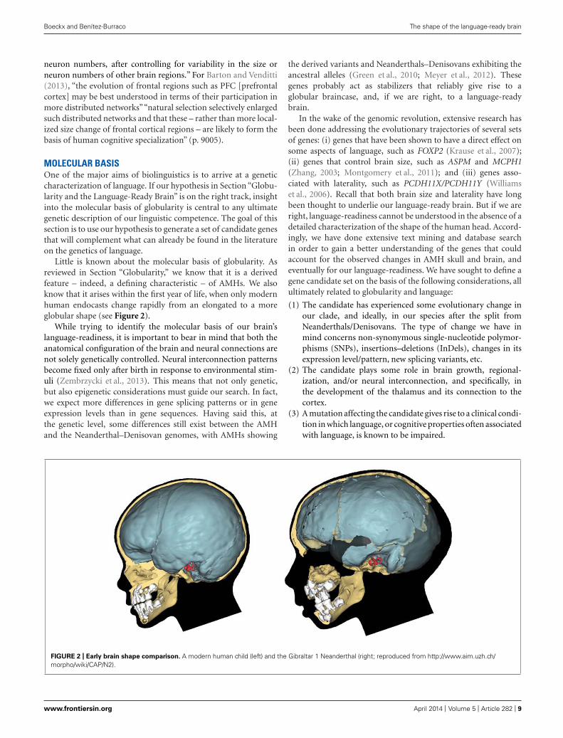
Little is known about the molecular basis of globularity. Asreviewed in Section “Globularity,” we know that it is a derivedfeature – indeed, a defining characteristic – of AMHs. We alsoknow that it arises within the first year of life, when only modernhuman endocasts change rapidly from an elongated to a moreglobular shape (see Figure 2).
While trying to identify the molecular basis of our brain’slanguage-readiness, it is important to bear in mind that both theanatomical configuration of the brain and neural connections arenot solely genetically controlled. Neural interconnection patternsbecome fixed only after birth in response to environmental stim-uli (Zembrzycki et al., 2013). This means that not only genetic,but also epigenetic considerations must guide our search. In fact,we expect more differences in gene splicing patterns or in geneexpression levels than in gene sequences. Having said this, atthe genetic level, some differences still exist between the AMHand the Neanderthal–Denisovan genomes, with AMHs showing
the derived variants and Neanderthals–Denisovans exhibiting theancestral alleles (Green et al., 2010; Meyer et al., 2012). Thesegenes probably act as stabilizers that reliably give rise to aglobular braincase, and, if we are right, to a language-readybrain.
In the wake of the genomic revolution, extensive research hasbeen done addressing the evolutionary trajectories of several setsof genes: (i) genes that have been shown to have a direct effect onsome aspects of language, such as FOXP2 (Krause et al., 2007);(ii) genes that control brain size, such as ASPM and MCPH1(Zhang, 2003; Montgomery et al., 2011); and (iii) genes asso-ciated with laterality, such as PCDH11X/PCDH11Y (Williamset al., 2006). Recall that both brain size and laterality have longbeen thought to underlie our language-ready brain. But if we areright, language-readiness cannot be understood in the absence of adetailed characterization of the shape of the human head. Accord-ingly, we have done extensive text mining and database searchin order to gain a better understanding of the genes that couldaccount for the observed changes in AMH skull and brain, andeventually for our language-readiness. We have sought to define agene candidate set on the basis of the following considerations, allultimately related to globularity and language:
(1) The candidate has experienced some evolutionary change inour clade, and ideally, in our species after the split fromNeanderthals/Denisovans. The type of change we have inmind concerns non-synonymous single-nucleotide polymor-phisms (SNPs), insertions–deletions (InDels), changes in itsexpression level/pattern, new splicing variants, etc.
(2) The candidate plays some role in brain growth, regional-ization, and/or neural interconnection, and specifically, inthe development of the thalamus and its connection to thecortex.
(3) A mutation affecting the candidate gives rise to a clinical condi-tion in which language, or cognitive properties often associatedwith language, is known to be impaired.
FIGURE 2 | Early brain shape comparison. A modern human child (left) and the Gibraltar 1 Neanderthal (right; reproduced from http://www.aim.uzh.ch/morpho/wiki/CAP/N2).
www.frontiersin.org April 2014 | Volume 5 | Article 282 | 9

Boeckx and Benítez-Burraco The shape of the language-ready brain
(4) The candidate is a candidate gene for craniosynostosis or someother similar condition at the phenotypic level such as clei-docranial dysplasia. This is clearly relevant to our hypothesisas the timing of suture closures clearly interacts with braingrowth.
It stands to reason that these four considerations are but pointsof entry into the molecular basis of globularity. We do not fora moment believe that we have reached an exhaustive list, but wethink that the genes we report on in this section can serve as a solidbasis to characterize the interactome that underlies the language-ready brain. Ultimately, the candidate set as a whole serves as anadditional testing ground for our hypothesis.
Concerning the methodological approach, our modusoperandi was the following:
(1) We first searched the literature for candidate genes for cran-iosynostosis and related diseases in which cranial suturesbecome prematurely fixed or are not fixed at the proper timeduring the ontogeny. We also searched for genes that have beenrelated to craniofacial development, or more generally, skullmorphology. We compiled a tentative list of putative genesrelated to these phenotypes.
(2) We searched the literature for genes that play some rolein the development of the thalamus, during fetal devel-opment or, preferably, after birth, given the timing ofthe globularization phase reported on in Section “Glob-ularity.” We also compiled a tentative list of candidategenes.
(3) We matched both lists and suggested a tentative list ofcandidate genes to be used for the phylogenetic analysis.
(4) We searched the Neanderthal and Denisovan genomes forchanges at the sequence level in any of our candidates com-pared to the human homologs. We explored the Neanderthalgenome using both the Ensembl1 and the UCSC2 GenomeBrowsers. We also relied on the paper and the raw materialdelivered by Green et al. (2010). Concerning the Denisovangenome, we made use of the material provided by Meyer et al.(2012), including the valuable information provided in thesupplementary materials.
(5) We also looked if our candidates have experienced somechange in their expression patterns and splicing profiles. Wemostly relied on the comparative analyses of the human vs.primate transcriptomes performed by Konopka et al. (2012).
(6) We improved the functional analyses of our candidates in silico,looking for:(a) their expression patterns at the brain level, both in the adult
brain and during development both before and after birth.For the adult brain we made use of the microarray databaseof the Allen Brain Atlas3, which we visualized via the BrainExplorer® 2 tool. For the developing brain we made usedof the Prenatal LMD Microarray search engine4 and the
1http://projects.ensembl.org/neandertal/2http://genome.ucsc.edu/Neandertal/3http://human.brain-map.org/microarray/search4http://www.brainspan.org/lcm/search/index.html
Developmental Transcriptome browser5 of the Allen BrainAtlas.
(b) their interactome. We searched for protein–protein knownand predicted interactions via the String 9.05 tool6. String9.05 predicts direct (i.e., physical) and indirect (i.e., func-tional) associations between proteins that derive fromfour sources: genomic context, high-throughput exper-iments, conserved coexpression, and the knowledge wehad previously gained from text mining. We also searchedextensively the literature looking for functional links ofinterest between our candidates and with other genesrelated to brain development, skull development, and tolanguage.
(c) the linguistic and cognitive deficits linked to their muta-tion. We extensively explored the existing literature aboutthis issue via the PubMed browser. We also searched theOMIM database, which is maintained by the NationalCenter for Biotechnology Information7.
(7) We tried to refine our search for candidate genes by testing ifsome of our candidates’ partners within their respective inter-actomes (as provided by String 9.05) satisfy some of our fourcriteria. As before, we were mostly interested in genes that haveexperienced some evolutionary change in our species.
(8) We tried to confirm the hypothesis that some or all of our can-didates played some important role also in the emergence oflanguage properties by determining if some functional link(s)exist(s) between (some of) them and any of the “languagegenes” already identified in the literature. For achieving thiswe tried to determine if:(a) they functionally interact at some level. We made use
of String 9.05 and performed multiple searches thatinclude the whole set of our candidates and the wholeset of language-related genes compiled by Benítez Burraco(2009). We wanted to see if our candidate’s network(s)interact(s) with those of other language-related genes, andparadigmatically with that of FOXP2.
(b) any of our candidates and any of these “language genes”belong to the same functional module(s) as proposed byKonopka et al. (2012). In this case, we focused especiallyon FOXP2 and its functional targets, both upstream anddownstream the gene within its regulatory network.
Based on these, we arrive at the following tentative candidate set:USF1, RUNX2, DLX1, DLX2, DLX5, DLX6, BMP2, BMP7,
DISP1.Below we briefly describe the biological relevance of each gene
in the context of our hypothesis. As a general remark, though,let us make clear that we are not suggesting that all these geneswere selected for allowing the emergence of the language-readybrain. Instead, as they are functionally connected, we expectthat some evolutionary change occurred in one (or some) ofthem, which would have affected the whole network they areengaged in.
5http://www.brainspan.org/rnaseq/search/index.html6http://string-db.org/7http://www.ncbi.nlm.nih.gov/omim
Frontiers in Psychology | Language Sciences April 2014 | Volume 5 | Article 282 | 10

Boeckx and Benítez-Burraco The shape of the language-ready brain
(1) USF1. This gene encodes a transcription factor involved inregulating synaptic plasticity, neuronal survival and differ-entiation (Tabuchi et al., 2002; Steiger et al., 2004), but alsolipid metabolism (Lee et al., 2006). Together with other relatedtranscription factors, this gene might be involved in thebasal transcriptional machinery of APOE (Salero et al., 2003).This latter gene has been consistently related to some of themetabolic changes that allowed bigger brains, and eventu-ally enhanced cognitive capacities, to evolve within hominins(Bufill and Carbonell, 2006). Interestingly, some polymor-phisms of USF1 have been related to Alzheimer’s disease(Isotalo et al., 2012). Moreover, USF1 binds to the promoterof FMR1 (Kumari and Usdin, 2001). The hypermethyla-tion (i.e., epigenetic silencing) of this promoter gives rise tofragile X syndrome, an extensively studied cognitive disor-der (O’Donnell and Warren, 2002). Additionally, accordingto String 9.05, two putative partners of USF1 are CTNNB1(interactors of this gene have been related to autism; O’Roaket al., 2012) and HRAS (the locus of the gene, 11p15, is alocus for dyslexia; the gene has also been linked to autismand encodes a GTPase involved in neural growth and dif-ferentiation, long-term potentiation, and synaptic plasticity;Comings et al., 1996). Another functional partner of USF1 isGTF2I (Roy et al., 1997). GTF2I has been related to cogni-tive disabilities and also to craniofacial abnormalities togetherwith two other genes of its family also located in the 7q11.23region in Williams syndrome (Morris et al., 2003; Tassabehjiet al., 2005). Interestingly, GTF2I represses RUNX2 (Lazeb-nik et al., 2009), one of our candidate genes (more on thisgene below). Importantly, the regulatory region of USF1 hasundergone 30 fixed or high frequency changes after our splitfrom Denisovans (Meyer et al., 2012).
(2) RUNX2. It controls different aspects of the morphology ofthe upper body and the cranium: closure of cranial sutures,clavicle development, rib cage formation, and dental growth(Stein et al., 2004). It is known to cause cleidocranial dyspla-sia (Yoshida et al., 2003), which is characterized by delayedclosure of cranial sutures, hypoplastic or aplastic clavicles, abell-shaped rib cage, and dental abnormalities (Mundlos et al.,1997). As a general rule, one can say that the greater amountof RUNX2 in the brain, the shorter interval time in whichskull sutures remain open. Additionally, the gene appears toplay an important role at the brain level. Significantly, it ishighly expressed throughout the thalamus (Reale et al., 2013)and is involved in the control of rhythmic behavior (Reale et al.,2013). It is significantly downregulated in the hippocampus ofbipolars and seems to play some important role in the devel-opment of GABAergic neurons in this area (Benes et al., 2007).RUNX2 indirectly interacts with β-catenin. In fact, β-catenin,RUNX2, and DLX1, DLX2 (two of our candidate genes) arekey components of the GAD67 regulatory network, which isimportant for the normal development of GABAergic neuronswithin the hippocampus (Pleasure et al., 2000).
There is solid evidence of a selective sweep in RUNX2 afterour split from Neanderthals (Green et al., 2010). Interestingly,RUNX2 is mentioned in Schlebusch et al. (2012), who, as partof their examination of the Khoe-San genome, performed a
search for unusual stretches of high-frequency derived vari-ants shared among extant population. [Due to their earlydivergence (Veeramah et al., 2012), signals of selection sharedbetween Khoe-San and other populations offer a window intothe evolutionary processes that occurred 100 kya, the criticalperiod for the origin of AMH].
RUNX2 is stabilized by a protein called PIN1, to the extentthat Pin1 mutations give also rise to cleidocranial dysplasia-like phenotypes in mice (Yoon et al., 2013). Interestingly, PIN1regulates neuronal differentiation (Nakamura et al., 2012) andit is also involved in the onset of Alzheimer’s disease, influ-encing tau phosphorylation and amyloid precursor proteinprocessing (Lonati et al., 2011; Arosio et al., 2012). [In the tha-lamus it is around birth when PIN1 expression levels changeduring development (as per the Human Brain Transcriptomedatabase8)]. We believe that this can contribute to supportingthe view that RUNX2 modifications prompted some change(s)in brain development and not just in the development of theskull.
(3) DLX1. This gene controls skull morphology, thalamic devel-opment, and brain development and interconnectivity. Inhumans DLX1, along with DLX2, is expressed in neocorticalGABAergic neurons (Letinic et al., 2002) and specifically regu-lates neuron differentiation in the ventral thalamus (Andrewset al., 2003; Jones and Rubenstein, 2004). It also contributes toconnect thalamic nuclei with different neocortical domains.Mouse Dlx1/2(−/−) embryos (i.e., embryos in which bothcopies of the genes are knocked out) exhibit a shifted topog-raphy, even when regionalization defects in the thalamus orneocortex are not observed (Garel et al., 2002). This shift isfirst observed inside the basal ganglia, which develop abnor-mally (Garel et al., 2002). A modification in the expressionpattern of transcription factors like DLX1 in the forebraincan actually explain the species-specific programs for the gen-eration of neocortical local circuit neurons. Dlx1 deletionin mice results in reduced glutamatergic input to the hip-pocampus (Jones et al., 2011). Moreover, the less Dlx1 (alongwith Dlx2) is expressed in the cortex, the fewer interneu-ron subtypes are generated and the more migration dis-turbances appear during brain development (Ghanem et al.,2008). Finally, DLX1 seems to be downregulated in autists(Voineagu et al., 2011).
(4) DLX2. This gene is required for tooth and craniofacial devel-opment (Jeong et al., 2008; Gordon et al., 2010). Along withDlx1 it is expressed in neocortical GABAergic neurons, butalso in the ventral thalamus (Jones and Rubenstein, 2004).Some parts of the ventral lateral geniculate nucleus of the tha-lamus derive from the prethalamic lineage expressing Dlx2(but also Dlx5/6; Jones and Rubenstein, 2004). As for DLX1,its mutations give rise to different anomalies in craniofacial,limb, and bone development (Kraus and Lufkin, 2006). Sim-ilarly, it has been linked to autism and psychosis (Liu et al.,2009). According to Johnson et al. (2009), DLX1 and DLX2are differentially expressed across the brain. This differentialexpression has been further confirmed by microarray analysis,
8http://hbatlas.org/
www.frontiersin.org April 2014 | Volume 5 | Article 282 | 11

Boeckx and Benítez-Burraco The shape of the language-ready brain
by qRT-PCR, and, in the case of DLX1, also by immuno-histochemistry (Johnson et al., 2009). McKinsey et al. (2013)suggest that Dlx1 and Dlx2 control via Zfhx1b some importantsteps of neuronal proliferation within the cortex. Interest-ingly, when Zfhx1b is downregulated, “cells that ordinarilywould become cortical interneurons appear to transformtoward a subtype of GABAergic striatal interneurons” (p. 83).This suggests that whenever DLX1 and/or DLX2 are upreg-ulated, more cortical neurons are expected to be generated(and vice versa). McKinsey et al. (2013) also posit an inter-esting link between mutations within Zfhx1b (and plausiblyDlx1/DLx2 as well) and epileptic behavior in people affectedby Mowat–Wilson syndrome. As is well-known, there is apervasive link between epilepsy and language disorders, usu-ally involving genes belonging to the FOXP2 network (Pal,2011). Moreover, Mowat–Wilson syndrome is characterizedby speech delay, mental retardation, microcephaly, delayedmotor development, and what may perhaps be an archaicfacial phenotype, to judge from the following descriptionin Adam et al. (2006): “All [patients] had a characteristicfacial feature of a prominent nasal tip with the columellaextending below the ala nasi. Other common facial featuresincluded cupped ears with fleshy, upturned lobules, deep-seteyes, hypertelorism, medially flared and broad eyebrows, andpointed chin.”
(5) DLX5/DLX6. These genes encode bone morphogenetic fac-tors that control different steps of skull development, but alsoof brain development (Kraus and Lufkin, 2006; Wang et al.,2010). As is true of other DLX factors, DLX5 is seeminglyinvolved in the regulation of the migration and differentia-tion of precursor cells that give rise to GABAergic neuronsin the forebrain. Specifically, DLX5 can contribute to identifydifferent interneuron subpopulations in the adult neocortex(Cobos et al., 2006). Dlx5 also exhibits restricted expressionin mouse prethalamus (Jones and Rubenstein, 2004), plausi-bly playing some relevant role in thalamic development. In anautistic proband, Poitras et al. (2010) report a mutation in anultraconserved cis-regulatory element of DLX5/DLX6 (knownas I56i and also a binding site for GTF2I) that affects neuronsthat are tangentially migrating to the cortex. Reduced activ-ity is also observed in GABAergic interneurons of the adultsomatosensory cortex. A link between DLX5 and autism hasalso been suggested by other authors (e.g., Nakashima et al.,2010). Another cis-regulatory element inside DLX5, namelyI56ii, is active in “GABAergic projection neurons that mayderive from progenitors found in the ventral LGE [lateralganglionic eminence] and then migrate tangentially follow-ing a dorsal-to-ventral route before they finally settle downbetween the SVZ [subventricular zone] and the globus pal-lidus in the deep mantle of the MGE [medial ganglioniceminence]” (Ghanem et al., 2008, p. 423). This means thatI56ii marks a subgroup of striatal projection neurons at leastin the early stages of development. It may be worth notingat this point that a growing number of authors implicatethe striatum as a key component of language (e.g., Ullman,2001; Lieberman, 2002). Significantly, Dlx5 and Foxp2 areexpressed in the same intercalated cell masses of the amygdala
in rats and non-human primates, and in almost the sameneuronal populations of the striatum (Kaoru et al., 2010).Moreover, mutations on DLX5 and DLX6 give rise to handand foot malformations, intellectual disability, craniofacialanomalies, and hearing loss (Kraus and Lufkin, 2006; Brownet al., 2010; Shamseldin et al., 2012). Importantly, DLX5 reg-ulate the expression of RUNX2 (Jang et al., 2011). As wepointed out above, GTF2I regulates in turn the expressionof both DLX5 and DLX6, and interacts as well with USF1.According to String 9.05 one of DLX5 partners within its net-work could be MECP2, the main candidate for Rett syndrome(Amir et al., 1999). Rett syndrome is a neurodegenerativecondition in which language loss, problems for motor coor-dination, microcephaly, and autistic behavior are prominentsymptoms (Uchino et al., 2001; Veenstra-VanderWeele andCook, 2004). Finally, in mice Foxp2 controls the expres-sion of both Dlx5 and Dlx6 via Shhrs, a non-coding RNAhighly specific to the ganglionic eminences (Vernes et al.,2011).
(6) BMP2. This gene encodes a bone morphogenetic protein thatplays an important role in skull development: human mes-enchymal cells in the primary sutures of the skull exhibitrobust responses to BMP2; the osteogenic effect of BMP2transforms muscle into bone (Dwivedi et al., 2012). Addition-ally, BMP2 plays some relevant role during brain morpho-genesis. For instance, normal neurogenesis in the ganglioniceminences and correct cortical neurogenesis depend on thetranscriptionally based regulation of BMP2/4 signaling bysome histone deacetylases (Shakèd et al., 2008). BMP2 hasalso been reported to be involved in the survival and differ-entiation of GABAergic neurons and dopaminergic neuronsin the embryonic brain, and also in promoting generationof astrocytes (Shakèd et al., 2008). Finally, BMP2 can affectneural migration and/or cell pattern formation in differ-ent brain areas via PTEN and/or β-catenin. For instance, itinhibits PTEN protein degradation, at least in some pathologi-cal/experimental conditions (Waite and Eng, 2003). Accordingto Beck and Carethers (2007) BMP2 could inhibit PTENexpression as well via the RAS/ERK pathway. Moreover, BMP2interacts with β-catenin, acting synergistically together withWnt proteins for antagonizing the sensory fate-inducing activ-ity of Wnt/β-catenin. A consequence of this is that celldifferentiation in the neural crest is suppressed (Kleber et al.,2005). Importantly, in mice Bmp2 is expressed in the postna-tal thalamus in a nucleus-specific fashion, suggesting that itplays some role in the postnatal thalamus unrelated to theirknown role in developmental patterning (Yuge et al., 2011).Although mutations in BMP2 are more frequently linked toosteoporosis (Styrkarsdottir et al., 2003) and bone formationdiseases, like brachydactyly (Dathe et al., 2009), the mutationof PTEN gives rise to an autism spectrum disorder that alsoencompasses macrocephaly (Butler et al., 2005). In affectedpeople, language acquisition is delayed and attention deficithyperactivity disorder (ADHD) symptoms are also commonlyobserved (Naqvi et al., 2000). Moreover, PTEN regulates neu-ral migration and cell pattern formation in different brainareas, particularly in the cerebellum (Marino et al., 2002).
Frontiers in Psychology | Language Sciences April 2014 | Volume 5 | Article 282 | 12

Boeckx and Benítez-Burraco The shape of the language-ready brain
In mice Bmp2 (and also Bmp7) upregulates Dlx1, Dlx2,Dlx5, and Runx2 (Bustos-Valenzuela et al., 2011). It is alsoworth noting that during tooth development Wnt5a increasesthe expression of DLX1, DLX2, and RUNX2 mRNA, suggest-ing a functional link among them (Peng et al., 2010). Amongthe BMP2 partners, as predicted by String 9.05, we also findCTNNB1 (as in the case of USF1), as well as SHH, a gene con-trolling brain size that is one candidate for microcephaly andhas been positively selected in our clade (Dorus et al., 2004).According to String 9.05 DLX2 is a SHH partner as well. Itis also a partner of FGF8 [FGF8 is one of FOXP2 as targets(Spiteri et al., 2007)], a protein involved in the regionalizationof brain tissues in mammals (Fukuchi-Shimogori and Grove,2001)], and of SMAD9 [the locus of the gene, AUTS3, is linkedto autism (Smith et al., 2002); MAD proteins usually regulatecell proliferation and differentiation (Massague, 1996)].
(7) BMP7. Like BMP2, this gene encodes a bone morphogeneticfactor (Ozkaynak et al., 1990). Much like BMP2, it plays a mainrole in osteogenesis (Cheng et al. (2003), but also pivotal rolesin skull and brain development (Segklia et al., 2012), includingthe thalamus (Yuge et al., 2011). Mutations in this gene give riseto eye anomalies, deafness, scoliosis, cleft palate and develop-mental delay, and even learning disabilities (Wyatt et al., 2010).As we pointed out above, there seems to be a close functionallink between BMP7 (and BMP2) and RUNX2, DLX1, andDLX2.
(8) DISP1. This gene is a key component of the SHH signalingnetwork, which plays a key role in thalamic development (Nak-agawa and Shimogori, 2012). DISP1 has experienced positiveselection in modern humans that resulted in a change V/M inthe protein (Green et al., 2010).
A close examination of Konopka et al. (2012) confirms thatall our candidates seem to be interconnected to some level. Forinstance, BMP2 and USF1 belong to the same module (labeled“darkviolet” in Konopka et al., 2012). Modules like this one resultfrom a coexpression network analysis that is based upon exonsrather than whole genes and that was performed to “uncover anenrichment of gene coexpression patterns based on alternativesplicing” (p. 608), whereas DLX1 and BMP7 plausibly interactstrongly within module olivedrab3. Moreover, RUNX2, DLX2,DLX5, and DLX6 strongly interact within module palegreen1.Interestingly, both DLX1 and RUNX2 are highly connected toother genes belonging to the module lavenderblush1.
Also according to the data generated by Konopka et al. (2012),all our candidates have experienced changes in their expressionlevels and/or splicing patterns and/or interconnection patternscompared to those of chimps and rhesus. For instance, USF1 andBMP2 have quite increased their connectivity within the mod-ule olivedrab2, while DLX1 have reduced its connectivity withinthis module compared to that of chimps and rhesus. Olive-drab2 is an important module within Konopka et al.’s (2012)analysis, as many of the genes comprising it have increasedtheir connectivity in humans and their connectivity patterns arealso less conserved than in other primates. Moreover, DLX1 isthe only gene among our candidates that shows an enrichmentof ELAVL2 binding motifs. ELAVL2 is a splicing factor that
interacts with different microRNAs to regulate cortical neuro-genesis via derepression of Foxg1 (Shibata et al., 2011). (FOXG1mutations in humans lead to a syndrome of microcephaly andsocial and language impairment; Kortüm et al., 2011). Accordingto Konopka et al. (2012) some of the changes in the splic-ing patterns observed in the genes belonging to this olivedrab2module could be explained by the evolutionary modificationin humans of the expression pattern of this regulatory fac-tor. Interestingly, FOXP2 and some of their functional partners(CNTNAP2, CMIP, and ELP4) belong to this olivedrab2 mod-ule. All of them have greatly increased their connectivity inhumans compared to chimps and rhesus. Moreover, both FOXP2and CNTNAP2 are enriched ELAVL2 target genes within thismodule.
On the whole, we think that our network could be primar-ily related to the specification, migration and interconnection ofGABAergic neurons within the forebrain, to skull morphogenesisand to thalamic development. Aberrant development of GABAer-gic interneurons has been linked to several conditions, as autism,epilepsy, Rett syndrome, and schizophrenia (e.g., Di Cristo, 2007).As one may expect given the general cognitive character of thesediseases, language is known to be impaired in most of these condi-tions (Uchino et al., 2001; Veenstra-VanderWeele and Cook, 2004;Tager-Flusberg et al., 2005; Radanovic et al., 2013). If we con-sider other members of their interactomes (for instance, PTEN,SHH, ELAVL2, FOXG1, etc.) this network could be involved inthe control of brain size as well. Eventually, some functional linkexists with networks that are important for language, paradig-matically that of FOXP2, of which some components have alsobeen positively selected in our clade. In some cases, differencesexist specifically between the AMHs and Denisovan proteins, asCNTNAP2 exemplifies (Krause et al., 2007; Meyer et al., 2012).[Links between our core network members and the FOXP2 net-work are further reinforced by genes such as SIRT1, which hasbeen linked to Alzheimer (Chang and Guarente, 2013) via RUNX2(Shakibaei et al., 2012; Srivastava et al., 2012). On globularity andAlzheimer, see also Bruner and Jacobs (2013)].
In addition to the candidate genes discussed so far, three moregenes suggest themselves in the context of our hypothesis.
The first one is MEF2A. According to Somel et al. (2013),the 50–100 kb region upstream the gene shows an indication ofrecent positive selection in AMHs. Considering the role playedby the gene at the brain level, a change in MEF2A expressioncould have potentially resulted in, or contributed to, the delayedpeak expression and the increase in the overall mRNA abun-dance of synaptic genes that is characteristic of the prefrontalcortex of modern humans. Consequently, although Neanderthalshad brains that were larger than modern humans’, the corti-cal synaptic development in them may have been faster (Liuet al., 2012). Our search has revealed that the highest levels ofMEF2A RNA are detected in the thalamus around birth (as perthe Human Brain Transcriptome database9). Additionally, thisgene functionally interacts with some of the components of ournetwork, at least outside the brain. For instance, in the car-diac muscle MEF2A binds USF1 and USF2 (Moore et al., 2003).
9http://hbatlas.org/
www.frontiersin.org April 2014 | Volume 5 | Article 282 | 13

Boeckx and Benítez-Burraco The shape of the language-ready brain
In turn, overexpression of the USF proteins in myocytes sig-nificantly reduces the functional interaction between MEF2Aand some of its functional partners (Moore et al., 2003). More-over, in zebrafish mef2a expression can be activated by bmp2signaling in neonatal cardiomyocytes to the extent that exoge-nous mef2a is sufficient to rescue bmp2 mutants (Wang et al.,2007). Finally, SIRT1 activation by resveratrol also affects aMAPK5/MEF2A dependent signaling pathway (Gracia-Sanchoet al., 2010).
The second gene of interest is TSC1. According to Normandet al. (2013), Tsc1 deletion in the developing thalamus disruptsthalamocortical circuitry, neural function, and behavior. Muta-tions on this gene give rise to tuberous sclerosis, a condition thatusually entails learning difficulties that affects language growth(de Vries et al., 2009). These outcomes reinforces the link betweenthalamic development, thalamocortical networks, autism, andepilepsy, a link characteristic of many language disorders. [Thala-mic volume is reduced in high-functioning autists (Tsatsanis et al.,2003); moreover, the thalamus is both anatomically and function-ally underconnected with different cortical regions in people withautism spectrum disorders (Nair et al., 2013)].
The third gene we would like to mention at this point is OTX2.We were led to this gene via DLX genes. According to Ma (2011),in zebrafish dlx genes are important for parvalbumin-positiveGABAergic neuron development. If correct, this provides anotherlink between our candidate gene set and FOXP2, given the conver-gent differential regulation of parvalbumin in the brains of vocallearners argued for Hara et al. (2012) and the role FOXP2 playsin the context of auditory–motor association learning (Kurt et al.,2012). Parvalbumin-positive cells are critically involved in cross-modal plasticity (Desgent and Ptito, 2012), and this is where OTX2comes into play. According to Sugiyama et al. (2009) Otx2 homeo-protein is an essential morphogen for embryonic head formationand is reused later in life as a “messenger” for critical period plas-ticity. Moreover, in the domain of vision, it “is stimulated by visualexperience to propagate into the visual cortex, where it is internal-ized by GABAergic interneurons, especially parvalbumin-positivecells” (p. 69). In the same vein, Omodei et al. (2008) suggest that alink exists between OTX2, dopaminergic neurons within differentsubcortical areas, motor and sensorimotor behaviors, and even-tually, Parkinson disease (a condition in which some aspects oflanguage are disordered; Grossman, 1999). Not surprisingly, thereseems to be a functional link as well between this hub OTX2 geneand our network. In particular, the interaction between BMP7 andOTX2 is important for the development of different brain regions:at least, the mid- and hindbrain (Tilleman et al., 2010) and theneuroepithelium (von Frowein et al., 2006; Müller et al., 2007).Moreover, the protease SPC7 cleaves the pro-BMP7 to releasethe corresponding active protein; in turn, SPC7 knockdown isclaimed to reduce the expression of OTX2 in the anterior brain(Senturker et al., 2012). Additionally, both DLX1 and OTX2 areregionally restricted brain genes, important for the first stages ofbrain development (Yamamoto and Vernier, 2011). In fact, OTX2is one of the genes involved in early thalamic development, andalso a component of the SHH signaling pathway that is a principalrequirement for cell fate specification during thalamic develop-ment (Scholpp and Lumsden, 2010). Hemizygotic people have
learning problems that impair language acquisition (homocygoticdouble mutants die early during development; Ragge et al., 2005).Finally, OTX2 has been proposed as a candidate for some psychi-atric disorders, particularly, bipolar disorder (Sabunciyan et al.,2007), in which some components of language processing areimpaired.
The network displayed in Figure 3, generated via String 9.05,summarizes all our findings in a graphic fashion.
Let us stress that the genes identified in this section are notintended to exhaust the factors entering into globularity andthe formation and maintenance of language-ready brain, but wehope that they can serve as solid candidates in the future char-acterization of these aspects of the modern human phenotype,together with the FOXP2 interactome and the genes that con-tribute to achieving a brain size like that characteristic of ourspecies.
It stands to reason that as new information about each genebecomes available, our candidate gene set will expand. To give butone example of this, as this article was under review, the mostcomplete sequence to date of a Neanderthal genome was released(Prüfer et al., 2014). This is likely to be a rich source of informationabout our candidate gene set, once all the search tools becomeavailable for it. Already now, the supplementary material of Prüferet al. (2014) contains relevant information. Prüfer et al. (2014)highlight a highly disruptive intergenic change near CITED2 thatis 99% derived in modern humans and ancestral in both AltaiNeanderthal and Denisovan. Importantly, CITED2 is a regulatorytarget of FOXP2 (Nelson et al., 2013). As we have reviewed above,we expect that our network is linked at some level to the FOXP2network. And in fact, according to the HBT database, CITED2is highly expressed in the mediodorsal nucleus of the thalamus.Actually, from the early childhood forward this is the brain areawhere the gene is most expressed. Additionally, Cited2 interactswith Lhx2 (Glenn and Maurer, 1999), a transcription factor thatcontrols thalamocortical axonal guidance by specific regulationof Robo1 and Robo2 receptors (Marcos-Mondéjar et al., 2012).Interestingly, ROBO1 is one of the best-known candidate genesfor dyslexia (Hannula-Jouppi et al., 2005), suggesting a link tosome aspects of language already. Furthermore, both Cited2 andRunx2 are regulated by TGF (Luo et al., 2005), again suggestinganother functional link between both our network and the FOXP2network.
OUTLOOKMuch as we began this article by saying that our emphasis onglobularity takes us away from the more standard anatomical char-acterization of the language-ready brain in terms of laterality, orsheer brain size, the hypothesis put forth here definitely downplaysthe role of standard language-brain areas: Broca’s region and Wer-nicke’s territory. We certainly recognize the linguistic role of theseareas, or, more accurately, of the networks for which these regionsserve as hubs. But we believe that they play a much more signif-icant role at the level of externalization, an aspect of languagethat we have kept distinct from our focus here (cf. Hypothe-sis and Overview). We agree with Fedorenko et al. (2010) andthe works cited in that study that high-level linguistic process-ing is accomplished by the joint engagement of two functionally
Frontiers in Psychology | Language Sciences April 2014 | Volume 5 | Article 282 | 14

Boeckx and Benítez-Burraco The shape of the language-ready brain
FIGURE 3 |The whole set of proteins encoded by the candidates genes
for the language-ready brain. The network was generated by String 9.05, adatabase of known and predicted protein interactions, either physical orfunctional (Szklarczyk et al., 2011). The medium confidence value was 0.04.
Nodes representing the proteins encompassing the network are coloredrandomly. In this confidence view, stronger associations between proteinsare represented by thicker lines. SPC7 and ZFHX1B are spelled according totheir official symbol (PCSK7 and ZEB2, respectively).
and computationally different brain systems: (i) the classic “lan-guage regions” on the lateral surfaces of left frontal and temporallobes that appear to be quite functionally specialized for linguisticprocessing and (ii) the fronto-parietal network, a set of corticalregions that is engaged across a wide range of cognitive demandsand that we have argued are crucially regulated by the thalamus.As Fedorenko et al. (2010) note, most past neuroimaging workon language processing has not explicitly distinguished between
these two systems, especially in the frontal lobes, where subsetsof each system reside side by side within the region referred toas “Broca’s area.” In addition, we believe that much work in neu-rolinguistics has unintentionally emphasized the externalizationcomponent of language, since morpho-phonology is perhaps theeasiest aspect to single out linguistic tasks, even if the word“syntax”was said to be the target of the relevant works. In so doing, workon neuroimaging biased the results toward the Broca–Wernicke
www.frontiersin.org April 2014 | Volume 5 | Article 282 | 15
Boeckx and Benítez-Burraco The shape of the language-ready brain
model, and all too quickly attributed “syntax” to Broca’s area(see Bornkessel-Schlesewsky and Schlesewsky, 2013 for a converg-ing view). When properly re-assessed in light of what theoreticallinguistics takes to be syntax as opposed to the externalizationcomponent (Berwick et al., 2013a), such works may well con-firm Broca’s initial intuition that these primarily pertained tothe faculty of articulate language, although of course we expectthat these areas eventually connect with the network envisagedhere for the syntax–semantics interface. We hope that future workwill elucidate the manner in which this connection takes placeonce the hypothesis put forth here is more firmly established inneurolinguistic circles.
To repeat comments we made in Section “Hypothesis andOverview,” this is not to deny the importance of morpho-syntaxor externalization in the context of the linguistic brain. These areimportant aspects of modern language, but we think it is use-ful to keep these aspects separate from those we have focusedon here. Also, we do not mean to exclude that a globular brainhad other consequences for cognition, besides those we discussedhere. For instance, Lieberman (2011) suggests that a more globularbrain case had important consequences for our phonetic inven-tory. We leave an investigation of such consequences for futurework.
The present hypothesis has clear implications in the contextof clinical linguistics as well. As we have shown in the previoussections, the emphasis laid on the regulatory role of the thala-mus in the account proposed here makes numerous connectionswith the literature on cognitive and language disorders such asautism, schizophrenia, etc. that view them as disconnection syn-dromes (hyper- and/or hypo-connectivity), inhibition imbalance,and the like (Neul, 2011). Perhaps the clearest and most imme-diate connection of our hypothesis with the clinical linguisticsliterature comes from the in-depth neurolinguistic analysis of thelanguage symptoms of a patient who incurred bilateral parame-dian ischemic damage of the thalamus, carried out by De Witteet al. (2006). Their results – “a marked simplification of syntax,characterized in the patient by simple sentences and sentence frag-ments with a complete absence of embedded clauses” – strike usas consistent with the expectations that can be formed from theaccount put forth here.
Our hypothesis also generates testable predictions that couldbe met by a detailed investigation of situations where humanskulls are artificially deformed, a practice attested in several cul-tures (Neumann, 1942) and perhaps even among Neanderthals(Trinkaus, 1982). Unfortunately, too little is known in this domainfor us to discuss this topic further at this point. We also believe thatultimately our analysis must be reconciled with the variation wefind at the population level regarding skull shape, although heretoo we find that too little is known at present for us to expand onthis topic. We would like to stress that the globularity hypothesismakes crucial reference to an early postnatal developmental stage,at which point the skull is most globular (Vannucci et al., 2013).Adult deviations from this pattern, though significant, may notbe the best data to use at first. Given that, as we have said, bothshape and size parameters must be taken into account to charac-terize the language-ready brain, we think that our account wouldalso benefit from a detailed investigation of the anatomical and
cognitive consequences of microcephaly, although we will have toleave this topic for future research.
Outside the human range, our claim that different brain shapesentail wiring differences suggests that we should find these in com-paring species that differ in brain shapes (for instance, dogs). At themoment, we do not know of studies that address this predictionof our hypothesis.
Our hypothesis will also benefit from future research on theontogenetic and phylogenetic trajectories of the brain structureswe have discussed. Insights into the gene expressions pertainingto these structures is likely to add significantly to the informationwe have already gathered.
To conclude this section, we would like to point out that ithas not escaped our attention that if the hypothesis advanced inthis work is on the right track, it makes it even more difficultto unravel the role natural selection may have played in the emer-gence of language, given the integrated nature of human head, welldocumented in Lieberman (2011). The human skull is a complexand highly integrated structure. Recent studies of the genetics ofcraniofacial variation reveal a very complex and multifactorial pic-ture, with various factors such as locomotion, diet, and, of course,cognition being worth taking into account (Willmore et al., 2005;Burgio et al., 2009; Martínez-Abadías et al., 2009, 2012). Thesefindings contrast with older ideas that posit much simpler devel-opmental bases for variation in cranial morphology such as thegrowth of the brain, the face or the chondrocranium. Selectivebiases, as Lieberman (2008) points out, may have come from var-ious domains, with brain growth being only one of them, makingthe adaptationist question one of those “we may never answer”(Lewontin, 1998).
ACKNOWLEDGMENTSThe present work was made possible through a Marie Curie Inter-national Reintegration Grant from the European Union (PIRG-GA-2009-256413), research funds from the Fundació Bosch i Gim-pera, and a grant from the Spanish Ministry of Economy andCompetitiveness (FFI-2010-20634; to Cedric Boeckx), and a grantfrom the Spanish Ministry of Economy and Competitiveness andFEDER (FFI2010-14955; to Antonio Benítez-Burraco). We havebeen unable to identify the original source of Figure 2, which isavailable on several blogs and websites on the internet with nocopyright information. We would be happy to acknowledge itsauthor as soon as we gather information about it.
REFERENCESAdam, M. P., Schelley, S., Gallagher, R., Brady, A. N., Barr, K., Blumberg, B., et al.
(2006). Clinical features and management issues in Mowat–Wilson syndrome.Am. J. Med. Genet. 140A, 2730–2741. doi: 10.1002/ajmg.a.31530
Alelu-Paz, R., and Giménez-Amaya, J. M. (2008). The mediodorsal thalamic nucleusand schizophrenia. J. Psychiatry Neurosci. 33, 489–498.
Amir, R. E., Van den Veyver, I. B., Wan, M., Tran, C. Q., Francke, U., and Zoghbi, H.Y. (1999). Rett syndrome is caused by mutations in X-linked MECP2, encod-ing methyl-CpG-binding protein 2. Nat. Genet. 23, 185–188. doi: 10.1038/13810
Andrews, G. L., Yun, K., Rubenstein, J. L., and Mastick, G. S. (2003). Dlx tran-scription factors regulate differentiation of dopaminergic neurons of the ventralthalamus. Mol. Cell. Neurosci. 23, 107–120. doi: 10.1016/S1044-7431(03)00016-2
Arbib, M. (2012). How the Brain Got Language. Oxford: Oxford University Press.Arosio, B., Bulbarelli, A., Bastias Candia, S., Lonati, E., Mastronardi, L., et al.
(2012). Pin1 contribution to Alzheimer’s disease: transcriptional and epigenetic
Frontiers in Psychology | Language Sciences April 2014 | Volume 5 | Article 282 | 16

Boeckx and Benítez-Burraco The shape of the language-ready brain
mechanisms in patients with late-onset Alzheimer’s disease. Neurodegener. Dis.10, 207–211. doi: 10.1159/000333799
Balari, S., Benítez-Burraco, A., Camps, M., Longa, V. M., Lorenzo, G., andUriagereka, J. (2011). The archaeological record speaks: bridging anthropologyand linguistics. Int. J. Evol. Biol. 2011, 382679. doi: 10.4061/2011/382679
Balari, S., and Lorenzo, G. (2013). Computational Phenotypes. Oxford: OxfordUniversity Press.
Barbey, A. K., Colom, R., Solomon, J., Krueger, F., Forbes, C., and Grafman, J.(2012). An integrative architecture for general intelligence and executive func-tion revealed by lesion mapping. Brain 135, 1154–1164. doi: 10.1093/brain/aws021
Barton, R. A., and Venditti, C. (2013). Human frontal lobes are not relatively large.Proc. Natl. Acad. Sci. U.S.A. 110, 9001–9006. doi: 10.1073/pnas.1215723110
Beck, S. E., and Carethers, J. M. (2007). BMP suppresses PTEN expressionvia RAS/ERK signaling. Cancer Biol. Ther. 6, 1313–1317. doi: 10.4161/cbt.6.8.4507
Bedny, M., Pascual-Leone, A., Dodell-Feder, D., Fedorenko, E., and Saxe, R. (2011).Language processing in the occipital cortex of congenitally blind adults. Proc.Natl. Acad. Sci. U.S.A. 108, 4429–4434. doi: 10.1073/pnas.1014818108
Benes, F. M., Lim, B., Matzilevich, D., Walsh, J. P., Subburaju, S., andMinns, M. (2007). Regulation of the GABA cell phenotype in hippocampus ofschizophrenics and bipolars. Proc. Natl. Acad. Sci. U.S.A. 104, 10164–10169. doi:10.1073/pnas.0703806104
Benítez Burraco, A. (2009). Genes y lenguaje. Aspectos ontogenéticos, filogenéticosy cognitivos [Genes and Language. Ontogenetic, Phylogenetic, and CognitiveConcerns]. Barcelona: Reverté.
Benítez-Burraco, A., and Barceló-Coblijn, L. (2013). Hominin interbreeding andlanguage evolution: fine-tuning the details. J. Anthropol. Sci. 91, 277–290.
Benítez-Burraco, A., and Longa, V. (2012). Right-handedness, lateralization andlanguage in Neanderthals: a comment on Frayer et al. (2010). J. Anthropol. Sci.90, 187–192. doi: 10.4436/jass.90002
Berwick, R., Beckers, G., Okanoya, K., and Bolhuis, J. (2012). A bird’seye view of human language evolution. Front. Evol. Neurosci. 4:5. doi:10.3389/fnevo.2012.00005
Berwick, R. C., Friederici, A. D., Chomsky, N., and Bolhuis, J. J. (2013a). Evo-lution, brain, and the nature of language. Trends Cogn. Sci. 17, 89–98. doi:10.1016/j.tics.2012.12.002
Berwick, R. C., Hauser, M. D., and Tattersall, I. (2013b). Neanderthal language? Just-so stories take center stage. Front Psychol. 4:671. doi: 10.3389/fpsyg.2013.00671
Berwick, R. C., Okanoya, K., Beckers, G., and Bolhuis, J. J. (2011). Songsto syntax: the linguistics of birdsong. Trends Cogn. Sci. 15, 113–121. doi:10.1016/j.tics.2011.01.002
Bishop, D. V. (2013). Cerebral asymmetry and language development: cause,correlate, or consequence? Science 340, 1230531. doi: 10.1126/science.1230531
Bishop, K. M., Goudreau, G., and O’Leary, D. D. (2000). Regulation of area identityin the mammalian neocortex by Emx2 and Pax6. Science 288, 344–349. doi:10.1126/science.288.5464.344
Boeckx, C. (2010). Language in Cognition: Uncovering Mental Structures and theRules Behind Them. Malden: Wiley–Blackwell.
Boeckx, C. (2011a). “Some reflections on Darwin’s problem in the context ofCartesian biolinguistics,” in The Biolinguistic Enterprise: New Perspectives on theEvolution and Nature of the Human Language Faculty, eds A.-M. Di Sciullo andC. Boeckx (Oxford: Oxford University Press), 42–64.
Boeckx, C. (2011b). “The emergence of the language faculty, from a biolinguisticpoint of view,” in Oxford Handbook of Language Evolution, eds M. Tallerman andK. Gibson (Oxford: Oxford University Press), 492–501.
Boeckx, C. (2013a). Biolinguistics: forays into human cognitive biology. J. Anthropol.Sci. 91, 63–89. doi: 10.4436/JASS.91009
Boeckx, C. (2013b). On Merge: biolinguistic considerations. English Linguist. 30,463–484.
Bohlken, M. M., Brouwer, R. M., Mandl, R. C., van Haren, N. E., Brans, R. G., vanBaal, G. C., et al. (2013). Genes contributing to subcortical volumes and intellec-tual ability implicate the thalamus. Hum. Brain Mapp. doi: 10.1002/hbm.22356[Epub ahead of print].
Bornkessel-Schlesewsky, I., and Schlesewsky, M. (2013). Reconciling time, space andfunction: a new dorsal–ventral stream model of sentence comprehension. BrainLang. 125, 60–76. doi: 10.1016/j.bandl.2013.01.010
Broca, P. (1861). Remarks on the seat of the faculty of articulated language, followingan observation of aphemia (loss of speech). Bull. Soc. Anat. 6, 330–357.
Brown, K. K., Reiss, J. A., Crow, K., Ferguson, H. L., Kelly, C., Fritzsch, B., et al.(2010). Deletion of an enhancer near DLX5 and DLX6 in a family with hearingloss, craniofacial defects, and an inv(7)(q21.3q35). Hum. Genet. 127, 19–31. doi:10.1007/s00439-009-0736-4
Bruner, E. (2004). Geometric morphometrics and paleoneurology: brainshape evolution in the genus homo. J. Hum. Evol. 47, 279–303. doi:10.1016/j.jhevol.2004.03.009
Bruner, E. (2010). Morphological differences in the parietal lobes within the humangenus. Curr. Anthropol. 51, S77–S88. doi: 10.1086/650729
Bruner, E., and Jacobs, H. I. (2013). Alzheimer’s disease: the downside of a highlyevolved parietal lobe? J. Alzheimers Dis. 35, 227–240. doi: 10.3233/JAD-122299
Bruner, E., and Manzi, G. (2008). Paleoneurology of an“early”Neandertal: endocra-nial size, shape, and features of Saccopastore 1. J. Hum. Evol. 54, 729–742. doi:10.1016/j.jhevol.2007.08.014
Bruner, E., Manzi, G., and Arsuaga, J. L. (2003). Encephalization and allometric tra-jectories in the genus Homo: evidence from the Neandertal and modern lineages.Proc. Natl. Acad. Sci. U.S.A. 100, 15335–15340. doi: 10.1073/pnas.2536671100
Bruner, E., Martin-Loeches, M., and Colom, R. (2010). Human midsagittal brainshape variation: patterns, allometry and integration. J. Anat. 216, 589–599. doi:10.1111/j.1469-7580.2010.01221.x
Bufill, E., and Carbonell, E. (2006). Apolipoprotein E polymorphism and neuronalplasticity. Am. J. Hum. Biol. 18, 556–558. doi: 10.1002/ajhb.20516
Burgio, G., Baylac, M., Heyer, E., and Montagutelli, X. (2009). Genetic analysis ofskull shape variation and morphological integration in the mouse using interspe-cific recombinant congenic strains between C57BL/6 and mice of the Mus spretusspecies. Evolution 63, 2668–2686. doi: 10.1111/j.1558-5646.2009.00737.x
Bustos-Valenzuela, J. C., Fujita, A., Halcsik, E., Granjeiro, J. M., and Sogayar, M. C.(2011). Unveiling novel genes upregulated by both rhBMP2 and rhBMP7 duringearly osteoblastic transdifferentiation of C2C12 cells. BMC Res. Notes 4:370. doi:10.1186/1756-0500-4-370
Butler, M. G., Dasouki, M. J., Zhou, X.-P., Talebizadeh, Z., Brown, M., Taka-hashi, T. N., et al. (2005). Subset of individuals with autism spectrum disordersand extreme macrocephaly associated with germline PTEN tumour suppres-sor gene mutations. J. Med. Genet. 42, 318–321. doi: 10.1136/jmg.2004.024646
Buzsaki, G. (2008). Rhythms of the Brain. Oxford: Oxford University Press.Byne, W., Buchsbaum, M. S., Kemether, E., Hazlett, E. A., Shinwari, A., Mitropoulou,
V., et al. (2001). Magnetic resonance imaging of the thalamic mediodorsal nucleusand pulvinar in schizophrenia and schizotypal personality disorder. Arch. Gen.Psychiatry 58, 133–140. doi: 10.1001/archpsyc.58.2.133
Cannon, J., McCarthy, M. M., Lee, S., Lee, J., Börgers, C., Whittington, M. A., et al.(2014). Neurosystems: brain rhythms and cognitive processing. Eur. J. Neurosci.39, 705–719. doi: 10.1111/ejn.12453
Carruthers, P. (2002). The cognitive functions of language. Behav. Brain Sci. 25,657–674. doi: 10.1017/S0140525X02000122
Carruthers, P. (2006). The Architecture of Mind. Oxford: Oxford University Press.doi: 10.1093/acprof:oso/9780199207077.001.0001
Chang, H. C., and Guarente, L. (2013). SIRT1 mediates central circadian controlin the SCN by a mechanism that decays with aging. Cell 153, 1448–1460. doi:10.1016/j.cell.2013.05.027
Cheng, H., Jiang, W., Phillips, F. M., Haydon, R. C., Peng, Y., Zhou, L., et al. (2003).Osteogenic activity of the fourteen types of human bone morphogenetic proteins(BMPs). J. Bone Joint Surg. Am. 85, 1544–1552.
Cheung, C., McAlonan, G. M., Fung, Y. Y., Fung, G., Yu, K. K., Tai, K.S., et al. (2011). MRI study of minor physical anomaly in childhood autismimplicates aberrant neurodevelopment in infancy. PLoS ONE 6:e20246. doi:10.1371/journal.pone.0020246
Chklovskii, D. B., Schikorski, T., and Stevens, C. F. (2002). Wiring optimization incortical circuits. Neuron 34, 341–347. doi: 10.1016/S0896-6273(02)00679-7
Chklovskii, D. B., and Stevens, C. F. (2000). Wiring optimization in the brain. Adv.Neurol. 12, 103–107.
Chomsky, N. (1965). Aspects of the Theory of Syntax. Cambridge: MIT Press.Chomsky, N. (1975). Reflections on Language. New York: Pantheon.Chomsky, N. (1995). The Minimalist Program. Cambridge: MIT Press.Chomsky, N. (2012). The Science of Language. Cambridge: Cambridge University
Press. doi: 10.1017/CBO9781139061018
www.frontiersin.org April 2014 | Volume 5 | Article 282 | 17

Boeckx and Benítez-Burraco The shape of the language-ready brain
Chou, S.-J., Babot, Z., Leingärtner, A., Studer, M., Nakagawa, Y., and O’Leary,D. D. M. (2013). Geniculocortical input drives genetic distinctions betweenprimary and higher-order visual areas. Science 340, 1239–1242. doi: 10.1126/sci-ence.1232806
Cobos, I., Long, J. E., Thwin, M. T., and Rubenstein, J. L. (2006). Cellular patterns oftranscription factor expression in developing cortical interneurons. Cereb. Cortex16, i82–i88. doi: 10.1093/cercor/bhk003
Cochet, H., and Byrne, R. W. (2013). Evolutionary origins of human hand-edness: evaluating contrasting hypotheses. Anim. Cogn. 16, 531–542. doi:10.1007/s10071-013-0626-y
Comings, D. E., Wu, S., Chiu, C., Muhleman, D., and Sverd, J. (1996). Studies ofthe c-Harvey-Ras gene in psychiatric disorders. Psychiatry Res. 63, 25–32. doi:10.1016/0165-1781(96)02829-6
Crick, F. (1984). Function of the thalamic reticular complex: the searchlighthypothesis. Proc. Natl. Acad. Sci. U.S.A. 81, 4586–4590. doi: 10.1073/pnas.81.14.4586
Crosson, B. (2013). Thalamic mechanisms in language: a reconsiderationbased on recent findings and concepts. Brain Lang. 126, 73–88. doi:10.1016/j.bandl.2012.06.011
Crow, T. J. (2008). The big bang theory of the origin of psychosis and the faculty oflanguage. Schizophr. Res. 102, 31–52. doi: 10.1016/j.schres.2008.03.010
Dagenbach, D., Absher, J. R., and Kubat-Silman, A. K. (2001). Human workingmemory impairments associated with thalamic damage. Int. J. Neurosci. 111,67–87. doi: 10.3109/00207450108986553
D’Anastasio, R., Wroe, S., Tuniz, C., Mancini, L., Cesana, D. T., Dreossi, D.,et al. (2013). Micro-biomechanics of the Kebara 2 hyoid and its implicationsfor speech in Neanderthals. PLoS ONE 8:e82261. doi: 10.1371/journal.pone.0082261
Darwin, C. (1871). The Descent of Man. London: D. Appleton and Co.Dathe, K., Kjaer, K. W., Brehm, A., Meinecke, P., Nurnberg, P., Neto, J. C., et al.
(2009). Duplications involving a conserved regulatory element downstream ofBMP2 are associated with brachydactyly type A2. Am. J. Hum. Genet. 84, 483–492.doi: 10.1016/j.ajhg.2009.03.001
David, O., Maess, B., Eckstein, K., and Friederici, A. D. (2011). Dynamic causalmodeling of subcortical connectivity of language. J. Neurosci. 31, 2712–2717. doi:10.1523/JNEUROSCI.3433-10.2011
Deacon, T. (1997). The Symbolic Species. New York: Norton.Deacon, T. (2010). A role for relaxed selection in the evolution of the language
capacity. Proc. Natl. Acad. Sci. U.S.A. 107, 9000–9006. doi: 10.1073/pnas.0914624107
Dediu, D., and Levinson, S. C. (2013). On the antiquity of language: the reinter-pretation of Neandertal linguistic capacities and its consequences. Front. Psychol.4:397. doi: 10.3389/fpsyg.2013.00397
Dehaene, S. (2009). Reading in the Brain. New York: Viking.Dehaene, S., Kerszberg, M., and Changeux, J.-P. (1998). A neuronal model of a
global workspace in effortful cognitive tasks. Proc. Natl. Acad. Sci. U.S.A. 95,14529–14534. doi: 10.1073/pnas.95.24.14529
Desgent, S., and Ptito, M. (2012). Cortical GABAergic interneurons in cross-modal plasticity following early blindness. Neural Plast. 2012, 590725. doi:10.1155/2012/590725
de Vries, P. J., Gardiner, J., and Bolton, P. F. (2009). Neuropsychological attentiondeficits in tuberous sclerosis complex (TSC). Am. J. Med. Genet. 149A, 387–395.doi: 10.1002/ajmg.a.32690
De Witte, L., Wilssens, I., Engelborghs, S., De Deyn, P. P., and Mariën, P. (2006).Impairment of syntax and lexical semantics in a patient with bilateral paramedianthalamic infarction. Brain Lang. 96, 69–77. doi: 10.1016/j.bandl.2005.08.011
Dick, A. S., and Tremblay, P. (2012). Beyond the arcuate fasciculus: consensus andcontroversy in the connectional anatomy of language. Brain 135, 3529–3550. doi:10.1093/brain/aws222
Di Cristo, G. (2007). Development of cortical GABAergic circuits and itsimplications for neurodevelopmental disorders. Clin. Genet. 72, 1–8. doi:10.1111/j.1399-0004.2007.00822.x
Dietrich, S., Hertrich, I., and Ackermann, H. (2013). Training of ultra-fast speechcomprehension induces functional reorganization of the central-visual system inlate-blind humans. Front. Hum. Neurosci. 7:701. doi: 10.3389/fnhum.2013.00701
Dorus, S., Vallender, E. J., Evans, P. D., Anderson, J. R., Gilbert, S. L., Mahowald,M., et al. (2004). Accelerated evolution of nervous system genes in the origin ofHomo sapiens. Cell 119, 1027–1040. doi: 10.1016/j.cell.2004.11.040
Duncan, J. (2010). The multiple-demand (MD) system of the primate brain: men-tal programs for intelligent behaviour. Trends Cogn. Sci. 14, 172–179. doi:10.1016/j.tics.2010.01.004
Duncan, J. (2013). The structure of cognition: attentional episodes in mind andbrain. Neuron 80, 35–50. doi: 10.1016/j.neuron.2013.09.015
Dwivedi, P. P., Anderson, P. J., and Powell, B. C. (2012). Development of an efficient,non-viral transfection method for studying gene function and bone growth inhuman primary cranial suture mesenchymal cells reveals that the cells respond toBMP2 and BMP3. BMC Biotechnol. 12:45. doi: 10.1186/1472-6750-12-45
Fedorenko, E., Hsieh, P.-J., Nieto-Castañón, A., Whitfield-Gabrieli, S., and Kan-wisher, N. (2010). New method for fMRI investigations of language: definingROIs functionally in individual subjects. J. Neurophysiol. 104, 1177–1194. doi:10.1152/jn.00032.2010
Fitch, W. T. (2009). Prolegomena to a future science of biolinguistics. Biolinguistics3, 283–320.
Fitch, W. T. (2010). The Evolution of Language. Cambridge: Cambridge UniversityPress. doi: 10.1017/CBO9780511817779
Fitch, W. T., and Braccini, S. N. (2013). Primate laterality and the biology andevolution of human handedness: a review and synthesis. Ann. N. Y. Acad. Sci.1288, 70–85. doi: 10.1111/nyas.12071
Fodor, J. (1983). The Modularity of Mind. Cambridge: MIT Press.Fukuchi-Shimogori, T., and Grove, E. A. (2001). Neocortex patterning by the
secreted signaling molecule FGF8. Science 294, 1071–1074. doi: 10.1126/sci-ence.1064252
Garel, S., Yun, K., Grosschedl, R., and Rubenstein, J. L. (2002). The early topog-raphy of thalamocortical projections is shifted in Ebf1 and Dlx1/2 mutant mice.Development 129, 5621–5634. doi: 10.1242/dev.00166
Genovesio, A., Wise, S. P., and Passingham, R. E. (2014). Prefrontal–parietalfunction: from foraging to foresight. Trends Cogn. Sci. 18, 72–81. doi:10.1016/j.tics.2013.11.007
Ghanem, N., Yu, M., Poitras, L., Rubenstein, J. L., and Ekker, M. (2008). Character-ization of a distinct subpopulation of striatal projection neurons expressing theDlx genes in the basal ganglia through the activity of the I56ii enhancer. Dev. Biol.322, 415–424. doi: 10.1016/j.ydbio.2008.07.029
Glenn, D. J., and Maurer, R. A. (1999). MRG1 binds to the LIM domain of Lhx2 andmay function as a coactivator to stimulate glycoprotein hormone alpha-subunitgene expression. J. Biol. Chem. 274, 36159–36167. doi: 10.1074/jbc.274.51.36159
Goldberg, J. H., and Fee, M. S. (2011). Vocal babbling in songbirds requires thebasal ganglia-recipient motor thalamus. J. Neurophysiol. 105, 2729–2739. doi:10.1152/jn.00823.2010
Goldberg, J. H., and Fee, M. S. (2012). A cortical motor nucleus drives the basalganglia-recipient thalamus in singing birds. Nat. Neurosci. 15, 620–627. doi:10.1038/nn.3047
Gómez-Robles, A., Hopkins, W. D., and Sherwood, C. C. (2013). Increased mor-phological asymmetry, evolvability and plasticity in human brain evolution. Proc.R. Soc. Lond. B Biol. Sci. 280, 20130575. doi: 10.1098/rspb.2013.0575
Gordon, C. T., Brinas, I. M., Rodda, F. A., Bendall, A. J., and Farlie, P. G. (2010). Roleof Dlx genes in craniofacial morphogenesis: Dlx2 influences skeletal patterningby inducing ectomesenchymal aggregation in ovo. Evol. Dev. 12, 459–473. doi:10.1111/j.1525-142X.2010.00432.x
Gracia-Sancho, J., Villarreal, G. Jr., Zhang, Y., and García-Cardeña, G. (2010). Acti-vation of SIRT1 by resveratrol induces KLF2 expression conferring an endothelialvasoprotective phenotype. Cardiovasc. Res. 85, 514–519. doi: 10.1093/cvr/cvp337
Green, R. E., Krause, J., Briggs, A. W., Maricic, T., Stenzel, U., Kircher, M., et al.(2010). A draft sequence of the Neandertal genome. Science 328, 710–722. doi:10.1126/science.1188021
Greve, D. N., Van der Haegen, L., Cai, Q., Stufflebeam, S., Sabuncu, M. R., Fisch,B., et al. (2013). A surface-based analysis of language lateralization and corticalasymmetry. J. Cogn. Neurosci. 25, 1477–1492. doi: 10.1162/jocn_a_00405
Grossman, M. (1999). Sentence processing in Parkinson’s disease. Brain Cogn. 40,387–413. doi: 10.1006/brcg.1999.1087
Gruberger, M., Ben-Simon, E., Levkovitz, Y., Zangen, A., and Hendler, T. (2011).Towards a neuroscience of mind-wandering. Front. Hum. Neurosci. 5:56. doi:10.3389/fnhum.2011.00056
Gunz, P., Neubauer, S., Golovanova, L., Doronichev, V., Maureille, B., andHublin, J.-J. (2012). A uniquely modern human pattern of endocranial devel-opment. Insights from a new cranial reconstruction of the Neandertal newborn
Frontiers in Psychology | Language Sciences April 2014 | Volume 5 | Article 282 | 18

Boeckx and Benítez-Burraco The shape of the language-ready brain
from Mezmaiskaya. J. Hum. Evol. 62, 300–313. doi: 10.1016/j.jhevol.2011.11.013
Gunz, P., Neubauer, S., Maureille, B., and Hublin, J.-J. (2010). Brain developmentafter birth differs between Neanderthals and modern humans. Curr. Biol. 20,R921–R922. doi: 10.1016/j.cub.2010.10.018
Hagoort, P. (2005). On Broca, brain, and binding: a new framework. Trends Cogn.Sci. 9, 416–423. doi: 10.1016/j.tics.2005.07.004
Hancock, R., and Bever, T. G. (2013). Genetic factors and normal variation in theorganization of language. Biolinguistics 7, 75–95.
Hannula-Jouppi, K., Kaminen-Ahola, N., Taipale, M., Eklund, R., Nopola-Hemmi, J., Kaariainen, H., et al. (2005). The axon guidance receptor geneROBO1 is a candidate gene for developmental dyslexia. PLoS Genet. 1:e50. doi:10.1371/journal.pgen.0010050
Hara, E., Rivas, M. V., Ward, J. M., Okanoya, K., and Jarvis, E. D. (2012). Convergentdifferential regulation of parvalbumin in the brains of vocal learners. PLoS ONE7:e29457. doi: 10.1371/journal.pone.0029457
Harrington, A. (1987). Medicine, Mind, and the Double Brain. Cambridge:Cambridge University Press.
Hauser, M. (2009). The possibility of impossible cultures. Nature 460, 190–196. doi:10.1038/460190a
Hebb, A., and Ojemann, G. (2013). The thalamus and language revisited. BrainLang. 126, 99–108. doi: 10.1016/j.bandl.2012.06.010
Hecht, E. E., Gutman, D. A., Preuss, T. M., Sánchez, M. M., Parr, L. A., andRilling, J. K. (2013). Process versus product in social learning: comparativediffusion tensor imaging of neural systems for action execution–observationmatching in macaques, chimpanzees, and humans. Cortex 23, 1014–1024. doi:10.1093/cercor/bhs097
Hofman, M. A. (1989). On the evolution and geometry of the brain in mammals.Prog. Neurobiol. 32, 137–158. doi: 10.1016/0301-0082(89)90013-0
Isotalo, K., Kok, E. H., Luoto, T. M., Haikonen, S., Haapasalo, H., Lehtimäki, T., et al.(2012). Upstream transcription factor 1 (USF1) polymorphisms associate withAlzheimer’s disease-related neuropathological lesions: Tampere Autopsy Study.Brain Pathol. 22, 765–775. doi: 10.1111/j.1750-3639.2012.00586.x
Jackendoff, R. (2002). Foundations of Language. Oxford: Oxford University Press.doi: 10.1093/acprof:oso/9780198270126.001.0001
Jang, W. G., Kim, E. J., Lee, K. N., Son, H. J., and Koh, J. T. (2011). AMP-activatedprotein kinase (AMPK) positively regulates osteoblast differentiation via induc-tion of Dlx5-dependent Runx2 expression in MC3T3E1 cells. Biochem. Biophys.Res. Commun. 404, 1004–1009. doi: 10.1016/j.bbrc.2010.12.099
Jarvis, E. D. (2004). Learned birdsong and the neurobiology of human language.Ann. N. Y. Acad. Sci. 1016, 746–777. doi: 10.1196/annals.1298.038
Jeong, J., Li, X., McEvilly, R. J., Rosenfeld, M. G., Lufkin, T., and Rubenstein, J.L. (2008). Dlx genes pattern mammalian jaw primordium by regulating bothlower jaw-specific and upper jaw-specific genetic programs. Development 135,2905–2916. doi: 10.1242/dev.019778
Johnson, M. B., Kawasawa, Y. I., Mason, C. E., Krsnik, Z., Coppola, G., Bog-danoviæ, D., et al. (2009). Functional and evolutionary insights into humanbrain development through global transcriptome analysis. Neuron 62, 494–509.doi: 10.1016/j.neuron.2009.03.027
Jones, D. L., Howard, M. A., Stanco, A., Rubenstein, J. L., and Baraban, S.C. (2011). Deletion of Dlx1 results in reduced glutamatergic input to hip-pocampal interneurons. J. Neurophysiol. 105, 1984–1991. doi: 10.1152/jn.00056.2011
Jones, E. G., and Rubenstein, J. L. (2004). Expression of regulatory genes duringdifferentiation of thalamic nuclei in mouse and monkey. J. Comp. Neurol. 477,55–80. doi: 10.1002/cne.20234
Kaoru, T., Liu, F. C., Ishida, M., Oishi, T., Hayashi, M., Kitagawa, M., et al. (2010).Molecular characterization of the intercalated cell masses of the amygdala: impli-cations for the relationship with the striatum. Neuroscience 166, 220–230. doi:10.1016/j.neuroscience.2009.12.004
Kegl, J. (2004). “Language emergence in a language-ready brain: acquisitionissues,” in Language Acquisition in Signed Languages, eds G. Morgan and B. Woll(Cambridge: Cambridge University Press), 207–254.
Kinzler, K. D., and Spelke, E. S. (2007). Core systems in human cognition. Prog.Brain Res. 164, 257–264. doi: 10.1016/S0079-6123(07)64014-X
Kirby, S. (2013). “Language, culture, and computation: an adaptive systemsapproach to biolinguistics,” in The Cambridge Handbook of Biolinguistics, eds C.Boeckx and K. K. Grohmann (Cambridge: Cambridge University Press), 460–477.
Kircher, A., and Glendenning, K. K. (2002). The evolution of human intelligenceand neural inhibition. Univ. Estad. Camp. Brain Mind Mag. 2002, 1–10.
Kleber, M., Lee, H.-Y., Wurdak, H., Buchstaller, J., Riccomagno, M. M., Ittner, L. M.,et al. (2005). Neural crest stem cell maintenance by combinatorial Wnt and BMPsignaling. J. Cell Biol. 169, 309–320. doi: 10.1083/jcb.200411095
Klostermann, F., Krugel, L. K., and Ehlen, F. (2013). Functional roles of the thala-mus in language capacities. Front. Syst. Neurosci. 7:32. doi: 10.3389/fnsys.2013.00032
Konopka, G., Friedrich, T., Davis-Turak, J., Winden, K., Oldham, M. C., Gao, F.,et al. (2012). Human-specific transcriptional networks in the brain. Neuron 75,601–617. doi: 10.1016/j.neuron.2012.05.034
Kortüm, F., Das, S., Flindt, M., Morris-Rosendahl, D. J., Stefanova, I., Goldstein,A., et al. (2011). The core FOXG1 syndrome phenotype consists of postnatalmicrocephaly, severe mental retardation, absent language, dyskinesia, and cor-pus callosum hypogenesis. J. Med. Genet. 48, 396–406. doi: 10.1136/jmg.2010.087528
Kovacs, G. G., Peden, A., Weis, S., Höftberger, R., Berghoff, A. S., Yull, H., et al.(2013). Rapidly progressive dementia with thalamic degeneration and peculiarcortical prion protein immunoreactivity, but absence of proteinase K resistantPrP: a new disease entity? Acta Neuropathol. Commun. 1, 72. doi: 10.1186/2051-5960-1-72
Kraus, P., and Lufkin, T. (2006). Dlx homeobox gene control of mammalian limband craniofacial development. Am. J. Med. Genet. A. 140, 1366–1374. doi:10.1002/ajmg.a.31252
Krause, J., Lalueza-Fox, C., Orlando, L., Enard, W., Green, R. E., Burbano, H. A.,et al. (2007). The derived FOXP2 variant of modern humans was shared withNeandertals. Curr. Biol. 17, 1908–1912. doi: 10.1016/j.cub.2007.10.008
Kumari, D., and Usdin, K. (2001). Interaction of the transcription factorsUSF1, USF2, and alpha-Pal/Nrf-1 with the FMR1 promoter. Implications forfragile X mental retardation syndrome. Biol. Chem. 276, 4357–4364. doi:10.1074/jbc.M009629200
Kurt, S., Fisher, S. E., and Ehret, G. (2012). Foxp2 mutations impair auditory–motorassociation learning. PLoS ONE 7:e33130. doi: 10.1371/journal.pone.0033130
Lazebnik, M. B., Tussie-Luna, M. I., Hinds, P. W., and Roy, A. L. (2009). Williams–Beuren syndrome-associated transcription factor TFII-I regulates osteogenicmarker genes. J. Biol. Chem. 284, 36234–36239. doi: 10.1074/jbc.C109.063115
Lee, J. C., Lusis, A. J., and Pajukanta, P. (2006). Familial combined hyperlipidemia:upstream transcription factor 1 and beyond. Curr. Opin. Lipidol. 17, 101–109.doi: 10.1097/01.mol.0000217890.54875.13
Letinic, K., Zoncu, R., and Rakic, P. (2002). Origin of GABAergic neurons in thehuman neocortex. Nature 417, 645–649. doi: 10.1038/nature00779
Lewontin, R. C. (1998). “The evolution of cognition,” in An Invitation to CognitiveScience: Methods, Models, and Conceptual Issues, Vol. 4, eds D. Scarborough andS. Sternberg (Cambridge: MIT Press), 107–132.
Lieberman, D. E. (2008). Speculations about the selective basis for modern humancraniofacial form. Evol. Anthropol. 17, 55–68. doi: 10.1002/evan.20154
Lieberman, D. E. (2011). The Evolution of the Human Head. Cambridge, MA:Harvard University Press.
Lieberman, P. (2002). On the nature and evolution of the neural bases of humanlanguage. Am. J. Phys. Anthropol. 35, 36–62. doi: 10.1002/ajpa.10171
Lisman, J. (2005). The theta/gamma discrete phase code occurring during thehippocampal phase precession may be a more general brain coding scheme.Hippocampus 15, 913–922. doi: 10.1002/hipo.20121
Liu, F., Porter, R. M., Wells, J., Glatt, V., Pilapil, C., and Evans, C. H. (2012).Evaluation of BMP-2 gene-activated muscle grafts for cranial defect repair. J.Orthop. Res. 30, 1095–1102. doi: 10.1002/jor.22038
Liu, X., Novosedlik, N., Wang, A., Hudson, M. L., Cohen, I. L., Chudley, A. E.,et al. (2009). The DLX1 and DLX2 genes and susceptibility to autism spectrumdisorders. Eur. J. Hum. Genet. 17, 228–235. doi: 10.1038/ejhg.2008.148
Liu, X., Somel, M., Tang, L., Yan, Z., Jiang, X., Guo, S., et al. (2012). Extensionof cortical synaptic development distinguishes humans from chimpanzees andmacaques. Genome Res. 22, 611–622. doi: 10.1101/gr.127324.111
Lonati, E., Masserini, M., and Bulbarelli, A. (2011). Pin1: a new out-look in Alzheimer’s disease. Curr. Alzheimer Res. 8, 615–622. doi:10.2174/156720511796717140
Longa, V. M. (2013). The evolution of the Faculty of Language from a Chomskyanperspective: bridging linguistics and biology. J. Anthropol. Sci. 91, 15–62. doi:10.4436/jass.91011
www.frontiersin.org April 2014 | Volume 5 | Article 282 | 19
Boeckx and Benítez-Burraco The shape of the language-ready brain
Luo, X., Ding, L., Xu, J., and Chegini, N. (2005). Gene expression profiling ofleiomyoma and myometrial smooth muscle cells in response to transforminggrowth factor-beta. Endocrinology 146, 1097–1118. doi: 10.1210/en.2004-1377
Ma, W. (2011). Dlx Gene Regulation of Zebrafish GABAergic Interneuron Develop-ment, M.Sc dissertation, University of Ottawa.
Marcos-Mondéjar, P., Peregrín, S., Li, J. Y., Carlsson, L., Tole, S., and López-Bendito,G. (2012). The lhx2 transcription factor controls thalamocortical axonal guidanceby specific regulation of robo1 and robo2 receptors. J. Neurosci. 32, 4372–4385.doi: 10.1523/JNEUROSCI.5851-11.2012
Marino, S., Krimpenfort, P., Leung, C., van der Korput, H. A., Trapman, J.,Camenisch, I., et al. (2002). PTEN is essential for cell migration but not forfate determination and tumourigenesis in the cerebellum. Development 129,3513–3522.
Martínez, I., Rosa, M., Arsuaga, J.-L., Jarabo, P., Quam, R., Lorenzo, C., et al.(2004). Auditory capacities in Middle Pleistocene humans from the Sierrade Atapuerca in Spain. Proc. Natl. Acad. Sci. U.S.A. 101, 9976–9981. doi:10.1073/pnas.0403595101
Martínez-Abadías, N., Esparza, M., Sjøvold, T., González-José, R., Santos,M., Hernández, M., et al. (2012). Pervasive genetic integration directs theevolution of human skull shape. Evolution 66, 1010–1023. doi: 10.1111/j.1558-5646.2011.01496.x
Martínez-Abadías, N., Paschetta, C., de Azevedo, S., Esparza, M., and González-José,R. (2009). Developmental and genetic constraints on neurocranial globularity:insights from analyses of deformed skulls and quantitative genetics. Evol. Biol. 36,37–56. doi: 10.1007/s11692-008-9045-4
Massague, J. (1996). TGFb signaling: receptors, transducers and Mad proteins. Cell85, 947–950. doi: 10.1016/S0092-8674(00)81296-9
McCarthy, R. C. (2001). Anthropoid cranial base architecture and scaling relation-ships. J. Hum. Evol. 40, 41–66. doi: 10.1006/jhev.2000.0446
McKinsey, G. L., Lindtner, S., Trzcinski, B., Visel, A., Pennacchio, L. A., Huylebroeck,D., et al. (2013). Dlx1&2-dependent expression of Zfhx1b (Sip1, Zeb2) regulatesthe fate switch between cortical and striatal interneurons. Neuron 77, 83–98. doi:10.1016/j.neuron.2012.11.035
Meyer, M., Kircher, M., Gansauge, M. T., Li, H., Racimo, F., Mallick, S., et al. (2012).A high-coverage genome sequence from an archaic Denisovan individual. Science338, 222–226. doi: 10.1126/science.1224344
Miller, E. K., and Buschman, T. J. (2013). Cortical circuits for the control of attention.Curr. Opin. Neurobiol. 23, 216–222. doi: 10.1016/j.conb.2012.11.011
Min, B.-K. (2010). A thalamic reticular networking model of consciousness. Theor.Biol. Med. Model. 7:10. doi: 10.1186/1742-4682-7-10
Minagawa-Kawai, Y., Cristià, A., and Dupoux, E. (2011). Cerebral lateralizationand early speech acquisition: a developmental scenario. Dev. Cogn. Neurosci. 1,217–232. doi: 10.1016/j.dcn.2011.03.005
Mithen, S. (1996). The Prehistory of the Mind. London: Thames and Hudson.Montgomery, S. H., Capellini, I., Venditti, C., Barton, R. A., and Mundy, N. I. (2011).
Adaptive evolution of four microcephaly genes and the evolution of brain size inanthropoid primates. Mol. Biol. Evol. 28, 625–638. doi: 10.1093/molbev/msq237
Moore, M. L., Park, E. A., and McMillin, J. B. (2003). Upstream stimulatory fac-tor represses the induction of carnitine palmitoyltransferase-Ibeta expression byPGC-1. J. Biol. Chem. 278, 17263–17268. doi: 10.1074/jbc.M210486200
Moorman, S., Gobes, S. M. H., Kuijpers, M., Kerkhofs, A., Zandbergen, M.A., and Bolhuis, J. J. (2012). Human-like brain hemispheric dominance inbirdsong learning. Proc. Natl. Acad. Sci. U.S.A. 109, 12782–12787. doi:10.1073/pnas.1207207109
Morrill, R. J., Paukner, A., Ferrari, P. F., and Ghazanfar, A. A. (2012). Monkeylipsmacking develops like the human speech rhythm. Dev. Sci. 15, 557–568. doi:10.1111/j.1467-7687.2012.01149.x
Morris, C. A., Mervis, C. B., Hobart, H. H., Gregg, R. G., Bertrand, J., Ens-ing, G. J., et al. (2003). GTF2I hemizygosity implicated in mental retardationin Williams syndrome: genotype–phenotype analysis of five families with dele-tions in the Williams syndrome region. Am. J. Med. Genet. 123A, 45–59. doi:10.1002/ajmg.a.20496
Müller, F., Rohrer, H., and Vogel-Höpker, A. (2007). Bone morphogenetic proteinsspecify the retinal pigment epithelium in the chick embryo. Development 134,3483–3493. doi: 10.1242/dev.02884
Mundlos, S., Otto, F., Mundlos, C., Mulliken, J. B., Aylsworth, A. S., Albright, S., et al.(1997). Mutations involving the transcription factor CBFA1 cause cleidocranialdysplasia. Cell 89, 773–779. doi: 10.1016/S0092-8674(00)80260-3
Murdoch, B. (2010). The cerebellum and language: historical perspective and review.Cortex 46, 858–868. doi: 10.1016/j.cortex.2009.07.018
Nair, A., Treiber, J. M., Shukla, D. K., Shih, P., and Müller, R.-A. (2013). Impairedthalamocortical connectivity in autism spectrum disorder: a study of func-tional and anatomical connectivity. Brain 136, 1942–1955. doi: 10.1093/brain/awt079
Nakagawa, Y., and Shimogori, T. (2012). Diversity of thalamic progenitor cellsand postmitotic neurons. Eur. J. Neurosci. 35, 1554–1562. doi: 10.1111/j.1460-9568.2012.08089.x
Nakamura, K., Kosugi, I., Lee, D. Y., Hafner, A., Sinclair, D. A., Ryo, A., et al. (2012).Prolyl isomerase Pin1 regulates neuronal differentiation via β-catenin. Mol. Cell.Biol. 32, 2966–2978. doi: 10.1128/MCB.05688-11
Nakashima, N., Yamagata, T., Mori, M., Kuwajima, M., Suwa, K., and Momoi, M.Y. (2010). Expression analysis and mutation detection of DLX5 and DLX6 inautism. Brain Dev. 32, 98–104. doi: 10.1016/j.braindev.2008.12.021
Naqvi, S., Cole, T., and Graham, J. M. Jr. (2000). Cole-Hughes macrocephaly syn-drome and associated autistic manifestations. Am. J. Med. Genet. 94, 149–152.doi: 10.1002/1096-8628(20000911)94:2<149::AID-AJMG7>3.0.CO;2-#
Nelson, C. S., Fuller, C. K., Fordyce, P. M., Greninger, A. L., Li, H., andDeRisi, J. L. (2013). Microfluidic affinity and ChIP-seq analyses converge ona conserved FOXP2-binding motif in chimp and human, which enables thedetection of evolutionarily novel targets. Nucleic Acids Res. 41, 5991–6004. doi:10.1093/nar/gkt259
Neubauer, S., Gunz, P., and Hublin, J.-J. (2010). Endocranial shape changes dur-ing growth in chimpanzees and humans: a morphometric analysis of uniqueand shared aspects. J. Hum. Evol. 59, 555–566. doi: 10.1016/j.jhevol.2010.06.011
Neul, J. L. (2011). Unfolding neurodevelopmental disorders: the mysteryof developing connections. Nat. Med. 17, 1353–1355. doi: 10.1038/nm.2552
Neumann, G. K. (1942). Types of artificial cranial deformation in the Eastern UnitedStates. Am. Antiq. 7, 306–310. doi: 10.2307/275486
Normand, E. A., Crandall, S. R., Thorn, C. A., Murphy, E. M., Voelcker, B., Browning,C., et al. (2013). Temporal and mosaic tsc1 deletion in the developing thalamusdisrupts thalamocortical circuitry, neural function, and behavior. Neuron 78,895–909. doi: 10.1016/j.neuron.2013.03.030
O’Donnell, W. T., and Warren, S. T. (2002). A decade of molecular stud-ies of fragile X syndrome. Annu. Rev. Neurosci. 25, 315–338. doi:10.1146/annurev.neuro.25.112701.142909
Okanoya, K. (2004). The Bengalese finch: a window on the behavioral neu-robiology of birdsong syntax. Ann. N. Y. Acad. Sci. 1016, 724–735. doi:10.1196/annals.1298.026
Okanoya, K. (2012). “Emergence of protolanguage via mutual segmentation of songstrings and behavioral contexts,” in Future Trends in the Biology of Language, edsS. Ojima, Y. Otsu, J. F. Connolly, and G. Thierry (Tokyo: Keio University Press),83–93.
Omodei, D., Acampora, D., Mancuso, P., Prakash, N., Di Giovannantonio, L.G., Wurst, W., et al. (2008). Anterior–posterior graded response to Otx2 con-trols proliferation and differentiation of dopaminergic progenitors in the ventralmesencephalon. Development 135, 3459–3470. doi: 10.1242/dev.027003
O’Roak, B. J., Vives, L., Girirajan, S., Karakoc, E., Krumm, N., Coe, B. P., et al.(2012). Sporadic autism exomes reveal a highly interconnected protein networkof de novo mutations. Nature 485, 246–250. doi: 10.1038/nature10989
Ozkaynak, E., Rueger, D. C., Drier, E. A., Corbett, C., Ridge, R. J., Sampath, T. K.,et al. (1990). OP-1 cDNA encodes an osteogenic protein in the TGF-beta family.EMBO J. 9, 2085–2093.
Pal, D. K. (2011). Epilepsy and neurodevelopmental disorders of language. Curr.Opin. Neurol. 24, 126–131. doi: 10.1097/WCO.0b013e328344634a
Parnaudeau, S., O’Neill, P. K., Bolkan, S. S., Ward, R. D., Abbas, A. I., Roth, B.L., et al. (2013). Inhibition of mediodorsal thalamus disrupts thalamofrontalconnectivity and cognition. Neuron 77, 1151–1162. doi: 10.1016/j.neuron.2013.01.038
Pearce, E., Stringer, C., and Dunbar, R. I. M. (2013). New insights into differencesin brain organization between Neanderthals and anatomically modern humans.Proc. Biol. Sci. 280, 20130168. doi: 10.1098/rspb.2013.0168
Peng, L., Dong, G., Xu, P., Ren, L. B., Wang, C. L., Aragon, M., et al. (2010).Expression of Wnt5a in tooth germs and the related signal transduction analysis.Arch. Oral Biol. 55, 108–114. doi: 10.1016/j.archoralbio.2009.12.002
Frontiers in Psychology | Language Sciences April 2014 | Volume 5 | Article 282 | 20

Boeckx and Benítez-Burraco The shape of the language-ready brain
Person, A. L., and Perkel, D. J. (2005). Unitary IPSPs drive precise thala-mic spiking in a circuit required for learning. Neuron 26, 129–140. doi:10.1016/j.neuron.2004.12.057
Pietroski, P. (2007). Systematicity via monadicity. Croat. J. Philos. 7, 343–374.Pinker, S. (1994). The Language Instinct. London: Penguin.Pleasure, S. J., Anderson, S., Hevner, R., Bagri, A., Marin, O., Lowenstein, D. H.,
et al. (2000). Cell migration from the ganglionic eminences is required for thedevelopment of hippocampal GABAergic interneurons. Neuron 28, 727–740. doi:10.1016/S0896-6273(00)00149-5
Poeppel, D. (2005). “The interdisciplinary study of language and its chal-lenges,” in Jahrbuch des Wissenschaftskollegs zu Berlin, ed. D. Grimm (Berlin:Wissenschaftkolleg), 1–14.
Poeppel, D. (2011). Genetics and language: a neurobiological perspective onthe missing link (-ing hypotheses). J. Neurodev. Disord. 3, 381–387. doi:10.1007/s11689-011-9097-0
Poeppel, D. (2012). The maps problem and the mapping problem: two challengesfor a cognitive neuroscience of speech and language. Cogn. Neuropsychol. 29,34–55. doi: 10.1080/02643294.2012.710600
Poitras, L., Yu, M., Lesage-Pelletier, C., Macdonald, R. B., Gagné, J. P., Hatch,G., et al. (2010). An SNP in an ultraconserved regulatory element affectsDlx5/Dlx6 regulation in the forebrain. Development 137, 3089–3097. doi:10.1242/dev.051052
Popken, G. J., Bunney, W. E., Potkin, S. G., and Jones, E. G. (2000). Subnucleus-specific loss of neurons in medial thalamus of schizophrenics. Proc. Natl. Acad.Sci. U.S.A. 97, 9276–9280. doi: 10.1073/pnas.150243397
Price, D. J., Kennedy, H., Dehay, C., Zhou, L., Mercier, M., Jossin, Y., et al. (2006).The development of cortical connections. Eur. J. Neurosci. 23, 910–920. doi:10.1111/j.1460-9568.2006.04620.x
Prüfer, K., Racimo, F., Patterson, N., Jay, F., Sankararaman, S., Sawyer, S.,et al. (2014). The complete genome sequence of a Neanderthal from the AltaiMountains. Nature 505, 43–49. doi: 10.1038/nature12886
Radanovic, M., Sousa, R. T., Valiengo, L., Gattaz, W. F., and Forlenza O. V.(2013). Formal Thought Disorder and language impairment in schizophrenia.Arq. Neuropsiquiatr. 71, 55–60. doi: 10.1590/S0004-282X2012005000015
Ragge, N. K., Brown, A. G., Poloschek, C. M., Lorenz, B., Henderson, R. A., Clarke,M. P., et al. (2005). Heterozygous mutations of OTX2 cause severe ocular mal-formations. Am. J. Hum. Genet. 76, 1008–1022. Erratum: Am. J. Hum. Genet. 77,334. doi: 10.1086/430721
Ramón y Cajal, S. (1909). Histologie du Systèm Nerveux de L’homme & des Vértébrates.Paris: Trans Azoulay L. Maloine.
Reale, M. E., Webb, I. C., Wang, X., Baltazar, R. M., Coolen, L. M., and Lehman,M. N. (2013). The transcription factor Runx2 is under circadian control in thesuprachiasmatic nucleus and functions in the control of rhythmic behavior. PLoSONE 8:e54317. doi: 10.1371/journal.pone.0054317
Reimers-Kipping, S., Hevers, W., Pääbo, S., and Enard, W. (2011). HumanizedFoxp2 specifically affects cortico-basal ganglia circuits. Neuroscience 175, 75–84.doi: 10.1016/j.neuroscience.2010.11.042
Rendu, W. Beauval, C., Crevecoeur, I., Bayle, P., Balzeau, A., Bismuth, T.,Bourguignon, L., et al. (2014). Evidence supporting an intentional Neandertalburial at La Chapelle-aux-Saints. Proc. Natl. Acad. Sci. U.S.A. 111, 81–86. doi:10.1073/pnas.1316780110
Roy, A. L., Du, H., Gregor, P. D., Novina, C. D., Martinez, E., and Roeder, R. G. (1997).Cloning of an Inr- and E-box binding protein, TFII-I, that interacts physicallyand functionally with USF1. EMBO J. 16, 7091–7104. doi: 10.1093/emboj/16.23.7091
Saalmann, Y. B., Pinsk, M. A., Wang, L., Li, X., and Kastner, S. (2012). The pulvinarregulates information transmission between cortical areas based on attentiondemands. Science 337, 753–756. doi: 10.1126/science.1223082
Sabunciyan, S., Yolken, R., Ragan, C. M., Potash, J. B., Nimgaonkar, V. L., Dickerson,F., et al. (2007). Polymorphisms in the homeobox gene OTX2 may be a risk factorfor bipolar disorder. Am. J. Med. Genet. B Neuropsychiatr. Genet. 144, 1083–1086.doi: 10.1002/ajmg.b.30523
Salero, E., Giménez, C., and Zafra, F. (2003). Identification of a non-canonicalE-box motif as a regulatory element in the proximal promoter region of theapolipoprotein E gene. Biochem. J. 370, 979–986. doi: 10.1042/BJ20021142
Schlebusch, C. M., Skoglund, P., Sjödin, P., Gattepaille, L. M., Hernández, D., Jay,F., et al. (2012). Genomic variation in seven Khoe-San groups reveals adaptationand complex African history. Science 338, 374–379. doi: 10.1126/science.1227721
Scholpp, S., and Lumsden, A. (2010). Building a bridal chamber: development ofthe thalamus. Trends Neurosci. 33, 373–380. doi: 10.1016/j.tins.2010.05.003
Segklia, A., Seuntjens, E., Elkouris, M., Tsalavos, S., Stappers, E., Mitsiadis, T. A., et al.(2012). Bmp7 regulates the survival, proliferation, and neurogenic properties ofneural progenitor cells during corticogenesis in the mouse. PLoS ONE 7:e34088.doi: 10.1371/journal.pone.0034088
Senturker, S., Thomas, J. T., Mateshaytis, J., and Moos, M. Jr. (2012). A homolog ofSubtilisin-like Proprotein Convertase 7 is essential to anterior neural developmentin Xenopus. PLoS ONE 7:e39380. doi: 10.1371/journal.pone.0039380
Shakèd, M., Weissmüller, K., Svoboda, H., Hortschansky, P., Nishino, N.,Wölfl, S., et al. (2008). Histone deacetylases control neurogenesis in embry-onic brain by inhibition of BMP2/4 signaling. PLoS ONE 3:e2668. doi:10.1371/journal.pone.0002668
Shakibaei, M., Shayan, P., Busch, F., Aldinger, C., Buhrmann, C., Lueders, C., et al.(2012). Resveratrol mediated modulation of Sirt-1/Runx2 promotes osteogenicdifferentiation of mesenchymal stem cells: potential role of Runx2 deacetylation.PLoS ONE 7:e35712. doi: 10.1371/journal.pone.0035712
Shamseldin, H. E., Faden, M. A., Alashram, W., and Alkuraya, F. S. (2012).Identification of a novel DLX5 mutation in a family with autosomal reces-sive split hand and foot malformation. J. Med. Genet. 49, 16–20. doi:10.1136/jmedgenet-2011-100556
Shanahan, M. (2012). The brain’s connective core and its role in animal cog-nition. Philos. Trans. R. Soc. Lond. B Biol. Sci. 367, 2704–2714. doi:10.1098/rstb.2012.0128
Sherwood, C. C., Subiaul, F., and Zawidzki, T. W. (2008). A natural history of thehuman mind: tracing evolutionary changes in brain and cognition. J. Anat. 212,426–454. doi: 10.1111/j.1469-7580.2008.00868.x
Shibata, M., Nakao, H., Kiyonari, H., Abe, T., and Aizawa, S. (2011). MicroRNA-9regulates neurogenesis in mouse telencephalon by targeting multiple transcrip-tion factors. J. Neurosci. 31, 3407–3422. doi: 10.1523/JNEUROSCI.5085-10.2011
Singer, W. (2013). Cortical dynamics revisited. Trends Cogn. Sci. 17, 616–626. doi:10.1016/j.tics.2013.09.006
Smaers, J. B., and Soligo, C. (2013). Brain reorganization, not relative brain size,primarily characterizes anthropoid brain evolution. Proc. R. Soc. Lond. B Biol. Sci.280, 20130269. doi: 10.1098/rspb.2013.0269
Smith, M., Woodroffe, A., Smith, R., Holguin, S., Martinez, J., Filipek, P. A., et al.(2002). Molecular genetic delineation of a deletion of chromosome 13q12-q13in a patient with autism and auditory processing deficits. Cytogenet. Genome Res.98, 233–239. doi: 10.1159/000071040
Somel, M., Liu, X., and Khaitovich, P. (2013). Human brain evolution: tran-scripts, metabolites and their regulators. Nat. Rev. Neurosci. 14, 112–127. doi:10.1038/nrn3372
Spelke, E. (2003). “What makes us smart?,” in Language in Mind, eds D. Gentnerand S. Goldin-Meadow (Cambridge, MA: MIT Press), 277–311.
Spelke, E. S. (1994). Initial knowledge: six suggestions. Cognition 50, 431–445. doi:10.1016/0010-0277(94)90039-6
Spelke, E. S. (2000). Core knowledge. Am. Psychol. 55, 1233–1243. doi:10.1037/0003-066X.55.11.1233
Spelke, E. S. (2004). “Core knowledge,” in Attention and Performance, Vol. 20,Functional Neuroimaging of Visual Cognition, eds N. Kanwisher and J. Duncan(Oxford: Oxford University Press), 29–56.
Spiteri, E., Konopka, G., Coppola, G., Bomar, J., Oldham, M., Ou, J., et al. (2007).Identification of the transcriptional targets of FOXP2, a gene linked to speech andlanguage, in developing human brain. Am. J. Hum. Genet. 81, 1144–1157. doi:10.1086/522237
Srivastava, S., Bedi, U., and Roy, P. (2012). Synergistic actions of insulin-sensitiveand Sirt1-mediated pathways in the differentiation of mouse embryonic stemcells to osteoblast. Mol. Cell. Endocrinol. 361, 153–164. doi: 10.1016/j.mce.2012.04.002
Steiger, J. L., Bandyopadhyay, S., Farb, D. H., and Russek, S. J. (2004). cAMP responseelement-binding protein, activating transcription factor-4, and upstream stimu-latory factor differentially control hippocampal GABABR1a and GABABR1b sub-unit gene expression through alternative promoters. J. Neurosci. 24, 6115–6126.doi: 10.1523/JNEUROSCI.1200-04.2004
Stein, G. S., Lian, J. B., van Wijnen, A. J., Stein, J. L., Montecino, M., Javed, A.,et al. (2004). Runx2 control of organization assembly and activity of the reg-ulatory machinery for skeletal gene expression. Oncogene 23, 4315–4329. doi:10.1038/sj.onc.1207676
www.frontiersin.org April 2014 | Volume 5 | Article 282 | 21
Boeckx and Benítez-Burraco The shape of the language-ready brain
Streidter, G. F. (2005). Principles of Brain Evolution. Sunderland, MA: SinauerAssociates.
Styrkarsdottir, U., Cazier J.-B., Kong, A., Rolfsson, O., Larsen, H., Bjarnadottir, E.,et al. (2003). Linkage of osteoporosis to chromosome 20p12 and association toBMP2. PLoS Biol. 1:e69. doi: 10.1371/journal.pbio.0000069
Sugiyama, S., Prochiantz, A., and Hensch, T. K. (2009). From brain formation toplasticity: insights on Otx2 homeoprotein. Dev. Growth Differ. 51, 369–377. doi:10.1111/j.1440-169X.2009.01093.x
Szklarczyk, D., Franceschini, A., Kuhn, M., Simonovic, M., Roth, A., Mínguez, P.,et al. (2011). The STRING database in 2011: functional interaction networks ofproteins, globally integrated and scored. Nucleic Acids Res. 39, D561–D568. doi:10.1093/nar/gkq973
Tabuchi, A., Sakaya, H., Kisukeda, T., Fushiki, H., and Tsuda, M. (2002). Involvementof an upstream stimulatory factor as well as cAMP-responsive element-bindingprotein in the activation of brain-derived neurotrophic factor gene promoter I. J.Biol. Chem. 277, 35920–35931. doi: 10.1074/jbc.M204784200
Tager-Flusberg, H., Paul, R., and Lord, C. (2005). “Language and communicationin autism,” in Handbook of Autism and Pervasive Developmental Disorders, eds F.R. Volkmar, R. Paul, A. Klin, and D. Cohen (New York: Wiley), 335–364.
Tassabehji, M., Hammond, P., Karmiloff-Smith, A., Thompson, P., Thorgeirsson,S. S., Durkin, M. E., et al. (2005). GTF2IRD1 in craniofacial development ofhumans and mice. Science 310, 1184–1187. doi: 10.1126/science.1116142
Theyel, B., Llano, D., and Sherman, S. (2009). The corticothalamocortical cir-cuit drives higher-order cortex in the mouse. Nat. Neurosci. 13, 84–88. doi:10.1038/nn.2449
Tilleman, H., Hakim, V., Novikov, O., Liser, K., Nashelsky, L., Di Salvio, M., et al.(2010). Bmp5/7 in concert with the mid-hindbrain organizer control develop-ment of noradrenergic locus coeruleus neurons. Mol. Cell. Neurosci. 45, 1–11.doi: 10.1016/j.mcn.2010.05.003
Toga, A. W., and Thompson, P. M. (2003). Mapping brain asymmetry. Nat. Rev.Neurosci. 4, 37–48. doi: 10.1038/nrn1009
Tomasello, M. (1999). The Cultural Origins of Human Cognition. Cambridge:Harvard University Press.
Tomasello, M. (2008). Origins of Human Communication. Cambridge: MIT Press.Tomasello, M. (2009). Why We Cooperate. Cambridge: MIT Press.Tononi, G., and Edelman, G. M. (1998). Consciousness and complexity. Science 282,
1846–1851. doi: 10.1126/science.282.5395.1846Trinkaus, E. (1982). Artificial cranial deformation in the Shanidar 1 and 5
Neandertals. Curr. Anthropol. 23, 198–199. doi: 10.1086/202808Tsatsanis, K. D., Rourke, B. P., Klin, A., Volkmar, F. R., Cicchetti, D., and Schultz, R.
T. (2003). Reduced thalamic volume in high-functioning individuals with autism.Biol. Psychiatry 53, 121–129. doi: 10.1016/S0006-3223(02)01530-5
Turken, A. U., and Dronkers, N. F. (2011). The neural architecture of the lan-guage comprehension network: converging evidence from lesion and connectivityanalyses. Front. Syst. Neurosci. 5:1. doi: 10.3389/fnsys.2011.00001
Uchino, J., Suzuki, M., Hoshino, K., Nomura, Y., and Segawa, M. (2001).Development of language in Rett syndrome. Brain Dev. 23, S233–S235. doi:10.1016/S0387-7604(01)00367-9
Uhlhaas, P. J., Roux, F., and Singer, W. (2013). Thalamocortical synchroniza-tion and cognition: implications for schizophrenia? Neuron 77, 997–999. doi:10.1016/j.neuron.2013.02.033
Ullman, M. T. (2001). The declarative/procedural model of lexicon and grammar.J. Psycholinguist. Res. 30, 37–69. doi: 10.1023/A:1005204207369
Vannucci, R., Baron, T. F., and Holloway, R. L. (2013). Frontal brain expansionduring development using MRI and endocasts: relation to microcephaly andHomo floresiensis. Anat. Rec. (Hoboken) 296, 630–637. doi: 10.1002/ar.22663
Vargha-Khadem, F., Gadian, D. G., Copp, A., and Mishkin, M. (2005). FOXP2 andthe neuroanatomy of speech and language. Nat. Rev. Neurosci. 6, 131–138. doi:10.1038/nrn1605
Veenstra-VanderWeele, J., and Cook, E. H. (2004). Molecular genetics of autismspectrum disorder. Mol. Psychiatry 9, 819–832. doi: 10.1038/sj.mp.4001505
Veeramah, K. R., Wegmann, D., Woerner, A., Mendez, F. L., Watkins, J. C., Destro-Bisol, G., et al. (2012). An early divergence of KhoeSan ancestors from thoseof other modern humans is supported by an ABC-based analysis of autoso-mal resequencing data. Mol. Biol. Evol. 29, 617–630. doi: 10.1093/molbev/msr212
Vernes, S. C., Oliver, P. L., Spiteri, E., Lockstone, H. E., Puliyadi, R., Taylor, J.M., et al. (2011). Foxp2 regulates gene networks implicated in neurite outgrowth
in the developing brain. PLoS Genet. 7:e1002145. doi: 10.1371/journal.pgen.1002145
Vincent, J. L., Kahn, I., Snyder, A. Z., Raichle, M. E., and Buckner, R. L. (2008).Evidence for a frontoparietal control system revealed by intrinsic functionalconnectivity. J. Neurophysiol. 100, 3328–3342. doi: 10.1152/jn.90355.2008
Voineagu, I., Wang, X., Johnston, P., Lowe, J. K., Tian, Y., Horvath, S., et al. (2011).Transcriptomic analysis of autistic brain reveals convergent molecular pathology.Nature 474, 380–384. doi: 10.1038/nature10110
von Frowein, J., Wizenmann, A., and Götz, M. (2006). The transcription fac-tors Emx1 and Emx2 suppress choroid plexus development and promoteneuroepithelial cell fate. Dev. Biol. 296, 239–252. doi: 10.1016/j.ydbio.2006.04.461
Wada, K., Hayase, S., Imai, R., Mori, C., Kobayashi, M., Liu W.-C., et al. (2013).Differential androgen receptor expression and DNA methylation state in striatumsong nucleus Area X between wild and domesticated songbird strains. Eur. J.Neurosci. 38, 2600–2610. doi: 10.1111/ejn.12258
Wahl, M., Marzinzik, F., Friederici, A. D., Hahne, A., Kupsch, A., Schneider, G.H., et al. (2008). The human thalamus processes syntactic and semantic languageviolations. Neuron 59, 695–707. doi: 10.1016/j.neuron.2008.07.011
Waite, K. A., and Eng, C. (2003). BMP2 exposure results in decreased PTEN proteindegradation and increased PTEN levels. Hum. Mol. Genet. 12, 679–684. doi:10.1093/hmg/ddg069
Wang, Y., Dye, C. A., Sohal, V., Long, J. E., Estrada, R. C., Roztocil, T., et al. (2010).Dlx5 and Dlx6 regulate the development of parvalbumin-expressing corticalinterneurons. J. Neurosci. 30, 5334–5345. doi: 10.1523/JNEUROSCI.5963-09.2010
Wang, Y. X., Qian, L. X., Liu, D., Yao, L. L., Jiang, Q., Yu, Z., et al. (2007). Bonemorphogenetic protein-2 acts upstream of myocyte-specific enhancer factor 2ato control embryonic cardiac contractility. Cardiovasc. Res. 74, 290–303. doi:10.1016/j.cardiores.2007.02.007
Whitman, J. C., Ward, L. M., and Woodward, T. S. (2013). Patterns of cortical oscilla-tions organize neural activity into whole-brain functional networks evident in thefMRI BOLD signal. Front. Hum. Neurosci. 7:80. doi: 10.3389/fnhum.2013.00080
Williams, N. A., Close, J. P., Giouzeli, M., and Crow, T. J. (2006). Acceleratedevolution of Protocadherin11X/Y: a candidate gene-pair for cerebral asymmetryand language. Am. J. Med. Genet. B Neuropsychiatr. Genet. 141, 623–633. doi:10.1002/ajmg.b.30357
Willmore, K. E., Klingenberg, C. P., and Hallgrímsson, B. (2005). The relationshipbetween fluctuating asymmetry and environmental variance in rhesus macaqueskulls. Evolution 59, 898–909.
Wyatt, A. W., Osborne, R. J., Stewart, H., and Ragge, N. K. (2010). Bone mor-phogenetic protein 7 (BMP7) mutations are associated with variable ocular,brain, ear, palate, and skeletal anomalies. Hum. Mutat. 31, 781–787. doi:10.1002/humu.21280
Wynn, T., and Coolidge, F. L. (2004). The skilled Neanderthal mind. J. Hum. Evol.46, 467–487. doi: 10.1016/j.jhevol.2004.01.005
Wynn, T., and Coolidge, F. (2011). How to Think Like a Neanderthal. Oxford: OxfordUniversity Press.
Yamamoto, K., and Vernier, P. (2011). The evolution of dopamine systems inchordates. Front. Neuroanat. 5:21. doi: 10.3389/fnana.2011.00021
Yoon, W. J., Islam, R., Cho, Y. D., Woo, K. M., Baek, J. H., Uchida, T., et al. (2013).Pin1-mediated Runx2 modification is critical for skeletal development. J. CellPhysiol. 228, 2377–2385. doi: 10.1002/jcp.24403
Yoshida, T., Kanegane, H., Osato, M., Yanagida, M., Miyawaki, T., Ito, Y., et al.(2003). Functional analysis of RUNX2 mutations in cleidocranial dysplasia: novelinsights into genotype–phenotype correlations. Blood Cells Mol. Dis. 30, 184–193.doi: 10.1016/S1079-9796(03)00020-2
Young, K. A., Holcomb, L. A., Yazdani, U., Hicks, P. B., and German, D. C. (2004).Elevated neuron number in the limbic thalamus in major depression. Am. J.Psychiatry 161, 1270–1277. doi: 10.1176/appi.ajp.161.7.1270
Yuge, K., Kataoka, A., Yoshida, A. C., Itoh, D., Aggarwal, M., Mori, S., et al. (2011).Region-specific gene expression in early postnatal mouse thalamus. J. Comp.Neurol. 519, 544–561. doi: 10.1002/cne.22532
Zembrzycki, A., Chou, S. J., Ashery-Padan, R., Stoykova, A., and O’Leary, D. D.(2013). Sensory cortex limits cortical maps and drives top-down plasticity inthalamocortical circuits. Nat. Neurosci. 16, 1060–1067. doi: 10.1038/nn.3454
Zhang, J. (2003). Evolution of the human ASPM gene, a major determinant of brainsize. Genetics 165, 2063–2070.
Frontiers in Psychology | Language Sciences April 2014 | Volume 5 | Article 282 | 22

Boeckx and Benítez-Burraco The shape of the language-ready brain
Zilhão, J., Angelucci, D. E., Badal-García, E., d’Errico, F., Daniel, F., Dayet,L., et al. (2010). Symbolic use of marine shells and mineral pigments byIberian Neandertals. Proc. Natl. Acad. Sci. U.S.A. 107, 1023–1028. doi:10.1073/pnas.0914088107
Zollikofer, C. P. E., and Ponce de León, M. S. (2013). Pandora’sgrowing box: inferring the evolution and development of homininbrains from endocasts. Evol. Anthropol. 22, 20–33. doi: 10.1002/evan.21333
Conflict of Interest Statement: The authors declare that the research was conductedin the absence of any commercial or financial relationships that could be construedas a potential conflict of interest.
Received: 14 November 2013; accepted: 17 March 2014; published online: 04 April2014.Citation: Boeckx C and Benítez-Burraco A (2014) The shape of the human language-ready brain. Front. Psychol. 5:282. doi: 10.3389/fpsyg.2014.00282This article was submitted to Language Sciences, a section of the journal Frontiers inPsychology.Copyright © 2014 Boeckx and Benítez-Burraco. This is an open-access article dis-tributed under the terms of the Creative Commons Attribution License (CC BY). Theuse, distribution or reproduction in other forums is permitted, provided the originalauthor(s) or licensor are credited and that the original publication in this journal is cited,in accordance with accepted academic practice. No use, distribution or reproduction ispermitted which does not comply with these terms.
www.frontiersin.org April 2014 | Volume 5 | Article 282 | 23