the skeletal system. the skeletal system the skeletal system includes: bones cartilages joints ...
TRANSCRIPT
THE SKELETAL SYSTEM
The Skeletal System
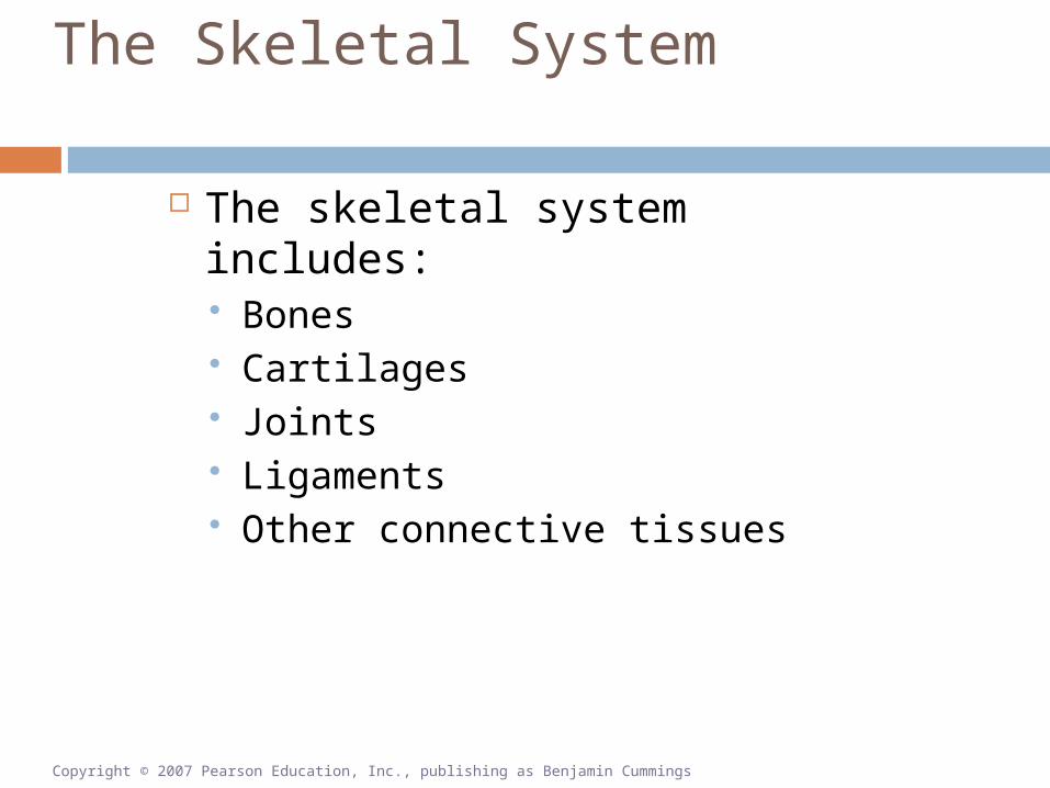
The skeletal system includes: Bones Cartilages Joints Ligaments Other connective tissues
Copyright © 2007 Pearson Education, Inc., publishing as Benjamin Cummings

The Skeletal System
Functions of the Skeletal System Support against gravity Storage
Calcium, phosphorous Fat
Blood cell production Protection of soft internal organs Leverage for muscle action
Copyright © 2007 Pearson Education, Inc., publishing as Benjamin Cummings

The Structure of Bone
Bone (Osseous Tissue) Specialized cells
2% of bone weight Strong flexible matrix
Calcium phosphate crystals Two-thirds of bone weight
Collagen fibers
Copyright © 2007 Pearson Education, Inc., publishing as Benjamin Cummings

The Structure of Bone
Macroscopic Features of Bone General shapes of bones
Long bones (e.g., humerus)
Short bones (e.g., carpal bones)
Flat bones (e.g., parietal bone)
Irregular bones (e.g., vertebra)
Copyright © 2007 Pearson Education, Inc., publishing as Benjamin Cummings

The Structure of Bone Shapes of Bones
Figure 6-1

The Structure of Bone
Features in a Long Bone Diaphysis (shaft)
Compact (dense) bone Marrow cavity
Epiphyses (ends) Spongy (cancellous) bone
Articular cartilage Periosteum (covering) Endosteum (lining)
Copyright © 2007 Pearson Education, Inc., publishing as Benjamin Cummings

The Structure of Bone
The Structure of a Long Bone
Figure 6-2

The Structure of Bone
Microscopic Features of Bone Periosteum
Outer fibrous layer Inner cellular layer
Osteocytes Within lacunae (holes) in matrix Between lamellae of matrix Branches within canaliculi
Copyright © 2007 Pearson Education, Inc., publishing as Benjamin Cummings

The Structure of Bone Microscopic Features of Bone
Osteon—Basic functional unit of compact bone; columnar in shape Strong in long axis of bone Concentric layers of osteocytes Concentric layers of matrix (lamellae) Central (Haversian) canal
Axial tunnel for blood vessels Perforating canal
Radial tunnel for blood vessels
Copyright © 2007 Pearson Education, Inc., publishing as Benjamin Cummings

The Structure of Bone Structure of a Typical Bone
Figure 6-3(a)

The Structure of Bone
Structure of a Typical Bone
Figure 6-3(b)

The Structure of Bone
Microscopic Features of Spongy Bone No osteons Lamellae as trabeculae
Arches, rods, plates of bone Branching network of bony tissue Strong in many directions Red marrow (blood forming) spaces
Copyright © 2007 Pearson Education, Inc., publishing as Benjamin Cummings

The Structure of Bone Cells in Bone
Osteocytes Mature bone cells between lamellae
Osteoclasts Source of acid, enzymes for osteolysis Calcium homeostasis
Osteoblasts Responsible for osteogenesis (new bone) Source of collagen, calcium salts
Copyright © 2007 Pearson Education, Inc., publishing as Benjamin Cummings

Bone Formation and Growth
Intramembranous Ossification Ossification—Process of converting other
tissues to bone Forms flat bones of skull, mandible, clavicle Stem cells differentiate to osteoblasts Produces spongy bone, then compact bone
Copyright © 2007 Pearson Education, Inc., publishing as Benjamin Cummings

Bone Formation and Growth
Bone Formation in 16-Week-Old Fetus
Figure 6-4

Bone Formation and Growth
Endochondral Ossification Most bones formed this way Cartilage model replaced by bone Replacement begins in middle
(diaphysis) Replacement follows in ends
(epiphyses)
Copyright © 2007 Pearson Education, Inc., publishing as Benjamin Cummings

Figure 6-52 of 6
Enlargingchondrocytes within
calcifying matrix
Chondrocytes at the center of the growing cartilage model enlarge and then die as the matrix calicifies.
Cartilagemodel
Copyright © 2007 Pearson Education, Inc., publishing as Benjamin Cummings

Figure 6-53 of 6
Enlargingchondrocytes within
calcifying matrix
Chondrocytes at the center of the growing cartilage model enlarge and then die as the matrix calicifies.
Newly derived osteoblasts cover the shaft of the cartilage in a thin layer of bone.
Cartilagemodel
Boneformation
Epiphysis
Diaphysis
Copyright © 2007 Pearson Education, Inc., publishing as Benjamin Cummings

Figure 6-54 of 6
Enlargingchondrocytes within
calcifying matrix
Chondrocytes at the center of the growing cartilage model enlarge and then die as the matrix calicifies.
Newly derived osteoblasts cover the shaft of the cartilage in a thin layer of bone.
Blood vessels penetrate the cartilage. New osteoblasts form a primary ossification center.
Cartilagemodel
Boneformation
Epiphysis
Diaphysis Marrowcavity
Primaryossificationcenter
Bloodvessel
Copyright © 2007 Pearson Education, Inc., publishing as Benjamin Cummings
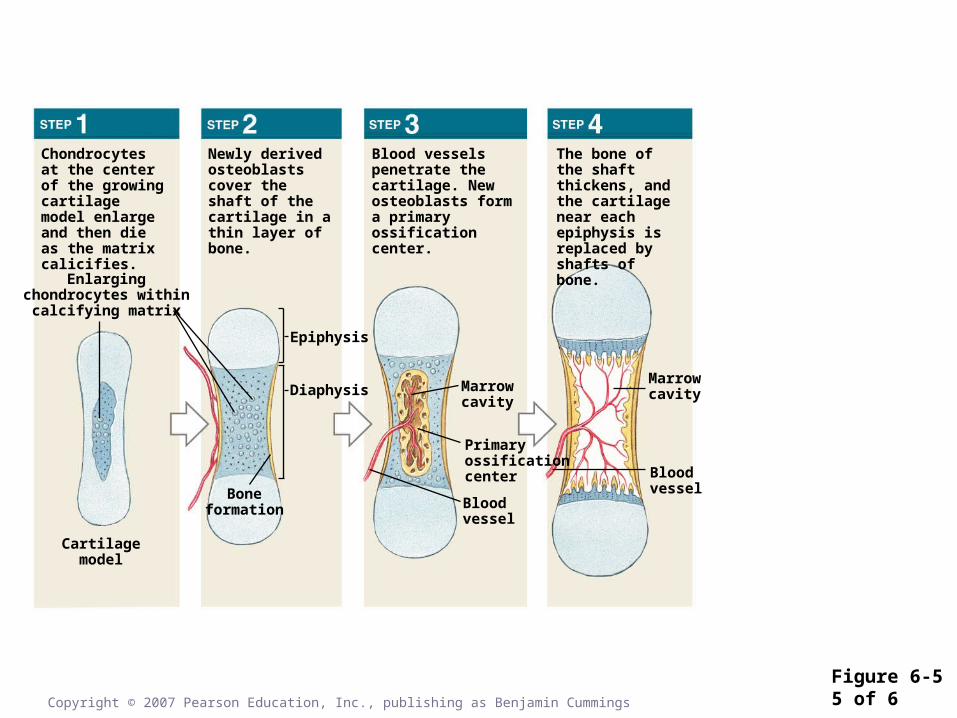
Figure 6-55 of 6
Enlargingchondrocytes within
calcifying matrix
Chondrocytes at the center of the growing cartilage model enlarge and then die as the matrix calicifies.
Newly derived osteoblasts cover the shaft of the cartilage in a thin layer of bone.
Blood vessels penetrate the cartilage. New osteoblasts form a primary ossification center.
The bone of the shaft thickens, and the cartilage near each epiphysis is replaced by shafts of bone.
Cartilagemodel
Boneformation
Epiphysis
Diaphysis Marrowcavity
Primaryossificationcenter
Bloodvessel
Marrowcavity
Bloodvessel
Copyright © 2007 Pearson Education, Inc., publishing as Benjamin Cummings

Figure 6-56 of 6
Enlargingchondrocytes within
calcifying matrix
Chondrocytes at the center of the growing cartilage model enlarge and then die as the matrix calicifies.
Newly derived osteoblasts cover the shaft of the cartilage in a thin layer of bone.
Blood vessels penetrate the cartilage. New osteoblasts form a primary ossification center.
The bone of the shaft thickens, and the cartilage near each epiphysis is replaced by shafts of bone.
Blood vessels invade the epiphyses and osteo-blasts form secondary centers of ossification.
Cartilagemodel
Boneformation
Epiphysis
Diaphysis Marrowcavity
Primaryossificationcenter
Bloodvessel
Marrowcavity
Bloodvessel
Secondaryossificationcenter
Epiphysealcartilage
Articularcartilage
Copyright © 2007 Pearson Education, Inc., publishing as Benjamin Cummings

Bone Formation and Growth
Appositional Bone Growth
Figure 6-6

Bone Formation and Growth Requirements for Normal Bone Growth
Minerals Calcium, phosphate
Vitamins Vitamin D3
Vitamin C Vitamin A
Hormones Growth Hormone Sex hormones, thyroid hormone, others
Copyright © 2007 Pearson Education, Inc., publishing as Benjamin Cummings
Bone Remodeling/Homeostasis
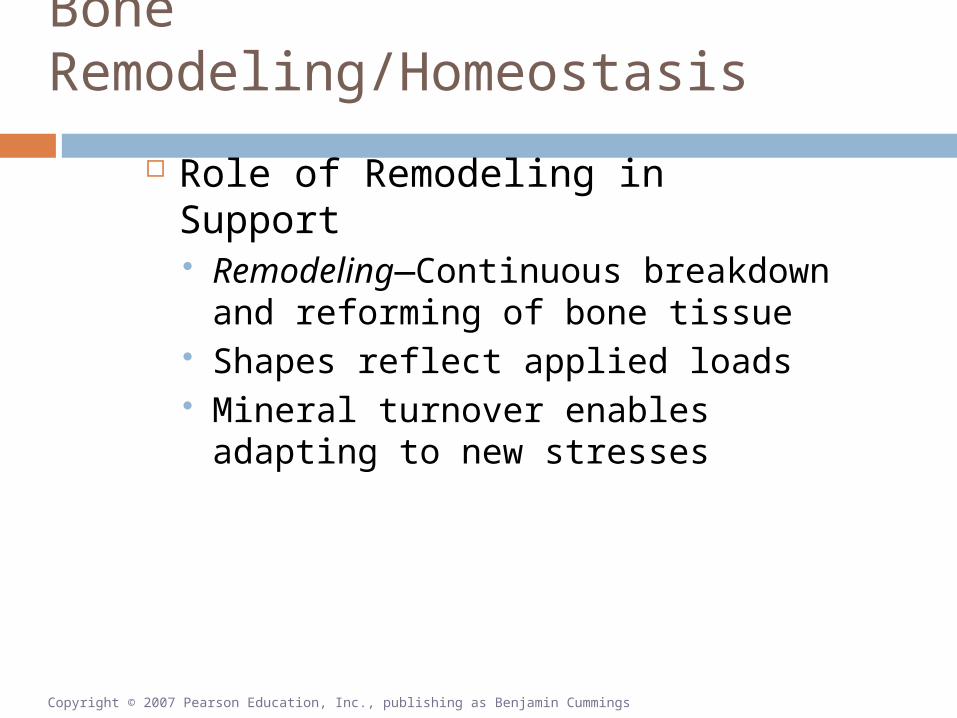
Role of Remodeling in Support Remodeling—Continuous
breakdown and reforming of bone tissue
Shapes reflect applied loads Mineral turnover enables adapting
to new stresses
Copyright © 2007 Pearson Education, Inc., publishing as Benjamin Cummings

Bone Remodeling/Homeostasis
What you don’t use, you lose.
Copyright © 2007 Pearson Education, Inc., publishing as Benjamin Cummings

Bone Remodeling/Homeostasis
Homeostasis and Mineral Storage Bones store calcium
Contain 99% of body calcium up to two kg calcium
Hormones control storage/release PTH, calcitriol release bone calcium Calcitonin stores bone calcium
Blood levels kept constant
Copyright © 2007 Pearson Education, Inc., publishing as Benjamin Cummings
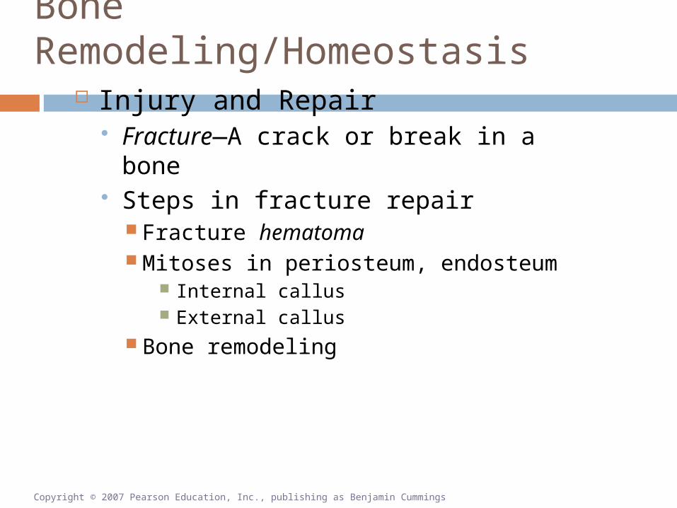
Bone Remodeling/Homeostasis
Injury and Repair Fracture—A crack or break in a bone Steps in fracture repair
Fracture hematoma Mitoses in periosteum, endosteum
Internal callus External callus
Bone remodeling
Copyright © 2007 Pearson Education, Inc., publishing as Benjamin Cummings

Figure 6-71 of 5
Bonefragments
Immediately after the fracture, extensive bleeding occurs. Over a period of several hours, a large blood clot, or fracture hematoma, develops.
Fracturehematoma
Deadbone
NewbonePeriosteum
Spongy boneof internal
callus
Cartilageof external
callus
An internal callus forms as a network of spongy bone unites the inner edges, and an external callus of cartilage and bone stabilizes the outer edges.
The cartilage of the external callus has been replaced by bone, and struts of spongy bone now unite the broken ends. Fragments of dead bone and the areas of bone closest to the break have been removed and replaced.
A swelling initially marks the location of the fracture. Over time, this region will be remodeled, and little evidence of the fracture will remain.
Internalcallus
Externalcallus
Externalcallus
Copyright © 2007 Pearson Education, Inc., publishing as Benjamin Cummings

Figure 6-72 of 5
Bonefragments
Immediately after the fracture, extensive bleeding occurs. Over a period of several hours, a large blood clot, or fracture hematoma, develops.
Fracturehematoma
Deadbone
Copyright © 2007 Pearson Education, Inc., publishing as Benjamin Cummings

Figure 6-73 of 5
Bonefragments
Immediately after the fracture, extensive bleeding occurs. Over a period of several hours, a large blood clot, or fracture hematoma, develops.
Fracturehematoma
Deadbone
NewbonePeriosteum
Spongy boneof internal
callus
Cartilageof external
callus
An internal callus forms as a network of spongy bone unites the inner edges, and an external callus of cartilage and bone stabilizes the outer edges.
Copyright © 2007 Pearson Education, Inc., publishing as Benjamin Cummings
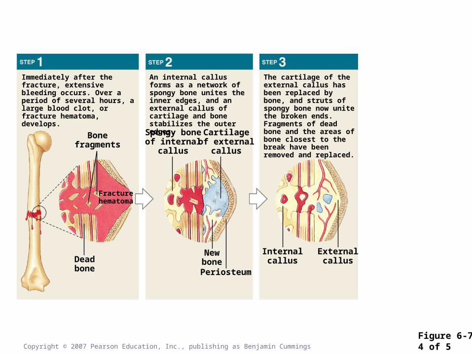
Figure 6-74 of 5
Bonefragments
Immediately after the fracture, extensive bleeding occurs. Over a period of several hours, a large blood clot, or fracture hematoma, develops.
Fracturehematoma
Deadbone
NewbonePeriosteum
Spongy boneof internal
callus
Cartilageof external
callus
An internal callus forms as a network of spongy bone unites the inner edges, and an external callus of cartilage and bone stabilizes the outer edges.
The cartilage of the external callus has been replaced by bone, and struts of spongy bone now unite the broken ends. Fragments of dead bone and the areas of bone closest to the break have been removed and replaced.
Internalcallus
Externalcallus
Copyright © 2007 Pearson Education, Inc., publishing as Benjamin Cummings
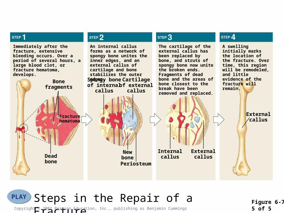
Figure 6-75 of 5
Steps in the Repair of a FracturePLAYPLAY
Bonefragments
Immediately after the fracture, extensive bleeding occurs. Over a period of several hours, a large blood clot, or fracture hematoma, develops.
Fracturehematoma
Deadbone
NewbonePeriosteum
Spongy boneof internal
callus
Cartilageof external
callus
An internal callus forms as a network of spongy bone unites the inner edges, and an external callus of cartilage and bone stabilizes the outer edges.
The cartilage of the external callus has been replaced by bone, and struts of spongy bone now unite the broken ends. Fragments of dead bone and the areas of bone closest to the break have been removed and replaced.
A swelling initially marks the location of the fracture. Over time, this region will be remodeled, and little evidence of the fracture will remain.
Internalcallus
Externalcallus
Externalcallus
Copyright © 2007 Pearson Education, Inc., publishing as Benjamin Cummings

Aging and the Skeletal System
Osteopenia—Less than normal ossification (mineral content) in bone Osteopenia starts before age 40
Women lose 8% per decade Men lose 3% per decade
Spongy bone most affected Epiphyses Vertebrae Jaws
Copyright © 2007 Pearson Education, Inc., publishing as Benjamin Cummings

An Overview of the Skeleton Surface Features of Bones
Table 6-1 (2 of 2)
An Overview of the Skeleton
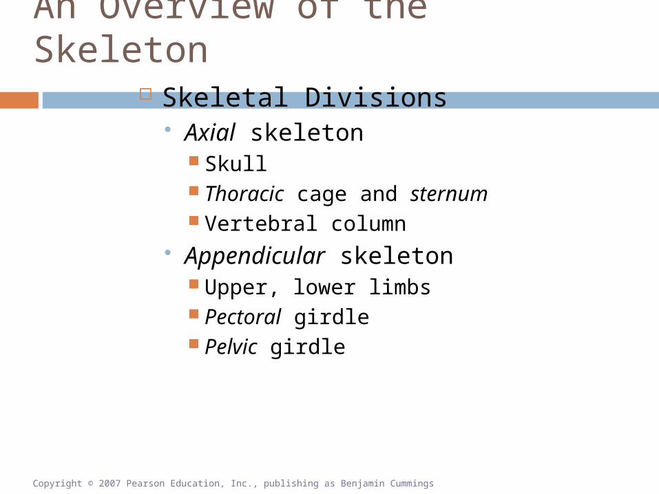
Skeletal Divisions Axial skeleton
Skull Thoracic cage and sternum Vertebral column
Appendicular skeleton Upper, lower limbs Pectoral girdle Pelvic girdle
Copyright © 2007 Pearson Education, Inc., publishing as Benjamin Cummings

An Overview of the Skeleton
The Skeleton
Figure 6-8(a)

An Overview of the Skeleton
The Skeleton
Figure 6-8(b)

An Overview of the Skeleton
The Axial and Appendicular Divisions of the Skeleton.
Figure 6-9

The Axial Division: The Skull
Bones of the Cranium Frontal bone
Forehead, superior surface of orbits Parietal bones
Sides, roof Occipital bone
Foramen magnum Temporal bones
Sides, base
Copyright © 2007 Pearson Education, Inc., publishing as Benjamin Cummings

The Axial Division: The Skull
Bones of the Cranium (continued) Sphenoid bone
Bridge between cranial and facial bones
Ethmoid bone Cribriform plate Nasal septum
Copyright © 2007 Pearson Education, Inc., publishing as Benjamin Cummings

The Axial Division: The Skull The Adult Skull (Part I)
Figure 6-10

The Axial Division: The Skull
Bones of the Face Maxillary bones Zygomatic bones
Zygomatic arch (with temporal bones)
Mandible
Copyright © 2007 Pearson Education, Inc., publishing as Benjamin Cummings

The Axial Division: The Skull
Bones of the Face (continued) Palatine bones The Vomer Nasal bones Lacrimal bones Inferior nasal conchae Nasal complex
Nasal septum
Copyright © 2007 Pearson Education, Inc., publishing as Benjamin Cummings

The Axial Division: The Skull
Bones of the Face (continued) Paranasal sinuses
Frontal Sphenoidal Ethmoidal Palatine Maxillary
Copyright © 2007 Pearson Education, Inc., publishing as Benjamin Cummings

The Axial Division: The Skull
The Adult Skull (Part II)
Figure 6-11(a)
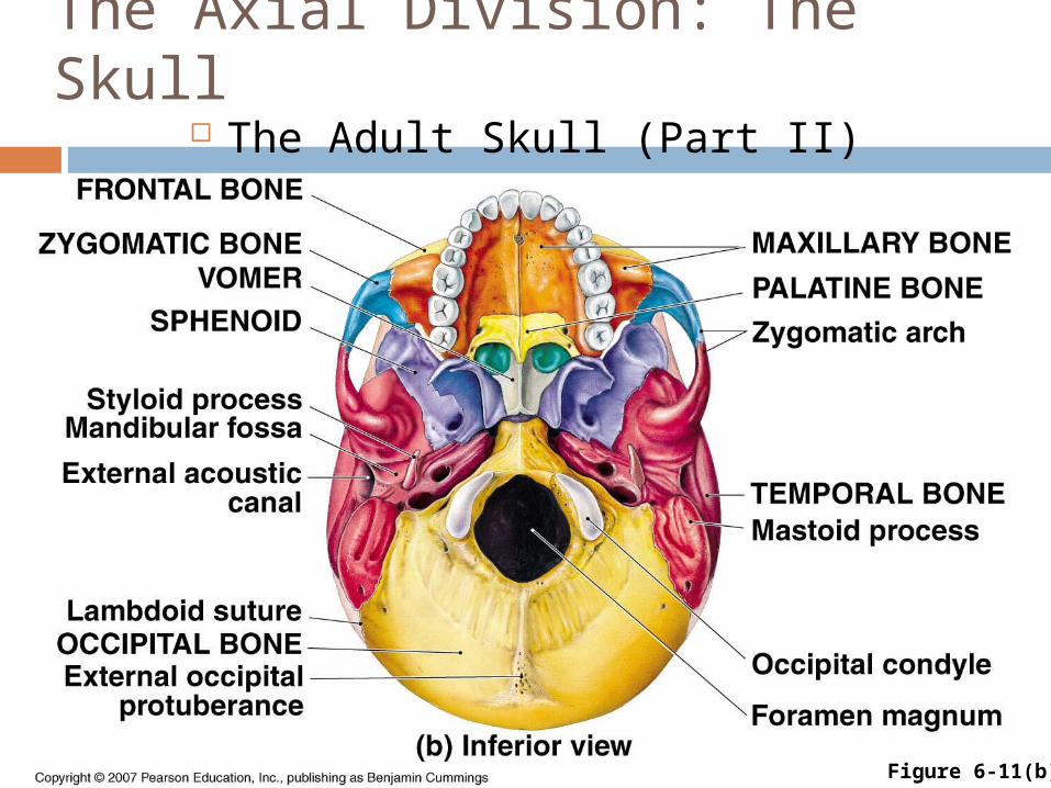
The Axial Division: The Skull The Adult Skull (Part II)
Figure 6-11(b)
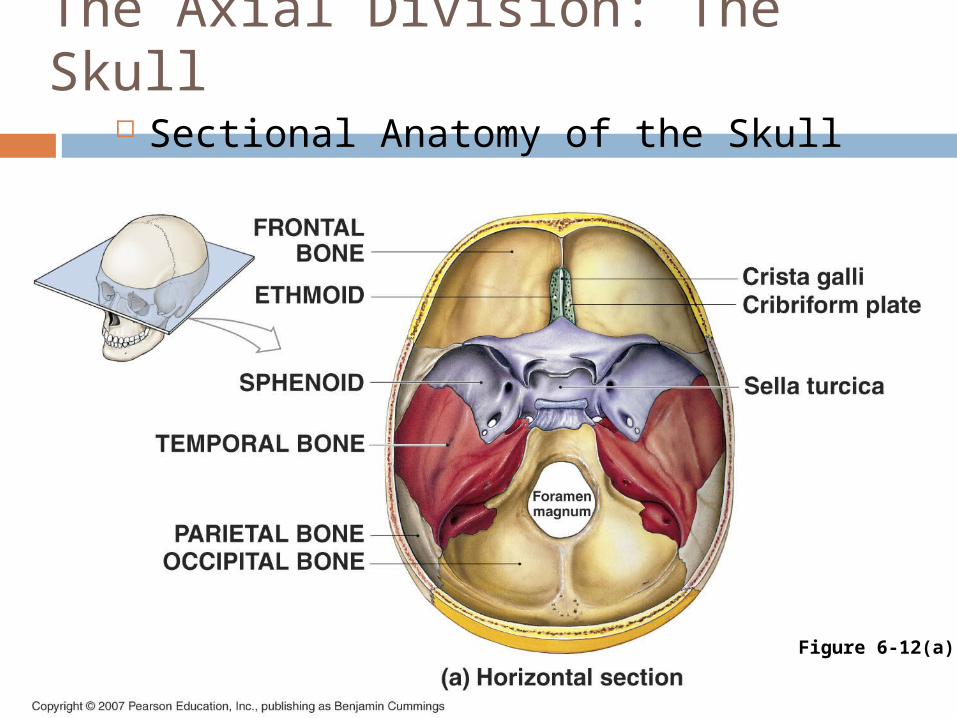
The Axial Division: The Skull
Sectional Anatomy of the Skull
Figure 6-12(a)

The Axial Division: The Skull
Sectional Anatomy of the Skull
Figure 6-12(b)

The Axial Division: The SkullSectional Anatomy of the Skull
Figure 6-12(c)

The Axial Division: The Skull The Paranasal Sinuses
Figure 6-13

Axial Division: The Skull The Skull of a Newborn
Figure 6-15(a)
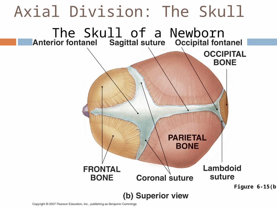
Axial Division: The SkullThe Skull of a Newborn
Figure 6-15(b)

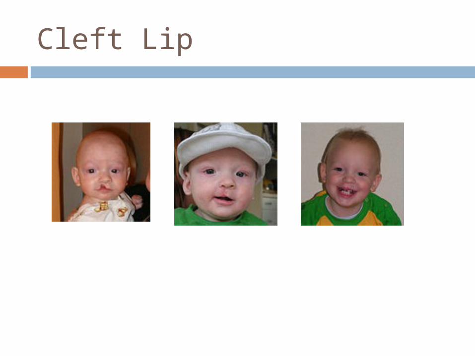
Cleft Lip
Unilateral incomplete
Unilateral complete
Bilateral Complete
Cleft Lip

Prevalence of cleft lip Native Americans: 3.74/1000 Japanese: 0.82/1000 to 3.36/1000 Chinese: 1.45/1000 to 4.04/1000 Caucasians: 1.43/1000 to 1.86/1000 Latin Americans: 1.04/1000 Africans: 0.18/1000 to 1.67/1000

Cleft Palate
Incomplete cleft palate
Complete unilateral lip & palate cleft
Complete bilateral lip & palate cleft

Vertebral Column/Thoracic Cage
Vertebral Column (Spine) 26 Bones
7 Cervical vertebrae (C1 to C7)
12 Thoracic vertebrae (T1 to T12)
5 Lumbar vertebrae (L1 to L5) Sacrum Coccyx (tailbone)
Copyright © 2007 Pearson Education, Inc., publishing as Benjamin Cummings

Vertebral Column/Thoracic Cage
Spinal Curvature Alignment of body weight Primary curves
Thoracic Sacral
Secondary curves Cervical Lumbar
Copyright © 2007 Pearson Education, Inc., publishing as Benjamin Cummings

Vertebral Column/Thoracic Cage
The Vertebral Column
Figure 6-16

Vertebral Column/Thoracic Cage
Vertebral Anatomy Body Arch
Transverse, spinous processes Pedicle, lamina Vertebral foramen
Vertebral canal
Articular processes Articular facets
Intervertebral discs
Copyright © 2007 Pearson Education, Inc., publishing as Benjamin Cummings

Vertebral Column/Thoracic Cage
Regional Differences in Vertebrae Cervical
Oval body Transverse foramina
Thoracic Heart-shaped body
Lumbar Massive (heaviest loading) Blade-like transverse processes
Copyright © 2007 Pearson Education, Inc., publishing as Benjamin Cummings

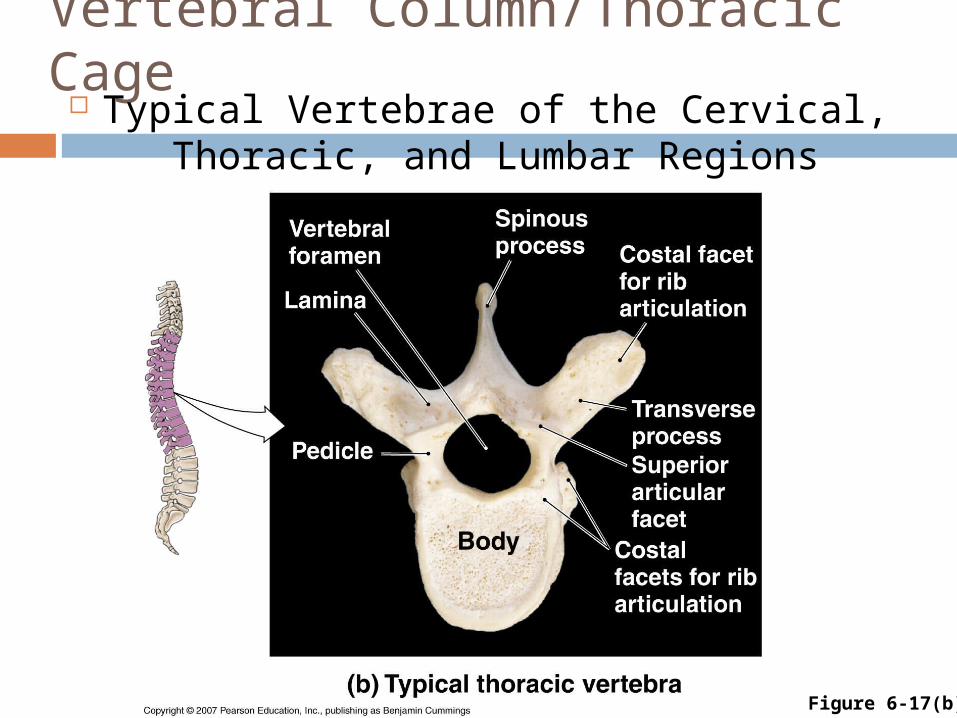
Vertebral Column/Thoracic Cage Typical Vertebrae of the Cervical, Thoracic,
and Lumbar Regions
Figure 6-17(a)
Vertebral Column/Thoracic Cage Typical Vertebrae of the Cervical, Thoracic,
and Lumbar Regions
Figure 6-17(b)

Vertebral Column/Thoracic Cage Typical Vertebrae of the Cervical, Thoracic,
and Lumbar Regions
Figure 6-17(c)

Vertebral Column/Thoracic Cage
The Atlas and Axis
Figure 6-18

Vertebral Column/Thoracic Cage
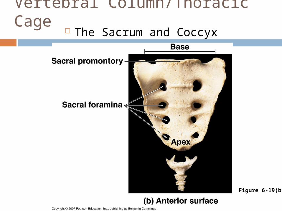
Functions of Sacrum Protects pelvic organs Base articulates with lumbar
vertebra Apex articulates with coccyx
Copyright © 2007 Pearson Education, Inc., publishing as Benjamin Cummings

Vertebral Column/Thoracic Cage
The Sacrum and Coccyx
Figure 6-19(a)
Vertebral Column/Thoracic Cage
The Sacrum and Coccyx
Figure 6-19(b)

Vertebral Column/Thoracic Cage
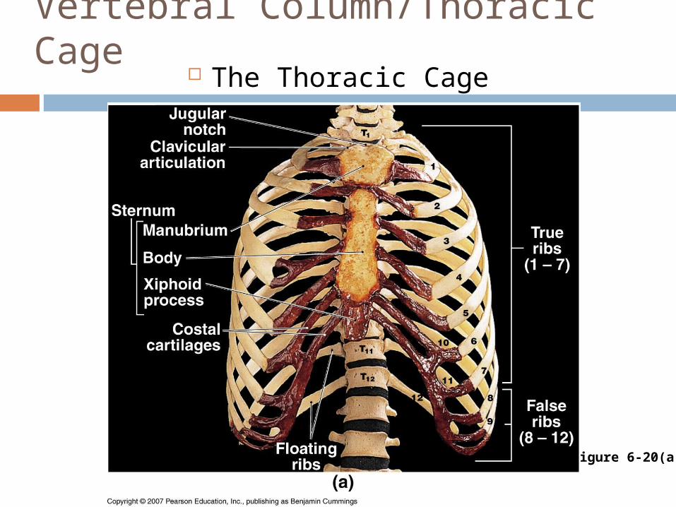
Components of Thoracic Cage Thoracic vertebrae Ribs
Seven pairs of true ribs Cartilaginous joint with sternum
Five pairs of false ribs Sternum
Manubrium, body, xiphoid process
Copyright © 2007 Pearson Education, Inc., publishing as Benjamin Cummings
Vertebral Column/Thoracic Cage
The Thoracic Cage
Figure 6-20(a)

Vertebral Column/Thoracic Cage
The Thoracic Cage
Figure 6-20(b)

Appendicular Division
Pectoral Girdle (Shoulder Girdle) Components
Scapulae (“shoulder blade”) Coracoid process Acromium Scapular spine
Clavicles (“collar bone”) Functions
Shoulder, arm movement Articulation for arm
Copyright © 2007 Pearson Education, Inc., publishing as Benjamin Cummings

Appendicular Division
The Clavicle
Figure 6-21

Appendicular Division The Scapula
Figure 6-22

Tuberosity Large, roughened elevation on a bony surface
Condyle Rounded articular projection on the surface of a bone
Trochlea A pulley
Fossa Shallow depression in surface of bone
Foramen Opening or passage through a bone
Sinus

Appendicular Division
Upper Limb Humerus
Head articulates with scapula Muscles attach to
Greater, lesser tubercles Deltoid tuberosity Medial, lateral epicondyles
Distal condyle articulates with forearm
Copyright © 2007 Pearson Education, Inc., publishing as Benjamin Cummings
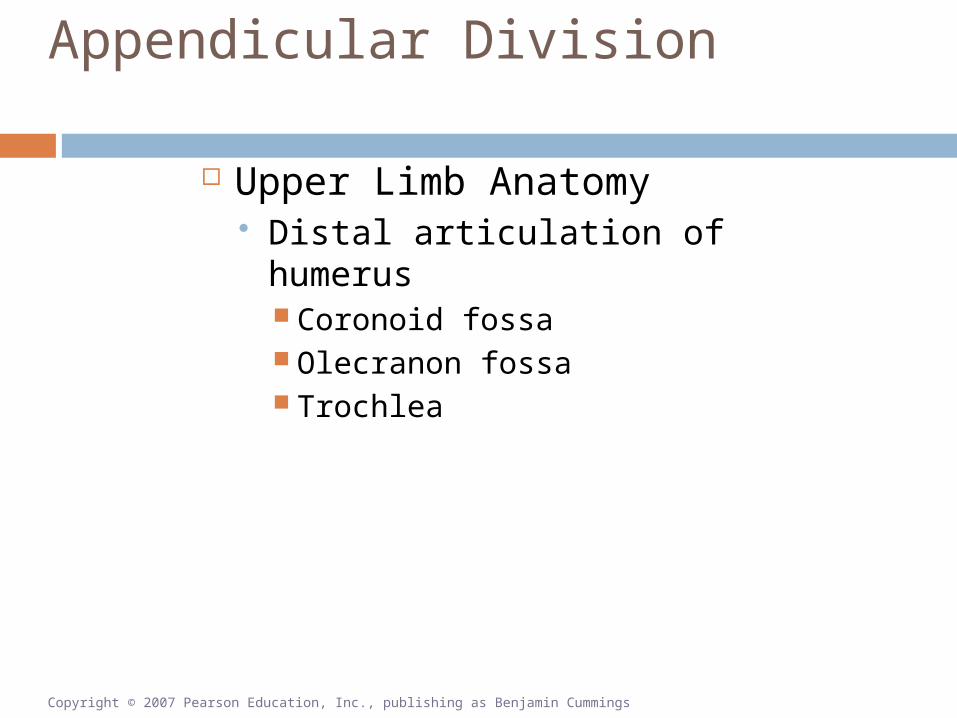
Appendicular Division
Upper Limb Anatomy Distal articulation of humerus
Coronoid fossa Olecranon fossa Trochlea
Copyright © 2007 Pearson Education, Inc., publishing as Benjamin Cummings

Appendicular Division
The Humerus
Figure 6-23

Appendicular Division
Bones of the Forearm Radius
Lateral (thumb side) Head articulates with humerus Radial tuberosity attaches biceps brachii Participates in wrist joint
Ulna Trochlear notch articulates with humerus Olecranon forms point of elbow
Copyright © 2007 Pearson Education, Inc., publishing as Benjamin Cummings

Appendicular Division
The Radius and Ulna
Figure 6-24

Appendicular Division
Bones of the Wrist and Hand Two rows of carpal bones
Proximal articulation with radius Distal articulation with metacarpal bones
Proximal phalanges (finger bones) articulate with metacarpals Three phalanges/finger Two phalanges/thumb (pollex)
Copyright © 2007 Pearson Education, Inc., publishing as Benjamin Cummings

Appendicular Division
Bones of the Wrist and Hand
Figure 6-25

Appendicular Division
The Pelvic Girdle Formed by two hipbones
hipbones formed by fusion of: Ilium Ischium Pubis
Pubic symphysis limit movement Pelvis formed by coxae, sacrum,
coccyx
Copyright © 2007 Pearson Education, Inc., publishing as Benjamin Cummings

Appendicular Division The Pelvis
Copyright © 2007 Pearson Education, Inc., publishing as Benjamin Cummings Figure 6-26(a)

Appendicular Division
The Pelvis
Figure 6-26(b)

Appendicular Division
The Pelvis
Figure 6-26(c)
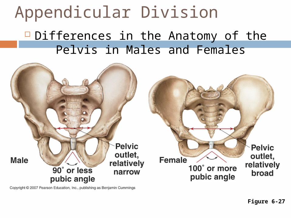
Appendicular Division Differences in the Anatomy of the Pelvis
in Males and Females
Figure 6-27

Appendicular Division
Bones of the Lower Limb Femur (thighbone) Patella (kneecap) Tibia (shinbone) Fibula Ankle bones Foot bones
Copyright © 2007 Pearson Education, Inc., publishing as Benjamin Cummings

Appendicular Division
The Femur
Figure 6-28
Appendicular Division
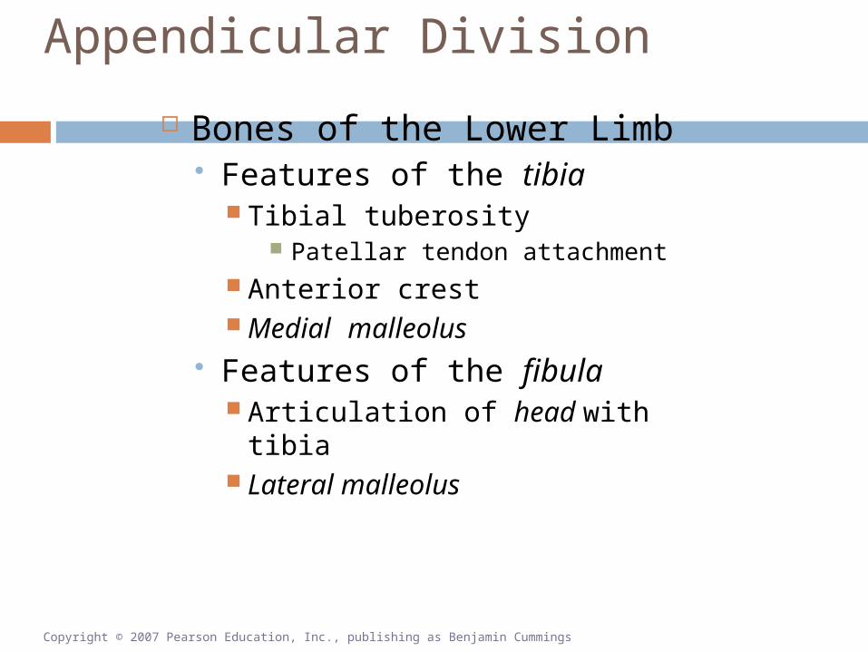
Bones of the Lower Limb Features of the tibia
Tibial tuberosity Patellar tendon attachment
Anterior crest Medial malleolus
Features of the fibula Articulation of head with tibia Lateral malleolus
Copyright © 2007 Pearson Education, Inc., publishing as Benjamin Cummings

Appendicular Division
The Right Tibia and Fibula
Figure 6-29

Appendicular Division
The Bones of the Ankle and Foot Ankle
Seven tarsal bones Talus
Joint with tibia, fibula
Foot Calcaneus (heel bone)
Major load-bearing bone Metatarsal bones Five phalanges (toes)
Copyright © 2007 Pearson Education, Inc., publishing as Benjamin Cummings

Appendicular Division
The Bones of the Ankle and Foot
Figure 6-30(a)

Appendicular Division
The Bones of the Ankle and Foot
Figure 6-30(b)

Articulations
Classification of Joints (Articulations) Joint—Where two bones interact Three functional classes of joint
Synarthroses Immovable
Amphiarthroses Slightly movable
Diarthroses Freely movable
Copyright © 2007 Pearson Education, Inc., publishing as Benjamin Cummings

Articulations Examples of Joints
Synarthroses Suture Gomphosis Synchondrosis
Amphiarthroses Syndesmosis Symphysis
Diarthroses Synovial joints
Copyright © 2007 Pearson Education, Inc., publishing as Benjamin Cummings

Articulations
Synovial Joints (Diarthroses) Epiphyses covered by articular cartilage Lubricated by synovial fluid Enclosed within joint capsule Other synovial structures include:
Menisci Bursae Fat pads Ligaments
Copyright © 2007 Pearson Education, Inc., publishing as Benjamin Cummings

Articulations The Structure of Synovial Joints
Figure 6-31(a)

Articulations
The Structure of Synovial Joints
Figure 6-31(b)

Articulations
Synovial Joints: Movements Flexion Extension Hyperextension Abduction Adduction Circumduction Rotation
Supination Pronation
Copyright © 2007 Pearson Education, Inc., publishing as Benjamin Cummings

Articulations
Angular Movements
Figure 6-32(a)

Articulations
Angular Movements
Figure 6-32(b)
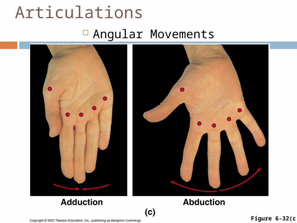
Articulations Angular Movements
Figure 6-32(c)

Articulations
Angular Movements
Figure 6-32(d)

Articulations
Rotational Movements
Figure 6-33(a)

Articulations
Rotational Movements
Figure 6-33(b)

Articulations
Special Movements Foot and ankle
Inversion, eversion Dorsiflexion, plantar flexion
Hand Opposition of thumb, palm
Head Protraction, retraction Depression, elevation (jaw)
Copyright © 2007 Pearson Education, Inc., publishing as Benjamin Cummings

Articulations Special Movements
Figure 6-34

Articulations
Structural Classification of Synovial Joints Gliding (e.g., vertebra–vertebra) Hinge (e.g., knee) Pivot (e.g., atlas–axis) Ellipsoidal (e.g., distal radius) Saddle (e.g., thumb) Ball-and-Socket (e.g., hip)
Copyright © 2007 Pearson Education, Inc., publishing as Benjamin Cummings
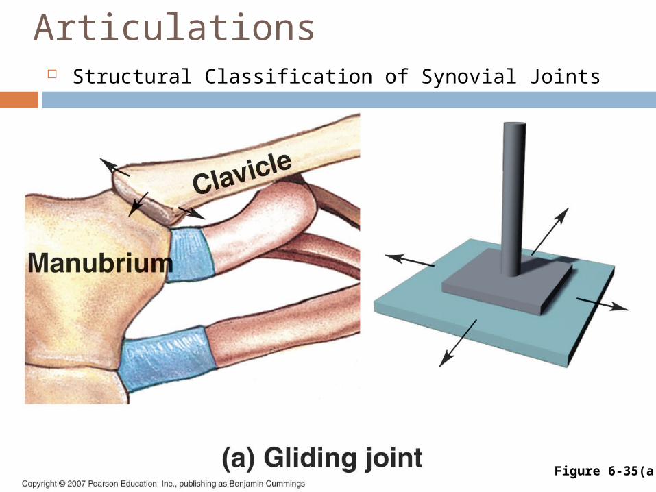
Articulations Structural Classification of Synovial Joints
Figure 6-35(a)
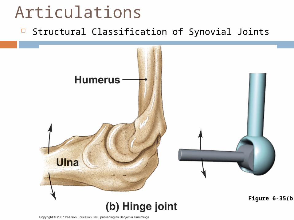
Articulations Structural Classification of Synovial Joints
Figure 6-35(b)

Articulations
Structural Classification of Synovial Joints
Figure 6-35(c)

Articulations
Figure 6-35(d)

Articulations
Structural Classification of Synovial Joints
Figure 6-35(e)

Articulations
Structural Classification of Synovial Joints
Figure 6-35(f)
Articulations
Greater mobility = weaker joint
Copyright © 2007 Pearson Education, Inc., publishing as Benjamin Cummings

Articulations
Intervertebral Articulations Two kinds join adjacent vertebrae
Gliding joints Between superior and inferior articular
processes Permit small movements
Symphyseal joints Intervertebral discs composed of
fibrocartilage Cushion and connect
Copyright © 2007 Pearson Education, Inc., publishing as Benjamin Cummings

Articulations
The Shoulder Joint Ball-and-socket design frees movement Joint dislocates easily Bursae reduce friction
Bursitis restricts motion, causes pain
Copyright © 2007 Pearson Education, Inc., publishing as Benjamin Cummings

Articulations
The Shoulder Joint
Figure 6-37
Articulations
The Elbow Joint Two articulations
Humerus–radius Humerus–ulna
Interlocking hinge design Limited movement Flexion and extension only Strong ligaments
Copyright © 2007 Pearson Education, Inc., publishing as Benjamin Cummings

Articulations The Elbow Joint
Figure 6-38

Articulations
The Hip Joint Extremely strong, stable joint
Many strong ligaments Tough joint capsule Bulky muscles
Versatile movements Flexion, extension, adduction,
abduction, circumduction, rotation
Copyright © 2007 Pearson Education, Inc., publishing as Benjamin Cummings

Articulations
The Hip Joint
Figure 6-39

Articulations
The Knee Joint Complex hinge joint
Three separate articulations Femur-tibia (between condyles—lateral
and medial) Femur-patella
Fibrocartilage pads Ligaments
Copyright © 2007 Pearson Education, Inc., publishing as Benjamin Cummings
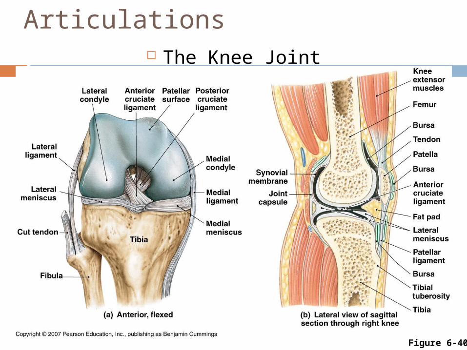
Articulations The Knee Joint
Figure 6-40

The Skeletal System in Perspective
Figure 6-411 of 11Copyright © 2007 Pearson Education, Inc., publishing as Benjamin Cummings

Figure 6-412 of 11Copyright © 2007 Pearson Education, Inc., publishing as Benjamin Cummings
The Integumentary System
• Synthesizes vitamin D3, essential for calcium and phosphorus absorption (bone maintenance and growth)
• Provides structural support

Figure 6-413 of 11Copyright © 2007 Pearson Education, Inc., publishing as Benjamin Cummings
The Muscular System
• Stabilizes bone positions; tension in tendons stimulates bone growth and maintenance
• Provides calcium needed for normal muscle contraction; bones act as levers to produce body movements

Figure 6-414 of 11Copyright © 2007 Pearson Education, Inc., publishing as Benjamin Cummings
The Nervous System
• Regulates bone position by controlling muscle contractions
• Provides calcium for neural function; protects brain, spinal cord; receptors at joints provide information about body position

Figure 6-415 of 11Copyright © 2007 Pearson Education, Inc., publishing as Benjamin Cummings
The Endocrine System
• Skeletal growth regulated by growth hormone, thyroid hormones, and sex hormones; calcium mobilization regulated by parathyroid hormone and calcitonin
• Protects endocrine organs, especially in brain, chest, and pelvic cavity

Figure 6-416 of 11Copyright © 2007 Pearson Education, Inc., publishing as Benjamin Cummings
The Cardiovascular System
• Provides oxygen, nutrients, hormones, blood cells; removes waste products and carbon dioxide
• Provides calcium needed for cardiac muscle contraction, blood cells produced in bone marrow

Figure 6-417 of 11Copyright © 2007 Pearson Education, Inc., publishing as Benjamin Cummings
The Lymphatic System
• Lymphocytes assist in the defense and repair of bone following injuries
• Lymphocytes and other cells of the immune response are produced and stored in bone marrow
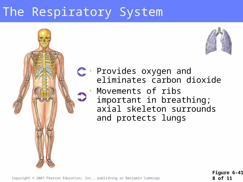
Figure 6-418 of 11Copyright © 2007 Pearson Education, Inc., publishing as Benjamin Cummings
The Respiratory System
• Provides oxygen and eliminates carbon dioxide
• Movements of ribs important in breathing; axial skeleton surrounds and protects lungs
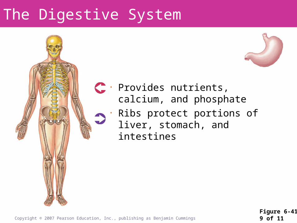
Figure 6-419 of 11Copyright © 2007 Pearson Education, Inc., publishing as Benjamin Cummings
The Digestive System
• Provides nutrients, calcium, and phosphate
• Ribs protect portions of liver, stomach, and intestines

Figure 6-4110 of 11Copyright © 2007 Pearson Education, Inc., publishing as Benjamin Cummings
The Urinary System
• Conserves calcium and phosphate needed for bone growth; disposes of waste products
• Axial skeleton provides some protection for kidneys and ureters; pelvis protects urinary bladder and proximal urethra

Figure 6-4111 of 11Copyright © 2007 Pearson Education, Inc., publishing as Benjamin Cummings
The Reproductive System
• Sex hormones stimulate growth and maintenance of bones; surge of sex hormones at puberty causes acceleration of growth and closure of epiphyseal cartilages
• Pelvis protects reproductive organs of female, protects portion of ductus deferens and accessory glands in males