the ste20/germinal center kinase pod6 interacts with …yardeno/odedspdfs/seiler2006.pdf · the...
TRANSCRIPT

Molecular Biology of the CellVol. 17, 4080–4092, September 2006
The STE20/Germinal Center Kinase POD6 Interacts withthe NDR Kinase COT1 and Is Involved in Polar TipExtension in Neurospora crassa□D
Stephan Seiler,*† Nico Vogt,† Carmit Ziv,‡ Rena Gorovits,‡ and Oded Yarden‡
*Deutsche Forschungsgemeinschaft Research Center of Molecular Physiology of the Brain (CMPB) and†Institut fur Mikrobiologie und Genetik, Abteilung Molekulare Mikrobiologie, Universitat Gottingen,D-37077 Gottingen, Germany; and ‡Department of Plant Pathology and Microbiology, The OttoWarburg Minerva Center for Agricultural Biotechnology, Faculty of Agricultural, Food andEnvironmental Quality Sciences, The Hebrew University of Jerusalem, Rehovot 76100, Israel
Submitted January 25, 2006; Revised June 19, 2006; Accepted June 28, 2006Monitoring Editor: David Drubin
Members of the Ste20 and NDR protein kinase families are important for normal cell differentiation and morphogenesisin various organisms. We characterized POD6 (NCU02537.2), a novel member of the GCK family of Ste20 kinases that isessential for hyphal tip extension and coordinated branch formation in the filamentous fungus Neurospora crassa. pod-6and the NDR kinase mutant cot-1 exhibit indistinguishable growth defects, characterized by cessation of cell elongation,hyperbranching, and altered cell-wall composition. We suggest that POD6 and COT1 act in the same genetic pathway,based on the fact that both pod-6 and cot-1 can be suppressed by 1) environmental stresses, 2) altering protein kinase Aactivity, and 3) common extragenic suppressors (ropy, as well as gul-1, which is characterized here as the ortholog of thebudding and fission yeasts SSD1 and Sts5, respectively). Unlinked noncomplementation of cot-1/pod-6 alleles indicatesa potential physical interaction between the two kinases, which is further supported by coimmunoprecipitation analyses,partial colocalization of both proteins in wild-type cells, and their common mislocalization in dynein/kinesin mutants.We conclude that POD6 acts together with COT1 and is essential for polar cell extension in a kinesin/dynein-dependentmanner in N. crassa.
INTRODUCTION
Factors that determine and modulate cell polarity have beenthe subject of extensive investigations in a variety of exper-imental organisms (Drubin and Nelson, 1996; Nelson, 2003),with the most progress having been made in the unicellularyeasts Saccharomyces cerevisiae and Schizosaccharomyces pombe(Bahler and Peter, 2000; Pruyne and Bretscher, 2000a, 2000b;Pruyne et al., 2004). The mechanisms by which polarity isestablished in filamentous fungi have remained largely ob-scure, but it is likely that the fundamental principles leadingto the initial polarization of the cell are conserved amongunicellular organisms (Bahler and Peter, 2000; Wendland,2001), filamentous fungi (Galagan et al., 2003; Borkovich etal., 2004; Harris and Momany, 2004), and animals (Hall,1998). But in contrast to baker’s yeast, where growth be-comes isotropic soon after bud emergence, the growth offilamentous fungi must stay highly polar to produce a tip-growing hypha that can extend at astonishing rates of morethan 1 �m/s (Lopez-Franco et al., 1994; Seiler and Plamann,2003; Harris et al., 2005). Thus, filamentous fungi presentgood model systems to study how this highly polar shape is
maintained over long distances, the way in which newbranch points are initiated and how their spatial relationshipis regulated.
In recent years, protein kinases of the NDR Ser/Thr pro-tein kinase family have emerged as being important fornormal cell differentiation and polar morphogenesis in var-ious organisms, yet their specific functions are still elusive(Tamaskovic et al., 2003; Hergovich et al., 2006). In Drosophilamelanogaster, the NDR kinases Tricornered and Warts arerequired for control of the extent and direction of cell pro-liferation as well as for neuronal morphogenesis (Justice etal., 1995; Xu et al., 1995; Geng et al., 2000; Emoto et al., 2004).The Caenorhabditis elegans homolog SAX1 regulates aspectsof neuronal cell shape and has been proposed to be involvedin cell spreading, neurite initiation, and dendritic tiling(Zallen et al., 2000; Gallegos and Bargmann, 2004). Verde etal. (1998) have shown that the fission yeast gene orb6 isrequired to maintain cell polarity during interphase. Thebudding yeast kinase Cbk1p is involved in cell separationand modulates cell shape (Racki et al., 2000; Bidlingmaier etal., 2001). A number of recent large-scale screens have iden-tified several proteins that interact with Cbk1p (Ito et al.,2001; Du and Novick, 2002; Ho et al., 2002), establishing theidea that Cbk1p and other interacting proteins may repre-sent the core components of a conserved complex requiredfor polarized morphogenesis. Further work in both yeasts aswell as in animal cells has resulted in an emerging network,which includes the NDR kinase and its binding partner andactivator MOB2, which are regulated through a Ste20 typekinase that interacts with a MO25- as well as a FURRY-like
This article was published online ahead of print in MBC in Press(http://www.molbiolcell.org/cgi/doi/10.1091/mbc.E06–01–0072)on July 5, 2006.□D The online version of this article contains supplemental materialat MBC Online (http://www.molbiolcell.org).
Address correspondence to: Stephan Seiler ([email protected]).
4080 © 2006 by The American Society for Cell Biology
scaffolding protein (Nelson et al., 2003; Kanai et al., 2005;Stegert et al., 2005; Hergovich et al., 2006).
The founding member of the NDR family, the kinaseCOT1 of the filamentous fungus Neurospora crassa, is re-quired for hyphal tip elongation (Collinge and Trinci, 1974;Collinge et al., 1978; Yarden et al., 1992), and the tempera-ture-sensitive cot-1 mutant ceases hyphal elongation afterbeing shifted to restrictive temperature. This is accompaniedby a massive induction of new hyphal tip formation, creat-ing the typical barbed-wired morphology of cot-1 cells. Asimilar branching and growth-termination phenotype hasbeen observed in neuronal cells of sax-1 and fry mutants in C.elegans and D. melanogaster (Geng et al., 2000; Zallen et al.,2000), suggesting an evolutionarily conserved function ofNDR kinases in the formation of branched cellular struc-tures. This may be linked to changes in a general stress-sensing response, similar to that reported for the mamma-lian NDR-related myotonic dystrophy kinase (Mounsey etal., 1995; Chahine and George, 1997; Kushnir et al., 1997).Evidence for this includes suppression of the cot-1 pheno-type by osmotic and other environmental stresses as well asby altering cAMP-dependent kinase (protein kinase A[PKA]) activity levels in the temperature-shifted cultures(Gorovits and Yarden, 2003).
Another large emerging group of kinases that have beenimplicated in various signaling pathways are the Ste20 ki-nases (Dan et al., 2001; Bokoch, 2003). Originally defined byS. cerevisiae Ste20p, an upstream kinase of the mitogen-acti-vated protein kinase pathway, the Ste20 group of kinases isdivided into the p21-activated (PAK) kinases and several ger-minal center kinase (GCK) subfamilies. The true PAKs, origi-nally characterized as the primary downstream effectorsof Rac/Cdc42-type GTPases, are defined by a C-terminalkinase domain and an N-terminally located Cdc42/Racinteracting/binding (CRIB) motif that mediates binding ofthe small G-protein to the kinase and its subsequent acti-vation. The GCK group differs from the PAKs in that thekinase domain is located N-terminally, they lack typicalCRIB domains, and their noncatalytic domains are highlyvariable. In contrast to the PAKs, the function of the GCKsis much less defined, but they have been implicated instress response, proliferation, and apoptosis (Dan et al.,2001; Bokoch, 2003).
Despite the relevance of an apically growing tip cell formembers of the fungal kingdom, the key components thatare specifically required for tip extension and branch-pointspecification are poorly understood, with COT1 being thebest-characterized player to date. An important link be-tween cytoskeleton assembly and function and COT1 activ-ity has been established by the analysis of cot-1 suppressormutants, which are defective in the microtubule-dependentmotor protein complex dynein/dynactin (Plamann et al.,1994; Bruno et al., 1996b), but the underlying molecularmechanisms remain unclear. Several other complementationgroups have been identified, which result in cot-1-likegrowth and defective hyphal elongation when these genesare mutated (Seiler and Plamann, 2003). Here, we report thecharacterization of POD6 (NCU02537.2), a novel member ofthe GCK family of Ste20 kinases that influences cell mor-phology in a manner highly similar to COT1 and acts inconcert, and perhaps in cooperation, with this NDR kinase.We also present evidence that POD6 is essential for cellu-lar extension in a motor-protein– dependent manner inN. crassa.
MATERIALS AND METHODS
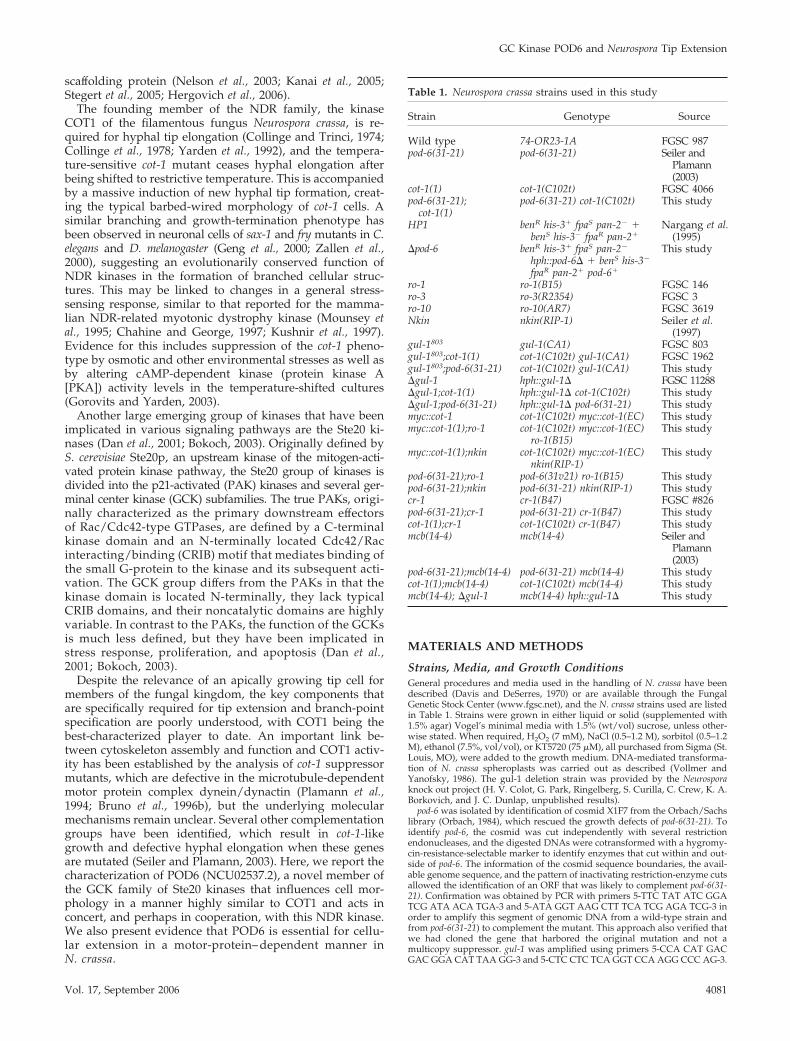
Strains, Media, and Growth ConditionsGeneral procedures and media used in the handling of N. crassa have beendescribed (Davis and DeSerres, 1970) or are available through the FungalGenetic Stock Center (www.fgsc.net), and the N. crassa strains used are listedin Table 1. Strains were grown in either liquid or solid (supplemented with1.5% agar) Vogel’s minimal media with 1.5% (wt/vol) sucrose, unless other-wise stated. When required, H2O2 (7 mM), NaCl (0.5–1.2 M), sorbitol (0.5–1.2M), ethanol (7.5%, vol/vol), or KT5720 (75 �M), all purchased from Sigma (St.Louis, MO), were added to the growth medium. DNA-mediated transforma-tion of N. crassa spheroplasts was carried out as described (Vollmer andYanofsky, 1986). The gul-1 deletion strain was provided by the Neurosporaknock out project (H. V. Colot, G. Park, Ringelberg, S. Curilla, C. Crew, K. A.Borkovich, and J. C. Dunlap, unpublished results).
pod-6 was isolated by identification of cosmid X1F7 from the Orbach/Sachslibrary (Orbach, 1984), which rescued the growth defects of pod-6(31-21). Toidentify pod-6, the cosmid was cut independently with several restrictionendonucleases, and the digested DNAs were cotransformed with a hygromy-cin-resistance-selectable marker to identify enzymes that cut within and out-side of pod-6. The information of the cosmid sequence boundaries, the avail-able genome sequence, and the pattern of inactivating restriction-enzyme cutsallowed the identification of an ORF that was likely to complement pod-6(31-21). Confirmation was obtained by PCR with primers 5-TTC TAT ATC GGATCG ATA ACA TGA-3 and 5-ATA GGT AAG CTT TCA TCG AGA TCG-3 inorder to amplify this segment of genomic DNA from a wild-type strain andfrom pod-6(31-21) to complement the mutant. This approach also verified thatwe had cloned the gene that harbored the original mutation and not amulticopy suppressor. gul-1 was amplified using primers 5-CCA CAT GACGAC GGA CAT TAA GG-3 and 5-CTC CTC TCA GGT CCA AGG CCC AG-3.
Table 1. Neurospora crassa strains used in this study
Strain Genotype Source
Wild type 74-OR23-1A FGSC 987pod-6(31-21) pod-6(31-21) Seiler and
Plamann(2003)
cot-1(1) cot-1(C102t) FGSC 4066pod-6(31-21);
cot-1(1)pod-6(31-21) cot-1(C102t) This study
HP1 benR his-3� fpaS pan-2� �benS his-3� fpaR pan-2�
Nargang et al.(1995)
�pod-6 benR his-3� fpaS pan-2�
hph::pod-6� � benS his-3�
fpaR pan-2� pod-6�
This study
ro-1 ro-1(B15) FGSC 146ro-3 ro-3(R2354) FGSC 3ro-10 ro-10(AR7) FGSC 3619Nkin nkin(RIP-1) Seiler et al.
(1997)gul-1803 gul-1(CA1) FGSC 803gul-1803;cot-1(1) cot-1(C102t) gul-1(CA1) FGSC 1962gul-1803;pod-6(31-21) cot-1(C102t) gul-1(CA1) This study�gul-1 hph::gul-1� FGSC 11288�gul-1;cot-1(1) hph::gul-1� cot-1(C102t) This study�gul-1;pod-6(31-21) hph::gul-1� pod-6(31-21) This studymyc::cot-1 cot-1(C102t) myc::cot-1(EC) This studymyc::cot-1(1);ro-1 cot-1(C102t) myc::cot-1(EC)
ro-1(B15)This study
myc::cot-1(1);nkin cot-1(C102t) myc::cot-1(EC)nkin(RIP-1)
This study
pod-6(31-21);ro-1 pod-6(31v21) ro-1(B15) This studypod-6(31-21);nkin pod-6(31-21) nkin(RIP-1) This studycr-1 cr-1(B47) FGSC #826pod-6(31-21);cr-1 pod-6(31-21) cr-1(B47) This studycot-1(1);cr-1 cot-1(C102t) cr-1(B47) This studymcb(14-4) mcb(14-4) Seiler and
Plamann(2003)
pod-6(31-21);mcb(14-4) pod-6(31-21) mcb(14-4) This studycot-1(1);mcb(14-4) cot-1(C102t) mcb(14-4) This studymcb(14-4); �gul-1 mcb(14-4) hph::gul-1� This study
GC Kinase POD6 and Neurospora Tip Extension
Vol. 17, September 2006 4081

To construct the resistance cassette for the generation of a �pod-6 strain, theAspergillus nidulans gpdA promoter was obtained as an 888-base pair SacI/NcoI fragment from pSM1 (Poggeler et al., 2003), fused via NcoI with the581-base pair nourseothricin resistance gene (NatR) from pAG25 (Goldsteinand McCusker, 1999) that was amplified with the primers 5-ACC CCA TGGCCA TGA CCA CTC TTG ACG AC-3 and 5-AGG GAA TTC TCA GGG GCAGGG CAT GC-3 and introduced together via SacI/EcoRI into pBluescriptSK� (Stratagene, La Jolla, CA). The nourseothricin concentration was ad-justed to 20 �g/ml to select for transformants.
The deletion cassette was obtained by plasmid gap repair in S. cerevisiae(Orr-Weaver and Szostak, 1983), with pRS416 (Sikorski and Hieter, 1989) asthe yeast vector cut with XbaI and XhoI and the following primers to generatethe NatR cassette and the 5� and 3� flanking regions of pod-6 by PCR withcosmid pXIF7 as template: 5-GCG AGC GGC AGG CGC TCT ACA TGA GCATGC CCT GCC CCT GAG GGA GGT AGG GTC TTG-3, 5-TGG AAT TGTGAG CGG ATA ACA ATT TCA CAC AGG AAA CAG CGC ATG TGC GGGTGG GTA ATG-3, 5-ATT AAG TTG CGT AAC GCC AGG GTT TTC CCAGTC ACG ACG CTA CAG CAC TTG TGA TGG TGC-3, 5-CTC CGC ATGCCA GAA AGA GTC ACC GGT CAC TGT ACA GAG CTG ACT GCC CCCGCA AGG CC-3, 5-TAC TAC CAA CAG TCT CCT CGG GTC CTT GCG GGGGCA GTC AGC TCT GTA CAG TGA CCG GT-3, and 5-TTC TGA GAC AAATAA CAT CCC GTT ACA AGA CCC TAC CTC CCT CAG GGG CAG GGCATG CT-3.
Homologous recombination events in N. crassa were verified by phenotypicanalysis of transformants grown on 400 �M p-fluorophenylalanine (fpa) and200 �g/ml histidine or 5 �g/ml benomyl and 200 �g/ml pantothenic acidand by the complementation of the growth defect of potential �pod-6 strainswith a pod-6-containing amplicon using the same primers as for the comple-mentation of pod-6(31-21).
An MYC-tagged version of COT1 was constructed, utilizing plasmidpOY18, containing the cot-1 wild-type allele, capable of complementing thecot-1 temperature-sensitive mutant (Yarden et al., 1992), along with plasmidpCM2-MT containing a 6MYC tag sequence (Cheng et al., 2001). The 6MYCtag sequence was previously PCR-amplified from pCM2-MT, while introduc-ing an NsiI restriction site at both ends of the product, and ligated into apDrive vector (Qiagen, Hilden, Germany) to create pME8 (Efrat, Gorovits,and Yarden, unpublished results). In this study, pME8 was used as a templatefor PCR amplification using the primers 5-GGT ATG CAT CGT TCG AAAGCT-3 and 5-TTG AAT TCG AAG CCT CAT GCA T-3, which introduced anNspV restriction site (underlined) at the PCR product ends, thereby facilitat-ing the cloning of the amplicon into the NspV site in pOY18 and creatingpCZ18. Correct integration of the MYC tag sequence at the 5� end of the cot-1gene coding region was verified by sequencing with the primer 5-TCT GGTTGT TGT TGG CAT TG-3. A 3.3-kb XbaI fragment from pMP6 containing ahygromycin-resistance cassette was finally introduced into the unique XbaIsite of pCZ218 creating pCZ218/hygR. To verify proper activity of the fusedMYC::COT1 protein, this construct was used to complement the growthdefects of cot-1(1). Western-blot analysis using anti-MYC antibodies verifiedthe presence of MYC-tagged COT1 with the expected molecular mass.
Immunological MethodsFor the preparation of polyclonal anti-POD6 antibodies, a POD6 fragmentcontaining amino acids 421–675 was expressed in Escherichia coli BL21(DE3)as a pQE60-based His6-fusion protein (Stratagene) and purified via the Ni-NTA purification system (Qiagen). Polyclonal antibodies were generated byPineda Antikorper Service (Berlin), and the serum was affinity-purified usingthe original antigen.
N. crassa mycelial samples were frozen in liquid nitrogen, suspended in 50mM phosphate buffer, pH 7.0, 150 mM KCl, 1 mM DTT, and protein inhibitorsand centrifuged, and the supernatant was used for further experiments.Proteins were separated by 7.5 or 10% SDS-PAGE. Western blotting wasperformed according to standard procedures. For immunoprecipitation (IP),protein G-Sepharose beads (Amersham Biosciences, Piscataway, NJ) wereincubated on a rotation device with an excess of monoclonal anti-MYCantibody (clone 9E10) in binding buffer (50 mM phosphate buffer, pH 7.0, 150mM KCl, 1 mM DTT, 0.2% wt/vol NP40, 1 mM EDTA, and protein inhibitors)for 2 h at 4°C. After the unbound antibodies were removed, the treated beadswere resuspended in binding buffer with 400–500 �g protein extract andincubated for 3 h at 4°C. Immunoprecipitated protein was eluted with 100mM glycine, boiled in Sample Buffer before SDS-PAGE, blotted, and probedwith �-MYC or �-POD6 antibodies.
MicroscopyImmunolocalization for N. crassa hyphae was conducted following a protocoladapted from Minke et al. (1999). Fresh conidia were spread on a small pieceof cellulose filter (GN-6, Gelman Sciences, Ann Arbor, MI) placed on thesurface of a sucrose agar plate, and incubated until small mycelia formed.Filters were plunge-frozen in liquid propane and transferred to fixative (3%formaldehyde in 100% ethanol precooled to �80°C). Samples were main-tained at �80°C for at least 2 d and then slowly transferred to room temper-ature (2 h at �20°C, 2 h at 4°C). Filters were rehydrated in a series ofethanol:buffer (100 mM phosphate, pH 7.0) solutions starting at a ratio of
90:10 and ending at 10:90. After several rinses in phosphate buffer, thesamples were incubated for 0.5–5 min in 2 mg/ml lysing enzymes (Sigma) in100 mM potassium citrate, pH 6.0, 20 mM EGTA to digest the cell wall. Afterseveral washes with phosphate buffer, filters were incubated in 1% BSA toblock nonspecific binding of antibody to the filter. Samples were immersed inthe primary antibody for at least 8 h, washed several times in phosphatebuffer and incubated in the secondary antibody for �8 h, and visualized usingstandard rhodamine and FITC filter sets. For light microscopy, samples wereviewed with a Zeiss Axioscope microscope (Oberkochen, Germany) and theImprovision Openlab 4.04 software (Lexington, MA). For the colocalization ofMYC::COT1 and POD6, 200-�m hyphal stacks were acquired and volume-deconvoluted using Openlab 4.04. Documentation was performed with aNikon DXM1200F digital camera (Melville, NY) or an Olympus SZX12 ste-reomicroscope (Hamburg, Germany), and a Kappa PS30 camera (Gleichen,Germany).
RESULTS
POD6 Belongs to the Germinal Center Family of the Ste20Group of KinasesTo identify critical components that contribute to polarizedgrowth in N. crassa, we recently developed a large-scalescreen for the isolation of conditional mutants defective inhyphal morphogenesis and isolated seven independent al-leles of pod-6 (polarity defective-6; Seiler and Plamann,2003). During the complementation of pod-6, we noticed thatthe N-terminal two thirds of the automatically annotatedORF NCU02537.2 were sufficient to complement pod-6. Fur-ther inspection of NCU02537.2 by BLAST analyses sug-gested that two ORFs were incorrectly fused during theannotation by the introduction of an erroneous intron, omit-ting the stop codon of the first ORF (pod-6) and joining itwith a second ORF (probably an ortholog of the translationinitiation factor SUA5). RT-PCR and sequencing experi-ments of cDNA with POD6-specific primers were used toconfirm this prediction. Thus, pod-6 encodes a 928-aminoacid protein containing an N-terminal kinase domain withthe highest sequence similarity to members of the Ste20group of kinases and a C-terminal region with no character-istic sequence motifs (Figure 1). Based on the homology ofthe kinase domain, its N-terminal localization and the lack ofdefined sequence motifs in the C-terminus, POD6 belongs tothe GCK-III subfamily of eukaryotic Ste20 kinases. Theknown vertebrate members of this subfamily, SOK1, MST3,and MST4, have been implicated in the regulation of stressresponse, apoptosis, and proliferation, but the molecularmechanisms involved are unknown (Dan et al., 2001, 2002;Lin et al., 2001; Qian et al., 2001). The most closely relatedbudding and fission yeast kinases are Kic1p and Nak1,respectively. Both kinases have been recently reported aspart of a morphogenetic network that also contains the NDRkinases Cbk1p and Orb6, respectively, that is important forcoordinating polarized growth with daughter cell specifictranscription and cell cycle progression (Nelson et al., 2003;Kanai et al., 2005; Leonhard and Nurse, 2005).
POD6 Is Essential for Hyphal Tip ExtensionWhen wild-type N. crassa conidia (asexual spores) germi-nate, they rehydrate and begin to grow isotropically.Growth soon becomes polarized, and usually one hyphal tipis generated. Continued polarized growth results in unidi-rectional extension of the straight primary hypha, and newhyphal tips are subsequently generated by branching fromsubapical compartments at intervals of 30–150 �m. Contin-uous hyphal elongation and branching results in the forma-tion of spreading colonies.
To determine the morphological changes conferred bypod-6, we conducted a microscopic analysis of pod-6 grownunder permissive and restrictive temperatures (Figure 2A).
S. Seiler et al.
Molecular Biology of the Cell4082

As no major morphological differences were observed amongthe seven isolated pod-6 alleles, we focused our analyses on thepod-6(31-21) allele. When grown on agar plates at 25°C, nodifferences in hyphal elongation or branching frequencywere detected between pod-6(31-21) and the wild type. How-ever, within 45 min of transferring pod-6(31-21) to 37°C, weobserved a rapid cessation of tip extension, whereas thetemperature shift had no effect on the morphology of wildtype (note that the agar plate served as a strong temperaturebuffer in these shift experiments). In contrast to a wild-typeapex, which is a dome-shaped structure, the tips of pod-6(31-21) have a characteristic pointed/needle-like shape after theshift to restrictive growth conditions. This cessation of tipextension was simultaneously accompanied by the appear-ance of numerous subapical branches 20–50 �m in lengththat also stopped growth with a pointed tip. After prolonged
incubation at restrictive temperature, these branches pro-duced secondary and tertiary branches, and the hyphaebegan to swell up in a bulbous and apolar manner. Transferof such a culture back to a permissive temperature resultedin all tips resuming normal growth rates, diameter, andmorphology within 2–5 min. Germination of pod-6(31-21) atrestrictive temperature resulted in the formation of com-pact colonies with multiple 10 –50-�m-long germ tubeswith pointed tips, which also produced secondary andtertiary branches. These experiments indicate that POD6is essential for tip extension, but is also required to inhibitexcessive branch formation in subapical regions of thehypha. Sequencing of pod-6(31-21) revealed a single aminoacid substitution of the conserved isoleucine 310 (ATA) atthe C-terminus of the kinase domain by a lysine (AAA;Figure 1).
Figure 1. POD6 is a GC kinase. The catalytic kinase domains of N. crassa POD6, S. cerevisiae Kic1p, S. pombe Nak1, human MST3 and SOK1,and two uncharacterized GCK-III kinases from D. melanogaster and C. elegans (Dan et al., 2001) were aligned using the CLUSTAL W alignmentalgorithm. Identical and conserved corresponding amino acids are shaded dark and light, respectively, the PAK signature motif “GTPFW-MAPE” is underlined and the position of the isoleucine-lysine substitution of pod-6(31-21) is marked with an asterisk.
GC Kinase POD6 and Neurospora Tip Extension
Vol. 17, September 2006 4083

To examine the phenotype of a pod-6 deletion strain, weconstructed a null mutant by using the sheltered disruptionmethod (Nargang et al., 1995), which takes advantage of thefact that N. crassa is a multinucleated cell. In brief, the pod-6gene was deleted via homologous recombination in the het-erokaryotic strain HP1, thereby allowing the generation ofmutants in genes that are essential or important for growth.The resulting mutant harbors two kinds of nuclei: one witha null allele of pod-6 and one with a wild-type copy. Thesenuclei contain selectable markers that allow a shift in thenuclear ratio within the heterokaryotic cells. Growth onmedia containing fpa and histidine favors the propagationof the wild-type nucleus. In contrast, growth of heterokary-otic cells on media containing benomyl and pantothenic acidforces the knockout nucleus to predominate and thus leadsto the depletion of POD6 and morphological defects that areindistinguishable from pod-6(31-21) germinated at or shiftedto restrictive temperatures (Figures 2B). These results indi-cate that pod-6(31-21) is a temperature-sensitive loss-of-func-tion allele of pod-6. Both mutants indicate an essential rolefor POD6 during the extension of the hyphal tip and incontrolling the number and position of subapical branches.
POD6 and COT1 Act in the Same Genetic Pathway inParallel to the PKA PathwayThe morphological defects of pod-6(31-21) and of �pod-6were strikingly similar to those of cot-1(1) and led us tohypothesize that POD6 and COT1 may have related func-tions. This was supported by the observation that several
recessive alleles of cot-1 and pod-6 showed unlinked non-complementation. This genetic phenomenon suggests thatthe two proteins are either part of independent pathways,which are equally necessary for the successful completion ofa process or that the two proteins interact physically. To furtheranalyze the relation between COT1 and POD6, double mutantswere generated (Figure 3A). cot-1(1), pod-6(31-21), and cot-1(1);pod-6(31-21) mutants showed identical phenotypes at restric-tive temperature. This provides further evidence of their in-volvement in a common genetic pathway and, possibly, of aphysical interaction between the two proteins.
The cot-1(1) mutation has been shown to result in increasedcell-wall thickness at restrictive temperature (Collinge et al.,1978; Gorovits et al., 2000), suggesting a defect in cell-wallmetabolism. We therefore tested chitin deposition by staininghyphae with Calcofluor White. Chitin is the primary compo-nent of the inner layers of the hyphal cell wall and is thereforeaccessible to the dye primarily at the hyphal tips. At permissivetemperature, hyphal tips (and to a minor extent also septa) ofpod-6(31-21) and also cot-1(1) are strongly labeled in a manneridentical to the wild type (Figure 3B). Within 5 h at 37°C,pod-6(31-21), cot-1(1), and the pod-6(31-21);cot-1(1) double mu-tant showed extensive label in a patchy, subapical mannerthroughout the hyphae, including strong septal staining, indi-cating excessive chitin deposition in all mutants, and furthersuggesting a functional connection between POD6 and COT1.Furthermore, it appears that the high calcofluor stain at the tipobserved in mutants grown at the permissive temperature isnearly absent when the strains are cultured at the restrictive
Figure 2. POD6 is essential for hyphal tip extension. (A) pod-6(31-21) was grown on minimal media plates at 25°C and shifted for 1, 3, and12 h to restrictive temperature to illustrate the cessation of tip extension with pointed, needle-like tips and the progressive hyperbranchingof the mutant. Hyphae that were shifted to the restrictive temperature and then shifted back to the permissive temperature resumed growthat all generated tips within 5 min (center panel). When germinated at the restrictive temperature, pod-6 exhibited a compact, hyperbranched,colonial morphology. (B) Growth of the HP1 pod-6� � HP1 �pod6 heterokaryotic strain on minimal medium, medium containing fpa andhistidine and medium containing benomyl and pantothenic acid (which forces the knockout nucleus to predominate), resulting inmorphological defects identical to pod-6(31-21) germinated at or shifted to restrictive temperature.
S. Seiler et al.
Molecular Biology of the Cell4084

temperature, indicating decreased chitin synthesis at thesenonelongating tips.
If POD6 and COT1 have related functions, they may sharethe same extragenic suppressors. Therefore, we testedwhether components of the dynein/dynactin complex couldpartially complement the growth defects of pod-6, similar to themechanism described for cot-1(1) (Plamann et al., 1994; Bruno etal., 1996b). We compared growth rates of ro-1;pod-6(31-21),ro-3;pod-6(31-21), and ro-10;pod-6(31-21) with pod-6(31-21)grown at restrictive temperatures and found that these mu-tations partially suppress the pod-6(31-21) phenotype (Figure3C; for hyphal phenotypes of the double mutants, see Sup-plementary Figure 1).
In addition to dynein/dynactin mutations, gul-1 has alsobeen described as a dominant modifier of cot-1 (Terenzi andReissig, 1967). Although the identity of the affected gene hasnot yet been determined, its close linkage with am-1 andace-5 (Perkins et al., 2001) and the available genome sequenceled us to suspect that GUL1 may be encoded byNCU01197.2. To test this, we amplified wild-type and mu-tant gul-1803 alleles by PCR and transformed cot-1(1) andpod-6(31-21) with both amplicons. While transformationwith gul-1� resulted in compact cot-1(1) or pod-6(31-21) col-onies, strains transformed with the mutant gul-1803 allelepartially suppressed the growth defects of cot-1(1) and pod-6(31-21), similar to the original cot-1;gul-1 strain. This indi-cated that NCU01197.2 codes for GUL1 and that gul-1 is acommon dominant suppressor of both cot-1(1) and pod-6(31-21). Database searches indicated that GUL1 is an evolution-arily conserved protein. Ssd1p and STS5, the budding andfission yeast orthologues, respectively, are naturally poly-morphic proteins and have been implicated in the mainte-nance of cell-wall integrity, RNA binding, and the TOR
pathway, as well as in protein phosphatase-associated func-tions, but the underlying molecular mechanisms are entirelyunknown (Matsusaka et al., 1995; Toda et al., 1996; Evans andStark, 1997; Uesono et al., 1997). To characterize the functionof GUL1 in apical tip extension, we compared the pheno-types of the dominant gul-1803 and a gul-1 deletion strain.Morphological characteristics of both strains suggested arole of GUL1 in the establishment of a functional tip bothduring germination as well as during the formation of newbranches (Supplementary Figure 1; Table 2).
According to Gorovits and Yarden (2003), the cot-1(1)defect can be suppressed by regaining partial COT1 activityin mutants of the dynein/dynactin (ropy) complex. Wetherefore tested gul-1803 and gul-1803;cot-1(1) cultures at 37°Cfor the presence/absence of the 67-kDa COT1 band anddetermined that the dominant mutation in gul-1803 elimi-nated the need for the 67-kDa COT1 form (Figure 3D),indicating that suppression by the dynein/dynactin muta-
Figure 3. COT1 and POD6 act in the same genetic pathway. (A) A cot-1;pod-6 double mutant displayed the same morphological defects asthe two parental strains. All three strains were grown on minimal media plates shifted to restrictive temperature for 5 h after growth atpermissive temperature. (B) Calcofluor White stained primarily the hyphal apex of mutant strains grown at permissive temperature butmutants shifted to restrictive temperature for 5 h showed an abnormal and patchy label throughout the hyphae, including strong septalstaining. (C) Suppressor analysis of cot-1(1) and pod-6(31-21) showed that gul-1 as well as components of the dynein/dynactin complexpartially suppress the cot-1(1) or pod-6(31-21) growth defects (ro-1: dynein heavy chain mutant, ro-3: p150glued mutant; ro-10: 24-kDa subunitof dynactin). (D) Western-blot analysis of cell extracts probed with anti-COT1 antibody indicated that suppression of the cot-1(1) phenotypeat restrictive temperature is independent of 67-kDa COT1 presence.
Table 2. Morphological characteristics of wild type and gul-1mutants
Wild type �gul-1 gul-1803
Growth rate(cm/d; n � 3)
6.2 � 0.2 5.6 � 0.3 5.5 � 0.2
Conidial germinationrate after 5 h(%; n � 150)
83 54 61
Distance betweenbranch points(�m; n � 80)
338 � 115 125 � 43 153 � 57
GC Kinase POD6 and Neurospora Tip Extension
Vol. 17, September 2006 4085

tions is different from that involving gul-1. Bypassing theneed for COT1 was also observed when the mutants weretreated with various environmental stresses that alter PKAactivity (see below), suggesting a potential link between theproposed functions of GUL1 and the PKA pathway. Theconsistency of the effects that common genetic suppressorshave on cot-1 and pod-6 morphology strongly support thatPOD6 and COT1 act in a common genetic pathway.
In addition to the suppression by extragenic mutations,cot-1(1) has been shown to undergo environmental stress-related suppression (Gorovits and Yarden, 2003). Therefore,we cultured pod-6(31-21) as well as pod-6(31-21);cot-1(1) un-der similar environmental stresses. When grown in the pres-ence of NaCl, a significant increase in radial growth ofpod-6(31-21) as well as pod-6(31-21);cot-1(1) was observed(Figure 4A). Furthermore, when either NaCl or ethanol wereadded to the medium, suppression of these strains wasobserved (Figure 4B). Similar results were obtained whenthe strains were cultured in the presence of sorbitol or H2O2.A common component of fungal stress response is the PKA-dependent signaling pathway (Thevelein, 1994; Gustin et al.,1998), and the stress-related suppression of cot-1(1) wasaccompanied by reduced levels of PKA activity (Gorovitsand Yarden, 2003). Thus, we inhibited PKA by amendingpod-6(31-21), cot-1(1) and the double mutant grown at restric-tive temperature with the PKA-specific inhibitor KT5720and observed partial suppression of the mutant phenotypesin a manner similar to that observed in the stressed cultures(Figure 5A), indicating that lowering PKA activity can by-pass the POD6/COT1 defects. This is further supported bythe analysis of double mutants between cot-1(1)/pod-6(31-21)and cr-1, an adenylate cyclase mutant with reduced levels ofcAMP that can be complemented by adding cAMP (Terenziet al., 1974). These double mutants showed normal tip growthat 37°C (Figure 5B). In contrast, when we generated doublemutants using a temperature-sensitive allele in the regulatorysubunit of PKA that displayed increased levels of PKA activity
at restrictive temperature (Bruno et al., 1996a; Seiler and Pla-mann, 2003) by crossing cot-1(1)/pod-6(31-21) with mcb, we ob-served a synthetic effect (Figure 5C). Unlike the single mutants,mcb(14-4);pod-6(31-21) and mcb(14-4);cot-1(1) showed slightgrowth abnormalities at permissive temperatures. After shift-ing to 37°C, the synthetic defects were even more pronounced,resulting in branched and swollen hyphal tips and markedseptation within 5 h in the double mutants, whereas mcb(14-4)displayed only minor abnormalities, such as some swollen tipsand branches. Overnight incubation at 37°C resulted in celllysis and subsequent death of double mutant cultures, whereasmcb(14-4) displayed a mixture of normally growing hyphaeand chains of spherical cells. As changes in cell polarity in cot-1and pod-6 are emphasized when PKA activity levels are altered,we concluded that COT1/POD6 and PKA act in parallel path-ways to regulate cell polarity in a positive or negative manner,respectively.
POD6 and COT1 Are Physically Associated and Dependon Opposing Microtubule-dependent Motor Proteins forCorrect LocalizationTo examine whether POD6 physically interacts with COT1,we generated and affinity-purified antisera against POD6that recognized a single polypeptide of ca. 100-kDa in wild-type extracts and generated a strain expressing aMYC::COT1 fusion protein (Figure 6A). The POD6 signalwas strongly reduced in extracts of the heterokaryotic�pod-6 strain grown on benomyl and pantothenic acid, in-dicating that the anti-POD6 antibodies are specific for POD6(Figure 6B). Reduced amounts of POD6 were also observedin pod-6(31-21) shifted to restrictive temperature, indicatingthat a reduction in POD6 protein levels accompanies thetemperature shift. Nevertheless, we observed no change inprotein abundance of either kinase in the other mutantgrown at restrictive temperature (Figure 6C). cot-1 wastagged N-terminally with a 6MYC tag under the control ofits own promoter. This construct fully complemented cot-
Figure 4. Environmental suppression of pod-6(31-21) and cot-1(1); pod-6(31-21). (A) NaCl amendment in the growth medium suppresses thecot-1, pod-6 and double mutant phenotype. Relative growth (expressed as percent of nonamended control) was determined on the basis ofradial growth of N. crassa strains in the presence of different concentrations of NaCl. Cultures were incubated at 34°C. The results are theaverage of at least three independent results. Error bars, SE. *, wild type; ● , cot-1(1); Œ, pod-6(31-21); f, cot-1(1);pod-6(31-21). (B) Pictures ofliquid cultures supplemented with either 1 M NaCl or 7.5% ethanol were taken 8 h after they were shifted from permissive to restrictivetemperatures.
S. Seiler et al.
Molecular Biology of the Cell4086

1(1) and ensured full activity of the modified protein. Anti-MYC, anti-POD6, and, as a control, anti-HA antibodies wereused for IP experiments from extracts of the MYC::COT1strain (Figure 6D). The anti-POD6 IP recovered a markedlyhigher portion of POD6 from the extracts when comparedwith the amount of coprecipitated COT1, whereas anti-MYCIP recovered an abundant quantity of COT1 and coprecipi-
tated much less of POD6. These results indicate that underthe conditions tested, low but significant portions of COT1and POD6 were associated with each other.
To determine the cellular distribution of MYC::COT1 andPOD6 in N. crassa hyphae, we used the anti-MYC and anti-POD6 antibodies, respectively (Figure 7A). Both proteinsexhibit a vesicular-reticulate distribution throughout the hy-
Figure 5. PKA and POD6/COT1 act in parallel pathways. (A) Effect of amending liquid growth media with the PKA inhibitor KT5720.Suppression of the growth defect was monitored after shifting the cultures to restrictive temperatures for 6 h. (B) The growth defect of cot-1(1)and pod-6(31-21) was suppressed in the cr-1 mutant background, whereas (C) mcb;cot-1/pod-6 double mutants are synthetically lethal. For Band C, the strains were grown on minimal media plates supplemented with 1% yeast extract and shifted for the indicated times.
GC Kinase POD6 and Neurospora Tip Extension
Vol. 17, September 2006 4087

pha that is, to a large extent, overlapping. In conjunctionwith the co-IP data, indicating a low but significant level ofinteraction, we concluded that COT1 and POD6 may be partof one or more dynamic protein complexes in the cell, butthat the proteins are not necessarily associated with eachother at all times. This punctate localization pattern and ourprevious results that both mutants can be suppressed bycomponents of the dynein/dynactin motor complex prompted
us to analyze the dependence of POD6 and COT1 on motorprotein function (Figure 7B). In ro-1 hyphae, POD6 was notevenly distributed, but rather accumulated in clusters at thehyphal apex. In contrast, we observed the opposite distributionin conventional kinesin mutants with both kinases accumu-lating subapically at the septae. Finally, we also tested theinterdependence of POD6 and COT1 for localization in therespective mutants shifted to restrictive temperatures. In
Figure 6. COT1 and POD6 interact. (A) COT1, POD6, and MYC::COT1 were detected in extracts prepared from wild-type cells orMYC::COT1 cells, respectively, to illustrate the specificity of the antibodies being used. (B) Extracts of the nuclear-ratio-modulatedheterokaryotic �pod-6 strain grown in appropriate media (see Materials and Methods) were adjusted to equal amounts of soluble protein andprobed to determine the effect of the predominance of �pod-6 nucleus (in the presence of benomyl and pantothenic acid) on POD6 abundance.(C) Reduced amounts of POD6 were also observed in pod-6(31-21) shifted to restrictive temperature for 5 h, but no alteration in the expressionlevel of either kinase in the other mutant grown at restrictive temperature was observed. (D) Cell extracts of MYC::COT1 cells were prepared,precipitated with �-MYC, �-POD6 or, as control, �-HA antibodies, separated by SDS-PAGE and probed with �-MYC or �-POD6 antibodies.
Figure 7. COT1 and POD6 colocalize and aredependent on microtubule-dependent motorproteins for their correct localization.MYC::COT1 and POD6 were colocalized ingrowing wild-type hyphae and exhibited punc-tate/reticulate staining that was evenly distrib-uted throughout the hypha. Note that the Spit-zenkorper region at the extreme hyphal apex isunlabeled. (B) This punctate distribution ofmyc::COT1 and POD6 was altered in dyneinheavy chain and conventional kinesin mutants,with myc::COT1 and POD6 accumulating at thehyphal tip in the minus-end-directed (�) ro-1mutant and in subapical areas/septae in theplus-end-directed (�) nkin mutant. In ro-1, thenuclei are counterstained with DAPI to illustratethat the accumulation at the tip is not due totheir clustering, whereas in nkin, septal associa-tion of the kinases is indicated by CalcufluorWhite staining.
S. Seiler et al.
Molecular Biology of the Cell4088
neither case were significant alterations in the kinase distri-bution patterns detected (unpublished data).
DISCUSSION
Our characterization of pod-6(31-21) and �pod-6 revealed thesame morphogenetic defects as those previously character-ized in the cot-1(1) mutant. These include the rapid cessationof tip extension, subsequent hyperbranching, and alteredcell-wall organization. In addition, we showed that pod-6(31-21);cot-1(1) double mutants exhibit phenotypic characteris-tics identical to the parental strains and that mutations inboth genes share common extragenic suppressors, whichinclude mutations in components of the dynein/dynactinmotor protein complex (ropy) as well as mutations in theconserved polymorphic protein GUL1. These resultsstrongly suggest that POD6 and COT1 function in the samegenetic pathway. This conclusion is further supported byour localization and IP results, which provide evidence thatPOD6 and COT1 can physically associate with each other inthe cell and in vitro. Finally, we showed that the correctdistribution of both kinases is dependent on the two oppos-ing motor proteins: conventional kinesin and cytoplasmicdynein.
Although our data clearly indicate that COT1 and POD6can, at least to some extent, physically interact with oneanother and act in the same genetic pathway, the hierarchi-cal relationship between COT1 and POD6 have yet to beelucidated. Neither kinase’s expression level was altered inthe other mutant grown at restrictive temperature. Further-more, overexpression of one kinase, driven by the strongmodified cpc-1 promoter in the other mutant did not alterthe mutant phenotypes either (unpublished data). In addi-tion, no codependence for localization was observed, sug-gesting that COT1 and POD6 operate in a network or inparallel, rather than in a linear genetic path. This view issupported by the observation that pod-6(31-21);cot-1(1) dou-ble mutants displayed a synthetic growth defect when cul-tivated at semirestrictive temperatures, i.e., between 30 and32°C. Similarly, Nelson et al. (2003) found no codependenceof Cbk1p localization on Kic1p in S. cerevisiae.
The morphological defects of both pod-6 and cot-1 can bepartially suppressed by various environmental stresses, whichhave been shown to decrease PKA activity and bypass therequirement for a functional COT1 or POD6. Based on the factthat in contrast to mutations in dynein/dynactin components,mutant gul-1803 eliminated the need for the 67-kDa COT1isoform, it is tempting to speculate that gul-1 suppression islinked with the environment- and PKA-activity-dependentsuppression mechanism. This is supported by S. cerevisiaessd1 mutants, which can suppress the absence of Bcy1p (theyeast PKA regulatory subunit; Sutton et al., 1991), as well asby the fact that the S. pombe homolog sts5 has been impli-cated in the maintenance of cell polarity involving stress-signaling pathways (Toda et al., 1996). Nevertheless, we didnot find any observable genetic interactions between gul-1and the PKA pathway in �gul-1;mcb(14-4) double mutants inN. crassa (unpublished data), suggesting a functional link ofGUL1 with COT1/POD6 and not PKA. This is supported bythe fact that budding yeast SSD1 was isolated in large scaletwo-hybrid and affinity purification approaches as a directinteraction partner of Cbk1p (Racki et al., 2000; Ho et al.,2002). Furthermore, mutations that compromise SSD1 func-tion or deletion of SSD1 result in viable Cbk1 pathwaydeletions (Jorgensen et al., 2002).
Based on our analysis, it is conceivable that both kinasesfunction to promote tip elongation, whereas at the same time
curb excessive branch formation in subapical regions of thehypha. This is further supported by a previous study dem-onstrating that COT1 distribution is not restricted to thehyphal tip and can be found in association with the plasmamembrane along the entire hyphal filament (Gorovits et al.,2000). Inhibiting PKA activity by amending the mediumwith KT5720 or in pod-6/cot-1;cr-1 double mutants resulted inpartial suppression of the pod-6/cot-1 defects. pod-6/cot-1;mcbdouble mutants displayed a synthetic phenotype, furthersuggesting that POD6/COT1 and PKA act in parallel path-ways that regulate polarity formation in a positive or nega-tive manner, respectively. A common downstream target ofboth pathways may involve the link between PKA activityand actin organization. Disruption of the N. crassa actincytoskeleton with actin depolymerizing drugs or mutantsaffecting the actin organization result in increased septationand the generation of spherical cells (Heath et al., 2000; Silver-man-Gavrila and Lew, 2001; Seiler and Plamann, 2003). Thisphenotype strongly resembles the phenotype of the latrunculinA-hypersensitive, and PKA-hyperactive mutant, mcb. PKA hasalso been shown to regulate actin polymerization in buddingyeast (Ho and Bretscher, 2001). Nonetheless, the cellular re-sponses of yeast and N. crassa are the opposite. Althoughactivation of PKA in yeast leads to increased polarization andpseudohyphal differentiation (Pan and Heitman, 1999), activa-tion of PKA in N. crassa results in apolar growth (Bruno et al.,1996a; Seiler and Plamann, 2003). Similar to the observation inthe filamentous fungus, PKA in animal cells has been shown topromote a round cell shape during cytokinesis and to phos-phorylate monomeric actin to prevent actin-fiber formation(Ohta et al., 1987; Prat et al., 1993). The fact the animal NDRkinase function has been implied in regulating actin organiza-tion (Geng et al., 2000; Zallen et al., 2000; Emoto et al., 2004;Stork et al., 2004) along with the stated effect of PKA on thecytoskeleton in a variety of organisms supports the possibilitythat the fungal components function in a similar manner. It isconceivable that COT1/POD6 may positively modulate actindynamics in parallel to the negatively acting PKA pathway.Thus, in N. crassa, polymerized actin is essential for polar tipextension (Heath et al., 2000), but subapical lowering or mod-ification of the F-actin pool would act as a cue for the initiationof new branches. Probably the best indication for regulation ofthe actin cytoskeleton through GC kinases comes from work infission yeast. Huang et al. (2005) have shown that the GC kinaseNak1 directly interacts with the actin-binding proteins Hob1(an Rvs167/amphiphysin homolog that stimulates actin poly-merization through Wsp1), and Leonhard and Nurse (2005)determined that F-actin localization at cell tips is Nak1 depen-dent.
Interestingly, in none of the organisms studied has thefunction of GC-III and NDR kinases yet been associated withthe microtubule cytoskeleton. However, where determined,their cellular localization is always punctate (Verde et al.,1998; Nelson et al., 2003; Gallegos and Bargmann, 2004; Storket al., 2004; He et al., 2005; Kanai et al., 2005), suggesting avesicular localization and therefore also motor-dependenttransport. Conventional kinesin and dynein drive apical andretrograde-directed transport in N. crassa, respectively (Seiler etal., 1997, 1999). The observed accumulation of COT1 and POD6in the two motor mutants in apical or distal regions (corre-sponding to microtubule � and � ends, respectively; Konzacket al., 2005; Schuchardt et al., 2005) indicates that the equaldistribution of both kinases in wild type is a result of theactivity of the two opposing motor proteins. An explanationfor the suppression of the kinase mutant phenotypes by dy-nein/dynactin mutations may be that the reduced retrogradetransport rate in dynein mutants and the subsequent accumu-
GC Kinase POD6 and Neurospora Tip Extension
Vol. 17, September 2006 4089

lation of mutant kinases in apical areas could result in higherlevels of partially functional protein at the hyphal tip. Theobserved enrichment of kinases in a 10–20-�m-broad tip re-gion and not specifically within the hyphal apex of ro-1 may beexplained by altered microtubule dynamics in addition to ret-rograde transport defects in dynein mutants, which has beenreported for all systems studied (e.g., Shaw et al., 1997; Han etal., 2001).
Earlier observations demonstrated that COT1 could befound in various subcellular compartments, including thecytoplasm and nuclei, and in association with the plasmamembrane (Gorovits et al., 2000). Since then, other NDRkinases have also been shown to be present in the mentionedcompartments (Chen and Dickman, 2002; Hergovich et al.,2005, 2006). The punctate label observed in the present studysuggests that COT1 and POD6 may also be associated withinternal membrane structures—a distribution pattern thatmay not be as evident in thin sections analyzed by electronmicroscopy. Nevertheless, some immunogold label was alsoassociated with internal membrane structures in the men-tioned study, suggesting that, depending on the strains,antibodies, and labeling techniques used, the kinase local-ization may shift from primarily plasma membrane to pri-marily vesicle associated. In nkin mutants, an increased cor-tical/plasma membrane localization of POD6 and COT1 wasfound, suggesting that altering membrane dynamics caninfluence kinase localization. Furthermore, the strong asso-ciation of the kinases with septae in the kinesin mutant mayindicate a function of both proteins during septum forma-tion in wild-type cells too, similar to what has been reportedfor S. pombe ORB6 and NAK1 (Verde et al., 1998; Leonhardand Nurse, 2005). This is in line with the altered ultrastruc-ture of septae in cot-1 hyphae shifted to restrictive temper-atures (Gorovits et al., 2000).
The variety of phenotypic alterations such as an alteredcell-wall structure, cytoskeletal organization, and transportdefects associated with dysfunctional COT1/POD6 andtheir homologues in other organisms (Jorgensen et al., 2002;Nelson et al., 2003; Kanai et al., 2005; Leonhard and Nurse,2005) strongly suggests that these kinases have multiplecellular functions, probably requiring additional compo-nents. In fact, several genetic screens in S. cerevisiae haveidentified proteins that could potentially interact withCbk1p, the yeast homolog of COT1 (Ito et al., 2001; Du andNovick, 2002; Ho et al., 2002; Kurischko et al., 2005; Voth etal., 2005). Analyses in fission yeast have identified additionalproteins that interact with the ORB6 (Verde et al., 1998; Houet al., 2003; Wiley et al., 2003). In N. crassa, co-IP experimentshave provided evidence for a potential physical interactionbetween COT1 and the type 2B phosphatase calcineurin(Gorovits et al., 1999). It is therefore likely that additionalproteins can interact with POD6/COT1 or their homo-logues. The added phenotypic complexity of filamentousfungi and cells of higher eukaryotes provides an opportu-nity to dissect the contribution of different COT1-interactingproteins to the establishment and maintenance of cell polar-ity. Given the similarity between NDR and GC-III kinases infilamentous fungi and animals together with the describeddifferences in COT1 complex regulation between yeasts andN. crassa/animals, they may serve as useful models to sep-arate signals required for branch formation and tip exten-sion in neuronal cells.
ACKNOWLEDGMENTS
We thank Yi Liu for providing us with plasmid pCM2-MT. Sequence data ofpod-6 have been deposited under GenBank accession number DQ336953. This
joint research project was financially supported by the German Bundesland ofLower Saxony and the Volkswagen Foundation, Hannover, Germany (S.S.and O.Y.) and by the Deutsche Forschungsgemeinschaft (DFG) through theDFG Research Center of Molecular Physiology of the Brain (CMPB) to S.S., theDFG Schwerpunkt Zellpolaritat (SPP1111) to S.S. and N.V., and The IsraelScience Foundation to O.Y.
REFERENCES
Bahler, J., and Peter, M. (2000). Cell polarity in yeast. In: Cell Polarity, ed.D. G. Drubin, Oxford: Oxford University Press, 21–77.
Bidlingmaier, S., Weiss, E. L., Seidel, C., Drubin, D. G., and Snyder, M. (2001).The Cbk1p pathway is important for polarized cell growth and cell separationin Saccharomyces cerevisiae. Mol. Cell. Biol. 21, 2449–2462.
Bokoch, G. M. (2003). Biology of the p21-activated kinases. Annu. Rev. Bio-chem. 72, 743–781.
Borkovich, K. A., et al. (2004). Lessons from the genome sequence of Neuros-pora crassa: tracing the path from genomic blueprint to multicellular organism.Microbiol. Mol. Biol. Rev. 68, 1–108.
Bruno, K. S., Aramayo, R., Minke, P. F., Metzenberg, R. L., and Plamann, M.(1996a). Loss of growth polarity and mislocalization of septa in a Neurosporamutant altered in the regulatory subunit of cAMP-dependent protein kinase.EMBO J. 15, 5772–5782.
Bruno, K. S., Tinsley, J. H., Minke, P. F., and Plamann, M. (1996b). Geneticinteractions among cytoplasmic dynein, dynactin, and nuclear distributionmutants of Neurospora crassa. Proc. Natl. Acad. Sci. USA 93, 4775–4780.
Caesar-Ton That, C., Rossier, C., Barja, F., Turian, G., and Ross, U.-P. (1988).Induction of multiple germ tubes in Neurospora crassa by antitubulin agents.Eur. J. Cell Biol. 46, 68–79.
Chahine, M., and George, A. L., Jr. (1997). Myotonic dystrophy kinase mod-ulates skeletal muscle but not cardiac voltage-gated sodium channels. FEBSLett. 412, 621–624.
Chen, C. B., and Cickmann, M. B. (2002). Colletotrichum trifolii TB3 kinase, aCOT1 homolog, is light inducible and becomes localized in the nucleusduring hyphal elongation. Eukaryot. Cell 1, 626–633.
Cheng, P., Yang, Y., Heintzen, C., and Liu, Y. (2001). Coiled-coil domain-mediated FRQ-FRQ interaction is essential for its circadian clock function inNeurospora. EMBO J. 20, 101–108.
Collinge, A. J., Fletcher, M. H., and Trinci, A.P.J. (1978). Physiological andcytology of septation and branching in a temperature-sensitive colonial mu-tant (cot-1) of Neurospora crassa. Trans. Br. Mycol. Soc. 71, 107–120.
Collinge, A. J., and Trinci, A. P. (1974). Hyphal tips of wild type and spreadingcolonial mutants of Neurospora crassa. Arch. Microbiol. 99, 353–368.
Dan, I., Ong, S. E., Watanabe, N. M., Blagoev, B., Nielsen, M. M., Kajikawa, E.,Kristiansen, T. Z., Mann, M., and Pandey, A. (2002). Cloning of MASK, anovel member of the mammalian germinal center kinase III subfamily, withapoptosis-inducing properties. J. Biol. Chem. 277, 5929–5939.
Dan, I., Watanabe, N. M., and Kusumi, A. (2001). The Ste20 group kinases asregulators of MAP kinase cascades. Trends Cell Biol. 11, 220–230.
Davis, R. D., and DeSerres, F. J. (1970). Genetic and microbiological researchtechniques for Neurospora crassa. Meth. Enzymol. 17, 79–143.
Drubin, D. G., and Nelson, W. J. (1996). Origins of cell polarity. Cell 84,335–344.
Du, L. L., and Novick, P. (2002). Pag1p, a novel protein associated withprotein kinase Cbk1p, is required for cell morphogenesis and proliferation inSaccharomyces cerevisiae. Mol. Biol. Cell 13, 503–514.
Emoto, K., He, Y., Ye, B., Grueber, W. B., Adler, P. N., Jan, L. Y., and Jan, Y. N.(2004). Control of dendritic branching and tiling by the Tricornered-kinase/Furry signaling pathway in Drosophila sensory neurons. Cell 119, 245–256.
Evans, D. R., and Stark, M. J. (1997). Mutations in the Saccharomyces cerevisiaetype 2A protein phosphatase catalytic subunit reveal roles in cell wall integ-rity, actin cytoskeleton organization and mitosis. Genetics 145, 227–241.
Galagan, J. E., et al. (2003). The genome sequence of the filamentous fungusNeurospora crassa. Nature 422, 859–868.
Gallegos, M. E., and Bargmann, C. I. (2004). Mechanosensory neurite termi-nation and tiling depend on SAX-2 and the SAX-1 kinase. Neuron 44, 239–249.
Geng, W., He, B., Wang, M., and Adler, P. N. (2000). The tricornered gene,which is required for the integrity of epidermal cell extensions, encodes theDrosophila nuclear DBF2-related kinase. Genetics 156, 1817–1828.
S. Seiler et al.
Molecular Biology of the Cell4090

Goldstein, A. L., and McCusker, J. H. (1999). Three new dominant drugresistance cassettes for gene disruption in Saccharomyces cerevisiae. Yeast 15,1541–1553.
Gorovits, R., Propheta, O., Kolot, M., Dombradi, V., and Yarden, O. (1999). Amutation within the catalytic domain of COT1 kinase confers changes in thepresence of two COT1 isoforms and in Ser/Thr protein kinase and phospha-tase activities in Neurospora crassa. Fungal Genet. Biol. 27, 264–274.
Gorovits, R., Sjollema, K. A., Sietsma, J. H., and Yarden, O. (2000). Cellulardistribution of COT1 kinase in Neurospora crassa. Fungal Genet. Biol. 30, 63–79.
Gorovits, R., and Yarden, O. (2003). Environmental suppression of Neurosporacrassa cot-1 hyperbranching: a link between COT1 kinase and stress sensing.Eukaryot. Cell 2, 699–707.
Gustin, M. C., Albertyn, J., Alexander, M., and Davenport, K. (1998). MAPkinase pathways in the yeast Saccharomyces cerevisiae. Microbiol. Mol. Biol.Rev. 62, 1264–1300.
Han, G., Liu, B., Zhang, J., Zuo, W., Morris, N. R., and Xiang, X. (2001). TheAspergillus cytoplasmic dynein heavy chain and NUDF localize to microtu-bule ends and affect microtubule dynamics. Curr. Biol. 11, 719–724.
Hall, A. (1998). Rho GTPases and the actin cytoskeleton. Science 279, 509–514.
Harris, S. D., and Momany, M. (2004). Polarity in filamentous fungi: movingbeyond the yeast paradigm. Fungal Genet. Biol. 41, 391–400.
Harris, S. D., Read, N. D., Roberson, R. W., Shaw, B., Seiler, S., Plamann, M.,and Momany, M. (2005). Polarisome meets spitzenkorper: microscopy, genet-ics, and genomics converge. Eukaryot. Cell 4, 225–229.
He, Y., Fang, X., Emoto, K., Jan, Y. N., and Adler, P. N. (2005). The tricorneredSer/Thr protein kinase is regulated by phosphorylation and interacts withfurry during Drosophila wing hair development. Mol. Biol. Cell 16, 689–700.
Heath, I. B., Gupta, G., and Bai, S. (2000). Plasma membrane-adjacent actinfilaments, but not microtubules, are essential for both polarization and hyphaltip morphogenesis in Saprolegnia ferax and Neurospora crassa. Fungal Genet.Biol. 30, 45–62.
Hergovich, A., Bichsel, S. J., and Hemmings, B. A. (2005). Human NDRkinases are rapidly activated by MOB proteins through recruitment to theplasma membrane and phosphorylation. Mol. Cell Biol. 25, 8259–8272.
Hergovich, A., Stegert, M. R., Schmitz, D., and Hemmings, B. A. (2006). NDRkinases regulate essential cell processes from yeast to humans. Nat. Rev. Mol.Cell Biol. 4, 253–264.
Ho, J., and Bretscher, A. (2001). Ras regulates the polarity of the yeast actincytoskeleton through the stress response pathway. Mol. Biol. Cell 12, 1541–1555.
Ho, Y., et al. (2002). Systematic identification of protein complexes in Saccha-romyces cerevisiae by mass spectrometry. Nature 415, 180–183.
Hou, M. C., Wiley, D. J., Verde, F., and McCollum, D. (2003). Mob2p interactswith the protein kinase Orb6p to promote coordination of cell polarity withcell cycle progression. J. Cell Sci. 116, 125–135.
Huang, T. Y., Renaud-Young, M., and Young, D. (2005). Nak1 interacts withHob1 and Wsp1 to regulate cell growth and polarity in Schizosaccharomycespombe. J. Cell Sci. 118, 199–210.
Ito, T., Chiba, T., Ozawa, R., Yoshida, M., Hattori, M., and Sakaki, Y. (2001).A comprehensive two-hybrid analysis to explore the yeast protein interac-tome. Proc. Natl. Acad. Sci. USA 98, 4569–4574.
Jorgensen, P., Nelson, B., Robinson, M. D., Chen, Y., Andrews, B., Tyers, M.,and Boone, C. (2002). High-resolution genetic mapping with ordered arrays ofSaccharomyces cerevisiae deletion mutants. Genetics 162, 1091–1099.
Justice, R. W., Zilian, O., Woods, D. F., Noll, M., and Bryant, P. J. (1995). TheDrosophila tumor suppressor gene warts encodes a homolog of human myo-tonic dystrophy kinase and is required for the control of cell shape andproliferation. Genes Dev. 9, 534–546.
Kanai, M., Kume, K., Miyahara, K., Sakai, K., Nakamura, K., Leonhard, K.,Wiley, D. J., Verde, F., Toda, T., and Hirata, D. (2005). Fission yeast MO25protein is localized at SPB and septum and is essential for cell morphogenesis.EMBO J. 24, 3012–3025.
Konzack, S., Rischitor, P. E., Enke, C., and Fischer, R. (2005). The role of thekinesin motor KipA in microtubule organization and polarized growth ofAspergillus nidulans. Mol. Biol. Cell 16, 497–506.
Kurischko, C., Weiss, G., Ottey, M., and Luca, F. C. (2005). A role for theSaccharomyces cerevisiae regulation of Ace2 and polarized morphogenesis sig-naling network in cell integrity. Genetics 171, 443–455.
Kushnir, T., Knubovets, T., Itzchak, Y., Eliav, U., Sadeh, M., Rapoport, L.,Kott, E., and Navon, G. (1997). In vivo 23Na NMR studies of myotonicdystrophy. Magn. Reson. Med. 37, 192–196.
Leonhard, K., and Nurse, P. (2005). Ste20/GCK kinase Nak1/Orb3 polarizesthe actin cytoskeleton in fission yeast during the cell cycle. J. Cell Sci. 118,1033–1044.
Lin, J. L., Chen, H. C., Fang, H. I., Robinson, D., Kung, H. J., and Shih, H. M.(2001). MST4, a new Ste20-related kinase that mediates cell growth andtransformation via modulating ERK pathway. Oncogene 20, 6559–6569.
Lopez-Franco, R., Bartnicki-Garcia, S., and Bracker, C. E. (1994). Pulsedgrowth of fungal hyphal tips. Proc. Natl. Acad. Sci. USA 91, 12228–12232.
Mahadevan, M. S., et al. (1993). Structure and genomic sequence of themyotonic dystrophy (DM kinase) gene. Hum. Mol. Genet. 2, 299–304.
Matsusaka, T., Hirata, D., Yanagida, M., and Toda, T. (1995). A novel proteinkinase gene ssp1� is required for alteration of growth polarity and actinlocalization in fission yeast. EMBO J. 14, 3325–3338.
Minke, P. F., Lee, I. H., and Plamann, M. (1999). Microscopic analysis ofNeurospora ropy mutants defective in nuclear distribution. Fungal Genet. Biol.28, 55–67.
Mounsey, J. P., Xu, P., John, J. E. 3rd, Horne, L. T., Gilbert, J., Roses, A. D., andMoorman, J. R. (1995). Modulation of skeletal muscle sodium channels byhuman myotonic protein kinase. J. Clin. Invest. 95, 2379–2384.
Nargang, F. E., Kunkele, K. P., Mayer, A., Ritzel, R. G., Neupert, W., and Lill,R. (1995). ‘Sheltered disruption’ of Neurospora crassa MOM22, an essentialcomponent of the mitochondrial protein import complex. EMBO J. 14, 1099–1108.
Nelson, B., Kurischko, C., Horecka, J., Mody, M., Nair, P., Pratt, L., Zougman,A., McBroom, L. D., Hughes, T. R., Boone, C., and Luca, F. C. (2003). RAM: aconserved signaling network that regulates Ace2p transcriptional activity andpolarized morphogenesis. Mol. Biol. Cell 14, 3782–3803.
Nelson, W. J. (2003). Adaptation of core mechanisms to generate cell polarity.Nature 422, 766–774.
Ohta, Y., Akiyama, T., Nishida, E., and Sakai, H. (1987). Protein kinase C andcAMP-dependent protein kinase induce opposite effects on actin polymeriz-ability. FEBS Lett. 222, 305–310.
Orbach, M. J. (1984). A cosmid with a HyR marker for fungal library construc-tion and screening. Gene 150, 159–162.
Orr-Weaver, T. L., and Szostak, J. W. (1983). Yeast recombination: the asso-ciation between double-strand gap repair and crossing-over. Proc. Natl. Acad.Sci. USA 80, 4417–4421.
Pan, X., and Heitman, J. (1999). Cyclic AMP-dependent protein kinase regu-lates pseudohyphal differentiation in Saccharomyces cerevisiae. Mol. Cell. Biol.19, 4874–4887.
Perkins, D. D., Radford, A., and Sachs, M. S. (2001). The Neurospora Compen-dium, San Diego: Academic Press.
Plamann, M., Minke, P. F., Tinsley, J. H., and Bruno, K. S. (1994). Cytoplasmicdynein and actin-related protein Arp1 are required for normal nuclear dis-tribution in filamentous fungi. J. Cell Biol. 127, 139–149.
Poggeler, S., Masloff, S., Hoff, B., Mayrhofer, S., and Kuck, U. (2003). VersatileEGFP reporter plasmids for cellular localization of recombinant gene prod-ucts in filamentous fungi. Curr. Genet. 43, 54–61.
Prat, A. G., Bertorello, A. M., Ausiello, D. A., and Cantiello, H. F. (1993).Activation of epithelial Na� channels by protein kinase A requires actinfilaments. Am. J. Physiol. 265, C224–C233.
Pruyne, D., and Bretscher, A. (2000a). Polarization of cell growth in yeast.J. Cell Sci. 113, 571–585.
Pruyne, D., and Bretscher, A. (2000b). Polarization of cell growth in yeast. I.Establishment and maintenance of polarity states. J. Cell Sci. 113, 365–375.
Pruyne, D., Legesse-Miller, A., Gao, L., Dong, Y., and Bretscher, A. (2004).Mechanisms of polarized growth and organelle segregation in yeast. Annu.Rev. Cell Dev. Biol. 20, 559–591.
Qian, Z., Lin, C., Espinosa, R., LeBeau, M., and Rosner, M. R. (2001). Cloningand characterization of MST4, a novel Ste20-like kinase. J. Biol. Chem. 276,22439–22445.
Racki, W. J., Becam, A. M., Nasr, F., and Herbert, C. J. (2000). Cbk1p, a proteinsimilar to the human myotonic dystrophy kinase, is essential for normalmorphogenesis in Saccharomyces cerevisiae. EMBO J. 19, 4524–4532.
Riquelme, M., Reynaga-Pena, C. G., Gierz, G., and Bartnicki-Garcia, S. (1998).What determines growth direction in fungal hyphae? Fungal Genet. Biol. 24,101–109.
Schuchardt, I., Assmann, D., Thines, E., Schuberth, C., and Steinberg, G.(2005). Myosin-V, Kinesin-1, and Kinesin-3 cooperate in hyphal growth of thefungus Ustilago maydis. Mol. Biol. Cell 16, 5191–5201.
GC Kinase POD6 and Neurospora Tip Extension
Vol. 17, September 2006 4091

Seiler, S., Nargang, F. E., Steinberg, G., and Schliwa, M. (1997). Kinesin isessential for cell morphogenesis and polarized secretion in Neurospora crassa.EMBO J. 16, 3025–3034.
Seiler, S., and Plamann, M. (2003). The genetic basis of cellular morphogenesisin the filamentous fungus Neurospora crassa. Mol. Biol. Cell 14, 4352–4364.
Seiler, S., Plamann, M., and Schliwa, M. (1999). Kinesin and dynein mutantsprovide novel insights into the roles of vesicle traffic during cell morphogen-esis in Neurospora. Curr. Biol. 9, 779–785.
Shaw, S. L., Yeh, E., Maddox, P., Salmon, E. D., and Bloom, K. (1997). Astralmicrotubule dynamics in yeast: a microtubule-based searching mechanism forspindle orientation and nuclear migration into the bud. J. Cell Biol. 139,985–994.
Sikorski, R. S., and Hieter, P. (1989). A system of shuttle vectors and yeast hoststrains designed for efficient manipulation of DNA in Saccharomyces cerevisiae.Genetics 122, 19–27.
Silverman-Gavrila, L. B., and Lew, R. R. (2001). Regulation of the tip-high[Ca2�] gradient in growing hyphae of the fungus Neurospora crassa. Eur.J. Cell Biol. 80, 379–390.
Stegert, M. R., Hergovich, A., Tamaskovic, R., Bichsel, S. J., and Hemmings,B. A. (2005). Regulation of NDR protein kinase by hydrophobic motif phos-phorylation mediated by the mammalian Ste20-like kinase MST3. Mol. Cell.Biol. 25, 11019–11029.
Stork, O., Zhdanov, A., Kudersky, A., Yoshikawa, T., Obata, K., and Pape,H. C. (2004). Neuronal functions of the novel serine/threonine kinase Ndr2.J. Biol. Chem. 279, 45773–45781.
Sutton, A., Immanuel, D., and Arndt, K. T. (1991). The SIT4 protein phospha-tase functions in late G1 for progression into S phase. Mol. Cell. Biol. 11,2133–2148.
Tamaskovic, R., Bichsel, S. J., and Hemmings, B. A. (2003). NDR family ofAGC kinases—essential regulators of the cell cycle and morphogenesis. FEBSLett. 546, 73–80.
Terenzi, H. F., Flawia, M. M., and Torres, H. N. (1974). A Neurospora crassamorphological mutant showing reduced adenylate cyclase activity. Biochem.Biophys. Res. Commun. 58, 990–996.
Terenzi, H. F., and Reissig, J. L. (1967). Modifiers of the cot gene in Neurospora:the gulliver mutants. Genetics 56, 321–329.
Thevelein, J. M. (1994). Signal transduction in yeast. Yeast 10, 1753–1790.
Toda, T., Niwa, H., Nemoto, T., Dhut, S., Eddison, M., Matsusaka, T.,Yanagida, M., and Hirata, D. (1996). The fission yeast sts5� gene is requiredfor maintenance of growth polarity and functionally interacts with proteinkinase C and an osmosensing MAP-kinase pathway. J. Cell Sci. 109, 2331–2342.
Uesono, Y., Toh-e, A., and Kikuchi, Y. (1997). Ssd1p of Saccharomyces cerevisiaeassociates with RNA. J. Biol. Chem. 272, 16103–16109.
Verde, F., Wiley, D. J., and Nurse, P. (1998). Fission yeast orb6, a ser/thrprotein kinase related to mammalian rho kinase and myotonic dystrophykinase, is required for maintenance of cell polarity and coordinates cellmorphogenesis with the cell cycle. Proc. Natl. Acad. Sci. USA 95, 7526–7531.
Vollmer, S. J., and Yanofsky, C. (1986). Efficient cloning of genes of Neurosporacrassa. Proc. Natl. Acad. Sci. USA 83, 4869–4873.
Voth, W. P., Olsen, A. E., Sbia, M., Freedman, K. H., and Stillman, D. J. (2005).ACE2, CBK1, and BUD4 in budding and cell separation. Eukaryot. Cell. 4,1018–1028.
Wendland, J. (2001). Comparison of morphogenetic networks of filamentousfungi and yeast. Fungal Genet. Biol. 34, 63–82.
Wiley, D. J., Marcus, S., D’Urso, G., and Verde, F. (2003). Control of cellpolarity in fission yeast by association of Orb6p kinase with the highlyconserved protein methyltransferase Skb1p. J. Biol. Chem. 278, 25256–25263.
Xu, T., Wang, W., Zhang, S., Stewart, R. A., and Yu, W. (1995). Identifyingtumor suppressors in genetic mosaics: the Drosophila lats gene encodes aputative protein kinase. Development 121, 1053–1063.
Yarden, O., Plamann, M., Ebbole, D. J., and Yanofsky, C. (1992). cot-1, a generequired for hyphal elongation in Neurospora crassa, encodes a protein kinase.EMBO J. 11, 2159–2166.
Zallen, J. A., Peckol, E. L., Tobin, D. M., and Bargmann, C. I. (2000). Neuronalcell shape and neurite initiation are regulated by the Ndr kinase SAX-1, amember of the Orb6/COT-1/warts serine/threonine kinase family. Mol. Biol.Cell 11, 3177–3190.
S. Seiler et al.
Molecular Biology of the Cell4092