the structure of plasmodesmata as revealed by plasmolysis ... filethe structure of plasmodesmata as...
TRANSCRIPT

The Structure of Plasmodesmata as Revealed by Plasmolysis, Detergent Extraction, and Protease Digestion Lewis G. T'flney,* Todd J. Cooke,~ Pa t r i c ia S. Connel ly ,* a n d M a r y S. Tilney*
* Department of Biology, University of Pennsylvania, Philadelphia, Pennsylvania 19104; and ¢ Department of Botany, University of Maryland, College Park, Maryland 20742
Abstract. Plasmodesmata or intercellular bridges that connect plant ceils are cylindrical channels •40 nm in diameter. Running through the center of each is a dense rod, the desmotubule, that is connected to the endoplasmic reticulum of adjacent ceils. Fern, Onoclea sensibilis, gametophytes were cut in half and the cut surfaces exposed to the detergent, Triton X 100, then fixed. Although the plasma membrane limiting the plasmodesma is solubilized partially or completely, the desmotubule remains intact. Alternatively, if the cut surface is exposed to papain, then fixed, the des- motubule disappears, but the plasma membrane limit- ing the plasmodesmata remains intact albeit swollen and irregular in profile. Gametophytes were plasmo-
lyzed, and then fixed. As the cells retract from their cell walls they leave behind the plasmodesmata still inserted in the cell wall. They can break cleanly when the cell proper retracts or can pull away portions of the plasma membrane of the cell with them. Where the desmotubule remains intact, the plasmodesma re- tains its shape. These images and the results with de- tergents and proteases indicate that the desmotubule provides a cytoskeletal element for each plasmodesma, an element that not only stabilizes the whole struc- ture, but also limits its size and porosity. It is likely to be composed in large part of protein. Suggestions are made as to why this structure has been selected for in evolution.
p LASMODESMATA are aqueous channels that connect the cytoplasm of adjacent cells and thereby unite a plant into an interconnected commune of living pro-
toplasts (see Ledbetter and Porter, 1970; Robards, 1976; Robards and Lucas, 1990). These structures are especially common in the walls of columns of cells that lead towards sites of intense secretion such as in nectar-secreting glands (Gunning and Hughes, 1976; Eleftheriou and Hall, i983). In these cells there may be 15 or more plasmodesmata per square micrometer of wall surface.
The fine structure of the plasmodesmata has been inves- tigated by numerous investigators over the past 30 years (see the excellent recent review of Robards and Lucas, 1990 for references), and interpretations as to the structure and com- position of the desmotubule have changed repeatedly back- wards and forwards. Thus, Lopez-Salz et al. (1966) sug- gested that because it was connected to the ER it was a lipid-containing tubule; Robards (1968) suggested that it was composed of protein subunits similar to a microtubule (an unfortunate misnomer as it has neither the diameter nor the shape of a microtubule); and Overall et al. (1982) rein- troduced the position of Lopez-Salz et al. (1966) arguing on the basis of staining reactions and positional relationships that it was a lipid containing tubule. Unfortunately, the struc- ture and composition of the desmotubule are still not resolved because of the relatively small size of this structure and the inability to be able to isolate the plasmodesmata
from plant tissue. Also the methods available to the electron microscopist are draconian.
At the same time increasing interest has focused on the structure of the plasmodesmata and the desmotubule be- cause virus infections pass through the plasmodesmata (see Robards and Lucas, 1990). Somehow these viruses increase the molecular size exclusion limits of the plasmodesmata. For example, recent work on tobacco mosaic virus (TMV) t has demonstrated that a 30,000-d protein, P30, encoded by the TMV virus genome is an essential component in the spreading of viruses from cell to cell. The TMV RNA as- sociated with P30 passes from cell to cell by diffusion as an informasome-like ribonucleoprotein particle. With other viruses, such as spherical viruses, the entire virus passes through the plasmodesmata (Kitajima and Lauritus, 1969), not just the nucleic acid attached to a protein.
Essential to understanding how viral infections spread through the plasmodesmata from cell to cell is understand- ing how the plasmodesma increases in size and what happens to the desmotubule. In some cases the desmotubule disap- pears (Kitajima and Lauritis, 1969); in other cases it appar- ently is still present, although micrographs have not been made available (Wolf et al., 1989). More basic than this must be the determination of the chemical nature of the desmotu- bule and what its function is in the plasmodesmata.
1. Abbreviation used in this paper: TMV, tobacco mosaic virus.
© The Rockefeller University Press, 0021-9525/91/02/739/9 $2.00 The Journal of Cell Biology, Volume 112, Number 4, February 1991 739-747 739
on Septem
ber 15, 2006 w
ww
.jcb.orgD
ownloaded from

The Journal of Cell Biology, Volume 112, 1991 740
on Septem
ber 15, 2006 w
ww
.jcb.orgD
ownloaded from
We have been investigating the heart-shaped fern gameto- phyte as a model system to try to understand the function of the plasmodesmata during development. The gametophyte is a particularly advantageous system as it is a plate of cells only one cell thick and in the meristematic region the density of plasmodesmata is comparable to that in nectar-secreting cells. Furthermore, there is evidence that some substance or substances must pass through these structures to coordinate the behavior of the entire gametophyte (see Tilney et al., 1990).
As a prelude to determining what the "coordinating sub- stance" or morphogen is that coordinates cells in this gametophyte, we examined the effects of protease, detergent, and plasmolysis on the structure of the plasmodesmata. These studies led us to a different view as to the function of the desmotubule and of its composition, and also expanded our ideas as to what might be the nature of the "coordinating substance" that passes from cell to cell in the garnetophyte.
Materials and Methods
Culture Conditions The culture conditions followed those described by Cooke and Paolillo (1979) for the preparation of Onoclea sensibilis L. gametophytes. Briefly, sporophylls were collected from Thompkins County, New York and stored in polyethylene bags at -20°C. Spores were wetted with 0.1% Triton X 405 (Sigma Chemical Co., St. Louis, MO) and then sterilized with 10% Clorox for 75 s. These spores were plated on 0.8 % agar made up in Voth's No. 5 medium with common inorganic salts (Voth, 1943) supplemented with 1% sucrose at pH 6.0. The spores were germinated under cool-white fluorescent lights with an intensity of 150 pE/m2.s for 24 h and maintained under the light regime at 25°C for 21-30 d.
Detergent Extraction Because of the impermeability of the cuticle of gametophytes another method had to be devised to extract the plasmodesmata. What we did was to remove the gametophytes from the agar in the petri plates by grasping their rhizoids with fine forceps and placing them on a sheet of dental wax. Then each gametophyte was cut in half with a clean razor blade from one side of the notch containing the apical cell to the basal end containing the rhizdids. These "halved" gametophytes were then completely immersed in a solution containing 1% Triton X 100, 3 mM MgCi2, and 30 mM Tris at pH 7.5 for 30 rain at 4°C and then fixed. The halved gametophytes often would not sink into the detergent solution because of attached air bubbles which had to be removed with forceps and by swirling the preparation. The cut edge of the "halves" were now in direct contact with the detergent solu- tion thus bypassing the cuticle. By 30 rain not only was the cut cell com- pletely extracted, but also several ceils deeper in from the cut surface as judged by the dispersal of chlorophyll. The remaining cells of the gameto- phyte appeared green and at higher magnification could be seen to have dis- crete chloroplasts.
Papain Digestion Freshly cut half gametophytes were immersed in a solution containing papain (type HI with an activity of 16-40 U/mg, obtained from Sigma Chemical Co. cat. no. P3125) in 20 mM imidazole at pH 6.8. 25/tl of the papain was added to 10 ml buffer. Digestion was carried out at room temper-
ature for 30 rain before fixation. The digested half gametophytes were swirled every few minutes. Bubbles were again removed with forceps under a dissecting microscope, although more kept appearing, probably because photosynthesis was continuing and generating 02.
Plasmolysis Fern gametophytes were placed in a solution of 0.5 M mannitol. The cells with the largest vacuoles, located near the basal end of the gametophyte, plasmolyzed first and this spread towards the apical end with the smallest cells pulling away from their cell walls last. The progression of plasmolysis was monitored with a dissecting microscope. After 40 rain in 0.5 M man- nitol the gametophytes were fixed. After embedding, the gametophytes were oriented so that thin sections could be cut from the apical to basal ends of the gametophyte, to one side of the apical notch. Thus, the sections were cut through the walls of cells that had a high density of plasmodesmata but not the highest as these might be incompletely plasmolyzed. This treatment inhibits dye transfer from cell to cell but does not kill the cells. When the mannitol is removed, the cells rehydrate and begin to form new gameto- phytes (Tucker, E. B., unpublished observations).
EM Gametophytes or half gametophytes were all fixed by immersion in freshly prepared fixative. The basic fixative contained 1% OsO4, 1% glutaralde- hyde (from an 8 % stock; Electron Microscope Sciences, Fort Washington, PA) and 0.05M phosphate buffer at pH 6.3. Fixation was carried out for 45 rain at 4°C. The preparation was then rinsed three times in water at 4°C and en bloc stained in 0.5% nranyl acetate for 3 h to overnight, washed, and then dehydrated in acetone and embedded in epoxy resin (Spurr, 1969). The early stages in the embedding procedure must be done slowly, from 0 to 10% resin, in order to avoid shrinkage artefacts. The gametophytes were fiat embedded and then oriented so that sections could be cut with a known orientation. Thus, the detergent-extracted or protease-digested half gametophytes could be sectioned parallel to their cut surfaces.
A variety of special fixation protocols was employed as well after one or another of the experimental treatments. Such protocols included: (a) fixation in 1% OsO4 in 0.1 M phosphate buffer at pH 6.3 for 45 min at 4°C; (b) 1% glutaraldehyde and 1% OsO4 in 0.05 M phosphate buffer for 30 rain at room temperature at pH 6.3; (c) 1% glutaraldehyde with 2% tannic acid in 0.05 M phosphate buffer at pH 6.8 for 30 rain at room temperature fol- lowed by 1% OsO4 in 0.1 M phosphate buffer at pH 6.3 for 45 min at 4°C (derived from the method of Overall et al., 1982); and (d) 1% glutaralde- hyde with 0.05 M phosphate buffer at pH 6.8 containing 2% tannic acid for 30 win at room temperature. After rinsing in water the tissue was treated with 2 % FeCI3 for 1 h.
Results
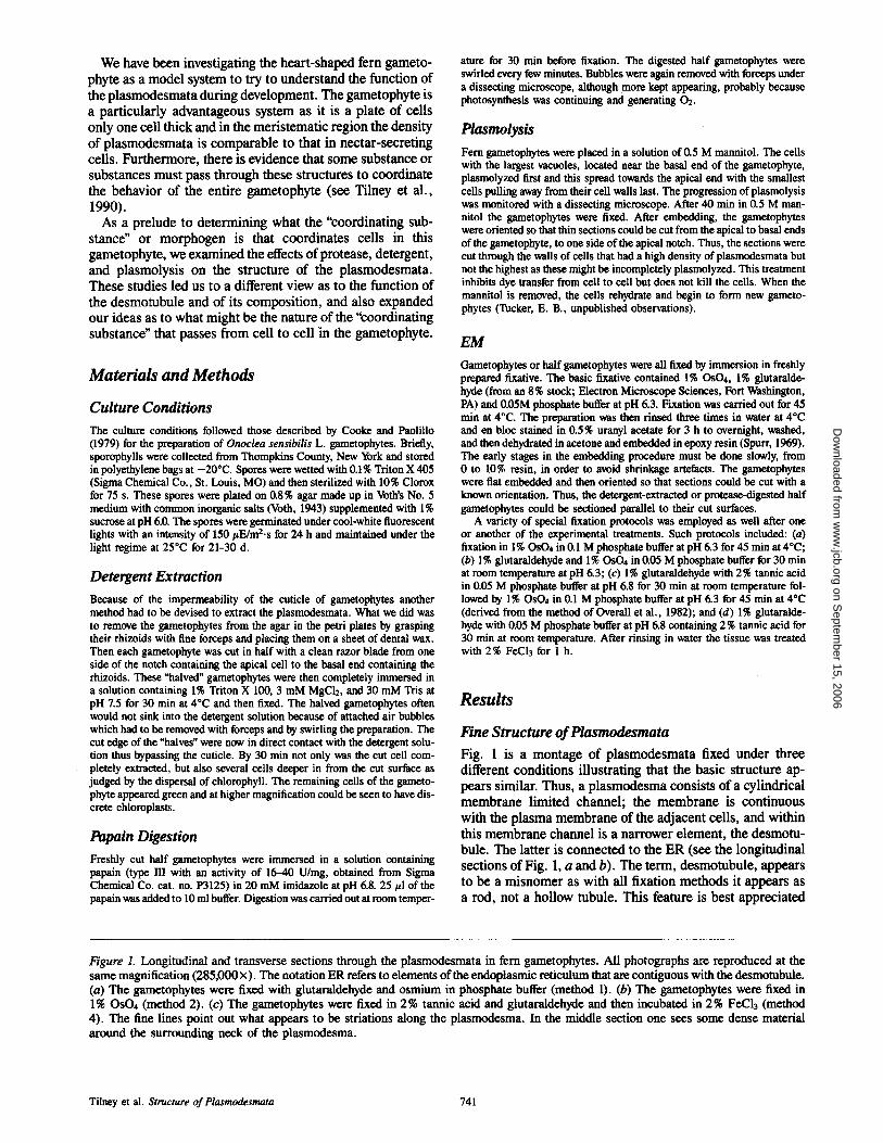
Fine Structure of Plasmodesmata Fig. 1 is a montage of plasmodesmata fixed under three different conditions illustrating that the basic structure ap- pears similar. Thus, a plasmodesma consists of a cylindrical membrane limited channel; the membrane is continuous with the plasma membrane of the adjacent cells, and within this membrane channel is a narrower element, the desmotu- bule. The latter is connected to the ER (see the longitudinal sections of Fig. 1, a and b). The term, desmotubule, appears to be a misnomer as with all fixation methods it appears as a rod, not a hollow tubule. This feature is best appreciated
Figure L Longitudinal and transverse sections through the plasmodesmata in fern gametophytes. All photographs are reproduced at the same magnification (285,000 x ). The notation ER refers to elements of the endoplasmic reticulum that are contiguous with the desmotubule. (a) The gametophytes were fixed with glutaraldehyde and osmium in phosphate buffer (method 1). (b) The gametophytes were fixed in 1% OsO4 (method 2). (c) The gametophytes were fixed in 2 % tannic acid and glutaraldehyde and then incubated in 2 % Feel3 (method 4). The fine lines point out what appears to be striations along the plasmodesma. In the middle section one sees some dense material around the surrounding neck of the plasmodesma.
Tilney et al. Structure of Plasmodesmata 741
on Septem
ber 15, 2006 w
ww
.jcb.orgD
ownloaded from
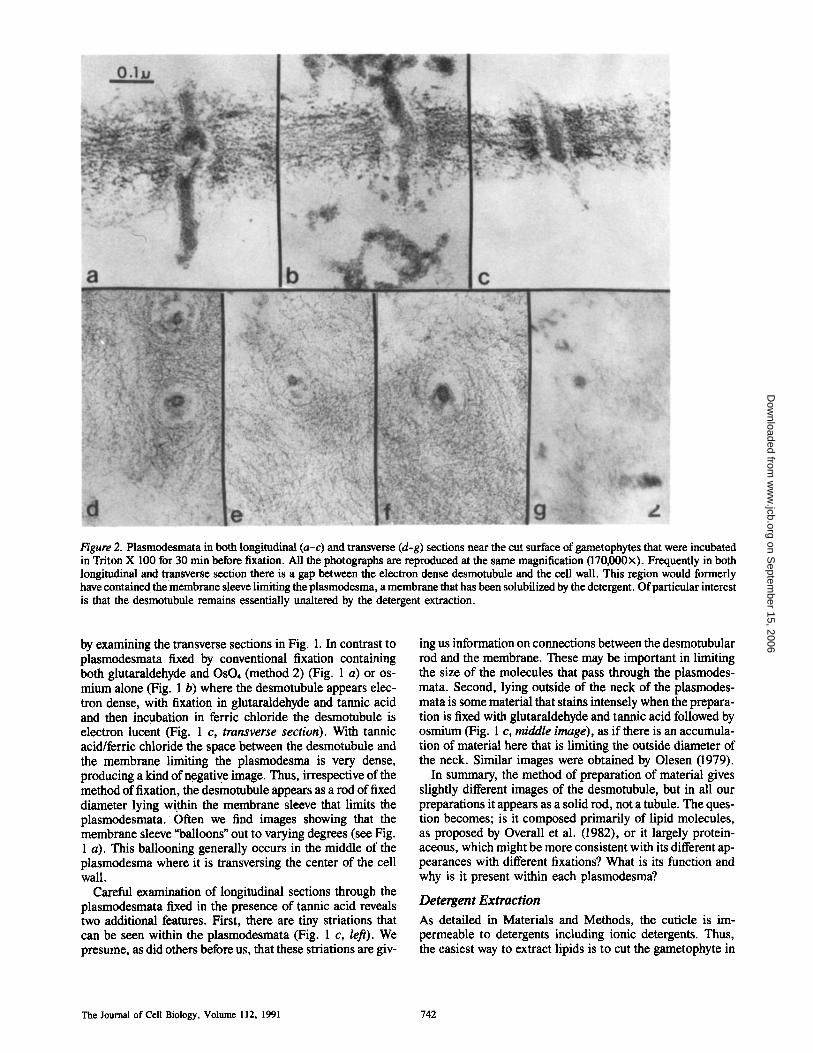
Figure 2. Plasmodesmata in both longitudinal (a-c) and transverse (d-g) sections near the cut surface of gametophytes that were incubated in Triton X 100 for 30 min before fixation. All the photographs are reproduced at the same magnification (170,000x). Frequently in both longitudinal and transverse section there is a gap between the electron dense desmotubule and the cell wall. This region would formerly have contained the membrane sleeve limiting the plasmodesma, a membrane that has been solubilized by the detergent. Of particular interest is that the desmotubule remains essentially unaltered by the detergent extraction.
by examining the transverse sections in Fig. 1. In contrast to plasmodesmata fixed by conventional fixation containing both glutaraldehyde and OsO4 (method 2) (Fig. 1 a) or os- mium alone (Fig. 1 b) where the desmotubule appears elec- tron dense, with fixation in glutaraldehyde and tannic acid and then incubation in ferric chloride the desmotubule is electron lucent (Fig. 1 c, transverse section). With tannic acid/ferric chloride the space between the desmotubule and the membrane limiting the plasmodesma is very dense, producing a kind of negative image. Thus, irrespective of the method of fixation, the desmotubule appears as a rod of fixed diameter lying within the membrane sleeve that limits the plasmodesmata. Often we find images showing that the membrane sleeve "balloons" out to varying degrees (see Fig. 1 a). This ballooning generally occurs in the middle of the plasmodesma where it is transversing the center of the cell wall.
Careful examination of longitudinal sections through the plasmodesmata fixed in the presence of tannic acid reveals two additional features. First, there are tiny striations that can be seen within the plasmodesmata (Fig. 1 c, left). We presume, as did others before us, that these striations are giv-
ing us information on connections between the desmotubular rod and the membrane. These may be important in limiting the size of the molecules that pass through the plasmodes- mata. Second, lying outside of the neck of the plasmodes- mata is some material that stains intensely when the prepara- tion is fixed with glutaraldehyde and tannic acid followed by osmium (Fig. 1 c, middle image), as if there is an accumula- tion of material here that is limiting the outside diameter of the neck. Similar images were obtained by Olesen (1979).
In summary, the method of preparation of material gives slightly different images of the desmotubule, but in all our preparations it appears as a solid rod, not a tubule. The ques- tion becomes; is it composed primarily of lipid molecules, as proposed by Overall et al. (1982), or it largely protein- aceous, which might be more consistent with its different ap- pearances with different fixations? What is its function and why is it present within each plasmodesma?
Detergent Extract ion
As detailed in Materials and Methods, the cuticle is im- permeable to detergents including ionic detergents. Thus, the easiest way to extract lipids is to cut the gametophyte in
The Journal of Cell Biology, Volume 112, 1991 742
on Septem
ber 15, 2006 w
ww
.jcb.orgD
ownloaded from

Figure 3. Halved gametophytes were incubated in papain and then fixed and thin sections were cut near the cut surface. In these six sections, all reproduced at 160,000, we see in the cell walls the remains of plasmodesmata. The desmotubule is partially digested in a. In b and f no obvious remnants of the desmotubule are present. Note that the membrane sleeve that limits the plasmodesma bulges outwards and is very irregular in profile.
half which allows the detergent to penetrate inwards starting from the cut surface.
In Fig. 2 we present a number of images of Triton- extracted plasmodesmata cut both in longitudinal (Fig. 2, a-c) and transverse section (Fig. 2, d-g). Running through the finely fibrillar primary cell wall are remnants of the plas- modesmata. The nearly cylindrical membrane sleeve that formerly limited the plasmodesma and was continuous with the plasma membrane of adjacent cells has dissolved par- tially or completely so that in certain images there is an empty space between the primary cell wall and the desmotu- bule (see Fig. 2, b, c, e, and g). Particularly relevant is that the desmotubule remains intact as it passes through the cell wall. Cisternae of ER which were formerly attached to the desmotubule on its cytoplasmic ends (see Fig. 1) have also dissolved. In some images a lumen of the desmotubule can be resolved (Fig. 2 d), but in other cases the desmotubule seems solid (Fig. 2, e-g). In either case the desmotubule re- mains as a morphologically defined structure, whereas most other membranes, including some of the membrane limiting the plasmodesmata, dissolve. Minimally, this indicates that the desmotubule contains a high concentration of tightly as- sociated protein. If lipid is present, it does not seem to be essential for the integrity of the desmotubule.
Protease Digestion As in the preceding section, we made use of halved gameto- phytes and examined the cell walls nearest the cut surface since if a cell is cut, the papain will have access not only to the rest of the cytoplasm of the cell but also to the plasmodes- mata which are connected to the cut cells.
From transverse sections through protease-treated plas- modesmata near the cut surface, we find that although the membraneous sleeve that limits the plasmodesma remains intact, it becomes variable in diameter and shape, bulging outwards at random positions. Even more interesting is that the desmotubule is completely missing in some (Fig. 3 b), and in others all that remains is a little dense material which is often plastered to one side of the membraneous sleeve (Fig. 3, a, b, e, and f ) . We found some plasmodesmata that look unchanged; these we interpret as not being thus far at- tacked by the papain. In summary, we find that the mem- brane sleeve of the plasmodesma is intact, albeit of variable diameter and shape, and the desmotubule has partially or completely disappeared. What this indicates is that the des- motubule is proteinaceous or protein is associated with the surface of the desmotubule and if this protein is digested, the membrane limiting the plasmodesma bulges out irregularly, perhaps even being capable of blebbing away.
Tilney et al. Structure of Plasmodesmata 743
on Septem
ber 15, 2006 w
ww
.jcb.orgD
ownloaded from

Figure 4. Montage of the remains ofplasmodesmata in gametophytes fixed 30 min after the initiation ofplasmolysis induced by 0.5 M mannit ol. (a) Low magnification of the cell wall between two plasmolyzed cells. Note that portions of the plasmadesmata still remain embedded in the cell wall. (b-g) Higher magnification images of the plasmodesmata of plasmolyzed gametophytes. All are reproduced at 150,000x. In b a portion of the surface ofa plasmolyzed cell shows parts of two plasmodesmata that have broken away from the rest of the plasmodesma that remains attached to the cell wall. In c-g are images of portions of plasmodesmata still embedded in the cell wall. Note that only when the desmotubule is intact does the membrane sleeve fit tightly.
The Journal of Cell Biology, Volume 112, 1991 744
on Septem
ber 15, 2006 w
ww
.jcb.orgD
ownloaded from
::. ~: (. .." .'.:fi".~. :. i. ::. i::: 5..
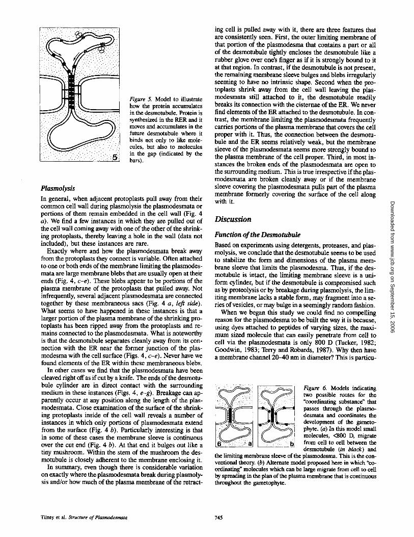
Figure 5. Model to illustrate how the protein accumulates in the desmotubule. Protein is synthesized in the RER and it moves and accumulates in the future desmotubule where it binds not only to like mole- cules, but also to molecules in the gap (indicated by the bars).
Plasmolysis
In general, when adjacent protoplasts pull away from their common cell wall during plasmolysis the plasmodesmata or portions of them remain embedded in the cell wall (Fig. 4 a). We find a few instances in which they are pulled out of the cell wall coming away with one of the other of the shrink- ing protoplasts, thereby leaving a hole in the wall (data not included), but these instances are rare.
Exactly where and how the plasmodesmata break away from the protoplasts they connect is variable. Often attached to one or both ends of the membrane limiting the plasmodes- mata are large membrane blebs that are usually open at their ends (Fig. 4, c-e). These blebs appear to be portions of the plasma membrane of the protoplasts that pulled away. Not infrequently, several adjacent plasmodesmata are connected together by these membraneous sacs (Fig. 4 a, leo side). What seems to have happened in these instances is that a larger portion of the plasma membrane of the shrinking pro- toplasts has been tipped away from the protoplasts and re- mains connected to the plasmodesmata. What is noteworthy is that the desmotubule separates cleanly away from its con- nection with the ER near the former junction of the plas- modesma with the cell surface (Figs. 4, c-e). Never have we found elements of the ER within these membraneous blebs.
In other cases we find that the plasmodesmata have been cleaved tight off as if cut by a knife. The ends of the desmotu- bule cylinder are in direct contact with the surrounding medium in these instances (Figs. 4, e-g). Breakage can ap- parently occur at any position along the length of the plas- modesmata. Close examination of the surface of the shrink- ing protoplasts inside of the cell wall reveals a number of instances in which only portions of plasmodesmata extend from the surface (Fig. 4 b). Particularly interesting is that in some of these cases the membrane sleeve is continuous over the cut end (Fig. 4 b). At that end it bulges out like a tiny mushroom. Within the stem of the mushroom the des- motubule is closely adherent to the membrane enclosing it.
In summary, even though there is considerable variation on exactly where the plasmodesmata break during plasmoly- sis and/or how much of the plasma membrane of the retract-
ing cell is pulled away with it, there are three features that are consistently seen. First, the outer limiting membrane of that portion of the plasmodesma that contains a part or all of the desmotubule tightly encloses the desmotubule like a rubber glove over one's finger as if it is strongly bound to it at that region. In contrast, if the desmotubule is not present, the remaining membrane sleeve bulges and blebs irregularly seeming to have no intrinsic shape. Second when the pro- toplasts shrink away from the cell wall leaving the plas- modesmata still attached to it, the desmotubule readily breaks its connection with the cisternae of the ER. We never find elements of the ER attached to the desmotubule. In con- trast, the membrane limiting the plasmodesmata frequently carries portions of the plasma membrane that covers the cell proper with it. Thus, the connection between the desmotu- bule and the ER seems relatively weak, but the membrane sleeve of the plasmodesmata seems more strongly bound to the plasma membrane of the cell proper. Third, in most in- stances the broken ends of the plasmodesmata are open to the surrounding medium. This is true irrespective if the plas- modesmata are broken cleanly away or if the membrane sleeve coveting the plasmodesmata pulls part of the plasma membrane formerly covering the surface of the cell along with it.
Discussion
Function o f the Desmotubule
Based on experiments using detergents, proteases, and plas- molysis, we conclude that the desmotubule seems to be used to stabilize the form and dimensions of the plasma mem- brane sleeve that limits the plasmodesma. Thus, if the des- motubule is intact, the limiting membrane sleeve is a uni- form cylinder, but if the desmotubule is compromised such as by proteolysis or by breakage during plasmolysis, the lim- iting membrane lacks a stable form, may fragment into a se- ries of vesicles, or may bulge in a seemingly random fashion.
When we began this study we could find no compelling reason for the plasmodesma to be built the way it is because, using dyes attached to peptides of varying sizes, the maxi- mum sized molecule that can easily penetrate from cell to cell via the plasmodesmata is only 800 D (Tucker, 1982; Goodwin, 1983; Terry and Robards, 1987). Why then have a membrane channel 20-40 nm in diameter? This is particu-
Figure 6. Models indicating two possible mutes for the "coordinating substance" that passes through the plasmo- desmata and coordinates the development of the gameto- phyte. (a) In this model small molecules, <BOO D, migrate from cell to cell between the desmotubule (in black) and
the limiting membrane sleeve of the plasmodesma. This is the con- ventional theory. (b) Alternate model proposed here in which "co- ordinating" molecules which can be large migrate from cell to cell by spreading in the plan of the plasma membrane that is continuous throughout the gametophyte.
Tilney et al. Structure of Pla~modesmata 745
on Septem
ber 15, 2006 w
ww
.jcb.orgD
ownloaded from

larly odd as in animal cells molecules of similar size pass from cell to cell via gap junctions with a channel <3 nm in diameter (Hertzberg et al., 1981). Part of the explanation may be that, unlike animal cells where the plasma mem- branes of adjacent cells approach each other closely, in plants adjacent cells are separated by a thick cell wall. It then becomes necessary to stabilize the tiny membraneous chan- nel that must span intervening cell walls. One way would be to include within it a cytoskeletal element. A logical choice would be existing cytoskeletal elements such as microtu- bules or actin filaments, but instead what was selected for was a new structure, the desmotubule. The term, desmotu- bole is an unfortunate one as it does not seem to be hollow and it can be confused with the term, "microtubule;' a com- mon organelle in plant cells. However, to change the term, desmotubule, at this stage seems hopeless as it is ingrained in the literature even though it clearly is not a tubule, nor is it in any way related to microtubules. What is curious is that the desmotubule is connected to the elements of the ER on each of its ends.
To understand why the desmotubule is connected to the ER requires a knowledge of how the plasmodesmata form. In an earlier report (Tilney et al., 1990), we demonstrated that in fern gametophytes, once the plasmodesmata have been formed during division, no subsequent change in num- ber of plasmodesmata occurs. This observation has been made earlier by Gunning (1970) on Azolla roots. Thus, in these systems, and probably in many others (although there are occasional situations where secondary plasmodesmata form in more mature walls [see Jones, 1976; Robards and Lucas, 1990]), plasmodesmata are only formed during cell plate formation. Steps in their formation have been doc- umented by Hepler (1982) who showed that the cell plate is formed by a lining up of vesicles containing cell wall mate- rial and a subsequent coalescence of these vesicles. Between vesicles he often found elements of the ER and, in fact, some of these were trapped there. A portion of these ER elements within newly forming phragmoplasts then would slim down and differentiate into the desmotubule which, even in mature cells, remains attached to the ER cisternae. Thus, the des- motubule was initially part of a cisternae of the ER. It is not surprising, therefore, that many studies have demonstrated this connection and have postulated that the desmotubule is a membraneous channel lying within the membrane limiting the plasmodesmata (Overall et al., 1982). However, what is required biologically, is not a membraneous tubule, another lipid channel, but a cytoskeletal structure. What must occur during desmotubule differentiation is an accumulation of protein into the former lipid channel, protein molecules that become bound to each other to form a cytoskeletal rod. Thus, transmembrane proteins are synthesized in the ER during aM/or shortly before phragmoplast formation. These accumulate in what will be the future desmotubule by bind- ing to each other (Fig. 5) and to molecules in the membrane of the vesicles which will ultimately form the membrane sleeve limiting the plasmodesmata. The latter we imagine are the striations shown in Fig. 1 c. They may be crucial for controlling the size of water soluble molecules being con- veyed through the plasmodesmata. It is obvious that more in- formation is needed about these striations. Because nothing is known about their composition, we have indicated them in the figure by bars. Steps in this direction have been at- tempted by freeze fracture studies (Thompson and Platt-
Aloia, 1985). (Parenthetically, it seems reasonable to sus- pect that the 30-kD movement protein of the TMV [Deom et al., 1987] permits the transport of this virus RNA by dis- rupting these protein crossbridges because the movement proteins apparently have no observable effect on the desmotu- bule structure [Wolf et al., 1989].)
We imagine that biologically this strategy (using a des- motubule) has been adopted during evolution because it will give rise to plasmodesmata of fixed diameters and thus fixed pore size and invariably there will be a cytoskeletal element within each plasmodesma, a feature which will stabilize the plasmodesm a so that it will not pinch into a series of vesicles and thus impede flow through the plasmodesmata.
What Might be the Nature of the Substance or Substances that Pass through the Plasmodesmata and Regulate Morphogenesis ?
Using water soluble dyes of varying sizes it has been shown that the aqueous channel in the plasmodesmata has a pore size that limits diffusion of water soluble molecules over 800 D. Since the desmotubule appears connected to the ER, a compartment not open to the bulk of the cytoplasm, and is likely to be a solid rod, diffusion of material through the plasmodesmata must occur in the space between the des- motubule and its encapsulating plasma membrane as di- agrammatically depicted in Fig. 6 a. However, there is an- other route for diffusion of materials from cell to cell through the plasmodesmata that should also be considered. This route would involve the migration of substances that live par- tially or even mostly in the bilayer of the plasma membrane (Fig. 6 b). Because of the fluid nature of membranes, sub- stances that are partitioned partially or nearly completely in the lipid bilayer could rapidly diffuse from cell to cell via the membrane sleeve of the plasmodesmata. This hypothesis is consistent with fluorescein redistribution after photobleach- ing to examine the movement of a fluorescent phospholipid analogue recently reported by Baron-Epel et al. (1988). They observed phospholipid exchange between contiguous cells which suggests that the plasmodesmata may provide a lipid soluble pathway for intercellular transport. Our results argue very strongly against the desmotubule as a conveyor of lipid soluble molecules, but instead point to the plasma mem- brane sleeve. Such a possibility has a number of interesting avenues to investigate. First, since plant cells have such large vacuoles and thus only a thin rim of cytoplasm, regulatory substances that are partitioned in the plasma membrane would be in intimate contact with this cytoplasm. Second, the size of the molecule that regulates morphogenesis would not be restricted to 800 D, but could be considerably larger, in fact, large, multisubunit complexes could move rapidly in the bilayer. These could have important enzymatic functions which in turn could regulate the activity of the cytoplasm as has been demonstrated for molecules that live in the bilayer, such as adenyl cyclase, phospholipase C, and their associated G proteins.
We wish to thank Bob Golder and Doug Rugh for the drawings in this manuscript.
This work was supported by a grant from National Institutes of Health HD144-74.
Received for publication 12 March 1990 and in revised form 12 November 1990.
The Journal of Cell Biology, Volume 112, 1991 746
on Septem
ber 15, 2006 w
ww
.jcb.orgD
ownloaded from

References
Baron-Epel, 0., D. Hernandez, L. W. Jiang, S. Meiners, and M. Schindler. 1988. Dynamic continuity of cytoplasmic and membrane compartments be- twecn plant cells. J. Cell Biol. 106:715-721.
Cooke, T. J., and D. J. Paolillo, Jr. 1979. The photobiology of fern gameto- phytes I. The phenomena of red-far-red and yellow far-red photoreversibil- ity. J. Exp. Bot. 30:71-80.
Doom, C. M., M. J. Oliver, and R. N. Beachy. 1987. The 30-kilodalton gene product of tobacco mosaic virus potentiates virus movement. Science (Wash. DC). 237:389-394.
Eleftheriou, E. P., and J. L. Hall. 1983. The extrafloral nectaries of cotton. I. Fine structure of secretory papillae. J. Exp. Bot. 34:103-119.
Goodwin, P. B. 1983. Molecular size limit for movement in the symplast of Elodea leaf. Planta. 157:124-130.
Gunning, B. E. S. 1970. Age-related and origin-related control of numbers of plasmodesmata in cell walls of developing Azolla roots. Planta. 143:181- 190.
Gunning, B. E. S., and J. E. Hughes. 1976. Quantitative assessment of sym- plastic transport of pre-nectar into the trichomes of Abuliton nectaries. Aust. .L Plant. PhysioL 43:619-637.
Hepler, P. K. 1982. Endoplasmic reticulum in the formation of the cell plant and plasmodesmata. Protoplasma. 111 : 121-133.
Hertzberg, E. L., T. S. Lawrence, and N. B. Gilula. Gap junctional communi- cation. Annu. Rev. Physiol. 43:479-491.
Jones, M. G. K. 1976. The origin and development of plasmodesmata. In Inter- cellular Communication in Plants: Studies on Plasmodesmata. B. E. S. Gun- ning and A. W. Robards, editors. Springer-Verlag, Berlin. 81-105.
Kitajima, E. W., and J. A. Lauritus. 1969. Plant virions in plasmodesmata. Virology. 37:681-685.
Ledbetter, M. C., and K. R. Porter. 1970. Introduction to the Fine Structure of Cells. Springer-Verlag, New York. 49-54.
Lopez-Salz, J. F., G. Gim6n6z-Martin, and M. C. Risfieno. 1966. Fine struc-
ture of the plasdesm. Protoplasma. 61:81-84. Olesen, P. 1979. The neck constriction in plasmodesmata; evidence for a pe-
ripheral sphincter-like structure revealed by fixation with tannic acid. Planta. 144:349-358.
Overall, R. L., J. Wolfe, and B. E. S. Gunning. 1982. Intercellular communica- tion in Azolla roots. I. Ultrastructure of plasmodesmata. Protoplasma. 111:134-150.
Robards, A. W. 1968. A new interpretation of plasmodesmatal ultrastructure. Ultrastruct. Pathol. 82:200-218.
Robards, A. W. 1976. Plasmodesmata in higher plants. In Intercellular Com- munication in Plants: Studies on Plasmodesmata. B. E. S. Gunning and A. W. Robards, editors. Springer-Verlag, Berlin. 15-57.
Robards, A. W., and W. J. Lucas. 1990. Plasmadesmata. Annu. Rev. Plant Physiol. Plant Mol. Biol. 41:369-419.
Spurr, A. R. 1969. A low-viscosity epoxy resin embedding medium for electron microscopy. J. Ultrastruct. Res. 26:31-43.
Terry, B. R., and A. W. Robards. 1987. Hydrodynamic radius alone governs the mobility of molecules through plasmodesmata. Planta. 171:145-157.
Thomson, W. W., and K. Platt-Aloia. 1985. The ultrastrncture of the plas- modesmata of the salt glands of Tamarix as revealed by transmission and freeze fracture electron microscopy. Protoplasma. 125:13-27.
Tilney, L. G., T. J. Cooke, P. S. Conneily, and M. S. Tilney. 1990. The distri- bution of plasmodesmata and its relationship to morphogenesis in fern gametophytes. Development (Camb.). In press.
Tucker, E. B. 1982. Translocation in the staminal hairs of Setcreaseapurpurea. I. A study of cell ultrastructure and cell to cell passage of molecular probes. Protoplasma. 113:193-201.
Voth, P. D. 1943. Effects of nutrient-solution concentration on the growth of Marehantia polymorpha. Bot. Gaz. 104:591-601.
Wolf, S., C. M. Deom, R. N. Beachy, and W. J. Lucas. 1989. Movement pro- tein of tobacco mosaic virus modifies plasmodesmatal size exclusion limit. Science (Wash. DC). 246:377-379.
Tilney et al. Structure of Plasmodesmata 747
on Septem
ber 15, 2006 w
ww
.jcb.orgD
ownloaded from