thematic review - virginia tech. lipid res.-2009-buczynski...of these enzymes produce multiple lipid...
TRANSCRIPT
thematic review
Thematic Review Series: Proteomics
An integrated omics analysis of eicosanoid biology1
Matthew W. Buczynski, Darren S. Dumlao, and Edward A. Dennis2
Department of Chemistry and Biochemistry, Department of Pharmacology, and School of Medicine,University of California, San Diego, La Jolla, CA 92093
Abstract Eicosanoids have been implicated in a vast numberof devastating inflammatory conditions, including arthritis,atherosclerosis, pain, and cancer. Currently, over a hundreddifferent eicosanoids have been identified, with many havingpotent bioactive signaling capacity. These lipid metabolitesare synthesized de novo by at least 50 unique enzymes, manyof which have been cloned and characterized. Due to the ex-tensive characterization of eicosanoid biosynthetic pathways,this field provides a unique framework for integrating geno-mics, proteomics, andmetabolomics toward the investigationof disease pathology. To facilitate a concerted systems biol-ogy approach, this review outlines the proteins implicated ineicosanoid biosynthesis and signaling in human, mouse, andrat. Applications of the extensive genomic and lipidomic re-search to date illustrate the questions in eicosanoid signalingthat could be uniquely addressed by a thorough analysis ofthe entire eicosanoid proteome.—Buczynski, M. W., D. S.Dumlao, and E. A. Dennis. An integrated omics analysis ofeicosanoid biology. J. Lipid Res. 2009. 50: 1015–1038.
Supplementary key words genomics • proteomics • lipidomics •
cyclooxygenase • lipoxygenase • cytochrome P450 • prostaglandin •
leukotriene • eicosanoid
SYSTEMS BIOLOGY AND EICOSANOID “OMICS”
Biological processes are comprised of numerous con-verging signals that concertedly create a coherent effect.Any individual signaling element may elicit a physiologicalresponse, yet its loss is not fatal to the organism. In fact, awide range of genetic variation can be observed within indi-vidual members of a given species without leading to a dra-matic loss of function. As these signals have redundant andemergent properties, it can be difficult to explain how abiological process works using a limited number of molecu-lar indicators. For this reason, systems biology has emerged
to address the question of how molecular biology works asan integrated process (1).
Systems biology has advanced exponentially during thepast two decades, with transcriptomics, proteomics, andmetabolomics each playing an integral role. Each of theseplatforms brings its own unique advantages and limitationsin facilitating the investigation of disease pathology. Atranscriptomic approach can detect the upregulation anddownregulation of important biosynthetic and signalinggenes; however, gene changes often donʼt directly corre-late with changes in protein levels (2). Proteomic analy-ses can identify enzymes and posttranslational proteinchanges in a cell or tissue, but fall short of determiningwhich particular enzymes actively produce metabolitesunder disease conditions; likewise, metabolic approachessuccessfully identify bioactive signaling molecules, buttherapeutic intervention generally requires definitive pro-tein targets.
The eicosanoid class of signaling molecules highlightsmany of the issues facing comprehensive systems biology
This work was supported by the National Institutes of Health LIPID MAPSLarge Scale Collaborative Grant GM069338 and R01 GM64611 andGM20501. M.W.B. was supported by Gastroenterology Pre-doctoral TrainingGrant 2T32DK007202-32 from the National Institutes of Health.
Manuscript received 3 February 2009 and in revised form 23 February 2009.
Published, JLR Papers in Press, February 24, 2009.DOI 10.1194/jlr.R900004-JLR200
Abbreviations: AKR, aldo-keto reductase; ALX, lipoxin A4 receptor;COX, cyclooxygenase; CRTH2, chemoattractant receptor-homologous,molecule expressed on Th2 cells; CYP, cytochrome P450; cysLT, cysteinylleukotrienes; DHET, dihydroxyeicosatrienoic acid; EET, epoxyeicosa-trienoic acid; EX, eoxin; FLAP, 5-lipoxygenase activating protein; GGL,g-glutamyl leukotrienase; GGT, g-glutamyl transpeptidase; GPCR, Gprotein-coupled receptor; HETE, hydroxyeicosatetraenoic acid; HpETE,hydroperoxy-eicosatetraenoic acid; HX, hepoxilin; 9K-PGR, 9-keto pros-taglandin reductase; LT, leukotriene; LTAH, leukotriene A4 hydrolase;LTB4DH, leukotriene B4 12-hydroxydehydrogenase; LTCS, leukotrieneC4 synthase; LIPID MAPS, Lipid Metabolites and Pathways Strategies;LOX, lipoxygenase; LX, lipoxin; MAPEG, membrane-associated proteinin eicosanoid and glutathione; MBD, membrane-bound dipeptidase;mGST, microsomal glutathione S-transferase; PG, prostaglandin; PGDH,prostaglandin dehydrogenase; PGDS, prostaglandin D synthase; PGES,PGE synthase; PGFS, prostaglandin F synthase; PGR, prostaglandinreductase; PLA2, phospholipase A2; PPAR, peroxisome proliferator-activated receptor; sEH, soluble epoxide hydrolase; TRPV4, vanilloid type 4receptor; TXA2, thromboxaneA2; 11-TXDH, 11-dehydroxythromboxaneB2 dehydrogenase.
1Guest editor for this article was Jay W. Heinecke, JLR AssociateEditor, University of Washington.
2 To whom correspondence should be addressed.e-mail: [email protected] online version of this article (available at http://www.jlr.org)
contains supplementary data in the form of seven tables.
Copyright © 2009 by the American Society for Biochemistry and Molecular Biology, Inc.
This article is available online at http://www.jlr.org Journal of Lipid Research Volume 50, 2009 1015
by guest, on January 13, 2017w
ww
.jlr.orgD
ownloaded from
0.DC1.html http://www.jlr.org/content/suppl/2009/09/16/R900004-JLR20Supplemental Material can be found at:
by guest, on January 13, 2017w
ww
.jlr.orgD
ownloaded from
0.DC1.html http://www.jlr.org/content/suppl/2009/09/16/R900004-JLR20Supplemental Material can be found at:
by guest, on January 13, 2017w
ww
.jlr.orgD
ownloaded from
0.DC1.html http://www.jlr.org/content/suppl/2009/09/16/R900004-JLR20Supplemental Material can be found at:
analyses. Eicosanoids comprise a class of bioactive lipidmediators derived from the metabolism of polyunsatu-rated fatty acids by cyclooxygenases (3–5), lipoxygenases(3, 6), cytochrome P450s, or nonenzymatic pathways(Fig. 1). Technically, “eicosanoids” refers to fatty acids con-taining 20 carbons (eicosa), but the field generally uses theterm eicosanoids more broadly to also include similar me-tabolites of other polyunsaturated fatty acids. This lipidclass has been intensely studied over the past 30 years be-cause of its contribution to the inflammatory response indiseases such as arthritis and asthma, yet the majority ofthis work has focused on a select few genes, proteins, ormetabolites within it. In many instances, the regulationof one arm has important regulatory implications for an-other arm of the eicosanoid biosynthetic pathway. Manyof these enzymes produce multiple lipid products; likewise,many lipid products can be formed by a number of differ-ent enzymes acting in parallel or in concert (Fig. 2). Forthese reasons, a comprehensive systems biology approachwould be invaluable for understanding and treating dis-eases implicating eicosanoid signaling.
Transcriptomic and lipidomic analyses of eicosanoidbiosynthesis can be performed using existing methodolo-gies. With the completion of the major mammalian ge-nome projects, commercial gene array technology hasrapidly progressed and allowed the generation of whole-genome data sets of mRNA changes to become a common-place tool at many research institutions. This proliferationallowed academic research groups to focus on the morechallenging task of interpreting this data and made tran-scriptomic analysis the gold standard in systems biology re-search. Due to its immense scope, metabolomics has not yetachieved this level of standardization in scientific practice.However, as a part of their goal to create the infrastructureto identify and quantify all lipid molecular species, the LipidMetabolites and Pathways Strategies (LIPID MAPS) consor-tium has developed liquid chromatography tandem massspectrometry methodology that comprehensively coversnearly all the known metabolites of this class (7, 8). Specifi-cally, a complete lipidomic analysis of eicosanoids is now
available (9), along with corresponding gene microarraydata (www.lipidmaps.org). Thus, is it timely to consider a pro-teomic analysis that could complete an integrated picture ofthe entire eicosanoid signaling network.
A quantitative proteomic analysis of the eicosanoid path-way has thus far lagged behind concomitant transcriptomicand lipidomic studies. These tandem mass spectrometrytechniques can bedivided into two philosophical approaches(10). The classically practiced MS1 and MS2 proteomic scan-ning techniques have demonstrated the potential to rapidlyidentify thousands of proteins from a single sample, yet theseoften represent only a fraction of the total protein content ofa cell or tissue, and the investigator has little control overwhich proteins make the cut (11). On the other hand, bymeasuring the levels of a selected population of proteinsusingmultiple reactionmonitoring, significantly lower limitsof detection for can be achieved (12). However, to create amethod to investigate a selected proteome, one must firsthave a fundamental understanding of what proteins couldbe involved andhow these proteins fit into the larger scheme.
In the book Functional Lipidomics, Bowers-Gentry et al.(7) give a broad outline for using a lipidomic approach toclassify and measure eicosanoids. To facilitate a concertedsystems biology approach, this review provides a systematicoverview of the proteins involved in eicosanoid biosynthesisand signaling. The genes and proteins responsible for theseactivities are listed in supplementary Tables I–VII online,provided one of the following pieces of evidence: 1) the genehas been expressed and characterized in vitro; 2) the puri-fied protein has been sequenced and characterized in vitro;3) the gene has been overexpressed (or for transcriptionfactors, expressed with a reporter gene) and characterizedin a cellular system; 4) loss of activity has been demonstratedin cells or tissue from gene knockout animals. Furthermore,as studies of human biology and disease commonly employmouse or rat models as a surrogate, important differencesbetween these species are highlighted.
PHOSPHOLIPASE A2
Arachidonic acid and other polyunsaturated fatty acidsserve as the metabolic precursors for eicosanoid synthesis(13, 14). Biologically, these molecules are generally notavailable in large quantities in the free acid form, but arestored at the sn-2 position on the glycerol backbone ofmembrane phospholipids. To be used for biosynthesis,phospholipase A2 (PLA2) liberates sn-2 fatty acids fromphospholipids at the membrane interface.
Phospholipase A2 represents a superfamily of at least 15groups that have wide-ranging roles in biological processes.These enzymes can be considered as five types: cytosolicPLA2 (cPLA2), secreted PLA2 (sPLA2), calcium-independentPLA2, platelet-activating factor acetylhydrolase, and lyso-somal PLA2; their classification and biological functionshave been extensively reviewed (13–15).
Current literature strongly implicates the Group IVAcPLA2 as the chief enzyme involved in polyunsaturatedfatty acid release for eicosanoid biosynthesis (16, 17), with
Fig. 1. Overview of eicosanoid biosynthesis through COX, LOX,CYP P450, and nonenzymatic pathways acting on arachidonic acid.
1016 Journal of Lipid Research Volume 50, 2009
by guest, on January 13, 2017w
ww
.jlr.orgD
ownloaded from
0.DC1.html http://www.jlr.org/content/suppl/2009/09/16/R900004-JLR20Supplemental Material can be found at:
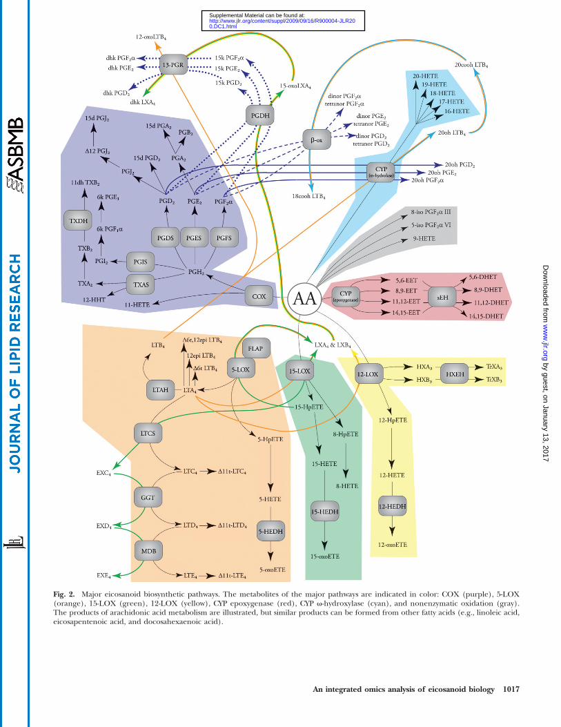
Fig. 2. Major eicosanoid biosynthetic pathways. The metabolites of the major pathways are indicated in color: COX (purple), 5-LOX(orange), 15-LOX (green), 12-LOX (yellow), CYP epoxygenase (red), CYP v-hydroxylase (cyan), and nonenzymatic oxidation (gray).The products of arachidonic acid metabolism are illustrated, but similar products can be formed from other fatty acids (e.g., linoleic acid,eicosapentenoic acid, and docosahexaenoic acid).
An integrated omics analysis of eicosanoid biology 1017
by guest, on January 13, 2017w
ww
.jlr.orgD
ownloaded from
0.DC1.html http://www.jlr.org/content/suppl/2009/09/16/R900004-JLR20Supplemental Material can be found at:
sPLA2s potentially playing a supplementary role (see supple-mentary Table I). Bonventre et al. (18) and Uozumi et al.(19) independently demonstrated that peritoneal macro-phages fromGroup IVA PLA2 knockout mice were incapableof producing eicosanoids. More recently, Adler et al. (20)identified a patient with inherited Group IVA deficiencyand determined that nearly all arachidonic acid used foreicosanoidproduction by platelets and circulating leukocyteswas attributable to this enzyme. The Group IVA cPLA2 usesa catalytic Ser/Asp dyad to hydrolyze fatty acids and containsa C2 domain that facilitates calcium-dependent translocationfrom the cytosol to themembrane surfacewhere it encountersphospholipid substrate. Overall, while cPLA2s have a broadrange of homology between human, mouse, and rat, theGroup IVA cPLA2 has 95% identity between these species.Other cPLA2 show between 54 and 82% identity in these spe-cies andhave been less well-characterized to date (13), yetmaystill play a supplementary role in eicosanoid biosynthesis.
While Group IVA cytosolic PLA2 is the primary enzymeinvolved in eicosanoid biosynthesis, sPLA2s have beendemonstrated to play a supplementary role, reviewedextensively by Lambeau and Gelb (21). In particular, theGroup IIA, Group V, and Group X PLA2s have all beenshown to modulate eicosanoid levels in mammalian cellu-lar systems. However, the majority of these studies havebeen performed by either overexpression or exogenoussupplementation of sPLA2, which complicates the investi-gation of its role in pathology. Future work using sPLA2
knockout mice may help further elucidate the role theseenzymes play in eicosanoid biosynthesis during an inflam-matory response.
CYCLOOXYGENASE METABOLITES
CyclooxygenasesProstaglandins (PGs) are bioactive signaling molecules
derived from cyclooxygenase (COX) and subsequent PGsynthase activity on arachidonic acid (4, 5, 22) (see sup-plementary Table II). COXs contain two distinct activesites, a COX and peroxidase site, both of which use thesame tyrosyl radical and heme-iron for catalysis. TheCOX site incorporates molecular O2 at the 11- and 15-carbon on arachidonic acid to form PGG2, which containsthe following moieties: a five-member ring linked at C-8and C-12, an endoperoxide bridge across C-9 and C-11,and a peroxide at C-15. The peroxidase site reduces theperoxide to a hydroxyl to form PGH2, the substrate forthe various PG synthases. In addition to forming PGH2,arachidonic acid can situate in the active site pocket in alimited number of suboptimal conformations that incor-porate a single molecular O2, forming trace amounts ofeither 11(R)-hydroxyeicosatetraenoic acid (HETE) or 15(S)-HETE. COX activity does not exhibit long-term stability,as the catalytic tyrosyl radical can be transferred to a near-by tyrosyl residue and cause “suicide inactivation” after?300 turnovers. Because the enzyme can perform onlya limited number of reactions, it must be constantly re-
expressed to generate metabolites. The details of thesemechanisms have been reviewed extensively (5).
COXs constitute two distinct genes, COX-1 and COX-2.These two isoforms exhibit ?60% identity and have nearlyidentical active site residues (4). The most significant struc-tural difference between these enzymes is an isoleucine tovaline substitution, which results in a larger COX-2 activesite pocket. This allows COX-2 to be more permissive inselecting substrates, and unlike COX-1, it can metabolizedihomo-g-linolenic and eicosapentaenoic acid in ad-dition to arachidonic acid. Pharmaceutical companieshave also taken advantage of the larger COX-2 active site,creating isoform selective inhibitors such as Celecoxib(Celebrex™). While COX-1 and COX-2 have similar struc-tural and catalytic features, these isozymes exhibit differentexpression patterns. COX-1 is constitutively expressed bymost cell types and has been implicated in a number ofhomeostatic processes, including stomach acidity control,endometrial cycling, and renal function. In contrast, COX-2expression is controlled by the pro-inflammatory transcrip-tion factor NF-kB and highly upregulated in response to in-fection, atherosclerosis, and a number of cancers.
ProstaglandinsPGH2 can form a number of different bioactive products
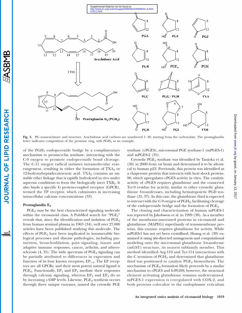
through the action of PG synthases. This includes a num-ber of important signaling molecules, including PGI2 (alsoknown as prostacyclin), thromboxane A2, PGE2 (alsoknown as dinoprostone), PGD2, and PGF2a. Followingthe PG abbreviation (Fig. 3), the nomenclature highlightsthe two important structural features: the componentsof the five-member prostane ring, as denoted by a letterA–K, and the number of double bonds, denoted by a sub-script number (23). The lone exception is thromboxane,which has a six-member oxane ring, and is abbreviated TX.
Prostaglandin I2 and thromboxane A2
PGI2 was first identified in 1976 (24) and is formed bythe prostacyclin synthase, a member of the cytochromeP450 monooxygenase superfamily (25). Structural elucida-tion of the human prostacyclin synthase was not obtaineduntil 2006 (26), confirming the mechanism of PGH2 endo-peroxide bridge rearrangement proposed by Hecker andUllrich in 1989 (27). The active site heme-iron interactswith the C-11 oxygen, promoting the hemolytic cleavageof the endoperoxide bond and the formation of an etherlinkage between C-9 and C-5. The PGI2 ring is highly labileand rapidly hydrolyzed to form the stable but biologicallyinactive 6-keto PGF1a, and because of this, PGI2 and 6-ketoPGF1a are often used interchangeably in the literature.PGI2 binds the G-coupled protein receptor IP (28, 29) aswell as the transcription factors peroxisome proliferator-activated receptor (PPAR) a, PPARy, and PPARg (30, 31).
Identified in 1975 (32), thromboxane A2 (TXA2) is thephysiological counterbalancing signal to PGI2. Similar toPGI2, its biosynthesis is catalyzed by a member of thecytochrome P450 superfamily member, thromboxane Asynthase (27, 33). This enzyme facilitates rearrangement
1018 Journal of Lipid Research Volume 50, 2009
by guest, on January 13, 2017w
ww
.jlr.orgD
ownloaded from
0.DC1.html http://www.jlr.org/content/suppl/2009/09/16/R900004-JLR20Supplemental Material can be found at:
of the PGH2 endoperoxide bridge by a complimentarymechanism to prostacyclin synthase, interacting with theC-9 oxygen to promote endoperoxide bond cleavage.The C-11 oxygen radical initiates intramolecular rear-rangement, resulting in either the formation of TXA2 or12-hydroxyheptadecatrienoic acid. TXA2 contains an un-stable ether linkage that is rapidly hydrolyzed in vivo underaqueous conditions to form the biologically inert TXB2. Italso binds a specific G protein-coupled receptor (GPCR),termed the TP receptor, which culminates in increasingintracellular calcium concentrations (33).
Prostaglandin E2
PGE2 may be the best characterized signaling moleculewithin the eicosanoid class. A PubMed search for “PGE2”reveals that, since the identification and isolation of PGE2
from human seminal plasma in 1963 (34), well over 27,000articles have been published studying this molecule. Theeffects of PGE2 have been implicated in innumerable bio-logical processes and disease pathologies, including par-turition, bronchodilation, pain signaling, innate andadaptive immune responses, cancer, arthritis, and athero-sclerosis (4, 35). The wide spectrum of PGE2 signaling canbe partially attributed to differences in expression andfunction of its four known receptors, EP1-4. The EP recep-tors are all GPCRs, and their most potent natural ligand isPGE2. Functionally, EP1 and EP3 mediate their responsesthrough calcium signaling, whereas EP2 and EP4 do soby increasing cAMP levels. Likewise, PGE2 synthesis occursthrough three unique enzymes, named the cytosolic PGE
synthase (cPGES), microsomal PGE synthase-1 (mPGES-1)and mPGES-2 (35).
Cytosolic PGE2 synthase was identified by Tanioka et al.(36) in 2000 from rat brain and determined it to be identi-cal to human p23. Previously, this protein was identified asa chaperone protein that interacts with heat shock protein-90, which upregulates cPGES activity in vitro. The catalyticactivity of cPGES requires glutathione and the conservedTyr-9 residue for activity, similar to other cytosolic gluta-thione S-transferases, including hematopoietic PGD syn-thase (35, 37). In this case, the glutathione thiol is expectedto interact with the C-9 oxygen of PGH2, facilitating cleavageof the endoperoxide bridge and the formation of PGE2.
The cloning and characterization of human mPGES-1was reported by Jakobsson et al. in 1999 (38). As a memberof the membrane-associated proteins in eicosanoid andglutathione (MAPEG) superfamily of transmembrane pro-teins, this enzyme requires glutathione for activity. WhilemPGES-1 has not yet been crystallized, Huang et al. (39) ex-amined it using site-directed mutagenesis and computationalmodeling onto the microsomal glutathione S-transferase(mGST) structure, its nearest subfamily member. Thismethod identified Arg-110 and Tyr-114 interactions withthe C terminus of PGH2 and determined that glutathionethiol was positioned to catalyze PGE2 biosynthesis. Themechanism of PGE2 formation likely proceeds by a similarmechanism to cPGES and h-PGDS; however, the structuralelement activating glutathione remains undetermined.mPGES-1 expression is coregulated with COX-2, andboth proteins colocalize to the endoplasmic reticulum
Fig. 3. PG nomenclature and structure. Arachidonic acid carbons are numbered 1–20, starting from the carboxylate. The prostaglandinletter indicates composition of the prostane ring, with PGH2 as an example.
An integrated omics analysis of eicosanoid biology 1019
by guest, on January 13, 2017w
ww
.jlr.orgD
ownloaded from
0.DC1.html http://www.jlr.org/content/suppl/2009/09/16/R900004-JLR20Supplemental Material can be found at:
(40); however, COX-2 resides in the lumen of the endo-plasmic reticulum, whereas themPGES-1 active site appearsto face the cytosol. A thorough examination of mPGES-1lature has been reviewed by Samuelsson, Morgenstern,and Jakobsson (40).
mPGES-2 activity was first reported by Wantanabe et al.(41) as a distinct PGES from cPGES and m-PGES-1 in rats.While it has no specific requirement for glutathione, its ac-tivity was enhanced by the presence of free thiols. The hu-man isoform was cloned in 2002 by Tanikawa et al. (42)and contains a putative C-x-x-C sequence found in glutar-edoxin and thioredoxin, but not cPGES or m-PGES-1. Sub-sequent crystallization by Yamada et al. in 2005 (43)confirmed it contained significant structural differencesfrom other PGES. Using molecular modeling, they pro-posed a mechanism for mPGES-2 catalysis. A hydrogenbonding network between Phe-112, Cys-110, Cys-113, andTyr-107 lowers the Pka of Cys-110 such that it can donateits thiol hydrogen to the C-11 oxygen. This leads to cleav-age of the endoperoxide bridge and the creation of a ke-tone by the C-9 oxygen, forming PGE2. The final step isfacilitated by free thiols, activated by Tyr-107; however, thisdoes not appear to be necessary for productive catalysis.
Prostaglandin D2
PGD2 is a structural isomer of PGE2, and early studies ofD-series PGs regarded them as side-products of E-seriesbiosynthesis (44). Whereas the prostane ring on PGE2
has a 9-keto and 11-hydroxy moiety, the positions of thesesubstituents are reversed on PGD2. In the late 1970s, PGD2
was identified as a major product in rat brain homogenates(45) and activated mast cells (46). PGD2 is formed by twoevolutionarily distinct, but functionally convergent, prosta-glandin D synthases (PGDS): the lipocalin-type (l-PGDS)and hematopoietic-type (h-PGDS). One critical differencebetween the PGDSs is the requirement for glutathione;l-PGDS can function without this cofactor, while h-PGDSuses it for catalysis. Urade et al. (47) proposed a mecha-nism for l-PGDS, whereby the thiol from Cys-65 forms atransient bond with the C-11 oxygen of PGH2, openingthe endoperoxide ring. Exogenous sulfhydryl compoundsthen remove the C-11 hydrogen, culminating in PGD2 pro-duction and release. NMR structural studies and molecularmodeling estimated a 5 Å distance between the Cys-65thiol and the C-11 oxygen, consistent with this model(48). Following crystallization in 1997, Kanaoka et al.(49) proposed that h-PGDS contains a pocket that activatesglutathione via Tyr-8, such that the glutathione thiol func-tions analogous to Cys-65 of l-PGDS.
PGD2 has two known receptors, DP1 and chemoattrac-tant receptor-homologous, molecule expressed on Th2cells (CRTH2) (50). DP1 belongs to the family of GPCR thatincludes IP, TP, EP1-4, and FP, and ligand binding leads tointracellular increases of cAMP. While exhibiting similarPGD2 binding, CRTH2 shares little homology with DP1
and appearsmore closely related to the leukotrieneB4 recep-tors BLT1 and BLT2. CRTH2 activation leads to elevation ofintracellular calcium and decreased cAMP levels (51).
Prostaglandin F2aPGF2a was isolated and structurally characterized in
1963 by Samuelsson (34) from human seminal fluid anddemonstrably stimulated smooth muscles from rabbit duo-denum. Since then, it has been implicated in a number ofphysiological processes and disease states (52), includingendometrial cycling, embryo development parturition,vasoconstriction, as well as acute inflammation, oxygen-depravation injury, and atherosclerosis. Only one PGF2a-specific receptor has been cloned (53), a GPCR termedFP, which upon binding ligand results in an elevation ofintracellular calcium. To date, three PGF2a biosyntheticenzymes have been cloned and characterized (54): prosta-glandin F synthase (PGFS) (55), prostamide/PGFS (56),and 9-keto prostaglandin reductase (9K-PGR).
The human PGFS was cloned in 1999 by Suzuki-Yamamotoet al. (55) and determined to be identical to aldo-keto re-ductase 1C3 (AKR1C3). Similar to mPGES-2 and h-PGDS,PGFS evolved from the thioredoxin protein family and con-tains the putative C-x-x-C motif. Crystal structures of PGFSwere published in 2004 (57) and 2006 (58) by Komotoet al. and used to propose a mechanism for PGF2a catalysis.In this model, no specific amino acids coordinate with thePGH2 endoperoxide; however, the reactive hydrogen ofNADPH interacts with the C-9 oxygen of PGH2. This leadsto a hydride shift and cleavage of the endoperoxide bond,whereby the C-11 oxygen can be protonated byH2O to com-plete the reaction. Interestingly, PGFS can also take PGD2
as a substrate. They suggest that His-117 and Tyr-55 formhydrogen bonds with the carbonyl at C-9, promoting ananalogous hydride shift from NADPH onto C-9. This formsa stable hydroxyl moiety and the creation of 11b-PGF2a.Structural comparisons show that PGFS is nearly identicalto AKR1C1 and AKR1C2, with only seven amino acid differ-ences, and their structures superimposable (57); however,these substitutions all incorporate smaller amino acids toform a larger active site cavity and mitigate potential sterichindrance with PG substrates predicted in AKR1C1 andAKR1C2. An in vitro analysis of purified AKR-1C1, AKR-1C2,and AKR-1C4 confirms they have no significant activity towardPG substrate (59). To date, neither the mouse or rat homo-logs of PGFS have been conclusively cloned and identified.
Prostamide/PGFS was identified in mouse and swinebrain in 2008 (56), and the recombinant murine proteinwas expressed and characterized. Like PGFS, prostamide/PGFS is classified as a thioredoxin protein, and its C-x-x-Cmotif is required for productive catalysis. This protein facil-itates the reduction of PGH2 to PGF2a by a similar mecha-nism to PGFS, involving a hydride transfer fromNADPH; incontrast to PGFS, it does not appear to productively reduceeither PGE2 or PGD2 but instead takes PGH2-ethanolamideas a substrate. A comparison between the swine and murineprotein demonstrated similar catalytic parameters, andbasedon sequence homology, it is predicted that its function is wellconserved in mammalian species, including human and rat.
In addition to using PGH2 and PGD2 as substrates forthe formation of PGF2a, it has been shown that PGE2
can be reduced by 9K-PGR activity to form this molecule(60). In 2000, Asselin and Fortier (61) identified and char-
1020 Journal of Lipid Research Volume 50, 2009
by guest, on January 13, 2017w
ww
.jlr.orgD
ownloaded from
0.DC1.html http://www.jlr.org/content/suppl/2009/09/16/R900004-JLR20Supplemental Material can be found at:
acterized bovine AKR1C5 as the putative 9K-PGR but haveyet to definitively characterize a functional homolog in hu-man, mouse, or rat. Since previous work has shown thatonly a few substitutions can radically alter the substratespecificity of enzymes in the AKR family, homology model-ing in absence of in vitro data must be viewed with caution.
Carbonyl reductase-1 (CBR-1)has also beendemonstratedto reduce a number of exogenous and endogenous meta-bolic substrates, including 9-keto reductase activity onPGE2 to form PGF2a (62, 63). Its high Km value comparedwith other substrates indicates that PGE2 may not be anendogenous substrate; however, a number of studies haveimplicated murine Carbonyl reductase-1 in vivo with lowerPGE2 levels (64) and alterations in the PGE2/PGF2a ratio(65), warranting more thorough investigation into its rolein eicosanoid metabolism.
Cyclopentenone prostaglandinsCyclopentenone PGs comprise a family of molecules
that are formed by dehydration of hydroxyl moieties PGE2
and PGD2 (66). Dehydration of PGE2 leads to PGA2, whichhas been shown to isomerize and form PGC2. This highlyunstable molecule rapidly undergoes a secondary isomeri-zation, culminating in PGB2. Analogous to PGE2, PGD2
dehydration of the prostane ring forms PGJ2. When PGJ2isomerizes to form D12-PGJ2, it promotes a secondary de-hydration of the C-15 hydroxyl culminating in 15d-PGJ2.Cyclopentenone PGs contain highly electrophilic a,b-unsaturated carbonyls that have been shown to react withthe thiol moiety on cysteinyl residues, either on glutathioneor cellular proteins. PGA2 and PGJ2 contain a single a,b-unsaturated carbonyl, while both D12-PGJ2 and 15d-PGJ2contain two of these electrophilic centers.
Cyclopentenones can exert their effects through bothreceptor-mediated signaling and covalent protein interac-tion. Dehydration dramatically lowers the affinity of a cy-clopentenone for the PG receptor of its precursor PGmolecule. However, 15d-PGJ2 has been identified as a highaffinity ligand for the transcription factor PPARg as well asa less potent activator of PPARa and PPARy. It is unclearwhether the levels of 15d-PGJ2 necessary to activate PPARsare high enough to elicit this response in vivo (67). Evi-dence suggests that nonreceptor-mediated effects of cy-clopentenones can be attributed to their electrophilica,b-unsaturated carbonyl centers. Many of these biologicalactions can be recapitulated by cyclopentenone, whilestructurally related molecules lacking a reactive center,such as PGB2, are unable to elicit these effects.
5-LIPOXYGENASE METABOLITES
5-Lipoxygenase and 5-lipoxygenase activating proteinMetabolites from the 5-lipoxygenase (5-LOX) pathway
were first structurally characterized in 1979, when Murphy,Hammarstrom, and Samuelsson (68) identified leuko-triene C4 as a component of the slow-reacting sub-stance of anaphylaxis. Leukotrienes comprise a family
of bioactive signaling molecules formed by the activity of5-LOX on arachidonic acid (see supplementary Table III).5-LOX contains a nonheme iron bound by three histidineresidues, which it uses to catalyze the addition of molecu-lar oxygen into arachidonic acid. 5-LOX was cloned in1988 (69), but a three-dimensional structure has not yetbeen determined. However, attempts have been madeto model the human 5-LOX sequence onto the rabbit15-LOX crystal structure, which has 57% sequence iden-tity. Similar to cPLA2, 5-LOX has a catalytic domainlinked to a C2 domain that facilitates translocation to themembrane surface. Phosphorylation of Ser271 and Ser663
increase activity, whereas Ser523 phosphorylation inac-tivates this enzyme. 5-LOX also appears to have an ATPbinding site that does not require hydrolysis, but insteadstabilizes its structure.
Unlike other lipoxygenases, 5-LOX requires the pres-ence of 5-LOX activating protein (FLAP) for productiveleukotriene synthesis in vivo. FLAP is a member of theMAPEG superfamily, localizing to the nuclear envelope.Recently, a group at Merck Research Laboratories led byJoseph W. Becker succeeded in crystallizing FLAP boundeither both MK-886 and MK-591, inhibitors of 5-LOX me-tabolism (70). They suggest FLAP exists as a trimer, creat-ing a 3200 Å binding pocket that allows arachidonic acidto laterally diffuse into the protein complex from themembrane. The cytosolic loops of FLAP interact with the5-LOX catalytic domain and transfer arachidonic acid intothe 5-LOX active site. The rat 5-LOX and FLAP have notbeen expressed for specific characterization by in vitro sys-tems; however, both the 5-LOX and FLAP have been stud-ied using chemical inhibitors, their sequences are highlyconserved (.90% identity with their human and mousehomologs) and are generally accepted to play a role in leu-kotriene biosynthesis.
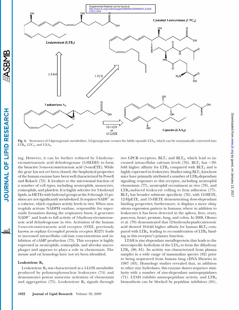
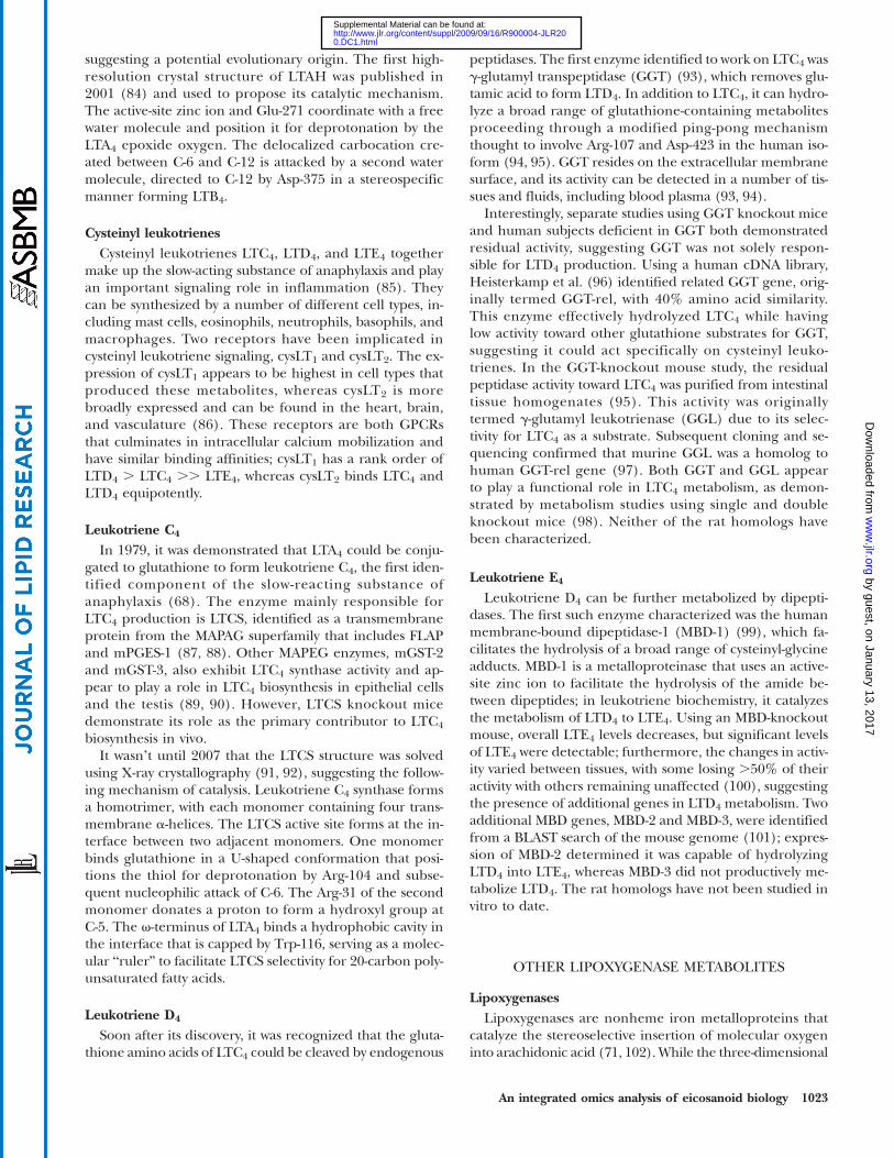
5-LOX performs the initial enzymatic step in leukotrienesynthesis (71), creating 5-hydroperoxy-eicosatetraenoicacid (5-HpETE) by incorporating one molecular oxygenat the C-5 position of arachidonic acid. Depending on cellu-lar conditions, 5-HpETE has a number of potential meta-bolic fates. It can be secreted in its peroxide form, reducedto 5-HETE, or undergo a catalytic rearrangement in the5-LOX active site to form leukotriene (LT) A4 (Fig. 4).When produced in vivo, LTA4 can be acted on by LTA4 hy-drolase (LTAH) to generate the stereospecific LTB4 or usedas a substrate by LTC4 synthase (LTCS) to catalyze the con-jugation of the glutathione to formLTC4. Additionally, LTA4
serves as a precursor for lipoxin biosynthesis, describedin greater detail in subsequent sections. Nonenzymatichydrolysis of the LTA4 epoxide creates all four potentialsteroisomers of LTB4, including D6-trans LTB4, 12-epiLTB4, and D6-trans, 12-epi LTB4. The enzymes involved inleukotrienemetabolism are described here, and a thoroughreview of the subject has been published by Murphy andGijon (71).
5-HETE and 5-oxoETECurrent literature suggests that 5-HETE itself does
not appear to play a significant role in biological signal-
An integrated omics analysis of eicosanoid biology 1021
by guest, on January 13, 2017w
ww
.jlr.orgD
ownloaded from
0.DC1.html http://www.jlr.org/content/suppl/2009/09/16/R900004-JLR20Supplemental Material can be found at:
ing. However, it can be further reduced by 5-hydroxy-eicosatetraenoic acid dehydrogenase (5-HEDH) to formthe bioactive 5-oxo-eicosatetraenoic acid (5-oxoETE). Whilethe gene has not yet been cloned, the biophysical propertiesof the human enzymehave beenwell characterized by Powelland Rokach (72). It localizes to the microsomal fraction ofa number of cell types, including neutrophils, monocytes,eosinophils, and platelets. It is highly selective for 5-hydroxyllipids, asHETEs with hydroxyl groups at the 8 through 15 po-sition are not significantlymetabolized. It requires NADP1 asa cofactor, which regulates activity levels in vivo. When neu-trophils activate NADPH oxidase, responsible for super-oxide formation during the respiratory burst, it generatesNADP1 and leads to full activity of 5-hydroxy-eicosatetrae-noic acid dehydrogenase in vivo. Activation of the human5-oxo-eicosatetraenoic acid receptor (OXE, previouslyknown as orphan G-coupled protein receptor R527) leadsto increased intracellular calcium concentrations and in-hibition of cAMP production (73). This receptor is highlyexpressed in neutrophils, eosinophils, and alveolar macro-phages and appears to plays a role in chemotaxis. Themouse and rat homologs have not yet been identified.
Leukotriene B4
Leukotriene B4 was characterized as a 5-LOX metaboliteproduced by polymorphonuclear leukocytes (74) anddemonstrates potent autocrine activation of chemotaxisand aggregation (75). Leukotriene B4 signals through
two GPCR receptors, BLT1 and BLT2, which lead to in-creased intracellular calcium levels (76). BLT1 has ?20-fold higher affinity for LTB4 compared with BLT2 and ishighly expressed in leukocytes. Studies using BLT1 knockoutmice have primarily attributed a number of LTB4-dependantsignaling responses to this receptor, including neutrophilchemotaxis (77), neutrophil recruitment in vivo (78), andLTB4-induced leukocyte rolling to firm adhesion (77).BLT2 has broader substrate specificity (76), with 12-HETE,12-HpETE, and 15-HETE demonstrating dose-dependantbinding properties; furthermore, it displays a more ubiq-uitous expression pattern in humans, where in addition toleukocytes it has been detected in the spleen, liver, ovary,pancreas, heart, prostate, lung, and colon. In 2008, Okunoet al. (79) demonstrated that 12-hydroxyheptadecatrienoicacid showed 10-fold higher affinity for human BLT2 com-pared with LTB4, leading to reconsideration of LTB4 bind-ing as this receptorʼs primary function.
LTAH is zinc-dependant metalloprotein that leads to thestereospecific hydrolysis of the LTA4 to form the dihydroxyLTB4 (80, 81). Its activity was characterized from plasmasamples in a wide range of mammalian species (82) priorto being sequenced from human lung cDNA libraries in1987 (83). Homology studies revealed that, in additionto other zinc hydrolases, this enzyme shares sequence simi-larity with a number of zinc-dependant aminopeptidases(71). LTAH exhibits aminopeptidase activity, and LTB4
biosynthesis can be blocked by peptidase inhibitors (81),
Fig. 4. Structures of 5-lipoxygenase metabolites. 5-Lipoxygenase creates the labile epoxide LTA4, which can be enzymatically converted intoLTB4, LTC4, and LXA4.
1022 Journal of Lipid Research Volume 50, 2009
by guest, on January 13, 2017w
ww
.jlr.orgD
ownloaded from
0.DC1.html http://www.jlr.org/content/suppl/2009/09/16/R900004-JLR20Supplemental Material can be found at:
suggesting a potential evolutionary origin. The first high-resolution crystal structure of LTAH was published in2001 (84) and used to propose its catalytic mechanism.The active-site zinc ion and Glu-271 coordinate with a freewater molecule and position it for deprotonation by theLTA4 epoxide oxygen. The delocalized carbocation cre-ated between C-6 and C-12 is attacked by a second watermolecule, directed to C-12 by Asp-375 in a stereospecificmanner forming LTB4.
Cysteinyl leukotrienesCysteinyl leukotrienes LTC4, LTD4, and LTE4 together
make up the slow-acting substance of anaphylaxis and playan important signaling role in inflammation (85). Theycan be synthesized by a number of different cell types, in-cluding mast cells, eosinophils, neutrophils, basophils, andmacrophages. Two receptors have been implicated incysteinyl leukotriene signaling, cysLT1 and cysLT2. The ex-pression of cysLT1 appears to be highest in cell types thatproduced these metabolites, whereas cysLT2 is morebroadly expressed and can be found in the heart, brain,and vasculature (86). These receptors are both GPCRsthat culminates in intracellular calcium mobilization andhave similar binding affinities; cysLT1 has a rank order ofLTD4 . LTC4 .. LTE4, whereas cysLT2 binds LTC4 andLTD4 equipotently.
Leukotriene C4
In 1979, it was demonstrated that LTA4 could be conju-gated to glutathione to form leukotriene C4, the first iden-tified component of the slow-reacting substance ofanaphylaxis (68). The enzyme mainly responsible forLTC4 production is LTCS, identified as a transmembraneprotein from the MAPAG superfamily that includes FLAPand mPGES-1 (87, 88). Other MAPEG enzymes, mGST-2and mGST-3, also exhibit LTC4 synthase activity and ap-pear to play a role in LTC4 biosynthesis in epithelial cellsand the testis (89, 90). However, LTCS knockout micedemonstrate its role as the primary contributor to LTC4
biosynthesis in vivo.It wasnʼt until 2007 that the LTCS structure was solved
using X-ray crystallography (91, 92), suggesting the follow-ing mechanism of catalysis. Leukotriene C4 synthase formsa homotrimer, with each monomer containing four trans-membrane a-helices. The LTCS active site forms at the in-terface between two adjacent monomers. One monomerbinds glutathione in a U-shaped conformation that posi-tions the thiol for deprotonation by Arg-104 and subse-quent nucleophilic attack of C-6. The Arg-31 of the secondmonomer donates a proton to form a hydroxyl group atC-5. The v-terminus of LTA4 binds a hydrophobic cavity inthe interface that is capped by Trp-116, serving as a molec-ular “ruler” to facilitate LTCS selectivity for 20-carbon poly-unsaturated fatty acids.
Leukotriene D4
Soon after its discovery, it was recognized that the gluta-thione amino acids of LTC4 could be cleaved by endogenous
peptidases. The first enzyme identified to work on LTC4 wasg-glutamyl transpeptidase (GGT) (93), which removes glu-tamic acid to form LTD4. In addition to LTC4, it can hydro-lyze a broad range of glutathione-containing metabolitesproceeding through a modified ping-pong mechanismthought to involve Arg-107 and Asp-423 in the human iso-form (94, 95). GGT resides on the extracellular membranesurface, and its activity can be detected in a number of tis-sues and fluids, including blood plasma (93, 94).
Interestingly, separate studies using GGT knockout miceand human subjects deficient in GGT both demonstratedresidual activity, suggesting GGT was not solely respon-sible for LTD4 production. Using a human cDNA library,Heisterkamp et al. (96) identified related GGT gene, orig-inally termed GGT-rel, with 40% amino acid similarity.This enzyme effectively hydrolyzed LTC4 while havinglow activity toward other glutathione substrates for GGT,suggesting it could act specifically on cysteinyl leuko-trienes. In the GGT-knockout mouse study, the residualpeptidase activity toward LTC4 was purified from intestinaltissue homogenates (95). This activity was originallytermed g-glutamyl leukotrienase (GGL) due to its selec-tivity for LTC4 as a substrate. Subsequent cloning and se-quencing confirmed that murine GGL was a homolog tohuman GGT-rel gene (97). Both GGT and GGL appearto play a functional role in LTC4 metabolism, as demon-strated by metabolism studies using single and doubleknockout mice (98). Neither of the rat homologs havebeen characterized.
Leukotriene E4
Leukotriene D4 can be further metabolized by dipepti-dases. The first such enzyme characterized was the humanmembrane-bound dipeptidase-1 (MBD-1) (99), which fa-cilitates the hydrolysis of a broad range of cysteinyl-glycineadducts. MBD-1 is a metalloproteinase that uses an active-site zinc ion to facilitate the hydrolysis of the amide be-tween dipeptides; in leukotriene biochemistry, it catalyzesthe metabolism of LTD4 to LTE4. Using an MBD-knockoutmouse, overall LTE4 levels decreases, but significant levelsof LTE4 were detectable; furthermore, the changes in activ-ity varied between tissues, with some losing .50% of theiractivity with others remaining unaffected (100), suggestingthe presence of additional genes in LTD4 metabolism. Twoadditional MBD genes, MBD-2 and MBD-3, were identifiedfrom a BLAST search of the mouse genome (101); expres-sion of MBD-2 determined it was capable of hydrolyzingLTD4 into LTE4, whereas MBD-3 did not productively me-tabolize LTD4. The rat homologs have not been studied invitro to date.
OTHER LIPOXYGENASE METABOLITES
LipoxygenasesLipoxygenases are nonheme iron metalloproteins that
catalyze the stereoselective insertion of molecular oxygeninto arachidonic acid (71, 102).While the three-dimensional
An integrated omics analysis of eicosanoid biology 1023
by guest, on January 13, 2017w
ww
.jlr.orgD
ownloaded from
0.DC1.html http://www.jlr.org/content/suppl/2009/09/16/R900004-JLR20Supplemental Material can be found at:
structures of most mammalian lipoxygenases have not beensolved, the structure of soybean LOX (103, 104) and rabbitleukocyte-type 12/15-LOX (105) have been reported andused as the basis for understanding other lipoxygenasesthrough homology modeling. The LOX reaction mecha-nism involves an iron-catalyzed hydrogen abstraction fromarachidonic acid at C-7, C-10, or C-13, forming a conjugatedradical reaching two carbons in either direction (106). Thestructure of a given LOX enzyme generally conforms tightlyaround the fatty acid, and small channels within the proteindirect molecular oxygen toward selected carbons, facili-tating the formation of the following HpETEs and corre-sponding HETEs: 8-HETE, 12-HETE, and 15-HETE (seesupplementary Table III). With the exception of 12R-LOX, mammalian LOXs typically direct the oxygen toattack the pro-S face of arachidonic acid. The positionalspecificity by which a LOX incorporates oxygen onto ara-chidonic acid is determined by how deeply it penetratesthe cavity, as well as the orientation (either C terminus orv terminus) it enters the active site. Work by Jisaka et al. sug-gests that two amino acid positions opposite of the catalyticiron ion determine the entry orientation. When a histidineand a bulky aromatic amino acid fill this position, arachi-donic acid C terminus enters the active side and promotes5-LOX and 8-LOX activity. Substitution of these amino acidswith either a glutamine or aspartate alongside an aliphaticresidue promotes the entry of the v terminus and facilitates12-LOX and 15-LOX activity.
Hydroxyeicosatetraenoic acidsIn mammalian systems, lipoxygenase activities have
been implicated in the biosynthesis of 5-HETE (see above),8-HETE, 12-HETE, and 15-HETE (71, 106). The HETEshave been implicated as potential ligands for PPARa(107) and PPARg (108). They exert numerous biologicaleffects, including, but not limited to, mitogen-activated pro-tein kinase signaling (109), monocyte chemoattractantprotein-1 expression (110), angiogenesis (111), cancer growthand metastasis (112, 113), and neuronal apoptosis (114);however, the precise molecular mechanisms behind these
effects remain unclear. Interestingly, 12- and 15-HpETE,but not the corresponding HETEs, have been shownto directly activate the capsaicin-sensitive vanilloid re-ceptor (VR1) (115), which is involved in inflammatorypain signaling.
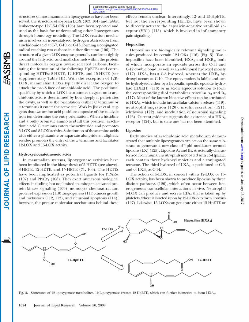
HepoxilinsHepoxilins are biologically relevant signaling mole-
cules produced by certain 12-LOXs (116) (Fig. 5). Two -hepoxilins have been identified, HXA3 and HXB3, bothof which incorporate an epoxide across the C-11 andC-12 double bond, as well as an additional hydroxyl moiety(117); HXA3 has a C-8 hydroxyl, whereas the HXB3 hy-droxyl occurs at C-10. The epoxy moiety is labile and canbe hydrolyzed either by a hepoxilin specific epoxide hydro-lase (HXEH) (118) or in acidic aqueous solution to formthe corresponding diol metabolites trioxilin A3 and B3
(117). Most of the known bioactive effects can be attributedto HXA3, which include intracellular calcium release (119),neutrophil migration (120), insulin secretion (121),ichthyosis (122), and modulation of neuronal signaling(123). Current evidence suggests the existence of a HXA3
receptor (124), but to date one has not been identified.
LipoxinsEarly studies of arachidonic acid metabolism demon-
strated that multiple lipoxygenases can act on the same sub-strate to generate a new class of lipid mediators termedlipoxins (LX) (125). Lipoxins A4 andB4, structurally charac-terized from human neutrophils incubated with 15-HpETE,each contain three hydroxyl moieties and a conjugatedtetraene. The third hydroxyl of LXA4 is positioned at C-6,and of LXB4 at C-14.
The action of 5-LOX, in concert with a 12-LOX or 15-LOX activity, has been shown to produce lipoxins by threedistinct pathways (126), which often occur between het-erogeneous transcellular interactions in vivo. Neutrophil5-LOX can produce and secrete LTA4 that is taken up byplatelets, where it is acted uponby 12-LOX-p to form lipoxins(127). Likewise, 15-LOXs can generate either 15-HpETE or
Fig. 5. Structures of 12-lipoxygenase metabolites. 12-Lipoxygenase creates 12-HpETE, which can further isomerize to form HXA3.
1024 Journal of Lipid Research Volume 50, 2009
by guest, on January 13, 2017w
ww
.jlr.orgD
ownloaded from
0.DC1.html http://www.jlr.org/content/suppl/2009/09/16/R900004-JLR20Supplemental Material can be found at:
15-HETE that can be taken up by monocytes and neutro-phils, where highly expressed 5-LOX uses it to generatelipoxins (125). Finally, aspirin acetylated COX-2, renderedunable to synthesize PGs, can act as a 15-LOX. This leads tothe formation of 15R-HETE and culminates in creation ofepi-lipoxins (128), which have altered stereochemistry atthe C-15 hydroxyl but similar biological potency.
Lipoxins are the first lipid mediators discovered thatdemonstrated anti-inflammatory activity as well as the ca-pacity to promote the resolution of inflammation and re-turn to tissue homeostasis (6, 126, 129). Their biologicalactivity has been shown to be mediated through the lipoxinA4 receptor (ALX), cysLT1, and the aryl hydrocarbon recep-tor, a nuclear transcription factor (129). The ALX receptoris a GPCR that upon binding ligand can lead to intracellularcalcium increases, PLD activation, and decreased NF-kB ac-tivity. Additionally, ALX cross-talks with vascular endothelialgrowth factor receptor, connective tissue growth factor re-ceptor, and platelet-derived growth factor receptor to down-regulate their activity. This receptor can also bind a numberof small peptides; some dock the LXA4 binding site, whileother others appear to function at a different site and leadto distinct downstream signaling events. LXA4 binds to re-combinant cysLT1 with approximately equal affinity asLTD4 (130) and has been suggested to function as a com-petitive inhibitor. Lipoxin binding to the aryl hydrocarbonreceptor transcription factor leads to the expression ofsuppressor of cytokine signaling-2 in dendritic cells (131,132). On a pathological level, the lipoxin signaling pro-motes inhibition of neutrophil migration, the recruitmentof nonphlogistic macrophages, and increased phagocytosisamong the countless documented effects (6, 126). The com-plex nature of lipoxin receptor signaling has been reviewedby Chiang et al. (129) in greater detail.
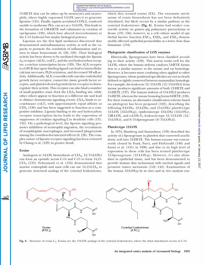
EoxinsAnalogous to 5-LOX biosynthesis of LTA4, 12/15-LOX-l
can form an epoxide across C-14 and C-15 to form 14,15-LTA4 (133). Feltenmark et al. (134) demonstrated thatmurine eosinophils and mast cells can use 14,15-LTA4 togenerate structural analogs of the cysteinyl leukotrienes,
which they termed eoxins (EX). The enzymatic mech-anism of eoxin biosynthesis has not been definitivelyelucidated, but likely occurs by a similar pathway as thecysteinyl leukotrienes (Fig. 6). Eoxins had weaker con-tractile activity on guinea pig pulmonary parenchyma orileum (135, 136); however, in a cell culture model of epi-thelial barrier function EXC4, EXD4, and EXE4 demon-strably effected epithelial permeability at a lower dose thanhistamine (134).
Phylogenetic classification of LOX enzymesHistorically, lipoxygenases have been classified accord-
ing to their activity (102). This system works well for the5-LOX, where the human isoform catalyzes 5-HETE forma-tion in a similar manner to the mouse and rat homologs.However, it becomes more confusing when applied to otherlipoxygenases, whose positional specificities are not as clearlydefined or tightly conserved betweenmammalian homologs.For example, the leukocyte 12/15-LOX (12/15-LOX-l) frommouse produces significant amounts of both 12-HETE and15-HETE (137). The human isoform of 15-LOX-2 produces15-HETE, whereas themouse homolog forms 8-HETE (138).For these reasons, an alternative classification scheme basedon phylogeny has been proposed (102), describing thefollowing 8-LOXs, 12-LOXs, and 15-LOXs: platelet-type12-LOX (12-LOX-p), epidermis-type 12-LOXs (12-LOX-e,12R-LOX, and e-LOX-3), leukocyte-type 12/15-LOX (12/15-LOX-l), and 8/15-lipoxygenase (8/15-LOX-2).
Platelet-type 12-LOXIn 1974, Hamberg and Samuelsson (139) described the
activity of a lipoxygenase in platelets that converted arachi-donic acid into 12-HETE. The human enzyme was concur-rently cloned by Funk, Furci, and FitzGerald (140) andIsumi et al. (141) in 1990, and due to its high level ofexpression in these cells has been termed platelet-type12-lipoxygenase (12-LOX-p). However, itʼs also abun-dant in epithelial tissue, and has been demonstrated toprovide human skin melanomas with survival signals andpromote tumor metastasis (142–144). Examination ofthe human 12-LOX-p by in vitro and in vivo analysis con-
Fig. 6. Structure of eoxin C4. Eoxins are the 15-LOX analogs of the cysteinyl leukotrienes, where the thiol attachment occurs at C-14.
An integrated omics analysis of eicosanoid biology 1025
by guest, on January 13, 2017w
ww
.jlr.orgD
ownloaded from
0.DC1.html http://www.jlr.org/content/suppl/2009/09/16/R900004-JLR20Supplemental Material can be found at:
firmed the native geneʼs positional specificity for produc-ing 12-HETE (140, 145). While mutations designed toknock 12-lipoxygenase activity into the rabbit leukocyte-type12/15-LOX (primarily a 15-LOX) succeeded (146, 147),the reverse mutagenesis experiments failed to convert12-LOX-p into an effective 15-lipoxygenase (145). Otherstudies indicate that 12-LOX-p can function as a hepoxilinsynthase on 12-HpETE (148), as well as a lipoxin synthaseon LTA4 (149).
Epidermal-type 12-LOXs (12-LOX-e, 12R-LOX,and e-LOX-3)
The epidermal type 12-lipoxygenase (12-LOX-e) wasseparately cloned from mice by Funk et al. in 1996 (150)and Kinzig et al. in 1997 (151). The gene was the same sizeas the platelet-type and leukocyte-type 12-LOX and had60% identity to these enzymes. Expression of the murine12-LOX-e gene confirmed its lipoxygenase activity, specifi-cally producing 12(S)-HETE (150). By analogy, 12-LOX-elikely functions by a similar catalytic mechanism as other12-LOX. Using in situ hybridization, 12-LOX-e expressionwas demonstrated in keratinocytes and targeted areas ofthe hair follicle. The human isoform of 12-LOX-e was iden-tified as a nonfunctional pseudogene (152), while in rat itsactivity has not been investigated.
As early as 1975, scaly lesions caused by psoriasis, an in-flammatory disorder that affects the skin and joints, wereknown to contain increased levels of stereochemically pure12-HETE (153). Distinct from other lipoxygenase metabo-lites, the hydroxyl moiety of 12-HETE found in these le-sions was in the R-configuration (154). In 1998, Boeglin,Kim, and Brash (155) discovered the source of the 12R-HETE originated from 12R-lipoxygenase (12R-LOX), thefirst known mammalian R-lipoxygenase. Expression of12R-LOX demonstrated that it produced 12R-HETE tothe exclusion of significant amounts of 12S-HETE, as wellas not making 5-HETE, 8-HETE, 9-HETE, 11-HETE, and15-HETE. Using mutants of two R-lipoxygenases (human12R-LOX and coral 8R-LOX) and two S-lipoxygenases (hu-man and mouse 8/15-LOX-2 isoforms), Coffa and Brash(156) identified a conserved amino acid, correspondingwith 12R-LOX Gly-441, that confers a substantial amountof stereochemical control to these enzymes. At this posi-tion, an alanine-to-glycine mutation in S-LOXs reversedthe chirality of lipid metabolites. However, while mutatingthe glycyl residue of coral 8R-LOX fully converted it to a12S-LOX, the 12R-LOXmutant had mixed activity. Meruvuet al. (157) identified Val631 as another important residuein facilitating the positional and stereochemical specificityof this enzyme.
Another epidermal-type LOX, e-LOX-3, has beencloned from both mouse (158) and human (159). Whileit contained the putative lipoxygenasesʼ structural featuresand had .50% homology with 12R-LOX, gene expressionin a number of cell lines indicated it had no activity towardarachidonic acid (158). However, mutations in both 12R-LOX and e-LOX-3 were identified in patients afflictedwith psoriasis (160), and in 2003, Yu et al. (161) identifiede-LOX-3 as a hepoxilin synthase that specifically used
12R-HpETE as a substrate. This suggests that impairmentof hepoxilin production by a 12R-LOX/e-LOX-3 pathwayleads to psoriasis in human patients.
Leukocyte-type 12/15-LOX (12/15-LOX-l)The human 12/15-LOX-l was originally cloned as a
15-LOX from a cDNA library in 1988 (162), followed byrabbit (163), rat (164), andmouse (165). The first structuralelucidation of a mammalian lipoxygenase was reportedusing X-ray crystallography on the rabbit 12/15-LOX-lhomolog (105). The substrate binding pocket is lined by hy-drophobic residues, and four residues comprise its base:Phe-353, Ile-418, Met-419, and Ile-593 (147). These residuesare conserved in the human isoform and are consideredcritical for promoting 15-LOX activity. By comparison, themouse and rat 12/15-LOX-l have amino acid substitutionsat each of these positions that could explain their predilec-tion toward 12-LOX activity. The rat 12/15-LOX-l also hasintrinsic hepoxilin synthase activity (148), whereas hu-man homolog does not. Based on the evidence that human12-LOX-p can synthesize hepoxilins, Nigam et al. (116) sur-mised that lipoxygenase positional specificity, and notgenetic homology, confer an enzyme with hepoxilin syn-thase activity. This would implicate the mouse 12/15-LOX-las a hepoxilin synthase, but to date it has not been ex-perimentally examined. The human 12/15-LOX-l exhibitseoxygenase activity that generates 14,15-LTs (134); the rab-bit (166), pig (133), and sheep (167) homologs also demon-strate this activity, suggesting that mouse and rat enzymesretain this capacity.
From even preliminary investigations (168), 12/15-LOX-l has demonstrated positional promiscuity when oxi-dizing arachidonic acid, forming 15-HETE, 12-HETE, and8,15-diHETE. The production of 12-HETE by 12/15-LOX-lhas been shown to increase expression of monocyte chemo-attractant protein-1, which increases vascular adhesion andthe recruitment of macrophages (168). In addition to ac-tion on free arachidonic acid, 12/15-LOX-l can functionat the membrane surface and on LDL to oxidize phospho-lipid and cholesterol ester fatty acids, leading to the devel-opment of atherosclerosis (169). In 2008, Harkewicz et al.(170) identified 12/15-LOX-l cholesterol ester peroxidesas the active components of minimally oxidized LDL. Incontrast to producing pro-inflammatory molecules, 12/15-LOX-l can take LTA4 as a substrate and produce the anti-inflammatory lipoxins (6, 129). Using both knockout andoverexpression transgenic mouse models, Merched et al.(171) propose that 12/15-LOX-l helps suppress athero-sclerosis. Undoubtedly, the capacity of 12/15-LOX-l tomake pro-inflammatory as well anti-inflammatory metabo-lites complicates the study of this enzyme at the whole-animal level, and future research will help elucidate its rolein signaling.
8/15-lipoxygenase-2In 1997, Brash, Boeglin, and Chang (172) reported the
discovery of a second 15-LOX gene in humans. Themurinehomolog has primarily 8-LOX activity (173), while the rat
1026 Journal of Lipid Research Volume 50, 2009
by guest, on January 13, 2017w
ww
.jlr.orgD
ownloaded from
0.DC1.html http://www.jlr.org/content/suppl/2009/09/16/R900004-JLR20Supplemental Material can be found at:
homolog has not been characterized to date. Mutationalanalyses of these enzymes determined that substitution ofTyr-603 and His-604 to the corresponding residues in thehuman isoform (aspartate and valine) confers 15-LOX ac-tivity (174). Likewise, mutation of the human enzyme tothe murine residues confers 8-LOX activity. Though theseenzymes appear to have different metabolic output, over-expression of either isoform leads to similar biological con-sequences in murine keratinocytes (138). In this model,8/15-LOX-2 expression, as well as exogenous addition of15-HETE and 8-HETE, blocked premalignant cell growthin a manner attributed to the inhibition of DNA synthesis.Interestingly, both human and murine 8/15-LOX-2 havebeen shown to produce 8,15-diHETE in vitro (175), a morepotent agonist of PPARa than 8-HETE. In these studies, themurine enzyme was capable of both reactions and sequen-tially oxygenates the C-8 and C-15 position. The humanhomolog could only perform the oxygenation of C-15 butwas able to use 8-HETE as a substrate. Future investigationsmay yet elucidate other novel metabolites produced by8/15-LOX-2 that have important bioactivities.
CYTOCHROME P450 METABOLITES
Cytochrome P450Cytochrome P450 (CYP) enzymes comprise a diverse
superfamily of enzymes present in bacteria, fungi, prostists,plants, and animals (176, 177). The name was coined in1962 to describe the first characterized cytochrome P450based on the unusual absorbance peak of 450 nm by itscarbon monoxide-bound form (178). CYP facilitate the ad-dition of oxygen on a wide range of substrates. To date,7232 genes in 781 families of have been categorized, andthe list is constantly growing as more complete genome se-quences become available; the generally accepted nomen-clature system and current list of known CYPs is curatedby Dr. David R. Nelson (http://drnelson.utmem.edu/CytochromeP450.html). They play an important role inthe metabolism of toxins in the liver as well as signaling
throughout the body. In eicosanoid biosynthesis, CYP activ-ities are classified by hydrolase and epoxygenase activities.
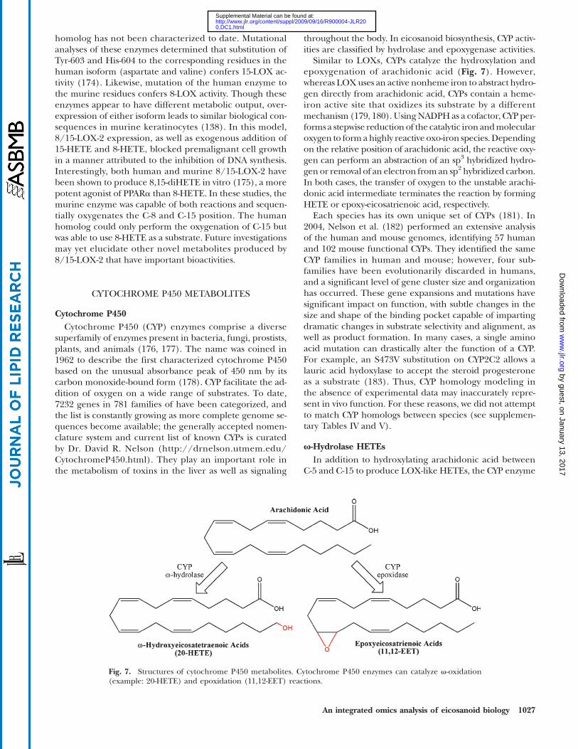
Similar to LOXs, CYPs catalyze the hydroxylation andepoxygenation of arachidonic acid (Fig. 7). However,whereas LOXuses an active nonheme iron to abstract hydro-gen directly from arachidonic acid, CYPs contain a heme-iron active site that oxidizes its substrate by a differentmechanism (179, 180). UsingNADPH as a cofactor, CYP per-forms a stepwise reduction of the catalytic iron andmolecularoxygen to formahighly reactive oxo-iron species.Dependingon the relative position of arachidonic acid, the reactive oxy-gen can perform an abstraction of an sp3 hybridized hydro-gen or removal of an electron froman sp2 hybridized carbon.In both cases, the transfer of oxygen to the unstable arachi-donic acid intermediate terminates the reaction by formingHETE or epoxy-eicosatrienoic acid, respectively.
Each species has its own unique set of CYPs (181). In2004, Nelson et al. (182) performed an extensive analysisof the human and mouse genomes, identifying 57 humanand 102 mouse functional CYPs. They identified the sameCYP families in human and mouse; however, four sub-families have been evolutionarily discarded in humans,and a significant level of gene cluster size and organizationhas occurred. These gene expansions and mutations havesignificant impact on function, with subtle changes in thesize and shape of the binding pocket capable of impartingdramatic changes in substrate selectivity and alignment, aswell as product formation. In many cases, a single aminoacid mutation can drastically alter the function of a CYP.For example, an S473V substitution on CYP2C2 allows alauric acid hydoxylase to accept the steroid progesteroneas a substrate (183). Thus, CYP homology modeling inthe absence of experimental data may inaccurately repre-sent in vivo function. For these reasons, we did not attemptto match CYP homologs between species (see supplemen-tary Tables IV and V).
v-Hydrolase HETEsIn addition to hydroxylating arachidonic acid between
C-5 and C-15 to produce LOX-like HETEs, the CYP enzyme
Fig. 7. Structures of cytochrome P450 metabolites. Cytochrome P450 enzymes can catalyze v-oxidation(example: 20-HETE) and epoxidation (11,12-EET) reactions.
An integrated omics analysis of eicosanoid biology 1027
by guest, on January 13, 2017w
ww
.jlr.orgD
ownloaded from
0.DC1.html http://www.jlr.org/content/suppl/2009/09/16/R900004-JLR20Supplemental Material can be found at:
can add a hydroxyl moiety to the sp3-hybridized v-carbonsto form a unique class of HETEs (177). Historically,v-hydrolase activity has been attributed to CYP4A and 4Fin mammalian systems, though CYP from other familieshave demonstrable activity as well (see supplementaryTable IV). The most well characterized of these is 20-HETE,hypothesized to play an important role in hypertension(184). 20-HETE has been shown promote systemic vaso-constriction by inhibiting KCa channel activity (185, 186),and in the kidney it blocks sodium reabsorbtion by inhibit-ing Na1-K1-ATPase activity (187). Other v-HETEs demon-strate considerable bioactivity that often act in opposition to20-HETE. For example, 18-HETE and 19-HETE dose-dependently induce vasodilatation by inhibiting the effectsof 20-HETE (188). They also, in addition to 16-HETE and17-HETE, have been shown to induce sodium reuptakein the kidney (188). More recently, 16-HETE has dem-onstrated the unique capacity among v-HETEs to in-hibit neutrophil adhesion, suggesting an important role ininflammation (189). Though it has been suggested thatv-HETEs signal through a putative receptor, neither thereceptor nor its active second-messenger component hasbe identified (177).
Epoxyeicosatrienoic acidsThe epoxidation of arachidonic acid by CYPs results in
the formation of unique bioactive lipid mediators termedepoxyeicosatrienoic acids (EETs) (190). Each double bondhas been shown to be susceptible to oxidation, resulting in5,6-EET, 8,9-EET, 11,12-EET, and 14,15-EET. In mammals,the most well documented CYP epoxygenases include the2C and 2J families, although currently numerous othershave been implicated in EET biosynthesis (see supplemen-tary Table V). The CYP epoxidases display significant pro-miscuity, with most enzymes making more than one EET,as well as retaining a significant level of v-hydrolase activity.
EETs have been implicated in a number of importantbiological processes, including vascular tone, renal func-tion, leukocyte adhesion, neuronal signaling, and angio-genesis. However, the effect of specific EET isomersoften remains uncertain. In 1999, Node et al. (191) deter-mined that 11,12-EET specifically block the expression ofvascular cell adhesion molecule-1 by inhibiting NF-kB, animportant mediator of leukocyte recruitment during in-flammation. Using a model of vascular laminar flow, Liuet al. (192) suggest NF-kB inhibition can be achieved byany of the four EET stereoisomers.
Current evidence suggests that EET signaling occursthrough a putative GPCR, though to date it has not beenidentified. Membranes from human U937 monocyte cellline were discovered by Wong et al. (193) to contain a highaffinity binding site for 11,12-EET and 14,15-EET, whichhas recently been characterized as a GPCR that inducesPKA activation and increases in cAMP (194). The EET-induced GPCR activity leads to the opening of BKCa chan-nels through the Gsa g-protein (195, 196). Falck et al.(197) synthesized a library of 11,12-EET analogs and, usingvascular cell adhesion molecule-1 expression in human
endothelial cells, identified a number of structural elementsrequired for functional recognition by the putative GPCRbinding site. A corresponding study measuring BKCa activ-ity induced by a series of 14,15-EET analogs identified simi-lar structural elements (198), suggesting these two EETsmay bind the same receptor in vivo.
In addition to GPCR signaling, EETs have been identi-fied as bioactive ligands for cation channels and PPARs.Watanabe et al. (199) identified 5,6-EET, and to a lesserdegree 8,9-EET, as an endogenous ligand for the vanilloidtype 4 receptor (TRPV4). When activated, TRPV4 acts as acation channel to allow the influx of extracellular calcium,and its activity has been implicated in rodent models of in-flammatory hyperalgesia (200–202). Downstream activa-tion of BKCa channels also result from TRPV4 (203).While these studies implicate EETs in the induction ofhyperalgesia, Inceoglu et al. (204) demonstrate that inhib-iting soluble epoxide hydrolase, which raises endogenouslevels of EETs, blocks inflammatory hyperalgesia in the ratmodel. Receptor binding assays identified micrometerEET affinity for cannabinoid CB2, neurokinin NK1, and do-pamine D3 receptors. In rat cardiomyocytes, EETs activateKATP channels by reducing their sensitivity to endogenousATP inhibition (205). Studies have also implicated EETs inthe direct activation of transcription factors PPARa andPPARg (192, 206). However, the selectivity of these recep-tors for specific EET regioisomers as well as the down-stream biological consequences have remained unclear,and undoubtedly further work will characterize the rolethat EETs play in biological signaling (207, 208).
Soluble epoxide hydrolase anddihydroxyeicosatrienoic acids
The majority of the EET biological activities are dimin-ished by the hydrolysis to the corresponding dihydroxy-eicosatrienoic acids (DHET). In mammals, this processis primarily catalyzed by the enzyme soluble epoxide hy-drolase (sEH). The murine and human isoforms havebeen cloned and structurally characterized by X-ray crystal-lography (209–211), allowing for a detailed mechanisticdescription of its catalytic function using the human se-quence (212). The sEH active site forms a deep, L-shapedhydrophobic pocket with a catalytic aspartyl at the L-turnaccessible to both openings. Upon entering the activesite, the EET epoxide moiety interacts with Tyr-382 andTyr-465. His-523 positions and activates Asp-334 for a nu-cleophilic attack on an epoxide carbon, opening the ringand facilitating water hydrolysis to the corresponding diol.Unlike other eicosanoid biosynthetic enzymes, sEH doesnot require any metal cofactor for catalysis. However, itsactivity can be inhibited by the presence of zinc, suggestinga straightforward mechanism for regulating EET metabo-lism during inflammation (213). Due to the loss of EETbioactivity, sEH formation of DHETs has been consideredthe metabolic inactivation step of EET signaling. However,recent work has demonstrated that DHETs can retain cer-tain EET signaling capacities, effectively activating BKCa
channels in smooth muscle cells (214), as well as PPARaand PPARg (192, 215).
1028 Journal of Lipid Research Volume 50, 2009
by guest, on January 13, 2017w
ww
.jlr.orgD
ownloaded from
0.DC1.html http://www.jlr.org/content/suppl/2009/09/16/R900004-JLR20Supplemental Material can be found at:
Nonenzymatic lipid metabolitesFree radicals generated by oxidative stress have long
been believed to participate in the development of a num-ber of neurodegenerative and inflammatory diseases bycausing damage to DNA, proteins, and lipids (216). In1990, Morrow et al. (217) described a class of PG-like com-pounds formed in vitro by free radical catalyzed peroxida-tion. Abstraction of hydrogen from C-13 forms a fivecarbon delocalized radical that can form a prostane ringwhen followed by the addition of molecular oxygen atthe activated C-11. Due to the lack of enzymatic controlin the reaction, these PG-like compounds comprise a seriesof stereoisomers termed 15-series due to the location ofthe nonprostane hydroxyl moiety. Furthermore, the reac-tion can also be initiated at C-7, leading to the 5-series,as well as at C-10, leading to both the 8- and 12-series ofisoprostanes. In total, 64 possible isoprostane isomers canbe generated by radical initiated peroxidation (216). Todate, no specific isoprostane receptors have been identi-fied. However, isoprostanes have demonstrated binding af-finity for PG receptors, including the FP and TP receptor(218, 219).
Lipid peroxidation can also proceed forward in alipoxygenase-like activity. Following the abstraction of hy-drogen, the peroxide formed by oxygenation of the radicalcan simply be reduced to a corresponding HETE. By thispathway, free radical reactions can produce all the HETEsgenerated by LOX and CYP. Additionally, nonenzymaticperoxidation can generate 9-HETE, which has no identifi-able biological activity and has been used as a marker ofnonenzymatic lipoxygenase-like activity (220). In practice,both 9-HETE and isoprostanes have been shown to serve asuseful biomarkers of oxidative stress in vivo. In a recentstudy of major coronary heart disease (221), human plasmalevels of isoprostanes and 9-HETE highly correlated withthe pathogenesis and may serve as an indicator in clinicalassessments of heart disease.
EICOSANOID CATABOLISM
Eicosanoid catabolism plays an important role in thecontrol of bioactive lipid signaling (see supplementaryTable VI). Not all molecules from the same class breakdown by the same pathway, which helps further differenti-ate the functional signaling role of each eicosanoid in vivo.In particular, the dysfunction of PG catabolizing enzymes,reviewed in detail by Tai et al. (222), has been implicatedin susceptibility to disease. Generally, the catabolism ofeicosanoids leads to diminished bioactive signaling and uri-nary excretion from the body by facilitating changes thatincrease their water solubility.
11-Hydroxythromboxane B2 reductaseStudies investigating thromboxane clearance indicate
that a significant portion undergoes dehydrogenation atC-11 to form 11dh-TXB2. This degradation metabolite isreadily detected in both human blood plasma and urine(223, 224). The enzyme responsible for catalysis in vivo
has been termed 11-dehydroxythromboxane B2 dehydro-genase (11-TXDH) and has two distinct isoforms. Usingamino acid composition analysis, the liver-type isoformfrom swine has been classified as a cytosolic aldehyde de-hydrogenase family member (225). The second enzyme,the kidney-type 11-TXDH, can catalyze the dehydrogena-tion but not the reverse reductions, distinguishing it fromthe liver type. While the human, mouse, and rat 11-TXDHisoforms have not been identified, 11dh-TXB2 has beendetected in urine samples from these species. Historically,TXB2 and 11dh-TXB2 have been considered biologicallyinert due to their inability to activate the thromboxaneTP receptor (226), but work by Böhm et al. (227) demon-strates that 11dh-TXB2 is an agonist of the CRTH2 receptor.
15-Prostaglandin dehydrogenaseThe catabolism of selected PGs, leukotrienes, and
HETEs is initiated by oxidation by 15-prostaglandin dehy-drogenase (15-PGDH). Two distinct enzymes have beenidentified with this activity, 15-PGDH-I and 15-PGDH-II;however, the high Km values of 15-PGDH-II for most PGssuggest that the majority of the in vivo activity can be attrib-uted to 15-PGDH-I (222). The 15-PGDH-I is NAD1 depen-dant and metabolizes E-series PGs, lipoxins, 15-HETE,5,15-diHETE, and 8,15-diHETE, while other PGs appearto be less susceptible substrates (228). In all likelihood,15-PGDH can metabolize a larger array of eicosanoidsthan those which have been tested.
The 15-PGDH-I genes have been cloned in human,mouse, and rat, but a crystal structure of the enzyme hasnot yet been determined. In 2006, Cho et al. (229) usedthe molecular modeling to develop a three-dimensionalstructure of the human protein and propose a catalyticmechanism. Following the sequential binding of NAD1
and PGE2, the 15-hydroxyl is coordinated with Tyr-151,Ser-138, andGln-148. Tyr-151 deprotonates the 15-hydroxyl,facilitating a hydride transfer from C-15 and the release of15k-PGE2. The oxidation to a 15-ketone reduces the bio-logical activity of most eicosanoid substrates, and loss offunction has been implicated in the development ofcertain cancers (230). However, in 2007, Chou et al. (231)identified 15k-PGE2 as a ligand for PPARg and an activecomponent of murine 3T3-L1 fibroblast differentiationinto adipocytes.
13-Prostaglandin reductaseThe 13-prostaglandin reductases (13-PGR) metabolize
eicosanoids by catalyzing NADH/NADPH-dependant dou-ble bond reduction. The gene was originally cloned fromhuman and pig sources as leukotriene B4 12-hydroxydehy-drogenase (LTB4DH) (232), but later discovered to havedual functionality as a PG reductase (13-PGR-1) (233). Inaddition to reducing PGs, 13-PGR has been shown to uselipoxins as substrates (234). The resulting metabolitesfrom 13-PGR activity show reduced biological activity,and PGE2 metabolites from this pathway are readily de-tected in urinary excretion (235). The rat protein hasLTB4DH and 13-PGR activities, suggesting that these func-
An integrated omics analysis of eicosanoid biology 1029
by guest, on January 13, 2017w
ww
.jlr.orgD
ownloaded from
0.DC1.html http://www.jlr.org/content/suppl/2009/09/16/R900004-JLR20Supplemental Material can be found at:
tions are retained in the murine homolog (see supplemen-tary Table VI).
Structural determinations of human 13-PGR-1 in com-plex with NADP1 and 15k-PGE2 were solved in 2004 andused to propose the following mechanism of catalysis(236). 13-PGR-1 forms a homodimer, with an active sitepocket formed at the dimer interface. The 2ʼ-hydroxylgroup of NADPH forms a hydrogen bond with the C-15oxygen on PGE2, promoting the stabilization of an enolateintermediate. The NADPH hydride attacks the carboca-tion at C-13, followed by ketone formation and the releaseof the reduced metabolite 13,14-dihydro,15-keto-PGE2
(dhk-PGE2). LTB4 oxidation likely proceeds by a similarmechanism as 15-PGDH, with a conserved tyrosine servingas the catalytic residue. In 2007, Chou et al. (231) reportedthe discovery of a second PG reductase gene (13-PGR-2) inmice that they demonstrate works by a similar reductivemechanism on 15k-PGE2 but have not exhaustively studiedits substrate selectivity or potential LTB4DH activity.
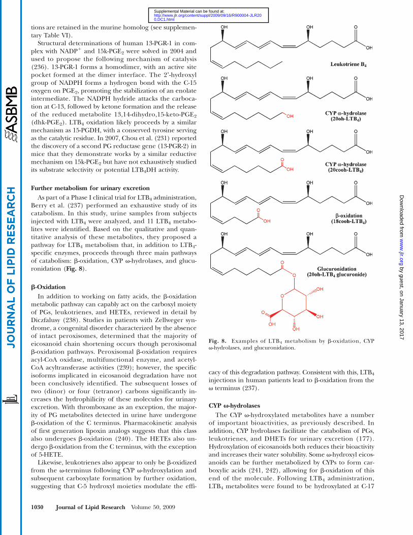
Further metabolism for urinary excretionAs part of a Phase I clinical trial for LTB4 administration,
Berry et al. (237) performed an exhaustive study of itscatabolism. In this study, urine samples from subjectsinjected with LTB4 were analyzed, and 11 LTB4 metabo-lites were identified. Based on the qualitative and quan-titative analysis of these metabolites, they proposed apathway for LTB4 metabolism that, in addition to LTB4-specific enzymes, proceeds through three main pathwaysof catabolism: b-oxidation, CYP v-hydrolases, and glucu-ronidation (Fig. 8).
b-OxidationIn addition to working on fatty acids, the b-oxidation
metabolic pathway can capably act on the carboxyl moietyof PGs, leukotrienes, and HETEs, reviewed in detail byDiczfalusy (238). Studies in patients with Zellweger syn-drome, a congenital disorder characterized by the absenceof intact peroxisomes, determined that the majority ofeicosanoid chain shortening occurs though peroxisomalb-oxidation pathways. Peroxisomal b-oxidation requiresacyl-CoA oxidase, multifunctional enzyme, and acetyl-CoA acyltransferase activities (239); however, the specificisoforms implicated in eicosanoid degradation have notbeen conclusively identified. The subsequent losses oftwo (dinor) or four (tetranor) carbons significantly in-creases the hydrophilicity of these molecules for urinaryexcretion. With thromboxane as an exception, the major-ity of PG metabolites detected in urine have undergoneb-oxidation of the C terminus. Pharmacokinetic analysisof first generation lipoxin analogs suggests that this classalso undergoes b-oxidation (240). The HETEs also un-dergo b-oxidation from the C terminus, with the exceptionof 5-HETE.
Likewise, leukotrienes also appear to only be b-oxidizedfrom the v-terminus following CYP v-hydroxylation andsubsequent carboxylate formation by further oxidation,suggesting that C-5 hydroxyl moieties modulate the effi-
cacy of this degradation pathway. Consistent with this, LTB4
injections in human patients lead to b-oxidation from thev terminus (237).
CYP v-hydrolasesThe CYP v-hydroxylated metabolites have a number
of important bioactivities, as previously described. Inaddition, CYP hydrolases facilitate the catabolism of PGs,leukotrienes, and DHETs for urinary excretion (177).Hydroxylation of eicosanoids both reduces their bioactivityand increases their water solubility. Some v-hydroxyl eicos-anoids can be further metabolized by CYPs to form car-boxylic acids (241, 242), allowing for b-oxidation of thisend of the molecule. Following LTB4 administration,LTB4 metabolites were found to be hydroxylated at C-17
Fig. 8. Examples of LTB4 metabolism by b-oxidation, CYPv-hydrolases, and glucuronidation.
1030 Journal of Lipid Research Volume 50, 2009
by guest, on January 13, 2017w
ww
.jlr.orgD
ownloaded from
0.DC1.html http://www.jlr.org/content/suppl/2009/09/16/R900004-JLR20Supplemental Material can be found at:
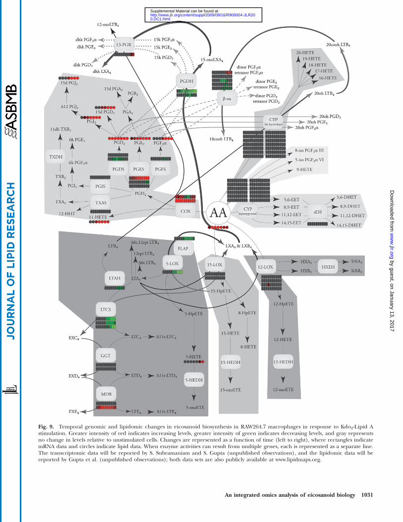
Fig. 9. Temporal genomic and lipidomic changes in eicosanoid biosynthesis in RAW264.7 macrophages in response to Kdo2-Lipid Astimulation. Greater intensity of red indicates increasing levels, greater intensity of green indicates decreasing levels, and gray representsno change in levels relative to unstimulated cells. Changes are represented as a function of time (left to right), where rectangles indicatemRNA data and circles indicate lipid data. When enzyme activities can result from multiple genes, each is represented as a separate line.The transcriptomic data will be reported by S. Subramaniam and S. Gupta (unpublished observations), and the lipidomic data will bereported by Gupta et al. (unpublished observations); both data sets are also publicly available at www.lipidmaps.org.
An integrated omics analysis of eicosanoid biology 1031
by guest, on January 13, 2017w
ww
.jlr.orgD
ownloaded from
0.DC1.html http://www.jlr.org/content/suppl/2009/09/16/R900004-JLR20Supplemental Material can be found at:
though C-20; however, the majority of these compoundshad undergone further catabolism to form glucuronidesat these sites (237).
GlucuronidationIn addition to action by CYPs, lipophilic molecules are
further catabolized into water-soluble metabolites throughthe addition of the sugar glucuronide (243). In mam-mals, this reaction is catalyzed by members of the UDP-glucuronosyltransferase enzymes. To date, 15 UDP enzymeshave been found in humans, and a study by Turgeon et al.(244) in 2003 using a HEK293 expression system found thatmost of them were capable of metabolizing LTB4 and LOX-derived HETEs. Glucuronidation also plays a significantrole in the catabolism of DHETs in humans, but rats excretethe diol form directly (245). In the human trials of LTB4
metabolism, nearly all metabolites with a hydroxyl moietywere found to be glucuronidated, suggesting nonspecificcatalysis of eicosanoids by UDPs plays a significant role intheir removal from the body (237).
APPLICATIONS OF A SYSTEMS APPROACHTO EICOSANOID BIOLOGY
The LIPID MAPS consortium has produced preliminarystudies comparing comprehensive lipidomic and transcrip-tomic changes in macrophages (7–9). In response to thelipopolysaccharide Kdo2-Lipid A, RAW264.7murinemacro-phage cells were analyzed for eicosanoids by LC/MS/MS,as well as transcriptomic changes using Affemetrix gene ar-rays. Using publicly available data (www.lipidmaps.org),these changes can be mapped together to investigate thecorrelation between genes and metabolites (Fig. 9). Thisstimulatory pathway causes upregulation of the numerousCOX genes and downregulation of the 5-LOX pathway,causing primarily increases in COXmetabolites (9); PGDH,which metabolically inactivates PGE2, was downregulated,suggesting that it plays an important role in eicosanoiddegradation. It remains to be addressed if other LOX orCYP enzymes are basally expressed and play an importantrole in this cell model. Work by Buczynski et al. (9) high-lights the issue of basal protein activity, demonstrating thatcalcium agonists can activate the constitutively expressed5-LOX pathway in RAW264.7 cells to make cysteinyl leu-kotrienes. Clearly, a proteomic analysis could addressa number of such remaining questions in this cellularsystem. Thus, it is timely to carry out a proteomic analysisof the eicosanoid proteins to give a fully integrated bio-logical picture.
In a similar murine macrophage model, Shui et al. (246)compared the efficacy of two state of the art quantitativeproteomic approaches. Using either the isobaric tag forrelative and absolute quantitation chemical tagging orthe stable isotope labeling with amino acids in cell culturelabeling technique, they identified 1286 total unique pro-teins from the entire activated macrophage proteome,with 512 of those being reliably detectable for quantita-tion. However, probing these data sets for specific changes
in the eicosanoid proteome only identified two proteins,FLAP and 13-PGR. None of the PG biosynthetic proteinswere identified using these approaches, including thehighly upregulated COX-2. In contrast, Jenkins et al.(247) have developed a selected population procedurefor quantifying changes in the entire CYP protein family.Combining the advantages of isobaric tag for relative andabsolute quantitation with the selectivity of multiple-reactionmonitoring mass spectrometry, they could measure 16 dif-ferent CYPs at femtomolar levels from murine liver micro-some samples. Provided purified protein standards areavailable, this methodology could be readily expanded toinvestigate the entire eicosanoid proteome.
The information contained herein provides the basisnecessary to decipher the eicosanoid proteome and com-plete the integrated pathway map. Microarray analysis ofgene regulation and mass spectrometry methodology forlipid analysis capable of comprehensively analyzing thesepathways now exist. This review provides the tools neces-sary to create a complimentary proteomic methodology.The sequential flow of information from gene to proteinto metabolite remains a central focus in biological re-search. However, the integration of eicosanoid transcrip-tomic, proteomic, and lipidomic platforms, if carried out,would represent the first time such an analysis was per-formed on any biological system. This would undoubtedlyhave enormous implications for our understanding of biol-ogy and disease pathology and serve as a model for futuresystems biology endeavors.
The authors thank Rebecca C. Bowers-Gentry for her invalu-able advice and input in drafting this manuscript. Sequencealignments found in the supplementary tables were done usingJalview 2.3 (248). The authors are also grateful to the entireLIPID MAPS Consortium for the use of their data, availableat www.lipidmaps.org for public use.
REFERENCES
1. Aderem, A. 2005. Systems biology: its practice and challenges. Cell.121: 511–513.
2. de Godoy, L. M., J. V. Olsen, J. Cox, M. L. Nielsen, N. C. Hubner,F. Frohlich, T. C. Walther, and M. Mann. 2008. Comprehensivemass-spectrometry-based proteome quantification of haploid versusdiploid yeast. Nature. 455: 1251–1254.
3. Funk, C. D. 2001. Prostaglandins and leukotrienes: advances ineicosanoid biology. Science. 294: 1871–1875.
4. Simmons, D. L., R. M. Botting, and T. Hla. 2004. Cyclooxygenaseisozymes: the biology of prostaglandin synthesis and inhibition.Pharmacol. Rev. 56: 387–437.
5. Smith, W. L., D. L. DeWitt, and R. M. Garavito. 2000. Cyclooxy-genases: structural, cellular, and molecular biology. Annu. Rev.Biochem. 69: 145–182.
6. Serhan, C. N., N. Chiang, and T. E. Van Dyke. 2008. Resolvinginflammation: dual anti-inflammatory and pro-resolution lipidmediators. Nat. Rev. Immunol. 8: 349–361.
7. Feng, L., and G. D. Prestwich. 2006. Functional Lipidomics. Taylor& Francis, Boca Raton, FL.
8. Schmelzer, K., E. Fahy, S. Subramaniam, and E. A. Dennis.2007. The lipid maps initiative in lipidomics. Methods Enzymol.432: 171–183.
9. Buczynski, M. W., D. L. Stephens, R. C. Bowers-Gentry, A. Grkovich,R. A. Deems, and E. A. Dennis. 2007. TLR-4 and sustained calcium
1032 Journal of Lipid Research Volume 50, 2009
by guest, on January 13, 2017w
ww
.jlr.orgD
ownloaded from
0.DC1.html http://www.jlr.org/content/suppl/2009/09/16/R900004-JLR20Supplemental Material can be found at:
agonists synergistically produce eicosanoids independent of pro-tein synthesis in RAW264.7 cells. J. Biol. Chem. 282: 22834–22847.
10. Domon, B., and R. Aebersold. 2006. Mass spectrometry and proteinanalysis. Science. 312: 212–217.
11. Bantscheff, M., M. Schirle, G. Sweetman, J. Rick, and B. Kuster.2007. Quantitative mass spectrometry in proteomics: a critical re-view. Anal. Bioanal. Chem. 389: 1017–1031.
12. Keshishian, H., T. Addona, M. Burgess, E. Kuhn, and S. A. Carr.2007. Quantitative, multiplexed assays for low abundance proteinsin plasma by targeted mass spectrometry and stable isotope dilu-tion. Mol. Cell. Proteomics. 6: 2212–2229.
13. Schaloske, R. H., and E. A. Dennis. 2006. The phospholipase A2superfamily and its group numbering system. Biochim. Biophys.Acta. 1761: 1246–1259.
14. Six, D. A., and E. A. Dennis. 2000. The expanding superfamily ofphospholipase A(2) enzymes: classification and characterization.Biochim. Biophys. Acta. 1488: 1–19.
15. Burke, J. E., and E. A.Dennis. Phospholipase A2 structure/function,mechanism and signaling. J. Lipid Res. Epub ahead of print.November, 14, 2008; doi: 10.1194/jlr.R800033-JLR200.
16. Uozumi, N., and T. Shimizu. 2002. Roles for cytosolic phospho-lipase A2alpha as revealed by gene-targeted mice. ProstaglandinsOther Lipid Mediat. 68–69: 59–69.
17. Ghosh, M., D. E. Tucker, S. A. Burchett, and C. C. Leslie. 2006.Properties of the Group IV phospholipase A2 family. Prog. LipidRes. 45: 487–510.
18. Bonventre, J. V., Z. Huang, M. R. Taheri, E. OʼLeary, E. Li, M. A.Moskowitz, and A. Sapirstein. 1997. Reduced fertility and post-ischaemic brain injury in mice deficient in cytosolic phospholipaseA2. Nature. 390: 622–625.
19. Uozumi, N., K. Kume, T. Nagase, N. Nakatani, S. Ishii, F. Tashiro,Y. Komagata, K. Maki, K. Ikuta, Y. Ouchi, et al. 1997. Role of cytosolicphospholipase A2 in allergic response and parturition. Nature. 390:618–622.
20. Adler, D. H., J. D. Cogan, J. A. Phillips, N. Schnetz-Boutaud, G. L.Milne, T. Iverson, J. A. Stein, D.A. Brenner, J. D.Morrow,O. Boutaud,et al. 2008. Inherited human cPLA(2alpha) deficiency is associatedwith impaired eicosanoid biosynthesis, small intestinal ulceration,and platelet dysfunction. J. Clin. Invest. 118: 2121–2131.
21. Lambeau, G., and M. H. Gelb. 2008. Biochemistry and physiologyof mammalian secreted phospholipases A2. Annu. Rev. Biochem. 77:495–520.
22. Rouzer, C. A., and L. J. Marnett. Cyclooxygenases: structural andfunctional insights. J. Lipid Res. Epub ahead of print. October 23,2008; doi10.1194/jlr.R800042-JLR200..
23. Nelson, N. A. 1974. Prostaglandin nomenclature. J. Med. Chem. 17:911–918.
24. Whittaker, N., S. Bunting, J. Salmon, S. Moncada, J. R. Vane, R. A.Johnson, D. R. Morton, J. H. Kinner, R. R. Gorman, J. C. McGuire,et al. 1976. The chemical structure of prostaglandin X (prostacyclin).Prostaglandins. 12: 915–928.
25. Wu, K. K., and J. Y. Liou. 2005. Cellular and molecular biology ofprostacyclin synthase. Biochem. Biophys. Res. Commun. 338: 45–52.
26. Chiang, C. W., H. C. Yeh, L. H. Wang, and N. L. Chan. 2006. Crystalstructure of the human prostacyclin synthase. J. Mol. Biol. 364:266–274.
27. Hecker, M., and V. Ullrich. 1989. On the mechanism of pros-tacyclin and thromboxane A2 biosynthesis. J. Biol. Chem. 264:141–150.
28. Katsuyama,M., Y. Sugimoto, T.Namba,A. Irie,M.Negishi, S.Narumiya,and A. Ichikawa. 1994. Cloning and expression of a cDNA for thehuman prostacyclin receptor. FEBS Lett. 344: 74–78.
29. Boie, Y., T. H. Rushmore, A. Darmon-Goodwin, R. Grygorczyk,D. M. Slipetz, K. M. Metters, and M. Abramovitz. 1994. Cloningand expression of a cDNA for the human prostanoid IP receptor.J. Biol. Chem. 269: 12173–12178.
30. Aubert, J., G. Ailhaud, and R. Negrel. 1996. Evidence for a novelregulatory pathway activated by (carba)prostacyclin in preadiposeand adipose cells. FEBS Lett. 397: 117–121.
31. Kojo,H.,M. Fukagawa, K. Tajima, A. Suzuki, T. Fujimura, I. Aramori,K. Hayashi, and S. Nishimura. 2003. Evaluation of human peroxi-some proliferator-activated receptor (PPAR) subtype selectivity ofa variety of anti-inflammatory drugs based on a novel assay for PPARdelta(beta). J. Pharmacol. Sci. 93: 347–355.
32. Hamberg, M., J. Svensson, and B. Samuelsson. 1975. Thromboxanes:a new group of biologically active compounds derived from prosta-glandin endoperoxides. Proc. Natl. Acad. Sci. USA. 72: 2994–2998.
33. Kinsella, B. T., D. J. OʼMahony, and G. A. Fitzgerald. 1997. Thehuman thromboxane A2 receptor alpha isoform (TP alpha) func-tionally couples to the G proteins Gq and G11 in vivo and is acti-vated by the isoprostane 8-epi prostaglandin F2 alpha. J. Pharmacol.Exp. Ther. 281: 957–964.
34. Samuelsson, B. 1963. Isolation and identification of prostaglandinsfrom human seminal plasma. 18. Prostaglandins and related fac-tors. J. Biol. Chem. 238: 3229–3234.
35. Park, J. Y., M. H. Pillinger, and S. B. Abramson. 2006. ProstaglandinE2 synthesis and secretion: the role of PGE2 synthases. Clin. Immunol.119: 229–240.
36. Tanioka, T., Y. Nakatani, N. Semmyo, M. Murakami, and I. Kudo.2000. Molecular identification of cytosolic prostaglandin E2 syn-thase that is functionally coupled with cyclooxygenase-1 in imme-diate prostaglandin E2 biosynthesis. J. Biol. Chem. 275: 32775–32782.
37. Murakami, M., Y. Nakatani, T. Tanioka, and I. Kudo. 2002. Pros-taglandin E synthase. Prostaglandins Other Lipid Mediat. 68–69:383–399.
38. Jakobsson, P. J., S. Thoren, R. Morgenstern, and B. Samuelsson.1999. Identification of human prostaglandin E synthase: a micro-somal, glutathione-dependent, inducible enzyme, constitutinga potential novel drug target. Proc. Natl. Acad. Sci. USA. 96:7220–7225.
39. Huang, X., W. Yan, D. Gao, M. Tong, H. H. Tai, and C. G. Zhan.2006. Structural and functional characterization of human micro-somal prostaglandin E synthase-1 by computational modeling andsite-directed mutagenesis. Bioorg. Med. Chem. 14: 3553–3562.
40. Samuelsson, B., R. Morgenstern, and P. J. Jakobsson. 2007. Mem-braneprostaglandinE synthase-1: a novel therapeutic target.Pharmacol.Rev. 59: 207–224.
41. Watanabe, K., K. Kurihara, Y. Tokunaga, and O. Hayaishi. 1997.Two types of microsomal prostaglandin E synthase: glutathione-dependent and -independent prostaglandin E synthases. Biochem.Biophys. Res. Commun. 235: 148–152.
42. Tanikawa, N., Y. Ohmiya, H. Ohkubo, K. Hashimoto, K. Kangawa,M. Kojima, S. Ito, and K. Watanabe. 2002. Identification and char-acterization of a novel type of membrane-associated prostaglandinE synthase. Biochem. Biophys. Res. Commun. 291: 884–889.
43. Yamada, T., J. Komoto, K. Watanabe, Y. Ohmiya, and F. Takusagawa.2005. Crystal structure and possible catalytic mechanism of micro-somal prostaglandin E synthase type 2 (mPGES-2). J. Mol. Biol. 348:1163–1176.
44. Ito, S., S. Narumiya, and O. Hayaishi. 1989. Prostaglandin D2: abiochemical perspective. Prostaglandins Leukot. Essent. Fatty Acids.37: 219–234.
45. Abdel-Halim, M. S., M. Hamberg, B. Sjoquist, and E. Anggard.1977. Identification of prostaglandin D2 as a major prostaglandinin homogenates of rat brain. Prostaglandins. 14: 633–643.
46. Roberts 2nd, L. J., R. A. Lewis, J. A. Oates, and K. F. Austen.1979. Prostaglandin thromboxane, and 12-hydroxy-5,8,10,14-eicosatetraenoic acid production by ionophore-stimulated ratserosal mast cells. Biochim. Biophys. Acta. 575: 185–192.
47. Urade, Y., T. Tanaka, N. Eguchi, M. Kikuchi, H. Kimura, H. Toh,and O. Hayaishi. 1995. Structural and functional significance ofcysteine residues of glutathione-independent prostaglandin Dsynthase. Identification of Cys65 as an essential thiol. J. Biol. Chem.270: 1422–1428.
48. Shimamoto, S., T. Yoshida, T. Inui, K. Gohda, Y. Kobayashi,K. Fujimori, T. Tsurumura, K. Aritake, Y. Urade, and T. Ohkubo.2007. NMR solution structure of lipocalin-type prostaglandin Dsynthase: evidence for partial overlapping of catalytic pocket andretinoic acid-binding pocket within the central cavity. J. Biol. Chem.282: 31373–31379.
49. Kanaoka, Y., H. Ago, E. Inagaki, T. Nanayama, M. Miyano, R. Kikuno,Y. Fujii, N. Eguchi, H. Toh, Y. Urade, et al. 1997. Cloning and crys-tal structure of hematopoietic prostaglandin D synthase. Cell. 90:1085–1095.
50. Pettipher, R. 2008. The roles of the prostaglandin D(2) receptorsDP(1) and CRTH2 in promoting allergic responses. Br. J. Pharmacol.153 (Suppl 1.): S191–S199.
51. Sawyer, N., E. Cauchon, A. Chateauneuf, R. P. Cruz, D.W. Nicholson,K.M.Metters, G. P. OʼNeill, and F. G. Gervais. 2002.Molecular phar-macology of the human prostaglandin D2 receptor, CRTH2. Br.J. Pharmacol. 137: 1163–1172.
52. Basu, S. 2007. Novel cyclooxygenase-catalyzed bioactive prosta-glandin F2alpha from physiology to new principles in inflamma-tion. Med. Res. Rev. 27: 435–468.
An integrated omics analysis of eicosanoid biology 1033
by guest, on January 13, 2017w
ww
.jlr.orgD
ownloaded from
0.DC1.html http://www.jlr.org/content/suppl/2009/09/16/R900004-JLR20Supplemental Material can be found at:
53. Abramovitz, M., Y. Boie, T. Nguyen, T. H. Rushmore, M. A. Bayne,K. M. Metters, D. M. Slipetz, and R. Grygorczyk. 1994. Cloningand expression of a cDNA for the human prostanoid FP receptor.J. Biol. Chem. 269: 2632–2636.
54. Daiyasu, H., K. Watanabe, and H. Toh. 2008. Recruitment ofthioredoxin-like domains into prostaglandin synthases. Biochem.Biophys. Res. Commun. 369: 281–286.
55. Suzuki-Yamamoto, T., M. Nishizawa, M. Fukui, E. Okuda-Ashitaka,T. Nakajima, S. Ito, and K. Watanabe. 1999. cDNA cloning, expres-sion and characterization of human prostaglandin F synthase.FEBS Lett. 462: 335–340.
56. Moriuchi, H., N. Koda, E. Okuda-Ashitaka, H. Daiyasu, K. Ogasawara,H. Toh, S. Ito, D. F. Woodward, and K. Watanabe. 2008. Molecularcharacterization of a novel type of prostamide/prostaglandin Fsynthase, belonging to the thioredoxin-like superfamily. J. Biol.Chem. 283: 792–801.
57. Komoto, J., T. Yamada, K. Watanabe, and F. Takusagawa. 2004.Crystal structure of human prostaglandin F synthase (AKR1C3).Biochemistry. 43: 2188–2198.
58. Komoto, J., T. Yamada, K. Watanabe, D. F. Woodward, andF. Takusagawa. 2006. Prostaglandin F2alpha formation from prosta-glandin H2 by prostaglandin F synthase (PGFS): crystal structure ofPGFS containing bimatoprost. Biochemistry. 45: 1987–1996.
59. Penning, T. M., S. Steckelbroeck, D. R. Bauman, M. W. Miller,Y. Jin, D. M. Peehl, K. M. Fung, and H. K. Lin. 2006. Aldo-ketoreductase (AKR) 1C3: role in prostate disease and the develop-ment of specific inhibitors. Mol. Cell. Endocrinol. 248: 182–191.
60. Levine, L., K. Y. Wu, and S. S. Pong. 1975. Stereospecificity of en-zymatic reduction of prostaglandin E2 to F2alpha. Prostaglandins.9: 531–544.
61. Asselin, E., and M. A. Fortier. 2000. Detection and regulation of themessenger for a putative bovine endometrial 9-keto-prostaglandinE(2) reductase: effect of oxytocin and interferon-tau. Biol. Reprod.62: 125–131.
62. Wermuth, B. 1981. Purification and properties of an NADPH-dependent carbonyl reductase from human brain. Relationshipto prostaglandin 9-ketoreductase and xenobiotic ketone reductase.J. Biol. Chem. 256: 1206–1213.
63. Miura, T., T. Nishinaka, and T. Terada. 2008. Different functionsbetween human monomeric carbonyl reductase 3 and carbonylreductase 1. Mol. Cell Biochem. 315: 113–121.
64. Yokoyama, Y., B. Xin, T. Shigeto, M. Umemoto, A. Kasai-Sakamoto,M. Futagami, S. Tsuchida, F. Al-Mulla, and H. Mizunuma. 2007.Clofibric acid, a peroxisome proliferator-activated receptor alphaligand, inhibits growth of human ovarian cancer. Mol. Cancer Ther.6: 1379–1386.
65. Waclawik, A., and A. J. Ziecik. 2007. Differential expression of pros-taglandin (PG) synthesis enzymes in conceptus during peri-implantation period and endometrial expression of carbonylreductase/PG 9-ketoreductase in the pig. J. Endocrinol. 194:499–510.
66. Straus, D. S., and C. K. Glass. 2001. Cyclopentenone prostaglan-dins: new insights on biological activities and cellular targets.Med. Res. Rev. 21: 185–210.
67. Powell, W. S. 2003. 15-Deoxy-delta12,14–PGJ2: endogenous PPAR-gamma ligand or minor eicosanoid degradation product? J. Clin.Invest. 112: 828–830.
68. Murphy, R. C., S. Hammarstrom, and B. Samuelsson. 1979. Leuko-triene C: a slow-reacting substance from murine mastocytomacells. Proc. Natl. Acad. Sci. USA. 76: 4275–4279.
69. Matsumoto, T., C. D. Funk, O. Radmark, J. O. Hoog, H. Jornvall, andB. Samuelsson. 1988. Molecular cloning and amino acid sequence ofhuman 5-lipoxygenase. Proc. Natl. Acad. Sci. USA. 85: 26–30.
70. Ferguson, A. D., B. M. McKeever, S. Xu, D. Wisniewski, D. K. Miller,T. T. Yamin, R. H. Spencer, L. Chu, F. Ujjainwalla, B. R. Cunningham,et al. 2007. Crystal structure of inhibitor-bound human 5-lipoxygenase-activating protein. Science. 317: 510–512.
71. Murphy, R. C., and M. A. Gijon. 2007. Biosynthesis and metabolismof leukotrienes. Biochem. J. 405: 379–395.
72. Powell, W. S., and J. Rokach. 2005. Biochemistry, biology andchemistry of the 5-lipoxygenase product 5-oxo-ETE. Prog. LipidRes. 44: 154–183.
73. Jones, C. E., S. Holden, L. Tenaillon, U. Bhatia, K. Seuwen,P. Tranter, J. Turner, R. Kettle, R. Bouhelal, S. Charlton, et al.2003. Expression and characterization of a 5-oxo-6E,8Z,11Z,14Z-eicosatetraenoic acid receptor highly expressed on human eosino-phils and neutrophils. Mol. Pharmacol. 63: 471–477.
74. Borgeat, P., and B. Samuelsson. 1979. Metabolism of arachidonicacid in polymorphonuclear leukocytes. Structural analysis of novelhydroxylated compounds. J. Biol. Chem. 254: 7865–7869.
75. Ford-Hutchinson, A. W., M. A. Bray, M. V. Doig, M. E. Shipley, andM. J. Smith. 1980. Leukotriene B, a potent chemokinetic and ag-gregating substance released from polymorphonuclear leukocytes.Nature. 286: 264–265.
76. Tager, A. M., and A. D. Luster. 2003. BLT1 and BLT2: the leuko-triene B(4) receptors. Prostaglandins Leukot. Essent. Fatty Acids. 69:123–134.
77. Tager, A. M., J. H. Dufour, K. Goodarzi, S. D. Bercury, U. H. vonAndrian, and A. D. Luster. 2000. BLTR mediates leukotriene B(4)-induced chemotaxis and adhesion and plays a dominant role ineosinophil accumulation in a murine model of peritonitis. J. Exp.Med. 192: 439–446.
78. Haribabu, B., M. W. Verghese, D. A. Steeber, D. D. Sellars, C. B.Bock, and R. Snyderman. 2000. Targeted disruption of the leuko-triene B(4) receptor in mice reveals its role in inflammation andplatelet-activating factor-induced anaphylaxis. J. Exp. Med. 192:433–438.
79. Okuno, T., Y. Iizuka, H. Okazaki, T. Yokomizo, R. Taguchi, andT. Shimizu. 2008. 12(S)-Hydroxyheptadeca-5Z, 8E, 10E-trienoicacid is a natural ligand for leukotriene B4 receptor 2. J. Exp.Med. 205: 759–766.
80. Haeggstrom, J. Z. 2004. Leukotriene A4 hydrolase/aminopeptidase,the gatekeeper of chemotactic leukotriene B4 biosynthesis. J. Biol.Chem. 279: 50639–50642.
81. Tholander, F., F. Kull, E. Ohlson, J. Shafqat, M. M. Thunnissen,and J. Z. Haeggstrom. 2005. Leukotriene A4 hydrolase, insightsinto the molecular evolution by homology modeling and muta-tional analysis of enzyme from Saccharomyces cerevisiae. J. Biol.Chem. 280: 33477–33486.
82. Fitzpatrick, F., J. Haeggstrom, E. Granstrom, and B. Samuelsson.1983. Metabolism of leukotriene A4 by an enzyme in blood plasma:a possible leukotactic mechanism. Proc. Natl. Acad. Sci. USA. 80:5425–5429.
83. Funk, C. D., O. Radmark, J. Y. Fu, T. Matsumoto, H. Jornvall,T. Shimizu, and B. Samuelsson. 1987. Molecular cloning and aminoacid sequence of leukotriene A4 hydrolase. Proc. Natl. Acad. Sci.USA. 84: 6677–6681.
84. Thunnissen, M. M., P. Nordlund, and J. Z. Haeggstrom. 2001. Crys-tal structure of human leukotriene A(4) hydrolase, a bifunctionalenzyme in inflammation. Nat. Struct. Biol. 8: 131–135.
85. Peters-Golden, M., M. M. Gleason, and A. Togias. 2006. Cysteinylleukotrienes: multi-functional mediators in allergic rhinitis. Clin.Exp. Allergy. 36: 689–703.
86. Rovati, G. E., and V. Capra. 2007. Cysteinyl-leukotriene receptorsand cellular signals. ScientificWorldJournal. 7: 1375–1392.
87. Lam, B. K., J. F. Penrose, G. J. Freeman, and K. F. Austen. 1994.Expression cloning of a cDNA for human leukotriene C4 synthase,an integral membrane protein conjugating reduced glutathione toleukotriene A4. Proc. Natl. Acad. Sci. USA. 91: 7663–7667.
88. Welsch, D. J., D. P. Creely, S. D. Hauser, K. J. Mathis, G. G. Krivi,and P. C. Isakson. 1994. Molecular cloning and expression ofhuman leukotriene-C4 synthase. Proc. Natl. Acad. Sci. USA. 91:9745–9749.
89. Scoggan, K. A., P. J. Jakobsson, and A. W. Ford-Hutchinson. 1997.Production of leukotriene C4 in different human tissues is attribut-able to distinct membrane bound biosynthetic enzymes. J. Biol.Chem. 272: 10182–10187.
90. Kanaoka, Y., A. Maekawa, J. F. Penrose, K. F. Austen, and B. K.Lam. 2001. Attenuated zymosan-induced peritoneal vascular per-meability and IgE-dependent passive cutaneous anaphylaxis in micelacking leukotriene C4 synthase. J. Biol. Chem. 276: 22608–22613.
91. Ago, H., Y. Kanaoka, D. Irikura, B. K. Lam, T. Shimamura, K. F.Austen, and M. Miyano. 2007. Crystal structure of a human mem-brane protein involved in cysteinyl leukotriene biosynthesis. Nature.448: 609–612.
92. Martinez Molina, D., A. Wetterholm, A. Kohl, A. A. McCarthy,D. Niegowski, E. Ohlson, T. Hammarberg, S. Eshaghi, J. Z.Haeggstrom, and P. Nordlund. 2007. Structural basis for synthesisof inflammatory mediators by human leukotriene C4 synthase.Nature. 448: 613–616.
93. Anderson, M. E., R. D. Allison, and A. Meister. 1982. Intercon-version of leukotrienes catalyzed by purified gamma-glutamyltranspeptidase: concomitant formation of leukotriene D4 andgamma-glutamyl amino acids.Proc. Natl. Acad. Sci. USA. 79: 1088–1091.
1034 Journal of Lipid Research Volume 50, 2009
by guest, on January 13, 2017w
ww
.jlr.orgD
ownloaded from
0.DC1.html http://www.jlr.org/content/suppl/2009/09/16/R900004-JLR20Supplemental Material can be found at:
94. Keillor, J.W., R. Castonguay, andC. Lherbet. 2005. Gamma-glutamyltranspeptidase substrate specificity and catalytic mechanism.Methods Enzymol. 401: 449–467.
95. Carter, B. Z., A. L. Wiseman, R. Orkiszewski, K. D. Ballard, C. N.Ou, and M. W. Lieberman. 1997. Metabolism of leukotriene C4 ingamma-glutamyl transpeptidase-deficient mice. J. Biol. Chem. 272:12305–12310.
96. Heisterkamp, N., E. Rajpert-De Meyts, L. Uribe, H. J. Forman, andJ. Groffen. 1991. Identification of a human gamma-glutamyl cleav-ing enzyme related to, but distinct from, gamma-glutamyl trans-peptidase. Proc. Natl. Acad. Sci. USA. 88: 6303–6307.
97. Carter, B. Z., Z. Z. Shi, R. Barrios, and M. W. Lieberman. 1998.Gamma-glutamyl leukotrienase, a gamma-glutamyl transpeptidasegene family member, is expressed primarily in spleen. J. Biol. Chem.273: 28277–28285.
98. Shi, Z. Z., B.Han, G.M.Habib,M.M.Matzuk, andM.W. Lieberman.2001. Disruption of gamma-glutamyl leukotrienase results in disrup-tion of leukotriene D(4) synthesis in vivo and attenuation of theacute inflammatory response. Mol. Cell. Biol. 21: 5389–5395.
99. Adachi, H., I. Kubota, N. Okamura, H. Iwata, M. Tsujimoto,H. Nakazato, T. Nishihara, and T. Noguchi. 1989. Purificationand characterization of human microsomal dipeptidase. J. Biochem.105: 957–961.
100. Habib, G. M., Z. Z. Shi, A. A. Cuevas, Q. Guo, M. M. Matzuk, andM. W. Lieberman. 1998. Leukotriene D4 and cystinyl-bis-glycinemetabolism in membrane-bound dipeptidase-deficient mice. Proc.Natl. Acad. Sci. USA. 95: 4859–4863.
101. Habib, G. M., Z. Z. Shi, A. A. Cuevas, and M. W. Lieberman. 2003.Identification of two additional members of the membrane-bounddipeptidase family. FASEB J. 17: 1313–1315.
102. Kuhn, H., and V. B. OʼDonnell. 2006. Inflammation and immuneregulation by 12/15-lipoxygenases. Prog. Lipid Res. 45: 334–356.
103. Minor, W., J. Steczko, B. Stec, Z. Otwinowski, J. T. Bolin, R. Walter,and B. Axelrod. 1996. Crystal structure of soybean lipoxygenaseL-1 at 1.4 A resolution. Biochemistry. 35: 10687–10701.
104. Skrzypczak-Jankun, E., L. M. Amzel, B. A. Kroa, and M. O. Funk, Jr.1997. Structure of soybean lipoxygenase L3 and a comparison withits L1 isoenzyme. Proteins. 29: 15–31.
105. Gillmor, S. A., A. Villasenor, R. Fletterick, E. Sigal, and M. F.Browner. 1997. The structure of mammalian 15-lipoxygenase re-veals similarity to the lipases and the determinants of substratespecificity. Nat. Struct. Biol. 4: 1003–1009.
106. Coffa, G., C. Schneider, and A. R. Brash. 2005. A comprehensivemodel of positional and stereo control in lipoxygenases. Biochem.Biophys. Res. Commun. 338: 87–92.
107. Murakami, K., T. Ide, M. Suzuki, T. Mochizuki, and T. Kadowaki.1999. Evidence for direct binding of fatty acids and eicosanoids tohuman peroxisome proliferators-activated receptor alpha. Biochem.Biophys. Res. Commun. 260: 609–613.
108. Huang, J. T., J. S. Welch, M. Ricote, C. J. Binder, T. M. Willson,C. Kelly, J. L. Witztum, C. D. Funk, D. Conrad, and C. K. Glass.1999. Interleukin-4-dependent production of PPAR-gamma ligandsin macrophages by 12/15-lipoxygenase. Nature. 400: 378–382.
109. Hsi, L. C., L. C. Wilson, and T. E. Eling. 2002. Opposing effects of15-lipoxygenase-1 and -2 metabolites on MAPK signaling in prostate.Alteration in peroxisome proliferator-activated receptor gamma.J. Biol. Chem. 277: 40549–40556.
110. Wen, Y., J. Gu, G. E. Vandenhoff, X. Liu, and J. L. Nadler. 2008.Role of 12/15-lipoxygenase in the expression of MCP-1 in mousemacrophages. Am. J. Physiol. Heart Circ. Physiol. 294: H1933–H1938.
111. Zhang, B., H. Cao, andG. N. Rao. 2005. 15(S)-hydroxyeicosatetraenoicacid induces angiogenesis via activation of PI3K-Akt-mTOR-S6K1signaling. Cancer Res. 65: 7283–7291.
112. Nie, D., S. Krishnamoorthy, R. Jin, K. Tang, Y. Chen, Y. Qiao,A. Zacharek, Y. Guo, J. Milanini, G. Pages, et al. 2006. Mechanismsregulating tumor angiogenesis by 12-lipoxygenase in prostate can-cer cells. J. Biol. Chem. 281: 18601–18609.
113. Kumar, K. A., K. M. Arunasree, K. R. Roy, N. P. Reddy,A. Aparna, G. V. Reddy, and P. Reddanna. 2009. Effects of (15S)-hydroperoxyeicosatetraenoic acid and (15S)-hydroxyeicosatetraenoicacid on the acute- lymphoblastic-leukaemia cell line Jurkat: activa-tion of the Fas-mediated death pathway. Biotechnol. Appl. Biochem.52: 121–133.
114. Kwon, K. J., Y. S. Jung, S. H. Lee, C. H. Moon, and E. J. Baik. 2005.Arachidonic acid induces neuronal death through lipoxygenaseand cytochrome P450 rather than cyclooxygenase. J. Neurosci.Res. 81: 73–84.
115. Hwang, S. W., H. Cho, J. Kwak, S. Y. Lee, C. J. Kang, J. Jung, S. Cho,K. H. Min, Y. G. Suh, D. Kim, et al. 2000. Direct activation ofcapsaicin receptors by products of lipoxygenases: endogenouscapsaicin-like substances. Proc. Natl. Acad. Sci. USA. 97: 6155–6160.
116. Nigam, S., M. P. Zafiriou, R. Deva, R. Ciccoli, and R. Roux-Van derMerwe. 2007. Structure, biochemistry and biology of hepoxilins:an update. FEBS J. 274: 3503–3512.
117. Pace-Asciak, C. R., E. Granstrom, and B. Samuelsson. 1983. Ara-chidonic acid epoxides. Isolation and structure of two hydroxyepoxide intermediates in the formation of 8,11,12- and 10,11,12-trihydroxyeicosatrienoic acids. J. Biol. Chem. 258: 6835–6840.
118. Pace-Asciak, C. R., and W. S. Lee. 1989. Purification of hepoxilinepoxide hydrolase from rat liver. J. Biol. Chem. 264: 9310–9313.
119. Dho, S., S. Grinstein, E. J. Corey, W. G. Su, and C. R. Pace-Asciak.1990. Hepoxilin A3 induces changes in cytosolic calcium, intracel-lular pH and membrane potential in human neutrophils. Biochem.J. 266: 63–68.
120. Mrsny, R. J., A. T. Gewirtz, D. Siccardi, T. Savidge, B. P. Hurley, J. L.Madara, and B. A. McCormick. 2004. Identification of hepoxilinA3 in inflammatory events: a required role in neutrophil migra-tion across intestinal epithelia. Proc. Natl. Acad. Sci. USA. 101:7421–7426.
121. Pace-Asciak, C. R., J. M. Martin, and E. J. Corey. 1986. Hepoxilins,potential endogenous mediators of insulin release. Prog. Lipid Res.25: 625–628.
122. Nigam, S., M. P. Zafiriou, R. Deva, N. Kerstin, C. Geilen, R. Ciccoli,M. Sczepanski, and M. Lohse. 2008. Hepoxilin A3 (HXA3)synthase deficiency is causative of a novel ichthyosis form. FEBSLett. 582: 279–285.
123. Carlen, P. L., N. Gurevich, L. Zhang, P. H. Wu, D. Reynaud, andC. R. Pace-Asciak. 1994. Formation and electrophysiological actionsof the arachidonic acid metabolites, hepoxilins, at nanomolar con-centrations in rat hippocampal slices. Neuroscience. 58: 493–502.
124. Reynaud, D., P. Demin, and C. R. Pace-Asciak. 1996. HepoxilinA3-specific binding in human neutrophils. Biochem. J. 313: 537–541.
125. Serhan, C. N., M. Hamberg, and B. Samuelsson. 1984. Lipoxins:novel series of biologically active compounds formed from arachi-donic acid in human leukocytes. Proc. Natl. Acad. Sci. USA. 81:5335–5339.
126. Serhan, C. N. 2005. Lipoxins and aspirin-triggered 15-epi-lipoxinsare the first lipid mediators of endogenous anti-inflammation andresolution. Prostaglandins Leukot. Essent. Fatty Acids. 73: 141–162.
127. Serhan, C. N., and K. A. Sheppard. 1990. Lipoxin formation dur-ing human neutrophil-platelet interactions. Evidence for the trans-formation of leukotriene A4 by platelet 12-lipoxygenase in vitro.J. Clin. Invest. 85: 772–780.
128. Claria, J., and C. N. Serhan. 1995. Aspirin triggers previously un-described bioactive eicosanoids by human endothelial cell-leukocyteinteractions. Proc. Natl. Acad. Sci. USA. 92: 9475–9479.
129. Chiang, N., C. N. Serhan, S. E. Dahlen, J. M. Drazen, D. W. Hay,G. E. Rovati, T. Shimizu, T. Yokomizo, and C. Brink. 2006. Thelipoxin receptor ALX: potent ligand-specific and stereoselectiveactions in vivo. Pharmacol. Rev. 58: 463–487.
130. Gronert, K., T. Martinsson-Niskanen, S. Ravasi, N. Chiang, andC. N. Serhan. 2001. Selectivity of recombinant human leukotrieneD(4), leukotriene B(4), and lipoxin A(4) receptors with aspirin-triggered 15-epi-LXA(4) and regulation of vascular and inflamma-tory responses. Am. J. Pathol. 158: 3–9.
131. Schaldach, C. M., J. Riby, and L. F. Bjeldanes. 1999. LipoxinA4: a new class of ligand for the Ah receptor. Biochemistry. 38:7594–7600.
132. Machado, F. S., J. E. Johndrow, L. Esper, A. Dias, A. Bafica, C. N.Serhan, and J. Aliberti. 2006. Anti-inflammatory actions of lipoxinA4 and aspirin-triggered lipoxin are SOCS-2 dependent. Nat. Med.12: 330–334.
133. Yokoyama, C., F. Shinjo, T. Yoshimoto, S. Yamamoto, J. A. Oates,and A. R. Brash. 1986. Arachidonate 12-lipoxygenase purifiedfrom porcine leukocytes by immunoaffinity chromatography andits reactivity with hydroperoxyeicosatetraenoic acids. J. Biol. Chem.261: 16714–16721.
134. Feltenmark, S., N. Gautam, A. Brunnstrom, W. Griffiths, L. Backman,C. Edenius, L. Lindbom, M. Bjorkholm, and H. E. Claesson. 2008.Eoxins are proinflammatory arachidonic acid metabolites producedvia the 15-lipoxygenase-1 pathway in human eosinophils and mastcells. Proc. Natl. Acad. Sci. USA. 105: 680–685.
135. Drazen, J. M., R. A. Lewis, K. F. Austen, M. Toda, F. Brion, A. Marfat,and E. J. Corey. 1981. Contractile activities of structural analogs of
An integrated omics analysis of eicosanoid biology 1035
by guest, on January 13, 2017w
ww
.jlr.orgD
ownloaded from
0.DC1.html http://www.jlr.org/content/suppl/2009/09/16/R900004-JLR20Supplemental Material can be found at:
leukotrienes C and D: necessity of a hydrophobic region. Proc. Natl.Acad. Sci. USA. 78: 3195–3198.
136. Sala, A., M. Civelli, D. Oliva, B. Spur, A. E. Crea, G. C. Folco, andS. Nicosia. 1990. Contractile and binding activities of structuralanalogues of LTC4 in the longitudinal muscle of guinea-pig ileum.Eicosanoids. 3: 105–110.
137. Burger, F., P. Krieg, F. Marks, andG. Furstenberger. 2000. Positional-and stereo-selectivity of fatty acid oxygenation catalysed by mouse(12S)-lipoxygenase isoenzymes. Biochem. J. 348: 329–335.
138. Schweiger, D., G. Furstenberger, and P. Krieg. 2007. Inducibleexpression of 15-lipoxygenase-2 and 8-lipoxygenase inhibits cellgrowth via common signaling pathways. J. Lipid Res. 48: 553–564.
139. Hamberg, M., and B. Samuelsson. 1974. Prostaglandin endo-peroxides. Novel transformations of arachidonic acid in humanplatelets. Proc. Natl. Acad. Sci. USA. 71: 3400–3404.
140. Funk, C. D., L. Furci, and G. A. FitzGerald. 1990. Molecular clon-ing, primary structure, and expression of the human platelet/erythroleukemia cell 12-lipoxygenase. Proc. Natl. Acad. Sci. USA.87: 5638–5642.
141. Izumi, T., S. Hoshiko, O. Radmark, and B. Samuelsson. 1990. Clon-ing of the cDNA for human 12-lipoxygenase. Proc. Natl. Acad. Sci.USA. 87: 7477–7481.
142. Virmani, J., E. N. Johnson, A. J. Klein-Szanto, and C. D. Funk. 2001.Role of ‘platelet-type’ 12-lipoxygenase in skin carcinogenesis.CancerLett. 162: 161–165.
143. Pidgeon, G. P., K. Tang, Y. L. Cai, E. Piasentin, and K. V. Honn.2003. Overexpression of platelet-type 12-lipoxygenase promotestumor cell survival by enhancing alpha(v)beta(3) and alpha(v)beta(5) integrin expression. Cancer Res. 63: 4258–4267.
144. Raso, E., B. Dome, B. Somlai, A. Zacharek, W. Hagmann, K. V.Honn, and J. Timar. 2004. Molecular identification, localizationand function of platelet-type 12-lipoxygenase in human melanomaprogression, under experimental and clinical conditions.MelanomaRes. 14: 245–250.
145. Chen, X. S., and C. D. Funk. 1993. Structure-function propertiesof human platelet 12-lipoxygenase: chimeric enzyme and in vitromutagenesis studies. FASEB J. 7: 694–701.
146. Sloane, D. L., R. Leung, J. Barnett, C. S. Craik, and E. Sigal. 1995.Conversion of human 15-lipoxygenase to an efficient 12-lipoxygenase:the side-chain geometry of amino acids 417 and 418 determine po-sitional specificity. Protein Eng. 8: 275–282.
147. Borngraber, S., M. Browner, S. Gillmor, C. Gerth, M. Anton, R.Fletterick, and H. Kuhn. 1999. Shape and specificity in mammalian15-lipoxygenase active site. The functional interplay of sequence de-terminants for the reaction specificity. J. Biol. Chem. 274: 37345–37350.
148. Nigam, S., S. Patabhiraman, R. Ciccoli, G. Ishdorj, K. Schwarz,B. Petrucev, H. Kuhn, and J. Z. Haeggstrom. 2004. The ratleukocyte-type 12-lipoxygenase exhibits an intrinsic hepoxilinA3 synthase activity. J. Biol. Chem. 279: 29023–29030.
149. Romano, M., X. S. Chen, Y. Takahashi, S. Yamamoto, C. D. Funk,and C. N. Serhan. 1993. Lipoxin synthase activity of human platelet12-lipoxygenase. Biochem. J. 296: 127–133.
150. Funk, C. D., D. S. Keeney, E. H. Oliw, W. E. Boeglin, and A. R.Brash. 1996. Functional expression and cellular localization of amouse epidermal lipoxygenase. J. Biol. Chem. 271: 23338–23344.
151. Kinzig, A., G. Furstenberger, F. Burger, S. Vogel, K. Muller-Decker,A. Mincheva, P. Lichter, F. Marks, and P. Krieg. 1997. Murine epi-dermal lipoxygenase (Aloxe) encodes a 12-lipoxygenase isoform.FEBS Lett. 402: 162–166.
152. Sun, D., S. H. Elsea, P. I. Patel, and C. D. Funk. 1998. Cloning of ahuman “epidermal-type” 12-lipoxygenase-related gene and chro-mosomal localization to 17p13. Cytogenet. Cell Genet. 81: 79–82.
153. Hammarstrom, S., M. Hamberg, B. Samuelsson, E. A. Duell,M. Stawiski, and J. J. Voorhees. 1975. Increased concentrations of non-esterified arachidonic acid, 12L-hydroxy-5,8,10,14-eicosatetraenoicacid, prostaglandin E2, and prostaglandin F2alpha in epidermisof psoriasis. Proc. Natl. Acad. Sci. USA. 72: 5130–5134.
154. Woollard, P.M. 1986. Stereochemical difference between 12-hydroxy-5,8,10,14-eicosatetraenoic acid in platelets and psoriatic lesions.Biochem. Biophys. Res. Commun. 136: 169–176.
155. Boeglin, W. E., R. B. Kim, and A. R. Brash. 1998. A 12R-lipoxygenasein human skin: mechanistic evidence, molecular cloning, and ex-pression. Proc. Natl. Acad. Sci. USA. 95: 6744–6749.
156. Coffa, G., and A. R. Brash. 2004. A single active site residue directsoxygenation stereospecificity in lipoxygenases: stereocontrol islinked to the position of oxygenation. Proc. Natl. Acad. Sci. USA.101: 15579–15584.
157. Meruvu, S.,M.Walther, I. Ivanov, S.Hammarstrom,G. Furstenberger,P. Krieg, P. Reddanna, and H. Kuhn. 2005. Sequence determinantsfor the reaction specificity of murine (12R)-lipoxygenase: targetedsubstrate modification and site-directed mutagenesis. J. Biol. Chem.280: 36633–36641.
158. Kinzig, A., M. Heidt, G. Furstenberger, F. Marks, and P. Krieg. 1999.cDNA cloning, genomic structure, and chromosomal localization ofa novel murine epidermis-type lipoxygenase. Genomics. 58: 158–164.
159. Krieg, P., F. Marks, and G. Furstenberger. 2001. A gene cluster en-coding human epidermis-type lipoxygenases at chromosome17p13.1: cloning, physical mapping, and expression. Genomics.73: 323–330.
160. Jobard, F., C. Lefevre, A. Karaduman, C. Blanchet-Bardon,S. Emre, J . Weissenbach, M. Ozguc, M. Lathrop, J . F.Prudʼhomme, and J. Fischer. 2002. Lipoxygenase-3 (ALOXE3)and 12(R)-lipoxygenase (ALOX12B) are mutated in non-bullouscongenital ichthyosiform erythroderma (NCIE) linked to chromo-some 17p13.1. Hum. Mol. Genet. 11: 107–113.
161. Yu, Z., C. Schneider, W. E. Boeglin, L. J. Marnett, and A. R. Brash.2003. The lipoxygenase gene ALOXE3 implicated in skin differen-tiation encodes a hydroperoxide isomerase. Proc. Natl. Acad. Sci.USA. 100: 9162–9167.
162. Sigal, E., C. S. Craik, E. Highland, D. Grunberger, L. L. Costello,R. A. Dixon, and J. A. Nadel. 1988. Molecular cloning and primarystructure of human 15-lipoxygenase. Biochem. Biophys. Res. Commun.157: 457–464.
163. Fleming, J., B. J. Thiele, J. Chester, J. OʼPrey, S. Janetzki, A. Aitken,I. A. Anton, S. M. Rapoport, and P. R. Harrison. 1989. The completesequence of the rabbit erythroid cell-specific 15-lipoxygenasemRNA:comparison of the predicted amino acid sequence of the erythrocytelipoxygenase with other lipoxygenases. Gene. 79: 181–188.
164. Watanabe, T., J. F. Medina, J. Z. Haeggstrom, O. Radmark, andB. Samuelsson. 1993. Molecular cloning of a 12-lipoxygenasecDNA from rat brain. Eur. J. Biochem. 212: 605–612.
165. Chen, X. S., U. Kurre, N. A. Jenkins, N. G. Copeland, and C. D.Funk. 1994. cDNA cloning, expression, mutagenesis of C-terminalisoleucine, genomic structure, and chromosomal localizations ofmurine 12-lipoxygenases. J. Biol. Chem. 269: 13979–13987.
166. Bryant, R. W., T. Schewe, S. M. Rapoport, and J. M. Bailey. 1985.Leukotriene formationby a purified reticulocyte lipoxygenase enzyme.Conversion of arachidonic acid and 15-hydroperoxyeicosatetraenoicacid to 14, 15-leukotriene A4. J. Biol. Chem. 260: 3548–3555.
167. Sailesh, S., Y. V. Kumar, M. Prasad, and P. Reddanna. 1994. Sheeputerus dual lipoxygenase in the synthesis of 14,15-leukotrienes.Arch. Biochem. Biophys. 315: 362–368.
168. Sigal, E., D. Grunberger, E. Highland, C. Gross, R. A. Dixon,and C. S. Craik. 1990. Expression of cloned human reticulocyte15-lipoxygenase and immunological evidence that 15-lipoxygenasesof different cell types are related. J. Biol. Chem. 265: 5113–5120.
169. Glass, C. K., and J. L. Witztum. 2001. Atherosclerosis. the roadahead. Cell. 104: 503–516.
170. Harkewicz, R., K. Hartvigsen, F. Almazan, E. A. Dennis, J. L. Witztum,and Y. I. Miller. 2008. Cholesteryl ester hydroperoxides are biolog-ically active components of minimally oxidized low density lipo-protein. J. Biol. Chem. 283: 10241–10251.
171. Merched, A. J., K. Ko, K. H. Gotlinger, C. N. Serhan, and L. Chan.2008. Atherosclerosis: evidence for impairment of resolution ofvascular inflammation governed by specific lipid mediators. FASEBJ. 22: 3595–3606.
172. Brash, A. R., W. E. Boeglin, and M. S. Chang. 1997. Discovery of asecond 15S-lipoxygenase in humans. Proc. Natl. Acad. Sci. USA. 94:6148–6152.
173. Jisaka, M., R. B. Kim, W. E. Boeglin, L. B. Nanney, and A. R. Brash.1997. Molecular cloning and functional expression of a phorbolester-inducible 8S-lipoxygenase from mouse skin. J. Biol. Chem.272: 24410–24416.
174. Jisaka, M., R. B. Kim, W. E. Boeglin, and A. R. Brash. 2000. Identi-fication of amino acid determinants of the positional specificity ofmouse 8S-lipoxygenase and human 15S-lipoxygenase-2. J. Biol.Chem. 275: 1287–1293.
175. Jisaka,M., C. Iwanaga,N. Takahashi, T.Goto, T. Kawada, T. Yamamoto,I. Ikeda, K. Nishimura, T. Nagaya, T. Fushiki, et al. 2005. Doubledioxygenation by mouse 8S-lipoxygenase: specific formation of apotent peroxisome proliferator-activated receptor alpha agonist.Biochem. Biophys. Res. Commun. 338: 136–143.
176. Nelson, D. R. 2006. Cytochrome P450 nomenclature, 2004.MethodsMol. Biol. 320: 1–10.
1036 Journal of Lipid Research Volume 50, 2009
by guest, on January 13, 2017w
ww
.jlr.orgD
ownloaded from
0.DC1.html http://www.jlr.org/content/suppl/2009/09/16/R900004-JLR20Supplemental Material can be found at:
177. Roman, R. J. 2002. P-450 metabolites of arachidonic acid in thecontrol of cardiovascular function. Physiol. Rev. 82: 131–185.
178. Omura, T. 1999. Forty years of cytochrome P450. Biochem. Biophys.Res. Commun. 266: 690–698.
179. Schlichting, I., J. Berendzen, K. Chu, A. M. Stock, S. A. Maves,D. E. Benson, R. M. Sweet, D. Ringe, G. A. Petsko, and S. G. Sligar.2000. The catalytic pathway of cytochrome p450cam at atomicresolution. Science. 287: 1615–1622.
180. Hirao, H., D. Kumar, W. Thiel, and S. Shaik. 2005. Two states andtwo more in the mechanisms of hydroxylation and epoxidation bycytochrome P450. J. Am. Chem. Soc. 127: 13007–13018.
181. Nelson, D. R. 1999. Cytochrome P450 and the individuality of species.Arch. Biochem. Biophys. 369: 1–10.
182. Nelson, D. R., D. C. Zeldin, S. M. Hoffman, L. J. Maltais, H. M.Wain, and D. W. Nebert. 2004. Comparison of cytochrome P450(CYP) genes from the mouse and human genomes, includingnomenclature recommendations for genes, pseudogenes andalternative-splice variants. Pharmacogenetics. 14: 1–18.
183. Ramarao, M., and B. Kemper. 1995. Substitution at residue 473confers progesterone 21-hydroxylase activity to cytochrome P4502C2. Mol. Pharmacol. 48: 417–424.
184. Fleming, I. 2005. Cytochrome P-450 under pressure: more evi-dence for a link between 20-hydroxyeicosatetraenoic acid and hy-pertension. Circulation. 111: 5–7.
185. Ma, Y. H., D. Gebremedhin, M. L. Schwartzman, J. R. Falck, J. E.Clark, B. S. Masters, D. R. Harder, and R. J. Roman. 1993. 20-Hydroxyeicosatetraenoic acid is an endogenous vasoconstrictorof canine renal arcuate arteries. Circ. Res. 72: 126–136.
186. Croft, K. D., J. C. McGiff, A. Sanchez-Mendoza, and M. A. Carroll.2000. Angiotensin II releases 20-HETE from rat renal microvessels.Am. J. Physiol. Renal Physiol. 279: F544–F551.
187. Schwartzman, M., N. R. Ferreri, M. A. Carroll, E. Songu-Mize, andJ. C. McGiff. 1985. Renal cytochrome P450-related arachidonatemetabolite inhibits (Na1 1 K1)ATPase. Nature. 314: 620–622.
188. Carroll, M. A., M. Balazy, P. Margiotta, D. D. Huang, J. R. Falck,and J. C. McGiff. 1996. Cytochrome P-450-dependent HETEs: pro-file of biological activity and stimulation by vasoactive peptides.Am. J. Physiol. 271: R863–R869.
189. Bednar, M. M., C. E. Gross, S. R. Russell, S. P. Fuller, T. P. Ahern,D. B. Howard, J. R. Falck, K. M. Reddy, and M. Balazy. 2000. 16(R)-hydroxyeicosatetraenoic acid, a novel cytochrome P450 product ofarachidonic acid, suppresses activation of human polymorpho-nuclear leukocyte and reduces intracranial pressure in a rabbitmodel of thromboembolic stroke. Neurosurgery. 47: 1410–1418 (dis-cussion 1418–1419).
190. Spector, A. A. Arachidonic acid cytochrome P450 epoxygenasepathway. J. Lipid Res. Epub ahead of print. October 23, 2008;doi: 10.1194/jlr.R800038-JLR200.
191. Node, K., Y. Huo, X. Ruan, B. Yang, M. Spiecker, K. Ley, D. C.Zeldin, and J. K. Liao. 1999. Anti-inflammatory properties of cy-tochrome P450 epoxygenase-derived eicosanoids. Science. 285:1276–1279.
192. Liu, Y., Y. Zhang, K. Schmelzer, T. S. Lee, X. Fang, Y. Zhu, A. A.Spector, S. Gill, C. Morisseau, B. D. Hammock, et al. 2005. Theantiinflammatory effect of laminar flow: the role of PPARgamma,epoxyeicosatrienoic acids, and soluble epoxide hydrolase. Proc.Natl. Acad. Sci. USA. 102: 16747–16752.
193. Wong, P. Y., P. S. Lai, S. Y. Shen, Y. Y. Belosludtsev, and J. R. Falck.1997. Post-receptor signal transduction and regulation of 14(R),15(S)-epoxyeicosatrienoic acid (14,15-EET) binding in U-937 cells.J. Lipid Mediat. Cell Signal. 16: 155–169.
194. Yang, W., V. R. Tuniki, S. Anjaiah, J. R. Falck, C. J. Hillard, andW. B. Campbell. 2008. Characterization of epoxyeicosatrienoicacid binding site in U937 membranes using a novel radiolabeledagonist, 20–125i-14,15-epoxyeicosa-8(Z)-enoic acid. J. Pharmacol.Exp. Ther. 324: 1019–1027.
195. Node, K., X. L. Ruan, J. Dai, S. X. Yang, L. Graham, D. C. Zeldin,and J. K. Liao. 2001. Activation of Galpha s mediates induction oftissue-type plasminogen activator gene transcription by epoxyeico-satrienoic acids. J. Biol. Chem. 276: 15983–15989.
196. Li, P. L., and W. B. Campbell. 1997. Epoxyeicosatrienoic acids ac-tivate K1 channels in coronary smooth muscle through a guaninenucleotide binding protein. Circ. Res. 80: 877–884.
197. Falck, J. R., L. M. Reddy, Y. K. Reddy, M. Bondlela, U. M. Krishna,Y. Ji, J. Sun, and J. K. Liao. 2003. 11,12-epoxyeicosatrienoic acid(11,12-EET): structural determinants for inhibition of TNF-alpha-induced VCAM-1 expression. Bioorg. Med. Chem. Lett. 13: 4011–4014.
198. Gauthier, K. M., J. R. Falck, L. M. Reddy, and W. B. Campbell.2004. 14,15-EET analogs: characterization of structural require-ments for agonist and antagonist activity in bovine coronary ar-teries. Pharmacol. Res. 49: 515–524.
199. Watanabe, H., J. Vriens, J. Prenen, G. Droogmans, T. Voets, andB. Nilius. 2003. Anandamide and arachidonic acid use epoxy-eicosatrienoic acids to activate TRPV4 channels. Nature. 424:434–438.
200. Alessandri-Haber, N., O. A. Dina, E. K. Joseph, D. Reichling,and J. D. Levine. 2006. A transient receptor potential vanilloid4-dependent mechanism of hyperalgesia is engaged by concertedaction of inflammatory mediators. J. Neurosci. 26: 3864–3874.
201. Alessandri-Haber, N., O. A. Dina, E. K. Joseph, D. B. Reichling,and J. D. Levine. 2008. Interaction of transient receptor potentialvanilloid 4, integrin, and SRC tyrosine kinase in mechanical hyper-algesia. J. Neurosci. 28: 1046–1057.
202. Sipe, W. E., S. M. Brierley, C. M. Martin, B. D. Phillis, F. B. Cruz,E. F. Grady, W. Liedtke, D. M. Cohen, S. Vanner, L. A. Blackshaw,et al. 2008. Transient receptor potential vanilloid 4 mediates pro-tease activated receptor 2-induced sensitization of colonic afferentnerves and visceral hyperalgesia. Am. J. Physiol. Gastrointest. LiverPhysiol. 294: G1288–G1298.
203. Earley, S., T. J. Heppner, M. T. Nelson, and J. E. Brayden. 2005.TRPV4 forms a novel Ca21 signaling complex with ryanodine re-ceptors and BKCa channels. Circ. Res. 97: 1270–1279.
204. Inceoglu, B., K. R. Schmelzer, C. Morisseau, S. L. Jinks, and B. D.Hammock. 2007. Soluble epoxide hydrolase inhibition revealsnovel biological functions of epoxyeicosatrienoic acids (EETs).Prostaglandins Other Lipid Mediat. 82: 42–49.
205. Lu, T., T. Hoshi, N. L. Weintraub, A. A. Spector, and H. C. Lee.2001. Activation of ATP-sensitive K(1) channels by epoxyeicosa-trienoic acids in rat cardiac ventricular myocytes. J. Physiol. 537:811–827.
206. Ng, V. Y., Y. Huang, L. M. Reddy, J. R. Falck, E. T. Lin, and D. L.Kroetz. 2007. Cytochrome P450 eicosanoids are activators of per-oxisome proliferator-activated receptor alpha. Drug Metab. Dispos.35: 1126–1134.
207. Kotlikoff, M. I. 2005. EDHF redux: EETs, TRPV4, and Ca21sparks. Circ. Res. 97: 1209–1210.
208. Spector, A. A., and A. W. Norris. 2007. Action of epoxyeicosa-trienoic acids on cellular function. Am. J. Physiol. Cell Physiol. 292:C996–C1012.
209. Beetham, J. K., T. Tian, and B. D. Hammock. 1993. cDNA cloningand expression of a soluble epoxide hydrolase from human liver.Arch. Biochem. Biophys. 305: 197–201.
210. Gomez, G. A., C. Morisseau, B. D.Hammock, andD.W. Christianson.2004. Structure of human epoxide hydrolase reveals mechanisticinferences on bifunctional catalysis in epoxide and phosphate esterhydrolysis. Biochemistry. 43: 4716–4723.
211. Argiriadi, M. A., C. Morisseau, B. D. Hammock, and D. W.Christianson. 1999. Detoxification of environmental mutagensand carcinogens: structure, mechanism, and evolution of liverepoxide hydrolase. Proc. Natl. Acad. Sci. USA. 96: 10637–10642.
212. Morisseau, C., and B. D. Hammock. 2005. Epoxide hydrolases:mechanisms, inhibitor designs, and biological roles. Annu. Rev.Pharmacol. Toxicol. 45: 311–333.
213. Draper, A. J., and B. D. Hammock. 1999. Inhibition of soluble andmicrosomal epoxide hydrolase by zinc and other metals. Toxicol.Sci. 52: 26–32.
214. Lu, T., P. V. Katakam, M. VanRollins, N. L. Weintraub, A. A. Spector,and H. C. Lee. 2001. Dihydroxyeicosatrienoic acids are potent acti-vators of Ca(21)-activated K(1) channels in isolated rat coronaryarterial myocytes. J. Physiol. 534: 651–667.
215. Fang, X., S. Hu, B. Xu, G. D. Snyder, S. Harmon, J. Yao, Y. Liu,B. Sangras, J. R. Falck, N. L. Weintraub, et al. 2006. 14,15-Dihydroxyeicosatrienoic acid activates peroxisome proliferator-activated receptor-alpha. Am. J. Physiol. Heart Circ. Physiol. 290:H55–H63.
216. Milne, G. L., H. Yin, and J. D. Morrow. 2008. Human biochemistryof the isoprostane pathway. J. Biol. Chem. 283: 15533–15537.
217. Morrow, J. D., K. E. Hill, R. F. Burk, T. M. Nammour, K. F. Badr,and L. J. Roberts 2nd. 1990. A series of prostaglandin F2-like com-pounds are produced in vivo in humans by a non-cyclooxygenase,free radical-catalyzed mechanism. Proc. Natl. Acad. Sci. USA. 87:9383–9387.
218. Kunapuli, P., J. A. Lawson, J. Rokach, and G. A. FitzGerald. 1997.Functional characterization of the ocular prostaglandin f2alpha
An integrated omics analysis of eicosanoid biology 1037
by guest, on January 13, 2017w
ww
.jlr.orgD
ownloaded from
0.DC1.html http://www.jlr.org/content/suppl/2009/09/16/R900004-JLR20Supplemental Material can be found at:
(PGF2alpha) receptor.Activationby the isoprostane, 12-iso-PGF2alpha.J. Biol. Chem. 272: 27147–27154.
219. Takahashi, K., T. M. Nammour, M. Fukunaga, J. Ebert, J. D. Morrow,L. J. Roberts 2nd, R. L. Hoover, and K. F. Badr. 1992. Glomer-ular actions of a free radical-generated novel prostaglandin,8-epi-prostaglandin F2 alpha, in the rat. Evidence for interactionwith thromboxane A2 receptors. J. Clin. Invest. 90: 136–141.
220. Guido, D. M., R. McKenna, and W. R. Mathews. 1993. Quantitationof hydroperoxy-eicosatetraenoic acids and hydroxy-eicosatetraenoicacids as indicators of lipid peroxidation using gas chromatography-mass spectrometry. Anal. Biochem. 209: 123–129.
221. Shishehbor, M. H., R. Zhang, H. Medina, M. L. Brennan, D. M.Brennan, S. G. Ellis, E. J. Topol, and S. L. Hazen. 2006. Systemicelevations of free radical oxidation products of arachidonic acidare associated with angiographic evidence of coronary artery disease.Free Radic. Biol. Med. 41: 1678–1683.
222. Tai, H. H., C. M. Ensor, M. Tong, H. Zhou, and F. Yan. 2002. Pros-taglandin catabolizing enzymes. Prostaglandins Other Lipid Mediat.68–69: 483–493.
223. Kumlin, M., and E. Granstrom. 1986. Radioimmunoassay for11-dehydro-TXB2: a method for monitoring thromboxane pro-duction in vivo. Prostaglandins. 32: 741–767.
224. Catella, F., D. Healy, J. A. Lawson, and G. A. FitzGerald. 1986.11-Dehydrothromboxane B2: a quantitative index of thromboxaneA2 formation in the human circulation. Proc. Natl. Acad. Sci. USA.83: 5861–5865.
225. Westlund, P., A. C. Fylling, E. Cederlund, and H. Jornvall. 1994.11-Hydroxythromboxane B2 dehydrogenase is identical to cytosolicaldehyde dehydrogenase. FEBS Lett. 345: 99–103.
226. Roberts 2nd, L. J., A. R. Brash, and J. A. Oates. 1982. Metabolic fateof thromboxane A2 and prostacyclin. Adv. Prostaglandin ThromboxaneLeukot. Res. 10: 211–225.
227. Böhm, E., G. J. Sturm, I. Weiglhofer, H. Sandig, M. Shichijo, A.McNamee, J. E. Pease, M. Kollroser, B. A. Peskar, and A. Heinemann.2004. 11-Dehydro-thromboxane B2, a stable thromboxane metabo-lite, is a full agonist of chemoattractant receptor-homologous mole-cule expressed on TH2 cells (CRTH2) in human eosinophilsand basophils. J. Biol. Chem. 279: 7663–7670.
228. Ensor, C. M., and H. H. Tai. 1995. 15-Hydroxyprostaglandin dehy-drogenase. J. Lipid Mediat. Cell Signal. 12: 313–319.
229. Cho, H., L. Huang, A. Hamza, D. Gao, C. G. Zhan, and H. H. Tai.2006. Role of glutamine 148 of human 15-hydroxyprostaglandindehydrogenase in catalytic oxidation of prostaglandin E2. Bioorg.Med. Chem. 14: 6486–6491.
230. Myung, S. J., R. M. Rerko, M. Yan, P. Platzer, K. Guda, A. Dotson,E. Lawrence, A. J. Dannenberg, A. K. Lovgren, G. Luo, et al. 2006.15-Hydroxyprostaglandin dehydrogenase is an in vivo suppressor ofcolon tumorigenesis. Proc. Natl. Acad. Sci. USA. 103: 12098–12102.
231. Chou, W. L., L. M. Chuang, C. C. Chou, A. H. Wang, J. A. Lawson,G. A. FitzGerald, and Z. F. Chang. 2007. Identification of a novelprostaglandin reductase reveals the involvement of prostaglandinE2 catabolism in regulation of peroxisome proliferator-activatedreceptor gamma activation. J. Biol. Chem. 282: 18162–18172.
232. Yokomizo, T., Y. Ogawa, N. Uozumi, K. Kume, T. Izumi, andT. Shimizu. 1996. cDNA cloning, expression, and mutagenesisstudy of leukotriene B4 12-hydroxydehydrogenase. J. Biol. Chem.271: 2844–2850.
233. Ensor, C. M., H. Zhang, and H. H. Tai. 1998. Purification, cDNAcloning and expression of 15-oxoprostaglandin 13-reductase frompig lung. Biochem. J. 330: 103–108.
234. Clish, C. B., B. D. Levy, N. Chiang, H. H. Tai, and C. N. Serhan.2000. Oxidoreductases in lipoxin A4 metabolic inactivation: anovel role for 15-onoprostaglandin 13-reductase/leukotriene B412-hydroxydehydrogenase in inflammation. J. Biol. Chem. 275:25372–25380.
235. Gross, N. D., J. O. Boyle, J. D. Morrow, M. K. Williams, C. S.Moskowitz, K. Subbaramaiah, A. J. Dannenberg, and A. J. Duffield-Lillico. 2005. Levels of prostaglandin Emetabolite, the major urinarymetabolite of prostaglandin E2, are increased in smokers.Clin. CancerRes. 11: 6087–6093.
236. Hori, T., T. Yokomizo, H. Ago,M. Sugahara, G. Ueno,M. Yamamoto,T. Kumasaka, T. Shimizu, and M. Miyano. 2004. Structural basis ofleukotriene B4 12-hydroxydehydrogenase/15-Oxo-prostaglandin13-reductase catalytic mechanism and a possible Src homology 3domain binding loop. J. Biol. Chem. 279: 22615–22623.
237. Berry, K. A., P. Borgeat, J. Gosselin, L. Flamand, and R. C. Murphy.2003. Urinary metabolites of leukotriene B4 in the human subject.J. Biol. Chem. 278: 24449–24460.
238. Diczfalusy, U. 1994. Beta-oxidation of eicosanoids. Prog. Lipid Res.33: 403–428.
239. Wanders, R. J., and H. R. Waterham. 2006. Biochemistry of mam-malian peroxisomes revisited. Annu. Rev. Biochem. 75: 295–332.
240. Guilford, W. J., and J. F. Parkinson. 2005. Second-generation beta-oxidation resistant 3-oxa-lipoxin A4 analogs. Prostaglandins Leukot.Essent. Fatty Acids. 73: 245–250.
241. Soberman, R. J., J. P. Sutyak, R. T. Okita, D. F. Wendelborn, L. J.Roberts 2nd, and K. F. Austen. 1988. The identification and forma-tion of 20-aldehyde leukotriene B4. J. Biol. Chem. 263: 7996–8002.
242. Sutyak, J., K. F. Austen, and R. J. Soberman. 1989. Identification ofan aldehyde dehydrogenase in the microsomes of human poly-morphonuclear leukocytes that metabolizes 20-aldehyde leuko-triene B4. J. Biol. Chem. 264: 14818–14823.
243. Tukey, R. H., and C. P. Strassburg. 2000. Human UDP-glucuronosyltransferases: metabolism, expression, and disease.Annu. Rev. Pharmacol. Toxicol. 40: 581–616.
244. Turgeon, D., S. Chouinard, P. Belanger, S. Picard, J. F. Labbe,P. Borgeat, and A. Belanger. 2003. Glucuronidation of arachidonicand linoleic acidmetabolites by humanUDP-glucuronosyltransferases.J. Lipid Res. 44: 1182–1191.
245. Newman, J. W., T. Watanabe, and B. D. Hammock. 2002. The si-multaneous quantification of cytochrome P450 dependent linoleateand arachidonate metabolites in urine by HPLC-MS/MS. J. Lipid Res.43: 1563–1578.
246. Shui, W., S. A. Gilmore, L. Sheu, J. Liu, J. D. Keasling, and C. R.Bertozzi. 2008. Quantitative proteomic profiling of host-pathogeninteractions: the macrophage response to mycobacterium tubercu-losis lipids. J. Proteome Res. 8: 282–289.
247. Jenkins, R. E., N. R. Kitteringham, C. L. Hunter, S. Webb, T. J.Hunt, R. Elsby, R. B. Watson, D. Williams, S. R. Pennington, andB. K. Park. 2006. Relative and absolute quantitative expression pro-filing of cytochromes P450 using isotope-coded affinity tags. Proteo-mics. 6: 1934–1947.
248. Clamp, M., J. Cuff, S. M. Searle, and G. J. Barton. 2004. The JalviewJava alignment editor. Bioinformatics. 20: 426–427.
1038 Journal of Lipid Research Volume 50, 2009
by guest, on January 13, 2017w
ww
.jlr.orgD
ownloaded from
0.DC1.html http://www.jlr.org/content/suppl/2009/09/16/R900004-JLR20Supplemental Material can be found at:
ERRATA
The authors of “An integrated omics analysis of eicosanoid biology” ( J. Lipid Res. 50:1015-1038) have advised theJournal that one of the grant numbers listed in the manuscript (GM 20508) is incorrect. It should read GM 20501.This incorrect grant number initially appeared online but has since been corrected.
DOI: 10.1194/jlr.R900004ERR-JLR200
Journal of Lipid Research Volume 50, 2009 1505