therapeutic efficacy of genistein-cytoreg® combination in breast
TRANSCRIPT

Functional Foods in Health and Disease 2012, 2(5):137-150 Page 137 of 150
Research Article Open Access
Therapeutic efficacy of Genistein-Cytoreg® combination in breast cancer cells
1Johnson MM*,
1Kumi-Diaka KJ,
2Zoeller R,
2Graves BS,
1Merchant KT and
1Hörmann
VP, 3Hassanhi M
1Department of Biological Sciences, Department of Biological Sciences, College of Sciences,
Florida Atlantic University at Davie, 3200 College Avenue Davie FL 33314 USA; 2Department
of Exercise and Health Sciences, Florida Atlantic University at Davie 3200 College Avenue
Davie FL 33314 USA; 3Department of Blood Bank, University of Zulia, Maracaibo, Venezuela
*Corresponding author: Johnson MM; 1Department of Biological Sciences, Department of
Biological Sciences, College of Sciences, Florida Atlantic University at Davie. 3200 College
Avenue Davie FL 33314 USA
Submission date: March 27, 2012, Acceptance date: May 19, 2012; Publication date: May 20,
2012
ABSTRACT:
Background: In spite the heavy investments in therapeutic research breast cancer still impacts
the lives of women globally. The projected incidence of new cases of in situ breast cancer in the
USA for 2011 is 57,650, with estimated 39,520 deaths. The phytoestrogen, genistein and the
synthetic compound, Cytoreg® have been shown to inhibit growth and proliferation in many
cancer cell lines.
Purpose of the Study: In this study, we investigated the therapeutic efficacy of Cytoreg®-
genistein combination on growth inhibition in the MCF-7 human breast cancer cells.
Method: MCF-7 cells were treated with genistein and Cytoreg® single and combination
treatments for 24-48hrs; and post treatment chemosensitivity assessed, using: Trypan Blue
exclusion and MTT assays for cell viability, Ethidium bromide/Acridine orange to assess
apoptosis induction, and FAM Poly-Caspase binding assay for mechanism of action.
Results: The overall data indicated dose- and time- dependent cell death in the MCF-cells and
apoptosis as the major means of treatment-induced growth inhibition with all the treatment
regimens.
Conclusion: Comparatively, the genistein-Cytoreg® combination treatment was significantly
more efficacious in growth inhibition in the MCF cells than either genistein or Cytoreg® alone.
Genistein seems to act additively with Cytoreg® in combination treatment-induced apoptosis in

Functional Foods in Health and Disease 2012, 2(5):137-150 Page 138 of 150
MCF-7 cells. The normal human breast epithelial cells were not significantly inhibited by either
single or the combination treatments.
Key words: Cytoreg®, Genistein, Combination treatment, MCF- cancer cells, apoptosis
INTRODUCTION:
Breast cancer is the most frequently diagnosed cancer in women and is three times more
common than all gynecologic malignancies combined. An estimated 230,480 women will be
diagnosed with invasive breast cancer in 2011 with an estimated 39,520 deaths [1, 9]. The factors
predicting the development of breast cancer are poorly understood and it is difficult to determine
why one individual is more susceptible to developing breast cancer than another [2]. Known risk
factors in breast cancer include: environmental effects, lifestyle, race/ethnicity, age, medical, and
genetic predisposition [22, 30]. Standard treatment regimens include chemotherapy, radiation
therapy and surgery, the choice of which depends on the stage of progression of the disease.
However, these treatments are flawed with mild to severe and often fatal, side effects due to
cytotoxicity. Currently attention is focused on phytochemicals as potential monotherapy or
adjuvant to chemotherapy and/or radiation therapy [3]. Two therapeutic agents currently under
study are genestein isoflavone, a phytochemical, and the chemical, Cytoreg®.
Genistein is an isoflavone found most commonly in soy products and displays structural
similarities to estrogen. Isoflavones benefit humans in four ways: as estrogens and anti-
estrogens, as cancer-enzyme inhibitors, as antioxidants, and as immune system enhancers or
stimulants [28]. The mechanism by which genistein induces cell proliferation is still unclear;
however, studies involving genistein have shown that phytochemicals can act in conjunction with
other agents to inhibit the growth of cancer cells [5, 6, 10]. Genistein can bind to estrogen
receptors, regulate gene expression and display both estrogen agonist and antagonist properties
[28]. This ability may allow genistein to control cell growth, thus preventing growth and
proliferation of hormone dependent tumors [2, 5]. Several mechanisms of action which may
contribute to its anticancer properties include: apoptosis upregulation, angiogenesis inhibition,
DNA topoisomerase II inhibition and protein tyrosine kinase inhibition [11]. Existing data shows
that genistein also exhibits a concentration dependent bi-phasic effect of ER- -
dependent cells such as MCF-7 [7, 17].
Cytoreg®
is a novel, synthetic, anti-tumor, pro-apoptotic, therapeutic agent that is
pharmacologically active and demonstrates a low pH (pH < 1.0) of which hydrofluoric (HF),
hydrochloric (HCL) and sulfuric acids (H2SO4) are active principles [8, 13, 27]. Cytoreg® also
acts as a cellular regulator and antioxidant agent [8, 11, 13]. Greater penetration and increased
regulation of cellular activity is achieved because Cytoreg® has a low molecular weight and high
oxidative activity with consequent, greater efficacy [8, 13, 27].
Cytoreg® functions to stimulate the immune system by the presence of the fluoride ion,
which induces the production of IL-1, IL-6, TNF-α, macrophages and granulocytes [8, 13, 27].
One desirable characteristic is its ability to cross the cell membrane of both quiescent and
proliferating cells. Presently, most therapeutic regimens are able to treat proliferating cells only.
Functional Foods in Health and Disease 2012, 2(5):137-150 Page 139 of 150
Existing data shows a dose- and time- dependent growth inhibition in MCF-7 breast cancer cells
with significant differences (P<0.05) in chemosensitivity between the different breast cancer cell
lines [8, 11, 13]. Furthermore, both hormone sensitive and hormone independent tumors display
susceptibility to Cytoreg®-induced growth inhibition, implying that the action of Cytoreg® does
not appear to be directly related to hormone regulation activities [8, 13].
Previous studies involving genistein and Cytoreg® combination have shown that the
presence of Cytoreg® in the combination appears to enhance the therapeutic efficacy of genistein
in an additive manner in PC3 and LNCaP cell lines [17, 25]. Genistein-arrested cancer cell
growth occurs at the G2/M phase of the cell cycle [5, 11]. Cytoreg® enters both proliferating and
non-proliferating cells, disrupting the mitochondrial transmembrane, leading to the release of
cytochrome c and the initiation of apoptosis. The modus operandi of both treatments differ and
this difference allows them to achieve a higher therapeutic efficacy when used in combination [8,
10, 12]. The present study is in conformity with the previous observation.
MATERIALS AND METHODS:
Materials
Cell lines and culture medium: Normal Human Breast Epithelial Cells (NHBC), and MCF-7
human, breast, adenocarcinoma cell line (ATCC, Manassas, Virginia USA) were utilized in this
study. The cells were maintained in RPMI-1640 medium (Sigma-Aldrich Chemical Co., St Louis
MO USA), supplemented with 10% fetal bovine serum (FBS) and100 IU/ml of penicillin and
100μg/ml of streptomycin (Sigma Aldrich). MCF-7 cells are well-characterized oestrogen
receptor positive (ER+) cells, and therefore are useful in vitro model for hormone-dependent
breast cancer studies. MCF-7 cells are also HER2/neu positive, positive for cytokeratin and
negative for desmin, endothelin, GFAP (Glial Fibrillary Acidic Protein), neurofilament and
vimentin; NHBC (ER+) normal human breast cancer cells.
Reagents and Bioassays: Dimethylsulfoxide (DMSO); MTT (3-(4, 5-dimethyl-thiozol-2-ayl)-2,
5-diphenyl tetrazolium bromide); Ethidium Bromide (EtBr), Acridine Orange (AcrO) were
purchased from Sigma Aldrich Chemical Co.; and FAM Poly-caspase (Invitrogen).
Test agents:
i) Genistein isoflavone (4’, 5’, 7-trihydroisoflavone) (Indofine Chemical Co, Summerville, NJ,
USA). Genistein isoflavone was constituted in DMSO solvent to make a stock solution of
10,000μM, from which aliquots of working concentrations of 10, 20, 30, 40, 50 and 60μM (G10 -
60) were made.
ii) Cytoreg® (complement of Cytorex Biosciences Inc.) was supplied as a stock solution. A
series of dilutions were prepared at six cytotoxic dose ranges of 0.0005, 0.001, 0.002, 0.005,
0.01, and 0.02μg/ml (Cyt.0005 - 0.02) for single treatments. Combination treatments consisted of
varying concentrations of genistein and Cytoreg® as shown in Table 1.

Functional Foods in Health and Disease 2012, 2(5):137-150 Page 140 of 150
Table 1 Chart of combination treatment (G10/Cyt0.0005–G60/Cyt0.02)
Combinations 1 2 3 4 5 6
Genistein M 10 20 30 40 50 60
Cytoreg® g/ml 0.0005 0.001 0.002 0.005 0.01 0.02
The combination treatment was chosen based on preliminary studies of several combinations to
choose the most efficacious and rational EC50.
Methods:
Cell culture and treatment
MCF-7 and NHBC cells were separately cultured in 75cm3 flasks at 37
°C, 5% CO2, and 89%
humidity, to achieve 80 - 90% confluence. The cells were harvested, centrifuged and
reconstituted into suspension with fresh RPMI 1640 media. Then 1.5 x 10³ cells (MCF-7 or
-well microtiter plate (MTP) and cultured
for two days to allow adherence and 80-90% confluence. The supernatants were discarded and
adhered cells were directly exposed to varying concentrations of the treatment agents as follows:
i) various concentrations of genistein (Gn10-60) in triplicates; ii) various concentrations/dilutions
of Cytoreg® (Cyt0.0005 -0.02) in triplicates, and iii) various concentrations of the Genistein-
Cytoreg® combination (Gn10-60+Cyt.0005-.02) as in Table1, in triplicates. The MTPs were
incubated for 24 to 48 hr. At each time point, the cells were processed with the various bioassays
as below:
The rationale for choosing MCF-7 cells: We have repeated these experiments in other breast
cancer cell lines (GI-101 and MDA-MB-435S), and the results were similar. MCF-7 expresses
the estrogen receptor and is hormone responsive. It is one of the classical cell line models used in
breast cancer research.
Cell proliferation Assay: Post-treatment determination of cell proliferation was performed
using standard MTT tetrazolium ELISA calorimetric assay as previously described [4, 11]. MTT
[3-(4, 5-dimethylthiazol-2-yl)-2, 5-diphenyltetrazolium bromide] assay, assesses the metabolic
status of cells.
The validity and consistency of the result was assessed using the standard Trypan Blue
exclusion – hemocytometer counting assay [11]. For morphological studies, the cells were
treated as described, and micro-photographed directly under inverted microscope using a digital
camera (Nikon: Coolpix VR & iso 2000, Japan).
Ethidium Bromide/Acridine Orange assay: AcrO/EtBr assay was used to determine the
presence of apoptosis. Briefly the cells were cultured and treated as described previously [29].
The cells were harvested, centrifuged and resuspended in Dulbecco’s phosphate buffered saline
for EtBr-AcrO staining [29]. A fresh cocktail of EtBr (10µl) and AcrO (75µl) was prepared

Functional Foods in Health and Disease 2012, 2(5):137-150 Page 141 of 150
according to the manufacturer’s protocol. About 1.5µl of EtBr-AcrO cocktail was added to 25µl
of cell suspension and the mixture incubated in the dark, at room temperature for 2 minutes. 10µl
of the stained cells were transferred onto microscopic slides and examined under fluorescence
microscope for apoptosis, necrotic and/or live cells based on morphologic and fluorescence
emission characteristics. Fifty to sixty cells in four different fields per slide were counted and
quantitative analysis done for percentage of apoptosis, necrosis and viable cells. Apoptosis cell
death was confirmed in parallel experiments using propidium iodide PI nuclear stain [16, 17, 23,
31].
FAM Poly-caspase binding assay: Treatment-induced caspase activation was determined using
the FAM Poly-caspase binding assay (FAM Poly-caspase; Invitrogen), which utilizes FAM-
VAD-FMK to measure caspase activation in situ. Briefly the cells were cultured and treated as
described previously then the caspase assay was carried out according to the manufacturer’s
protocol. The cells were then examined under fluorescent microscope using a band-pass filter
(excitation 490nm, emission>520nm). Quantitative analysis of apoptosis was determined by
counting all cells in 4-5 fields per slide, and recording the percentage of viable, apoptotic and
necrotic cells based on fluorescent characteristics.
Statistical analysis: All experiments were performed in triplicates, and repeated twice to ensure
comparative results. Linear regression analysis was used to identify trends within treatments.
Differences between treatments were assessed by ANOVA and significant differences were
investigated with post-hoc analysis. Alpha was set at <0.05 for all analyses.
RESULTS:
The effects of genistein and Cytoreg® single treatments on MCF-7 cell growth and
proliferation:
MCF-7 cells were treated with various concentrations of genistein or Cytoreg® for up to 48 hr.
NHBC were treated for comparison. Both genistein and Cytoreg® had no significant inhibitory
effects on NHBC cells at the dose ranges used in this experiment. Slight growth inhibition was
manifested at 60µg/ml Cytoreg®.
Functional Foods in Health and Disease 2012, 2(5):137-150 Page 142 of 150
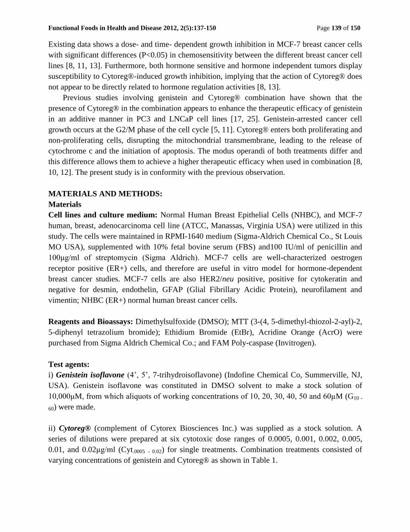
Figure 1. MTT assay was performed to assess the viability and chemosensitivity of normal cells. Cells
were exposed to varying concentrations of genistein (Gn0 – Gn60) for 24 - 48hr, incubated at 37°C, 5%
CO2, and 89% humidity, as previously described. Similar results were observed for Cytoreg® (Cyt0.0005 –
Cyt0.02). Percentage viability between the dosage levels were significantly different (P<0.05). Data are the
mean ± SEM (Standard Error of the Mean) of two independent experiments performed in triplicate. Bar =
SEM.
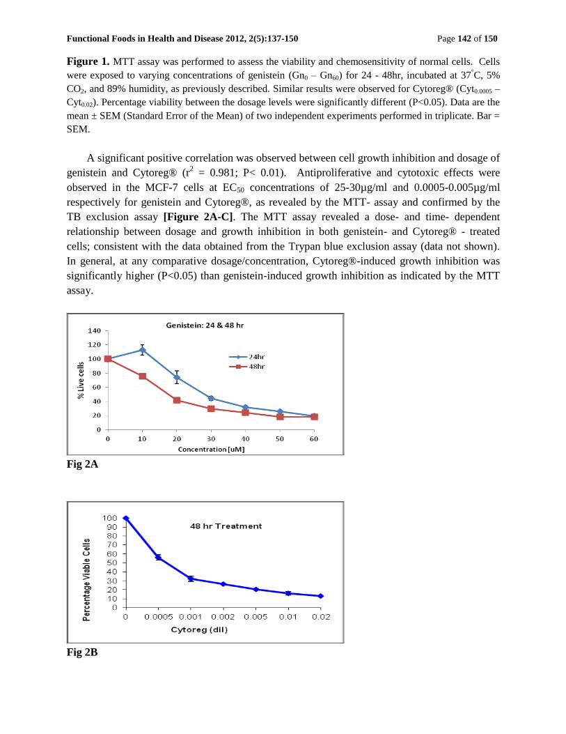
A significant positive correlation was observed between cell growth inhibition and dosage of
genistein and Cytoreg® (r2 = 0.981; P< 0.01). Antiproliferative and cytotoxic effects were
observed in the MCF-7 cells at EC50 concentrations of 25-30µg/ml and 0.0005-0.005µg/ml
respectively for genistein and Cytoreg®, as revealed by the MTT- assay and confirmed by the
TB exclusion assay [Figure 2A-C]. The MTT assay revealed a dose- and time- dependent
relationship between dosage and growth inhibition in both genistein- and Cytoreg® - treated
cells; consistent with the data obtained from the Trypan blue exclusion assay (data not shown).
In general, at any comparative dosage/concentration, Cytoreg®-induced growth inhibition was
significantly higher (P<0.05) than genistein-induced growth inhibition as indicated by the MTT
assay.
Fig 2A
Fig 2B
Functional Foods in Health and Disease 2012, 2(5):137-150 Page 143 of 150
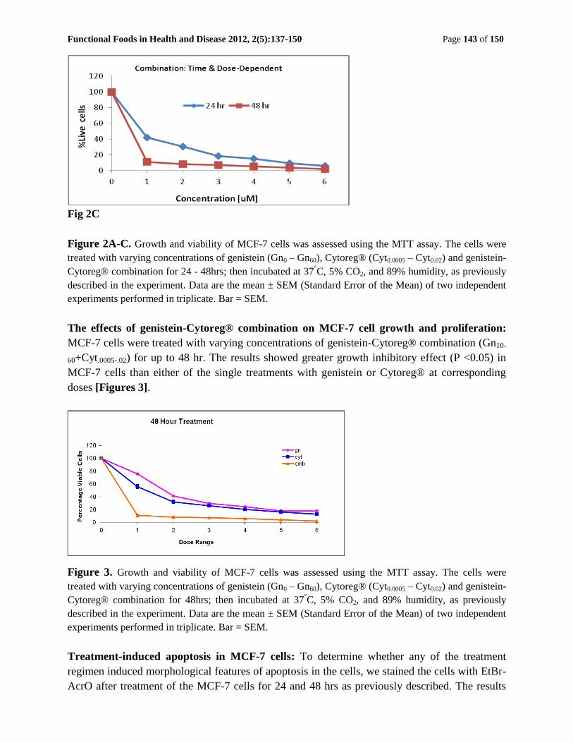
Fig 2C
Figure 2A-C. Growth and viability of MCF-7 cells was assessed using the MTT assay. The cells were
treated with varying concentrations of genistein (Gn0 – Gn60), Cytoreg® (Cyt0.0005 – Cyt0.02) and genistein-
Cytoreg® combination for 24 - 48hrs; then incubated at 37°C, 5% CO2, and 89% humidity, as previously
described in the experiment. Data are the mean ± SEM (Standard Error of the Mean) of two independent
experiments performed in triplicate. Bar = SEM.
The effects of genistein-Cytoreg® combination on MCF-7 cell growth and proliferation:
MCF-7 cells were treated with varying concentrations of genistein-Cytoreg® combination (Gn10-
60+Cyt.0005-.02) for up to 48 hr. The results showed greater growth inhibitory effect (P <0.05) in
MCF-7 cells than either of the single treatments with genistein or Cytoreg® at corresponding
doses [Figures 3].
Figure 3. Growth and viability of MCF-7 cells was assessed using the MTT assay. The cells were
treated with varying concentrations of genistein (Gn0 – Gn60), Cytoreg® (Cyt0.0005 – Cyt0.02) and genistein-
Cytoreg® combination for 48hrs; then incubated at 37°C, 5% CO2, and 89% humidity, as previously
described in the experiment. Data are the mean ± SEM (Standard Error of the Mean) of two independent
experiments performed in triplicate. Bar = SEM.
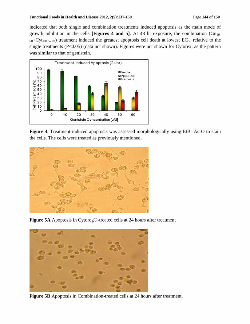
Treatment-induced apoptosis in MCF-7 cells: To determine whether any of the treatment
regimen induced morphological features of apoptosis in the cells, we stained the cells with EtBr-
AcrO after treatment of the MCF-7 cells for 24 and 48 hrs as previously described. The results
Functional Foods in Health and Disease 2012, 2(5):137-150 Page 144 of 150
indicated that both single and combination treatments induced apoptosis as the main mode of
growth inhibition in the cells [Figures 4 and 5]. At 48 hr exposure, the combination (Gn10-
60+Cyt.0005-.02) treatment induced the greatest apoptosis cell death at lowest EC50 relative to the
single treatments (P<0.05) (data not shown). Figures were not shown for Cytorex, as the pattern
was similar to that of genistein.
Figure 4. Treatment-induced apoptosis was assessed morphologically using EtBr-AcrO to stain
the cells. The cells were treated as previously mentioned.
Figure 5A Apoptosis in Cytoreg®-treated cells at 24 hours after treatment
Figure 5B Apoptosis in Combination-treated cells at 24 hours after treatment.
Functional Foods in Health and Disease 2012, 2(5):137-150 Page 145 of 150
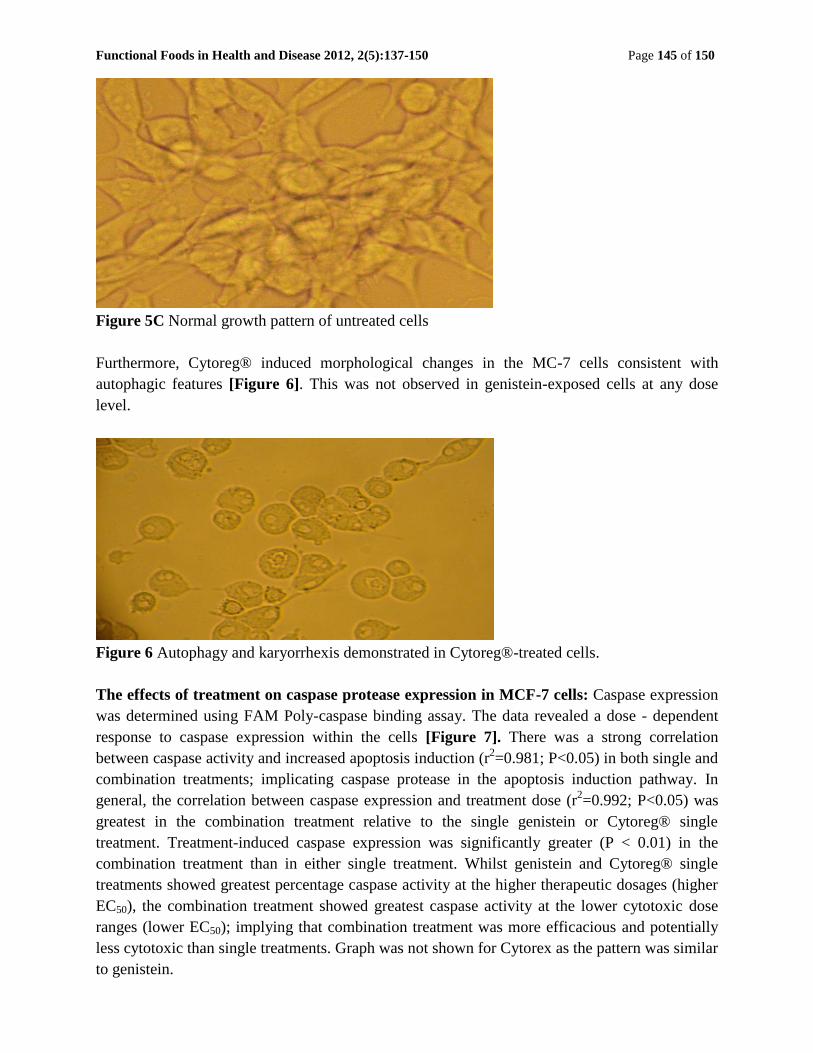
Figure 5C Normal growth pattern of untreated cells
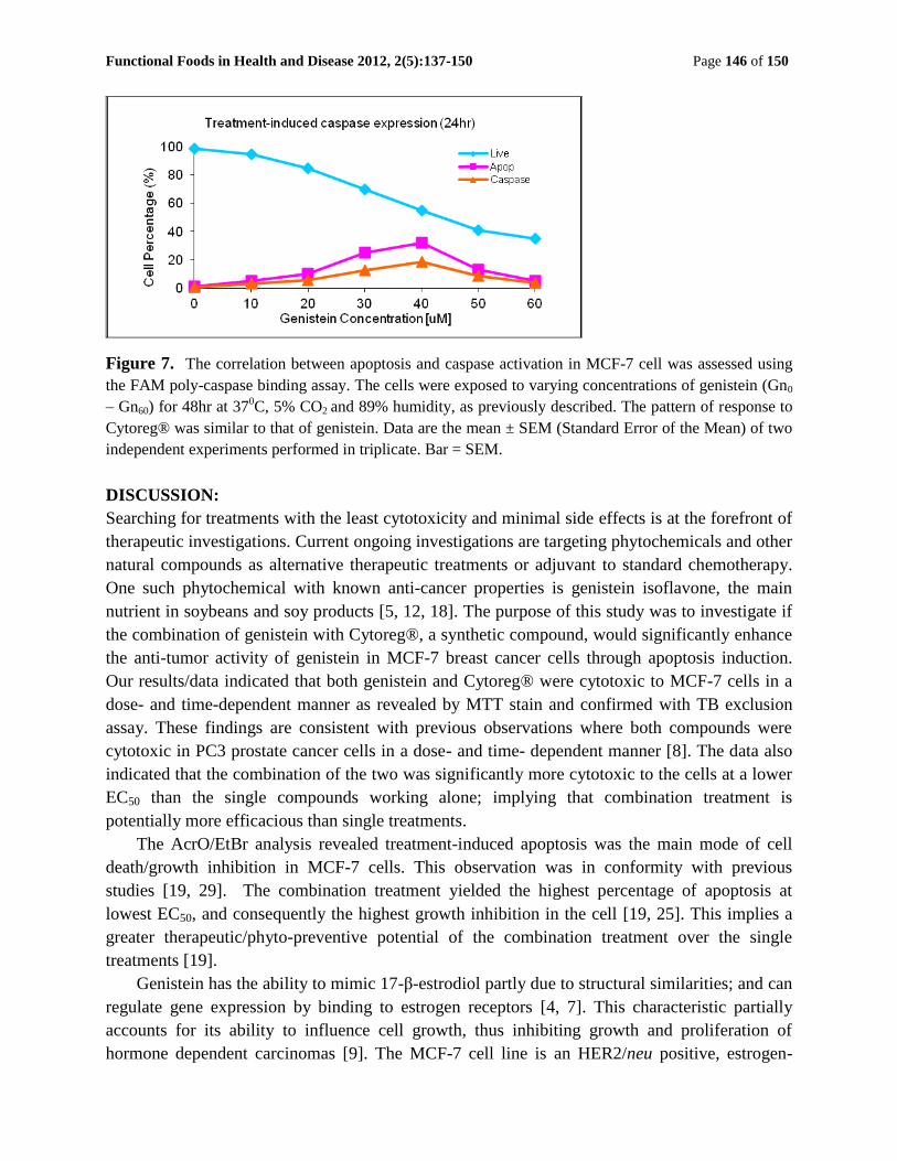
Furthermore, Cytoreg® induced morphological changes in the MC-7 cells consistent with
autophagic features [Figure 6]. This was not observed in genistein-exposed cells at any dose
level.
Figure 6 Autophagy and karyorrhexis demonstrated in Cytoreg®-treated cells.
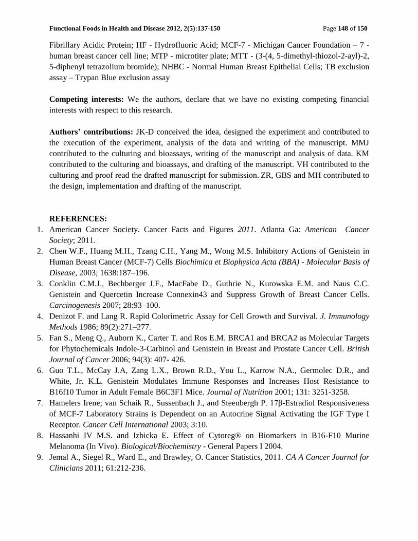
The effects of treatment on caspase protease expression in MCF-7 cells: Caspase expression
was determined using FAM Poly-caspase binding assay. The data revealed a dose - dependent
response to caspase expression within the cells [Figure 7]. There was a strong correlation
between caspase activity and increased apoptosis induction (r2=0.981; P<0.05) in both single and
combination treatments; implicating caspase protease in the apoptosis induction pathway. In
general, the correlation between caspase expression and treatment dose (r2=0.992; P<0.05) was
greatest in the combination treatment relative to the single genistein or Cytoreg® single
treatment. Treatment-induced caspase expression was significantly greater (P < 0.01) in the
combination treatment than in either single treatment. Whilst genistein and Cytoreg® single
treatments showed greatest percentage caspase activity at the higher therapeutic dosages (higher
EC50), the combination treatment showed greatest caspase activity at the lower cytotoxic dose
ranges (lower EC50); implying that combination treatment was more efficacious and potentially
less cytotoxic than single treatments. Graph was not shown for Cytorex as the pattern was similar
to genistein.
Functional Foods in Health and Disease 2012, 2(5):137-150 Page 146 of 150
Figure 7. The correlation between apoptosis and caspase activation in MCF-7 cell was assessed using
the FAM poly-caspase binding assay. The cells were exposed to varying concentrations of genistein (Gn0
– Gn60) for 48hr at 370C, 5% CO2 and 89% humidity, as previously described. The pattern of response to
Cytoreg® was similar to that of genistein. Data are the mean ± SEM (Standard Error of the Mean) of two
independent experiments performed in triplicate. Bar = SEM.
DISCUSSION:
Searching for treatments with the least cytotoxicity and minimal side effects is at the forefront of
therapeutic investigations. Current ongoing investigations are targeting phytochemicals and other
natural compounds as alternative therapeutic treatments or adjuvant to standard chemotherapy.
One such phytochemical with known anti-cancer properties is genistein isoflavone, the main
nutrient in soybeans and soy products [5, 12, 18]. The purpose of this study was to investigate if
the combination of genistein with Cytoreg®, a synthetic compound, would significantly enhance
the anti-tumor activity of genistein in MCF-7 breast cancer cells through apoptosis induction.
Our results/data indicated that both genistein and Cytoreg® were cytotoxic to MCF-7 cells in a
dose- and time-dependent manner as revealed by MTT stain and confirmed with TB exclusion
assay. These findings are consistent with previous observations where both compounds were
cytotoxic in PC3 prostate cancer cells in a dose- and time- dependent manner [8]. The data also
indicated that the combination of the two was significantly more cytotoxic to the cells at a lower
EC50 than the single compounds working alone; implying that combination treatment is
potentially more efficacious than single treatments.
The AcrO/EtBr analysis revealed treatment-induced apoptosis was the main mode of cell
death/growth inhibition in MCF-7 cells. This observation was in conformity with previous
studies [19, 29]. The combination treatment yielded the highest percentage of apoptosis at
lowest EC50, and consequently the highest growth inhibition in the cell [19, 25]. This implies a
greater therapeutic/phyto-preventive potential of the combination treatment over the single
treatments [19].
Genistein has the ability to mimic 17-β-estrodiol partly due to structural similarities; and can
regulate gene expression by binding to estrogen receptors [4, 7]. This characteristic partially
accounts for its ability to influence cell growth, thus inhibiting growth and proliferation of
hormone dependent carcinomas [9]. The MCF-7 cell line is an HER2/neu positive, estrogen-

Functional Foods in Health and Disease 2012, 2(5):137-150 Page 147 of 150
dependent human breast cancer cell line. Genistein demonstrates increased effectiveness against
MCF-7 cells whereby when genistein-induced apoptosis occurs; this triggers a dose-dependent
increase in BRCA1 and BRCA2 protein levels in MCF-7 cells [20]. It appears that the addition
of Cytoreg® to genistein additively enhanced/increased the effectiveness of genistein in the
MCF-7 cells.
Cytoreg® induced other morphologic features/changes in the MCF-7 cells consistent with
autophagy, a form of cellular degradation that leads to lysis of cellular components which in turn
may lead to karyorrhexis [22]. Genistein-treated cells did not show any evidence of autophagy at
any dosage level used in this study. This observation implies that one of the mechanisms by
which Cytoreg®-induced cell death occurs, may be initiation of autophagy, leading to apoptosis
and/or necrosis in the cells. We are hypothesizing that autophagy may release apoptotic inducing
factors/proteins with subsequent induction of apoptosis; bypassing the caspase-protease signaling
pathway. The alternate possibility is that such released factors could cause releasing of caspase
protease cascade, leading to induced apoptosis. In previous studies in our laboratory, blocking of
caspase did not totally abrogate Cytoreg®-induced apoptosis in the cells [8].
In the present investigation, caspase protease activation/expression was the major signaling
pathway in treatment-induced apoptosis in the cells [20]. Previous studies reported caspase 3
expression in genistein- and Cytoreg®- induced apoptosis in PC3 prostate cancer and testicular
cells [23, 24, 25]. Caspase protease activation was confirmed with data obtained from the results.
A strong, positive correlation (r2 = 0.981; P<0.05) between caspase expression and apoptosis
induction was identified. Of the three treatment groups, combination treatment showed the most
significant apoptotic response with respect to caspase activity. The combination treatment group
was more sensitive to caspase expression than either single treatment group. Significant
apoptosis induction was manifested in the former group at lower caspase expression levels.
However, the overall results indicated caspase protease as the major pathway in apoptosis
induction in all treatment groups. These results were in agreement with findings in previous
studies [25, 26].
CONCLUSION: Both genistein and Cytoreg® induced growth inhibition through apoptotic cell
death in MCF-7 cells with evidence of autophagy in Cytoreg® - exposed cells. The apoptotic cell
death was mostly caspase-dependent. The genistein-cytoreg® combination was significantly
more efficacious at lower EC50 (lower cytotoxic level) than either compound alone; implying a
greater therapeutic potential of the combination.
Acknowledgements: The authors acknowledge the technical staff in our research laboratory for
their assistance. Florida Atlantic University is acknowledged for providing the facilities and
partial funding of this project. Rambaugh-Goodwin Cancer Research Institute of Nova
Southeastern University is also acknowledged for allowing the use of its facilities. Cytorex
Biosciences Inc. is highly acknowledged for supplying the synthetic drug - Cytoreg®.
List of Abbreviations: AcrO - Acridine Orange; DMSO – Dimethylsulfoxide; ER- positive –
estrogen-dependent cells; EtBr - Ethidium Bromide; FBS - fetal bovine serum; GFAP - Glial
Functional Foods in Health and Disease 2012, 2(5):137-150 Page 148 of 150
Fibrillary Acidic Protein; HF - Hydrofluoric Acid; MCF-7 - Michigan Cancer Foundation – 7 -
human breast cancer cell line; MTP - microtiter plate; MTT - (3-(4, 5-dimethyl-thiozol-2-ayl)-2,
5-diphenyl tetrazolium bromide); NHBC - Normal Human Breast Epithelial Cells; TB exclusion
assay – Trypan Blue exclusion assay
Competing interests: We the authors, declare that we have no existing competing financial
interests with respect to this research.
Authors’ contributions: JK-D conceived the idea, designed the experiment and contributed to
the execution of the experiment, analysis of the data and writing of the manuscript. MMJ
contributed to the culturing and bioassays, writing of the manuscript and analysis of data. KM
contributed to the culturing and bioassays, and drafting of the manuscript. VH contributed to the
culturing and proof read the drafted manuscript for submission. ZR, GBS and MH contributed to
the design, implementation and drafting of the manuscript.
REFERENCES:
1. American Cancer Society. Cancer Facts and Figures 2011. Atlanta Ga: American Cancer
Society; 2011.
2. Chen W.F., Huang M.H., Tzang C.H., Yang M., Wong M.S. Inhibitory Actions of Genistein in
Human Breast Cancer (MCF-7) Cells Biochimica et Biophysica Acta (BBA) - Molecular Basis of
Disease, 2003; 1638:187–196.
3. Conklin C.M.J., Bechberger J.F., MacFabe D., Guthrie N., Kurowska E.M. and Naus C.C.
Genistein and Quercetin Increase Connexin43 and Suppress Growth of Breast Cancer Cells.
Carcinogenesis 2007; 28:93–100.
4. Denizot F. and Lang R. Rapid Colorimetric Assay for Cell Growth and Survival. J. Immunology
Methods 1986; 89(2):271–277.
5. Fan S., Meng Q., Auborn K., Carter T. and Ros E.M. BRCA1 and BRCA2 as Molecular Targets
for Phytochemicals Indole-3-Carbinol and Genistein in Breast and Prostate Cancer Cell. British
Journal of Cancer 2006; 94(3): 407- 426.
6. Guo T.L., McCay J.A, Zang L.X., Brown R.D., You L., Karrow N.A., Germolec D.R., and
White, Jr. K.L. Genistein Modulates Immune Responses and Increases Host Resistance to
B16f10 Tumor in Adult Female B6C3F1 Mice. Journal of Nutrition 2001; 131: 3251-3258.
7. Hamelers Irene; van Schaik R., Sussenbach J., and Steenbergh P. 17β-Estradiol Responsiveness
of MCF-7 Laboratory Strains is Dependent on an Autocrine Signal Activating the IGF Type I
Receptor. Cancer Cell International 2003; 3:10.
8. Hassanhi IV M.S. and Izbicka E. Effect of Cytoreg® on Biomarkers in B16-F10 Murine
Melanoma (In Vivo). Biological/Biochemistry - General Papers I 2004.
9. Jemal A., Siegel R., Ward E., and Brawley, O. Cancer Statistics, 2011. CA A Cancer Journal for
Clinicians 2011; 61:212-236.

Functional Foods in Health and Disease 2012, 2(5):137-150 Page 149 of 150
10. Khoshyomn S., Nathan D., Manske G.C., Osler T.M., and Penar P.L. Synergistic Effect of
Genistein and BCNU on Growth Inhibition and Cytotoxicity of Glioblastoma cells. Journal of
Neuro-Oncology 2002; 57(3):193-200.
11. Kumi-Diaka J. and Butler A. Caspase-3 protease activation during the process of genistein-
induced apoptosis in TM4 testicular cells. Biology of the Cell 2000; 92:115-124.
12. Kumi-Diaka J., Sanderson N.A., Hall A. The Mediating Role of Caspase-3 Protease in the
Intracellular Mechanism of Genistein-Induced Apoptosis in Human Prostatic Carcinoma Cell
Lines, DU145 and LNCaP. Biology of the Cell 2000; 92(8-9): 595−604.
13. Kumi-Diaka J., Hassanhi M., Brown J., Merchant K., Garcia C., and Jimenez W. Cytoreg™
Inhibits Growth and Proliferation of Human Adenocarcinoma Cells via Induction of Apoptosis.
Journal of Carcinogenesis 2006; 5:1-8.
14. Levine B. Cell Biology: Autophagy and Cancer. Nature 2007; 446:745-747.
15. Linford N.J. and Dorsa D.M. 17 beta-Estradiol and the Phytoestrogen Genistein Attenuate
Neuronal Apoptosis Induced by the Endoplasmic Reticulum Calcium-ATPase Inhibitor
Thapsigargin. Steroids 2002; 67(13-14): 1029–1040.
16. Majno, G. and Jori, I. Review: Apoptosis, Oncosis, and Necrosis. An Overview of Cell Death.
American Journal of Pathology 1995; 146(1): 3-15.
17. Maggiolini M., Vivacqua A., Carpino A., Bonofiglio D., Fasanella G., Salerno M., Picard D.,
and Ando S. The Mutant Androgen Receptor T877A Mediates the Proliferative but not the
Cytotoxic Dose-Dependent Effects of Genistein and Quercetin on Human LNCaP Prostate
Cancer Cells. Molecular Pharmacology 2002; 62(5): 1027–1035.
18. Alshatwi AA, Shafi G, Hasan TN, Al-Hazzani AA, Alsaif MA, Alfawaz MA, Lei KY, and
Munshi A. Apoptosis-mediated inhibition of human breast cancer cell proliferation by lemon
citrus extract. Asian Pac J Cancer Prev. 2011; 12(6): 1555-1559.
19. Nakagawa H., Yamamoto D., Kiyozuka Y., Tsuta K., Uemura Y., Hioki K., Tsutsui Y., and
Tsubura A. Effects of Genistein and Synergistic Action in Combination with Eicosapentaenoic
Acid on the Growth of Breast Cancer Cell Lines. Journal of Cancer Research Clinical Oncology
2000; 126: 448–454.
20. Pei W., Liou A.K.F. and Chen J. Two Caspase-Mediated Apoptotic Pathways Induced by
Rotenone Toxicity in Cortical Neuronal Cells. FASEB Journal 2003; 17:520-522.
21. Porter A.G. and Janicke R.U. Emerging Roles of Caspase-3 in Apoptosis. Cell Death Differ
1999; 6:99 –104.
22. Ramanakumar, A.V. Need for Epidemiological Evidence from the Developing World to know
the Cancer-Related Risk Factors. Journal of Cancer Research and Therapeutics 2007; 3(1): 29-
33.
23. Riccardi C. and Ildo N. Analysis of Apoptosis by Propidium Iodide Staining and Flow
Cytometry. Nature Protocols 2006; 1:1458-1461.
24. Ruddon, R.W. Cancer Biology. New York: Oxford University Press, Inc. (1995).
25. Sakamoto, K. Synergistic Effects of Thearubigin and Genistein on Human Prostate Tumor Cell
(PC-3) Growth via Cell Cycle Arrest. Cancer Letters 2000; 151(7): 103-109.

Functional Foods in Health and Disease 2012, 2(5):137-150 Page 150 of 150
26. Vicencio J.M., Galluzzi L., Tajeddine N., Ortiz C., Criollo A., Tasdemir E., Morselli E., Younes
A.B., Maiuri M.C., Lavandero S., and Kroemer G. Senescence, Apoptosis or Autophagy? When
a Damaged Cell Must Decide Its Path – A Mini-Review. Gerontology 2008; 54: 92-99.
27. Villalobos A. Cytoreg Recibió Patente de Invención. Medicamento Venezolano Contra el
Cáncer. Diario El Carabobeño 2005.
28. Yang S., Zhou Q. and Yang X. Caspase-3 Status is a Determinant of the Differential Responses
to Genistein Between MDA-MB-231 and MCF-7 Breast Cancer Cells. Biochem Biophys Acta
2007; 1773: 903–911.
29. Ariffin, S., Omar, W., Ariffin, Z., Safian, M., Senafi, S., and Wahab, R. Intrinsic
Anticarcinogenic Effects of Piper sarmentosum Ethanolic Extract on a Human Hepatoma Cell
Line. Cancer Cell International 2009; 9:6.
30. McPherson, K. ABC of breast diseases: Breast cancer epidemiology, risk factors, and genetics.
BMJ 2000; 321:1198.3.
31. Mannick, J.B., Miao, X. Q., and Stamler, J.S. Communication: Nitric Oxide Inhibits Fas-
induced. Apoptosis. The Journal of Biological Chemistry1997; 272(39):24125-24128.