thesis - surgery.uonbi.ac.ke ent.pdf · fuad farah maalim h58/71002/2009 masters in medicine in ent...
TRANSCRIPT

THESIS
Anatomic variations of paranasal sinuses in patients undergoing CT scan
evaluation at the Kenyatta National Hospital
PRINCIPAL INVESTIGATOR
DR. FUAD FARAH MAALIM
H58/71002/2009
MASTERS IN MEDICINE IN ENT HEAD AND NECK SURGERY
A THESIS FOR DESERTATIONAS PARTIAL FULFILLMENT OF THE
REQUIREMENTS BY THE UNIVERSITY OF NAIROBI FOR THE AWARD OF
THE DEGREE OF MASTERS IN MEDICINE IN ENT HEAD AND NECK
SURGERY.
DECLARATION
This is my original work which has not been presented for a degree award at any other
university.
Signed Date .
Dr Fuad Farah.
This thesis is supervised by:
Prof. H.O Oburra
Professor
Department of Surgery. ENT Head and Neck Surgery.
University of Nairobi.
Signed Date .
Dr Jane Thinwa.
Consultant.
Department of Radiology. Kenyatta National Hospital.
Signed Date .
Dr John Ayugi.
Lecturer and consultant.
Department of Surgery. ENT Head and Neck Surgery
University of Nairobi.
Signed Date .

1.0 ACRONYMS AND ABBREVIATIONS:
CT Scan : Computed Tomographic Scan.
ENT HN: Ear, Nose, Throat, Head and Neck Surgery.
FESS : Functional Endoscopic Sinus Surgery.
KNH : Kenyatta National hospital.
PNS : Paranasal sinus.
2.0 ABSTRACT
Objective: To determine radiologic variants of paranasal air sinuses among patients
undergoing CT scan evaluation at KNH. To determine demographic characteristics of patients
requiring paranasal CT scanning at KNH.
Study Design: Prospective cross sectional study
Methods: This is a prospective cross sectional study that was conducted at the Department of
Radiology in Kenyatta National Hospital comprising of 100 patients’ computed tomography
scans of paranasal sinuses. The machine that was used is Phillips Brilliance CT scanner, 16
slice. Model 45356702331, serial number 729. CT scans were taken in axial plane and then
three millimeter slices reconfigured into coronal and sagittal planes. Anatomical variants
assessed are agger nasi, haller cells, onodi cells, concha bullosa, and deviated nasal septum.
Other uncommon variants were also detected.
Study duration: From October 2013 and completion will be in October 2014.
Data Management and Analysis: The data collected was transferred into Microsoft Access
database and then analyzed using STATA version 13 (Stata Corp, College Station, Texas)
statistical software. Descriptive analysis was used to determine the means, frequencies and
proportions of the various anatomical variations. The proportions of the various anatomical
variations further have the 95% CI. Results are presented in the form of tables and graphs
together with their descriptions.
Results: Sample size of 100 patients, 56 females and 44 males. Of the common variants,
91(91%) patients have agger nasi cells, 13(13%) patients have haller cells, 44(44%) patients
have concha bullosa, 41(41%) patients have onodi cells while 16(16%) have septal nasal
deviation.
Of the uncommon variants, maxillary sinus hypoplasia was identified in 1(1%) patient.
Superior concha bullosa was identified in 4(4%) patients. Inferior concha bullosa was identified
in 1(1%) patient. Bilateral paradoxical middle turbinate was present in 5(5%) patients.
Paradoxical superior turbinate was present in the right in 1(1%) patient. Septate sphenoid was
present in 14(14%) patients.
Keros type 1 in 25(25%) patients; keros type 2 in 67(67%) patients, keros type 3 in 7(7%)
patients and keros type 4 in 1(1%) patient.
Conclusion: anatomical variations are best viewed and appreciated in the coronal plane. The
Keros classification of olfactory fossa depth is best calculated in coronal plane. Both sagittal
and axial planes don’t give adequate information with regards to the anatomy of paranasal
sinuses and the presence of anatomical variations.
Gender had significant correlation (Pearson chi-square p-value=0.043) with Onodi cell, which
implied that the variant was mainly dominant on males as compared to females.

3.0 INTRODUCTION
Evaluation of anatomic variations of the paranasal air sinuses is important in patients who are
undergoing CT scan evaluation for various rhinologic reasons. Knowledge of the anatomic
variations does reduces the surgical complication rates during FESS, helps explain recurrence
of disease and allows one to change the operative technique.
The ethmoid air sinuses play a crucial role in FESS. The posterior ethmoids and the sphenoid
sinuses can be accessed via the anterior ethmoid cells. Anterior ethmoidectomy helps improve
frontal air sinus drainage hence minimizing the need for opening the frontal sinus during
surgery.
The anatomic variations of paranasal sinus make the approach toFESS much more
complex.1Some anatomic variants are probably not responsible for development of chronic
rhinosinusitis, but knowledge of their presence is paramount. Variations in the pneumatization
of ethmoid sinuses may disturb sinus ventilation.2,3 These can be the etiological factor for
sinusitis and spread of infection to adjacent structures.
During FESS, straying beyond the surgical field may lead to serious complications such as
cerebrospinal fluid leak, meningitis, or blindness, so a detailed knowledge of the possible
anatomical variations is essential.
Computer Tomography scan is a mandatory radiological investigation for patients undergoing
FESS. CT scan helps identify these anatomic variations. Many centers use the three millimeter
cuts for all views – coronal, sagittal and axial to assess the different anatomical structures of
the lateral nasal wall and the paranasal sinuses. The coronal views are best for the sphenoid
and the ethmoid cell variants such as the onodi or sphenoethmoidal cell.4
The development and refinement of CT scans has allowed extensive assessment of patients’
paranasal sinuses thus providing a guide map for FESS surgeons to operate.5
4.0 BACKGROUND
4.1 ANATOMY
Understanding the embryology and gross anatomy of the paranasal sinuses is of paramount
importance in the surgical aspect due to its close proximity to vital structures such as the orbit
and optic nerve.
Embryology
The frontal, maxillary and ethmoid sinuses arise from evaginations of the lateral nasal wall,
whereas the sphenoid sinuses arise from a posterior evagination of the nasal capsule.
Embryological development of ethmoid sinus begins during the fifth month of intrauterine life
as numerous evaginations from the fetal nasal chamber into the anlage of the ethmoid bone. At
first this evaginations are mere slits, but they quickly grow into a tubular form before assuming
a round or globular shape at term. The expansion of each air cell with rapid growth of the cells
is at the expense of the ethmoid bone. The haphazard growth of cells makes each persons

ethmoid labyrinth different from the next. The expansion of these cells continues until late
puberty or until the wall of the sinus strikes a layer of compact bone or another sinus. By
adulthood, the ethmoid block averages 3.3 by 2.7 by 1.4 cm in size, with the longest dimension
being the anteroposterior. The ethmoids complete pneumatization by the age of twelve years.
Frontal sinus are either absent or insignificant at birth, but gradually increase in size and reach
maximum dimension at around age twenty years.6 Further pneumatization due to atrophic
changes may occur in old age.7 Structural changes can only be caused by altercation due to
tumours, fractures or severe infections.8The two develop independently hence often
asymmetry.
Maxillary sinus development begins at tenth week gestation as some mucosal invagination of
primitive ethmoid infundibulum. Shaping of maxillary sinus starts by fusion of multiple
invaginations into a single cavity from week eleven. Growth is progressive in all dimensions
more so anteroposteriorly, similar to the cranial elongation.9It undergoes two periods of rapid
growth, between birth and age of three years, also between ages seven and eighteen years. A
poorly developed infundibular passage is associated with hypoplasia.10
Sphenoid sinus develops from evagination of sphenoethmoid recess at birth. Pneumatization
begins age three and reaches sellar by age seven and proceeds rapidly after. Development
continues until age twelve to fifteen years.
Gross Anatomy:
The ethmoid bone is a light, papyraceous, osseous structure attached to each edge of the
cribriform plate as a block of ethmoid sinuses. Each block is made of a group of three to fifteen
air cells. The ethmoidal cell block or labyrinth is pyramidal in shape, being wider posteriorly
(where it abuts the sphenoid bone) than anteriorly (where it contacts the lacrimal bone). The
ostia of the ethmoidal sinuses are the smallest found in any of the paranasal sinuses, measuring
one to two millimeters in diameter.To understand the anatomic relations of the ethmoid
labyrinth its best to consider it as a box.The roof of the box is the top wall or roof of the
ethmoidal air cell box and is termed the fovea ethmoidalis. The fovea is not a flat plate of bone
but undulating, because the domes of the topmost ethmoidal cells bulge into it and mold it into
such a contour. The anterior part of the fovea lies more superior to the posterior part, since the
anterior cranial fossa descends approximately fifteen degrees from a horizontal plane as it
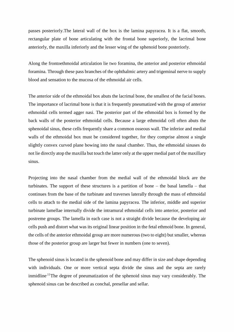
passes posteriorly.The lateral wall of the box is the lamina papyracea. It is a flat, smooth,
rectangular plate of bone articulating with the frontal bone superiorly, the lacrimal bone
anteriorly, the maxilla inferiorly and the lesser wing of the sphenoid bone posteriorly.
Along the frontoethmoidal articulation lie two foramina, the anterior and posterior ethmoidal
foramina. Through these pass branches of the ophthalmic artery and trigeminal nerve to supply
blood and sensation to the mucosa of the ethmoidal air cells.
The anterior side of the ethmoidal box abuts the lacrimal bone, the smallest of the facial bones.
The importance of lacrimal bone is that it is frequently pneumatized with the group of anterior
ethmoidal cells termed agger nasi. The posterior part of the ethmoidal box is formed by the
back walls of the posterior ethmoidal cells. Because a large ethmoidal cell often abuts the
sphenoidal sinus, these cells frequently share a common osseous wall. The inferior and medial
walls of the ethmoidal box must be considered together, for they comprise almost a single
slightly convex curved plane bowing into the nasal chamber. Thus, the ethmoidal sinuses do
not lie directly atop the maxilla but touch the latter only at the upper medial part of the maxillary
sinus.
Projecting into the nasal chamber from the medial wall of the ethmoidal block are the
turbinates. The support of these structures is a partition of bone – the basal lamella – that
continues from the base of the turbinate and traverses laterally through the mass of ethmoidal
cells to attach to the medial side of the lamina papyracea. The inferior, middle and superior
turbinate lamellae internally divide the intramural ethmoidal cells into anterior, posterior and
postreme groups. The lamella in each case is not a straight divide because the developing air
cells push and distort what was its original linear position in the fetal ethmoid bone. In general,
the cells of the anterior ethmoidal group are more numerous (two to eight) but smaller, whereas
those of the posterior group are larger but fewer in numbers (one to seven).
The sphenoid sinus is located in the sphenoid bone and may differ in size and shape depending
with individuals. One or more vertical septa divide the sinus and the septa are rarely
inmidline.11The degree of pneumatization of the sphenoid sinus may vary considerably. The
sphenoid sinus can be described as conchal, presellar and sellar.
Radiological anatomy:
CT scan is the investigation of choice for paranasal sinuses. Modern multi slice CT scanner
allow thin axial plane slices to be obtained, from which reconstruction to sagittal and coronal
planes can be made. The coronal plane best displays the osteomeatal complex. The axial plane
helps identify the basal lamella of middle turbinate which is the dividing point of anterior and
posterior ethmoid sinuses.4
Ethmoid air sinuses are the most developed paranasal sinus at birth but are not evident on X-
ray until the age of 1year. Anatomical boundaries of anterior ethmoid cells are: anteriorly, the
ethmoid infundibulum; posteriorly, the basal lamella; laterally, the lamina papyracea;
superiorly, the skull base. Anatomical boundaries of posterior ethmoid cells are: anteriorly, the
basal lamella; posteriorly, the anterior sphenoid wall; medially, the superior turbinate; laterally,
the lamina papyracea; superiorly, the skull base.4
Agger nasi is the most anterior ethmoidal air cells. It forms the anterior inferior border of frontal
recess. Its degree of pneumatization has significant effect on frontal sinus drainage and surgical
access. Frontal recess may be displaced medially and posteriorly, depending on the size of
agger nasi cell. Onodi cell is assessed by comparing both coronal and sagittal cuts carefully.
They pneumatize superolateral to the sphenoid sinus: hence surgical clearance should be in the
inferomedial direction, due to optic nerve and internal carotid relations.
Sphenoid sinus’ most reliable landmark is the ostium which lies approx 1cm superior to the
posterior inferior end of superior turbinate and approx 1.5cm superior to the choana. CT scan
evaluation focuses on pneumatization and the relation to both internal carotid artery and optic
nerve.
The boundaries of the frontal sinus tract/ recess are: anteriorly, the aggernasi cell; posteriorly,
the ethmoid bulla and skull base; laterally, the lamina papyracea and medially, the middle
turbinate.
Anatomic Variants:

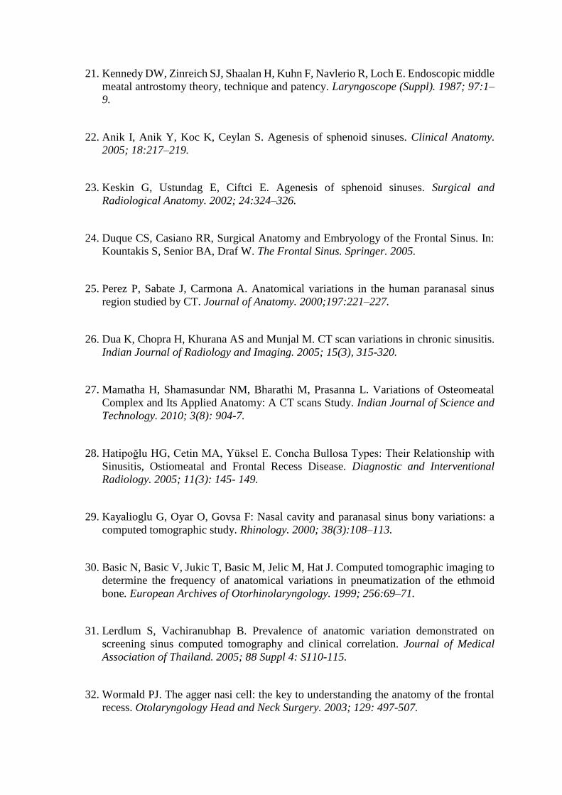
Concha bullosa: – it is pneumatization of the concha. The middle concha is the most
commonly involved. Bolger et al classified concha bullosa pneumatization as per the location
into lamellar, bulbous and extensive. The extensive type is the combination of both the lamellar
and bulbous type. Pneumatization of the middle concha is the extension of normal
pneumatization of the ethmoid cells.12This is usually by the anterior ethmoid cells but the
posterior ethmoid cells have also been documented or by both groups. Concha bullosa is
implicated in the pathogenesis of rhinosinusitis because of its tendency to narrow the middle
meatus and the infundibulum.Some studies have found the prevalence of concha bullosa to be
the same in patients with and without sinus disease symptoms.1
Superior Concha bullosa: - pneumatisation of superior turbinate is extremely rare condition.
Inferior concha bullosa: - pneumatization of the inferior turbinate as well is very rare.13
Keros classification: the length of the lateral lamella of the cribriform plate, defining the type
of olfactory fossa. In most of the cases, the cribriform plate is symmetrical; however, an
asymmetric skull base is an anatomic variant.44
Type I: cribriform plate 1-3 mm below fovea ethmoidalis.
Type II: 4-7 mm below.
Type III: 8-16 mm below.
Nasal septal deviation: - asymmetric nasal septum position which can put force on nasal
turbinates.
Haller cells: – pneumatization of ethmoid cells extends into anterior roof of maxillary sinus,
along the floor of the orbit. They are an etiological factor for sinusitis since they vary in size
and when large can narrow the ostium of the maxillary sinus or the ethmoid infundibulum. The
presence of an infraorbital cell may increase the risk of injury to the orbit during FESS, an
unrecognized cell masking the position of the orbital wall.14
Bolger et al defined a haller cell as any cell located beneath ethmoid bulla, lamina papyracea
or orbital floor.1
Agger nasi cells: – most anterior ethmoid air cell located below the frontal sinus and are
intimately related to the frontonasal recess, reaching the lacrimal fossa inferolaterally. They
are anterosuperior to the hiatus semilunaris. The agger nasi cell may form a significant part of
the anterior wall of the frontal recess and impinge upon the frontal sinus drainage tract.
Recognition of this relationship is crucial in management of chronic frontal sinusitis.15
Onodi cells: – posterior ethmoid cells that extend posterior, lateral and always superior to
sphenoid sinus, lying medial to the optic nerve.First described by Adolf Onodi in 1903.
Extensive pneumatization can expose the circumference of optic nerve, which is surrounded
by air spaces, mainly the inferior half.16Onodi cells are also known as sphenoethmoid cells and
are important to the surgeon in FESS. When present, both optic nerve and carotid artery may
be exposed in the posterior ethmoid cell.17The best orientation is on axial cuts where the course
of optic nerve can be followed as a relation.18
Uncinate bulla:- pneumatization of the uncinate process and is a rare entity. Uncinate process
projects from the ethmoid bone to the ethmoid process of the inferior nasal concha.
Pneumatization can result in narrowing of infundibulum and sinus drainage occlusion.20
Maxillary sinus hypoplasia:-uncommon condition that is misdiagnosed as chronic
sinusitis.19In its severe form, it impedes mucociliary clearance and is liable to retention of
mucus. Failure to recognize maxillary sinus hypoplasia intraoperation can lead to injury to the
medial orbital wall.20Finding the maxillary sinus ostium may be difficult.21
Classification of maxillary sinus hypoplasia:
Type 1. Mild sinus hypoplasia:
Normally developed uncinate process.
Well-developed infundibular passage.
Varying degree of mucosal thickening of affected sinus.
Type 2. Significant sinus hypoplasia:
Hypoplastic or absent uncinate process.
Ill-defined or absent infundibular passage.
Total opacification of affected sinus.
Type 3. Profound sinus hypoplasia:
Sinus represented by shallow cleft in the lateral nasal wall of the nose.
Absent uncinate process.
Sphenoid sinus hypoplasia:-defined as an oval-shaped sinus with pneumatization limited to
the presphenoid that is anterior to the vertical plane of the tuberculum sellar. It is an uncommon

condition. Few cases of agenesis have been reported.22Mainly seen in patients with craniofacial
anomalies and skeletal diseases.23
Sphenoid sinus pneumatization:-can be divided intoconchal, presellar or postsellar. Conchal:
the area below the sellar is a solid block of bone without pneumatization. Presellar: the
sphenoid is pneumatized to the level of the frontal plane of the sellar and not beyond. Sellar:
the most common type, where pneumatization extends into the body of the sphenoid beyond
the floor of sellar and reaches clivus.
Frontal sinus hypoplasia:-it is uncommon to find one dominant, one hypoplastic or both
hypoplastic frontal sinus. Rarely both aplastic frontal sinuses.24
5.0 LITERATURE REVIEW
Paranasal sinus anatomy is variable from individual to individual and so is the incidence of the
anatomical variations. Different studies around the globe demonstrate different percentages for
the anatomic variations. As for CT scan, it being the imaging modality of choice, most authors
use three millimeter coronal plane cuts as it gives similarity with surgical orientation. Axial
plane cuts are used for a clearer, exact diagnosis when coronal plane is not clear.
Stammberger and Hawke have shown that CT scan of PNS provides an anatomic road map to
identify presence of abnormalities, the location and severity of disease and exact location of
obstruction.19CT scan in a coronal plane is more informative than on an axial plane as indicated
by Perez P et al. This shows anatomical structures progressively as a surgeon’s visualization
and the relationships of sinus cavities and adjacent structures. Axial cuts are also imperative,
since they give anatomical relations of the paranasal sinuses.25Perez had a sample size of one
hundred patients of whom nearly all had agger nasi cells while 73% had concha bullosa and
3% had haller cells.
Removal of disease in the osteomeatal complex region is the basic principle of FESS, which is
best appreciated on CT scan as documented by Dua et al26whose sample size was fifty patients.
Fourty percent had agger nasi cell while both haller cells and concha bullosa had 16% each.
Mamatha H did re-emphasize the concept that osteomeatal complex is the key factor in the
causation of chronic sinusitis.This study also gave justification as to why the coronal plane is
preferred, since it displays the osteomeatal complex best.27Mamatha had a sample size of forty
patients and nearly half of them had agger nasi cells while 17.5% had haller cells and 15% had
concha bullosa. In a study by Hatipoglu, he looks at the statistical relationship between concha
bullosa type and osteomeatal disease, hence concluded that pneumatization of the inferior part
of the middle concha plays a major role in osteomeatal disease development.28
A comparison of anatomic variants in patients with sinus disease and patients without was
made by Kayalioglu. Clearly anatomical variants were common in patients with sinus
disease.29His sample size was ninety sinus patients and eighty two non sinus patients. Concha
bullosa which was close was seen 28.8% in sinus patients and 26.8% in non sinus patients.
Basic who did a CT scan study to determine frequency of anatomic variations in mainly
ethmoid pneumatization and did indicate it is imperative to adopt standardized classification
and definition of paranasal sinus variations. This would avoid discrepancies amongst various
authors.30A study by Arslan H et al had a large sample size of two hundred patients and looked
into anatomical variants of the paranasal sinus on two millimeter CT scan cuts where he found
that 30% had concha bullosa while onodi cells at 12% and haller cells were 6%.14
A comparison of prevalence of anatomic variation on CT scan and clinical findings was made
by Lerdlum, who observed that anatomic variation can compromise mucociliary drainage.
However, only large agger nasi had significant correlation to inflammatory sinus disease.31 A
study on agger nasi cell, the key to understanding anatomy of the frontal recess was done by
Wormald The anatomy and variations in the area are poorly understood by FESS surgeons.
Reconstructed CT scan cuts of PNS, mainly coronal and sagittal aid in identification of each
cell and assist the surgeon to formulate a clear, precise surgical plan.32
Onodi cell is an obstacle to sellar lesions with a transphenoidal surgical approach. Sinusitis in
the sphenoid region can present with visual symptoms due to its close relations to the optic
nerve. Intraoperatively, the Onodi cell can be easily mistaken for sphenoid sinus, hence
incomplete procedures sometimes. Risk of injury to both the optic nerve and internal carotid
artery are high as seen in the study by Ji-hyeon Shin.17 A CT scan study done on patients
requiring revision FESS by Bradley et al found agger nasi cell present in 93%, mainly the
patients with frontal rhinosinusitis. Frontal sinus disease correlates with severity of overall
sinus disease.33
A comparison study of anatomic variations of lateral nasal wall of patients undergoing FESS
was done by Badia L et al . Caucasians and Chinese were compared. Concha bullosa incidence
was higher in the Caucasian population, though incidence of Onodi cells was much greater in
Chinese population. There was no difference in presence of agger nasi cells.34 A study on
anatomic variations of lateral nasal wall in Thai patients undergoing FESS was done by
Nitinavakarn B et al and the most common site of sinus infection and inflammation was the
anterior ethmoid sinuses. Thai FESS surgeons should be informed about the variants, which
might be different if compared to western incidence.35 Vincent T et al did a study on association
of concha bullosa and deviated nasal septum with chronic rhinosinusitis in FESS patients. The
two anatomic variants and agger nasi are very common. In Malaysia, concha bullosa was
statistically more common among females and the Indian and Chinese ethnic groups.36A study

done by Baradaranfar et al looked into the frequency of anatomical variations in patients with
chronic rhinosinusitis who underwent sinus surgery. A total of 120 patients and he found agger
nasi in 36%, concha bullosa in 12.5%, septal deviation in 45% and haller cells in 4%.37
Maxillary sinus hypoplasia has been reported as being very uncommon and mainly noticed on
the coronal cuts of the CT scan. Bolger et al reported prevalence of unilateral hypoplastic
maxillary sinus to be 10.4% while Kantarci et al reported 7% in a study of 512 patients.38It is
imperative to look for other anomalies on the lateral nasal wall, especially the uncinate process
which can impede mucociliary clearance of the sinuses. Secondary middle turbinate is rare
anomaly characterized by a bony projection covered by soft tissue, arising from the lateral wall
of the middle meatus, studies done by Khanobthamchai et al put the incidence at 1.5%,39 while
Aykut et al found it to be 6.8%40 and Aksungur et al at 0.8%.41
A retrospective study on sphenoid sinus hyperplasia and agenesis was done by Binal C et al
who looked into 384 patients and found unilateral agenesis in 0.26%, unilateral sphenoid sinus
hypoplasia in 0.26% and bilateral sphenoid sinus hypoplasia in 0.26%. Bilateral sphenoid sinus
agenesis was not seen.42
6.0 STUDY JUSTIFICATION
This study offers local statistics on paranasal sinus anatomy and its variants. It acts as a baseline
research for anyone who wishes to carry further studies on particular paranasal sinuses. This
data will help the otorhinolaryngologist to build a complication free learning curve, assist in
local training and also emphasize on the need for radiological evaluation. This study helps in
standardization of the cuts size and view to request for in terms of paranasal sinus CT scans.
Clear information on trends of variation among the local population can be obtained in the
study. Paranasal air sinuses are important in FESS and knowledge of its anatomical variants
helps a surgeon in his/ her orientation during FESS.
7.0 AIMS AND OBJECTIVES
To determine the incidence of paranasal air sinus variations in patients undergoing CT scan
evaluation at Kenyatta National Hospital.
Specific Objectives
1. To determine demographic characteristics of patients requiring paranasal CT scanning
at KNH.
2. To determine radiologic variants of paranasal air sinuses among patients undergoing CT
scan evaluation at KNH.
8.0 STUDY DESIGN
Prospective cross sectional study.
8.1 Sample size
We want to estimate the proportion of population with anatomic variations of paranasal sinuses
in patients undergoing CT scan evaluation
The incidence of paranasal air sinus variations in patients undergoing CT scan is varying for
the different parameters of our interest i.e. Agger nasi cells (50%), Haller cells (17.5%) and

Concha bullosa (15%) and further there are no documentation/ publication in Africa/ Kenya –
the proportion of anatomic variations
For the cross sectional study and since we do not know the estimated population proportion or
prevalence we use p=0.05
We use a margin of error of 10% to allow us estimate the sample size that fits our estimated
resources
Based on the 95% confidence interval, a precision (margin of error) 10%d and that the
estimated population prevalence proportion is unknown we use p=0.50
Significance level 0.05 implying 1 1.96Z i.e. 95 % confidence level
The following Sample size determination formula for incidence studies (Lwanga SK &
Lameshow S)43was used to estimate the proportion of population study size.
22
1
2
(1 )Z p pn
d
2
2
1.96 0.50 (0.50)96
0.1n
The estimated sample size of 96 will be expanded to 106 to allow for 10% possible non
respondent / missing data.
8.2 Sampling method
Consecutive sampling
8.3 Study setting
Kenyatta National Hospital, Department of Radiology
8.4 Study Period
October, 2013 –November, 2014.
8.5 Inclusion Criteria
Patients above fifteen years age. Paranasal sinuses are developed.

Patients who consent to be included in the study.
CT scans of three millimeter cuts.
8.6Exclusion Criteria
Patients with history of previous nasal surgery or trauma.
Patients who do not consent to be included in the study.
Patients below the age of fifteen years.
8.7Confounding Factors
Presence of nasal polyposis may hinder visualization of some of the anatomic variations of
ethmoid sinuses.
8.8Clinical Evaluation
CT scans of the paranasal sinuses were taken in axial position. 3mm reconstruction cuts to
sagittal and coronal view were made of patients in the study.
8.9 Data Management and Analysis
Data Management
The collected data in the questionnaires was entered into Microsoft office access 2007 database
and then transferred to STATA version 13 (Stata Corp, College Station, Texas) for cleaning,
validation, coding and analysis. It was be checked for any wrong entry and double entry and
corrected. Back up was created in an external hard disk in case of damage and/or loss of original
data and then password protected. All data was stored under lock and key and with password
protected files under the custody of the principal investigator to prevent any illicit access to the
data. Use of coded data was done to ensure maximum confidentiality. At the end of the study,
the raw data was stored in one soft copy storage device.
Data Analysis
Data analysis was done using the STATA version 13 (Stata Corp, College Station, Texas)
statistical software. Descriptive analysis was used to determine the mean, frequency and
proportion of variables describing variations. 95% Confidence Interval for the proportions was

presented. Results was presented as table and graphs together with brief descriptions.
Confidence level was taken as 95% (p <0.05) where applicable.
9.0 MATERIALS AND EQUIPMENT
Materials and equipment that was used include:-
1. Phillips Brilliance CT scanner, 16 slice. Model 45356702331, serial number 729.
Manufactured by Phillips Medical Systems, Cleveland USA in 2007.
2. Compact Disks for storing soft copy of the patient’s paranasal CT scans.
Procedure
The study was mainly based at the department of radiology in Kenyatta National Hospital. It
is an outpatient setting and patients come for the procedure which does not take a long time. It
takes half an hour to do CT scanning. After the procedure, it takes time for the radiologist to
review and report on the various findings on the CT scan, hence patients are requested to either
wait or come back for the CT scan films and report on the following day. No extra time of the
patients was required to conduct this study.
The targeted patients in this study were the ones who were requested to undergo paranasal sinus
CT scan for various rhinologic reasons mainly from different clinics in Kenyatta National
Hospital. Hence patient recruitment was at the Radiology Department in Kenyatta National
Hospital. The principal investigator had to explain to the patients the study in order for them to
agree to consent to be included in the study. Patients incur the cost of doing the paranasal CT
scan, but no extra cost was incurred by the patients in regard to this particular study. Patients
were expected to give a short bio data on their ages and home. This took place prior to the
patient undergoing the CT scanning.
The CT scan machine at Kenyatta National Hospital takes axial cuts initially in order to save
hospital time for patients and also be able to have a high turnover with regards to the workload.
The reconfiguration into both coronal and sagittal planes was done to the specified three
millimeter cuts by the machine. The machine is capable of producing whichever requested
information in terms of size and planes. The radiologist took time to look at the scans in
different views and cuts in order to diagnose and report on the pathology and disease process
that is deduced from the images. The films produced eventually give a picture that helps best
to display the pathological findings.
After the CT scanning, the primary investigator accessed the films with the radiologist
supervisor and filled in the anatomical variants visualized on a check list with regards to the
plane and side, whether left or right or bilateral. The information and data was stored at the
radiology department. The primary investigator stored a soft copy of all the CT scan films of
the patients recruited in the study and that extra cost was not incurred neither by the patients
nor the Department of Radiology. The data collected from this study will be used by the
Departments of Radiology and Ear, Nose and Throat Surgery in conjunction with Kenyatta
National Hospital as a benchmark and reference for other related studies in the future.
Quality Control
1. The primary investigator did the enlisting of patients undergoing CT scan evaluation.
2. The primary investigator with one supervisor radiologist reviewed the scans and the
variations noted down in the check list.
3. The CT scans was standardized in size (3mm cuts) and all views requested.

10.0 ETHICAL CONSIDERATION
1. The study was carried out only after approval by Kenyatta National Hospital and
University of Nairobi Ethics and Research committees.
2. Those included in the study were required to give an informed consent either by
themselves or guardians in cases below 18 years.
3. Patients incurred no extra financial costs and their confidentiality was maintained.
4. There was no monetary gain by the primary investigator from this study.
5. There was no penalty for declining to participate in the study.
6. The study results shall be published in reputable journals and periodical publications
for the benefit of the medical fraternity, the study subjects and general population.
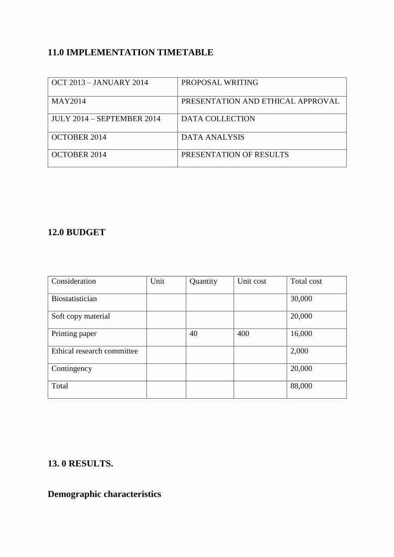
11.0 IMPLEMENTATION TIMETABLE
OCT 2013 – JANUARY 2014 PROPOSAL WRITING
MAY2014 PRESENTATION AND ETHICAL APPROVAL
JULY 2014 – SEPTEMBER 2014 DATA COLLECTION
OCTOBER 2014 DATA ANALYSIS
OCTOBER 2014 PRESENTATION OF RESULTS
12.0 BUDGET
Consideration Unit Quantity Unit cost Total cost
Biostatistician 30,000
Soft copy material 20,000
Printing paper 40 400 16,000
Ethical research committee 2,000
Contingency 20,000
Total 88,000
13. 0 RESULTS.
Demographic characteristics

In the study to determine the incidence of paranasal air sinus variations in patients undergoing
CT scan evaluation at Kenyatta National Hospital a sample of 100 respondents was used. We
had 56 females and 44 males. The mean age of the respondents was 36.9(±14.0) years within
the range of 17 to 79 years. Age had no effect on any of the anatomical variants (P-
values>0.05).
Anatomic
variants
Categories Age Total P-value
17-26 27-36 37-46 47-56 57-66 67-76 77-86
Concha
bullosa
No 11 19 10 9 4 2 1 56 0.081
Yes 16 14 4 6 3 1 0 44
Haller No 26 28 11 11 7 3 1 87 0.255
Yes 1 5 3 4 0 0 0 13
Agger
nasi
No 3 4 1 0 1 0 0 9 0.347
Yes 24 29 13 15 6 3 1 91
Onodi No 11 23 12 8 4 1 0 59 0.378
Yes 16 10 2 7 3 2 1 41
TABLE 1
Keros
classification
Categories Age Total P-value
17-
26
27-
36
37-46 47-56 57-66 67-76 77-86
Keros 1 Absent 20 21 12 13 5 3 1 75 0.215
Present 7 12 2 2 2 0 0 25
Keros 2 Absent 9 13 3 4 4 0 0 33 0.62
Present 18 20 11 11 3 3 1 67
Keros 3 Absent 26 32 13 13 5 3 1 93 0.089
Present 1 1 1 2 2 0 0 7
Keros 4 Absent 26 33 14 15 7 3 1 99 0.193
Present 1 0 0 0 0 0 0 1
TABLE 2
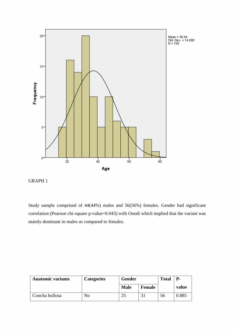
GRAPH 1
Study sample comprised of 44(44%) males and 56(56%) females. Gender had significant
correlation (Pearson chi-square p-value=0.043) with Onodi which implied that the variant was
mainly dominant in males as compared to females.
Anatomic variants Categories Gender Total P-
value Male Female
Concha bullosa No 25 31 56 0.885
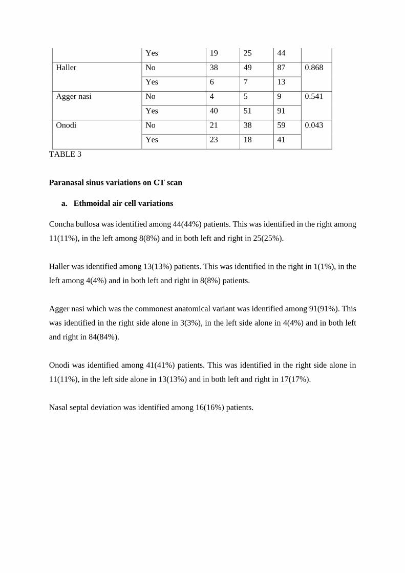
Yes 19 25 44
Haller No 38 49 87 0.868
Yes 6 7 13
Agger nasi No 4 5 9 0.541
Yes 40 51 91
Onodi No 21 38 59 0.043
Yes 23 18 41
TABLE 3
Paranasal sinus variations on CT scan
a. Ethmoidal air cell variations
Concha bullosa was identified among 44(44%) patients. This was identified in the right among
11(11%), in the left among 8(8%) and in both left and right in 25(25%).
Haller was identified among 13(13%) patients. This was identified in the right in 1(1%), in the
left among 4(4%) and in both left and right in 8(8%) patients.
Agger nasi which was the commonest anatomical variant was identified among 91(91%). This
was identified in the right side alone in 3(3%), in the left side alone in 4(4%) and in both left
and right in 84(84%).
Onodi was identified among 41(41%) patients. This was identified in the right side alone in
11(11%), in the left side alone in 13(13%) and in both left and right in 17(17%).
Nasal septal deviation was identified among 16(16%) patients.

GRAPH 2
Symptoms for CT scanning were indicated in all patients and mainly included nasal blockage
34.7%, headache 34.1% and rhinorrhea 18.2% among others as indicated below.
GRAPH 3
Symptoms Vs. Anatomic variants
Symptoms for CT scanning
Concha bullosa Haller Agger nasi Onodi
No Yes No Yes No Yes No Yes
Nasal blockage 28 24 47 5 6 46 34 18
Watery rhinorrhea 1 0 1 0 1 0 0 1
Fre
qu
en
cy
Anatomical variants
Anatomical variants
Percent
Sym
pto
ms
Symptoms of patients undergoing CT scan

Headache 18 14 24 8 1 31 17 15
Rhinorrhea 4 3 7 0 1 6 3 4
Epistaxis 1 1 2 0 0 2 1 1
Poor visibility 4 2 6 0 0 6 4 2
P-value 0.663 0.720 0.268 0.261
TABLE 4
b. Keros Classification.
Keros 1 was present in 25(25%) patients; keros 2 was present in 67(67%) patients, keros 3 was
present in 7(7%) patients and keros 4 was present in 1(1%) patient.
GRAPH 4
Keros Frequency Percent
Keros 1 25 25.0
Keros 2 67 67.0
Keros 3 7 7.0
Series1, Keros 1, 25.0, 25%
Series1, Keros 2, 67.0, 67%
Series1, Keros 3, 7.0,
7%
Series1, Keros 4, 1.0, 1%
Keros classification
Keros 4 1 1.0
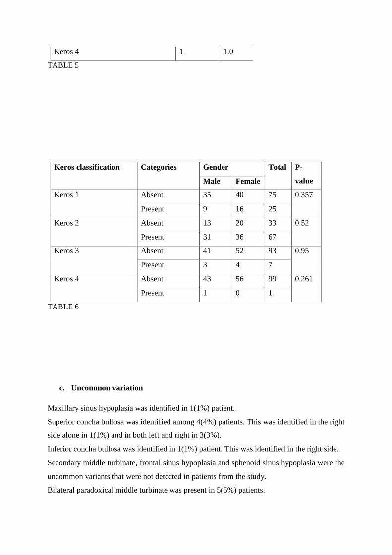
TABLE 5
Keros classification Categories Gender Total P-
value Male Female
Keros 1 Absent 35 40 75 0.357
Present 9 16 25
Keros 2 Absent 13 20 33 0.52
Present 31 36 67
Keros 3 Absent 41 52 93 0.95
Present 3 4 7
Keros 4 Absent 43 56 99 0.261
Present 1 0 1
TABLE 6
c. Uncommon variation
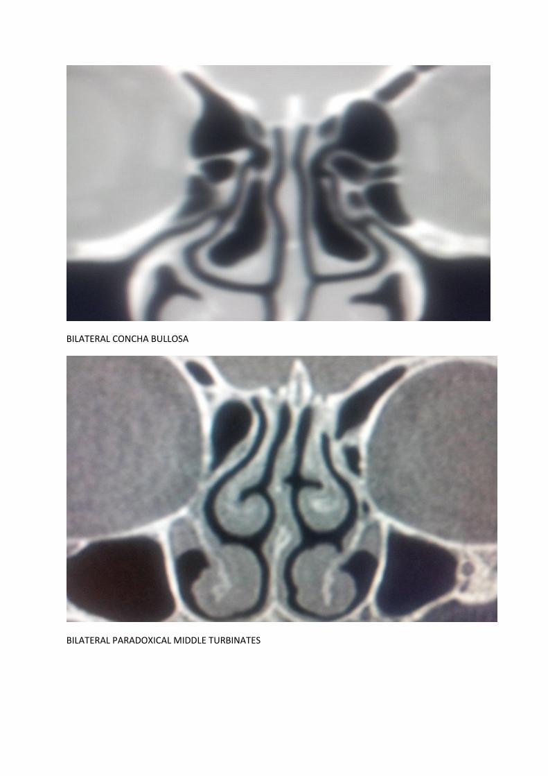
Maxillary sinus hypoplasia was identified in 1(1%) patient.
Superior concha bullosa was identified among 4(4%) patients. This was identified in the right
side alone in 1(1%) and in both left and right in 3(3%).
Inferior concha bullosa was identified in 1(1%) patient. This was identified in the right side.
Secondary middle turbinate, frontal sinus hypoplasia and sphenoid sinus hypoplasia were the
uncommon variants that were not detected in patients from the study.
Bilateral paradoxical middle turbinate was present in 5(5%) patients.

Paradoxical superior turbinate was present in the right in 1(1%) patient.
Septate sphenoid was present in 14(14%) patients.
14.0 CONCLUSION.
Anatomical variations are best viewed and appreciated in the coronal plane. Agger nasi is the
commonest anatomical variation of the paranasal sinuses followed by Concha bullosa, then
Onodi cells and Haller cells respectively.
Few of the very uncommon variations were also seen, namely paradoxical middle turbinate,
superior and inferior concha bullosa.
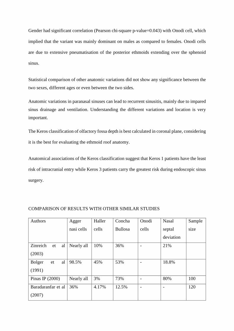
Gender had significant correlation (Pearson chi-square p-value=0.043) with Onodi cell, which
implied that the variant was mainly dominant on males as compared to females. Onodi cells
are due to extensive pneumatisation of the posterior ethmoids extending over the sphenoid
sinus.
Statistical comparison of other anatomic variations did not show any significance between the
two sexes, different ages or even between the two sides.
Anatomic variations in paranasal sinuses can lead to recurrent sinusitis, mainly due to impared
sinus drainage and ventilation. Understanding the different variations and location is very
important.
The Keros classification of olfactory fossa depth is best calculated in coronal plane, considering
it is the best for evaluating the ethmoid roof anatomy.
Anatomical associations of the Keros classification suggest that Keros 1 patients have the least
risk of intracranial entry while Keros 3 patients carry the greatest risk during endoscopic sinus
surgery.
COMPARISON OF RESULTS WITH OTHER SIMILAR STUDIES
Authors Agger
nasi cells
Haller
cells
Concha
Bullosa
Onodi
cells
Nasal
septal
deviation
Sample
size
Zinreich et al
(2003)
Nearly all 10% 36% - 21%
Bolger et al
(1991)
98.5% 45% 53% - 18.8%
Pinas IP (2000) Nearly all 3% 73% - 80% 100
Baradaranfar et al
(2007)
36% 4.17% 12.5% - - 120
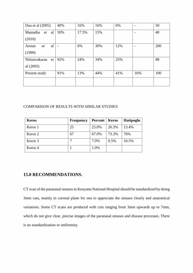
Dua et al (2005) 40% 16% 16% 6% - 50
Mamatha et al
(2010)
50% 17.5% 15% - 40
Arstan et al
(1999)
- 6% 30% 12% - 200
Nitinavakaran et
al (2005)
92% 24% 34% 25% 88
Present study 91% 13% 44% 41% 16% 100
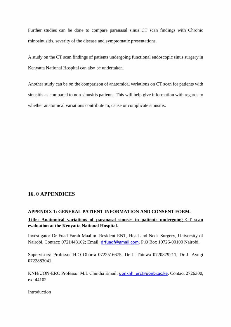
COMPARISON OF RESULTS WITH SIMILAR STUDIES
Keros Frequency Percent Keros Hatipoglu
Keros 1 25 25.0% 26.3% 13.4%
Keros 2 67 67.0% 73.3% 76%
Keros 3 7 7.0% 0.5% 10.5%
Keros 4 1 1.0%
15.0 RECOMMENDATIONS.
CT scan of the paranasal sinuses in Kenyatta National Hospital should be standardized by doing
3mm cuts, mainly in coronal plane for one to appreciate the sinuses clearly and anatomical
variations. Some CT scans are produced with cuts ranging from 3mm upwards up to 7mm,
which do not give clear, precise images of the paranasal sinuses and disease processes. There
is no standardization or uniformity.
Further studies can be done to compare paranasal sinus CT scan findings with Chronic
rhinosinusitis, severity of the disease and symptomatic presentations.
A study on the CT scan findings of patients undergoing functional endoscopic sinus surgery in
Kenyatta National Hospital can also be undertaken.
Another study can be on the comparison of anatomical variations on CT scan for patients with
sinusitis as compared to non-sinusitis patients. This will help give information with regards to
whether anatomical variations contribute to, cause or complicate sinusitis.
16. 0 APPENDICES
APPENDIX 1: GENERAL PATIENT INFORMATION AND CONSENT FORM.
Title: Anatomical variations of paranasal sinuses in patients undergoing CT scan
evaluation at the Kenyatta National Hospital.
Investigator Dr Fuad Farah Maalim. Resident ENT, Head and Neck Surgery, University of
Nairobi. Contact: 0721448162; Email: [email protected]. P.O Box 10726-00100 Nairobi.
Supervisors: Professor H.O Oburra 0722516675, Dr J. Thinwa 0720879211, Dr J. Ayugi
0722883041.
KNH/UON-ERC Professor M.L Chindia Email: [email protected]. Contact 2726300,
ext 44102.
Introduction
Participation in this study is voluntary. We aim to find out the anatomical variations of the
paranasal air sinuses in patients having CT scan evaluation, presenting to Kenyatta National
Hospital.
What is involved in this study?
Once you consent to participate, we will take a short medical history. The paranasal sinus CT
scan will be evaluated with a consultant radiologist.
Are there any risks involved?
There are no risks involved in participating in this study.
Will I be penalized for not participating?
No, you will receive the same attention and treatment as those who choose to participate in this
study.
What benefits will I get if I participate?
There will be no benefits accorded for your participation in this particular study.
The benefits of the study will be towards assisting in local training, emphasis in radiological
evaluation of paranasal sinus of patients. Information of ethnic variation trends will eventually
be obtained.
What about confidentiality?
All the information we obtain from you will be kept confidential.
How much will it cost me?
No extra cost will be incurred
What are my rights as a participant?
Participation in the study is voluntary. Once inducted in the study, you can choose to
discontinue at any time. This will not cause discrimination.
What do you do with the information you get?
This information will help us find out more about anatomical variations of the paranasal air
sinuses in patients undergoing CT scan evaluation. The data will act as a baseline research for
anyone who wishes to carry further studies on the various other paranasal sinuses.
Are you satisfied with the information given?
If yes and you are willing to participate or let child participate, please fill in and sign the consent
below.
APPENDIX 2: CONSENT FOR THE STUDY (ADULT).
Your participation in this study is entirely voluntary.
I………………………………………………………… Of…………………
Address…………………
study no………………… do hereby consent for myself to be included in this study on
anatomical variations of the ethmoid air sinuses in patients having CT scan evaluation for
rhinosinusitis presenting to Kenyatta national hospital.
The nature of the study has been fully explained to me by Dr Fuad Farah Maalim. Telephone
number 0721448162.
I have not been promised any material gain to participate.
I hereby agree to a soft copy of my CT scan be utilized for the study.
Signed…………………………………. (Self)
Date…………………………………
KIBALI CHA UTAFITI.
Kushiriki kwako katika utafiti huu ni kwa hiari yako.
Mimi …………………………………………………………. Anwani………..…….
Nambariyautafiti……………. Nimekubali kuhusishwa katika utafiti huu unaongalia utofauti
ya anatomia wa ethmoid sinasi kwa kutumia picha ya CT scan.
Nimefahamu baadaya kusoma na kufahamishwa na Dr Fuad Farah M, nambari ya simu
0721448162.
Hakuna malipo nitakayopewa.
Nimekubali picha yangu ya CT scan kutumika katika utafiti huu.
Sahihi…………………………………….. (Mgonjwa)
Tarehe……………………………………
CONSENT FOR THE STUDY (CHILD).
The participation of your child in this study is entirely voluntary.
I………………………………………………………………. Address
………………………..

Parent or guardian of ………………………………………….. study no………………… do
hereby consent for my child to be included in this study on anatomical variations of the ethmoid
air sinuses in patients having CT scan evaluation for rhinosinusitis presenting to Kenyatta
national hospital.
The nature of the study has been fully explained to me by Dr Fuad Farah Maalim. Telephone
number 0721448162.
I have not been promised any material gain to participate.
I hereby agree to a soft copy of my child’s CT scan be utilized for the study.
Signed…………………………………. (Parent/guardian)
Date……………………………….
ASSENT FORM FOR MINORS (age 8 – 17 years).
I voluntarily agree to participate in the research study, having had the information and
procedure being read out to me by the principal investigator Dr Fuad Farah. I have understood
the nature of the study and whatever information given will remain confidential.
Sign your name here (minor) …………………….. Date …………….
KIBALI CHA UTAFITI (MTOTO).
Kushiriki kwa motto wako katika utafiti huu ni kwa hiari yako.
Mimi …………………………………………………………… Anwani………………
Mzazi ama msimamizi wa .....................................................nambari ya utafiti…………….
Nimekubali mtoto wangu kuhusishwa katika utafiti huu unaongalia utofauti ya anatomia wa
ethmoid sinasi kwa kutumia picha ya CT scan.
Nimefahamu baada ya kusoma na kufahamishwa na Dr Fuad Farah Maalim, nambari ya simu
0721448162.
Hakuna malipo nitakayopewa.
Nimekubali picha ya mtoto wangu ya CT scan kutumika katika utafiti huu.
Sahihi…………………………… (Mzazi/ Msimamizi)
Tarehe……………………………………
KIBALI CHA MTOTO (miaka 8 – 17).
Nimekubali kushiriki katika utafiti huu kwa hiari yangu, baada ya Daktari Fuad Farah kunielezea na
kusoma utaratibu utakaofuatwa. Nime elewa na kufahamu utafiti huu.
Sahihi ya mtoto …………………………………………. Tarehe …………………………
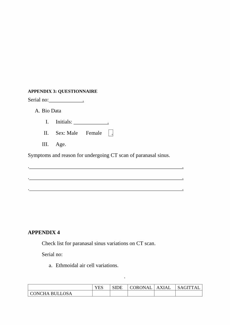
APPENDIX 3: QUESTIONNAIRE
Serial no: .
A. Bio Data
I. Initials: .
II. Sex: Male Female .
III. Age.
Symptoms and reason for undergoing CT scan of paranasal sinus.
. .
. .
. .
APPENDIX 4
Check list for paranasal sinus variations on CT scan.
Serial no:
a. Ethmoidal air cell variations.
.
YES SIDE CORONAL AXIAL SAGITTAL
CONCHA BULLOSA
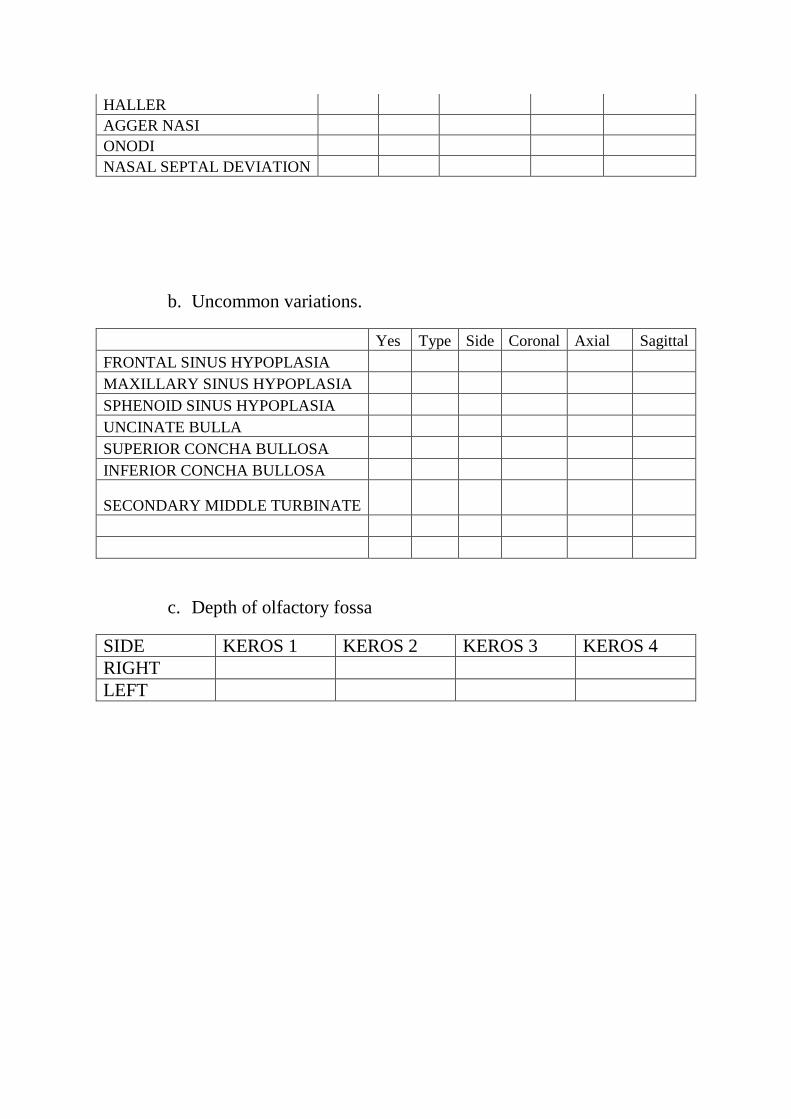
HALLER
AGGER NASI
ONODI
NASAL SEPTAL DEVIATION
b. Uncommon variations.
Yes Type Side Coronal Axial Sagittal
FRONTAL SINUS HYPOPLASIA
MAXILLARY SINUS HYPOPLASIA
SPHENOID SINUS HYPOPLASIA
UNCINATE BULLA
SUPERIOR CONCHA BULLOSA
INFERIOR CONCHA BULLOSA
SECONDARY MIDDLE TURBINATE
c. Depth of olfactory fossa
SIDE KEROS 1 KEROS 2 KEROS 3 KEROS 4
RIGHT
LEFT
16.0 REFERENCES
1. Bolger W. E., Butzin C. A., Parsons D. S. Paranasal sinus bony anatomic variations and
mucosal abnormalities: CT analysis for endoscopic sinus surgery. Laryngoscope. 1991;
101:56–64.
2. Kennedy DW, Zinreich SJ: Functional endoscopic approach to inflammatory sinus
disease: Current perspectives and technique modifications. American Journal of
Rhinology. 1988; 2, 89 – 96.
3. Mosher HP: Symposium on the ethmoid: the surgical anatomy of the ethmoidal
labyrinth. Trans American Academy of Ophthalmology and Otolaryngology. 1929:
376–410.
4. Y Ramakrishnan, I Zammit-Maempel, N S Jones. Paransal sinus computed tomography
anatomy: a surgeon’s perspective. The Journal of Laryngology & Otology. 2011; 125,
1141 – 1147.
5. Hudgins P. Complications of endoscopic sinus surgery: the role of the radiologist in
prevention. Radiologic Clinics of North America. 1993; 31:21–32.
6. Ribeiro Fde A. Standardized measurements of radiographic films of the frontal sinuses:
an aid to identifying unknown persons. Ear Nose Throat Journal. 2000; 79:26–33.
7. Yoshino M, Miyasaka S, Sato H, Seta S. Classification system of frontal sinus patterns
by radiography. Its application to identification of unknown skeletal remains. Forensic
Science International. 1987; 34:289–299.
8. Tatlisumak E, Yilmaz Ovali G, Aslan A, Asirdizer M, Zeyfeoglu Y, Tarhan S.
Identification of unknown bodies by using CT images of frontal sinus. Forensic Science
International. 2007; 166:428.
9. Morimoto N, Ogihara N, Katayama K, Shiota K. Threedimensional ontogenetic shape
changes in the human cranium during the fetal period. Journal of Anatomy. 2008;
212:627-635.
10. Hanna EH, Levine HL, Timen S, Kotton B. Hypoplasia of the maxillary antrum:
anatomic abnormalities, diagnostic difficulties and surgical implications. American
Journal of Rhinology. 1993; 7:105-110.
11. Yonetsu K, Watanabe M, Nakamura T. Age-related expansion and reduction in aeration
of the sphenoid sinus: volume assessment by helical CT scanning. American Journal of
Neuroradiology. 2000; 21:179–182.
12. Zinreich SJ, Mattox DE, Kennedy DW, Chisholm HL, Diffley DM, Rosenbaum AE.
Concha bullosa: CT evaluation. Journal of Computer Assisted Tomography. 1988;
12:778–84.
13. Dogru H, Doner F, Uygur K, Gedikli O, Cetin M. Pneumatized inferior turbinate.
American Journal of Otolaryngology. 1999; 20:139–41.
14. Arslan H, Aydinlioglu A, Bozkurt M, Egeli E. Anatomical variations of the paranasal
sinuses: CT examination for endoscopic sinus surgery. Auris Nasus Larynx. 1999;
26:39–48.
15. Calhoun KH, Waggenspack GA. CT evaluation of the paranasal sinuses in symptomatic
and asymptomatic populations. Otolaryngology Head and Neck Surgery. 1991;
104:480 –3.
16. Meloni F, Mini R, Rovasio S, Stomeo F, Teatini GP. Anatomic variations of surgical
importance in ethmoid labyrinth and sphenoid sinus. A study of radiological anatomy.
Surgical and Radiological Anatomy. 1992: 14:65–70.
17. Ji-Hyeon Shin, Sung Won Kim,Yong Kil Hong. The Onodi Cell: An Obstacle to Sellar
Lesions with a Transsphenoidal Approach. Otolaryngology Head and Neck Surgery.
2011; 145(6) 1040–1042.
18. Driben JS, Bolger WE, Robles HA, Cable B, Zinreich SJ. The reliability of
computerized tomographic detection of the Onodi (sphenoethmoid) cell. American
Journal of Rhinology. 1998; 12:105–11.
19. Stammberger H. Endoscopic sinus surgery-concepts in treatment of recurring
rhinosinusitis. Part II. Surgical technique. Otolaryngology Head and Neck Surgery.
1986; 94:147–56.
20. Bolger WE, Woodruff Jr WW, Morehead J, Parsons DS. Maxillary sinus hypoplasia
classification and description of associated uncinate process hypoplasia.
Otolaryngology Head and Neck Surgery. 1990; 103:759–65.
21. Kennedy DW, Zinreich SJ, Shaalan H, Kuhn F, Navlerio R, Loch E. Endoscopic middle
meatal antrostomy theory, technique and patency. Laryngoscope (Suppl). 1987; 97:1–
9.
22. Anik I, Anik Y, Koc K, Ceylan S. Agenesis of sphenoid sinuses. Clinical Anatomy.
2005; 18:217–219.
23. Keskin G, Ustundag E, Ciftci E. Agenesis of sphenoid sinuses. Surgical and
Radiological Anatomy. 2002; 24:324–326.
24. Duque CS, Casiano RR, Surgical Anatomy and Embryology of the Frontal Sinus. In:
Kountakis S, Senior BA, Draf W. The Frontal Sinus. Springer. 2005.
25. Perez P, Sabate J, Carmona A. Anatomical variations in the human paranasal sinus
region studied by CT. Journal of Anatomy. 2000;197:221–227.
26. Dua K, Chopra H, Khurana AS and Munjal M. CT scan variations in chronic sinusitis.
Indian Journal of Radiology and Imaging. 2005; 15(3), 315-320.
27. Mamatha H, Shamasundar NM, Bharathi M, Prasanna L. Variations of Osteomeatal
Complex and Its Applied Anatomy: A CT scans Study. Indian Journal of Science and
Technology. 2010; 3(8): 904-7.
28. Hatipoğlu HG, Cetin MA, Yüksel E. Concha Bullosa Types: Their Relationship with
Sinusitis, Ostiomeatal and Frontal Recess Disease. Diagnostic and Interventional
Radiology. 2005; 11(3): 145- 149.
29. Kayalioglu G, Oyar O, Govsa F: Nasal cavity and paranasal sinus bony variations: a
computed tomographic study. Rhinology. 2000; 38(3):108–113.
30. Basic N, Basic V, Jukic T, Basic M, Jelic M, Hat J. Computed tomographic imaging to
determine the frequency of anatomical variations in pneumatization of the ethmoid
bone. European Archives of Otorhinolaryngology. 1999; 256:69–71.
31. Lerdlum S, Vachiranubhap B. Prevalence of anatomic variation demonstrated on
screening sinus computed tomography and clinical correlation. Journal of Medical
Association of Thailand. 2005; 88 Suppl 4: S110-115.
32. Wormald PJ. The agger nasi cell: the key to understanding the anatomy of the frontal
recess. Otolaryngology Head and Neck Surgery. 2003; 129: 497-507.

33. Bradley DT, Kountakis SE. The role of agger nasi air cells in patients requiring revision
endoscopic frontal sinus surgery. Otolaryngology Head and Neck Surgery. 2004; 131:
525-527.
34. Badia L, Lund VJ, Wei W, Ho WK. Ethnic variation in sinonasal anatomy on CT-
scanning. Rhinology. 2005; 43: 210-214.
35. Nitinavakarn B, Thanaviratananich S, Sangsilp N. Anatomical variations of the lateral
nasal wall and paranasal sinuses: a CT study for endoscopic sinus surgery (ESS) in Thai
patients. Journal of Medical Association of Thailand. 2005; 88(6): 763-8.
36. Vincent TE, Gendeh BS. The Association of Concha Bullosa and Deviated Nasal
Septum with Chronic Rhinosinusitis in Functional Endoscopic Sinus Surgery Patients.
Medical Journal of Malaysia. 2010; 65(2): 108-11.
37. Baradaranfar MH, Labibi M. Anatomic variations of paranasal sinuses in patients with
chronic sinusitis and their correlation with CT scan staging. Acta Medica Iranica. 2007;
45:477-80.
38. Kantarci M, R. Murat Karasen, Fatih A, Omer O, Adnan O. Remarkable anatomic
variations in paranasal sinus region and their clinical importance. European Journal of
Radiology. 2004 (50)296–302.
39. Khanobthamchai K, Shankar L, Hawke M, Bingham B. The secondary middle
turbinate. Journal of Otolaryngology. 1991; 20:412–3.
40. Aykut M, Gümüsburun E, Müderris S, Adigüzel E. The secondary nasal middle
concha. Surgical and Radiologic Anatomy. 1994; 16:307–9.
41. Aksungur EH, Biçakçi K, Inal M, Akgül E, Binokay F, Aydogan B. CT demonstration
of accessory nasal turbinates: secondary middle turbinate and bifid inferior turbinate.
European Journal of Radiology. 1999; 31:174–6.
42. Binal C, Muhammed AS, Ahmet BY. A retrospective analysis of sphenoid sinus
hypoplasia and agenesis using dental volumetric CT scan in Turkish individuals:
Diagnostic and Interventional Radiology. 2011; 17:205 – 208.
43. Wanga SK, Lemeshow S. Sample size determination in health studies. A practical
manual. Ginebra: World Health Organization, 1991.

44. Keros P. On the practical value of differences in the level of the lamina cribrosa of the
ethmoid. Z Laryngology Rhinology Otology. 1962; 41:809–13.

KEROS TYPE 1
KEROS TYPE 2
BILATERAL CONCHA BULLOSA
BILATERAL PARADOXICAL MIDDLE TURBINATES

ONODI CELLS.
AGGER NASI CELLS.
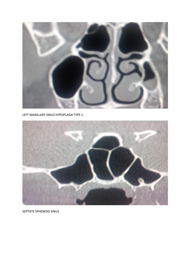
LEFT MAXILLARY SINUS HYPOPLASIA TYPE 1.
SEPTATE SPHENOID SINUS