thorax & lungs
DESCRIPTION
fTRANSCRIPT
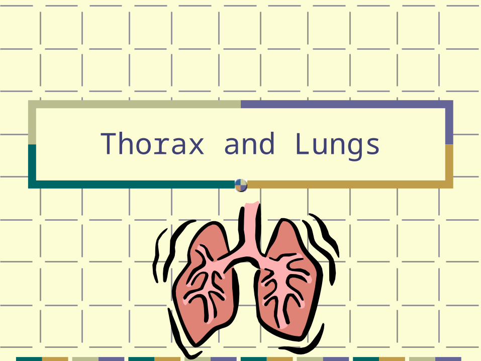
Thorax and Lungs
Outline
Structure and Function
Subjective Data
Objective Data
Abnormal Findings
Structure and Function
Thoracic Cage /Cavity
Shape- bony, conical shape, narrower at top borders – it is defined by:
Sternum – 3 parts: manubrium, body, xiphoid process Ribs – 12 pairs, 1st seven attach to the sternum (costal
cartilages) Ribs 8,9,&10 attach to the costal cartilage above, Ribs 11 & 12 are floating ribs
12 Thoracic vertebrae Diaphragm – the floor, separates the thoracic cavity from
the abdomen
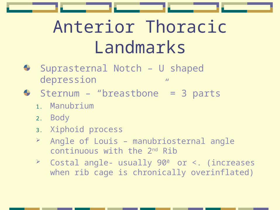
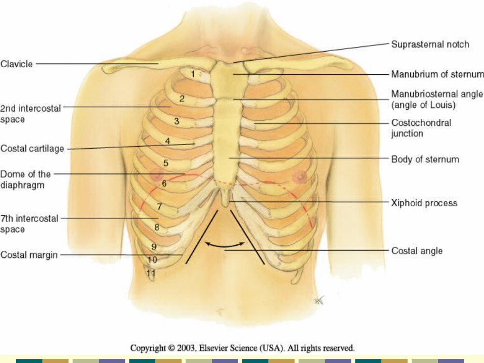
Anterior Thoracic Landmarks
Suprasternal Notch – U shaped depression
Sternum – “breastbone” = 3 parts1. Manubrium
2. Body
3. Xiphoid process Angle of Louis – manubriosternal angle
continuous with the 2nd Rib Costal angle- usually 900 or <. (increases when
rib cage is chronically overinflated)
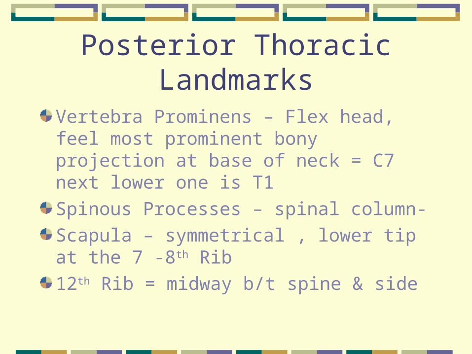
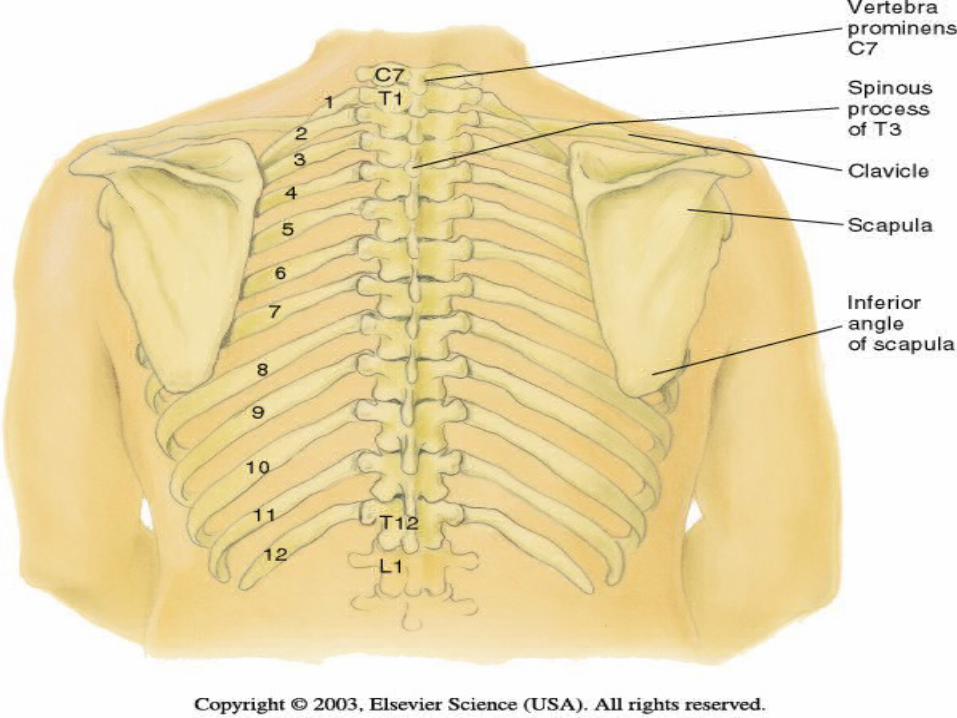
Posterior Thoracic Landmarks
Vertebra Prominens – Flex head, feel most prominent bony projection at base of neck = C7 next lower one is T1
Spinous Processes – spinal column-
Scapula – symmetrical , lower tip at the 7 -8th Rib
12th Rib = midway b/t spine & side
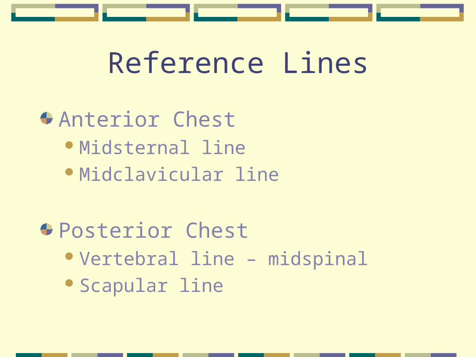
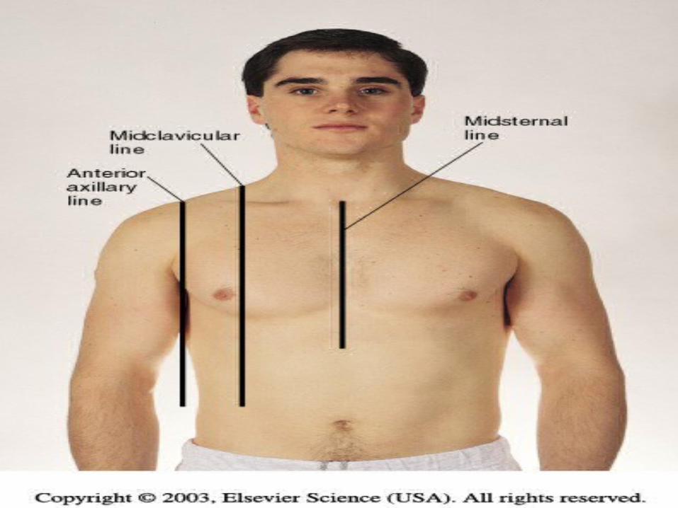
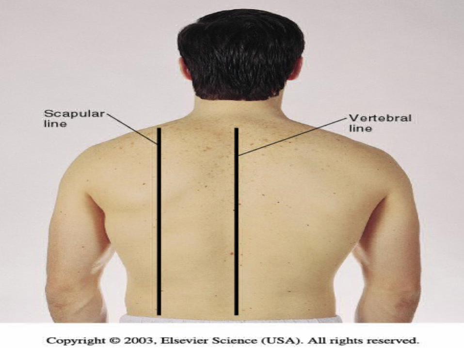
Reference Lines
Anterior ChestMidsternal lineMidclavicular line
Posterior ChestVertebral line – midspinalScapular line

Lateral ChestAnterior Axillary linePosterior Axillary lineMid–axillary line

The Thoracic Cavity
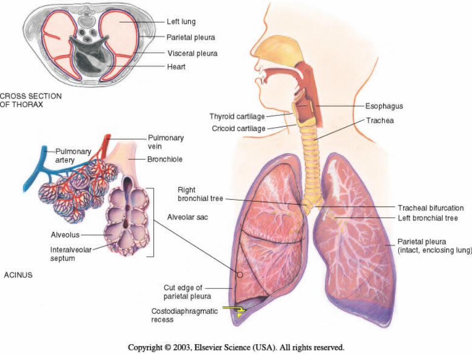
Mediastinum middle of the thoracic cavity & contains; Esophagus Trachea Heart Great Vessels
Pleural Cavities on either side of the mediastinum contain the lungs
Lung Borders
Anterior Chest – Apex 3 -4 cm. ↑ inner 1/3 of the clavicles Base – rests on the diaphragm, 6th rib, MCL
Lateral Chest Extends from Axilla apex to 7th –8th rib
Posteriorly Apex of lung is at C7 – Base T10 (on deep
inspiration to T12)
Lobes of Lung
Right Lung3 lobes, upper, middle , lowerShorter due to liver
Left LungLUL = Left Upper and Lower ( 2 lobes)Narrower due to heart
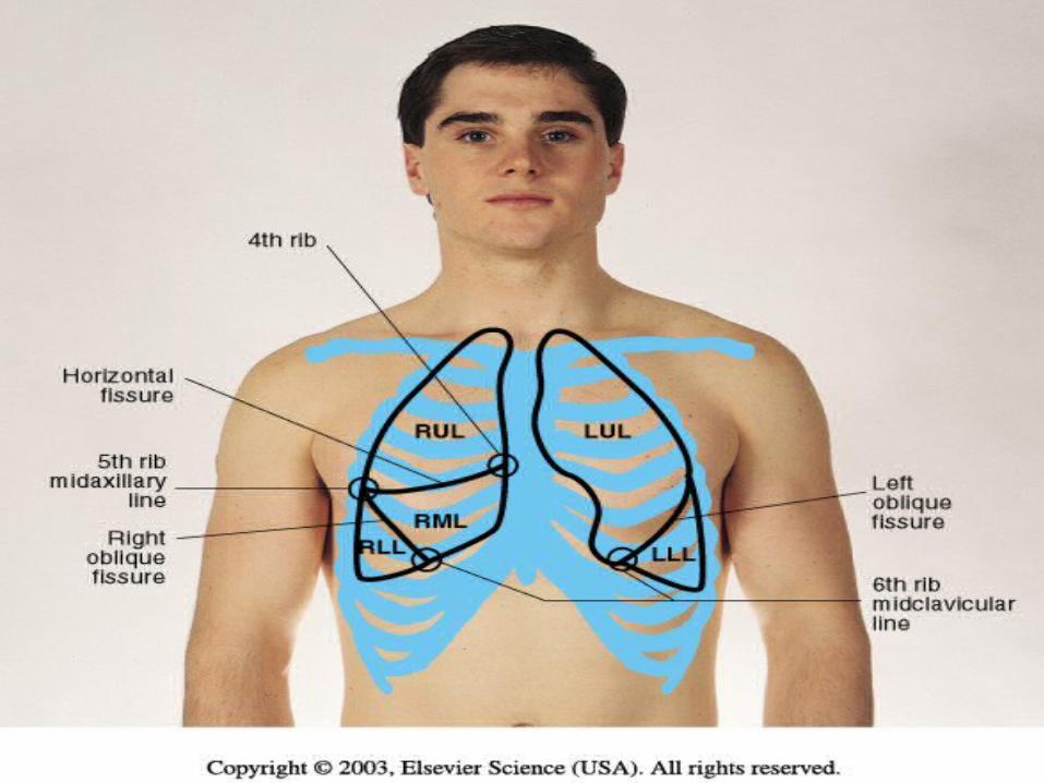
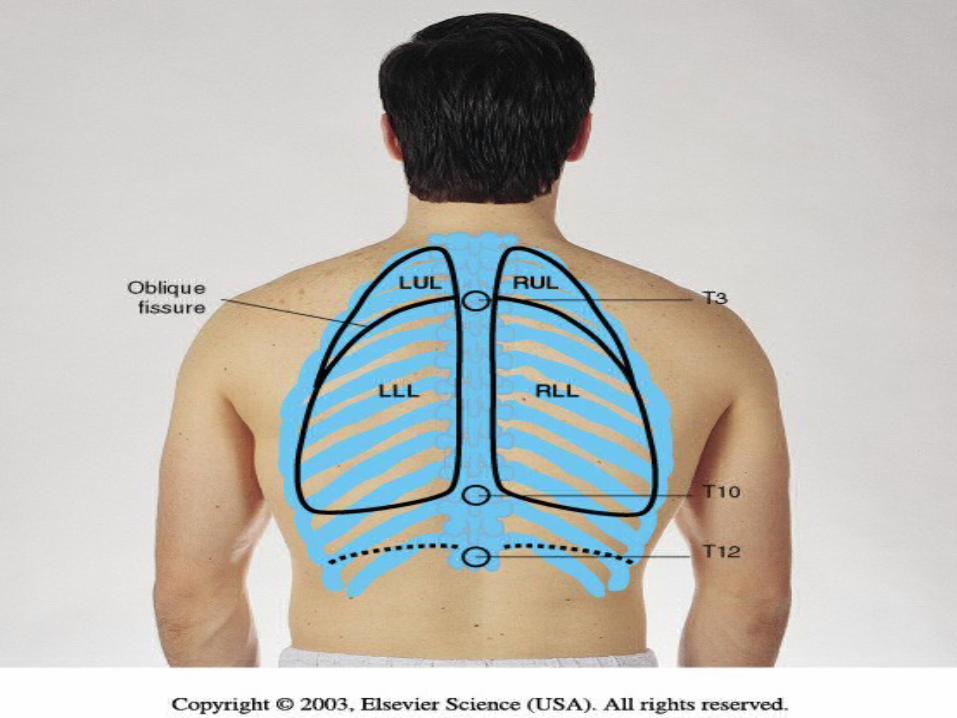
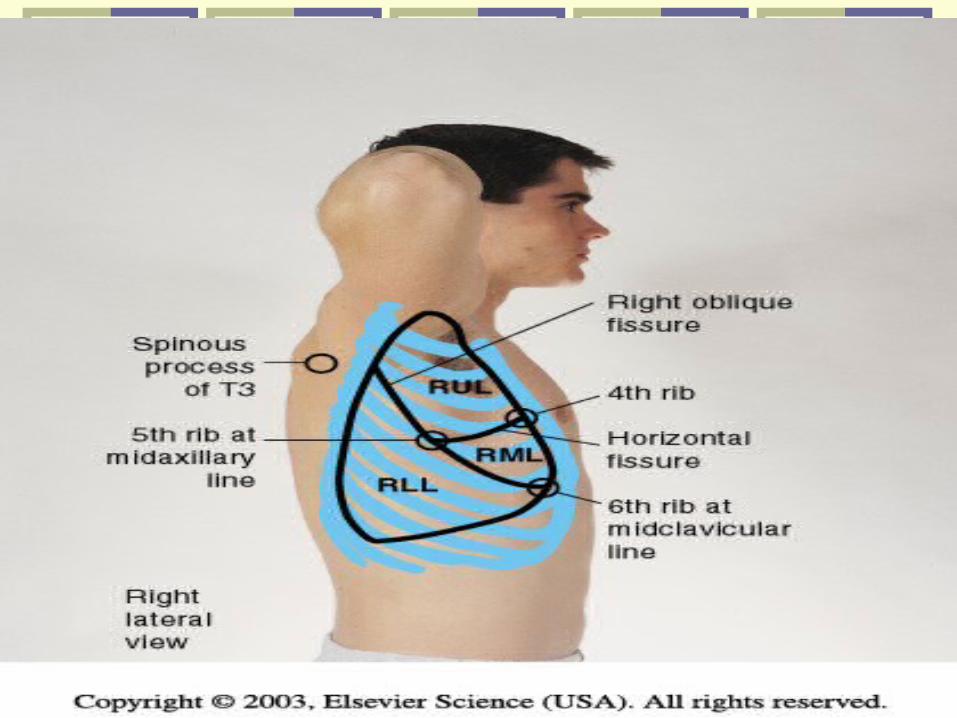
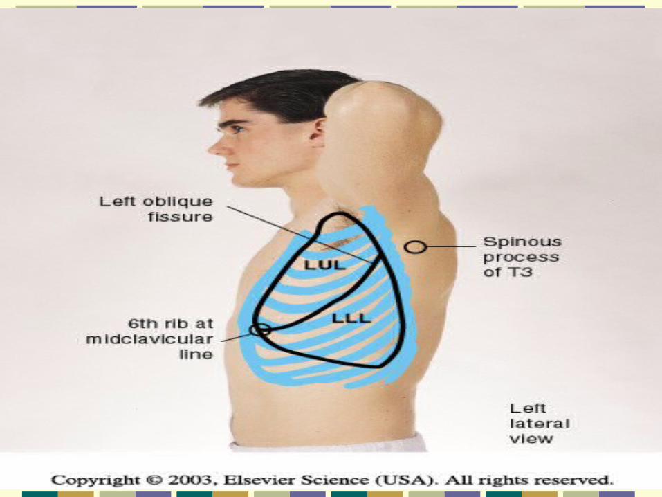
Lobes Diagonal sloping segmentsOblique fissures
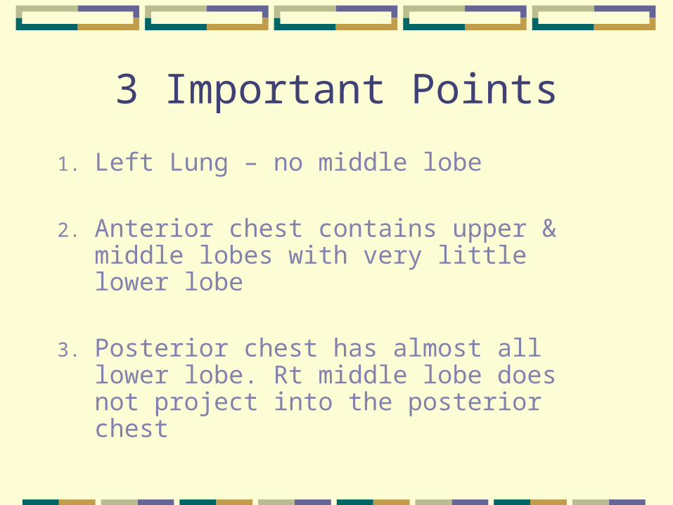
3 Important Points
1. Left Lung – no middle lobe
2. Anterior chest contains upper & middle lobes with very little lower lobe
3. Posterior chest has almost all lower lobe. Rt middle lobe does not project into the posterior chest
Pleurae
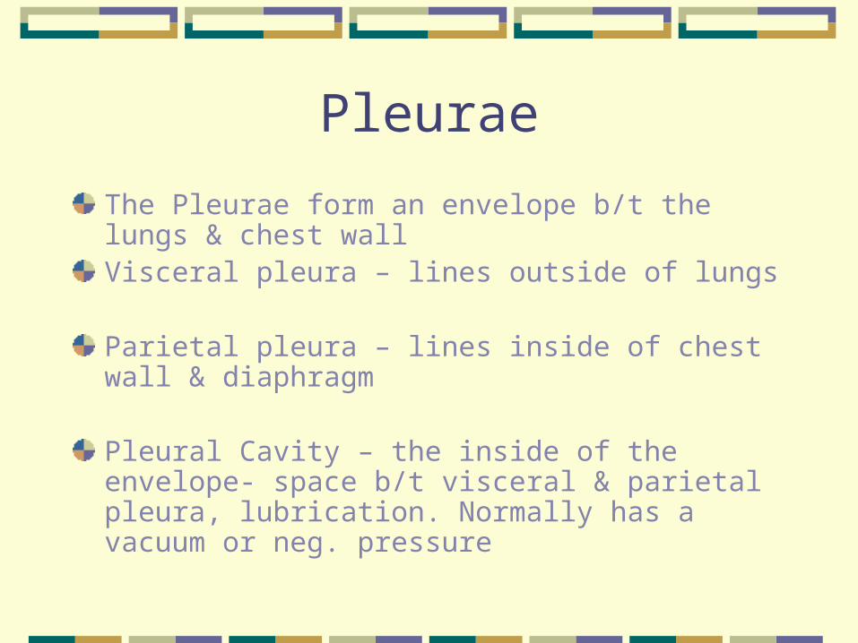
The Pleurae form an envelope b/t the lungs & chest wallVisceral pleura – lines outside of lungs
Parietal pleura – lines inside of chest wall & diaphragm
Pleural Cavity – the inside of the envelope- space b/t visceral & parietal pleura, lubrication. Normally has a vacuum or neg. pressure
Tracheal & Bronchial Tree
Trachea – anterior to esophagus- 10-11 cm.long, begins at cricoid cartilageBifurcates just below the sternal angle
( AKA angle of Louis, manubriosternal angle) into the
Right Main Stem Bronchus – shorter, wider, more vertical ( Intubation – listen to breath sounds bilaterally)
Left Main Stem Bronchus
Tracheal & Bronchial Tree
The trachea & bronchi provide the passage for air to get into the lungs from the environment = Dead Space (no air exchange takes place here)
Bronchi Secrete mucus – captures particles Cilia – moves the trapped particles up to be
expelled or swallowed
Acinus Functional respiratory unit consisting of, Bronchioles, alveolar ducts, alveolar sacs, &
alveoli Gaseous exchange in alveolar duct & alveoli
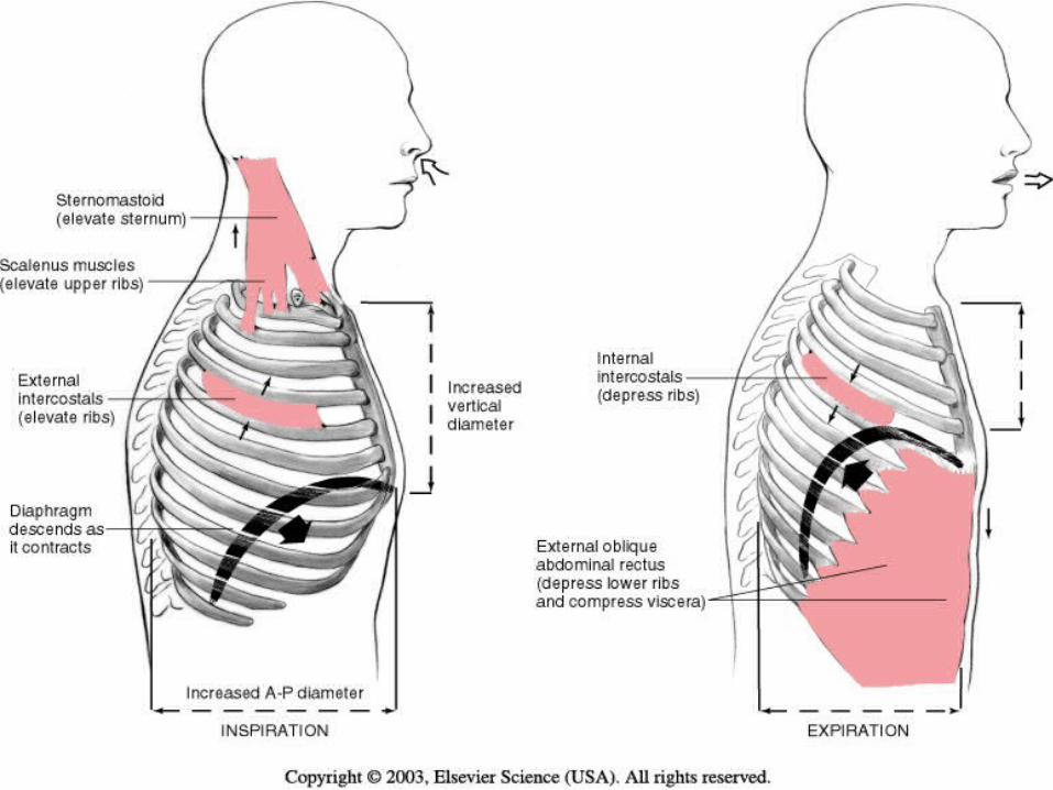
Mechanics of Respiration
4 Major Functions of the Respiratory System
1. Supply O2 for energy production
2. Remove CO2 , waste product of energy reactions
3. Homeostasis, acid-base balance of arterial blood
4. Heat exchange
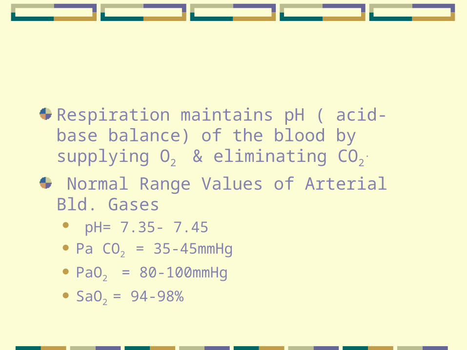
Respiration maintains pH ( acid- base balance) of the blood by supplying O2 & eliminating CO2
.
Normal Range Values of Arterial Bld. Gases pH= 7.35- 7.45 Pa CO2 = 35-45mmHg
PaO2 = 80-100mmHg
SaO2 = 94-98%
Lungs help to maintain the pH balance by adjusting the amt. of CO2 through:
HypoventilationHyperventilation
Respiration = breathing
Inspiration
Expiration
Control of Respiration Involuntary control by respiratory center in the
brain stem consisting of the pons & medulla Hypercapnia is an ↑ in CO2 in the Bld. And
provides the normal stimulus to breath Hypoxemia
Subjective Data
Cough
SOB
Chest Pain
Respiratory Infections
Smoking
Environmental Exposure
Self-care behaviors

Objective Data
Inspect
Palpate
Percuss
Auscultate
After Posterior Thyroid Exam
Posterior chest, Lateral chest, then Anterior chest
Remember to clean stethoscope end piece and warm prior to use on client.
Quiet environment conducive to hearing lung sounds
Equipment for Exam
Stethoscope
Ruler – 15cm.
Tape measure
Washable marker
Alcohol swabs
Posterior Chest
Inspect Thoracic CageShape and configuration Anteroposterior Diameter should be <
Transverse Diameter = Ratio 1:2 to 5:7Note Position of Person to breathe.
? orthopneaSkin Color & Condition, nail color

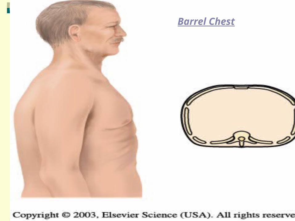
Barrel Chest
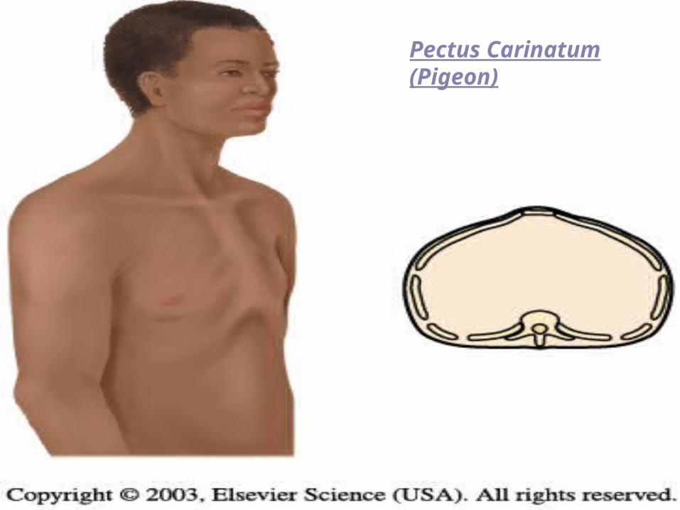
Pectus Carinatum (Pigeon)
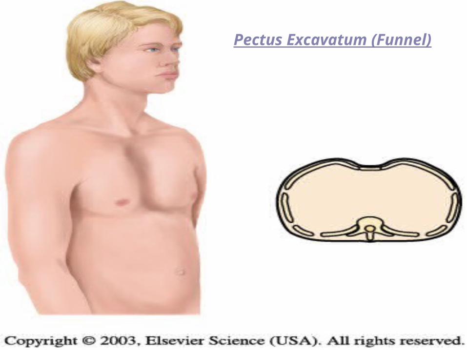
Pectus Excavatum (Funnel)
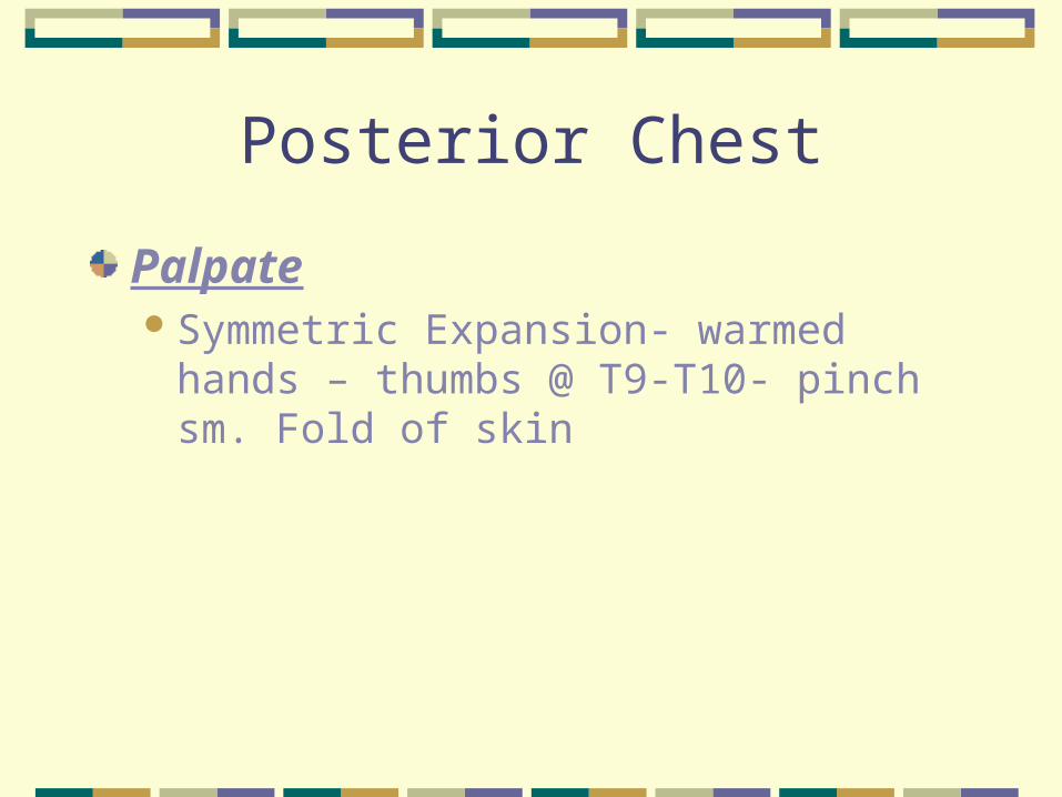
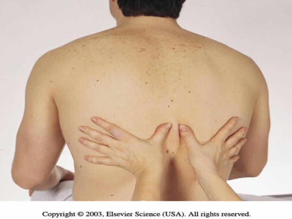
Posterior Chest
PalpateSymmetric Expansion- warmed hands –
thumbs @ T9-T10- pinch sm. Fold of skin
Posterior chest
Tactile Fremitus – palpable vibration of sound from the larynx- use palmer base of fingers- “99” or Blue Moon
Symmetry important – vibration should feel the same bilaterally.
Avoid palpating over scapulae because bone dampens out sound

↓ fremitus = obstructed bronchi, pleural effusion, pneumothorax or emphysema
Note any barrier that is b/t the sound and your hand will↓ fremitus
↑ fremitus occurs only with gross changes (Lobar pneumonia).

Entire Chest wall – gently palpate. NoteTenderness, skin temp., moisture, lumps,
lesionsCrepitus = coarse crackling sensation
palpable over skin surface. (Subcutaneous emphysema when air escapes from lung into S/C tissue)
Posterior Chest
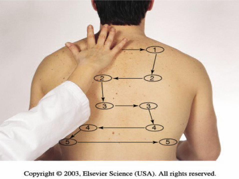
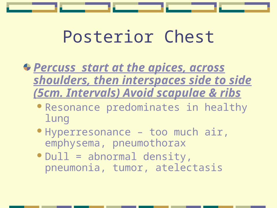
Percuss start at the apices, across shoulders, then interspaces side to side (5cm. Intervals) Avoid scapulae & ribsResonance predominates in healthy lungHyperresonance – too much air,
emphysema, pneumothoraxDull = abnormal density, pneumonia,
tumor, atelectasis
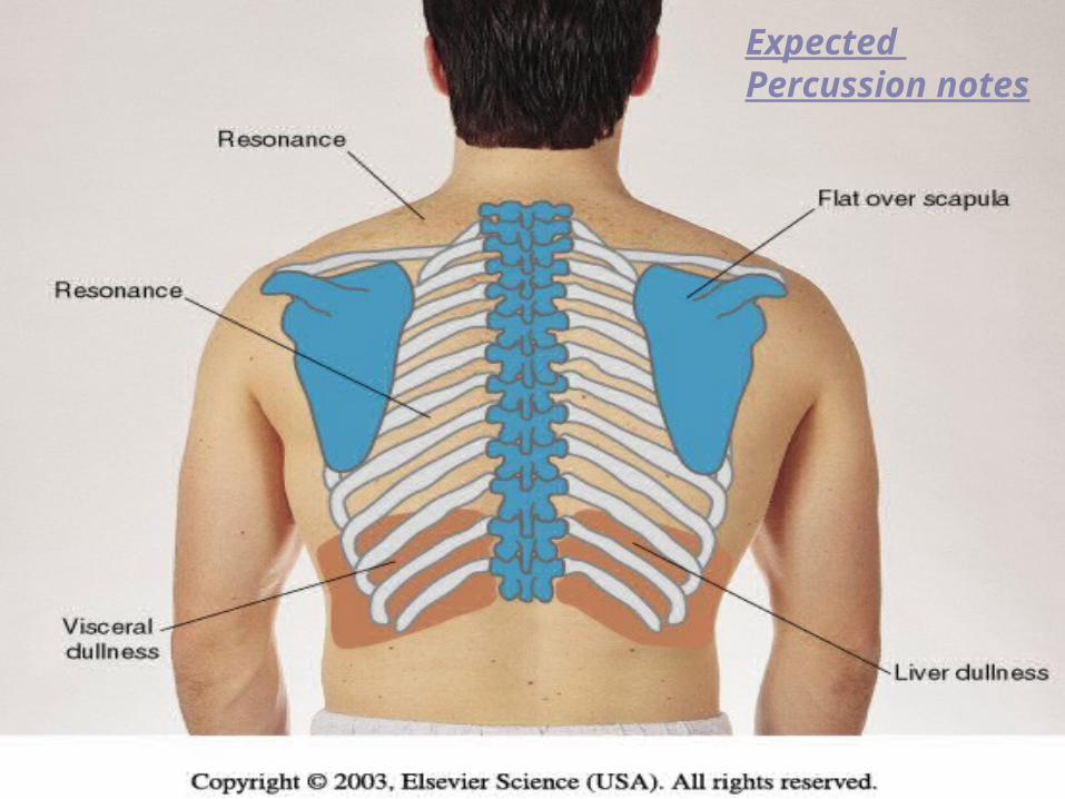
Expected Percussion notes
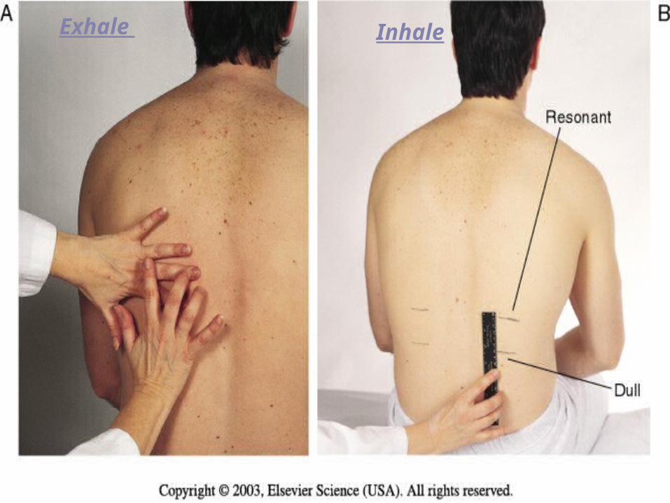
Diaphragmatic Expansion
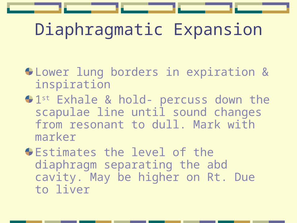
Lower lung borders in expiration & inspiration1st Exhale & hold- percuss down the scapulae line until sound changes from resonant to dull. Mark with markerEstimates the level of the diaphragm separating the abd cavity. May be higher on Rt. Due to liver
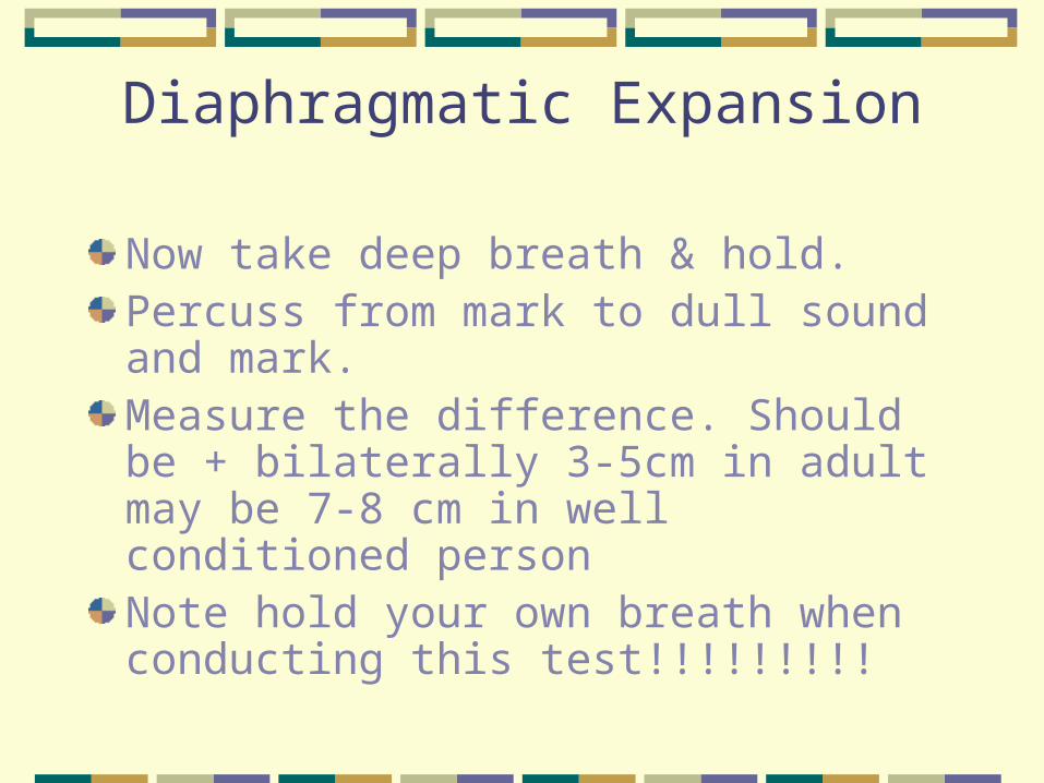
Diaphragmatic Expansion
Now take deep breath & hold.Percuss from mark to dull sound and mark.Measure the difference. Should be + bilaterally 3-5cm in adult may be 7-8 cm in well conditioned personNote hold your own breath when conducting this test!!!!!!!!!
Exhale Inhale
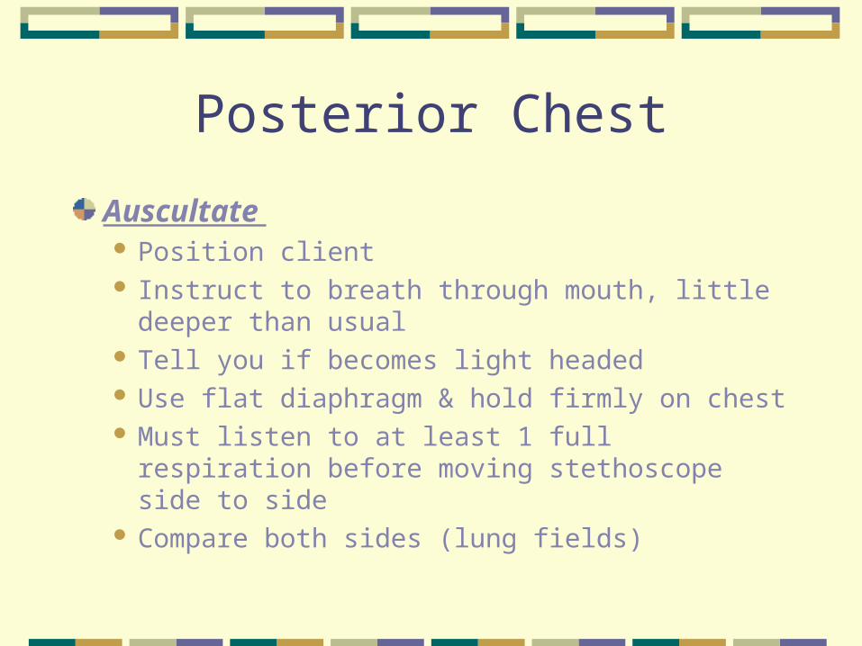
Posterior Chest
Auscultate Position client Instruct to breath through mouth, little deeper than
usual Tell you if becomes light headed Use flat diaphragm & hold firmly on chest Must listen to at least 1 full respiration before
moving stethoscope side to side Compare both sides (lung fields)
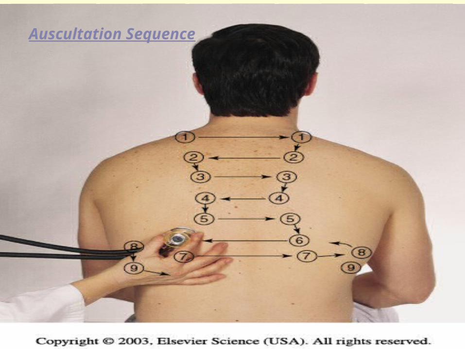
Auscultation Sequence
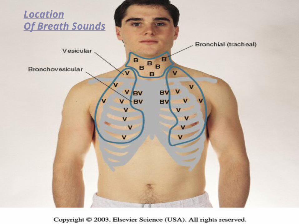
Normal Breath Sounds
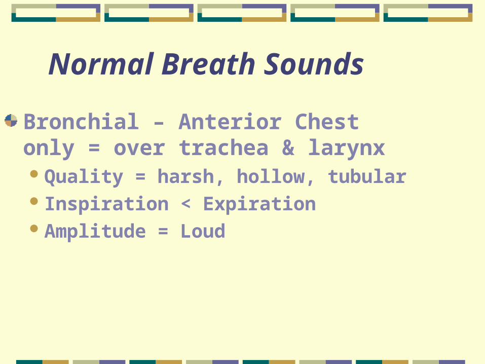
Bronchial – Anterior Chest only = over trachea & larynx Quality = harsh, hollow, tubular Inspiration < ExpirationAmplitude = Loud

Breath Sounds
Bronchovesicular both anterior & posteriorOver major bronchi, posterior b/t
scapulae, anterior upper sternum, 1st & 2nd ICS
Pitch = high Inspiration = ExpirationModerate amplitude

Vesicular – Anterior & posteriorQuality = rustling, wind in trees Inspiration > ExpirationSoft amplitude
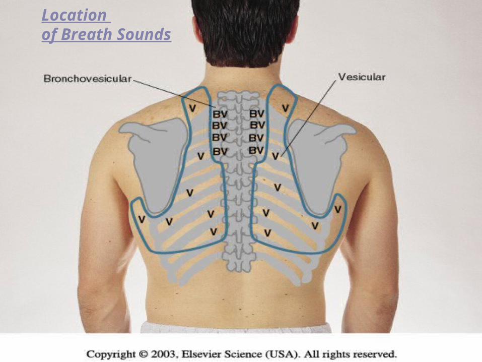
Location of Breath Sounds

Decreased or Absent Breath Sounds Causes = obstruction of the bronchial tree by secretions,
mucous plug, F.B ↓ lung elasticity, emphysema = lungs hyperinflated Pleurisy, pleural thickening, pneumothorax (air),
pleural effusion (fld.) in the pleural space
Increased Breath Sounds = dense lung tissue enhances sound transmission as in consolidation ie. pneumonia
Silent chest = ominous
Adventitious Sounds
Not normally heard in the lungs. Caused by moving air colliding with secretions or by popping open of previously deflated airwaysCrackles (Rales) Fine – high pitched popping- not cleared by coughing.
Simulate sound by rolling strand of hair b/t fingers near ear or moisten thumb& index finger & separate them near your ear
Course crackles- (opening a velcro fastener)
Pleural Friction Rub – coarse & low pitched, 2 pieces of leather rubbed together close to ear
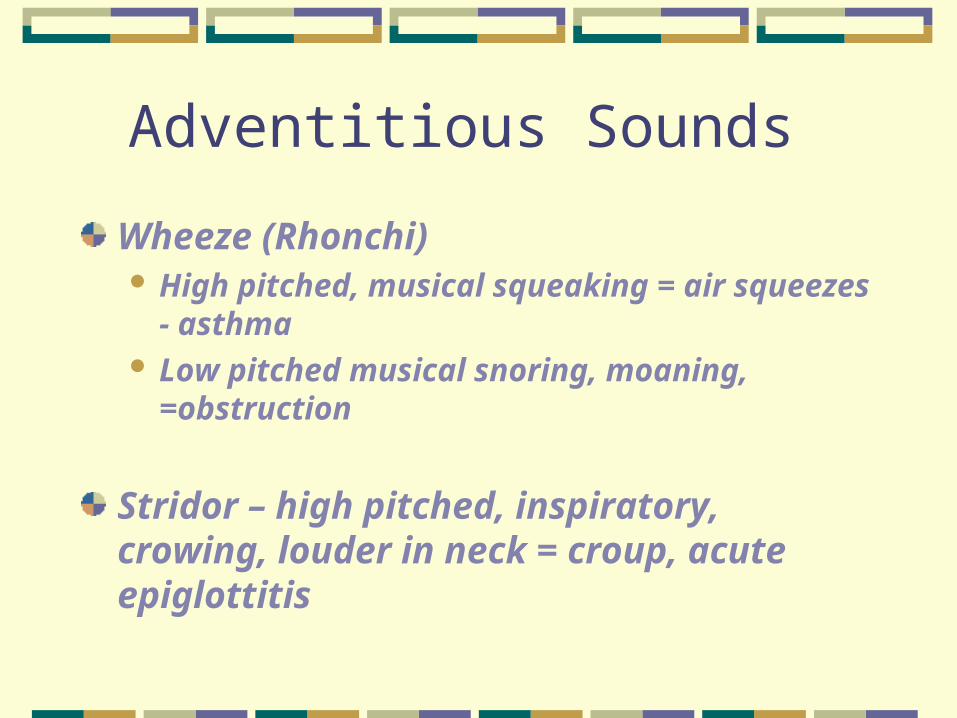
Adventitious Sounds
Wheeze (Rhonchi) High pitched, musical squeaking = air
squeezes - asthma Low pitched musical snoring, moaning,
=obstruction
Stridor – high pitched, inspiratory, crowing, louder in neck = croup, acute epiglottitis
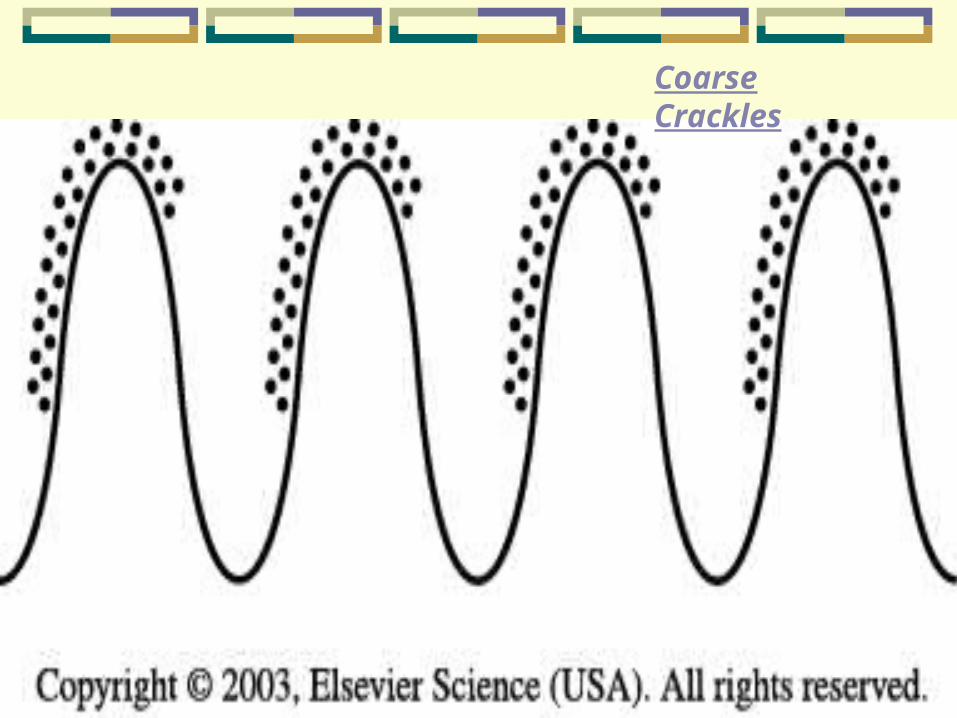
Coarse Crackles
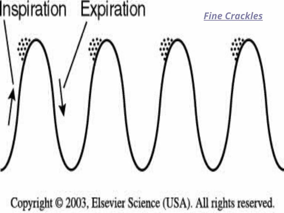
Fine Crackles
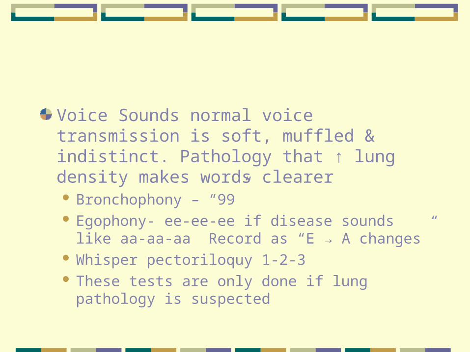
Voice Sounds normal voice transmission is soft, muffled & indistinct. Pathology that ↑ lung density makes words clearer Bronchophony – “99” Egophony- ee-ee-ee if disease sounds like aa-aa-
aa Record as “E → A changes” Whisper pectoriloquy 1-2-3 These tests are only done if lung pathology is
suspected

Anterior Chest
Inspect Shape & ConfigurationExpression- relaxedLOC – alert & cooperativeSkin color & conditionQuality of Respirations – reg. & even, no
retraction or use of accessory muscles
Anterior Chest
Palpate Symmetric Chest ExpansionTenderness, turgor, temp., moisture
Tactile FremitusCompare both sides
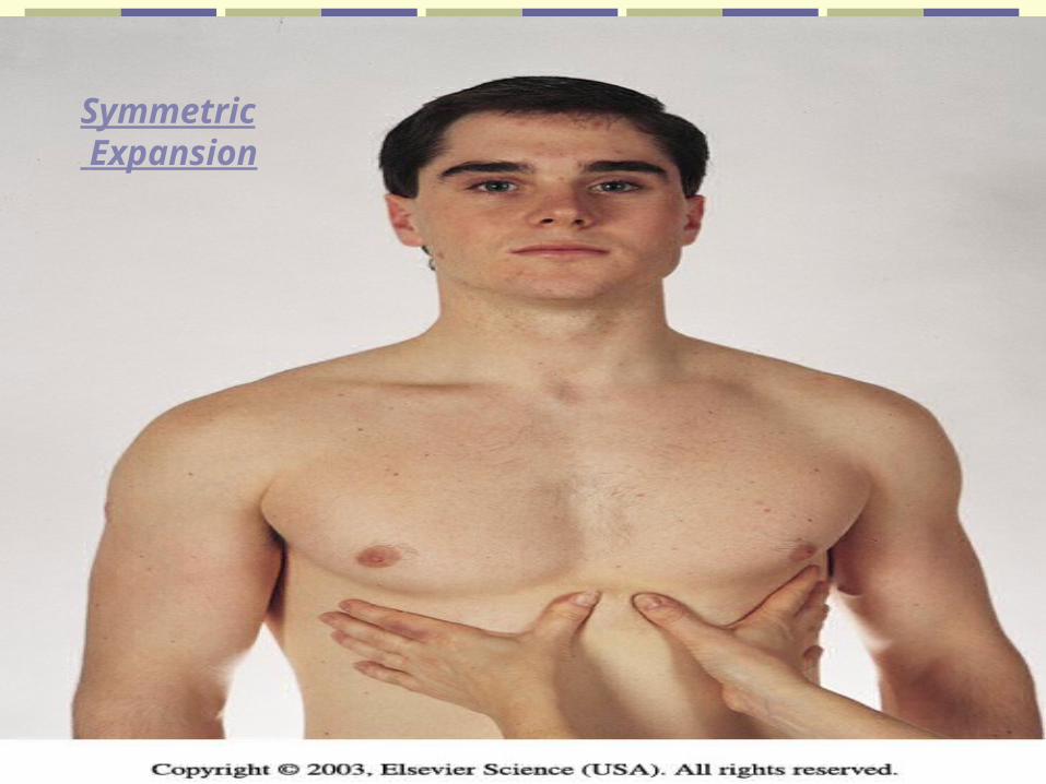
Symmetric Expansion
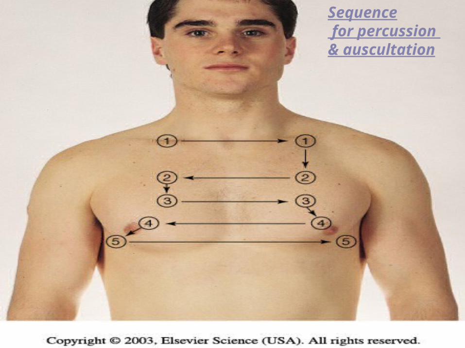
Sequence for percussion & auscultation
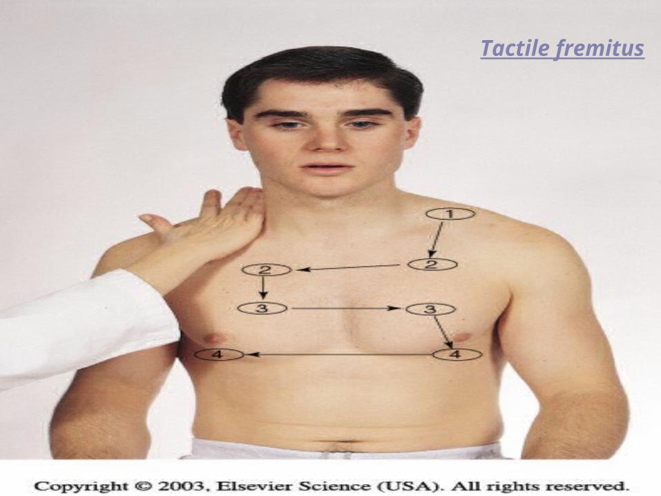
Tactile fremitus
Percussion
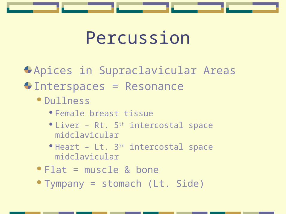
Apices in Supraclavicular Areas
Interspaces = ResonanceDullness
Female breast tissueLiver – Rt. 5th intercostal space midclavicularHeart – Lt. 3rd intercostal space midclavicular
Flat = muscle & boneTympany = stomach (Lt. Side)
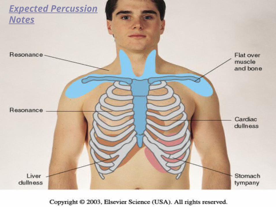
Expected PercussionNotes
Auscultate
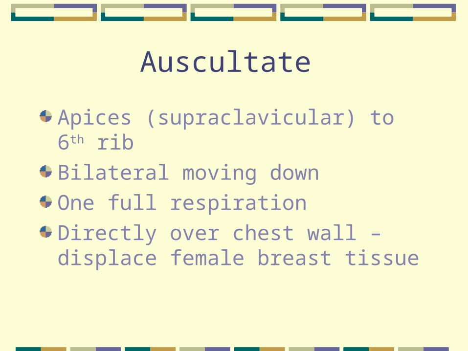
Apices (supraclavicular) to 6th rib
Bilateral moving down
One full respiration
Directly over chest wall – displace female breast tissue
LocationOf Breath Sounds
Pulse Oximeter
Noninvasive measurement of arterial oxygen saturation = SpO2 by measuring the relative amt. of light absorbed by oxyhemoglobin and unoxygenated hemoglobin. It compares light emitted to amt absorbed. Normally 97 -98%
Terms for Documentation
Rate Eupnea 12 – 20 bpm normalTachypnea > 24, rapid, shallowBradypnea < 10Apnea = No respirations for 10 sec. or
more
Pattern = breathing rhythm. Normal respirations are regular and even.Cheyne – stokes = resp wax & wane in reg
pattern with periods of apnea(20sec)Biot’s or ataxisic Sim. To cheyne –stokes
but pattern irreg.
Depth – on inspiration the normal depth is nonexaggerated and effortless.Shallow
Sighing – purposeful to expand the alveoli
Symmetry – bilateral rise and fall of the chest with respiration
Audibility – normally be heard by the unaided ear several centimeters from the patient’s nose/mouth
Patient position – healthy person breathes comfortably in supine, prone or upright positionOrthopnea
Mode of Breathing – normally inhale/exhale through nose
Sputum SampleColor
Mucoid, yellow/green, rust/blood tinged, black, pink
OdorAmountConsistency