three-dimensional models of cancer for pharmacology … cascade of cancer drug discovery depends...
TRANSCRIPT
© 2014 Wiley-VCH Verlag GmbH & Co. KGaA, Weinheim 1115
1 Introduction
In this review, we attempt to survey the current status ofin vitro/ex vivo models of human cancer that try to betterreproduce the complexity and heterogeneity of a humancancer in situ, with a perspective of their use in medicine.A historical background to the use of tumor cell lines in
cancer drug discovery is followed by a survey of morerecent attempts to mimic in vitro the three-dimensionalcharacteristics of solid tumors, including those modelswhich add elements of the important tumor stroma suchas fibroblasts. Studies using bioreactors are reviewed;these not only permit changes in scalability but are per-missive for dynamic, controlled flow-through conditionsof cell culture. Although cell lines with or without stromamay be studied and used in three dimensions, they nev-ertheless constitute attempts to re-construct only some ofthe complexity of a complex and often heterogeneoustumor, and essentially remain reductionist models. Theuse of precision-cut tumor slices potentially permits allaspects of tumor complexity and heterogeneity to be cap-tured in vitro, sometimes referred to as ex vivo, but in
Review
Three-dimensional models of cancer for pharmacology andcancer cell biology: Capturing tumor complexity in vitro/ex vivo
John A. Hickman1, Ralph Graeser2, Ronald de Hoogt2, Suzana Vidic2, Catarina Brito3,4, Matthias Gutekunst5
and Heiko van der Kuip5 on behalf of the IMI PREDECT consortium
1 IMI PREDECT, Paris, France2 Janssen Pharmaceutica NV, Beerse, Belgium3 iBET – Instituto de Biologia Experimental e Tecnológica, Oeiras, Portugal4 Instituto de Tecnologia Química e Biológica, Universidade Nova de Lisboa, Oeiras, Portugal5 Dr. Margarete Fischer-Bosch Institute of Clinical Pharmacology, University of Tübingen, Stuttgart, Germany
Cancers are complex and heterogeneous pathological “organs” in a dynamic interplay with theirhost. Models of human cancer in vitro, used in cancer biology and drug discovery, are generallyhighly reductionist. These cancer models do not incorporate complexity or heterogeneity. This raises the question as to whether the cancer models’ biochemical circuitry (not their genome) rep-resents, with sufficient fidelity, a tumor in situ. Around 95% of new anticancer drugs eventually failin clinical trial, despite robust indications of activity in existing in vitro pre-clinical models. Inno-vative models are required that better capture tumor biology. An important feature of all tissues,and tumors, is that cells grow in three dimensions. Advances in generating and characterizing sim-ple and complex (with added stromal components) three-dimensional in vitro models (3D mod-els) are reviewed in this article. The application of stirred bioreactors to permit both scale-up/scale-down of these cancer models and, importantly, methods to permit controlled changes in envi-ronment (pH, nutrients, and oxygen) are also described. The challenges of generating thin tumorslices, their utility, and potential advantages and disadvantages are discussed. These in vitro/exvivo models represent a distinct move to capture the realities of tumor biology in situ, but signif-icant characterization work still remains to be done in order to show that their biochemical cir-cuitry accurately reflects that of a tumor.
Keywords: Bioreactors · Cancer · Drug discovery · 3D models · Tissue slices
Correspondence: Prof. John Hickman, Coordinator, IMI PREDECT, 126 blvd Pereire, 75017 Paris, FranceE-mail: [email protected]
Abbreviations: ECM, extracellular matrix; EGF, epidermal growth factor;FGF, fibroblast growth factor; GF, growth factor; HGF, hepatocyte growthfactor; RCCS, rotary cell culture systems
Biotechnol. J. 2014, 9, 1115–1128 DOI 10.1002/biot.201300492
www.biotechnology-journal.com
BiotechnologyJournal
Received 11 MAR 2014Revised 11 JUL 2014Accepted 05 AUG 2014
1116 © 2014 Wiley-VCH Verlag GmbH & Co. KGaA, Weinheim
reviewing progress with this technological platform,some important caveats are articulated. Finally, weaddress the rather vexed question of how any model maybe “validated” sufficiently well so that it may be incorpo-rated into mainstream activities not only relating to drugdiscovery but also to the study of tumor biology in vitro/exvivo.
1.1 The current problems of pre-clinical models of cancer
In a recent and self-revelatory article addressing some ofthe current malaise in the pharmaceutical industry andthe difficulties of novel drug discovery [1], it was sug-gested that ultimate project success in drug discoverydepended very heavily on the confidence that teams hadin studies of target validation. This critical first step in thepre-clinical cascade of cancer drug discovery dependsalmost entirely on the use of in vitro models, cell lines, thatattempt to recapitulate pathology in a reductionist way(reviewed in [2]). These same models, expressing a drugtarget of interest, generally become incorporated intosubsequent steps of the pre-clinical cascade of drug dis-covery, providing essential tools for compound screeningand refinement, until candidate molecules are chosen forin vivo assays of efficacy; sometimes, the same long-established cell lines are grafted into mice for such a pur-pose. There has been a growing realization that the work-horse of robust reproducible cell lines, growing in sus-pension or in two-dimensions on plastic, amenable tohigh-throughput screening, is limited in its ability to cap-ture the complexity and heterogeneity of a cancer. Thecompelling evidence for this lies in clinical trial data: thegreatest source of failure in phase 2 is a lack of efficacy [1,3–5] an efficacy that had presumably been robustlydemonstrated in pre-clinical studies. Novel cancer thera-peutics are the worst candidate drugs within all thera-peutic areas which lack of efficacy in clinical trial [3] andthus, one could postulate that the pre-clinical models ofcancer, including in vitro models, are among the least pre-dictive.
1.2 Cell lines: Does genotype capture phenotype?
One defense of the use of single cell lines, growing in sus-pension culture or on plastic, as sufficient models ofhuman cancers, largely rests on the data emerging frommassive efforts to fully sequence their genomes anddescribe their transcriptomes [6, 7]. Representation ofintra-tumoral heterogeneity, within a pathology, has beenargued to be satisfied by increasing the numbers of celllines representing that pathology (e.g. colon cancer) (see[2]). Each cell lines’ “representivity” of pathology has beenclaimed to be reflected in the fidelity by which they carrysignature patterns of genetic alterations. In addition, theresponse of cell lines to pharmacological agents was
claimed to reflect clinical sensitivity patterns [6, 7]although this is controversial [8]. What these studies donot address is whether the biochemical circuitry of thesecells, such as signaling cascades initiated by growth fac-tors (GFs), cytokines and heterogeneous cell contacts, isrepresentative of a complex tumor in situ? Tumors haverightly been described as “organs” [9]. It is difficult toimagine how cell lines growing on plastic, lacking all theinterplay with a dynamic and complex stroma, may berepresentative of a tumor. In addition, solid tumors growin three dimensions, with homo- or hetero-intercellularcontacts. However, in reviewing three-dimensional in vitro models of cancer, recently also addressed elsewhere[10, 11], and precision-cut slices, these same questions,about how representative their circuitry is, must also beposed. This challenge is addressed at the end of this review.
2 2D and 3D cell line models
2.1 Some history of 2D cancer models
In the 1950s, cell culture was introduced as a tool toscreen compounds as potential drugs in vitro, using cellsgrown and passaged infinitely on glass in flasks in semi-synthetic medium ([12, 13] and references therein). How-ever, the correlation between in vitro culture, mouse invivo results, and clinical responses was very poor. Thiswas not seen too negatively – rather it was suggested tocompare the models and make use of their diversity: “Atthe present imperfect state of our knowledge, the mostfruitful approach to the complexities of cancer chemo -therapy may well be the imaginative exploitation of thediscrepancies, which are bound to emerge from thesecomparisons” [13].
Perhaps the next major advance was the colony form-ing assay, developed by Hamburger and Salmon [14] inthe 1970s, which allowed growing fresh human patientmaterial embedded as single cell solution in soft agar. Inan NCI-funded study, four laboratories evaluated the tech-nology and its use in drug testing. Six tumor entities,breast, colorectal, kidney, lung, melanoma, and ovarianyielded cultures that passed the quality controls estab-lished by the laboratories, for 20–38% of patients tested atsamples sizes of >160 patients each [15]. The protocol wasthen used to re-test 79 compounds, which had been inac-tive in the in vivo screening assay, on 15 tumors each.Fourteen compounds were found to be active in at leasttwo tumors. Due to the low plating efficiency, with a con-comitant bias of the assay toward tumors that grew in softagar, and the requirement for a constant supply of freshpatient material, the assay was considered impractical forlarger compound screens. The plating efficiency could beimproved to 70–80%, at least for certain pathologies [16],but a review on clinical studies indicated rather limitedsuccess of in vitro soft agar assays to predict a patient’s
www.biotechnology-journal.com www.biotecvisions.com
BiotechnologyJournal Biotechnol. J. 2014, 9, 1115–1128
© 2014 Wiley-VCH Verlag GmbH & Co. KGaA, Weinheim 1117
tumor’s sensitivity toward clinically approved drugs.Twelve prospective studies were investigated, includinga total of 506 patients (33%) that had been treated withchemotherapy chosen via clonogenic assays. The meanresponse rate for these patients was 27% (range 10–100%;n = 12 studies) compared to 18% (range, 0–100%; n = 7studies) for patients treated with empiric therapy. Theeffect on overall survival had not been properly addressed[17]. The absence of a functional tumor microenvironmentin the assays – extracellular matrix, stromal cells – andselection for cells within the tumor that survive in softagar may be explanations of poor predictivity.
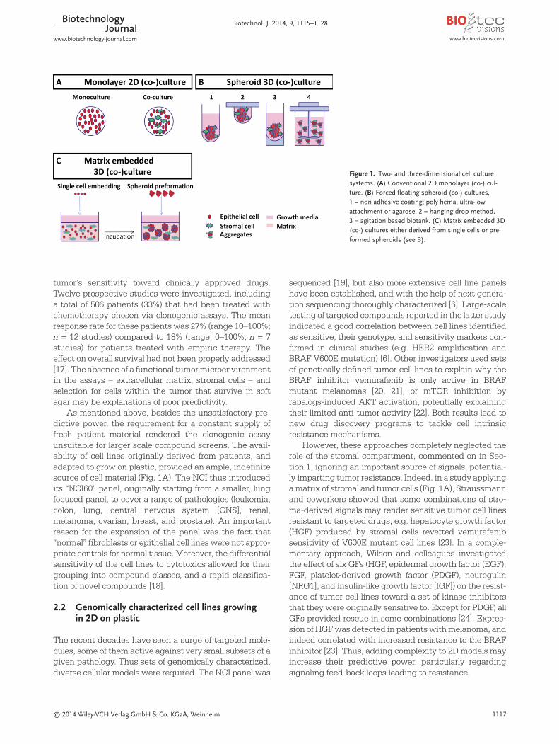
As mentioned above, besides the unsatisfactory pre-dictive power, the requirement for a constant supply offresh patient material rendered the clonogenic assayunsuitable for larger scale compound screens. The avail-ability of cell lines originally derived from patients, andadapted to grow on plastic, provided an ample, indefinitesource of cell material (Fig. 1A). The NCI thus introducedits “NCI60” panel, originally starting from a smaller, lungfocused panel, to cover a range of pathologies (leukemia,colon, lung, central nervous system [CNS], renal,melanoma, ovarian, breast, and prostate). An importantreason for the expansion of the panel was the fact that“normal” fibroblasts or epithelial cell lines were not appro-priate controls for normal tissue. Moreover, the differentialsensitivity of the cell lines to cytotoxics allowed for theirgrouping into compound classes, and a rapid classifica-tion of novel compounds [18].
2.2 Genomically characterized cell lines growing in 2D on plastic
The recent decades have seen a surge of targeted mole-cules, some of them active against very small subsets of agiven pathology. Thus sets of genomically characterized,diverse cellular models were required. The NCI panel was
sequenced [19], but also more extensive cell line panelshave been established, and with the help of next genera-tion sequencing thoroughly characterized [6]. Large-scaletesting of targeted compounds reported in the latter studyindicated a good correlation between cell lines identifiedas sensitive, their genotype, and sensitivity markers con-firmed in clinical studies (e.g. HER2 amplification andBRAF V600E mutation) [6]. Other investigators used setsof genetically defined tumor cell lines to explain why theBRAF inhibitor vemurafenib is only active in BRAFmutant melanomas [20, 21], or mTOR inhibition byrapalogs-induced AKT activation, potentially explainingtheir limited anti-tumor activity [22]. Both results lead tonew drug discovery programs to tackle cell intrinsicresistance mechanisms.
However, these approaches completely neglected therole of the stromal compartment, commented on in Sec-tion 1, ignoring an important source of signals, potential-ly imparting tumor resistance. Indeed, in a study applyinga matrix of stromal and tumor cells (Fig. 1A), Straussmannand coworkers showed that some combinations of stro-ma-derived signals may render sensitive tumor cell linesresistant to targeted drugs, e.g. hepatocyte growth factor(HGF) produced by stromal cells reverted vemurafenibsensitivity of V600E mutant cell lines [23]. In a comple-mentary approach, Wilson and colleagues investigatedthe effect of six GFs (HGF, epidermal growth factor (EGF),FGF, platelet-derived growth factor (PDGF), neuregulin[NRG1], and insulin-like growth factor [IGF]) on the resist-ance of tumor cell lines toward a set of kinase inhibitorsthat they were originally sensitive to. Except for PDGF, allGFs provided rescue in some combinations [24]. Expres-sion of HGF was detected in patients with melanoma, andindeed correlated with increased resistance to the BRAFinhibitor [23]. Thus, adding complexity to 2D models mayincrease their predictive power, particularly regardingsignaling feed-back loops leading to resistance.
www.biotecvisions.comwww.biotechnology-journal.com
BiotechnologyJournal Biotechnol. J. 2014, 9, 1115–1128
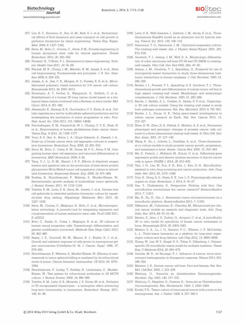
Figure 1. Two- and three-dimensional cell culturesystems. (A) Conventional 2D monolayer (co-) cul-ture. (B) Forced floating spheroid (co-) cultures,1 = non adhesive coating; poly hema, ultra-lowattachment or agarose, 2 = hanging drop method,3 = agitation based biotank. (C) Matrix embedded 3D(co-) cultures either derived from single cells or pre-formed spheroids (see B).
1118 © 2014 Wiley-VCH Verlag GmbH & Co. KGaA, Weinheim
2.3 Moving into 3D cancer models
Even complex 2D models still ignore an important fact –solid tumors grow in three dimensions (3D). A number oftechniques have been developed to cultivate cells as 3Dcultures. These include aggregating cells at the bottom ofa drop (“hanging drop”) [25], a liquid overlay technique,which prevents cells from attaching to the vessel surfaceusing a non-adherent coating (agarose- or poly-HEMAtreatment, ultra-low attachment plates with round bot-tom) [26–28], or growing cells in stirred culture systems,such as spinner flasks [29] (Fig. 1B), or stirred-tank biore-actors (Section 3). After initial aggregation, cells general-ly started to secrete extracellular matrix (ECM) compo-nents and upregulated proteins mediating cell–cell inter-actions, e.g. E-cadherin, and many, but not all, cell typesformed tight cell–cell contacts [30]. The resulting 3D sphe-roids were compact, stable and showed decreasing gra-dients of proliferating cells, oxygen levels, and nutritionsupplies from the periphery toward the spheroid’s center[27, 28, 31, 32]. These physiological characteristics of the3D spheroid closely resembled avascular tumor nodules,micro-metastases, and inter-vascular regions of large sol-id tumors as presented by Sutherland [33].
Culturing cells in 3D tumor spheroids altered theirsensitivity to cytotoxic agents such as 5-fluorouracil, cis-platin, or doxorubicin [34–38]. Several factors might con-fer this resistance to cytotoxic agents including – but notlimited to – insufficient distribution of the drugs withinthe spheroid [39], non-proliferative and hypoxic cells inthe core of the spheroid [40], induction of GF secretion(e.g. transforming growth factor [TGF]β [41]), cell–cellinteractions mediated by E-cadherin [42], and productionof extracellular matrix proteins as well as expressingresistance markers like ABCC1 multi-drug resistancepumps [37]. On the other hand, some targeted com-pounds appeared to be effective in 3D spheroids whilehaving significantly reduced or no effect in 2D monocul-tures [38, 40, 43]. For example the PI-3 kinase inhibitor PX-866 suppressed the growth of U87 glioblastoma, T47Dbreast and HCT116 colon cancer cells in 3D spheroids butnot when grown on plastic as monolayers [43]. PX-866 hasdemonstrated anti-tumor activity both as a single agentand in combination with other agents in a number ofhuman tumor models [44, 45] and is currently being eval-uated in clinical trials [46]. At least in some cases, 3Dspheroids thus seem to predict in vivo and potentiallyclinical efficacy better than 2D models.
For the above reason, and since they are highly repro-ducible, robust, easy to use and suitable for high-through-put screening, 3D spheroids represent an important 3Dplatform in the biopharmaceutical drug discovery [11].The size of cell aggregates as well as their proliferation,apoptosis, or drug responses may be measured based onfluorescence, luminescence, and absorbance by high-content imaging systems [40]. Also functional analyses
based on phenotypic RNAi screening proved to be possi-ble in high-throughput 3D spheroid systems [47].
2.4 3D and the stroma
A spheroid mono-culture system, however, still lacks thestromal compartment of a tumor. Investigators haveaddressed this issue by mixing tumor cells and fibroblastssuspensions, tumor spheroids with fibroblast/monocytesuspensions, tumor spheroids with embryonic bodies, orcombining tumor spheroids with a fibroblast/endothelialcell monolayer. Paracrine interactions, and effects of stro-mal cells on tumor cell behavior and/or vice versa havebeen reported [48–53]. This heterotypic cell–cell crosstalkwas shown to induce the formation of more compact 3Dspheroids [54] and affect inter-cellular signaling and geneexpression of the tumor cells, resulting in altered tumorcell proliferation and migration [49, 52, 55].
However, extensive characterization and validation ofthe 3D spheroid models for different cancer pathologiesare still required in order to understand how closely theyresemble the biological properties of in vivo tissue, includ-ing growth kinetics, gene expression, the architecture ofsignaling cascades, and drug treatment responses.
A first step toward a systematic comparison of pre-clinical models, including 3D spheroids, was the studyperformed by Lee et al. [27]. Histological and molecularfeatures of 29 different ovarian cancer cell lines were com-pared between 3D spheroids, 2D monocultures, andxenografts, including a correlation with literature dataavailable for the primary tumors. Comparison of 3D with2D cultures suggested up-regulation of E-cadherin, down-regulation of vimentin, decreased expression of the prolif-eration marker MIB1, and increased expression of apop-totic marker caspase-3 in spheroids. While most of the celllines formed poorly differentiated, high grade histologyspheroids, the cell line OAW42 histologically resembled awell-differentiated (grade 1) serous ovarian tumor, andUWB1.289 and BRC1 resembled moderately well differen-tiated (grade 2) serous ovarian tumors. No differentiationwas observed in 2D. The histology of the spheroidsmatched those of the original tumor in some cases, butthere was a clear selection for the poorly differentiatedhigh grade histology [27]. Adequate characterization ofthese models and their molecular signaling processes, aswell as the addition and functional analysis of the stromalcells in co-culture with tumor cells, will allow for a moredefined role of these models in the drug discovery process.
2.5 Incorporating the extracellular matrix (ECM)
Models based on spheroids growing in liquid medium stillmiss an essential component of tumor biology: the ECM.Pre-formed 3D spheroids embedded into, or sitting on topof, a gel matrix could be analyzed for cell invasion andmigration upon treatment with compounds. Using this
www.biotechnology-journal.com www.biotecvisions.com
BiotechnologyJournal Biotechnol. J. 2014, 9, 1115–1128
© 2014 Wiley-VCH Verlag GmbH & Co. KGaA, Weinheim 1119
assay, the HSP90 inhibitor 17-AAG and a phospholipaseC (PLC) γ inhibitor could be shown to prevent dissemina-tion on Matrigel™ (a laminin-rich basement membraneextract [BME] – see below) at concentrations below thatto inhibit 50% of growth (GI50). The former was also shownto interfere with invasion of U87 MG glioblastoma sphe-roids into Matrigel, again at sub GI50 concentrations [28].Cell lines like VCaP that do not form spheroids in ECM [56]can be pre-grown in medium before implantation intoECM (Fig. 1C). However, the ECM is not just an inertmatrix that provides a 3D scaffold for tumor cells to growand invade, but also plays an essential role in differentia-tion and maintenance of tissues, which may be missed insuch hybrid systems. Moreover, the ECM has been shownto provide survival and drug resistance signals in cancer(for a review, see [57]). β1 integrin signaling was shown toprotect small cell lung cancer (SCLC) cells in clonogenicsoft agar assays with added fibronectin from DNA dam-aging agents [58] breast cancer cells plated on fibronectinor collagen I from paclitaxel [59] and HER2 amplifiedbreast cancer cell lines in Matrigel from trastuzumab, per-tuzumab, as well as lapatinib [60]. Activation of theMAPK-pathway by integrin αv3β also protected breastcancer cells from paclitaxel toxicity [61]. Multiple inte-grin-targeted small molecules and antibodies are current-ly in clinical development [62].
Assays including ECM components may be performedin 2D, by coating plates with the respective proteins, butdue to the above outlined advantages of growing cells inthree dimensions, and the impact of matrix stiffness on cellgrowth and differentiation, embedding of the cells in thematrix is preferred. Indeed, matrix stiffness was shown toaffect stem cell differentiation [63, 64], tumor progression[65], invasion [66, 67], and drug sensitivity [68, 69]. 2D cul-tures have a stiffness of 1–2 GPa compared to normalbreast tissue (160 Pa), and breast cancer tissue (4 kPa).Matrigel and collagen are around 200–400 Pa [57].
Commonly used matrices in 3D cell culture are alaminin-rich BME (also called Matrigel), purified fromEngelbreth–Holm–Swarm (EHS) mouse sarcoma, com-posed mainly of laminin-111, collagen IV, and heparan sul-fate proteoglycan [70, 71], or collagen I. Mina Bissell’s lab-oratory spearheaded much of the research on 3D extra-cellular matrix embedded models (Fig. 1C). Mouse mammary epithelial cells grown on floating collagenmembrane, but not grown in 2D plastic, were shown todifferentiate to form acini, and secrete casein [72, 73].Embedding in Matrigel allowed them to form alveoli-likestructures that secreted milk proteins into a hollow lumen[74]. Whereas normal mammary cells formed hollow aciniformed by a single layer of epithelial cells, and stoppedproliferating after 10–12 days in culture, mammary tumorcells continued to proliferate, generating large spheres[75]. Genes that were down-regulated in normal humanmammary epithelial cells during differentiation in 3D cul-tures predicted for a poor outcome in breast cancer
patients [76, 77]. Moreover, the morphology of the spheresformed by the 3D Matrigel-associated cultures allowed fora clustering into clinically relevant subgroups. Most lumi-nal B subtype cell lines formed invasive stellar structuresin Matrigel, whereas luminal A and basal subtypes formedround or grape-like structures. When correlated with geneexpression analysis, cell lines forming stellar structureslacked E-cadherin and EGFR/ErbB2 expression, whereasgrape-shaped colony forming cell lines mostly over-expressed ErbB2. Strikingly, when 3D gene expressionwas compared to 2D gene expression, the main differen-tiator was the culture system rather than the cell lines[78]. For other tumor entities, similar observations weremade, e.g. prostate [56], colorectal cancer [79], and lungcancer [80]. A gene signature based on lung cancer celllines growing in 3D as smooth versus branched coloniescould classify lung cancer patients into good and badprognosis groups, albeit on a limited patient set [80].Moreover, not only primary normal and tumor cells fromthe colon may be grown in Matrigel, when supplied witha mix of GFs including R-spondin to stimulate WNT sig-naling, but potentially many other tumor types as well,thus allowing for the generation of large sets of patient-derived primary organoid cultures as potentially morepredictive tumor models [81].
In an effort to compare the effect of cell culture meth-ods on gene expression and drug responsiveness, JIMT1cell cultures were grown in 2D, as spheroids and as a 3Dmodel in Matrigel, as well as in a mouse xenograft model.Gene expression profiles revealed that JIMT1 cells cul-tured in Matrigel more closely resembled the xenograftmodel than the spheroid model. Pathway analysis of 54 genes down-regulated compared to 2D indicated thathormone (endoplasmic reticulum [ER]) and GF signaling(HGF and TGF) were most affected. One hundred and twodrugs were tested against the models, and 63 showed aneffect in at least one model. The 3D model in Matrigel wasgenerally more sensitive against the selection of com-pounds tested [82].
This latter study is another important step toward avalidation of 3D models for target validation and drug dis-covery. A recent Innovative Medicines Initiative (IMI)(www.imi.europa.eu) funded effort of a large consortiumof Academic, SME and pharmaceutical partners, PRE-DECT (www.predect.eu), aims to provide better charac-terized in vitro 3D platforms for both target validation andsubsequent pre-clinical studies with the goal to increasepredictivity of drug efficacy in patients.
3 Bioreactors: Controlled culture systems for accurate recapitulation of tumormicroenvironment
In order to increase the relevance of in vitro tumor model-ing approaches, discussed above, recreation of cellular
www.biotecvisions.comwww.biotechnology-journal.com
BiotechnologyJournal Biotechnol. J. 2014, 9, 1115–1128

1120 © 2014 Wiley-VCH Verlag GmbH & Co. KGaA, Weinheim
architecture and the microenvironment is essential. Thisincludes not only the recapitulation of 3D cellular archi-tecture and the presence of different cellular players, ECMcomponents, and nutrients, but also the maintenance atphysiological levels of physicochemical environmentalparameters known to influence drug response, such astemperature, pH, and oxygen [83–86], which could beachieved through utilization of bioreactors (Table 1).
Bioreactors are culture systems designed to provideefficient mass transfer; computer-controlled systems pro-vide on-line monitoring and automated control of envi-ronmental culture variables (temperature, pH, and dis-solved oxygen). Furthermore, the use of perfusion opera-tion modes allows simulation in vitro of consequences ofcirculatory system connections, such as shear stress andpressure, nutrient and O2 supply, and metabolite clear-ance [87], as well as supply of cytokines and GFs. Biore-actors have been extensively applied in biopharmaceuti-cal industrial processing. The knowledge accumulatedfacilitated the translation to the production of advancedmedical therapeutic products, such as stem cells for celltherapy [88] and tissue engineering applications [89], inwhich cells and their microenvironment are the productof interest. In recent years, bioreactor design and devel-opment have been toward improved accuracy in controlof cellular microenvironment with reduced shear, reducedworking volumes and parallelization.
Currently, the range of bioreactor types with potentialapplication in cancer in vitro models is wide: from classi-cal stirred culture vessels and rotary cell culture systems(RCCS) to last-generation microfluidic devices [88, 90]
(Fig. 2), plus an array of custom-made designs [91–93],which have been applied for the generation of 3D cellularstructures, as well as for their culture under dynamic andcontrolled conditions.
3.1 Stirred culture vessels
These include spinner vessels and computer-controlledstirred-tank bioreactors (Fig. 2), providing a dynamicstirred environment, overcoming mass transport and gastransfer limitations, as well as culture heterogeneity pre-sented by other systems [88] (Fig. 2A, Table 1). Stirredvessels are scalable systems, with simple design andoperation, extensively characterized hydrodynamically.Manipulation of parameters such as vessel and impellerdesign and stirring rate allow applicability to an array ofcell types with distinct aggregative capabilities and sen-sitivities to shear stress, including a large panel of tumorcell lines [94–97].
Another important feature is the feasibility to performnon-destructive sampling along time, enabling continu-ous monitorization/characterization as well as retrieval ofmaterial for further applications [98, 99]. Importantly,stirred-tank bioreactors are highly flexible, and canaccommodate different 3D culture strategies, from cellspheroids, to microcarrier/scaffold and microencapsulat-ed mono and co-cultures [99–102], presenting widespreadpotential. Hirschhaeuser et al. [103, 104], evaluated theefficacy of catumaxomab (anti-epithelial cell adhesionmolecule [EpCAM] × anti-CD3 bispecific antibody) in co-culture of head and neck squamous cell carcinoma sphe-roids and human peripheral blood mononuclear cells inspinner vessels. A dose-dependency effect was observed,including reduced spheroid volume, increased immune-cell infiltration and cytokine secretion, decreased propor-tion of proliferating cells, and reduced ability of colony for-mation.
The main limitations of stirred-culture vessels are thehydrodynamic shear force-related cellular stress, promot-ed by stirring, and the high culture volumes associatedwith these systems (minimum of 50–80 mL). Microencap-sulation and scaffold strategies can minimize cell expo-
www.biotechnology-journal.com www.biotecvisions.com
BiotechnologyJournal Biotechnol. J. 2014, 9, 1115–1128
Table 1. Overview over advantages and disadvantages of different in vitro/ex vivo models discussed in the review
2D Spheroid Soft agar Complex 3D Bioreactors Tissue slicescontrolled culture
systems
High throughput +++ +++ ++ + − to +++ −Long-term cultivation +++ ++ ++ + +++ −Reproducibility +++ ++ ++ ++ +++ −Handling read out +++ + + + + +Handling of cultivation +++ ++ ++ ++ +++ ++Tumor microenvironment −/+ −/+ −/+ ++ ++ +++Resemblance of in vivo − + + ++ ++ +++
Figure 2. Bioreactor systems for culture of in vitro tumor models. (A) Stirred culture vessels; (B) rotary cell culture systems; (C) microfluidicdevices.

© 2014 Wiley-VCH Verlag GmbH & Co. KGaA, Weinheim 1121
sure to shear-stress [100] and improve microenvironmentrecapitulation by accumulation of secreted factors andECM components [105]. Recent efforts have been madetoward the development of smaller-scale systems, withcommercially available solutions starting to emerge, suchas the Ambr® system (TAP Biosystems, recently acquiredby Sartorius), with working volumes of 10–15 mL.
The characteristics described make stirred-tankbioreactors particularly suitable for long term [106] and/orrepeated-dose studies [99], as well for large-scale produc-tion for feeding of high-throughput and parallel testing inminiaturized devices, such as microfluidic-based biore-actors (see below). Nevertheless, these systems have notbeen used to their full potential in cancer research.
3.2 Rotary cell culture systems (RCCS)
These are based on the rotating wall vessel bioreactor(RWV) developed by National Aeronautics and SpaceAdministration (NASA) for studying tissue generationand cell behavior under microgravity conditions(reviewed in [107]) and are commercialized by Synthecon(www.synthecon.com). RWV are horizontally rotatingcylindrical culture vessels with no internal stirring mech-anisms (Fig. 2B), providing low shear and turbulence.Cells remain suspended in near free-fall, simulatingmicrogravity conditions, which promote cell–cell interac-tions and minimize mechanical cell damage [108, 109], inthe absence or presence of scaffolds [110, 111]. Gas trans-fer is achieved by diffusion through a central silicon mem-brane and RCCS are compatible with perfusion operationmodes. The main disadvantages are associated withmass transfer limitations throughout the vessel, due torestricted control of nutrient/gas concentrations, whichmay result in heterogeneous microenvironments [88].RCCS have been applied to 3D cancer cell culture foralmost 40 years, to study tumor formation and progressionand tumor microenvironment [112]. Numerous co-culturemodels in which the stromal compartment is representedhave been described, such as colon and prostate, usingadenocarcinoma cell lines with human colonic fibroblasts[108] and human prostate carcinoma together withprostate or bone stromal cells [113]. These induced genet-ic, morphologic, and behavioral changes [114], namelystructural organization similar to normal colon cryptdevelopment and maintenance of androgen responsive-ness and prostate-specific antigen (PSA) production,respectively. Evidence cumulated through the years pointto altered gene expression and cell behavior due to micro-gravity [112]; a recent study focused on characterizationof poorly differentiated thyroid cancer cells cultured dur-ing the Shenzhou-8 space mission, under real micrograv-ity conditions, identified mechanisms of growth inhibi-tion by cytokine secretion and gene expression profiling[115]. Microgravity culture is an interesting and usefultool to study cancer mechanisms; nevertheless transla-
tion of findings under these conditions into human dis-ease models may not be easy.
3.3 Microfluidic devices
Microfluidic devices, or micro-bioreactors, are efficientsmall-scale systems with precise control of cell microen-vironment (Fig. 2C). Recent advances in microfluidictechnology boosted the development of novel in vitro drugscreening methods compatible with high-throughputapplications [116] and the development of “organs-on-chips,” composed of biologically functional tissue mimet-ics, in which inter-tissue interactions can be reconstitut-ed [117]. Microenvironment can be controlled by preciseadjustment of fluid flows, allowing perfusion operationmodes, resulting in shear stress and mechanical strain.The main limitations of these systems are associated withlow scalability and high complexity.
An array of designs have been proposed in recentyears, with highly diverse innovative approaches to tumormicroenvironment recapitulation [116, 118–120]. Recent-ly, a microfluidic model to analyze the specificity ofhuman breast cancer metastases to bone has beendescribed [121]. By recreating a vascularized osteo-cellconditioned microenvironment using bone marrow-derived mesenchymal stem cells and endothelial cells, theauthors identified molecular pathways critical for extrava-sation of breast cancer cells, involving breast cancer cellsurface receptor CXCR2 and bone-secreted chemokineCXCL5 [121]. Microfluidic systems for culture of patient-derived material have also been described, such as the“thick-tissue bioreactor” developed by McCawley and co-workers [122] for culture of biopsied portion of breasttumor tissue in parallel microchambers. As a proof-of-con-cept for drug testing by delivery, the chemotherapeuticagent docetaxel and protease inhibitor compounds wereadministered to breast tumor cell lines cultured inMatrigel [122]. Zhang et al. [123] described a microfluidicplatform for culture of patient-derived myeloma cells byemulating the dynamic physiology of the bone marrowmicroenvironment. The system uses a three-dimensionalossified tissue to mimic the tumor niche and recapitulateinteractions between bone marrow cells and osteoblasts[123]. Many of these technical advances should now begradually incorporated into standard practice.
4 Tumor tissue slices: Culture systempreserving the individual tumormicroenvironment
In contrast to more simplistic models, tissue slices pro-vide the opportunity to study tumor cells in the context ofan intact microenvironment. This includes all other celltypes of the particular tissue as well as the extracellularmatrix, which results in the conservation of naturally
www.biotecvisions.comwww.biotechnology-journal.com
BiotechnologyJournal Biotechnol. J. 2014, 9, 1115–1128

1122 © 2014 Wiley-VCH Verlag GmbH & Co. KGaA, Weinheim
occurring interactions in slice culture (Table 1). Given theinfluence of the tumor environment on drug sensitivityand other aspects of tumor biology [124], fresh tumor sam-ples would appear more suitable to study individualpatient tumors than established immortalized cell lines.Furthermore, tissue slice culture has the potential to cov-er the whole clinical spectrum of solid tumors including allstages and grades from well-differentiated slow-growingto poorly differentiated fast-growing subtypes, whereasthe vast majority of cell lines represent only high-stageand poorly differentiated tumors [125]. Consequently,short-term cultivated tumor tissue slices could mirrormore closely the intra- and inter-tumoral variability of sol-id tumors typically observed in vivo including prolifera-tive aspects as well as the response to drug treatment.The complexity of this model system comes along withchallenges that have to be addressed. In the followingsection, we discuss these challenges together with thepotential and limitations of tissue slice culture.
4.1 History of “organotypic tissue slices”
The idea of cultivating “organotypic tissue slices” is cer-tainly not new. In the 1920s, decades before the firstimmortalized cell line was established, free-hand cutpieces of tissue from rat and human carcinomas havealready been used to study cell metabolism [126, 127].This was followed by a variety of studies with explants ofhuman tumors cultivated in hanging drops [128], on plas-ma clots [129, 130] or in liquid media [131] many of themwith the aim to use this model for prediction of individualdrug sensitivity. However, after establishment of the HeLacell line in 1952 [132], immortalized tumor cell linesbecame the most frequently used model system world-wide and still represent the backbone of basic cancerresearch (see Section 2). With the introduction of high-precision tissue slicers [133], which allowed preparationof slices with constant thickness and therefore improvedreproducibility, tissue slice culture experienced a “renais-sance”. Consequently, tissue slices became an attractivealternative to immortalized cell lines since at the time itwas widely recognized that there is a need for more com-plex model systems. In the last decades, precision-cutslices from various patient tumors such as carcinomasfrom breast [134–139], lung [138, 140–142], brain [143,144], ovary [145, 146], cervix [147], kidney [148], andprostate [149] were introduced and used in various fieldsof cancer research.
4.2 Tumor tissue slice cultivation and applications
Primarily, tumor tissue slices have been used to deter-mine drug responses including responses to cytotoxicchemotherapeutics, small molecule inhibitors, and otherdrugs [135–138, 141, 142, 144, 146, 147, 150–154]. Cellularresponses were assessed on different levels ranging from
drug uptake, proliferation and induction of cell death tochanges in protein levels, and gene expression. Most ofthese studies are restricted to the analysis of the tumorcells’ response to drug treatment, whereas some includethe investigation of the role of cancer associated fibrob-lasts in the tumor’s response to cytotoxic substances [138,142]. Furthermore, localization and migration of immunecells in tumor tissue was examined using tissue slice cul-ture techniques [155]. Successful infections of cancercells in tissue slices with different viruses have beendescribed showing that this ex vivo model is also suitablefor the validation of alternative anti-cancer approachessuch as oncolytic viral infection or gene therapy [143,156–160]. In addition, tissue slice culture has been usedfor validation purposes in studies utilizing immortalizedcell lines from different tumor entities grown in 2D [139,145, 148]. Meanwhile, drug testing services based onpatient-derived tumor tissue slices are commerciallyavailable (http://indivumed.com).
Despite the prevalent use of tissue slices in cancerresearch, a systematic characterization and a comparisonwith the “original” tumor has only been performed at afairly rudimentary level so far. If at all, such comparisonsare restricted to viability [135, 140, 146], proliferation sta-tus [140, 142], apoptosis [135, 140], and marker expression[140]. All these studies come to the conclusion that tis-sues from different tumor entities can be maintained exvivo for 3–7 days without significant loss of viability/pro-liferation or induction of apoptosis and the maintenanceof overall tissue architecture. Whether their detailed bio-chemistry continued to reflect, with fidelity, that of thetumor from which they were derived is addressed below.
4.3 Factors that influence “tissue slice quality”,limitations and suggested solutions
In principle, the criteria for successful tumor slice cultiva-tion were summarized previously in an excellent reviewon tissue slices from non-malignant organs by Fisher andVickers [161]. However, due to different intraoperativemanipulation and pathology processing it is challengingto meet such “minimum” standard criteria for tumor tis-sue slices. Even with minimized warm ischemia times,optimized preservation solution and rapid slicing proce-dures there will be a variability that is beyond the controlof the researcher (Fig. 3). Furthermore, inter- and intra-tumoral heterogeneity [162] complicates slicing and thecultivation procedure itself. In different tumors from thesame entity and even within an individual tumor one hasto deal with variability in terms of consistency [135, 136,140], but also regarding occurrence of necrosis, prolifera-tion [140, 142] and cell composition [142]. Consequently,the question arises if a “standard operating” procedure oftumor tissue slice culture is feasible or if individuallyadapted procedures might be necessary. Even though anadequate characterization of the “original” tumor tissue
www.biotechnology-journal.com www.biotecvisions.com
BiotechnologyJournal Biotechnol. J. 2014, 9, 1115–1128

© 2014 Wiley-VCH Verlag GmbH & Co. KGaA, Weinheim 1123
cannot be done before starting tissue slice culture, thisshould be a pre-requisite for the interpretation of any dataobtained from cultivated tissue slices. Furthermore, everyexperiment should be performed in replicates with slicesrepresenting different sites of the tumor.
Besides availability of sufficient tissue material andethical issues raised by research with patient tumors, themost relevant disadvantage of this cultivation techniqueis the difficulty to define adequate controls. This is simplybased on the fact that ex vivo results from individualtumors cannot be reproduced in vivo. For this reason andin analogy to clinical studies, a considerable amount ofpatient samples are needed to achieve reliable results.Ultimately, to answer the question if tumor tissue sliceswill really be predictive of the patients’ therapy response,a systematic comparison with clinical trials using thesame drugs in vivo and ex vivo will be required. Suchcomparisons have not been performed so far.
There are also important technical issues to be con-sidered. Various mechanical stresses can influence tissuequality during surgery, pathology processing, and slicing.In addition, changes in temperature, oxygen levels andthe availability of nutrients definitely occur both duringthe preparation of the slice and its maintenance (Fig. 3).Obviously, such non-reversible perturbations do result ina restriction of tissue slice culture to short-term experi-ments. These factors not only determine the tissue quali-ty at the starting point of cultivation, and thereon, butmight also influence both the drug sensitivity of cancercells ex vivo and their use to investigate tumor biochem-istry. It is therefore essential to optimize slicing proce-dures as well as culture conditions to minimize such arti-factually induced changes (Fig. 3). Different slicing pro-cedures with slicers such as Krumdieck [135, 138, 146,151, 157, 163], manual choppers [153, 154], and vibra -tomes [140, 153, 154, 163] as well as different culture con-ditions including floating [135, 136, 138, 142, 152], filter-supported [140, 148, 153, 154], and rotating [149] systemshave been published. However, to date no systematicanalysis or comparison of the different preparation andculture conditions is available. Such “basic validation”studies could be performed with tumors from cell linederived xenografts, or even syngeneic models of mouse
tumors, providing relatively infinite and homogenoussource of tissue material. The syngeneic models wouldhave the advantage of providing tumors with a completestroma, particularly if genetically engineered mouse mod-els were to be investigated as surrogates for humantumors.
What remains to be established, permitting tissueslices to be a valuable adjunct to a variety of 3D platformsin vitro, is with what fidelity the biochemical circuitry ofa slice represents that of the in situ tumor from which itwas derived. What is the impact of the slicing procedureitself on the activation of stress and inflammatoryresponses? Do these procedures “re-wire” the biochemi-cal circuitry, both of the tumor cells and their stroma, suf-ficiently to influence canonical pathways that determinedrug response, for example by modulating DNA repairpathways or signaling cascades? Then, on incubation, atwhat period can an optimal window of fidelity to the orig-inal tumor be described permitting investigation of eitherdrug responses or the circuitry associated with a novel,potential drug target? Until these questions are addressedin some depth, tumor slices should be used with circum-spection. Claims that fresh slices reflect the in situ tumor,permitting programs of personalized medicine(http://indivumed.com, [139, 140, 153, 154]) should becarefully scrutinized. The IMI program PREDECT(www.predect.eu) has one of its goals the task of compre-hensively characterizing the biochemistry of tumor tissueslices. It hopes to define the limits of this exciting techni-cal platform, which should capture tumor complexity andheterogeneity in vitro.
5 Concluding remarks
In reviewing the considerable progress that has beenmade in attempts to create in vitro models that are morerepresentative of tumor complexity, particularly with cellsthat are surviving and/or growing in three dimensionswith stroma, a number of caveats were highlighted.These complex in vitro platforms are surely an advance,recognizing that existing models have limitations (Table 1). Several novel models have been shown to differ
www.biotecvisions.comwww.biotechnology-journal.com
BiotechnologyJournal Biotechnol. J. 2014, 9, 1115–1128
Figure 3. Flow diagram illustrating the steps in-betweenthe in vivo tumor and the cultivated and analyzed tumortissue slice cultivated ex vivo.
1124 © 2014 Wiley-VCH Verlag GmbH & Co. KGaA, Weinheim
in phenotypic and molecular characteristics from “stan-dard” two-dimensional cultures. This is encouraging. Isthis enough to move these advanced models alongsideexisting models or even replace them? The former, yes.The latter, no. These “new” models should be embracedby industry and the academic community so that theycan be characterized, tested and refined further by thecommunity. “Validation” that these novel models will bemore predictive of clinical outcome will require a retro-spective analysis of their utility based on clinical results.
What remains to be done? A two-pronged approach isrequired: first, there are many continuing technical ques-tions to address. How to grow mixed stromal-tumor com-plexes in appropriate ratios reflecting pathology? Whattype of ECM to add – such as collagen – Matrigel mixes orhow to move toward defined ECM components and whatare the flow and physico-chemical environmental charac-teristics of a particular pathology/cancer stage? How tooptimize tissue slice preparation and maintenance? Theword “optimize” is key. Optimize toward what? This iswhere considerable effort is required. While cancer celllines have been characterized for changes in theirgenomes and compared with human tumors, what has notbeen sufficiently characterized is how the biochemical cir-cuitry of a novel in vitro platform (and even the 2D celllines) compares with a tumor in situ. Demonstrating thata novel platform is “different” is insufficient. As it becomesclear that a single target approach to drug discovery isunsustainable, the ever increasing power of systems biol-ogy should allow hypotheses to be tested regarding therational drugging of complimentary “nodes” in a network[164]. A systems approach is only viable if models ofpathology and biology, used for validation, are representa-tive of in vivo conditions. While very important strideshave been made in modeling biology and pathology in ani-mals, for example creating models to validate novel targetsusing genetically engineered mice [165], there is a contin-ued need to develop and advance the use of appropriate invitro models, such as those described here. However, thecommunity should be prudent about the use of novel plat-forms which often remain as “black boxes” and shouldcontribute to their further characterization.
The authors thank all partners especially the postdocs inPREDECT for their great contributions to the project. Thiswork has been supported by the Innovative Medicines Initiative Joint Undertaking under grant agreement n° 115188, resources of which are composed of financialcontribution from the European Union’s Seventh Frame-work Programme (FP7/2007-2013) and EFPIA companiesin kind contribution. We apologize to the many authorswhom we have not cited due to limits to the length of thisreview. Work Package and Platform Leaders and Repre-sentatives of the Partners in IMI PREDECT: ElizabethAnderson, Simon Barry, Erwin Boghaert, Cathrin Brisken,
Mike Burbridge, Yolanda Chong, Sylvia Grünewald, Gui-do Jenster, Pekka Kallio, Olli Kallioniemi, Juha Klefström,Johan Lundin, Outi Monni, Matthias Nees, Adam Nopora,Moshe Oren, Varda Rotter, Jack Schalken, Julia Schüler,Matt Smalley, Wolfgang Sommergruber, Jan Trapman,Wytske van Weerden, Emmy Verschuren, Loredana Vesci,Jaak Vilo.
The authors declare no financial or commercial conflict ofinterest.
6 References
[1] Cook, D., Brown, D., Alexander, R., March, R. et al., Lessons learnedfrom the fate of AstraZeneca’s drug pipeline: A five-dimensionalframework. Nat. Rev. Drug Discov. 2014, 13, 419–431.
[2] Wilding, J. L., Bodmer, W. F., Cancer cell lines for drug discovery anddevelopment. Cancer Res. 2014, 74, 2377–2384.
[3] Hay, M., Thomas, D. W., Craighead, J. L., Economides, C., Rosenthal,J., Clinical development success rates for investigational drugs. Nat.Biotechnol. 2014, 32, 40–51.
[4] Bhattacharjee, Y., Biomedicine. Pharma firms push for sharing ofcancer trial data. Science 2012, 338.
[5] Arrowsmith, J., Trial watch: Phase III and submission failures:2007–2010. Nat. Rev. Drug Discov. 2011, 10, 87.
[6] Barretina, J., Caponigro, G., Stransky, N., Venkatesan, K. et al., TheCancer Cell Line Encyclopedia enables predictive modelling of anti-cancer drug sensitivity. Nature 2012, 483, 603–607.
[7] Garnett, M. J., Edelman, E. J., Heidorn, S. J., Greenman, C. D. et al.,Systematic identification of genomic markers of drug sensitivity incancer cells. Nature 2012, 483, 570–575.
www.biotechnology-journal.com www.biotecvisions.com
BiotechnologyJournal Biotechnol. J. 2014, 9, 1115–1128
PREDECT is one of the major Innovative Medicines Initiatives (IMI) of
the European Union joining nine academic laboratories with three
small biotech enterprises and seven major pharmaceutical companies,
aiming to investigate and characterize in vitro/ex vivo platforms for
target validation and drug discovery. The consortium is a response to
the alarmingly high rate of initially promising drugs failing in clinical
trials – which may reflect the currently used laboratory cancer models
that (more or less) fail to represent the complexity and heterogeneity
of human tumors. Professor John Hickman coordinates the PREDECT
consortium, Dr. Ralph Graeser is the deputy coordinator.
© 2014 Wiley-VCH Verlag GmbH & Co. KGaA, Weinheim 1125
[8] Haibe-Kains, B., El-Hachem, N., Birkbak, N. J., Jin, A. C. et al., Incon-sistency in large pharmacogenomic studies. Nature 2013, 504,389–393.
[9] Egeblad, M., Nakasone, E. S., Werb, Z., Tumors as organs: Complextissues that interface with the entire organism. Dev. Cell 2010, 18,884–901.
[10] Grainger, D. W., Cell-based drug testing; this world is not flat. Adv.Drug Deliv. Rev. 2014, 69–70, vii–xi.
[11] Thoma, C. R., Zimmermann, M., Agarkova, I., Kelm, J. M., Krek, W.,3D cell culture systems modeling tumor growth determinants incancer target discovery. Adv. Drug Deliv. Rev. 2014, 69–70, 29–41.
[12] Eagle, H., Foley, G. E., Cytotoxicity in human cell cultures as a pri-mary screen for the detection of anti-tumor agents. Cancer Res.1958, 18, 1017–1025.
[13] Hirschberg, E., Tissue culture in cancer chemotherapy screening.Cancer Res. 1958, 18, 869–878.
[14] Hamburger, A. W., Salmon, S. E., Primary bioassay of human tumorstem cells. Science 1977, 197, 461–463.
[15] Shoemaker, R. H., Wolpert-DeFilippes, M. K., Kern, D. H., Lieber, M. M. et al., Application of a human tumor colony-forming assay tonew drug screening. Cancer Res. 1985, 45, 2145–2153.
[16] Fiebig, H. H., Maier, A., Burger, A. M., Clonogenic assay with estab-lished human tumour xenografts: Correlation of in vitro to in vivoactivity as a basis for anticancer drug discovery. Eur. J. Cancer(Oxford Engl.) 2004, 40, 802–820.
[17] Cortazar, P., Johnson, B. E., Review of the efficacy of individualizedchemotherapy selected by in vitro drug sensitivity testing forpatients with cancer. J. Clin. Oncol. 1999, 17, 1625–1631.
[18] Shoemaker, R. H., The NCI60 human tumour cell line anticancerdrug screen. Nat. Rev. Cancer 2006, 6, 813–823.
[19] Abaan, O. D., Polley, E. C., Davis, S. R., Zhu, Y. J. et al., The exomesof the NCI-60 panel: A genomic resource for cancer biology and sys-tems pharmacology. Cancer Res. 2013, 73, 4372–4382.
[20] Hatzivassiliou, G., Song, K., Yen, I., Brandhuber, B. J. et al., RAFinhibitors prime wild-type RAF to activate the MAPK pathway andenhance growth. Nature 2010, 464, 431–435.
[21] Poulikakos, P. I., Zhang, C., Bollag, G., Shokat, K. M., Rosen, N., RAFinhibitors transactivate RAF dimers and ERK signalling in cells withwild-type BRAF. Nature 2010, 464, 427–430.
[22] O’Reilly, K. E., Rojo, F., She, Q.-B., Solit, D. et al., mTOR inhibitioninduces upstream receptor tyrosine kinase signaling and activatesAkt. Cancer Res. 2006, 66, 1500–1508.
[23] Straussman, R., Morikawa, T., Shee, K., Barzily-Rokni, M. et al.,Tumour micro-environment elicits innate resistance to RAFinhibitors through HGF secretion. Nature 2012, 487, 500–504.
[24] Wilson, T. R., Fridlyand, J., Yan, Y., Penuel, E. et al., Widespreadpotential for growth-factor-driven resistance to anticancer kinaseinhibitors. Nature 2012, 487, 505–509.
[25] Kelm, J. M., Timmins, N. E., Brown, C. J., Fussenegger, M., Nielsen,L. K., Method for generation of homogeneous multicellular tumorspheroids applicable to a wide variety of cell types. Biotechnol. Bio-eng. 2003, 83, 173–180.
[26] Friedrich, J., Seidel, C., Ebner, R., Kunz-Schughart, L. A., Spheroid-based drug screen: Considerations and practical approach. Nat. Pro-toc. 2009, 4, 309–324.
[27] Lee, J. M., Mhawech-Fauceglia, P., Lee, N., Parsanian, L. C. et al., A three-dimensional microenvironment alters protein expressionand chemosensitivity of epithelial ovarian cancer cells in vitro. Lab.Invest. 2013, 93, 528–542.
[28] Vinci, M., Gowan, S., Boxall, F., Patterson, L. et al., Advances inestablishment and analysis of three-dimensional tumor spheroid-based functional assays for target validation and drug evaluation.BMC Biol. 2012, 10, 29.
[29] Song, K.-D., Liu, T.-Q., Li, X.-Q., Cui, Z.-F. et al., Three-dimensionalexpansion: In suspension culture of SD rat’s osteoblasts in a rotatingwall vessel bioreactor. Biomed. Environ. Sci. 2007, 20, 91–98.
[30] Lin, R.-Z., Lin, R.-Z., Chang, H.-Y., Recent advances in three-dimen-sional multicellular spheroid culture for biomedical research.Biotechnol. J. 2008, 3, 1172–1184.
[31] Lin, R.-Z., Chou, L.-F., Chien, C.-C. M., Chang, H.-Y., Dynamic analy-sis of hepatoma spheroid formation: Roles of E-cadherin and beta1-integrin. Cell Tissue Res. 2006, 324, 411–422.
[32] Kunz-Schughart, L. A., Multicellular tumor spheroids: Intermediatesbetween monolayer culture and in vivo tumor. Cell Biol. Int. 1999, 23,157–161.
[33] Sutherland, R. M., Cell and environment interactions in tumormicroregions: The multicell spheroid model. Science 1988, 240,177–184.
[34] Frankel, A., Buckman, R., Kerbel, R. S., Abrogation of taxol-inducedG2-M arrest and apoptosis in human ovarian cancer cells grown asmulticellular tumor spheroids. Cancer Res. 1997, 57, 2388–2393.
[35] Kerr, D. J., Wheldon, T. E., Kerr, A. M., Freshney, R. I., Kaye, S. B., Theeffect of adriamycin and 4′-deoxydoxorubicin on cell survival ofhuman lung tumour cells grown in monolayer and as spheroids. Br.J. Cancer 1986, 54, 423–429.
[36] Kobayashi, H., Man, S., Graham, C. H., Kapitain, S. J. et al., Acquiredmulticellular-mediated resistance to alkylating agents in cancer.Proc. Natl. Acad. Sci. USA 1993, 90, 3294–3298.
[37] Longati, P., Jia, X., Eimer, J., Wagman, A. et al., 3D pancreatic carci-noma spheroids induce a matrix-rich, chemoresistant phenotypeoffering a better model for drug testing. BMC Cancer 2013, 13, 95.
[38] Tung, Y.-C., Hsiao, A. Y., Allen, S. G., Torisawa, Y. et al., High-throughput 3D spheroid culture and drug testing using a 384 hang-ing drop array. The Analyst 2011, 136, 473–478.
[39] Tunggal, J. K., Cowan, D. S., Shaikh, H., Tannock, I. F., Penetration ofanticancer drugs through solid tissue: A factor that limits the effec-tiveness of chemotherapy for solid tumors. Clin. Cancer Res. 1999, 5,1583–1586.
[40] Wenzel, C., Riefke, B., Gründemann, S., Krebs, A. et al., 3D high-con-tent screening for the identification of compounds that target cellsin dormant tumor spheroid regions. Exp. Cell Res. 2014, 323, 131–143.
[41] Ohmori, T., Yang, J. L., Price, J. O., Arteaga, C. L., Blockade of tumorcell transforming growth factor-betas enhances cell cycle progres-sion and sensitizes human breast carcinoma cells to cytotoxicchemotherapy. Exp. Cell Res. 1998, 245, 350–359.
[42] Green, S. K., Francia, G., Isidoro, C., Kerbel, R. S., Antiadhesive anti-bodies targeting E-cadherin sensitize multicellular tumor spheroidsto chemotherapy in vitro. Mol. Cancer Ther. 2004, 3, 149–159.
[43] Howes, A. L., Chiang, G. G., Lang, E. S., Ho, C. B. et al., The phos-phatidylinositol 3-kinase inhibitor, PX-866, is a potent inhibitor ofcancer cell motility and growth in three-dimensional cultures. Mol.Cancer Ther. 2007, 6, 2505–2514.
[44] Ihle, N. T., Williams, R., Chow, S., Chew, W. et al., Molecular phar-macology and antitumor activity of PX-866, a novel inhibitor of phos-phoinositide-3-kinase signaling. Mol. Cancer Ther. 2004, 3, 763–772.
[45] Koul, D., Shen, R., Kim, Y.-W., Kondo, Y. et al., Cellular and in vivoactivity of a novel PI3K inhibitor, PX-866, against human glioblas-toma. Neuro-Oncology 2010, 12, 559–569.
[46] Rodon, J., Dienstmann, R., Serra, V., Tabernero, J., Development ofPI3K inhibitors: Lessons learned from early clinical trials. Nat. Rev.Clin. Oncol. 2013, 10, 143–153.
[47] Thoma, C. R., Stroebel, S., Rösch, N., Calpe, B. et al., A high-through-put-compatible 3D microtissue co-culture system for phenotypicRNAi screening applications. J. Biomol. Screen. 2013, 18, 1330–1337.
[48] Gottfried, E., Kunz-Schughart, L. A., Andreesen, R., Kreutz, M.,Brave little world: Spheroids as an in vitro model to study tumor-immune-cell interactions. Cell Cycle (Georgetown Tex.) 2006, 5,691–695.
[49] Hauptmann, S., Zwadlo-Klarwasser, G., Jansen, M., Klosterhalfen,B., Kirkpatrick, C. J., Macrophages and multicellular tumor sphe-
www.biotecvisions.comwww.biotechnology-journal.com
BiotechnologyJournal Biotechnol. J. 2014, 9, 1115–1128
1126 © 2014 Wiley-VCH Verlag GmbH & Co. KGaA, Weinheim
roids in co-culture: A three-dimensional model to study tumor-hostinteractions. Evidence for macrophage-mediated tumor cell prolifer-ation and migration. Am. J. Pathol. 1993, 143, 1406–1415.
[50] Kunz-Schughart, L. A., Schroeder, J. A., Wondrak, M., van Rey, F. et al., Potential of fibroblasts to regulate the formation of three-dimensional vessel-like structures from endothelial cells in vitro.Am. J. Physiol. Cell Physiol. 2006, 290, C1385–C1398.
[51] Kunz-Schughart, L. A., Heyder, P., Schroeder, J., Knuechel, R., A het-erologous 3-D coculture model of breast tumor cells and fibroblaststo study tumor-associated fibroblast differentiation. Exp. Cell Res.2001, 266, 74–86.
[52] Seidl, P., Huettinger, R., Knuechel, R., Kunz-Schughart, L. A., Three-dimensional fibroblast-tumor cell interaction causes downregula-tion of RACK1 mRNA expression in breast cancer cells in vitro. Int.J. Cancer 2002, 102, 129–136.
[53] Brouty-Boyé, D., Magnien, V., Myofibroblast and concurrent ED-Bfibronectin phenotype in human stromal cells cultured from non-malignant and malignant breast tissue. Eur. J. Cancer (Oxford Engl.)1990 1994, 30A, 66–73.
[54] Costa, E. C., Gaspar, V. M., Coutinho, P., Correia, I. J., Optimizationof liquid overlay technique to formulate heterogenic 3D co-culturesmodels. Biotechnol. Bioeng. 2014, 111, 1672–1685.
[55] Amann, A., Zwierzina, M., Gamerith, G., Bitsche, M. et al., Develop-ment of an innovative 3D cell culture system to study tumour–stro-ma interactions in non-small cell lung cancer cells. PLoS ONE 2014,9, e92511.
[56] Härmä, V., Virtanen, J., Mäkelä, R., Happonen, A. et al., A compre-hensive panel of three-dimensional models for studies of prostatecancer growth, invasion and drug responses. PLoS ONE 2010, 5,e10431.
[57] Correia, A. L., Bissell, M. J., The tumor microenvironment is a dom-inant force in multidrug resistance. Drug Resist. Updat. 2012, 15,39–49.
[58] Sethi, T., Rintoul, R. C., Moore, S. M., MacKinnon, A. C. et al., Extra-cellular matrix proteins protect small cell lung cancer cells againstapoptosis: A mechanism for small cell lung cancer growth and drugresistance in vivo. Nat. Med. 1999, 5, 662–668.
[59] Aoudjit, F., Vuori, K., Integrin signaling inhibits paclitaxel-inducedapoptosis in breast cancer cells. Oncogene 2001, 20, 4995–5004.
[60] Huang, C., Park, C. C., Hilsenbeck, S. G., Ward, R. et al., β1 integrinmediates an alternative survival pathway in breast cancer cellsresistant to lapatinib. Breast Cancer Res. 2011, 13, R84.
[61] Menendez, J. A., Vellon, L., Mehmi, I., Teng, P. K. et al., A novelCYR61-triggered “CYR61-alphavbeta3 integrin loop” regulatesbreast cancer cell survival and chemosensitivity through activationof ERK1/ERK2 MAPK signaling pathway. Oncogene 2005, 24, 761–779.
[62] Danen, E. H. J., Integrin signaling as a cancer drug target. ISRN CellBiol. 2013, 2013, 1–14.
[63] McBeath, R., Pirone, D. M., Nelson, C. M., Bhadriraju, K., Chen, C. S.,Cell shape, cytoskeletal tension, and RhoA regulate stem cell lineagecommitment. Dev. Cell 2004, 6, 483–495.
[64] Park, J. S., Chu, J. S., Tsou, A. D., Diop, R. et al., The effect of matrixstiffness on the differentiation of mesenchymal stem cells inresponse to TGF-β. Biomaterials 2011, 32, 3921–3930.
[65] Levental, K. R., Yu, H., Kass, L., Lakins, J. N. et al., Matrix crosslink-ing forces tumor progression by enhancing integrin signaling. Cell2009, 139, 891–906.
[66] Pathak, A., Kumar, S., Independent regulation of tumor cell migra-tion by matrix stiffness and confinement. Proc. Natl. Acad. Sci. USA2012, 109, 10334–10339.
[67] Soman, P., Kelber, J. A., Lee, J. W., Wright, T. N. et al., Cancer cellmigration within 3D layer-by-layer microfabricated pho-tocrosslinked PEG scaffolds with tunable stiffness. Biomaterials2012, 33, 7064–7070.
[68] Schrader, J., Gordon-Walker, T. T., Aucott, R. L., van Deemter, M. et al., Matrix stiffness modulates proliferation, chemotherapeuticresponse, and dormancy in hepatocellular carcinoma cells. Hepatol-ogy (Baltimore Md.) 2011, 53, 1192–1205.
[69] Zustiak, S., Nossal, R., Sackett, D. L., Multiwell stiffness assay for thestudy of cell responsiveness to cytotoxic drugs. Biotechnol. Bioeng.2014, 111, 396–403.
[70] Orkin, R. W., Gehron, P., McGoodwin, E. B., Martin, G. R. et al., A murine tumor producing a matrix of basement membrane. J. Exp.Med. 1977, 145, 204–220.
[71] Hughes, C. S., Postovit, L. M., Lajoie, G. A., Matrigel: A complex pro-tein mixture required for optimal growth of cell culture. Proteomics2010, 10, 1886–1890.
[72] Emerman, J. T., Enami, J., Pitelka, D. R., Nandi, S., Hormonal effectson intracellular and secreted casein in cultures of mouse mammaryepithelial cells on floating collagen membranes. Proc. Natl. Acad.Sci. USA 1977, 74, 4466–4470.
[73] Emerman, J. T., Pitelka, D. R., Maintenance and induction of mor-phological differentiation in dissociated mammary epithelium onfloating collagen membranes. In Vitro 1977, 13, 316–328.
[74] Barcellos-Hoff, M. H., Aggeler, J., Ram, T. G., Bissell, M. J., Func-tional differentiation and alveolar morphogenesis of primary mam-mary cultures on reconstituted basement membrane. Dev. (Cam-bridge England) 1989, 105, 223–235.
[75] Petersen, O. W., Rønnov-Jessen, L., Howlett, A. R., Bissell, M. J.,Interaction with basement membrane serves to rapidly distinguishgrowth and differentiation pattern of normal and malignant humanbreast epithelial cells. Proc. Natl. Acad. Sci. USA 1992, 89, 9064–9068.
[76] Fournier, M. V., Martin, K. J., Kenny, P. A., Xhaja, K. et al., Geneexpression signature in organized and growth-arrested mammaryacini predicts good outcome in breast cancer. Cancer Res. 2006, 66,7095–7102.
[77] Martin, K. J., Patrick, D. R., Bissell, M. J., Fournier, M. V., Prognosticbreast cancer signature identified from 3D culture model accuratelypredicts clinical outcome across independent datasets. PLoS ONE2008, 3, e2994.
[78] Kenny, P. A., Lee, G. Y., Myers, C. A., Neve, R. M. et al., The mor-phologies of breast cancer cell lines in three-dimensional assays cor-relate with their profiles of gene expression. Mol. Oncol. 2007, 1,84–96.
[79] Luca, A. C., Mersch, S., Deenen, R., Schmidt, S. et al., Impact of the3D microenvironment on phenotype, gene expression, and EGFRinhibition of colorectal cancer cell lines. PLoS ONE 2013, 8, e59689.
[80] Cichon, M. A., Gainullin, V. G., Zhang, Y., Radisky, D. C., Growth oflung cancer cells in three-dimensional microenvironments revealskey features of tumor malignancy. Integr. Biol. Quant. Biosci. NanoMacro 2012, 4, 440–448.
[81] Sato, T., Vries, R. G., Snippert, H. J., van de Wetering, M. et al., Sin-gle Lgr5 stem cells build crypt-villus structures in vitro without amesenchymal niche. Nature 2009, 459, 262–265.
[82] Hongisto, V., Jernström, S., Fey, V., Mpindi, J.-P. et al., High-through-put 3D screening reveals differences in drug sensitivities betweenculture models of JIMT1 breast cancer cells. PLoS ONE 2013, 8,e77232.
[83] Song, C. W., Griffin, R., Park, H. J., Influence of Tumor pH on Thera-peutic Response. In: Teicher, B. A. (Ed.), Cancer drug discovery anddevelopment: Cancer drug resistance. Humana Press Inc., TotowaNJ 2006, pp. 21–42.
[84] Khramtsov, V. V., Gillies, R. J., Janus-faced tumor microenvironmentand redox. Antioxid. Redox Signal. 2014, 21, 723–729.
[85] Zeng, W., Liu, P., Pan, W., Singh, S. R., Wei, Y., Hypoxia and hypoxiainducible factors in tumor metabolism. Cancer Lett. 2014, in press.
[86] Getzenberg, R. H., Coffey, D. S., Changing the energy habitat of thecancer cell in order to impact therapeutic resistance. Mol. Pharm.2011, 8, 2089–2093.
www.biotechnology-journal.com www.biotecvisions.com
BiotechnologyJournal Biotechnol. J. 2014, 9, 1115–1128
© 2014 Wiley-VCH Verlag GmbH & Co. KGaA, Weinheim 1127
[87] Lim, K.-T., Seonwoo, H., Son, H.-M., Baik, S.-J. et al., Biomechani-cal effects of fluid dynamics and mass transport on cell growth inperfusion bioreactors for tissue engineering. Tissue Eng. Regen.Med. 2009, 6, 1327–1342.
[88] Serra, M., Brito, C., Correia, C., Alves, P. M., Process engineering ofhuman pluripotent stem cells for clinical application. TrendsBiotechnol. 2012, 30, 350–359.
[89] Plunkett, N., O’Brien, F. J., Bioreactors in tissue engineering. Tech-nol. Health Care 2011, 19, 55–69.
[90] Placzek, M. R., Chung, I.-M., Macedo, H. M., Ismail, S. et al., Stemcell bioprocessing: Fundamentals and principles. J. R. Soc. Inter-face 2009, 6, 209–232.
[91] Jaeger, A. A., Das, C. K., Morgan, N. Y., Pursley, R. H. et al., Micro-fabricated polymeric vessel mimetics for 3-D cancer cell culture.Biomaterials 2013, 34, 8301–8313.
[92] Stratmann, A. T., Fecher, D., Wangorsch, G., Göttlich, C. et al.,Establishment of a human 3D lung cancer model based on a bio-logical tissue matrix combined with a Boolean in silico model. Mol.Oncol. 2014, 8, 351–365.
[93] Alessandri, K., Sarangi, B. R., Gurchenkov, V. V., Sinha, B. et al., Cel-lular capsules as a tool for multicellular spheroid production and forinvestigating the mechanics of tumor progression in vitro. Proc.Natl. Acad. Sci. USA 2013, 110, 14843–14848.
[94] Panchalingam, K. M., Paramchuk, W. J., Chiang, C.-Y. K., Shah, N.et al., Bioprocessing of human glioblastoma brain cancer tissue.Tissue Eng. A 2010, 16, 1169–1177.
[95] Youn, B. S., Sen, A., Behie, L. A., Girgis-Gabardo, A., Hassell, J. A.,Scale-up of breast cancer stem cell aggregate cultures to suspen-sion bioreactors. Biotechnol. Prog. 2006, 22, 801–810.
[96] Serra, M., Brito, C., Costa, E. M., Sousa, M. F. Q., Alves, P. M., Inte-grating human stem cell expansion and neuronal differentiation inbioreactors. BMC Biotechnol. 2009, 9, 82.
[97] Tang, Y.-J., Li, H.-M., Hamel, J.-F. P., Effects of dissolved oxygentension and agitation rate on the production of heat-shock proteinglycoprotein 96 by MethA tumor cell suspension culture in stirred-tank bioreactors. Bioprocess Biosyst. Eng. 2008, 32, 475–484.
[98] Rodday, B., Hirschhaeuser, F., Walenta, S., Mueller-Klieser, W.,Semiautomatic growth analysis of multicellular tumor spheroids. J. Biomol. Screen. 2011, 16, 1119–1124.
[99] Tostões, R. M., Leite, S. B., Serra, M., Jensen, J. et al., Human livercell spheroids in extended perfusion bioreactor culture for repeat-ed-dose drug testing. Hepatology (Baltimore Md.) 2012, 55,1227–1236.
[100] Serra, M., Correia, C., Malpique, R., Brito, C. et al., Microencapsu-lation technology: A powerful tool for integrating expansion andcryopreservation of human embryonic stem cells. PLoS ONE 2011,6, e23212.
[101] Brito, C., Simão, D., Costa, I., Malpique, R. et al., 3D cultures ofhuman neural progenitor cells: Dopaminergic differentiation andgenetic modification [corrected]. Methods (San Diego Calif.) 2012,56, 452–460.
[102] Rasey, J. S., Cornwell, M. M., Maurer, B. J., Boyles, D. J. et al.,Growth and radiation response of cells grown in macroporous gel-atin microcarriers (CultiSpher-G). Br. J. Cancer. Suppl. 1996, 27,S78–S81.
[103] Hirschhaeuser, F., Walenta, S., Mueller-Klieser, W., Efficacy of catu-maxomab in tumor spheroid killing is mediated by its trifunctionalmode of action. Cancer Immunol. Immunother. CII 2010, 59, 1675–1684.
[104] Hirschhaeuser, F., Leidig, T., Rodday, B., Lindemann, C., Mueller-Klieser, W., Test system for trifunctional antibodies in 3D MCTSculture. J. Biomol. Screen. 2009, 14, 980–990.
[105] Tostões, R. M., Leite, S. B., Miranda, J. P., Sousa, M. et al., Perfusionof 3D encapsulated hepatocytes – a synergistic effect enhancinglong-term functionality in bioreactors. Biotechnol. Bioeng. 2011,108, 41–49.
[106] Leite, S. B., Wilk-Zasadna, I., Zaldivar, J. M., Airola, E. et al., Three-dimensional HepaRG model as an attractive tool for toxicity test-ing. Toxicol. Sci. 2012, 130, 106–116.
[107] Hammond, T. G., Hammond, J. M., Optimized suspension culture:The rotating-wall vessel. Am. J. Physiol.-Renal Physiol. 2001, 281,F12–F25.
[108] Goodwin, T. J., Jessup, J. M., Wolf, D. A., Morphologic differentia-tion of colon carcinoma cell lines HT-29 and HT-29KM in rotating-wall vessels. Vitro Cell. Dev. Biol.1992, 28A, 47–60.
[109] Jessup, J. M., Goodwin, T. J., Spaulding, G., Prospects for use ofmicrogravity-based bioreactors to study three-dimensional host-tumor interactions in human neoplasia. J. Cell. Biochem. 1993, 51,290–300.
[110] Becker, J. L., Prewett, T. L., Spaulding, G. F., Goodwin, T. J., Three-dimensional growth and differentiation of ovarian tumor cell line inhigh aspect rotating-wall vessel: Morphologic and embryologicconsiderations. J. Cell. Biochem. 1993, 51, 283–289.
[111] Barrila, J., Radtke, A. L., Crabbé, A., Sarker, S. F. et al., Organotyp-ic 3D cell culture models: Using the rotating wall vessel to studyhost–pathogen interactions. Nat. Rev. Microbiol. 2010, 8, 791–801.
[112] Becker, J. L., Souza, G. R., Using space-based investigations toinform cancer research on Earth. Nat. Rev. Cancer 2013, 13,315–327.
[113] Rhee, H. W., Zhau, H. E., Pathak, S., Multani, A. S. et al., Permanentphenotypic and genotypic changes of prostate cancer cells cul-tured in a three-dimensional rotating-wall vessel. In Vitro Cell. Dev.Biol. Anim. 2001, 37, 127–140.
[114] Wang, R., Xu, J., Juliette, L., Castilleja, A. et al., Three-dimension-al co-culture models to study prostate cancer growth, progression,and metastasis to bone. Semin. Cancer Biol. 2005, 15, 353–364.
[115] Ma, X., Pietsch, J., Wehland, M., Schulz, H. et al., Differential geneexpression profile and altered cytokine secretion of thyroid cancercells in space. FASEB J. 2014, 28, 813–835.
[116] Tsui, J. H., Lee, W., Pun, S. H., Kim, J., Kim, D.-H., Microfluidics-assisted in vitro drug screening and carrier production. Adv. DrugDeliv. Rev. 2013, 65, 1575–1588.
[117] Yum, K., Hong, S. G., Healy, K. E., Lee, L. P., Physiologically relevantorgans on chips. Biotechnol. J. 2014, 9, 16–27.
[118] Das, T., Chakraborty, S., Perspective: Flicking with flow: Canmicrofluidics revolutionize the cancer research? Biomicrofluidics2013, 7, 11811.
[119] Ma, H., Xu, H., Qin, J., Biomimetic tumor microenvironment on amicrofluidic platform. Biomicrofluidics 2013, 7, 11501.
[120] Håkanson, M., Cukierman, E., Charnley, M., Miniaturized pre-clin-ical cancer models as research and diagnostic tools. Adv. DrugDeliv. Rev. 2014, 69–70, 52–66.
[121] Bersini, S., Jeon, J. S., Dubini, G., Arrigoni, C. et al., A microfluidic3D in vitro model for specificity of breast cancer metastasis tobone. Biomaterials 2014, 35, 2454–2461.
[122] Markov, D. A., Lu, J. Q., Samson, P. C., Wikswo, J. P., McCawley, L. J., Thick-tissue bioreactor as a platform for long-term organ-otypic culture and drug delivery. Lab Chip 2012, 12, 4560–4568.
[123] Zhang, W., Lee, W. Y., Siegel, D. S., Tolias, P., Zilberberg, J., Patient-specific 3D microfluidic tissue model for multiple myeloma. TissueEng. C Methods 2014, 20, 663–670.
[124] Junttila, M. R., de Sauvage, F. J., Influence of tumour micro-envi-ronment heterogeneity on therapeutic response. Nature 2013, 501,346–354.
[125] Masters, J. R., Human cancer cell lines: Fact and fantasy. Nat. Rev.Mol. Cell Biol. 2000, 1, 233–236.
[126] Warburg, O., Versuche an überlebendem Karzinomgewebe.Biochem. Ztg. 1923, 142, 317–333.
[127] Warburg, O., Negelein, E., Posener, K., Versuche an ÜberlebendemCarcinomgewebe. Klin. Wochenschr. 1924, 3, 1062–1064.
[128] Kredel, F. E., Tissue culture of intracranial tumors with a note on themeningiomas. Am. J. Pathol. 1928, 4, 337–340.3.
www.biotecvisions.comwww.biotechnology-journal.com
BiotechnologyJournal Biotechnol. J. 2014, 9, 1115–1128

1128 © 2014 Wiley-VCH Verlag GmbH & Co. KGaA, Weinheim
[129] Wright, J. C., Cobb, J. P., Gumport, S. L., Safadi, D. et al., Furtherinvestigation of the relation between the clinical and tissue cultureresponse to chemotherapeutic agents on human cancer. Cancer1962, 15, 284–293.
[130] Hurley, J. D., Yount, L. J., Selection of anticancer drug for palliationusing tissue culture sensitivity studies. Am. J. Surg. 1965, 109,39–42.
[131] Röller, M. R., Owen, S. P., Heidelberger, C., Studies on the organ cul-ture of human tumors. Cancer Res. 1966, 26, 626–637.
[132] Gey, G. O., Coffman, W. D., Kubicek, M. T., Tissue culture studiesof the proliferative capacity of cervical carcinoma and normalepithelium. Cancer Res. 1952, 12, 264–265.
[133] Krumdieck, C. L., dos Santos, J. E., Ho, K. J., A new instrument forthe rapid preparation of tissue slices. Anal. Biochem. 1980, 104,118–123.
[134] Mira-y-Lopez, R., Ossowski, L., Preservation of steroid hormonereceptors in organ cultures of human breast carcinomas. CancerRes. 1990, 50, 78–83.
[135] van der Kuip, H., Mürdter, T. E., Sonnenberg, M., McClellan, M. etal., Short term culture of breast cancer tissues to study the activi-ty of the anticancer drug taxol in an intact tumor environment.BMC Cancer 2006, 6, 86.
[136] Holliday, D. L., Moss, M. A., Pollock, S., Lane, S. et al., The practi-calities of using tissue slices as preclinical organotypic breast can-cer models. J. Clin. Pathol. 2013, 66, 253–255.
[137] Milani, C., Welsh, J., Katayama, M. L. H., Lyra, E. C. et al., Humanbreast tumor slices: A model for identification of vitamin D regu-lated genes in the tumor microenvironment. J. Steroid Biochem.Mol. Biol. 2010, 121, 151–155.
[138] Sonnenberg, M., van der Kuip, H., Haubeis, S., Fritz, P. et al., High-ly variable response to cytotoxic chemotherapy in carcinoma-asso-ciated fibroblasts (CAFs) from lung and breast. BMC Cancer 2008,8, 364.
[139] Séveno, C., Loussouarn, D., Bréchet, S., Campone, M. et al., γ-Sec-retase inhibition promotes cell death, Noxa upregulation, and sen-sitization to BH3 mimetic ABT-737 in human breast cancer cells.Breast Cancer Res. 2012, 14, R96.
[140] Vaira, V., Fedele, G., Pyne, S., Fasoli, E. et al., Preclinical model oforganotypic culture for pharmacodynamic profiling of humantumors. Proc. Natl. Acad. Sci. USA 2010, 107, 8352–8356.
[141] Dong, M., Philippi, C., Loretz, B., Nafee, N. et al., Tissue slice mod-el of human lung cancer to investigate telomerase inhibition bynanoparticle delivery of antisense 2′-O-methyl-RNA. Int. J. Pharm.2011, 419, 33–42.
[142] Schmid, J. O., Dong, M., Haubeiss, S., Friedel, G. et al., Cancer cellscue the p53 response of cancer-associated fibroblasts to cisplatin.Cancer Res. 2012, 72, 5824–5832.
[143] Liu, Y., Lang, F., Xie, X., Prabhu, S. et al., Efficacy of adenovirallyexpressed soluble TRAIL in human glioma organotypic slice cul-ture and glioma xenografts. Cell Death Dis. 2011, 2, e121.
[144] Merz, F., Müller, M., Taucher-Scholz, G., Rödel, F. et al., Tissue slicecultures from humans or rodents: A new tool to evaluate biologicaleffects of heavy ions. Radiat. Environ. Biophys. 2010, 49, 457–462.
[145] Meijer, A., Kruyt, F. A. E., van der Zee, A. G. J., Hollema, H. et al.,Nutlin-3 preferentially sensitises wild-type p53-expressing cancercells to DR5-selective TRAIL over rhTRAIL. Br. J. Cancer 2013, 109,2685–2695.
[146] Estes, J. M., Oliver, P. G., Straughn, J. M., Jr., Zhou, T. et al., Effica-cy of anti-death receptor 5 (DR5) antibody (TRA-8) against primaryhuman ovarian carcinoma using a novel ex vivo tissue slice model.Gynecol. Oncol. 2007, 105, 291–298.
[147] Kendrick, J. E., Straughn, J. M., Jr., Oliver, P. G., Wang, W. et al.,Anti-tumor activity of the TRA-8 anti-DR5 antibody in combinationwith cisplatin in an ex vivo human cervical cancer model. Gynecol.Oncol. 2008, 108, 591–597.
[148] Weissinger, D., Tagscherer, K. E., Macher-Göppinger, S., Hafer-kamp, A. et al., The soluble decoy receptor 3 is regulated by a PI3K-dependent mechanism and promotes migration and invasion inrenal cell carcinoma. Mol. Cancer 2013, 12, 120.
[149] Kiviharju-af Hällström, T. M., Jäämaa, S., Mönkkönen, M., Peltonen,K. et al., Human prostate epithelium lacks Wee1A-mediated DNAdamage-induced checkpoint enforcement. Proc. Natl. Acad. Sci.USA 2007, 104, 7211–7216.
[150] Kern, M. A., Haugg, A. M., Eiteneuer, E., Konze, E. et al., Ex vivoanalysis of antineoplastic agents in precision-cut tissue slices ofhuman origin: Effects of cyclooxygenase-2 inhibition in hepatocel-lular carcinoma. Liver Int. 2006, 26, 604–612.
[151] Frederick, P. J., Kendrick, J. E., Straughn, J. M., Jr., Della Manna, D. L. et al., Effect of TRA-8 anti-death receptor 5 antibody in com-bination with chemotherapy in an ex vivo human ovarian cancermodel. Int. J. Gynecol. Cancer 2009, 19, 814–819.
[152] Grosso, S. H. G., Katayama, M. L. H., Roela, R. A., Nonogaki, S. et al., Breast cancer tissue slices as a model for evaluation ofresponse to rapamycin. Cell Tissue Res. 2013, 352, 671–684.
[153] Gerlach, M. M., Merz, F., Wichmann, G., Kubick, C. et al., Slice cul-tures from head and neck squamous cell carcinoma: A novel testsystem for drug susceptibility and mechanisms of resistance. Br. J.Cancer 2014, 110, 479–488.
[154] Merz, F., Gaunitz, F., Dehghani, F., Renner, C. et al., Organotypicslice cultures of human glioblastoma reveal different susceptibili-ties to treatments. Neuro-Oncology 2013, 15, 670–681.
[155] Salmon, H., Franciszkiewicz, K., Damotte, D., Dieu-Nosjean, M.-C.et al., Matrix architecture defines the preferential localization andmigration of T cells into the stroma of human lung tumors. J. Clin.Invest. 2012, 122, 899–910.
[156] Kirby, T. O., Rivera, A., Rein, D., Wang, M. et al., A novel ex vivomodel system for evaluation of conditionally replicative aden-oviruses therapeutic efficacy and toxicity. Clin. Cancer Res. 2004,10, 8697–8703.
[157] Stoff-Khalili, M. A., Stoff, A., Rivera, A. A., Mathis, J. M. et al., Genetransfer to carcinoma of the breast with fiber-modified adenoviralvectors in a tissue slice model system. Cancer Biol. Ther. 2005, 4,1203–1210.
[158] Zimmermann, M., Armeanu, S., Smirnow, I., Kupka, S. et al.,Human precision-cut liver tumor slices as a tumor patient-individ-ual predictive test system for oncolytic measles vaccine viruses.Int. J. Oncol. 2009, 34, 1247–1256.
[159] Van Geer, M.-A., Kuhlmann, K. F. D., Bakker, C. T., ten Kate, F. J. W.et al., Ex-vivo evaluation of gene therapy vectors in human pan-creatic (cancer) tissue slices. World J. Gastroenterol. 2009, 15,1359–1366.
[160] Pennington, K., Chu, Q. D., Curiel, D. T., Li, B. D. L., Mathis, J. M.,The utility of a tissue slice model system to determine breast can-cer infectivity by oncolytic adenoviruses. J. Surg. Res. 2010, 163,270–275.
[161] Fisher, R. L., Vickers, A. E. M., Preparation and culture of precision-cut organ slices from human and animal. Xenobiotica 2013, 43,8–14.
[162] Swanton, C., Intratumor heterogeneity: Evolution through spaceand time. Cancer Res. 2012, 72, 4875–4882.
[163] Zimmermann, M., Lampe, J., Lange, S., Smirnow, I. et al., Improvedreproducibility in preparing precision-cut liver tissue slices.Cytotechnology 2009, 61, 145–152.
[164] Young, M., Zimmer, S., Whitmore, A., Drug molecules and biology:Network and systems aspects. In: Morphy, R., Harris, C. J. (Eds.),Designing multi-target drugs. Royal Society of Chemistry, Cam-bridge, UK 2012, pp. 32–49.
[165] Huijbers, I. J., Bin Ali, R., Pritchard, C., Cozijnsen, M. et al., Rapidtarget gene validation in complex cancer mouse models using re-derived embryonic stem cells. EMBO Mol. Med. 2014, 6, 212–225.
www.biotechnology-journal.com www.biotecvisions.com
BiotechnologyJournal Biotechnol. J. 2014, 9, 1115–1128
© 2014 Wiley-VCH Verlag GmbH & Co. KGaA, Weinheim www.biotechnology-journal.com
Editorial: The Innovative Medicines Initiative − collaborations are key to innovating R&D processesHugh Laverty and Michel Goldmanhttp://dx.doi.org/10.1002/biot.201400487
ReviewPersonalized medicine approaches for colon cancer driven by genomics and systems biology: OncoTrack David Henderson, Lesley A. Ogilvie, Nicholas Hoyle,Ulrich Keilholz, Bodo Lange, Hans Lehrach, on behalf of the OncoTrack Consortium
http://dx.doi.org/10.1002/biot.201400109
ReviewThree-dimensional models of cancer for pharmacology and cancer cell biology: Capturing tumor complexity in vitro/ex vivoJohn A. Hickman, Ralph Graeser, Ronald de Hoogt,Suzana Vidic, Catarina Brito, Matthias Gutekunst and Heiko van der Kuip on behalf of the IMI PREDECTconsortium
http://dx.doi.org/10.1002/biot.201300492
Research ArticleA reconstructed metastasis model to recapitulate themetastatic spread in vitroMukti R. Parikh, Kayla E. Minser, Laura M. Rank, Carlotta A. Glackin, and Julia Kirshner
http://dx.doi.org/10.1002/biot.201400121
Research ArticleOverexpression of microRNAs enhances recombinant proteinproduction in Chinese hamster ovary cellsWan Ping Loh, Bernard Loo, Lihan Zhou, Peiqing Zhang,Dong-Yup Lee, Yuansheng Yang and Kong Peng Lam
http://dx.doi.org/10.1002/biot.201400050
Research ArticleModeling of cell culture damage and recovery leads to increased antibody and biomass productivity in CHO cell culturesSaeideh Naderi, Ali Nikdel, Mukesh Meshram, Brendan McConkey, Brian Ingalls, Hector Budman and Jeno Scharer
http://dx.doi.org/10.1002/biot.201300287
Research ArticleEnhanced transient recombinant protein production in CHO cells through the co-transfection of the product genewith Bcl-xLMatthew P. Zustiak, Lisa Jose, Yueqing Xie, Jianwei Zhuand Micheal J. Betenbaugh
http://dx.doi.org/10.1002/biot.201300468
Research ArticleA novel dual-flow bioreactor simulates increased fluoresceinpermeability in epithelial tissue barriersSerena Giusti, Tommaso Sbrana, Margherita La Marca,Valentina Di Patria, Valentina Martinucci, Annalisa Tirella, Claudio Domenici and Arti Ahluwalia
http://dx.doi.org/10.1002/biot.201400004
Research ArticleReconstruction of a high-quality metabolic model enables theidentification of gene overexpression targets for enhancedantibiotic production in Streptomyces coelicolor A3(2)Minsuk Kim, Jeong Sang Yi, Joonwon Kim, Ji-Nu Kim, Min Woo Kim and Byung-Gee Kim
http://dx.doi.org/10.1002/biot.201300539
Research ArticleAvoiding antibody aggregation during processing:Establishing hold timesVarsha Joshi, Tarun Shivach, Vijesh Kumar, Nitin Yadavand Anurag Rathore
http://dx.doi.org/10.1002/biot.201400052
Research ArticleMinimizing fucosylation in insect cell-derived glycoproteinsreduces binding to IgE antibodies from the sera of patientswith allergyDieter Palmberger, Kazem Ashjaei, Stephanie Strell, Karin Hoffmann-Sommergruber and Reingard Grabherr
http://dx.doi.org/10.1002/biot.201400061
Technical ReportEngineering of a bispecific affibody molecule towards HER2and HER3 by addition of an albumin-binding domain allowsfor affinity purification and in vivo half-life extensionMagdalena Malm, Tarek Bass, Lindvi Gudmundsdotter,Martin Lord, Fredrik Y. Frejd, Stefan Ståhl and John Löfblom
http://dx.doi.org/10.1002/biot.201400009
Biotechnology Journal – list of articles published in the September 2014 issue.
Special issue: Medical Biotechnology. This Special Issue contains articles by the Innovative Medicines Initiative (IMI) (edited by Hugh Laverty and Michel Goldman) as well as other hot topics in medical biotechnology, such as tumor models,antibody production and CHO cell culture. The cover image shows a composite immunofluorescence image showing a patient derived colon tumor “canceroid” grown in a 3D in vitro matrix. Cells are imaged at 40X magni fication and stainedfor the stem cell regulatory gene BMI-1 (green), actin (red), and nuclei (blue) are stained with 4’,6-diamidino-2-phenylin-dole (blue). Image Joseph L. Regan (Bayer Pharma AG).See the article by Henderson et al. http://dx.doi.org/10.1002/biot.201400109
Systems & Synthetic Biology ·Nanobiotech · Medicine
ISSN 1860-6768 · BJIOAM 9 (9) 1095–1224 (2014) · Vol. 9 · September 2014
9/2014Tumor modelsAntibodiesCHO cell culture
www.biotechnology-journal.com
Medicalbiotechnology