throwing injuries of the shoulder*rules for the bowler to throw the ball. it could be assumed that a...
TRANSCRIPT
Br J Sp Med 1993; 27(4)
Review
Throwing injuries of the shoulder*Stephen Copeland FRCSRoyal Berkshire Hospital, London Road, Reading, UK
A review of the mechanism of throwing is presented andsplit into the different phases. The presentation andhistory is often more important than the clinical signswhich may only be present after repetitive throwingactivity. The pathological relationship between instabilityand impingement is discussed. Arthroscopy has beenfound to be extremely helpful in extending knowledge ofpathological anatomy as well as defining diagnosis andaiding treatment.
induced velocity of hammer throwing (Figure 2), theexplosive push of putting the shot (Figure 3) and thespinning pull of the discus throw (Figure 4). Themajority of basic biomechanical investigation hasbeen done on the baseball pitcher's throwing actionand the study of this mechanism can help us analyseother throwing mechanisms.
Keywords: Shoulder, throwing injuries, arthroscopy,impingement and instability
Injuries related to the throwing sports are commonlyseen in the general sports injury clinic. The upperextremity accounts for 75% of these injuries with theshoulder being the most common joint involved.The act of throwing means different things to
different sportsmen and in cricket it is against therules for the bowler to throw the ball. It could beassumed that a throw is the action we make whenthrowing a pebble into the sea but in fact thesequence of events and biomechanics of this arecompletely different from that of the straight armthrow of javelin throwing (Figure 1), the centrifugally
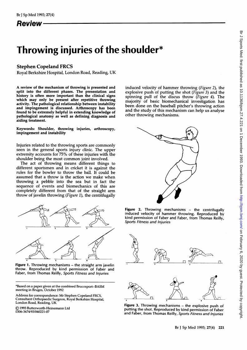
Figure 2. Throwing mechanisms - the centrifugallyinduced velocity of hammer throwing. Reproduced bykind permission of Faber and Faber, from Thomas Reilly,Sports Fitness and Injuries
N>1 AL
4 & n-
3
Figure 1. Throwing mechanisms - the straight arm javelinthrow. Reproduced by kind permission of Faber andFaber, from Thomas Reilly, Sports Fitness and Injuries
*Based on a paper given at the combined Brucosport-BASMmeeting in Bruges, October 1992Address for correspondence: Mr Stephen Copeland FRCS,Consultant Orthopaedic Surgeon, Royal Berkshire Hospital,London Road, Reading, UK
(© 1993 Butterworth-Heinemann Ltd0306-3674/93/040221-07
2 :
1 tY 0\n~~~
Figure 3. Throwing mechanisms - the explosive push ofputting the shot. Reproduced by kind permission of Faberand Faber, from Thomas Reilly, Sports Fitness and Injuries
Br J Sp Med 1993; 27(4) 221
2
5
1
4 6
on February 6, 2020 by guest. P
rotected by copyright.http://bjsm
.bmj.com
/B
r J Sports M
ed: first published as 10.1136/bjsm.27.4.221 on 1 D
ecember 1993. D
ownloaded from

Throwing injuries of the shoulder: S. Copeland
Figure 4. Throwing mechanisms - the spinning pull of thediscus throw. Reproduced by kind permission of Faberand Faber, from Thomas Reilly, Sports Fitness and Injuries
The throwing mechanismTo understand how to prevent and treat overuseinjuries resulting from a throwing action, it isimportant to understand the anatomical and physio-logical sequence of events (Figure 5).
Wind-up phaseWeight is transferred to the back foot, the trunklaterally flexed and arm extended. This can beseparated into two parts, early and late cocking. Inearly cocking the arm is abducted at 900 andhorizontally extended 30°. Late cocking involvesexternal rotation of the shoulder and elbow flexion inthe already abducted and extended arm. Infraspina-tus and teres minor provide external rotation and alsocontribute to glenohumeral stability by drawing thehumeral head towards the glenoid fossa. Subscapu-laris produces its peak electromyographic activity in
s74
Figure 5. Phases of throwing: (1) wind-up; (2) earlycocking; (3) late cocking; (4) acceleration; (5) follow-through. Reproduced by kind permission of Martin Dunitzfrom Phillip J. Marone, Shoulder Injuries in Sports
late cocking when contracting eccentrically to deceler-ate shoulder external rotation and protect the anteriorstructures of the shoulder which are under extremetension at this point'. Supraspinatus activity peaks inlate cocking as it contributes to stability by drawingthe humeral head towards the glenoid preventingtranslation which could compromise the volume ofthe subacromial space. There is a significantly greateractivity of the supraspinatus muscle in amateurswhen tested against professionals, hence fatigueproblems are much greater in the amateur. Thisindicates that teaching of technique has significantpotential for treatment. Professional baseball pitchersare more selective, economical and proficient con-cerning rotator cuff and supraspinatus activities2.
Acceleration phaseActivity of serratus anterior is maximal during thislate cocking phase along with pectoralis major andlatissimus dorsi. Pectoralis major and latissimus dorsiare the two muscles that actively impart velocity tothe ball during the acceleration phase of throwing.This phase of throwing is explosive and impels thehumerus into rapid internal rotation by means of aconcentric contraction. Subscapularis appears to actas a steering muscle to position the head in theglenoid. The acceleration phase includes elbowextension, forearm pronation and wrist flexion.
Follow-through phaseThe muscles now contract to decelerate the armwhich is adducted, internally rotated and forwardflexed at the end of acceleration. Posterior fibres ofdeltoid, supraspinatus, teres minor and infraspinatusall contract in this phase of throwing. Trapezius andthe rhomboids have a high activity in deceleratingscapular protraction and the biceps works in deceler-ating elbow extension and forearm pronation.
Presenting symptomsPainThe most common symptom in any sports clinic ispain and in relation to the throwing sports the historymust be detailed enough to determine at what phaseof the throw the pain occurs as this one fact alone canoften lead to a diagnosis and treatment.
ClickingIf this is a painless click then it is probably of nosignificance and purely a vacuum click alone. If it is apainful click then there may well be a history of director indirect mechanical injury and a possible tornintra-articular structure, e.g. labrum.
'Dead-arm'The 'dead-arm' syndrome is a clearly describedclinical entity in which in the acceleration phase ofthrowing the patient complains of the arm becomingcompletely useless and dropping down by the side
222 Br J Sp Med 1993; 27(4)
on February 6, 2020 by guest. P
rotected by copyright.http://bjsm
.bmj.com
/B
r J Sports M
ed: first published as 10.1136/bjsm.27.4.221 on 1 D
ecember 1993. D
ownloaded from

Throwing injuries of the shoulder: S. Copeland
sometimes associated with pins and needles but moregenerally a completely useless arm which can takefrom a few seconds only to minutes to recover. The'dead-arm' syndrome is due to a momentary subluxa-tion of the glenohumeral joint associated withcompression of the brachial plexus. Treatment musthence be directed towards stabilizing the joint bywhatever means.
Night pain and acheThis is frequently associated with inflammatorychange in the rotator cuff and pain is referred to thedeltoid insertion. The patients complain they cannotsleep on the affected side and if they were toinadvertently roll on to that shoulder the pain wakesthem up. If it is an ache only preventing them fromgoing to sleep then tendinitis must be suspected. If itis night pain that wakes them up then the possibilityof a complete rotator cuff tear must be borne in mind.
Overuse/fatigue tendinitisThe rotator cuff positions the humeral head in theglenoid to provide optimum mobility and stability butis dependent on intact static stabilizers and scapularrotators for this to occur. If the static stabilizers fail tocontain the head the rotator cuff must compensate forthis. Similarly scapular malposition places the glen-oid at a disadvantage and the rotator cuff isover-stretched to complete the throw. Elevatedmuscle loads lead to early fatigue and inflammatorychange. Overuse injuries are most common in thesupraspinatus muscle and tendon. This muscle hasthe greatest role in head depression during shoulderabduction and in a healthy shoulder can adequatelymaintain the subacromial space preventing impinge-ment (Figure 6). However, in fatigue or overload theshoulder can no longer resist superior translation andintrinsic changes in the tendon may occur (Figure 7).
Figure 7. Dysfunction of the rotator cuff casues imbalanceand upward subluxation of the head with impingement
Infraspinatus tendinitis may also occur and is due toinequality of strength between the internal andexternal rotators. Infraspinatus and teres minor workmainly to decelerate the internal rotation of thehumerus in follow through but may be damaged ifthey are relatively weak. It is this delicate balance ofequal and opposite muscle strength that maintainsthe asymptomatic shoulder. If one muscle is toostrong or another too weak or fatigued thensymptoms result.
ImpingementImpingement (Figure 8) is the term used to describerubbing of the bursal surface of the rotator cuff on theunder surface of the coracoacromial arch (acromion,coracoacromial ligament and coracoid (Figure 9))3.The mechanism of how this occurs is a matter of somedispute but undoubtedly involves mechanical consti-tutional and degenerative factors. The importance ofthese different elements varies with age. In the
Figure 6. Normal balance of deltoid and rotator cuff snugsthe humeral head down into the glenoid
Figure 8. Impingement test. Elevation causes mid-rangepain worse on resistance. Reproduced by kind permissionof Churchill Livingstone, from Carter Rowe, ed, TheShoulder
Br J Sp Med 1993; 27(4) 223
on February 6, 2020 by guest. P
rotected by copyright.http://bjsm
.bmj.com
/B
r J Sports M
ed: first published as 10.1136/bjsm.27.4.221 on 1 D
ecember 1993. D
ownloaded from

Throwing injuries of the shoulder: S. Copeland
B ..
Figure 9. Radiograph showing dye injected into supra-spinatus impinging during abduction against the coraco-acromial ligament
younger patient (under 35) presenting with symp-toms of impingement. It is commonly secondary toan underlying instability. In those over 35 the morecommon elements are fatigue/degenerative change. Ifthe static restraint (capsule and ligaments) of theshoulder are ineffective the workload of the rotatorcuff is increased. This overuse leads to fatigue anddysfunction. The humeral head can no longer beeffectively depressed in the glenoid, superior headtranslation occurs and the subacromial space is
Figure 10. Arthroscopic view of impingement; A, acro-mion; RC, rotator cuff. Reproduced by kind permission ofChurchill Livingstone, from Carter Rowe, ed, TheShoulder
reduced. Once this has occurred the cyclical failure ofdegenerative change within the rotator cuff occurs.During the early stages this is a completely reversibleprocess as inflammatory and oedematous changesare the predominant feature but as this becomes achronic feature this secondary fibrotic thickeningbecomes more of a mechanical problem (Figure 10).Interestingly the association of partial thicknessrotator cuff tears with impingement cannot beexplained on a purely mechanical base alone as thesetears are frequently either intrasubstance or on thejoint surface side rather than on the expected bursalsurface of the cuff. Uhtoff has shown a relativeavascularity of the cuff on the articular surfacecompared with the bursal surface4.
Rotator cuff tearsThis is most commonly the end result of chronicimpingement. It may occur as a one-off mechanicalinjury. An unsuspected rotator cuff tear may often bethe cause of continued symptoms following isolateddislocation of the shoulder. Prolonged overuse leadsto fibre failure. A tear may occur through anaccumulation of microtears. Propagation of a partialthickness tear may progress to a complete tear withadditional trauma and tension placed upon it.
InstabilityThis term is used to describe excessive symptomaticdisplacement of the humeral head in its relationshipto the glenoid fossa and is thought to be theunderlying cause of many problems in the throwingshoulder (Figure 11). Stresses during the phase ofthrowing can cause repeated microtrauma leading tolabral changes and stretching of the glenohumeralligaments. As increases in translation occur due tothe above process subluxation can occur. Theincreased stress placed on the rotator cuff to controlthe position of the humeral head leads to eccentricoveruse and damage. With inadequate static stabili-zation and rotator cuff dysfunction the humeral headcan translate superiorly causing impingement. Post-erior instability occurs commonly in throwers duringfollow through when the posterior capsule andlabrum are stretched5. The posterior labrum may beabraded and the capsule restraints are stretchedwhich permits posterior translation of the humeralhead causing stretching and fatigue in the rotatorcuff. This may lead to overuse tendinitis of theinfraspinatus but in general posterior instability andsubluxation are usually a problem that is welltolerated by the athlete and may be remedied byappropriate rehabilitation.
Impingement versus subluxationAs discussed above the history and age are importantfactors in delineating the two, however, specific testsmay be helpful.
If the patient lies supine with the arm abducted to900 and externally rotated (Figure 12), this does notusually cause pain with the primary impingement.
224 Br J Sp Med 1993; 27(4)
on February 6, 2020 by guest. P
rotected by copyright.http://bjsm
.bmj.com
/B
r J Sports M
ed: first published as 10.1136/bjsm.27.4.221 on 1 D
ecember 1993. D
ownloaded from

Throwing injuries of the shoulder: S. Copeland
Figure 11. Stability of the humeral head in the glenoid canbe likened to a seal balancing a ball. Reproduced by kindpermission of Churchill Livingstone, from Carter Rowe,ed, The Shoulder
However, if the impingement is secondary to aninstability, abduction and external rotation mayinduce pain and more importantly can be relieved byapplying pressure anterior to the joint to attempt tosublux the humeral head within the glenoid post-eriorly. The mechanism of this is thought to be anover-lax anterior capsule with a relatively tightposterior capsule which allows the forward subluxa-
Figure 12. Relocation test. Pain on forced external rotationin abduction - relieved by posteriorly directed forcesuggests impingement secondary to instability. Repro-duced by kind permission of Smith and Nephew Dyonics,Cambridge, UK
tion in abduction/external rotation, e.g. the youngtennis player after repetitive serving. If there is anydoubt as to the origin of the site of the pain localanaesthetic may be injected into the subacromialbursa which should relieve pain from both primaryand secondary impingement.
TreatmentAs has been discussed, the two main problems in thethrowing athlete appear to be a combination ofimpingement and instability. In the older athlete trueimpingement may be present but in the youngerpatient impingement secondary to instability must besuspected. From the history and examination thesetwo components must be delineated. If impingementis thought to be the primary cause of the problemthen this must be staged according to the usual Neerclassification3. In the early stages, rest and anti-inflammatory treatment alone may suffice. In long-standing cases steroid injection and non-steroidalanti-inflammatories may be helpful. The usualrecommended regimen for this is never more thanthree injections at 3-weekly intervals and rarely to beused in the young. The steroid injection is helpfuldiagnostically and also prognostically. The effect ofthe steroid is to reduce inflammation and hencethickening due to oedema. If the steroid is mixedwith local anaesthetic and injected into the subacro-mial bursa (Figure 13) then three reactions may occur.
1. Complete resolution of symptoms but return ofsymptoms after a few hours, i.e. the time taken forthe local anaesthetic to wear off. This confirms thatthe diagnosis of the site of pain is correct but thatprobably the secondary inflammatory changes are
@198\
I.
)i!-1
Jji0 \
Figure 13. Painful arc can be relieved by local anaestheticinjection. Reproduced by kind permission of ChurchillLivingstone, from Carter Rowe, ed, The Shoulder
Br J Sp Med 1993; 27(4) 225
J processCoracoid
on February 6, 2020 by guest. P
rotected by copyright.http://bjsm
.bmj.com
/B
r J Sports M
ed: first published as 10.1136/bjsm.27.4.221 on 1 D
ecember 1993. D
ownloaded from

Throwing injuries of the shoulder: S. Copeland
only a minimal part of the symptoms and that thesecondary fibrotic changes are probably predomi-nant. It is therefore probably useless to give anyfurther injections.
-f~~~~~~~~~~~~~~~~~~~~I
a
.- .-4.0 mm acromionizer
ir blade.."... ---nterior acromion
b' Figure 14. a Three portal arthroscopic subacromialdecompression - shaver, 'scope, drain; b Arthroscopicsubacromial decompression - the anterior and inferiorone-third of acromion is removed with a shaver. Repro-duced by kind permission of Smith and Nephew Dyonics,Cambridge, UK
2. Resolution of symptoms which lasts for a variableperiod of time, e.g. a few weeks, but then thesymptoms return. One can conclude from this thatinflammatory change is at least part of the problemand hence is reversible. A further injection wouldcertainly be worthwhile with hopefully furtherprolonged symptomatic relief. If, after the secondinjection, the symptoms return after a similar timeperiod then it is probably pointless going on to athird injection. However, if the second injectionbrings prolongation of relief then a third injectionmay well be indicated and may induce permanentrelief of symptoms. However, if injection therapyhas only a temporary benefit then if circumstancesdictate this may be the group that would benefitfrom surgical decompression. This may now bedone arthroscopically with minimal perioperativemorbidity and equal success to open procedures(Figure 14a,b).
3. If injection induces complete resolution of symp-toms then obviously no further treatment isnecessary.
If instability is thought to be the prime cause ofsymptoms the exact degree and direction of instab-ility must be elicited by examining the phase ofthrowing and stress testing. The weak and fatiguingmuscles must be strengthened, the posterior capsuleshould be stretched and that particular athlete'sthrowing technique carefully monitored with approp-riate coaching. It may take a year to reverse badhabits in capsular stretching.However, if all these measures fail then surgical
stabilization may well be considered. Capsular shiftprocedures correctly indicated are reliable and neednot limit the range of motion. Arthroscopy enablessubtle changes to be seen which confirm the clinical
i:... ar e
il.... -,jI) iS .
Figure 15. SLAP lesions. Superior anterior/posterior labral tears - worse on follow through
226 Br J Sp Med 1993; 27(4)
on February 6, 2020 by guest. P
rotected by copyright.http://bjsm
.bmj.com
/B
r J Sports M
ed: first published as 10.1136/bjsm.27.4.221 on 1 D
ecember 1993. D
ownloaded from

Throwing injuries of the shoulder: S. Copeland
diagnosis of instability. Lamination of the antero-inferior capsule indicate overstretching and in con-junction with chondromalacia seen on the postero-superior humeral head is diagnostic of anteroinferiorsubluxation. Arthroscopy has been enormously help-ful in recognizing previously unrecognized problems,e.g. superior labral anterior and posterior tears (slaplesions described by Snyder et al.6). These occur inthe follow-through phase of throwing when thebiceps tendon is avulsed from the superior glenoidtaking with it either part of the anterior or posteriorlabrum. The mechanism of this has been confirmedby Andrews who has electrically stimulated thebiceps in cadavers and reproduced this lesion6.
Arthroscopic methods of shoulder stabilization arenot yet as reliable as open methods but hold promisefor the future.
References1 Glousman R, Jobe F, Tibone J, Moynes M, Antonelli D, Perry J.
Dynamic EMG analysis of the throwing shoulder withgleno-humeral instability. J Bone Joint Surg [Am] 1989; 70-A:220.
2 Townsend A. EMG analysis of the gleno-humeral musclesduring a baseball rehabilitation programme. Am J Sports Med1991; 19: 3.
3 Neer C. Anterior acromioplasty for chronic impingementsyndrome in the shoulder. I Bone Joint Surg [Am] 1972; 54-A:
4 Uhtoff HK, Sarkar K, Ogatas L, Loehr J. Degenerativetendinopathies of the rotator cuff. Acta Orthop Scand 1990; 239:2-3.
5 Poppen N et al. Normal and abnormal motion of the shoulder.I Bone Joint Surg [Am] 1976; 58-A: 195.
6 Snyder SJ et al. SLAP. Lesions of the shoulder. Arthroscopy1990; 6: 274-9.
IN THE TREATMENT OF SOFT TISSUE INJURIES
Ibuleve Sports Gel harnesses the analgesic and anti- * jinflammatory power of ibuprofen in a topical gel.Ibuleve Sports Gel's soothing and fragrance free
formulation has been shown to be both easy to useand highly effective in bringing relief to the painand inflammation associated with soft tissueinjuries.
Suitable for use over several weeks,,Ibuleve Sports Gel can, in appropriatecases, also be used as an ultra-soundcoupling agent.Availability without prescrip-
tion allows your patients tocontinue their topical ttherapy between = < ibuprofen gelvisits. A_ portge.
A HELPING HAND IN THE TREATMENT OFMUSCULAR SPORTS INJURIES.
Ibuleve Sports Gel is availablefrom pharmacies at a price below the NHS prescription charge.IBULEVE Trademark and Product Licence held by Diomed Developments Ltd., Hitchin, UK.
DISTRIBUTED BY DDD LTD., 94 RICKMANSWORTH ROAD, WATFORD, HERTS, WD1 7JJ.Acne Ingredient ibuprofen BP 5.0% w/w. Directions: Lightly apply a thin layen of gel over he affected area. Massage gently until absorbed. Wash hands after use. Repeat as required up to three times daily. Indications: To relieve pain and reduce inflammation in muscularsports injuries. Precaeons: If symptoms perist for more than a few weeks consult doctor. Not recomnmended for children under 14 years. Patients with a history of kidney problems,astha oaspirin sensitivityshould seek medical adice beore using blee Sports Gel. Keep awayfroe broken skin, lips and eyes. Notto be used during pregnancy or lactation. Keep all medicines out ofthe reach of children. Do not use if sensitive to any ofthe ingredients. FOR EXTERNAL USE ONLY. Legal categoryiB Packs: Tubes of 3tgPL0173/0 0). Price t.95.
Br J Sp Med 1993; 27(4) 227
on February 6, 2020 by guest. P
rotected by copyright.http://bjsm
.bmj.com
/B
r J Sports M
ed: first published as 10.1136/bjsm.27.4.221 on 1 D
ecember 1993. D
ownloaded from