tian, ai-ling and lu, mingmin and calderón-mantilla, guillermo...
TRANSCRIPT

RESEARCH Open Access
A recombinant Fasciola gigantica 14-3-3epsilon protein (rFg14-3-3e) modulatesvarious functions of goat peripheral bloodmononuclear cellsAi-Ling Tian1, MingMin Lu2, Guillermo Calderón-Mantilla3, Evangelia Petsalaki3, Tania Dottorini4, XiaoWei Tian2,YuJian Wang2, Si-Yang Huang1,5, Jun-Ling Hou1, XiangRui Li2, Hany M. Elsheikha4* and Xing-Quan Zhu1*
Abstract
Background: The molecular structure of Fasciola gigantica 14-3-3 protein has been characterized. However, theinvolvement of this protein in parasite pathogenesis remains elusive and its effect on the functions of innateimmune cells is unknown. We report on the cloning and expression of a recombinant F. gigantica 14-3-3 epsilonprotein (rFg14-3-3e), and testing its effects on specific functions of goat peripheral blood mononuclear cells(PBMCs).
Methods: rFg14-3-3e protein was expressed in Pichia pastoris. Western blot and immunofluorescence assay (IFA)were used to examine the reactivity of rFg14-3-3e protein to anti-F. gigantica and anti-rFg14-3-3e antibodies,respectively. Various assays were used to investigate the stimulatory effects of the purified rFg14-3-3e protein onspecific functions of goat PBMCs, including cytokine secretion, proliferation, migration, nitric oxide (NO) production,phagocytosis, and apoptotic capabilities. Potential protein interactors of rFg14-3-3e were identified by querying thedatabases Intact, String, BioPlex and BioGrid. A Total Energy analysis of each of the identified interaction wasperformed. Gene Ontology (GO) enrichment analysis was conducted using Funcassociate 3.0.(Continued on next page)
* Correspondence: [email protected];[email protected] of Medicine and Health Sciences, School of Veterinary Medicine andScience, University of Nottingham, Sutton Bonington Campus,Loughborough LE12 5RD, UK1State Key Laboratory of Veterinary Etiological Biology, Key Laboratory ofVeterinary Parasitology of Gansu Province, Lanzhou Veterinary ResearchInstitute, Chinese Academy of Agricultural Sciences, Lanzhou, Gansu Province730046, People’s Republic of ChinaFull list of author information is available at the end of the article
© The Author(s). 2018 Open Access This article is distributed under the terms of the Creative Commons Attribution 4.0International License (http://creativecommons.org/licenses/by/4.0/), which permits unrestricted use, distribution, andreproduction in any medium, provided you give appropriate credit to the original author(s) and the source, provide a link tothe Creative Commons license, and indicate if changes were made. The Creative Commons Public Domain Dedication waiver(http://creativecommons.org/publicdomain/zero/1.0/) applies to the data made available in this article, unless otherwise stated.
Tian et al. Parasites & Vectors (2018) 11:152 https://doi.org/10.1186/s13071-018-2745-4

(Continued from previous page)
Results: Sequence analysis revealed that rFg14-3-3e protein had 100% identity to 14-3-3 protein from Fasciolahepatica. Western blot analysis showed that rFg14-3-3e protein is recognized by sera from goats experimentallyinfected with F. gigantica and immunofluorescence staining using rat anti-rFg14-3-3e antibodies demonstrated thespecific binding of rFg14-3-3e protein to the surface of goat PBMCs. rFg14-3-3e protein stimulated goat PBMCs toproduce interleukin-10 (IL-10) and transforming growth factor beta (TGF-β), corresponding with low levels of IL-4and interferon gamma (IFN-γ). Also, this recombinant protein promoted the release of NO and cell apoptosis, andinhibited the proliferation and migration of goat PBMCs and suppressed monocyte phagocytosis. Homologymodelling revealed 65% identity between rFg14-3-3e and human 14-3-3 protein YWHAE. GO enrichment analysis ofthe interacting proteins identified terms related to apoptosis, protein binding, locomotion, hippo signalling andleukocyte and lymphocyte differentiation, supporting the experimental findings.
Conclusions: Our data suggest that rFg14-3-3e protein can influence various cellular and immunological functionsof goat PBMCs in vitro and may be involved in mediating F. gigantica pathogenesis. Because of its involvement in F.gigantica recognition by innate immune cells, rFg14-3-3e protein may have applications for development ofdiagnostics and therapeutic interventions.
Keywords: Fasciola gigantica, 14-3-3 epsilon protein isoform, Homology modelling, Goat, Peripheral bloodmononuclear cells
BackgroundFasciola gigantica is a worldwide distributed liver flukethat can infect a wide range of livestock of economic im-portance such as cattle, sheep and goats [1]. Infectionswith Fasciola spp. result in reduced meat, milk and woolproduction [2], with an estimated economic loss morethan 3 billion USD per annum worldwide [3, 4]. Inaddition, disease caused by Fasciola infection (known asfasciolosis) in humans is included in the WHO top listedneglected tropical diseases [5, 6], and it is estimated thatat least 2.4 million people are infected worldwide and180 million are at risk of infections [2].The mammalian definitive hosts of F. gigantica be-
come infected by ingestion of vegetation containingencysted metacercariae. Infective metacercariae emergein the duodenum, rapidly traverse the intestinal wall,and migrate through the peritoneal cavity to reach theliver capsule. Following a period of burrowing, feedingand growth within the liver parenchyma, the flukesreach their final destination within the bile ducts wherethey attain their sexual maturity and produce eggs [7, 8].During their migration and development, liver flukes en-counter various host tissue types, distinct physiologicalmicroenvironments, and a combination of both humoraland cellular immune responses. F. gigantica can remark-ably tackle these challenges and survive for extended pe-riods inside its host [9].Although host immune evasion is a common strategy
used by successful parasites, Fasciola spp. provoke rec-ognition by host immune cells less capable of destroyingthem. To accomplish this, they use virulence-associatedexcretory-secretory products (ESPs) to manipulate cellu-lar processes [10], such as immune evasion and immunesubversion [11, 12] to promote parasite survival. Among
the virulence factors in the ESPs that are involved inhost-parasite interaction, the 14-3-3 proteins are of in-terests given their ability to bind to many signaling pro-teins, such as kinases and phosphatases, and to mediatemany regulatory processes, metabolism and signal trans-duction pathways [13, 14]. The 14-3-3 family encom-passes a group of conserved intracellular polypeptideswith molecular weight of 28–33 kDa, which are found inall eukaryotic organisms [15]. These proteins have beenthe focus of much research due to their roles in modu-lating various protein-protein interactions [16, 17].Various structural and functional aspects of 14-3-3
protein have been studied in Toxoplasma gondii [18],Schistosoma japonicum [19], Eimeria tenella [20],Echinococcus granulosus [21], Haemonchus contortus[22] and Trichinella spiralis [23], and the putativerole of 14-3-3 protein in parasite pathogenesis hasbeen reported [24–27]. However, the biological effectsof F. gigantica 14-3-3 protein on the host innate im-mune system are still largely unknown.In the present study, the gene encoding F. gigantica
14-3-3 epsilon protein (Fg14-3-3e) isoform was clonedand expressed in Pichia pastoris. The modulatory ef-fects of the purified rFg14-3-3e on various functionsof the goat peripheral blood mononuclear cells(PBMCs), including cytokine secretion, proliferation,migration, nitric oxide (NO) production, phagocytosisand apoptosis were studied using a range of assays.These experimental studies were complemented by insilico homology modelling analysis. Our data suggestthat rFg14-3-3e protein can significantly influence thecellular and immunological functions of goat PBMCs,all critical pieces of the immunopathogenesis of F.gigantica infection.
Tian et al. Parasites & Vectors (2018) 11:152 Page 2 of 14

MethodsAnimals used in the experimentsLocal, 3- to 6-month-old crossbred goats were obtainedfrom the teaching and research flock at Nanjing Agricul-tural University. Goats were fed with hay and wholeshelled corn, and were housed indoor in pens and pro-vided with water ad libitum. All goats were treated withtriclabendazole (50 mg/kg body weight) in order to elim-inate any potential previous infection with Fasciola spp.A fecal sample from each goat was microscopically ex-amined for helminth eggs 2 weeks after treatment. Onlygoats demonstrating no eggs were included in the studyand daily health checks were performed throughout theexperiment. Three biological replicates (three goats),each consisting of three technical replicates (i.e. threesamples from each goat), were used in this study. Forthe production of antibodies, female Sprague Dawley(SD) rats (body weight ~150 g) were purchased from theExperimental Animal Center of Jiangsu, PR China(Qualified Certificate: SCXK 2008-0004) and werehoused under specific-pathogen-free conditions in thelab animal facility and fed sterilized food and water adlibitum. All efforts were made to minimize suffering ofanimals.
Isolation of goat PBMCsGoat peripheral venous blood samples were collectedfrom healthy goats. The goat PBMCs were isolated fromfreshly collected blood samples using commerciallyavailable goat PBMCs’ isolation kit (TBD, Tianjin,China). The number of goat PBMCs were adjusted to adensity of 1 × 10⁶ cells/ml in RPMI 1640 medium con-taining 10% fetal bovine serum (FBS), 1% penicillin-streptomycin (Gibco, New York, USA). Goat PBMCswere maintained in a humidified atmosphere of 5%CO2 at 37 °C. Cells viability was determined by try-pan blue dye exclusion, and only cells with more than95% viability were used in the experiments. Mono-cytes were isolated from goat PBMCs based on theirtendency to adhere to the plastic surface of the cellculture flasks [28].
Parasite strainAdult flukes of F. gigantica were collected from the gall-bladder of naturally infected buffaloes slaughtered forhuman consumption at local abattoirs in GuangxiZhuang Autonomous Region, PR China. After washingin phosphate buffered saline (PBS, pH 7.4), the flukeswere collected and immediately used for RNA isolationor stored at -80 °C with RNA stabilizer until use. Thespecies identity was confirmed as F. gigantica based onPCR and sequencing of the second internal transcribedspacer (ITS2) of ribosomal DNA [29], which yielded ab-solute homology to ITS2 sequence of F. gigantica from
Guangxi Zhuang Autonomous Region (GenBank:AJ557569).
Recombinant plasmid construction and sequencing ofFg14-3-3e geneDue to the limited genomic data on F. gigantica, wesearched Fasciola hepatica ESPs dataset acquired by li-quid chromatography-tandem mass spectrometry (LC-MS/MS) and F. hepatica cDNA library from previousproteomic studies to screen for homologous 14-3-3 pro-tein sequences using the BLAST program (https://blas-t.ncbi.nlm.nih.gov/Blast.cgi). This analysis identified F.hepatica 14-3-3 epsilon protein isoform (Fh14-3-3e) se-quence, which was used to design primers to amplify F.gigantica 14-3-3 epsilon protein isoform (Fg14-3-3e) se-quence. First-strand cDNA was synthesized from total F.gigantica RNA, isolated from 30 mg of adult F. giganticaflukes using Trizol reagent (Invitrogen, San Diego,USA), by reverse transcription polymerase chain reac-tion (RT-PCR) using RevertAid First Strand cDNA Syn-thesis Kit (Thermo Scientific (EU), Vilnius, Lithuania).The cDNA was used for amplification of Fg14-3-3e geneusing a forward primer (5'-CCG GAA TTC GAG GAGGTG AAT ATC G-3') and a reverse primer (5'-ATT T GCGG CCG CCT AAG CCT TTT TCT CCT C-3').These specific primers of Fg14-3-3e gene were designedbased on the published sequence of Fh14-3-3e gene(GenBank: LN629113.1). EcoRI and NotI restriction sites,indicated by underline, were incorporated into theprimers to facilitate cloning. The amplified Fg14-3-3egene was digested with EcoRI and NotI and spliced intothe corresponding cloning sites in the plasmid vectorpMD19-T (Takara, Dalian, Liaoning, China). The recom-binant plasmid was transformed into Trans5α chem-ically competent cells (TransGen Biotech, Beijing,China). Several positive clones were selected and se-quenced by GenScript (Nanjing, Jiangsu, China) toconfirm the correct insertion/orientation of Fg14-3-3egene in the vector.
Characterization of the Fg14-3-3e gene sequenceWe characterized the Fg14-3-3e gene sequence by trans-lating the cDNA sequence into the amino acid se-quences using Seaview [30]. Then, three differentapproaches were used to identify and annotate signa-tures of functional mechanisms present in the aminoacidic sequence. Specifically, the protein sequence wasfirst subjected to the prediction of sequence domain,followed by comparative analysis and 3D modelling. Thepresence of sequence domain was performed usingSMART (Simple Modular Architecture Research Tool)[31]. Next, a comparative analysis was done to search forsimilarities in protein sequence databases (NCBI non-redundant protein sequence) and to identify possible
Tian et al. Parasites & Vectors (2018) 11:152 Page 3 of 14

homologous proteins. Furthermore, we predicted thethree-dimensional model of the homo-dimer usinghomology modelling (Swissmodel) [32]. The signalpeptide was predicted by SignalP 4.1 Server (http://www.cbs.dtu.dk/services/SignalP/), the transmembraneregion was predicted by TMHMM Server v. 2.0 (http://www.cbs.dtu.dk/services/TMHMM/) and the potentialN-glycosylation sites were predicted by NetNGlyc 1.0 Ser-ver (http://www.cbs.dtu.dk/services/NetNGlyc/).
Expression of rFg14-3-3e proteinPositive clones, containing the Fg14-3-3e gene in thecorrect orientation were selected. The purified Fg14-3-3egene fragment was sub-cloned into pPIC9K vector. Theconstructs were designed with appropriate restrictionsites and a carboxyl-terminal His6 tag to enable purifica-tion. The plasmid pPIC9K-Fg14-3-3e was linearized withSalI to transform the Methylotrophic Yeast P. pastorisGS115 strain by electroporation using a GenePulserXcellTM (Bio-Rad, Hercules, California, USA). Positiverecombinant P. pastoris clones containing the insertwere selected for expression by inoculating into 15 ml ofbuffered complex medium containing glycerol (BMGY).The inoculated BMGY medium [1% (wt/vol) yeast ex-tract, 2% (wt/vol) peptone, 1% (wt/vol) yeast nitrogenbase, 1% (wt/vol) glycerol, 0.00004% (wt/vol) biotin, and0.1 M potassium phosphate (pH 6.0)] in 100 ml conicalflasks was grown at 28 °C with vigorous shaking for 24h. The cells were harvested by centrifugation (250× g for10 min), resuspended in 20 ml of buffered complexmedium containing methanol (BMMY; BMGY mediumwith 1% methanol substituted for glycerol). The culturewas allowed to continue growing for four days. Duringthis Fg14-3-3e gene expression induction period, metha-nol was added every 24 h to maintain a final concentra-tion of 1% (v/v). The cells were then pelleted bycentrifugation (2,500× g at 4 °C for 10 min) and the cul-ture supernatant was harvested for protein extraction. P.pastoris cells transformed with vector pPIC9K (withoutinsert) were similarly treated and analyzed as a negativecontrol.
Protein extraction and SDS-PAGEFollowing extraction of the total protein from the yeastculture supernatant by precipitation with 10%trichloroacetic acid, the protein pellet was washed sev-eral times with acetone and finally dissolved in distilledwater. The size of the isolated rFg14-3-3e protein wasanalyzed in 12% sodium dodecyl sulfate-polyacrylamidegel electrophoresis (SDS-PAGE) gels, followed by Coo-massie blue staining, and the concentration of the pro-tein was determined using the Bradford method, usingbovine serum albumin (BSA) as the standard. Purified
proteins were stored frozen in 1× PBS at -80 °C untilfurther analysis.
Preparation of polyclonal antibodiesFive goats, raised in helminth-free conditions, were in-fected orally with 500 viable encysted metacercariae peranimal. One month later, the goat antisera were col-lected and stored frozen in aliquots at -80 °C until use inWestern blot analysis. To generate specific antibodiesagainst rFg14-3-3e protein, 300 μg of the purified pro-tein mixed with complete Freund’s adjuvant (1:1) wereinjected subcutaneously into SD rats for primaryimmunization. SD rats were boosted four times with thesame dose of the recombinant protein in incompleteFreund’s adjuvant at 2-week intervals. One week afterthe last injection, serum containing specific anti-rFg14-3-3e antibodies was collected and stored frozen at -80 °Cfor later use in immunofluorescence visualization ofrFg14-3-3e protein binding to the surface of the goatPBMCs.
Western blottingExtracted rFg14-3-3e protein samples (20 μg) were re-solved on 12% SDS-PAGE gels and transferred ontoHybond-C extra nitrocellulose membrane (Amersham,London, UK). Non-specific binding sites were blockedby immersing the membranes in 5% skim (non-fat) milkin Tris-buffered saline containing 0.1% Tween-20(TBST) for 1 h at ambient temperature. The membraneswere then washed 5 times (5 min each) in TBST. Subse-quently, the membranes were incubated with the pri-mary antibodies (antiserum from goats experimentallyinfected with F. gigantica) for 1 h at 37 °C (1:100 inTBST). After being washed 5 times with TBST, themembranes were incubated with HRP-conjugated rabbitanti-goat IgG (Sigma, St. Louis, MO, USA) for 1 h at 37°C (1:2000 in TBST). Finally, the immunoreaction wasvisualized using freshly prepared 3,3′-diaminobenzidine(DAB, Sigma) as a chromogenic substrate after 5 min.
Immunofluorescence staining of rFg14-3-3e protein bind-ing to goat PBMCsFreshly isolated goat PBMCs were incubated with rFg14-3-3e protein for 1 h in a humidified atmosphere of 5%CO2 at 37 °C. To minimize background staining, goatPBMCs were fixed with 4% paraformaldehyde, washed 3times in PBS (5 min each) and were treated with block-ing solution (4% BSA in PBS) for 1 h at ambienttemperature. rFg14-3-3e-treated or non-treated controlgoat PBMCs were incubated with rat anti-rFg14-3-3eantibody (1:100) for 1 h at 37 °C, followed by stainingwith Cy3 conjugated goat anti-rat IgG secondary anti-body (1:500) (Beyotime, Haimen, Jiangsu, China) for 1 hat 37°C. Hoechst 33342 (Invitrogen, Eugene, Oregon,
Tian et al. Parasites & Vectors (2018) 11:152 Page 4 of 14

USA) was used for nuclear staining. Stained cells wereimaged with 100× magnification using a Zeiss laser scan-ning confocal microscope (LSM710, Zeiss, Jena,Germany) and digital images were analyzed by Zen 2012imaging software.
ELISA analysis of the levels of cytokinesGoat PBMCs were seeded into a 24-well tissue cultureplate at 106 cells/well in 1 ml medium consisting ofRPMI 1640 medium. Serial concentrations (10 μg/ml, 20μg/ml, 40 μg/ml and 80 μg/ml) of rFg14-3-3e proteinwere added to the wells. The sham-treated PBMCsserved as controls, and the plate was incubated in a hu-midified atmosphere of 5% CO2 at 37 °C for 24 h. Thesupernatants were collected, and assayed for cytokineproduction using the goat enzyme linked immunosorb-ent assay (ELISA) kits (Mlbio, Shanghai, China). GoatPBMCs supernatants were assayed for the production ofinterleukin-4 (IL-4), IL-10, interferon gamma (IFN-γ),and transforming growth factor beta (TGF-β). Limits ofdetection were between 2 and 800 pg/ml depending onthe analytic assay.
Inhibitory effect of rFg14-3-3e protein on cellproliferationAnti-proliferative effects of rFg14-3-3e protein on goatPBMCs were determined by performing Cell CountingKit-8 (CCK-8) assay (Beyotime, Haimen, Jiangsu, China).Goat PBMCs were seeded on 96-well tissue cultureplates (104 cells /well in 100 μl), with three replicatesused for each rFg14-3-3e protein concentration (10 μg/ml, 20 μg/ml, 40 μg/ml, and 80 μg/ml). Cell culturedplates were incubated at 37 °C in a humidified atmos-phere with 5% CO2. Following 72 h incubation, 10 μl ofCCK-8 reagent was added to each well and the cultureswere further incubated at 37 °C in a humidified atmos-phere of 5% CO2 at 37 °C for 4 h in the dark. The op-tical density at 450 nm (OD450) was measured using amicroplate reader (Bio-Rad).
In vitro cell migration assayCell migration assay was performed using 6.5 mmTranswell® with 8.0 μm-pore-size polyester membraneinserts (Corning, Kennebunk, USA). Aliquots of 106 goatPBMCs in 1 ml RPMI culture medium were placed intothe top chamber of the transwell in a 24-well tissue cul-ture plate. Cells were stimulated with serial concentra-tions of rFg14-3-3e protein and incubated in ahumidified atmosphere of 5% CO2 at 37 °C for 24 h.After incubation, the cells were collected from the bot-tom chamber and the number of cells that migratedthrough the membrane into the lower chamber was de-termined using Automated Cell Counter (Countstar,
Shanghai, China). The results were presented as percent-ages of the seeded goat PBMCs.
Generation of nitric oxideNitric oxide production by goat PBMCs was evaluatedby measuring intracellular nitrite by Griess reaction fol-lowing the protocol of Total Nitric Oxide Assay Kit(Beyotime, Haimen, Jiangsu, China). Briefly, goat PBMCswere seeded at 106 cells/well into a 24-well tissue cultureplate. Cells were incubated for 24 h with various concen-trations of rFg14-3-3e protein in RPMI 1640 medium at37 °C in a humidified atmosphere with 5% CO2. Absorb-ance values of the colored solution were measured usinga plate microplate reader (Bio-Rad) at 540 nm. NO levelswere calculated against a standard curve generated using0 to 80 μM/l sodium nitrites.
FITC-dextran uptakeThe phagocytic activity of goat monocytes was assessedby measuring the uptake of fluorescein isothiocyanate(FITC)-dextran according to a previously describedmethod [33]. Briefly, freshly isolated goat monocyteswere seeded at 106 cells/well into a 24-well tissue cultureplate. Goat monocytes were incubated for 48 h withvarious concentrations of rFg14-3-3e protein in RPMI1640 medium in a humidified atmosphere of 5% CO2 at37 °C. Then, goat monocytes were re-suspended in 100μl of cold PBS, and incubated with 1 mg/ml FITC-dextran (average molecular weight of dextran, 59,000 to77,000; Sigma-Aldrich) in RPMI 1640 medium at 4 or37 °C for 1 h. After incubation, chilled PBS containing2% FBS was added to halt phagocytosis and then cellswere washed (three times with ice-cold PBS) and resus-pended in ice-cold PBS containing 2% paraformalde-hyde. FITC-dextran internalization of monocytes wasanalyzed by flow cytometry (BD Biosciences, San Jose,California, USA). Data were analyzed using FlowJo 10.1software (Tree Star, Ashland, Oregon, USA). Goatmonocytes FITC-dextran (phagocytosis index) was cal-culated as the change in Median Fluorescence Intensity(MFI) between cell samples incubated at 37 °C and 4 °C.
Apoptosis assay using Annexin V/PI stainingApoptosis in goat PBMCs was evaluated and quantifiedby flow cytometry (BD Biosciences) using the AnnexinV-FITC kit (Miltenyi Biotec, Bergisch Gladbach,Nordrhein-Westfalen, Germany). Briefly, PBMCs (106
cells/ml) were treated with different concentrations ofrFg14-3-3e protein and incubated in a humidified atmos-phere of 5% CO2 at 37 °C for 24 h. Then, PBMCs werestained with Annexin V and Propidium Iodide (PI) ac-cording to the manufacturer’s instructions. The non-treated goat PBMCs were used as a control. Annexin V-positive/PI-negative goat PBMCs were considered to be
Tian et al. Parasites & Vectors (2018) 11:152 Page 5 of 14
apoptotic cells and were analyzed as a percentage of theentire goat PBMCs population.
Homology modelling of rFg14-3-3e and potential com-plexes with host proteinsUsing protein blast [34] against the UniprotKB database[35] we retrieved the most similar human protein ofrFg14-3-3e fasta protein sequence from uniprot. P62258(YWHAE) had a Max score of 313, a Query coverage of89% and 65% identity. Using P62258 (YWHAE), wequeried the following interactions databases: Intact [36],String [37], BioPlex [38], BioGrid [39]. The resultinginteracting pairs were used as input for obtaining the ex-perimental and homology modelled structure interac-tions using Interactome3D [40]. All experimental andmodelled interactions were used to model potentialrFg14-3-3e-host interactions using FoldX [41]. Thealignment of the human YWHAE with rFg14-3-3e wasdone using Clustal Omega [42]. Finally, a total energyanalysis was done to identify stabilized and destabilizedinteractions. All complex structures and energy calcula-tion files can be found in Additional file 1: Dataset S1.
Statistical analysisAll data analyses and graphs were performed withGraphPad Premier 6.0 software package (GraphPadPrism, San Diego, California, USA). Statistical analyseswere done from raw data with a one-way ANOVA, andStudent’s t-tests, where applicable. Data values wereexpressed as means ± standard deviation (SD). Differ-ences between tested groups were considered significantif the P value was ≤ 0.05, and this is indicated in the fig-ures by asterisks (*P < 0.05; **P < 0.01; ***P < 0.001; or****P < 0.0001). All experiments were repeated a mini-mum of three separate times.
ResultsIdentification, cloning and expression of rFg14-3-3eproteinTo identify homologs of F. gigantica 14-3-3 protein inthe F. hepatica genome, we performed a BLAST search(https://blast.ncbi.nlm.nih.gov/Blast.cgi) to screen forhomologous 14-3-3 protein sequences. This analysisidentified Fh14-3-3e gene sequence (GenBank locus:LN629113.1), which was used to design 5' and 3' primersto amplify Fg14-3-3e. The Fg14-3-3e gene was amplifiedand the cDNA fragment was successfully cloned into thepMD19-T cloning vector.Nucleic acid sequencing identified positive pMD19-T-
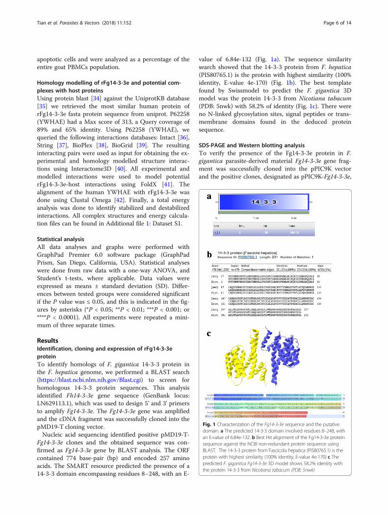
Fg14-3-3e clones and the obtained sequence was con-firmed as Fg14-3-3e gene by BLAST analysis. The ORFcontained 774 base-pair (bp) and encoded 257 aminoacids. The SMART resource predicted the presence of a14-3-3 domain encompassing residues 8–248, with an E-
value of 6.84e-132 (Fig. 1a). The sequence similaritysearch showed that the 14-3-3 protein from F. hepatica(PIS80765.1) is the protein with highest similarity (100%identity, E-value 4e-170) (Fig. 1b). The best templatefound by Swissmodel to predict the F. gigantica 3Dmodel was the protein 14-3-3 from Nicotiana tabacum(PDB: 5nwk) with 58.2% of identity (Fig. 1c). There wereno N-linked glycosylation sites, signal peptides or trans-membrane domains found in the deduced proteinsequence.
SDS-PAGE and Western blotting analysisTo verify the presence of the Fg14-3-3e protein in F.gigantica parasite-derived material Fg14-3-3e gene frag-ment was successfully cloned into the pPIC9K vectorand the positive clones, designated as pPIC9K-Fg14-3-3e,
Fig. 1 Characterization of the Fg14-3-3e sequence and the putativedomain. a The predicted 14-3-3 domain involved residues 8–248, withan E-value of 6.84e-132. b Best Hit alignment of the Fg14-3-3e proteinsequence against the NCBI non-redundant protein sequence usingBLAST. The 14-3-3 protein from Fascicola hepatica (PIS80765.1) is theprotein with highest similarity (100% identity, E-value 4e-170) c Thepredicted F. gigantica Fg14-3-3e 3D model shows 58.2% identity withthe protein 14-3-3 from Nicotiana tabacum (PDB: 5nwk)
Tian et al. Parasites & Vectors (2018) 11:152 Page 6 of 14

were transformed into P. pastoris strain GS115. The re-combinant protein (rFg14-3-3e) was successfullyexpressed in the culture supernatant of P. pastoris. Therecombinant clones showed high expression of a novelprotein of approximately 29 kDa on SDS-PAGE gels(Fig. 2a) after 72 h of induction with 1% methanol,which was absent in control samples. The specificity ofthis rFg14-3-3e protein was confirmed by Western blot-ting, which was probed with serum from goats experi-mentally infected with F. gigantica. IgG antibodies in theserum of infected goats recognized rFg14-3-3e proteinmonospecifically as it reacted to a single ~29 kDa band,which was absent when the Western blot was probedwith serum of normal goats and in the supernatant of P.pastoris cells transformed with the pPIC9K vector with-out the Fg14-3-3e insert (Fig. 2b).
Binding affinity of rFg14-3-3e protein to goat PBMCsBinding of rFg14-3-3e protein to the surface of goatPBMCs was assessed by immunofluorescent staining.Tagging rFg14-3-3e-treated goat PBMCs with specific
rat anti-rFg14-3-3e antibodies, as indicated by the con-centration of red Cy3 dye, showed successful binding ofrFg14-3-3e to the cell surface (Fig. 3), whereas, no fluor-escence was observed in the non-treated cells.
rFg14-3-3e protein modulated cytokine production bygoat PBMCsTo gain insight into how rFg14-3-3e protein modulatescytokine profiles of goat PBMCs, the levels of four cyto-kines, IL-4, IL-10, IFN-γ, and TGF-β, were determined.As shown in Fig. 4, when goat PBMCs were treated withserial concentrations of rFg14-3-3e protein, the produc-tion of IL-4 (10 μg/ml: ANOVA, F(4, 10) = 21.77, P =0.0089; 20 μg/ml: ANOVA, F(4, 10) = 21.77, P = 0.0021;40 μg/ml: ANOVA, F(4, 10) = 21.77, P = 0.0004; 80 μg/ml: ANOVA, F(4, 10) = 21.77, P < 0.0001) and IFN-γ (10μg/ml: ANOVA, F(4, 10) = 292.5, P = 0.5123; 20 μg/ml:ANOVA, F(4, 10) = 292.5, P < 0.0001; 40 μg/ml: ANOVA,F(4, 10) = 292.5, P < 0.0001; 80 μg/ml: ANOVA, F(4, 10) =292.5, P < 0.0001) were significantly decreased comparedto the control goat PBMCs. On the other hand, the pro-duction of IL-10 (10 μg/ml: ANOVA, F(4, 10) = 79.19, P= 0.2803; 20 μg/ml: ANOVA, F(4, 10) = 79.19, P < 0.0001;40 μg/ml: ANOVA, F(4, 10) = 79.19, P < 0.0001; 80 μg/ml: ANOVA, F(4, 10) = 79.19, P < 0.0001) and TGF-β (10μg/ml: ANOVA, F(4, 10) = 74.03, P = 0.0027; 20 μg/ml:ANOVA, F(4, 10) = 74.03, P < 0.0001; 40 μg/ml: ANOVA,F(4, 10) = 74.03, P < 0.0001; 80 μg/ml: ANOVA, F(4, 10) =74.03, P < 0.0001) were increased.
rFg14-3-3e protein inhibited cell proliferationTo determine whether rFg14-3-3e protein has any influ-ence on the proliferation of goat PBMCs, CCK-8 assaywas used. As shown in Fig. 5, the proliferation of goatPBMCs was significantly inhibited following treatmentwith rFg14-3-3e protein, at all tested protein concentra-tions (10 μg/ml: ANOVA, F(4, 25) = 157.2, P < 0.0001; 20μg/ml: ANOVA, F(4, 25) = 157.2, P < 0.0001; 40 μg/ml:ANOVA, F(4, 25) = 157.2, P < 0.0001; 80 μg/ml: ANOVA,F(4, 25) = 157.2, P < 0.0001). The anti-proliferative effectof rFg14-3-3e protein was concentration-dependent.
rFg14-3-3e protein did not stimulate cell migrationHere, we tested whether rFg14-3-3e protein stimulatesthe migration of goat PBMCs using 6.5 mm Transwell®with 8.0 μm Pore Polyester Membrane Inserts. As shownin Fig. 6, 40 μg/ml and 80 μg/ml of rFg14-3-3e proteinsignificantly suppressed the migration of goat PBMCscompared with the controls, but the 10 μg/ml and 20μg/ml did not (10 μg/ml: ANOVA, F(4, 10) = 361.4, P =0.9902; 20 μg/ml: ANOVA, F(4, 10) = 361.4, P = 0.0011;40 μg/ml: ANOVA, F(4, 10) = 361.4, P < 0.0001; 80 μg/ml: ANOVA, F(4, 10) = 361.4, P < 0.0001).
Fig. 2 SDS-PAGE and Western blotting analyses of the extractedrFg14-3-3e protein from the culture supernatant of P. pastoris. aProteins were resolved on 12% acrylamide gels and stained withCoomassie brilliant blue R250. Lane M: protein molecular weightmarker in kDa; Lane P: purified recombinant protein, rFg14-3-3e,which appeared as a single band of approximately 29 kDa(arrow). b The protein of interest was run under non-reducingconditions, and visualized by immunodetection using specificantibodies and enhanced chemiluminescence. Lane M: proteinmolecular weight marker in kDa; Lane P: loaded with recombinantprotein extracted from P. pastoris. Serum from F. gigantica-infectedgoats detected a single band of approximately 29 kDa (arrow); LaneC1: loaded with recombinant protein extracted from P. pastoris thatdid not react with the serum of normal, pre-immunized, goats; LaneC2: loaded with protein extracted from the supernatant of P. pastoriscells transformed with the pPIC9K vector without Fg14-3-3e gene insert,which did not react with goat serum containing anti-F. giganticaIgG antibodies
Tian et al. Parasites & Vectors (2018) 11:152 Page 7 of 14

Nitric oxide productionAs shown in Fig. 7, NO release was slightly increased bygoat PBMCs in the presence of rFg14-3-3e protein at 80μg/ml dose compared to the control, but not at 10 μg/ml,20 μg/ml or 40 μg/ml (10 μg/ml: ANOVA, F(4, 40) = 8.695,
P = 0.4307; 20 μg/ml: ANOVA, F(4, 40) = 8.695, P = 0.3221;40 μg/ml: ANOVA, F(4, 40) = 8.695, P = 0.5012; 80 μg/ml:ANOVA, F(4, 40) = 8.695, P = 0.0063). These results suggestthat increased NO production may become apparent onlywith exposure to a larg rFg14-3-3e protein concentration.
Fig. 3 Fasciola gigantica-derived rFg14-3-3e protein binds to the surface of goat PBMCs. Visualization of rFg14-3-3e protein attachment to goatPBMCs surface was carried out by incubation of goat PBMCs treated or untreated with rFg14-3-3e protein with rat anti-rFg14-3-3e primaryantibody. Hoechst (blue) and Cy3-conjugated secondary antibody (red) were used to stain host cell nuclei and rFg14-3-3e protein, respectively.Surface staining was detected in rFg14-3-3e-treated cells. No staining was detectable in untreated cells. Scale-bars: 10 μm
Fig. 4 rFg14-3-3e protein induced polarized patterns of cytokine secretion. Goat PBMCs were incubated for 24 h in the presence or absence ofserial concentrations of rFg14-3-3e protein. The levels of cytokine concentration in the supernatant of cultured goat PBMCs was quantified byELISA. Graphs represent means ± standard deviations of data from 3 independent biological replicates. Asterisks indicate statistical significancebetween treated and untreated control goat PBMCs (*P < 0.05; **P < 0.01; ***P < 0.001; ****P < 0.0001; ns, non-significant)
Tian et al. Parasites & Vectors (2018) 11:152 Page 8 of 14

rFg14-3-3e protein reduced cell phagocytosisThe phagocytic ability of goat monocytes was examinedvia assessment of FITC-dextran uptake. Results revealedthat treatment with rFg14-3-3e protein at all tested pro-tein concentrations suppressed goat monocyte phagocyt-osis (10 μg/ml: ANOVA, F(4, 25) = 59.52, P < 0.0001; 20μg/ml: ANOVA, F(4, 25) = 59.52, P < 0.0001; 40 μg/ml:ANOVA, F(4, 25) = 59.52, P < 0.0001; 80 μg/ml: ANOVA,
F(4, 25) = 59.52, P < 0.0001) (Fig. 8). rFg14-3-3e proteinreduced FITC-dextran phagocytosis by approximately32%, 42%, 65% and 70% at 10 μg/ml, 20 μg/ml, 40 μg/mland 80 μg/ml, respectively.
rFg14-3-3e protein stimulated cell apoptosisTo explore whether rFg14-3-3e protein has an apoptoticeffect on goat PBMCs, anenxin V-FITC/PI double stain-ing apoptosis assay was used. The results showed that
Fig. 5 rFg14-3-3e protein inhibited goat PBMCs proliferation.Goat PBMCs were sham-treated with control buffer or with serialconcentrations of rFg14-3-3e protein and incubated for 72 h at37 °C at 5% CO2. Proliferation of cells was determined using CCK-8 assay.Results indicate that rFg14-3-3e protein significantly inhibited goatPBMCs proliferation. Graphs represent means ± standard deviations ofdata from 3 independent biological replicates. Asterisks indicatesignificance difference between treated cells and control cells(****P < 0.0001)
Fig. 6 rFg14-3-3e protein suppressed goat PBMCs migration.Goat PBMCs were sham-treated with control buffer or with serialconcentrations of rFg14-3-3e protein, then the cell migration percentage(%) was determined. Graphs represent means ± standard deviations ofdata from 3 independent biological replicates. The asterisks indicatesignificant difference between treated and sham-treated control cells(**P < 0.001; ****P < 0.0001; ns, non-significant)
Fig. 7 Effects of rFg14-3-3e protein on intracellular NO production.Goat PBMCs were sham-treated with control buffer or with serialconcentrations of rFg14-3-3e protein and maintained at 37 °C. NOconcentration in the goat PBMCs was measured by Griess assay. Graphsrepresent means ± standard deviations of data from 3 independentbiological replicates. Asterisks indicate significant difference betweentreated and non-treated control cells (**P < 0.01; ns, non-significant).The inhibitory effect was only statistically significant at the highestconcentration (80 μm/ml)
Fig. 8 rFg14-3-3e protein suppressed goat cell phagocytosis.rFg14-3-3e protein inhibited the phagocytic ability of monocytesas indicated by the reduction in FITC-dextran uptake in a dose-dependent manner. Graphs represent means ± standard deviationsof data from 3 independent biological replicates. Significance wasset at ****P < 0.0001 compared to sham-treated monocytes
Tian et al. Parasites & Vectors (2018) 11:152 Page 9 of 14

rFg14-3-3e protein significantly induced, in a dose-dependent manner, apoptosis in goat PBMCs at alltested concentrations compared to untreated controlgoat PBMCs (10 μg/ml: ANOVA, F(4, 37) = 104.4, P <0.0001; 20 μg/ml: ANOVA, F(4, 37) = 104.4, P < 0.0001;40 μg/ml: ANOVA, F(4, 37) = 104.4, P < 0.0001; 80 μg/ml: ANOVA, F(4, 37) = 104.4, P < 0.0001) (Fig. 9).
Modelled protein interactions suggest host interactingpartners of the rFg14-3-3e proteinWe performed homology modelling using the human14-3-3 protein YWHAE, which had 65% identity to therFg14-3-3e using FoldX (Fig. 10). We then used Interac-tome3D [40] to model protein complexes involvingYWHAE and its human interactors. Interactome3D re-trieved 52 complex models between P62258 and other18 human proteins. Multiple models were made percomplex based on different complex templates that wereavailable in the PDB [43]. We then used FoldX [41] tomodel the interaction between the rFg14-3-3e and thesehuman proteins, and to calculate the changes in free en-ergy for these complexes. Additional file 2: Table S1shows the list of interactions with its respective total en-ergy for the human YWHAE protein and the modelledrFg14-3-3e protein, and the difference of the previousvalues. More negative energy values for the rFg14-3-3ecompared to the human YWHAE indicate increasedcomplex stability, and may suggest potential interactorsof the parasite protein in the host. Positive energy valuesindicate unstable complexes and thus unfavorable inter-actions. We observed negative values for 13 out of the
18 potentially interacting proteins. Enrichment analysisof the interacting proteins using Funcassociate 3.0 [44]identified Gene Ontology (GO) terms related to apop-tosis, protein binding, locomotion, hippo signalling and
Fig. 9 rFg14-3-3e protein induced apoptosis in goat PBMCs. Apoptotic cells were determined by Annexin V/PI staining and flow cytometryanalysis. a Dot plot showing death of goat PBMCs in response to exposure to rFg14-3-3e protein. b Apoptotic cells (Annexin V+/PI-) were plottedand compared with percentage of cell population. Graphs represent means ± standard deviations of data from 3 independent biological replicates.The asterisks indicate significant differences between treated and untreated control goat PBMCs (****P < 0.0001)
Fig. 10 3D visualization of the rFg14-3-3e molecule. Molecular homologymodelling using FoldX was used to predict a three-dimensionalrepresentation of the rFg14-3-3e protein. The predicted rFg14-3-3e3D model shows 65% identity with the human 14-3-3 protein YWHAE
Tian et al. Parasites & Vectors (2018) 11:152 Page 10 of 14

leukocyte, and lymphocyte differentiation (Additional file3: Table S2).
DiscussionHere, Fg14-3-3e gene was successfully cloned, expressedand the recombinant protein (rFg14-3-3e) was obtained.Sequence analysis of this recombinant protein indicatedthat rFg14-3-3e protein is a member of the 14-3-3 fam-ily, which mediates metabolism and signal transductionnetworks through binding to hundreds of other proteinpartners [15, 18–20]. The 14-3-3 protein has been stud-ied in other parasitic helminths [45] and Fh14-3-3e pro-tein was able to bind to goat PBMCs in vivo [46].The aim of the present study was to investigate the
immunomodulatory effects of rFg14-3-3e protein ongoat innate immunity. To achieve this, we cloned andexpressed Fg14-3-3 protein in a heterologous system.Our results demonstrated the successful expression ofrFg14-3-3e with a molecular mass of approximately29kDa, which was validated by Western blotting usingsera from F. gigantica-experimentally-infected goats.This rFg14-3-3e protein also reacted with the rat anti-rFg14-3-3e antibodies as indicated by specific binding ofrFg14-3-3e protein to the surface of goat PBMCs, sug-gesting that rFg14-3-3e protein might be involved ininteraction with cellular elements of the innate immunesystem during F. gigantica infection in goats. This im-munogenicity of rFg14-3-3e protein makes it a goodcandidate for the development of a diagnostic assay forF. gigantica infection.Our data also showed that goat PBMCs exposed to
rFg14-3-3e protein change their cytokine productionprofile and undergo apoptosis in vitro. In particular,rFg14-3-3e-induced the production of IL-10 and TGF-β,while reduced the production of IL-4 and IFN-γ, sug-gesting that rFg14-3-3e protein has polarized the goatPBMCs’ immune cytokine pattern. Earlier studies indi-cated that apoptotic cells may be involved in suppressinginflammatory responses by inducing the anti-inflammatory cytokines, IL-10 [47, 48] and TGF-β [49,50]. IL-10 and TGF-β secreted by allergen-specific type1 regulatory (Tr1) cells were found to cause immuno-suppression via inhibition of T-cell activation and differ-entiation [51]. Therefore, both IL-10 and TGF-βcytokines released from apoptotic goat PBMCs are likelyto modulate immune-inflammatory responses during F.gigantica infection.Our observation that rFg14-3-3e protein suppresses
immune response is in line with what we described pre-viously during F. gigantica infection in buffaloes. An‘early’ TGF-β-associated immuno-suppressive responsetogether with upregulation of liver IL-10 mRNA expres-sion [52] and increased level of serum IL-10 cytokine[53] have been observed in buffaloes during early F.
gigantica infection. In another study, we reported down-regulation of MHC-II related genes and suppression ofthe host pro-inflammatory (Th1) immune response dur-ing early F. gigantica infection in buffalo liver [54]. Thisobservation extends to F. hepatica, where immunosup-pression, mediated by IL-4 and IL-10, was reported inexperimentally infected rats during the early stage ofliver penetration [55]. In the context of F. gigantica in-fection, suppression of immune response may havebeneficial effects in terms of promoting parasite survival,while minimizing the inflammatory pathological damagein the host.We subsequently showed that rFg14-3-3e protein sig-
nificantly inhibited the proliferation and migration oftreated goat PBMCs compared to untreated cells. Prolif-eration and migration of host immune cells and produc-tion of cytokines are essential for the control of parasiteinfection [56, 57]. Also, we found that rFg14-3-3e pro-tein inhibits the phagocytic activity of monocytes, andincreases the NO release only at a high concentration.The abomasal nematode Haemonchus contortus was alsofound to inhibit phagocytosis, production of NO andfree radicals of host monocytes [58]. These previous re-sults and our findings indicate that similar to14-3-3 pro-tein of other parasites, rFg14-3-3e protein can activelysuppress host monocyte phagocytosis, and inhibits theproliferation and migration of treated goat PBMCs topromote the survival of the parasite.Next, we detected a significantly high degree of rFg14-
3-3e-induced apoptosis in goat PBMCs. 14-3-3 proteinsare key regulators of protein kinase signaling cascadesand can play many roles in apoptosis, signal transduc-tion, and cell cycle regulation [59, 60]. Induction ofapoptosis has also been reported in the peritoneal leuco-cytes of sheep experimentally infected with F. hepaticaduring early infection in an effort to support the juvenileflukes’ survival during the peritoneal migration to theliver [61]. Transcriptomic analysis showed that F. hepat-ica may attenuate the inflammatory response throughinduction of apoptosis in goat PBMCs [62]. Therefore, itcan be assumed that reduced proliferation and increasedapoptosis of goat PBMCs in response to exposure torFg14-3-3e may be survival mechanisms used by F.gigantica to evade host immune defence mechanisms.Both the death receptor pathway (extrinsic) and themitochondrial pathway (intrinsic) seemed to contributeto F. hepatica-induced apoptosis in goat PBMCs [62].However, apoptotic mechanisms vary in response to dif-ferent parasite species or host cell types. Therefore, fur-ther studies are required to investigate the process bywhich rFg14-3-3e protein causes death of goat PBMCs.The knowledge of the 3D structure of rFg14-3-3e pro-
tein can help in the understanding of its function andknowledge of the extent of molecular interaction
Tian et al. Parasites & Vectors (2018) 11:152 Page 11 of 14

between this protein and other host proteins may helpin future design of novel molecules useful to modulateits activity. In our work, we predicted the three-dimensional model of the homo-dimer using homologymodelling (Swissmodel). This prediction strategy identi-fied the protein 14-3-3 from Nicotiana tabacum (PDB:5nwk) as the best template to predict the F. gigantica 3Dmodel with 58.2% identity. We were also interested instudying potential host protein interactors with rFg14-3-3e protein. Therefore, we searched the UniprotKB data-base in order to identify a human protein that has thehighest similarity to rFg14-3-3e fasta protein sequence.This analysis revealed 65% identity between rFg14-3-3eand human P62258 (YWHAE). Human YWHAE genecodes for 14-3-3 protein epsilon, which is implicated inthe regulation of a large spectrum of signaling pathways.YWHAE was used as input to identify potential interac-tions of YWHAE protein with other human proteins invarious interaction databases. The interacting pairs werethen used to obtain experimental and homology mod-elled structure interactions using Interactome3D. Allidentified experimental and modelled interactions wereused to model potential rFg14-3-3e-host interactionsusing FoldX. The majority (13 of the 18) of the poten-tially interacting proteins of rFg14-3-3e compared to thehuman YWHAE were negative, suggesting more inter-connectivity of the parasite protein with the host pro-tein. We also, performed GO enrichment analysis, whichidentified terms related to apoptosis, protein binding,locomotion, hippo signaling, and leukocyte and lympho-cyte differentiation, all critical pieces of the immuno-pathogenesis of F. gigantica infection. These findingsindicate that results obtained by in silico analysis sup-port the reliability of the experimental findings and dem-onstrate that rFg14-3-3e is involved in a number ofimmunoregulatory activities.
ConclusionsThe results obtained in this study showed clear modula-tory effects of rFg14-3-3e protein on multiple functionsof goat PBMCs in vitro. rFg14-3-3e protein interactedwith sera from goats experimentally infected with F.gigantica and with rat anti-rFg14-3-3e antibody. Also,rFg14-3-3e protein stimulated the concurrent produc-tion of IL-10 and TGF-β, and promoted apoptosis ofgoat PBMCs. In addition, rFg14-3-3e protein reducedthe production of IL-4 and IFN-γ, inhibited proliferationand migration of goat PBMCs, and suppressed monocytephagocytic ability. Results obtained from experimentalstudies were supported by homology modelling and GOenrichment analysis. These findings indicate that F.gigantica rFg14-3-3e protein, may play important rolesin the interaction of F. gigantica with goat PBMCs. Ourdata may lead to a better understanding of F. gigantica
interactions with goat PBMCs and fasciolosis pathogen-esis. These results may be relevant for the identificationof potential novel immunomodulatory therapies and pre-ventative strategies, since antibodies or chemical inhibi-tors against rFg14-3-3e protein can interfere withimportant mechanisms in parasite survival. Further stud-ies are warranted to determine the immunomodulatoryrole of rFg14-3-3e protein in the context of F. giganticainfection in goats.
Additional files
Additional file 1: Dataset S1. Structure files, FoldX configuration filesand associated energy calculations for complexes between YWAHE andrFg14-3-3, and the 18 human protein interactors of YWAHE. (TGZ 5273 kb)
Additional file 2: Table S1. The list of interactions of rFg14-3-3e proteinwith its respective total energy for a the human YWHAE protein, b themodelled rFg14-3-3e protein and the difference of the previous values.(XLSX 15 kb)
Additional file 3: Table S2. Enrichment analysis results of theinteracting proteins using Funcassociate 3.0 [44]. (XLSM 18 kb)
AbbreviationsBSA: Bovine serum albumin; cDNA: Complementary deoxyribonucleicacid; DAB: 3,3′-diaminobenzidine; ELISA: Enzyme linked immunosorbentassay; ESP: Excretory and secretory product; FBS: Fetal bovine serum;FITC: Fluorescein isothiocyanate; GO: Gene Ontology; IFA: Immunofluorescenceassay; IFN-γ: Interferon gamma; IgG: Immunoglobulin G; IL-10: Interleukin 10;IL-4: Interleukin 4; ITS: Internal transcribed spacer; LC-MS/MS: Liquidchromatography-tandem mass spectrometry; MFI: Median fluorescenceintensity; MHC: Major histocompatibility complex; NO: Nitric oxide;ORF: Open reading frame; PBMC: Peripheral blood mononuclear cell;PBS: Phosphate-buffered solution; PI: Propidium iodide; PS: Penicillin-streptomycin; RNA: Ribonucleic acid; RT-PCR: Reverse transcriptionpolymerase chain reaction; SD: Sprague Dawley; SD: Standard deviation;SDS-PAGE: Sodium dodecyl sulfate polyacrylamide gel electrophoresis;SMART: Simple modular architecture research tool; TBST: Tris-bufferedsaline containing 0.1% tween-20; TGF-β: Transforming growth factorbeta; Treg: T regulatory cells
AcknowledgementsThe authors thank Ms Rong Li for technical assistance.
FundingProject financial support was provided by the National Key Basic ResearchProgram (973 Program) of China (Grant No. 2015CB150300).
Availability of data and materialsThe datasets supporting the findings of this article are included within thearticle and its additional files. The nucleotide sequence of the Fasciolagigantica 14-3-3 epsilon isoform was deposited in GenBank with the accessionnumber MG437303.
Authors’ contributionsXQZ, XRL and HME conceived and designed the study, and critically revisedthe manuscript. ALT performed the experiment, analyzed the data anddrafted the manuscript. MML, XWT, YJW, SYH and JLH helped in theimplementation of the study. GCM, EP, TD and HME performed andinterpreted the computational analysis. All authors read and approved thefinal manuscript.
Ethics approvalAll protocols were reviewed and approved by the Science and TechnologyAgency of Jiangsu Province (Approval number: SYXK (SU) 2010–0005).
Tian et al. Parasites & Vectors (2018) 11:152 Page 12 of 14

Consent for publicationNot applicable.
Competing interestsThe authors declare that they have no competing interests.
Publisher’s NoteSpringer Nature remains neutral with regard to jurisdictional claims inpublished maps and institutional affiliations.
Author details1State Key Laboratory of Veterinary Etiological Biology, Key Laboratory ofVeterinary Parasitology of Gansu Province, Lanzhou Veterinary ResearchInstitute, Chinese Academy of Agricultural Sciences, Lanzhou, Gansu Province730046, People’s Republic of China. 2College of Veterinary Medicine, NanjingAgricultural University, Nanjing 210095, People’s Republic of China.3European Molecular Biology Laboratory-European Bioinformatics Institute,Wellcome Genome Campus, Hinxton CB10 1SD, UK. 4Faculty of Medicineand Health Sciences, School of Veterinary Medicine and Science, Universityof Nottingham, Sutton Bonington Campus, Loughborough LE12 5RD, UK.5Jiangsu Co-innovation Center for the Prevention and Control of ImportantAnimal Infectious Diseases and Zoonoses, Yangzhou University College ofVeterinary Medicine, Yangzhou, Jiangsu Province 225009, People’s Republicof China.
Received: 9 December 2017 Accepted: 26 February 2018
References1. Mas-Coma S, Bargues MD, Valero MA. Fascioliasis and other plant-borne
trematode zoonoses. Int J Parasitol. 2005;35:1255–78.2. Meemon K, Sobhon P. Juvenile-specific cathepsin proteases in Fasciola
spp.: their characteristics and vaccine efficacies. Parasitol Res. 2015;114:2807–13.
3. Knox DP, Redmond DL, Skuce PJ, Newlands GF. The contribution ofmolecular biology to the development of vaccines against nematode andtrematode parasites of domestic ruminants. Vet Parasitol. 2001;101:311–35.
4. Piedrafita D, Spithill TW, Smith RE, Raadsma HW. Improving animal andhuman health through understanding liver fluke immunology. ParasiteImmunol. 2010;32:572–81.
5. World Health Organization. Report of the WHO informal meeting on use oftriclabendazole in fascioliasis control. Geneva, Switzerland: WHO; 2006.
6. World Health Organization. Accelerating work to overcome the global impactof neglected tropical diseases: a roadmap for implementation - executivesummary. Geneva, Switzerland: WHO; 2012.
7. Cancela M, Ruétalo N, Dell’Oca N, da Silva E, Smircich P, Rinaldi G, et al.Survey of transcripts expressed by the invasive juvenile stage of the liverfluke Fasciola hepatica. BMC Genomics. 2010;11:227.
8. Young ND, Jex AR, Cantacessi C, Hall RS, Campbell BE, Spithill TW, et al. Aportrait of the transcriptome of the neglected trematode, Fasciola gigantica -biological and biotechnological implications. PLoS Negl Trop Dis. 2011;5:e1004.
9. Robinson MW, Dalton JP, O’Brien BA, Donnelly S. Fasciola hepatica: thetherapeutic potential of a worm secretome. Int J Parasitol. 2013;43:283–91.
10. Jefferies JR, Campbell AM, van Rossum AJ, Barrett J, Brophy PM. Proteomicanalysis of Fasciola hepatica excretory-secretory products. Proteomics. 2001;1:1128–32.
11. Brophy PM, Patterson LH, Pritchard DI. Offensive secretory SODs? Parasitol.1995;11:112.
12. Berasain P, Carmona C, Frangione B, Dalton JP, Goni F. Fasciola hepatica:parasite-secreted proteinases degrade all human IgG subclasses:determination of the specific cleavage sites and identification of theimmunoglobulin fragments produced. Exp Parasitol. 2000;94:99–110.
13. Fu H, Subramanian RR, Masters SC. 14-3-3 protein: structure, function, andregulation. Annu Rev Pharmacol Toxicol. 2000;40:617–47.
14. Chaithirayanon K, Grams R, Vichasri-Grams S, Hofmann A, Korge G, ViyanantV, et al. Molecular and immunological characterization of encoding geneand 14-3-3 protein 1 in Fasciola gigantica. Parasitology. 2006;133:763–75.
15. Shi H, Wang X, Li D, Tang W, Wang H, Xu W, et al. Molecularcharacterization of cotton 14-3-3L gene preferentially expressed during fiberelongation. J Genet Genomics. 2007;34:151–9.
16. Assossou O, Besson F, Rouault JP, Persat F, Brisson C, Duret L, et al. Subcellularlocalization of 14-3-3 proteins in Toxoplasma gondii tachyzoites and evidencefor a lipid raftassociated form. FEMS Microbiol Lett. 2003;224:161–8.
17. Mancini M, Leo E, Takemaru K, Campi V, Castagnetti F, Soverini S, et al. 14-3-3binding and sumoylation concur to the down-modulation of beta-cateninantagonist chibby 1 in chronic myeloid leukemia. PLoS One. 2015;10:e0131074.
18. Meng M, He S, Zhao GH, Bai Y, Zhou HY, Cong H, et al. Evaluation ofprotective immune responses induced by DNA vaccines encodingToxoplasma gondii surface antigen 1 (SAG1) and 14-3-3 protein in BALB/cmice. Parasit Vectors. 2012;5:273.
19. Zhang N, Li WG, Wang M, Cai SF. Construction and expression of therecombinant plasmid pGEX-Sj14-3-3 of Schistosoma japonicum in BL21(DE3).Sichuan Da Xue Xue Bao Yi Xue Ban. 2012;43:310–3. (In Chinese).
20. Zhao N, Gong P, Cheng B, Li J, Yang Z, Li H, et al. Eimeria tenella: 14-3-3protein interacts with telomerase. Parasitol Res. 2014;113:3885–9.
21. Teichmann A, Vargas DM, Monteiro KM, Meneghetti BV, Dutra CS, Paredes R,et al. Characterization of 14-3-3 isoforms expressed in the Echinococcusgranulosus pathogenic larval stage. J Proteome Res. 2015;14:1700–15.
22. Gadahi JA, Ehsan M, Wang S, Zhang Z, Wang Y, Yan R, et al. Recombinantprotein of Haemonchus contortus 14-3-3 isoform 2 (rHcftt-2) decreased theproduction of IL-4 and suppressed the proliferation of goat PBMCs in vitro.Exp Parasitol. 2016;171:57–66.
23. Yang J, Zhu W, Huang J, Wang X, Sun X, Zhan B, et al. Partially protectiveimmunity induced by the 14-3-3 protein from Trichinella spiralis. VetParasitol. 2016;231:63–8.
24. Siles-Lucas M, Nunes CP, Zaha A, Breijo M. The 14-3-3 protein is secreted bythe adult worm of Echinococcus granulosus. Parasite Immunol. 2000;22:521–8.
25. Assossou O, Besson F, Rouault JP, Persat F, Ferrandiz J, Mayencon M,et al. Characterization of an excreted/secreted antigen form of 14-3-3protein in Toxoplasma gondii tachyzoites. FEMS Microbiol Lett. 2004;234:19–25.
26. Wang X, Chen W, Li X, Zhou C, Deng C, Lv X, et al. Identification and molecularcharacterization of a novel signaling molecule 14-3-3 epsilon in Clonorchissinensis excretory/secretory products. Parasitol Res. 2012;110:1411–20.
27. Wang T, Van Steendam K, Dhaenens M, Vlaminck J, Deforce D, Jex AR, et al.Proteomic analysis of the excretory-secretory products from larval stages ofAscaris suum reveals high abundance of glycosyl hydrolases. PLoSNeglected Trop Dis. 2013;7:e2467.
28. Kaleab B, Ottenoff T, Converse P, Halapi E, Tadesse G, Rottenberg M, et al.Mycobacterial-induced cytotoxic T cells as well as nonspecific killer cellsderived from healthy individuals and leprosy patients. Eur J Immunol. 1990;20:2651–9.
29. Huang WY, He B, Wang CR, Zhu XQ. Characterisation of Fasciola speciesfrom Mainland China by ITS-2 ribosomal DNA sequence. Vet Parasitol. 2004;120:75–83.
30. Gouy M, Guindon S, Gascuel O. SeaView version 4: a multiplatform graphicaluser interface for sequence alignment and phylogenetic tree building. MolBiol Evol. 2010;27:221–224.
31. Letunic I, Bork P. 20 years of the SMART protein domain annotation. NucleicAcids Res. 2017;46:D493–6.
32. Biasini M, Bienert S, Waterhouse A, Arnold K, Studer G, Schmidt T, et al.SWISS-MODEL: modelling protein tertiary and quaternary structure usingevolutionary information. Nucleic Acids Res. 2014;42:W252–8.
33. Li Y, Yuan C, Wang LK, Lu MM, Wang YJ, Wen YL, et al. Transmembraneprotein 147 (TMEM147): another partner protein of Haemonchus contortusgalectin on the goat peripheral blood mononuclear cells (PBMC). ParasitVectors. 2016;9:355.
34. Camacho C, Coulouris G, Avagyan V, Ma N, Papadopoulos J, Bealer K, et al.BLAST+: architecture and applications. BMC Bioinformatics. 2009;10:421.
35. The UniProt Consortium. UniProt: the universal protein knowledgebase.Nucleic Acids Res. 2017;45:D158–69.
36. Orchard S, Ammari M, Aranda B, Breuza L, Briganti L, Broackes-Carter F, et al.The MIntAct project - IntAct as a common curation platform for 11molecular interaction databases. Nucleic Acids Res. 2014;42:D358–63.
37. Szklarczyk D, Morris JH, Cook H, Kuhn M, Wyder S, Simonovic M, et al.The STRING database in 2017: quality-controlled protein-proteinassociation networks, made broadly accessible. Nucleic Acids Res.2017;45:D362–8.
38. Huttlin EL, Bruckner RJ, Paulo JA, Cannon JR, Ting L, Baltier K, et al.Architecture of the human interactome defines protein communities anddisease networks. Nature. 2017;545:505–9.
Tian et al. Parasites & Vectors (2018) 11:152 Page 13 of 14
39. Chatr-Aryamontri A, Breitkreutz BJ, Oughtred R, Boucher L, Heinicke S, Chen D,et al. The BioGRID interaction database: 2015 update. Nucleic Acids Res. 2015;43:D470–8.
40. Mosca R, Céol A, Aloy P. Interactome3D: adding structural details to proteinnetworks. Nat Methods. 2013;10:47–53.
41. Schymkowitz J, Borg J, Stricher F, Nys R, Rousseau F, Serrano L. The FoldXweb server: an online force field. Nucleic Acids Res. 2005;33:W382–8.
42. Sievers F, Wilm A, Dineen D, Gibson TJ, Karplus K, Li WZ, et al. Fast, scalablegeneration of high - quality protein multiple sequence alignments usingclustal omega. Mol Syst Biol. 2014;7:539.
43. Berman HM, Kleywegt GJ, Nakamura H, Markley JL. The Protein Data Bankarchive as an open data resource. J Comput Aided Mol Des. 2014;28:1009–14.
44. Berriz GF, Beaver JE, Cenik C, Tasan M, Roth FP. Next generation software forfunctional trend analysis. Bioinformatics. 2009;25:3043–4.
45. Siles-Lucas MM, Gottstein B. The 14-3-3 protein: a key molecule in parasitesas in other organisms. Trends Parasitol. 2003;19:575–81.
46. Liu Q, Huang SY, Yue DM, Wang JL, Wang Y, Li X, et al. Proteomic analysisof Fasciola hepatica excretory and secretory products (FhESPs) involved ininteracting with host PBMCs and cytokines by shotgun LC-MS/MS. ParasitolRes. 2016;116:627–35.
47. Voll RE, Herrmann EA, Roth C, Stach C, Kalden JR, Girkontaite I.Immunosuppressive effects of apoptotic cells. Nature. 1997;390:350–1.
48. Bzowska M, Guzik K, Barczyk K, Ernst M, Flad HD, Pryjma J. Increased IL-10production during spontaneous apoptosis of monocytes. Eur J Immunol.2002;32:2011–20.
49. Fadok VA, Bratton DL, Konowal A, Freed PW, Westcott JY, Henson PM.Macrophages that have ingested apoptotic cells in vitro inhibit proinflammatorycytokine production through autocrine/paracrine mechanisms involvingTGF-beta, PGE2, and PAF. J Clin Invest. 1998;101:890–8.
50. Chen W, Frank ME, Jin W, Wahl SM. TGF-beta released by apoptotic T cellscontributes to an immunosuppressive milieu. Immunity. 2001;14:715–25.
51. Taylor A, Verhagen J, Blaser K, Akdis M, Akdis CA. Mechanisms of immunesuppression by interleukin-10 and transforming growth factor-beta: the roleof T regulatory cells. Immunology. 2006;117:433–42.
52. Shi W, Wei ZY, Elsheikha HM, Zhang FK, Sheng ZA. Lu KJ, et al. Dynamicexpression of cytokine and transcription factor genes during experimentalFasciola gigantica infection in buffaloes. Parasit Vectors. 2017;10(1):602.
53. Zhang FK, Guo AJ, Hou JL, Sun MM, Sheng ZA, Zhang XX, et al. Serumlevels of cytokines in water buffaloes experimentally infected with Fasciolagigantica. Vet Parasitol. 2017;244:97–101.
54. Zhang FK, Zhang XX, Elsheikha HM, He JJ, Sheng ZA, Zheng WB, et al.Transcriptomic responses of water buffalo liver to infection with thedigenetic fluke Fasciola gigantica. Parasit Vectors. 2017;10:56.
55. Cervi L, Cejas H, Masih DT. Cytokines involved in the immunosuppressorperiod in experimental fasciolosis in rats. Int J Parasitol. 2001;31:1467–73.
56. Ortolani EL, Leal ML, Minervino AH, Aires AR, Coop RL, Jackson F, et al.Effects of parasitism on cellular immune response in sheep experimentallyinfected with Haemonchus contortus. Vet Parasitol. 2013;196:230–4.
57. Guo Z, González JF, Hernandez JN, Mcneilly TN, Corripiomiyar Y, Frew D, et al.Possible mechanisms of host resistance to Haemonchus contortus infection insheep breeds native to the Canary Islands. Sci Rep. 2016;6:26200.
58. Rathore D, Suchitra S, Saini M, Singh B, Joshi P. Identification of a 66kDaHaemonchus contortus excretory/secretory antigen that inhibits hostmonocytes. Vet Parasitol. 2006;138:291–300.
59. van Hemert MJ, Steensma HY, van Heusden GP. 14-3-3 protein: key regulatorsof cell division, signalling and apoptosis. Bioessays. 2001;23:936–46.
60. Gardino AK, Yaffe MB. 14-3-3 protein as signaling integration points for cellcycle control and apoptosis. Semin Cell Dev Biol. 2011;22:688–95.
61. Escamilla A, Pérez-Caballero R, Zafra R, Bautista MJ, Pacheco IL, Ruiz MT,et al. Apoptosis of peritoneal leucocytes during early stages of Fasciolahepatica infections in sheep. Vet Parasitol. 2017;238:49–53.
62. Fu Y, Chryssafidis AL, Browne JA, O'Sullivan J, McGettigan PA, Mulcahy G.Transcriptomic study on ovine immune responses to Fasciola hepaticainfection. PLoS Negl Trop Dis. 2016;10:e000e5015.
• We accept pre-submission inquiries
• Our selector tool helps you to find the most relevant journal
• We provide round the clock customer support
• Convenient online submission
• Thorough peer review
• Inclusion in PubMed and all major indexing services
• Maximum visibility for your research
Submit your manuscript atwww.biomedcentral.com/submit
Submit your next manuscript to BioMed Central and we will help you at every step:
Tian et al. Parasites & Vectors (2018) 11:152 Page 14 of 14