title parieto-frontal network in humans studied by cortico...
TRANSCRIPT

Title Parieto-frontal network in humans studied by cortico-corticalevoked potential.
Author(s)
Matsumoto, Riki; Nair, Dileep R; Ikeda, Akio; Fumuro,Tomoyuki; Lapresto, Eric; Mikuni, Nobuhiro; Bingaman,William; Miyamoto, Susumu; Fukuyama, Hidenao; Takahashi,Ryosuke; Najm, Imad; Shibasaki, Hiroshi; Lüders, Hans O
Citation Human brain mapping (2011), 33(12): 2856-2872
Issue Date 2011-12
URL http://hdl.handle.net/2433/195915
Right
© 2011 Wiley Periodicals, Inc.; This is the accepted version ofthe following article: Human Brain Mapping, 33(12) 2856-2872, which has been published in final form athttp://dx.doi.org/10.1002/hbm.2140.; この論文は出版社版でありません。引用の際には出版社版をご確認ご利用ください。This is not the published version. Please cite only thepublished version.
Type Journal Article
Textversion author
Kyoto University
Parieto-frontal network studied by CCEP Matsumoto R 1
Title
Parieto-frontal network in humans studied by cortico-cortical evoked
potential
Riki Matsumoto1,2, Dileep.R.Nair1, Akio Ikeda2, Tomoyuki Fumuro2, Eric
Lapresto1, Nobuhiro Mikuni3,7, William Bingaman1, Susumu Miyamoto3, Hidenao
Fukuyama4, RyosukeTakahashi2, Imad Najm1, Hiroshi Shibasaki2,5, Hans O.
Lüders6
Epilepsy Center1, Neurological institute, Cleveland Clinic, Cleveland, Ohio
Departments of Neurology2 and Neurosurgery3, Human Brain Research Center4,
Kyoto University Graduate School of Medicine, Kyoto, Japan
Department of Neurology5, Takeda General Hospital, Kyoto, Japan
Epilepsy Center6, University Hospitals Case Medical Center, Cleveland, Ohio
Department of Neurosurgery7, Sapporo Medical University
Address correspondence to
Riki Matsumoto M.D., Ph.D.
Department of Neurology, Kyoto University Graduate School of Medicine
54 Kawahara-cho, Shogoin, Sakyo, Kyoto, JAPAN 606-8507
Tel: +81-75-751-3772, Fax: +81-75-751-9416
Email: [email protected]
Running title: Parieto-frontal network studied by CCEP

Parieto-frontal network studied by CCEP Matsumoto R 2
Abstract
Parieto−frontal network is essential for sensorimotor integration in various
complex behaviors, and its disruption is associated with pathophysiology of
apraxia and visuo-spatial disorders. Despite advances in knowledge regarding
specialized cortical areas for various sensorimotor transformations, little is
known about the underlying cortico-cortical connectivity in humans. We
investigated inter-areal connections of the lateral parieto−frontal network in vivo
by means of cortico-cortical evoked potentials (CCEPs). Six patients with
epilepsy and one with brain tumor were studied. With the use of subdural
electrodes implanted for presurgical evaluation, network configuration was
investigated by tracking the connections from the parietal stimulus site to the
frontal site where the maximum CCEP was recorded. It was characterized by i) a
near-to-near and distant-to-distant, mirror symmetric configuration across the
central sulcus, ii) preserved dorso-ventral organization (the inferior parietal
lobule to the ventral premotor area and the superior parietal lobule to the dorsal
premotor area) and iii) projections to more than one frontal cortical sites in 56%
of explored connections. These findings were also confirmed by the
standardized parieto-frontal CCEP connectivity map constructed in reference to
the Jülich cytoarchitectonic atlas in the MNI standard space. The present CCEP
study provided an anatomical blueprint underlying the lateral parieto-frontal
network and demonstrated a connectivity pattern similar to non-human primates
in the newly developed inferior parietal lobule in humans.
Key words:

Parieto-frontal network studied by CCEP Matsumoto R 3
apraxia, cortico-cortical evoked potential, epilepsy, parieto-frontal network,
functional connectivity, praxis movement
Abbreviations:
AD = afterdischarge; AIP = anterior intraparietal area; BA = Brodmann’s area;
CCEP = cortico-cortical evoked potential; DCR = direct cortical response; ECoG
= electrocorticogram; FCD = focal cortical dysplasia; IFS = inferior frontal sulcus;
IPS = intraparietal sulcus; IPL = inferior parietal lobule; MI = primary motor
cortex; MIP = medial intraparietal ara; PM = lateral premotor cortex; PMd =
dorsal premotor cortex; PMv = ventral premotor cortex; PPC = posterior parietal
cortex; SFS = superior frontal sulcus; SI = primary somatosensory cortex; SPL =
superior parietal lobule; SLF = superior longitudinal fasciculus; PE, PEc, PEip,
PF, PFG, PGm of the macaque brain, according to Pandya and Seltzer (1982);
5M, 5L, 7PC, 7A, 7P, hIP1-3, OP1-4, PF, PFcm, PFm, PFt, PFop, PGa, PGp of
the human brain, according to Caspers et al. (2008), Eickhoff et al. (2006) and
Scheperjans et al. (2008)

Parieto-frontal network studied by CCEP Matsumoto R 4
Introduction
Parieto-frontal network is essential for sensorimotor integration in various
complex behaviors, and its disruption is associated with pathophysiology of
apraxia. Apraxia comprises a wide spectrum of impairment of skilled, learned
movements that result from acquired brain diseases. It was a landmark proposal
by Hugo Liepmann and Norman Geschwind when they posited that apraxia
occurred as a result of disconnection of anatomically separate cortical regions
(Liepmann, 1920; Geschwind 1965a,b). This concept was further developed by
Damasio, Mesulam and colleagues (Geschwind and Damasio 1985; Mesulam
2000), in which the contemporary neuroanatomical framework underlying
apraxia involved the parieto-frontal network connecting multiple specialized
cortical areas. These areas were further grouped into territories which were
thought to be connected through parallel, bidirectional pathways (see Catani and
ffytche 2005; Leiguarda and Marsden 2000 for review). In monkeys, multiple
parallel parieto-frontal circuits engaged in specific sensorimotor transformations
have been described (Rizzolatti, et al. 1998); for example, visual and
somatosensory transformations for reaching [MIP-F2]; transformation about the
location of body parts necessary for the control of movements [PE-F1];
visuomotor transformation for grasping [AIP-F5]; and internal representation of
action [PF-F5]. It has been hypothesized that unimodal apraxia such as optic
ataxia (Balint 1909) and tactile apraxia (Binkofski, et al. 2001) is due to
impairment of modality-selective sensorimotor transformation, while supramodal
apraxia such as ideational and ideomotor apraxia is due to impairment of
supra-modal sensory integration (Freund 2001) .

Parieto-frontal network studied by CCEP Matsumoto R 5
Lesion studies and functional activation studies have provided evidence
for such a modular organization in the cortex (see Leiguarda and Marsden 2000;
Freund 2001; Wheaton and Hallett 2007 for review). These studies also
revealed functional lateralization of the parieto-frontal network: left hemisphere
dominance for supramodal ideational and ideomotor apraxia and right
hemisphere dominance for impairment of spatial awareness or hemispatial
neglect (Corbetta and Shulman 2002). In contrast, anatomical knowledge of
connecting pathways has, until recently, relied on tracer studies in non-human
primates (Mesulam 2005). However, the development of a higher-order
association area in the human parietal lobe, especially the inferior parietal lobule
(IPL: Brodmann’s area (BA) 39 and 40), made it difficult, if not impossible, to
directly extrapolate the connectivity findings obtained in non-human primates
(Brodmann 1905; Eidelberg and Galaburda 1984; Grefkes and Fink 2005; Van
Essen, et al. 2001).
In vivo connectivity studies in humans have only recently begun using
non-invasive methods, diffusion tractography in particular (Behrens, et al. 2003;
Catani, et al. 2002; Wakana, et al. 2004). With the development of new
algorithms such as multifiber probabilistic diffusion tractography, investigation of
detailed cortico-cortical pathways, not only limited to the dense white mater
bundles, has just begun (Tomassini, et al. 2007). These pathways, however, are
solely determined by mathematical calculation of anisotropy of water molecules.
Thus, further work is needed to understand the anatomical organization of the
parieto-frontal network by means of different modalities. We have recently
developed an in vivo electrical tract tracing method to study cortico-cortical
Parieto-frontal network studied by CCEP Matsumoto R 6
connections in humans [cortico-cortical evoked potential (CCEP)] (Matsumoto,
et al. 2004a, 2004b, 2007a). By means of subdural electrodes implanted for
presurgical evaluation of patients with intractable partial epilepsy or brain tumor,
electrical pulses were applied directly to the cortex, and evoked cortical
potentials were recorded from remote cortical regions. This technique provides a
unique opportunity to electrophysiologically track functional connectivity among
different cortical regions. In this study, we investigated the lateral parieto-frontal
network underlying execution of praxis movements and its disturbance, apraxia
by means of CCEP. We also attempted to provide a standardized connectivity
map in reference to the Jülich cytoarchitectonic atlas by coregistering CCEP
connectivity findings obtained upon individual basis into the Montreal
Neurological Institute (MNI) standard space.
Material and Methods
Subjects
Seven patients, six with partial epilepsy and one with brain tumor, who
underwent chronic subdural electrode placement covering the lateral
fronto-parietal area for the presurgical evaluation, were studied (Table 1). In
Patient 1-3, the epileptic foci were outside the lateral fronto-parietal area. Patient
4 had astrocytoma in the middle third of the left precentral gyrus. Patient 5-7 had
the epileptic foci in the lateral fronto-parietal area: in the superior frontal gyrus
(dysembryoplastic neuroepithelial tumor, Patient 5), the basal temporal region
and the posterior part of the lateral temporo-parietal region (Patient 6), and the
frontal operculum (Patient 7). Neurological examination was unremarkable

Parieto-frontal network studied by CCEP Matsumoto R 7
except Patient 4 who showed mild weakness of the right hand and dysarthria.
The implanted electrodes were made of platinum, measuring 3.97 mm
(Cleveland Clinic, Cleveland, OH) (Patient 1-3) or 2.3 mm (Ad-Tech, Rachine,
WI) (Patient 4-7) in diameter with a center-to-center interelectrode distance of 1
cm. As a part of the routine presurgical evaluation, high frequency (50 Hz)
electrical stimulation was performed for the purpose of functional cortical
mapping (Lüders, et al. 1987). Cortical regions for electrical stimulation were
determined solely for clinical purposes, and cortical mapping of the perirolandic
area was performed in Patient 1 and 4-7. Normal configuration of the
sensorimotor cortices was observed except for Patient 4 in whom the
sensorimotor area was spilt by the tumor in the precentral gyrus. To define the
precise location of each electrode on the surface of the brain, subdural
electrodes were co-registered to three-dimensional volume-rendered MRIs,
which were reconstructed from MPRAGE (Patient 1-3, 5-7) or FSPGR (Patient
4) sequences (1.5 T) performed after grid implantation. The location of each
electrode was identified on the 2D MRIs using its signal void due to the property
of the platinum alloy. Details of the methodology have been described elsewhere
(Matsumoto, et al. 2003, 2004b). Major sulci in the lateral convexity were
identified using the atlas of the cerebral sulci by Ono et al. (1990) as a reference.
The present study was approved by the Institutional Review Board
Committee at Cleveland Clinic (IRB No. 4513) and by the Ethics Committee of
Kyoto University Graduate School of Medicine (No. 443). Informed consent was
obtained from all patients. Patient 2, 3, 5 and 7 have been reported elsewhere
for entirely different purposes (Ikeda, et al. 2009; Matsumoto, et al. 2004b,
Parieto-frontal network studied by CCEP Matsumoto R 8
2007a, 2011)
[Table 1 Patient profile]
Stimulus condition and data acquisition of CCEP
Details of the CCEP methodology have been described elsewhere (Matsumoto,
et al. 2004a, 2004b, 2007a). In brief, electrical stimulation was applied in a
bipolar manner to a pair of adjacently placed subdural electrodes by a
constant-current stimulator (Grass S88, Astro-Med, Inc., RI, or Electrical
Stimulator SEN-7203, Nihon Kohden, Tokyo, Japan). The electrical stimulus
consisted of a square wave pulse of 0.3 ms duration, which was given at a fixed
frequency of 1 Hz in alternating polarity. The stimulus strength was 80–100% of
the intensity that produced either clinical signs or afterdischarges (ADs) upon 50
Hz stimulation. If no clinical signs or ADs were elicited at 15 mA, the intensity
was set at 12–15 mA. In the cortical areas where 50 Hz stimulation was not
performed for clinical purposes, the current intensity was set at 12-15 mA after
confirming the absence of ADs at 1 Hz stimulation frequency. In cases in which
excessive artifacts obscured the evoked potential recordings, the intensity was
lowered stepwise by 1 mA until artifacts became small enough to visualize the
evoked responses.
Electrocorticograms (ECoGs) were recorded with a bandpass filter of
1–1000 Hz and a sampling rate of 2500 Hz in Patient 1-3 (Axon Epoch 2000
Neurological Workstation, Axon Systems Inc., NY), with the filter of 0.5–1500 Hz
at a sampling rate of 5000 Hz in Patient 4 and 5 (Biotop, NEC-Sanei, Tokyo,
Japan), and with the filter of 0.08–300/600 Hz at a sampling rate or 1000/2000

Parieto-frontal network studied by CCEP Matsumoto R 9
Hz in Patient 6 and 7 (EEG-1100, Nihon Kohden, Tokyo, Japan). Recordings
from subdural electrodes were referenced to a scalp electrode placed on the
skin over the mastoid process contralateral to the side of electrode implantation.
CCEPs were obtained by averaging ECoGs with a time window of 200 ms,
time-locked to the stimulus onset. In each session, at least two trials of 20–100
responses each were averaged separately to confirm the reproducibility of the
responses. During the recording of CCEPs, the patients were requested not to
perform any specific task. They were typically lying or sitting on the bed.
Electrode pairs in the lateral parietal area were stimulated and CCEPs
were recorded from the lateral convexity of the frontal lobe. Four to 14 pairs of
adjacent electrodes were stimulated per patient, amounting to 64 pairs in total
(Table 1). Stimulation was performed in the postcentral gyrus (i.e., primary
somatosensory cortex (SI)) at 18 stimulus sites and in the parietal area caudal to
the postcentral sulcus (i.e., posterior parietal cortex (PPC)) at 46 stimulus sites.
CCEPs were recorded from subdural electrodes (11-23 electrodes per patient)
placed on the lateral premotor (PM) and primary motor (MI) cortices (Table 1).
Since the extent of coverage by subdural electrodes was determined solely by
clinical needs, the grids mainly covered the ventral half of the lateral
parieto-frontal area in Patient 2, 6, 7, and the dorsal half in Patient 5, while the
other patients had coverage over both the dorsal and ventral regions. Possible
reciprocal connections from the frontal cortex to the parietal cortex were not
investigated in this study, because of the limited time allowed for the clinical
study. The reciprocal connections seem to be a general rule in humans
according to our previous CCEP studies on cortical motor and language

Parieto-frontal network studied by CCEP Matsumoto R 10
networks (Matsumoto, et al. 2004b, 2007a).
CCEP analysis
In the previous CCEP study on the language system (Matsumoto, et al. 2004b),
the CCEP consisted of an early (N1) and a late (N2) negative potential. In this
study, we focused on the analysis of the N1 potential since not all the responses
showed a clear N2 peak. The N1 peak was visually identified as the first
negative deflection that was clearly distinguishable from the stimulus artifact.
The N1 amplitude was measured as reported elsewhere (Matsumoto, et al.
2004b). In brief, the amplitude was measured from the line connecting the
preceding and following troughs to the N1 peak. This way of measurement was
employed because the conventional trough-to-peak measurement was difficult
due to the preceding stimulus artifact in some records.
The parieto-frontal connections from the site of stimulation were traced
according to the distribution of the CCEP field. The predominant connection was
defined based on the most prominent CCEP field with the maximum N1
response. Besides the most prominent field, if any other field with a discrete N1
potential was spatially separated from the main field, that field was considered
as a separate CCEP field, indicating multiple divergent connections.
To investigate the anatomical relationship between the site of stimulation
and that of the maximum response, these sites from each patient were plotted
on common coordinates. The stimulus site was defined as the midpoint of the
pair of stimulating electrodes, and the site of the maximum response as the
center of the electrode showing the maximum N1 potential (Fig. 1). For both the

Parieto-frontal network studied by CCEP Matsumoto R 11
parietal stimulus sites and the frontal recording sites, the relationship was
displayed in the rostro-caudal dimension (Fig. 1A) as well as in the dorso-ventral
dimension (Fig. 1B). For the rostro-caudal dimension, the surface distance of the
cortical sites from the central sulcus was displayed. To better describe the
functional property in the dorso-ventral dimension, the distance of the parietal
stimulus sites was measured from the border between superior (SPL) and
inferior (IPL) parietal lobules, namely intraparietal sulcus (IPS), and the distance
of the frontal recording sites was measured from the border between the dorsal
(PMd) and ventral (PMv) premotor areas. Because the functional motor mapping
was not performed in all the patients and no macroscopic anatomical landmark
existed to locate the border between PMd and PMv (Geyer, et al. 2000; Picard
and Strick 2001; Rizzolatti, et al. 1998), the virtual border was set arbitrarily as
the line parallel to the AC-PC line crossing the midpoint between the caudal
ends of the superior (SFS) and inferior (IFS) frontal sulci. When the stimulus and
response sites were situated, respectively, on the post- and pre-central gyri, the
distance was calculated from the extrapolated border drawn from the
above-described borderline toward the central sulcus.
[Figure 1 inserted here]
Standardization of the connectivity findings obtained by CCEP
In order to better delineate the anatomical localization of the parietal stimulus
and frontal response sites in the MNI standard space, electrodes identified on
the T1 volume acquisition (MPRAGE or FSPGR) taken after grid implantation
were non-linearly co-registered to the T1 volume acquisition taken before

Parieto-frontal network studied by CCEP Matsumoto R 12
implantation, then to the MNI standard space (ICBM-152) using FNIRT of the
FSL software (www.fmrib.ox.ac.uk/fsl/fnirt/). This method has been reported
elsewhere for standardization of the electrode locations (Matsumoto, et al. 2011).
Anatomical parcellation of the parietal stimulus site was identified in reference to
the location of the midpoint of the pair of stimulating electrodes in the
cytoarchitectonic probabilistic map of the Jülich histological atlas (Caspers, et al.
2008; Eickhoff, et al. 2006a; Scheperjans, et al. 2008). The parietal label having
the highest probability was taken as the label of the stimulus site using Eickhoff’s
anatomy toolbox v1.5 incorporated in FSLView (www.fmrib.ox.ac.uk/fsl/fslview/)
(Eickhoff, et al. 2006b). The label was chosen from the parietal labels on the
lateral convexity: BA1, BA2, 5M, 5L, 7PC, 7A, 7P, hIP1-3, OP1-4, PF, PFcm,
PFm, PFt, PFop, PGa, PGp. When the MNI coordinate of the stimulus site did
not provide any probability of the cortical labels, i.e., the coordinate was on or
slightly above the surface of the MNI standard brain, the location of the
coordinate was shifted perpendicular toward the cortical surface (<5 mm) until
the good probability was obtained in the probabilistic map. Regarding the frontal
CCEP responses, the location of the electrode showing the maximum response,
i.e., the target site of the predominant connection from the stimulus site, was
identified in the same manner. If there were additional, separate CCEP fields,
the electrode showing the maximum response of each additional field, i.e., the
target site of the additional divergent connection from the stimulus site, was also
identified. Because of our interest in parcellation along the dorsoventral axis,
BA1, BA2 and BA6 were further divided into their dorsal and ventral subdivisions
(e.g., BA1d, BA1v). The border between the dorsal and ventral subdivisions was

Parieto-frontal network studied by CCEP Matsumoto R 13
set at z = 48 according to the recent parcellation of the premotor areas by
probabilistic diffusion tractography (Tomassini, et al. 2007). BA6 was further
divided into its rostral (BA6dr) and caudal subdivisions (BA6dc, BA6vc) in
reference to the precentral sulcus (Matsumoto, et al. 2003, 2007a; Picard and
Strick 2001). Since cytoarchitechtonic parcellation was not completed in the
frontal lobe in the Jülich histological atlas, we defined the dorsal half of the most
caudal part of the middle frontal gyrus as BA6dr (the dorso-rostral division of the
premotor area). Accordingly, the frontal labels on the crown part of the lateral
convexity were categorized into BA6dr, BA6dc, BA6vc, BA44, and BA45.
Because BA44 was situated immediately rostral to the inferior precentral suclus,
BA44 and 45 were regarded as the rostral part of PMv in this study.
For the 3D display purpose, the baview software (Yamamoto, et al.
2011) was used to show the 3D view of the predominant and additional
connections (i.e., stimulus and response sites) in the fsaverage brain that is
constructed compatible to the initial MNI template (MNI305) in the FreeSurfer
software (http://surfer.nmr.mgh.harvard.edu/).
Results
Analysis of the parieto-frontal network in the individual space
In all the patients investigated, stimulation of the parietal lobe elicited CCEPs in
the frontal lobe. CCEPs were recognized in a total of 52 out of 64 stimulus sites
(81%, 2-14 sites per patient). As shown in a representative case (Fig. 2), the
majority of CCEPs consisted of a surface negative potential (N1), and when the
stimulus artifact was relatively small, a preceding small positive deflection could

Parieto-frontal network studied by CCEP Matsumoto R 14
be recognized. The predominant parieto-frontal connections from the stimulus
sites were determined based on the maximum N1 response. In terms of the
CCEP distribution along the rostro-caudal dimension, stimulation of the
postcentral gyrus (18 sites across all subjects) elicited the largest N1 almost
exclusively in the precentral gyrus (14 sites; see Fig. 2C, 2F for example) and
rarely in PM rostral to the precentral sulcus (2 site). In contrast, upon stimulation
of PPC (46 sites), the majority of CCEPs were observed in PM rostral to the
precentral sulcus (32 sites; Fig. 2A, D, E), and only a few responses were seen
maximum in the precentral gyrus (4 sites; Fig. 2B). This near-to-near and
distant-to-distant, mirror-symmetric configuration across the central sulcus was
substantiated in the regression analysis (Fig. 3A). In terms of the distance from
the central sulcus along the rostro-caudal dimension, a positive correlation was
observed between the sites of stimulation and maximum response (R = 0.791, p
< 0.0001). Namely, as the parietal stimulus site was more distant from the
central sulcus, the maximum response recorded from the frontal area was more
distant from the central sulcus. This mirror-symmetric configuration was further
supported by a significant positive correlation between the N1 peak latency and
the surface distance from the parietal stimulus sites to the frontal recording sites
(Fig. 3C, R = 0.683, p < 0.0001); the N1 latency was longer in proportion to the
distance between the parietal stimulus sites and frontal recording sites.
Regarding the network configuration in the dorso-ventral dimension, the
regression analysis showed a significant correlation between the parietal
stimulus sites and the frontal recording sites in terms of the distance from the
dorso-ventral border in the same direction (either ventral or dorsal) (Fig. 3B, R =

Parieto-frontal network studied by CCEP Matsumoto R 15
0.758, p < 0.0001). The more dorsal parietal stimulation resulted in the more
dorsal frontal response, and vice versa. In other words, the dorso-ventral
configuration (i.e., dorsal parietal to dorsal frontal and ventral parietal to ventral
frontal areas) was preserved across the central sulcus.
Besides the predominant parieto-frontal connections as judged by the
maximum CCEP response, additional frontal CCEP fields were identified in 29
out of 52 (56%) stimulus sites that elicited CCEPs upon stimulation (Fig. 2A-E,
Fig. 4). In 13 out of the 29 stimulus sites (25% of total), three (12 sites) or four (1)
independent fields were identified in the lateral frontal area (see Fig. 2A, B, Fig
4).
In Patient 4 who had a tumor in the precentral gyrus, a ventral
parieto-frontal connection was preserved in the vicinity of the tumor (Fig. 5).
Stimulation of the supramarginal gyrus elicited a maximum response at PMv
(supramarginal gyrus - PMv circuit). The circuit configuration was similar to
those found in other epilepsy patients (e.g., Fig 2E). In none of the patients,
seizures were provoked by single pulse stimulation of the lateral parietal area.
[Figure 2-5 inserted here, please make Fig. 2 in a large size (using the
whole width of the page) so that the readers can see the minute and thin
waveforms]
Analysis of the parieto-frontal network in the standardized space
The locations of the parietal stimulus and frontal response sites in the MNI
standard space were labeled according to the Jülich cytoarchitectonic atlas
(Table 2). Cortical regions located within the sulcal part of the parietal lobe (e.g.,

Parieto-frontal network studied by CCEP Matsumoto R 16
PFcm, hIP 1-3, OP1-3) were not identified as the most probable label.
Stimulation of the postcentral gyrus (BA1, BA2) almost exclusively elicited the
maximum CCEP responses in the precentral gyrus (at BA6dc, 6vc in 15/17
stimulus sites) with the preserved dorso-ventral organization, i.e., from BA1v&2v
to the ventral frontal areas, and from BA1d&2d to the dorsal frontal areas. In the
inferior parietal lobule (30 stimulus sites), predominant connections were
observed from the angular gyrus [PGa (3), PGp (3)] mostly to BA44 (3) and
BA6dr (2); from the supramarginal gyrus [PFop (1), PFt (1), PF (12), PFm (7)] to
BA44 (11), BA6dr (4), BA45 (3) and caudal BA6 (BA6dc, 6vc) (3); and from the
parietal operculum [OP4 (3)] to BA6vc (2) and BA44 (1). The superior parietal
lobule [7PC (2), 7A (3)] connected with BA6dr (2), caudal BA6 (2) and BA44 (1).
Results of the additional connections revealed by additional, separate CCEP
fields are also summarized in Table 2. The parietal stimulus and frontal response
sites from all the subjects (Patient 1-7) are displayed together in the MNI
standard space (Fig. 6). The two major configurations of the parieto-frontal
network – mirror symmetry across the central sulcus and preserved
dorso-ventral organization – were also confirmed in this 3D view. The MNI
coordinates and cytoarchitectonic labels of the parietal stimulus and frontal
response sites are available for all the patients investigated in the present study
in Table 3.
[Figure 6 and Table 2 inserted here, please make Fig. 6 in a large size
(using the whole width of the page) so that the readers can recognize each
small points on the 3D brain]

Parieto-frontal network studied by CCEP Matsumoto R 17
[Please insert Table 3 here using the whole width of the page]
Discussion
In this study, we were able to map a global topographical geometry of the lateral
parieto-frontal network. It was characterized by: i) mirror symmetry across the
central sulcus (the more caudal the parietal stimulus site, the more rostral the
frontal response site, and vice versa), ii) preserved dorso-ventral organization of
the predominant circuits (dorsal parietal to dorsal frontal and ventral parietal to
ventral frontal areas), and iii) projections to more than one frontal cortical sites
(predominant and additional circuits) in 56% of the explored connections. By
incorporating CCEP connectivity findings into the MNI standard space, we were
able to clarify the anatomical parcellation of the parietal stimulus and frontal
response sites in reference to the Jülich cytoarchitectonic atlas. The modes of
connectivity are discussed in the context of the functional organization of the
parieto-frontal circuits in special relation to apraxia.
Implication and limitation of CCEP
Knowledge of cortico-cortical connections in humans has been limited until very
recently because of the paucity of the available in vivo techniques in humans
(Mesulam 2005). The CCEP technique provides a unique opportunity to
electrophysiologically track cortico-cortical connections in vivo by stimulating a
part of the cortex through subdural or depth electrodes and recording evoked
cortical potentials that emanate from the remote cortical regions. This method
has been successfully applied to delineate cortico-cortical networks involved in

Parieto-frontal network studied by CCEP Matsumoto R 18
language and motor systems (Matsumoto, et al. 2004b, 2007a) as well as
subcortico-cortical networks (Lacruz, et al. 2007; Rosenberg, et al. 2009) . In
contrast to diffusion tractography that is calculated solely by mathematical
calculation of anisotropy of water molecules, the CCEP technique has an
advantage of tracking the inter-areal connectivity physiologically, providing
directional as well as temporal information. In this regard CCEP may well be
regarded as ‘functional tractography’ as compared with ‘anatomical fiber
tractography’ visualized by diffusion tractography. By means of precise electrode
localization in relation to individual sulci on 3D MRI, the functional tractography
allows delineation of cortico-cortical connectivity with spatial resolution of 1 cm.
However, as the stimulation and recording was performed by subdural
electrodes, the investigation was limited to the cortical surface, i.e., the crown
part, and little information was available about the sulcal part such as IPS.
Furthermore, this technique cannot identify the underlying anatomical pathways
between the stimulus and response sites.
The precise generator mechanisms of CCEP still remain unknown. The
possible mechanisms have been extensively discussed elsewhere (Matsumoto
and Nair 2007; Matsumoto, et al. 2004b, 2007a). In brief, we speculate that,
upon cortical stimulation, orthodromic excitation of cortico-cortical projection
neurons occurs through direct depolarization of the initial segment as well as
synaptic excitation in the local circuit. The latter is mainly mediated by ascending
recurrent axon collaterals of the pyramidal neurons and partly by excitatory
interneurons. With “oligo-synaptic” excitation of the cortico-cortical projection
neurons, the impulse travels through cortico-cortical projection fibers to the

Parieto-frontal network studied by CCEP Matsumoto R 19
target cortex with some jitter, and then generates a relatively blunt N1 potential.
Indeed, a similar blunt cortical potential called ‘direct cortical response (DCR)’
was recorded in the immediately adjacent cortex upon local cortical stimulation
in animals and humans (Adrian 1936; Goldring, et al. 1994). Of note, DCR
shares the morphology and latency (~10 ms in humans) with the CCEP recorded
from the very adjacent cortex to the stimulus site. Animal experiments have
shown that DCR is oligosynaptic and local in origin since it is observed in
completely isolated cortex (Jerva, et al. 1960; Li and Chou 1962). The present
findings provided some insight into the underlying anatomical pathway between
the stimulus and target cortices. The linear correlation between the N1 peak
latency and the surface distance from the parietal stimulus site to the frontal
response site (Fig. 3C) favors the direct cortico-cortical white matter pathway,
because, as the surface distance is longer, the actual white matter pathway
connecting the two cortical sites and accordingly its traveling time is expected to
be proportionally longer. By contrast, this would not be the case for the indirect
cortico-subcortico-cortical pathways. Since the distance between the cortical
surface and the subcortical structures such as thalamus may not significantly
differ depending on the location of the stimulus or response sites, the latency of
CCEP may fail to correlate with the surface distance between the two cortical
sites in that case.
Blueprint of the human lateral parieto-frontal network
By means of direct electrical cortical stimulation, the present CCEP study
provided blueprint of the lateral parieto-frontal network in humans. Based upon
Parieto-frontal network studied by CCEP Matsumoto R 20
the predominant circuits revealed by the maximum CCEP response, the
dorso-ventral configuration was preserved across the central sulcus (i.e., dorsal
parietal to dorsal frontal area, and vice versa). This relationship is consistent with
the known organization of the lateral parieto-frontal circuits in monkeys: strong
inputs from the superior parietal areas (MIP, PEip, posterior SPL, medial SPL,
PEc, PGm and V6A) to PMd (Fang, et al. 2005; Johnson, et al. 1996; Matelli, et
al. 1998; Schmahmann and Pandya 2006; Stepniewska, et al. 2006; Tanne, et al.
1995; Wise, et al. 1997) and from the inferior parietal areas (AIP, PF and PFG)
to PMv (Cavada and Goldman-Rakic 1989; Lewis and Van Essen 2000; Luppino,
et al. 1999; Matelli, et al. 1986; Rizzolatti, et al. 1998; Rozzi, et al. 2006;
Schmahmann and Pandya 2006). The preserved dorso-ventral organization
noted in the functional tractography study reported here complements the recent
similar associations revealed by probabilistic diffusion tractography (Tomassini,
et al. 2007). It serves as anatomical substrates for segregated parieto-frontal
circuits revealed by fMRI activation studies: SPL-PMd circuit for reaching and
arbitrary action selection (Astafiev, et al. 2003; Grol, et al. 2006; Prado, et al.
2005; Simon, et al. 2002), AIP-PMv for grasping and manual exploration of 3D
objects (Binkofski, et al. 1999; Binkofski, et al. 1998; Decety, et al. 1994; Grafton,
et al. 1996; Hattori, et al. 2009; Simon, et al. 2002), left IPL-PMv for tool-use
planning and execution (Fridman, et al. 2006; Johnson-Frey, et al. 2005), and
right IPL-PMv and the adjacent prefrontal cortex for spatial awareness (Corbetta
and Shulman 2002). Furthermore, the present study demonstrated that the
similar principle might be applied to the newly emerged inferior parietal regions
in humans (BA 39 and 40; see Fig 2D-F, 4A, 6 for example), the key structures

Parieto-frontal network studied by CCEP Matsumoto R 21
for multimodal convergence essential for higher cognitive functions such as
tool-use, language and calculation (Catani and ffytche 2005; Matsuhashi, et al.
2004). In fact, the ventral parieto-premotor network is also activated during
calculation and language tasks (Dehaene, et al. 1999; Simon, et al. 2004;
Vigneau, et al. 2006).
Regarding connectivity along the rostro-caudal axis, the present study
showed predominant connections from SI to MI for final execution of limb
movements as expected, and from PPC to PM for sensorimotor transformations.
Furthermore, regression analysis of the distance from the central sulcus to the
stimulus sites and to the recording sites revealed that the more caudal PPC
connected to the more rostral PM and vice versa. This mirror-symmetric
organization provides additional insights into the functional organization of the
lateral parieto-frontal network. PM, being situated between the prefrontal area
and MI, plays a role in mediating the transition from cognitive to motor functions,
with the more rostral parts primarily related to sensory or cognitive aspects of
motor behavior and the more caudal parts to the movement execution itself.
Microelectrode recording in monkeys and subdural macroelectrode recording in
humans have revealed that neurons possessing sensory properties are more
frequently found in the rostral PM and those possessing motor properties more
frequently in the caudal PM (Johnson, et al. 1996; Matsumoto, et al. 2003;
Weinrich, et al. 1984). A corresponding but oppositely oriented gradient was
observed in SPL in an instructed-delay reaching task in monkeys (Johnson, et al.
1996). Of note, in this task neurons of similar properties in PM and SPL were
interconnected by cortico-cortical projections. Interestingly, a similar reverse of

Parieto-frontal network studied by CCEP Matsumoto R 22
functional gradient was observed in humans for tool-use gestures in the left
hemisphere (Fridman, et al. 2006; Johnson-Frey, et al. 2005). In PPC,
planning-related activation was located more caudal and ventral to that
associated with execution, while in PMv the former was situated more rostrally to
the latter. The mirror-symmetric configuration seems to be a general
organizational framework in the lateral parieto-frontal network according to the
fMRI analysis of various cognitive tasks using an automatized clustering
algorithm (Simon, et al. 2004). A recent hypothesis of tension-based cortical
folding may further support the mirror-symmetric organization, a principle that
serves to minimize connection lengths in the brain (Van Essen 1997).
In order to further generalize the connectivity findings obtained by
CCEPs in individual patients, we attempted to provide the standardized
connectivity map by gathering the sites of stimulation and responses from all the
patients in the present study, and by coregistering them into the MNI standard
space (see Table 3 for individual MNI coordinates). By employing the Jülich
cytoarchitectonic probabilistic map that is based on quantitative, observer
independent definitions of cytoarchitectonic borders, we were able to integrate
physiological CCEP findings with the state-of-art anatomical parcellation of the
human parietal lobe. The present standardized connectivity map would help us
delineate the segregation of information flow for specific functions and
sensorimotor integration in the parieto-frontal network. In particular, by providing
the origins and terminations of the parieto-frontal connections (see Table 2, 3
and Fig. 6), the present connectivity findings complement the recent
segmentations of the superior longitudinal fasciculus (SLF) by means of diffusion
Parieto-frontal network studied by CCEP Matsumoto R 23
tractography (Makris, et al. 2005; Rushworth, et al. 2006). Based on the
knowledge of SLF subcomponents (SLF I, II, III) as revealed by invasive tracer
studies in the monkey brain (Petrides and Pandya 1984; Schmahmann and
Pandya 2006), fiber pathways were tracked from the stem portion of each
subcomponent that had characteristic orientation. SLF I was located in the white
matter of the superior parietal and superior frontal areas and extended from the
superior and medial parietal areas to the dorsal premotor and dorsolateral
prefrontal areas. SLF II occupied the central core of the white matter above the
insula and extended from the posterior IPL to the caudal–lateral prefrontal or
premotor regions. SLF III was situated in the white matter of the parietal and
frontal opercula and extended from the anterior IPL to the ventral premotor and
prefrontal regions.
Several limitations should be noted to interpret the present connectivity
findings. First, the extent of coverage by subdural electrodes was determined
due to clinical needs by all means; SPL was less covered than IPL in the parietal
lobe, and BA45 was less covered than BA44 in the frontal lobe. Second, the
gender as well as hemispheric difference of the parieto-frontal network could not
be evaluated due to the limited number of patients. Further case accumulation is
needed to investigate these differences. Third, possible effects of the pathology
on the network should be noted in patients having a brain tumor or epileptic
focus in the lateral parieto-frontal area. This matter is discussed in the next
section.
Clinical relevance

Parieto-frontal network studied by CCEP Matsumoto R 24
In the sequential control of actions, sensorimotor integration leading to action is
mainly processed in the dorsal pathway (PPC and PM), while the pragmatic
process of visual information about object attributes or object recognition occurs
through the ventral pathway (occipito-temporal cortex) (Goodale and Milner
1992). Consequently, lesions of the ventral stream produce visual agnosia, while
those of the dorsal stream (parieto-frontal circuits) give rise to deficits in
sensorimotor transformation, namely, apraxia. Lesion studies show that a wide
range of sensorimotor functions can be selectively disturbed in patients with
parietal lobe damage. Lesions of the rostral and caudal SPL cause unimodal,
somatosensorimotor-selective, tactile apraxia (Binkofski, et al. 2001) and
visuomotor-selective, optic ataxia (Balint 1909; Battaglia-Mayer and Caminiti
2002; Karnath and Perenin 2005; Perenin and Vighetto 1988), respectively.
Lesions involving the anterior lateral bank of IPS (human homologue of the
anterior intraparietal area (AIP)) cause selective deficits in coordination of the
finger movements required for grasping (Binkofski, et al. 1998). Lesions in IPL,
IPS and partly SPL of the left hemisphere produced supramodal ideomotor
apraxia (Haaland, et al. 2000; Halsband, et al. 2001; Heilman and Rothi 2003).
Ideomotor apraxia is characterized by impairment of skilled actions that cannot
be explained by lower-level perceptual or motor deficits (Leiguarda and Marsden
2000; Wheaton and Hallett 2007).
In contrast to discrete parietal lesions showing distinct apraxia, studies
exploring possible clinical-anatomical correlations in the frontal lobe largely have
failed to unveil a consistent and specific lesion site (Leiguarda and Marsden
2000). Only a few studies have clarified PM lesions responsible for ideomotor

Parieto-frontal network studied by CCEP Matsumoto R 25
(Faglioni and Basso 1985; Haaland, et al. 2000; Raymer, et al. 1999) and
limb-kinetic (Kleist 1931; Luria 1980) apraxia. This is partly because lesions that
involve the motor area projecting the corticospinal pathway (i.e., BA4 or caudal
BA6) and Broca’s area cause concomitant paresis and aphasia, respectively.
Moreover, relative lack of selectivity of the frontal lesions for producing discrete
apraxia could be ascribed to the divergent parieto-frontal connections as seen in
56% of the connections in the present CCEP study. The divergent connections
would lead to ‘cross-talk’ with the predominant parieto-frontal connections. It
could be argued that praxis movement is conducted by parallel
anatomofunctional neuronal systems, each controlling specific processes,
working in concert. The significance of the whole network in action generation is
further supported by the fact that lesions not only in the cortex but also in the
white matter in the left hemisphere could cause ideomotor apraxia (Papagno, et
al. 1993). Moreover, increased cortico-cortical coherence of
electroencephalogram (EEG) between the left PPC and PM was observed
during preparation and execution of praxis movements (Wheaton, et al. 2005),
indicating the importance of dynamic synchronization of the parieto-frontal
network in action generation.
Significance of the parieto-frontal network is also appreciated for other
higher cognitive functions, such as language and spatial awareness. The left
ventral parieto-frontal connections observed between the supramarginal and the
ventral frontal cortex (i.e., connections from PF/PFm in Fig. 6) have a striking
similarity with the anterior segment of the arcuate fasciculus of Catani et al.
(2005). Indeed, intraoperative electrical stimulation of the underlying subcortical

Parieto-frontal network studied by CCEP Matsumoto R 26
pathway as well as the ventral parieto-frontal cortices produced impairment of
verbal fluency abilities (Duffau et al., 2003). While the language and praxis
functions are largely lateralized to the left hemisphere, spatial awareness is the
right-sided higher cognitive function involving the parieto-frontal network
(Corbetta and Shulman 2002; Thiebaut de Schotten et al. 2011). Not only the
lesions in the parieto-frontal cortex but also the lesions in SLF II and III are
reported to produce hemispatial neglect (Doricchi, et al. 2008; Doricchi and
Tomaiuolo 2003; He, et al. 2007). Furthermore, intraoperative electrical
stimulation of SLF II produced transient hemispatial neglect, supporting the
importance of the parieto-frontal connection in spatial awareness (Thiebaut de
Schotten, et al. 2005).
In the present study, repetitive single pulse electrical stimulation was
employed to map inter-areal connectivity. Single pulse stimulation is usually not
intense enough to produce any deficits in higher brain functions. For future
clinical application to map out the network for praxis movement and other higher
cognitive functions, high frequency stimulation is needed to better delineate
behavior impairment. It is expected to complement the present CCEP study by
causing transient functional impairment of individual functions involved in the
parieto-frontal network. Although the present investigation was limited to the
patients undergoing invasive presurgical evaluation, a combined CCEP and
diffusion tractography study in this patient population will provide a rare yet
valuable opportunity to better understand the contemporary disconnection
framework that highlights both specialization of the association cortices and
connections between those brain regions (Catani and ffytche 2005). Recently

Parieto-frontal network studied by CCEP Matsumoto R 27
developed elaborate algorithms such as probabilistic diffusion tractography that
incorporates multiple-fiber estimation will be of significant benefit for delineating
subcortical pathways linking the stimulus and recording cortical sites (Behrens,
et al. 2007). For elucidating roles of the medial parieto-frontal circuits as well as
hemispheric difference of the parieto-frontal network related to praxis
movements and other cognitive functions, a future combined study based on the
accumulation of further cases is warranted.
It should be noted that the present study was carried out in patients with
intractable partial epilepsy or brain tumor. While the parieto-frontal connectivity
was studied in the presumably normal cortex away from the epileptic focus in
Patient 1-3, Patient 5-7 had the foci within the lateral parieto-frontal area. While
Patient 5 had dysembryoplastic neuroepithelial tumor 2 cm rostral to the
precentral sulcus in the superior frontal gyrus, Patient 6 and 7 had normal MRI
and the pathological diagnosis of focal cortical dysplasia (FCD) type 1A. Since
‘MRI-negative’ FCD are reported to have normal cortical functions and
cortico-cortical connections (Marusic, et al. 2002; Matsumoto, et al. 2007a) and
normal somatotopy was indeed observed in the sensorimotor strip upon high
frequency electrical stimulation in all the three patients, these patients most
likely had normal connectivity pattern in the parieto-frontal area. In Patient 4, the
connectivity was investigated close to a perirolandic tumor. In this particular
case, the CCEP investigation delineated a ventral parieto-frontal circuit
preserved in the vicinity of the lesion with the same global geometry as observed
in other patients (Fig. 5). Since CCEP studies are relatively easy to perform
(each average from a given stimulus site takes only 1–2 m in and does not

Parieto-frontal network studied by CCEP Matsumoto R 28
require much of the patients’ cooperation) and a chance of provoking seizures is
extremely low (Matsumoto and Nair 2007), the CCEP technique could be
applicable in an intraoperative setting to identify and monitor the functionally
important network in the vicinity of lesions such as tumors. In the present study,
we did not recruit epilepsy patients who had core epileptogenic areas in the
lateral parietal area. It is through the parieto-frontal network that, in patients with
parieto-occipital lobe epilepsy, epileptic discharges spread to the frontal lobe
and manifest ictal semiology of frontal lobe epilepsy (Ajmone-Marsan and
Ralston 1957; Ikeda, et al. 2002). By applying single pulse stimulation to the ictal
onset zone, we could delineate cortico-cortical network involved in spike
propagation in each individual patient (Matsumoto, et al. 2007b). This would be
clinically useful to differentiate “green” spread spikes from “red” spikes
originating from the epileptogenic focus.
Acknowledgments
We wish to thank Timothy O'Connor, Karl Horning, Mary Jo Sullivan, Pin Liu and
Dr. Rei Enatsu for technical assistance. We are also grateful for Dr. Hiroki
Yamamoto for his assistance in standardization of the CCEP connectivity
findings. This work was supported by Cleveland Clinic (the Advanced
International Clinical Fellowship Award to R.M.), the Japan Ministry of Education,
Culture, Sports, Science and Technology (MEXT) (Grants-in-Aid for Scientific
Research (C) 20591022 to R.M.) and the Japan Epilepsy Research Foundation
(the Research Grant to R.M.).
Conflicts of Interests: None declared.

Parieto-frontal network studied by CCEP Matsumoto R 29

Parieto-frontal network studied by CCEP Matsumoto R 30
References
Adrian E. (1936): The spread of activity in the cerebral cortex. J Physiol (Lond)
88:127-61.
Ajmone-Marsan C, Ralston B. (1957): The Epileptic Seizure. Its Functional
Morphology and Diagnostic Significance. Springfield: Charles C Thomas.
Astafiev SV, Shulman GL, Stanley CM, Snyder AZ, Van Essen DC, Corbetta M.
(2003): Functional organization of human intraparietal and frontal cortex
for attending, looking, and pointing. J Neurosci 23(11):4689-99.
Balint R. (1909): Seelenla¨hmung des Schauens, optische Ataxie,ra¨umliche
Sto¨rung der Aufmerksamkeit. Monatsschr. Psychiatr. Neurol. 25:51-81.
Battaglia-Mayer A, Caminiti R. (2002): Optic ataxia as a result of the breakdown
of the global tuning fields of parietal neurones. Brain 125(Pt 2):225-37.
Behrens TE, Berg HJ, Jbabdi S, Rushworth MF, Woolrich MW. (2007):
Probabilistic diffusion tractography with multiple fibre orientations: What
can we gain? Neuroimage 34(1):144-55.
Behrens TEJ, Woolrich MW, Jenkinson M, Johansen-Berg H, Nunes RG, Clare
S, Matthews PM, Brady JM, Smith SM. (2003): Characterization and
Propagation of Uncertainty in Diffusion-Weighted MR Imaging. Magnetic
Resonance in Medicine 50(5):1077-1088.
Binkofski F, Buccino G, Posse S, Seitz RJ, Rizzolatti G, Freund H. (1999): A
fronto-parietal circuit for object manipulation in man: evidence from an
fMRI-study. Eur J Neurosci 11(9):3276-86.
Binkofski F, Dohle C, Posse S, Stephan KM, Hefter H, Seitz RJ, Freund HJ.
(1998): Human anterior intraparietal area subserves prehension: a

Parieto-frontal network studied by CCEP Matsumoto R 31
combined lesion and functional MRI activation study. Neurology
50(5):1253-9.
Binkofski F, Kunesch E, Classen J, Seitz RJ, Freund HJ. (2001): Tactile apraxia:
unimodal apractic disorder of tactile object exploration associated with
parietal lobe lesions. Brain 124(Pt 1):132-44.
Brodmann K. (1905): Beitrage zu¨r histologischen localisation der grosshirnrinde,
dritte mitteilung: die rinderfelder der niederen affen. J Psychol Neurol
4:177-226.
Caspers S, Eickhoff SB, Geyer S, Scheperjans F, Mohlberg H, Zilles K, Amunts
K. (2008): The human inferior parietal lobule in stereotaxic space. Brain
Struct Funct 212(6):481-95.
Catani M, ffytche DH. (2005): The rises and falls of disconnection syndromes.
Brain 128(Pt 10):2224-39.
Catani M, Howard RJ, Pajevic S, Jones DK. (2002): Virtual in vivo interactive
dissection of white matter fasciculi in the human brain. Neuroimage
17(1):77-94.
Catani, M., D. K. Jones, et al. (2005): Perisylvian language networks of the
human brain. Ann Neurol 57(1): 8-16.
Cavada C, Goldman-Rakic PS. (1989): Posterior parietal cortex in rhesus
monkey: II. Evidence for segregated corticocortical networks linking
sensory and limbic areas with the frontal lobe. J Comp Neurol
287(4):422-45.
Corbetta M, Shulman GL. (2002): Control of goal-directed and stimulus-driven
attention in the brain. Nat Rev Neurosci 3(3):201-15.

Parieto-frontal network studied by CCEP Matsumoto R 32
Duffau, H., P. Gatignol, et al. (2003): The articulatory loop: study of the
subcortical connectivity by electrostimulation. Neuroreport 14(15):
2005-2008.
Decety J, Perani D, Jeannerod M, Bettinardi V, Tadary B, Woods R, Mazziotta
JC, Fazio F. (1994): Mapping motor representations with positron
emission tomography. Nature 371(6498):600-2.
Dehaene S, Spelke E, Pinel P, Stanescu R, Tsivkin S. (1999): Sources of
mathematical thinking: behavioral and brain-imaging evidence. Science
284(5416):970-4.
Doricchi F, Thiebaut de Schotten M, Tomaiuolo F, Bartolomeo P. (2008): White
matter (dis)connections and gray matter (dys)functions in visual neglect:
gaining insights into the brain networks of spatial awareness. Cortex
44(8):983-95.
Doricchi F, Tomaiuolo F. (2003): The anatomy of neglect without hemianopia: a
key role for parietal-frontal disconnection? Neuroreport 14(17):2239-43.
Eickhoff SB, Amunts K, Mohlberg H, Zilles K. (2006a): The human parietal
operculum. II. Stereotaxic maps and correlation with functional imaging
results. Cereb Cortex 16(2):268-79.
Eickhoff SB, Heim S, Zilles K, Amunts K. (2006b): Testing anatomically specified
hypotheses in functional imaging using cytoarchitectonic maps.
Neuroimage 32(2):570-82.
Eidelberg D, Galaburda AM. (1984): Inferior parietal lobule. Divergent
architectonic asymmetries in the human brain. Arch Neurol 41(8):843-52.
Faglioni P, Basso A. (1985): Historical perspectives on neuroanatomical

Parieto-frontal network studied by CCEP Matsumoto R 33
correlates of limb apraxia. In: Roy E, editor. Neuropsychological studies
of apraxia and related disorders. Amsterdam: North-Holland. p 3-44.
Fang PC, Stepniewska I, Kaas JH. (2005): Ipsilateral cortical connections of
motor, premotor, frontal eye, and posterior parietal fields in a prosimian
primate, Otolemur garnetti. J Comp Neurol 490(3):305-33.
Freund HJ. (2001): The parietal lobe as a sensorimotor interface: a perspective
from clinical and neuroimaging data. Neuroimage 14(1 Pt 2):S142-6.
Fridman EA, Immisch I, Hanakawa T, Bohlhalter S, Waldvogel D, Kansaku K,
Wheaton L, Wu T, Hallett M. (2006): The role of the dorsal stream for
gesture production. Neuroimage 29(2):417-28.
Geschwind N. (1965a): Disconnexion syndromes in animals and man. I. Brain
88(2):237-94.
Geschwind N. (1965b): Disconnexion syndromes in animals and man. II. Brain
88(3):585-644.
Geschwind N, Damasio A. (1985): Apraxia. In: Vinken P, Bruyn G, Klawans H,
editors. Handbook of clinical neurology. Amsterdam: Elsevier. p 423-32.
Geyer S, Matelli M, Luppino G, Zilles K. (2000): Functional neuroanatomy of the
primate isocortical motor system. Anat Embryol (Berl) 202(6):443-74.
Goldring S, Harding GW, Gregorie EM. (1994): Distinctive electrophysiological
characteristics of functionally discrete brain areas: a tenable approach to
functional localization. J Neurosurg 80(4):701-9.
Goodale MA, Milner AD. (1992): Separate visual pathways for perception and
action. Trends Neurosci 15(1):20-5.
Grafton ST, Fagg AH, Woods RP, Arbib MA. (1996): Functional anatomy of

Parieto-frontal network studied by CCEP Matsumoto R 34
pointing and grasping in humans. Cereb Cortex 6(2):226-37.
Grefkes C, Fink GR. (2005): The functional organization of the intraparietal
sulcus in humans and monkeys. J Anat 207(1):3-17.
Grol MJ, de Lange FP, Verstraten FA, Passingham RE, Toni I. (2006): Cerebral
changes during performance of overlearned arbitrary visuomotor
associations. J Neurosci 26(1):117-25.
Haaland KY, Harrington DL, Knight RT. (2000): Neural representations of skilled
movement. Brain 123 (Pt 11):2306-13.
Halsband U, Schmitt J, Weyers M, Binkofski F, Grutzner G, Freund HJ. (2001):
Recognition and imitation of pantomimed motor acts after unilateral
parietal and premotor lesions: a perspective on apraxia.
Neuropsychologia 39(2):200-16.
Hattori N, Shibasaki H, Wheaton L, Wu T, Matsuhashi M, Hallett M. (2009):
Discrete parieto-frontal functional connectivity related to grasping. J
Neurophysiol 101(3):1267-82.
He BJ, Snyder AZ, Vincent JL, Epstein A, Shulman GL, Corbetta M. (2007):
Breakdown of functional connectivity in frontoparietal networks underlies
behavioral deficits in spatial neglect. Neuron 53(6):905-18.
Heilman KM, Rothi LJG. (2003): Apraxia. In: Heilman KM, Valenstein E, editors.
Clinical Neuropsychology (4 ed.). New York: Oxford University Press. p
215-35.
Ikeda A, Hirasawa K, Kinoshita M, Hitomi T, Matsumoto R, Mitsueda T, Taki JY,
Inouch M, Mikuni N, Hori T, Fukuyama H, Hashimoto N, Shibasaki H,
Takahashi R. (2009): Negative motor seizure arising from the negative

Parieto-frontal network studied by CCEP Matsumoto R 35
motor area: is it ictal apraxia? Epilepsia 50(9):2072-84.
Ikeda A, Sato T, Ohara S, Matsuhashi M, Yamamoto J, Takayama M,
Matsumoto R, Mikuni N, Takahashi J, Miyamoto S, Taki W, Hashimoto N,
Shibasaki H. (2002): "Supplementary motor area (SMA) seizure" rather
than "SMA epilepsy" in optimal surgical candidates: a document of
subdural mapping. J Neurol Sci 202(1-2):43-52.
Jerva M, Holmes T, Goldring S, O'Leary J. (1960): Comparison of nembutal and
procaine effects on direct cortical response in isolated cat cortex
[abstract]. Fed Proc 19:291.
Johnson PB, Ferraina S, Bianchi L, Caminiti R. (1996): Cortical networks for
visual reaching: physiological and anatomical organization of frontal and
parietal lobe arm regions. Cereb Cortex 6(2):102-19.
Johnson-Frey SH, Newman-Norlund R, Grafton ST. (2005): A distributed left
hemisphere network active during planning of everyday tool use skills.
Cereb Cortex 15(6):681-95.
Karnath HO, Perenin MT. (2005): Cortical control of visually guided reaching:
evidence from patients with optic ataxia. Cereb Cortex 15(10):1561-9.
Kleist K. (1931): Gehirnpathologische und lokalisatorische Ergenbnisse: das
Stirnhirn im engeren Sinne und seine Storungen. Z ges Neurol Psychiat
131:442-8.
Lacruz ME, Garcia Seoane JJ, Valentin A, Selway R, Alarcon G. (2007): Frontal
and temporal functional connections of the living human brain. Eur J
Neurosci 26(5):1357-70.
Leiguarda RC, Marsden CD. (2000): Limb apraxias: higher-order disorders of

Parieto-frontal network studied by CCEP Matsumoto R 36
sensorimotor integration. Brain 123 (Pt 5):860-79.
Lewis JW, Van Essen DC. (2000): Corticocortical connections of visual,
sensorimotor, and multimodal processing areas in the parietal lobe of the
macaque monkey. J Comp Neurol 428(1):112-37.
Li C, Chou S. (1962): Cortical intracellular synaptic potentials and direct cortical
stimulation. J Cell Comp Physiol 60:1-16.
Liepmann H. (1920): Apraxie. In: Brugsh H, editor. Ergebnisse der gesamten
Medizin. Wien Berlin: Urban&Schwarzenberg. p 516-543.
Lüders H, Lesser R, Dinner D, Morris H, Hahn J, Friedman L, others. (1987):
Commentary: chronic intracranial recording and stimulation with subdural
electrodes. In: Engel JJ, editor. Surgical treatment of the epilepsies. New
York: Raven Press. p 297-321.
Luppino G, Murata A, Govoni P, Matelli M. (1999): Largely segregated
parietofrontal connections linking rostral intraparietal cortex (areas AIP
and VIP) and the ventral premotor cortex (areas F5 and F4). Exp Brain
Res 128(1-2):181-7.
Luria A. (1980): Higher cortical functions in man. 2nd ed. New York: Basic
Books.
Makris N, Kennedy DN, McInerney S, Sorensen AG, Wang R, Caviness VS, Jr.,
Pandya DN. (2005): Segmentation of subcomponents within the superior
longitudinal fascicle in humans: a quantitative, in vivo, DT-MRI study.
Cereb Cortex 15(6):854-69.
Marusic P, Najm IM, Ying Z, Prayson R, Rona S, Nair D, Hadar E, Kotagal P, Bej
MD, Wyllie E, Bingaman W, Lüders H. (2002): Focal cortical dysplasias in

Parieto-frontal network studied by CCEP Matsumoto R 37
eloquent cortex: functional characteristics and correlation with MRI and
histopathologic changes. Epilepsia 43(1):27-32.
Matelli M, Camarda R, Glickstein M, Rizzolatti G. (1986): Afferent and efferent
projections of the inferior area 6 in the macaque monkey. J Comp Neurol
251(3):281-98.
Matelli M, Govoni P, Galletti C, Kutz DF, Luppino G. (1998): Superior area 6
afferents from the superior parietal lobule in the macaque monkey. J
Comp Neurol 402(3):327-52.
Matsuhashi M, Ikeda A, Ohara S, Matsumoto R, Yamamoto J, Takayama M,
Satow T, Begum T, Usui K, Nagamine T, Mikuni N, Takahashi J,
Miyamoto S, Fukuyama H, Shibasaki H. (2004): Multisensory
convergence at human temporo-parietal junction - epicortical recording of
evoked responses. Clin Neurophysiol 115(5):1145-60.
Matsumoto R, Ikeda A, Ohara S, Matsuhashi M, Baba K, Yamane F, Hori T,
Mihara T, Nagamine T, Shibasaki H. (2003): Motor-related functional
subdivisions of human lateral premotor cortex: epicortical recording in
conditional visuomotor task. Clin Neurophysiol 114(6):1102-15.
Matsumoto R, Imamura H, Inouch M, Nakagawa T, Yokoyama Y, Matsuhashi M,
Mikuni N, Miyamoto S, Fukuyama H, Takahashi R, Ikeda A. (2011): Left
anterior temporal cortex actively engages in speech perception: A direct
cortical stimulation study. Neuropsychologia 49 (5): 1350-4.
Matsumoto R, Nair D. (2007): Cortico-cortical evoked potentials to define
eloquent cortex. In: Lüders H, editor. Textbook of epilepsy surgery.
Abington: Taylor&Francis Books Ltd. p 1049-59.

Parieto-frontal network studied by CCEP Matsumoto R 38
Matsumoto R, Nair DR, LaPresto E, Bingaman W, Shibasaki H, Lüders HO.
(2007a): Functional connectivity in human cortical motor system: a
cortico-cortical evoked potential study. Brain 130(Pt 1):181-97.
Matsumoto R, Nair DR, LaPresto E, Najm I, Bingaman W, Lüders HO. (2004a):
Cortico-cortical evoked potentials. In: Lüders HO, editor. Deep brain
stimulation and epilepsy. London: Martin Dunitz. p 105-111.
Matsumoto R, Nair DR, LaPresto E, Najm I, Bingaman W, Shibasaki H, Lüders
HO. (2004b): Functional connectivity in the human language system: a
cortico-cortical evoked potential study. Brain 127(Pt 10):2316-30.
Matsumoto R, Sawamoto N, Taki J, Mitsueda T, Inouchi M, Hitomi T, Kinoshita
M, Urayama S, Mikuni N, Behrens T, Fukuyama H, Takahashi R, Ikeda A.
(2007b): Cortico-cortical network involved in secondary epileptogenesis:
a combined study of functional and anatomical tractography. Epilepsia
48(suppl 7):44 [abstract].
Mesulam M. (2000): Principles of behavioral and cognitive neurology. Oxford:
Oxford university press.
Mesulam M. (2005): Imaging connectivity in the human cerebral cortex: the next
frontier? Ann Neurol 57(1):5-7.
Papagno C, Della Sala S, Basso A. (1993): Ideomotor apraxia without aphasia
and aphasia without apraxia: the anatomical support for a double
dissociation. J Neurol Neurosurg Psychiatry 56(3):286-9.
Perenin MT, Vighetto A. (1988): Optic ataxia: a specific disruption in visuomotor
mechanisms. I. Different aspects of the deficit in reaching for objects.
Brain 111 (Pt 3):643-74.

Parieto-frontal network studied by CCEP Matsumoto R 39
Petrides M, Pandya DN. (1984): Projections to the frontal cortex from the
posterior parietal region in the rhesus monkey. J Comp Neurol
228(1):105-16.
Picard N, Strick PL. (2001): Imaging the premotor areas. Curr Opin Neurobiol
11(6):663-72.
Prado J, Clavagnier S, Otzenberger H, Scheiber C, Kennedy H, Perenin MT.
(2005): Two cortical systems for reaching in central and peripheral vision.
Neuron 48(5):849-58.
Raymer AM, Merians AS, Adair JC, Schwartz RL, Williamson DJ, Rothi LJ,
Poizner H, Heilman KM. (1999): Crossed apraxia: implications for
handedness. Cortex 35(2):183-99.
Rizzolatti G, Luppino G, Matelli M. (1998): The organization of the cortical motor
system: new concepts. Electroencephalogr Clin Neurophysiol
106(4):283-96.
Rosenberg DS, Mauguiere F, Catenoix H, Faillenot I, Magnin M. (2009):
Reciprocal thalamocortical connectivity of the medial pulvinar: a depth
stimulation and evoked potential study in human brain. Cereb Cortex
19(6):1462-73.
Rozzi S, Calzavara R, Belmalih A, Borra E, Gregoriou GG, Matelli M, Luppino G.
(2006): Cortical connections of the inferior parietal cortical convexity of
the macaque monkey. Cereb Cortex 16(10):1389-417.
Rushworth MF, Behrens TE, Johansen-Berg H. (2006): Connection patterns
distinguish 3 regions of human parietal cortex. Cereb Cortex
16(10):1418-30.
Parieto-frontal network studied by CCEP Matsumoto R 40
Scheperjans F, Eickhoff SB, Homke L, Mohlberg H, Hermann K, Amunts K,
Zilles K. (2008): Probabilistic maps, morphometry, and variability of
cytoarchitectonic areas in the human superior parietal cortex. Cereb
Cortex 18(9):2141-57.
Schmahmann J, Pandya D. (2006): Superior longitudinal fasciculus and arcuate
fasciculus. In: Schmahmann J, Pandya D, editors. Fiber pathways of the
brain. Oxford: Oxford university press. p 393-408.
Simon O, Kherif F, Flandin G, Poline JB, Riviere D, Mangin JF, Le Bihan D,
Dehaene S. (2004): Automatized clustering and functional geometry of
human parietofrontal networks for language, space, and number.
Neuroimage 23(3):1192-202.
Simon O, Mangin JF, Cohen L, Le Bihan D, Dehaene S. (2002): Topographical
layout of hand, eye, calculation, and language-related areas in the human
parietal lobe. Neuron 33(3):475-87.
Stepniewska I, Preuss TM, Kaas JH. (2006): Ipsilateral cortical connections of
dorsal and ventral premotor areas in New World owl monkeys. J Comp
Neurol 495(6):691-708.
Tanne J, Boussaoud D, Boyer-Zeller N, Rouiller EM. (1995): Direct visual
pathways for reaching movements in the macaque monkey. Neuroreport
7(1):267-72.
Thiebaut de Schotten M, Urbanski M, Duffau H, Volle E, Levy R, Dubois B,
Bartolomeo P. (2005): Direct evidence for a parietal-frontal pathway
subserving spatial awareness in humans. Science 309(5744):2226-8.
Thiebaut de Schotten M, Dell’Acqua F, Forkel S, Simmons A, Vergani F, Murphy

Parieto-frontal network studied by CCEP Matsumoto R 41
DGM, Catani M. A Lateralized Brain Network for Visuo-Spatial Attention.
(2011): Nature Precedings
<http://hdl.handle.net/10101/npre.2011.5549.1>
Tomassini V, Jbabdi S, Klein JC, Behrens TE, Pozzilli C, Matthews PM,
Rushworth MF, Johansen-Berg H. (2007): Diffusion-weighted imaging
tractography-based parcellation of the human lateral premotor cortex
identifies dorsal and ventral subregions with anatomical and functional
specializations. J Neurosci 27(38):10259-69.
Van Essen DC. (1997): A tension-based theory of morphogenesis and compact
wiring in the central nervous system. Nature 385(6614):313-8.
Van Essen DC, Lewis JW, Drury HA, Hadjikhani N, Tootell RB, Bakircioglu M,
Miller MI. (2001): Mapping visual cortex in monkeys and humans using
surface-based atlases. Vision Res 41(10-11):1359-78.
Vigneau M, Beaucousin V, Herve PY, Duffau H, Crivello F, Houde O, Mazoyer B,
Tzourio-Mazoyer N. (2006): Meta-analyzing left hemisphere language
areas: phonology, semantics, and sentence processing. Neuroimage
30(4):1414-32.
Wakana S, Jiang H, Nagae-Poetscher LM, van Zijl PC, Mori S. (2004): Fiber
tract-based atlas of human white matter anatomy. Radiology
230(1):77-87.
Weinrich M, Wise SP, Mauritz KH. (1984): A neurophysiological study of the
premotor cortex in the rhesus monkey. Brain 107 (Pt 2):385-414.
Wheaton LA, Hallett M. (2007): Ideomotor apraxia: a review. J Neurol Sci
260(1-2):1-10.

Parieto-frontal network studied by CCEP Matsumoto R 42
Wheaton LA, Nolte G, Bohlhalter S, Fridman E, Hallett M. (2005):
Synchronization of parietal and premotor areas during preparation and
execution of praxis hand movements. Clin Neurophysiol 116(6):1382-90.
Wise SP, Boussaoud D, Johnson PB, Caminiti R. (1997): Premotor and parietal
cortex: corticocortical connectivity and combinatorial computations. Annu
Rev Neurosci 20:25-42.
Yamamoto H, Fukunaga M, Takahashi S, Mano H, Tanaka C, Umeda M, Ejima
Y. (2011): Inconsistency and uncertainty of the human visual area loci
following surface-based registration: probability and entropy maps
Human Brain Mapp. 32: n/a. doi: 10.1002/hbm.21200

Parieto-frontal network studied by CCEP Matsumoto R 43
Figure legends
Figure 1
Schematic diagram illustrating the coordinates for displaying the sites of
stimulation and maximum CCEP response in the lateral parietal and frontal
area.
(A) Distances from the central sulcus along the rostro-caudal dimension are
plotted for the parietal stimulus sites in the abscissa and for the frontal
recording sites in the ordinate. The distance from the central sulcus was
measured on a line drawn parallel to the AC-PC line.
(B) Along the dorso-ventral dimension, the parietal stimulus sites were
measured from the intraparietal sulcus (IPS, dotted line) and the frontal
recording sites were measured from the border between the dorsal and
ventral premotor areas (broken line).
CS = central sulcus; IFS = inferior frontal sulcus; IPS = intraparietal sulcus;
PrCS = precentral sulcus; PMd = dorsal premotor area; PMv = ventral
premotor area; SFS = superior frontal sulcus
Figure 2
CCEPs recorded from the lateral premotor area in a representative case
(Patient 1). CCEPs are plotted with subaverages (black and grey waveforms)
in reference to the major sulci identified on 3D MRI (left lower corner of each
figure). The vertical line corresponds to the time of single pulse stimulation.
On 3D MRI, each parietal stimulus site is shown as a pair of interconnected
black electrodes and the whole area covered by the recording electrodes is

Parieto-frontal network studied by CCEP Matsumoto R 44
shaded white. Maximum response of the main CCEP field is plotted as a
white circle, while that of smaller, separate CCEP fields, if present, as a black
circle. CCEPs elicited by parietal stimulation of the same dorso-ventral
division are displayed in the same row (A-C and D-F, respectively). Note that
the more caudal the parietal stimulus site is located, the more rostral the
frontal maximum response site is located (i.e., more distant from CS). Other
conventions are the same as for Fig. 1 except for Sylv = Sylvian fissure.
Figure 3
Spatial relationship between the sites of stimulation and maximum CCEP
response in the rostro-caudal coordinate (A) and in the dorso-ventral
coordinate (B). In (A), the distance of the parietal stimulus sites (abscissa)
and of the frontal response sites (ordinate) was measured from the central
sulcus. In (B), the distance of the parietal stimulus sites (abscissa) was
measured from the intraparietal sulcus (IPS) that is the border between SPL
and IPL, and that of the frontal response sites (ordinate) was measured from
the border between PMv and PMd (see text for more details). In terms of the
distance from the stimulus and response sites, regression analysis showed a
positive correlation between the sites of stimulation and maximum response
both for the rostrocaudal (A) and dorsoventral (B) axes. Consistent with the
mirror-symmetric configuration across the central sulcus as shown in (A), a
positive correlation was observed between the surface distance from the
parietal stimulus sites to the frontal response sites and the N1 peak latency of
the maximum response (C). Conventions are the same as for Fig. 1.
Parieto-frontal network studied by CCEP Matsumoto R 45
Figure 4
Representative CCEP distribution indicative of multiple parieto-frontal
connections in Patient 2 (left) and 3 (right). Besides the predominant
parieto-frontal connection as judged by the prominent CCEP field (maximum
response shown in white circle), additional connections were identified based
on spatially separate fields (maximum response in black circle). Three
separate connections were traced from the supramarginal gyrus to the ventral
premotor area in Patient 2, while in Patient 3 stimulation of the postcentral
gyrus revealed two separate connections to the adjacent precentral gyrus and
ventral premotor area. Implanted hemispheres are shown on the same side
for the sake of presentation by flipping the sides (Patient 2). See Fig. 2 also
for multiple connections in Patient 1. Conventions are the same as for Fig. 2.
Figure 5
Functional connections from the ventral parietal area to the premotor area
revealed in the vicinity of astrocytoma (shadowed) located in the precentral
gyrus in Patient 5. Stimulation at the supramarginal gyrus elicited CCEP
ventral to the tumor at around the ventral ramus of the precentral sulcus.
CCEP was not observed rostral to the tumor. Note the network configuration
was similar to that seen in an epilepsy patient (e.g., Fig. 2E), in whom
epileptic foci were away from the area of investigation. Conventions are the
same as for Fig. 2.

Parieto-frontal network studied by CCEP Matsumoto R 46
Figure 6
3D display of the stimulus and response sites in the MNI standard space.
The stimulus and response sites are accumulated from all the patients and
coregistered into the MNI standard space. All the stimulus and response sites
are collapsed onto the left convexity for a display purpose. Stimulus sites are
labeled and displayed together according to the Jülich cytoarchitectonic atlas
on the same 3D brain (see the label in the left upper corner of each 3D brain).
Regarding the frontal CCEP responses, a large sphere represents the
location of the electrode showing the maximum CCEP response, i.e., the
target site of the predominant connection from the stimulus site. A small
sphere represents the electrode showing the maximum response of the
additional, separate CCEP field, i.e., the target site of the additional divergent
connection from the stimulus site. Note the location of the maximum and
additional response sites may be overlapped within the lateral frontal area.
Also note that the parieto-frontal connections were not equally distributed
along the central sulcus partly due to the less electrode coverage in the dorsal
part than the ventral part. The lateral view of the 3D brain in the right lower
corner indicates the parietal segmentations of the Jülich atlas where electrical
stimuli were applied.

Table 1 Patient profile
Patient 1 2 3 4 5 6 7
Age, gender, handedness 33M R 37F R 39M R 54M R 17M R 32F R 23F R
Etiology MTLE MTLE partial epilepsy*
astrocytoma DNT FCD Type IA
FCD Type IA
Implanted hemisphere R L R L R L R
Number of electrode pairs stimulated on the parietal cortex
14 11 12 4 4 10 9
Number of recording electrodes placed on the frontal cortex
15 23 22 19 13 11 17
Neurological examination
normal
normal
normal
mild R hand weakness & dysarthria
normal
normal
normal
MTLE = mesial temporal lobe epilepsy; DNT = dysembryoplastic neuroepithelial tumor; FCD = focal cortical dysplasia
* epileptic focus was non-localizable

Table 2
Summary of the parietal stimulus and frontal response sites in reference to the Jülich cytoarchitectonic atlas in the MNI standard space
Number of the maximum (left, bold) and additional (right) responses is shown in each cell, -: no response See definitions of BA1v, BA1d, BA2d, BA2v, BA6dr, BA6dc, BA6vc in the text.
FRONTAL RESPONSE SITE
BA6dc BA6vc BA6dr BA44 BA45 subtotal
P
AR
IETA
L S
TIM
ULU
S S
ITE
BA1v -/- 3/2 -/- -/1 -/1 3/4
BA2v -/- 1/1 -/- -/1 1/1 2/3
OP4 -/1 2/- -/- 1/3 -/- 3/4
PFop -/- -/- -/- 1/- -/- 1/-
PFt -/- 1/- -/- -/1 -/- 1/1
PF 1/2 1/8 1/1 6/2 3/2 12/15
PFm -/- -/- 3/3 4/3 -/3 7/9
PGa -/- 1/- 2/- -/1 -/- 3/1
PGp -/- -/- -/- 3/- -/- 3/-
BA1d 9/- -/1 1/2 -/1 -/- 10/4
BA2d 2/1 -/- -/- -/1 -/- 2/2
7PC 1/- 1/- -/- -/- -/- 2/-
7A -/- -/- 2/- 1/- -/- 3/-
subtotal 13/4 10/12 9/6 16/14 4/7
total 52/43

Table 3 Individual cytoarchitectonic labels and MNI coordinates of the stimulus and response sites
Parietal stimulus site Target site of the predominant connection Target sites of additional connections Subject Label MNI coordinate (x,y,z) Label MNI coordinate Label MNI coordinate Label MNI coordinate Label MNI coordinate
Patient 1 BA1d 52 -22 58 BA6dr 30 -2 64 Patient 1 BA1d 36 -38 70 BA6dc 34 -26 72 Patient 3 BA1d 30 -43 75 BA6dc 26 -14 76 Patient 3 BA1d 48 -34 62 BA6dc 34 -14 70 BA6vc 60 6 36 Patient 3 BA1d 30 -33 73 BA6dc 36 -24 72 Patient 3 BA1d 50 -24 62 BA6dc 44 -14 66 BA6vc 60 6 36
Patient 4 BA1d -53 -21 57 BA6dc -44 -10 57 Patient 5 BA1d 23 -34 77 BA6dc 28 -24 74 BA6dr 30 -4 70 Patient 5 BA1d 35 -37 70 BA6dc 28 -24 74 BA6dr 20 -4 74 Patient 6 BA1d -53 -19 54 BA6dc -49 -6 54 BA44 -62 10 12 Patient 6 BA1v -64 -7 27 BA6vc -63 0 16 BA44 -62 10 12 Patient 7 BA1v 65 -5 33 BA6vc 61 2 35 BA45 58 36 2 BA6vc 58 14 34
Patient 7 BA1v 60 -10 43 BA6vc 61 2 35 BA6vc 58 14 34 Patient 1 BA2d 31 -47 70 BA6dc 34 -26 72 Patient 6 BA2d -52 -33 55 BA6dc -49 -6 54 BA44 -62 10 12 BA6dc -48 4 49 Patient 1 BA2v 60 -21 48 BA6vc 56 -8 48 BA44 60 18 20 Patient 7 BA2v 61 -22 47 BA45 62 20 22 BA45 58 36 2 BA6vc 61 2 35 Patient 1 OP4 67 -7 25 BA6vc 60 8 34 BA44 60 18 20
Patient 6 OP4 -65 -16 30 BA6vc -62 4 27 BA44 -62 10 12 BA44 -49 12 47 BA6dc -49 -6 54 Patient 7 OP4 69 -7 17 BA44 62 16 12 Patient 1 PF 67 -30 33 BA44 60 18 20 BA6vc 58 0 46 Patient 4 PF -64 -32 37 BA44 -58 14 22 Patient 6 PF -61 -39 40 BA44 -62 10 12 BA6vc -58 10 38 BA6dc -49 -6 54 Patient 6 PF -64 -28 38 BA44 -62 10 12 BA6dc -49 -6 54 BA6vc -62 4 27
Patient 7 PF 68 -30 24 BA44 62 16 12 Patient 7 PF 68 -23 29 BA44 62 16 12 BA45 58 36 2 BA6vc 61 2 35 Patient 2 PF -63 -48 29 BA45 -49 34 22 BA44 -58 16 5 Patient 7 PF 68 -37 18 BA45 58 30 16 Patient 7 PF 60 -34 48 BA45 62 20 22 BA45 56 30 -8 Patient 1 PF 65 -40 35 BA6dr 46 3 54 BA6vc 58 0 46
Patient 1 PF 57 -32 52 BA6dc 45 -18 62 BA44 60 18 20 BA6dr 48 14 52 Patient 6 PF -47 -45 57 BA6vc -58 10 38 BA6vc -63 0 16 BA6vc -58 2 41

Patient 1 PFm 53 -44 55 BA44 60 18 20 BA6dr 48 14 52 BA6dr 30 -2 64 Patient 2 PFm -59 -57 33 BA44 -49 18 44 BA44 -58 16 5 BA45 -49 34 22
Patient 6 PFm -54 -59 44 BA44 -55 18 33 Patient 6 PFm -58 -49 42 BA44 -62 10 12 BA44 -55 18 33 Patient 1 PFm 49 -52 54 BA6dr 30 -2 64 BA44 60 18 20 BA6dr 48 14 52 Patient 1 PFm 61 -52 37 BA6dr 35 8 62 Patient 2 PFm -55 -53 43 BA6dr -47 4 51 BA45 -53 26 -2 BA45 -46 34 32 Patient 7 PFop 69 -18 22 BA44 62 16 12
Patient 1 PFt 67 -19 30 BA6vc 60 8 34 BA44 60 18 20 Patient 1 PGa 42 -63 55 BA6dr 30 -2 64 BA44 60 18 20 Patient 3 PGa 44 -55 56 BA6dr 30 -2 67 Patient 6 PGa -41 -55 59 BA6vc -58 10 38 Patient 2 PGp -42 -70 49 BA44 -55 16 37 Patient 2 PGp -45 -73 42 BA44 -49 18 44
Patient 2 PGp -49 -76 30 BA44 -49 18 44 Patient 2 7A -34 -69 57 BA44 -55 16 37 Patient 1 7A 26 -56 70 BA6dr 30 -2 64 Patient 5 7A 23 -53 74 BA6dr 20 -4 74 Patient 3 7PC 45 -44 60 BA6dc 34 -14 70 Patient 2 7PC -42 -50 60 BA6vc -62 4 32

Figure 1
A
Fron
tal r
ecor
ding
site
x0
Parietal stimulus siterostral caudal
rost
ral
caud
al
X
(cm)
Y
Distance from CSy(cm)
Fron
tal r
ecor
ding
site
Distance from the dorso-ventral border
CSPrCS
SFS
IFSIPS
X Y
IFS
CSPrCS
SFS
IPSX Y
B
electrode pair of stimulationelectrode with maximum CCEP
border between PMd-PMv Parietal stimulus site
x
0
dors
alve
ntra
l
0PMv-PMd
border
Y
(cm)
ventral dorsal
X
IPS
y(cm)

CS PrCS
SFS
IFS
SylvPrCS
-100 ms
100 μV
12 ms
18 ms
20 ms
IFS-100 ms
100 μV
CS PrCS
SFS
SylvPrCS
13 ms
24 ms
24 ms
CS PrCS
SFS
IFS
SylvPrCS
-100 ms
150 μV
16 ms
14 ms
CS PrCS
SFS
IFS
SylvPrCS
-100 ms
150 μV
12 ms
CS
IFS
PrCS
SFS
SylvPrCS
-100 ms
150 μV
15 ms
21 ms
CS PrCS
SFS
IFS
SylvPrCS
-100 ms
150 μV
27 ms
12 ms
26 ms
stimulated pairof electrodes
area ofrecording electrodes
maximum response ofthe main CCEP field
maximum response of the smaller, separate CCEP field
n.a.
CS
Sylv
Figure 2 A. B. C.
D. E. F.
* *
*
*
( 200 μV)*
( 200 μV)*
CS
Sylv
CS
Sylv
CS
Sylv
CS
Sylv
CS
Sylv

rostral caudalParietal stimulus site
Fron
tal r
espo
nse
site
rost
ral
caud
al
Parietal stimulus siteFr
onta
l res
pons
e si
te
dors
alve
ntra
lventral dorsal
Dorso-ventral coordinate
PMv-PMd border
IPS
cm
cm
N1
peak
late
ncy
Figure 3
A. B.
C.
R = 0.791, p < 0.0001 R = 0.758, p < 0.0001
msec
cmDistance parietal stimulus to frontal response sites
R = 0.683, p < 0.0001
Correlation N1 latency vs. distance
cm
cm
Rostro-caudal coordinate
-4
-3
-2
-1
0
1
2
3
4
-5 -4 -3 -2 -1 0 1 2 3 4
0
10
20
30
40
50
0 1 2 3 4 5 6 7 8 9 10
0
1
2
3
4
5
0 1 2 3 4 5 6
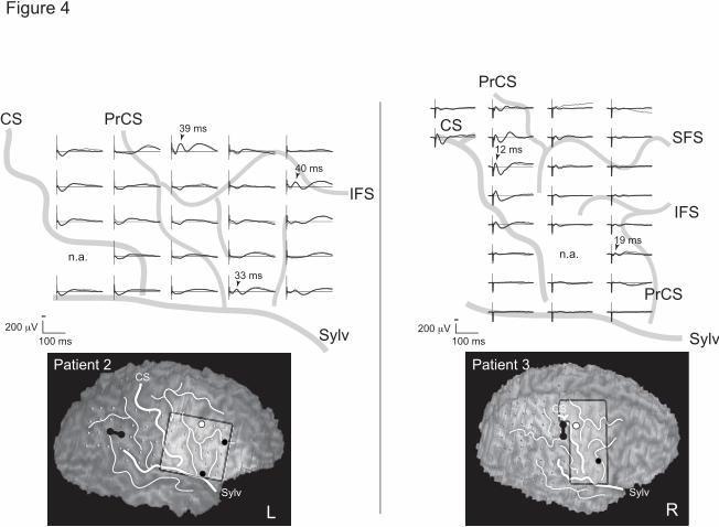
PrCS
IFS
CS
Sylv
39 ms
33 ms
40 ms
-
100 ms200 μV
-100 ms
200 μV
n.a. n.a.
PrCS
CS
PrCS
IFS
SFS12 ms
19 ms
Sylv
CS
Sylv
L
CS
Sylv
CSSC
R
Figure 4
Patient 2 Patient 3

Figure 5
PrCS
IFS
CS
Sylv
27 ms
-
100 ms200 μV
CS
Sylv
SFS
PrCS
L

BA1d BA2d 7PC 7A
PFBA1v BA2v
PFm
PGa
PGpPFt PFop
OP4
Parietal parcellation in Jülich Atlas
7PC7A
PGpPGa
PFmPF
BA1v BA2v
BA1dBA2d
PFtPFopOP4
Example
stimulus site
maximum CCEP site
additional (2nd/3rd)CCEP site
central sulcus