topographical layout of hand, eye, calculation - meteore service
TRANSCRIPT

Neuron, Vol. 33, 475–487, January 31, 2002, Copyright 2002 by Cell Press
Topographical Layout of Hand, Eye,Calculation, and Language-RelatedAreas in the Human Parietal Lobe
ization might be masked by interindividual variability andthe lack of anatomical precision inherent in comparinggroup studies from different neuroimaging laboratories.In support of this possibility, neurophysiological studiesin monkeys have revealed a parcellation of the parietal
Olivier Simon,1 Jean-Francois Mangin,2
Laurent Cohen,1 Denis Le Bihan,2
and Stanislas Dehaene1,3
1Unite INSERM 3342 Unite de Neuro-Activation FonctionnelleIFR 49 lobe into several specialized areas whose neurons ex-
hibit distinct response properties (Rizzolatti et al., 1998).Service Hospitalier Frederic JoliotCEA/DSV, Orsay Furthermore, neuroimaging studies have also begun to
reveal functional specialization in the human parietalFrancelobe, for instance, hand versus eye movement (Kawa-shima et al., 1996) or grasping versus pointing (Graftonet al., 1996).Summary
The aim of the present study is to further characterizethe functional organization of the human parietal lobe.To identify subdivisions of the human parietal cortex,
we collected fMRI data while ten subjects performed We collected functional magnetic resonance imageswhile subjects performed six different tasks: pointing,six tasks: grasping, pointing, saccades, attention, cal-
culation, and phoneme detection. Examination of task grasping, attention orienting, saccades, calculation, andphoneme detection. We did not design new tasks, butintersections revealed a systematic anterior-to-poste-
rior organization of activations associated with grasp- rather, replicated and extended existing tasks in orderto identify all six task-related activations within a subjecting only, grasping and pointing, all visuomotor tasks,
attention and saccades, and saccades only. Calcula- in a few minutes. This allowed us to examine the geomet-rical relations of those activations. In particular, we ex-tion yielded two distinct activations: one unique to
calculation in the bilateral anterior IPS mesial to the amined the overlap between the areas engaged in calcu-lation and in visuospatial tasks. The use of simplesupramarginal gyrus and the other shared with pho-
neme detection in the left IPS mesial to the angular grasping, pointing, and saccades tasks allowed us toidentify putative human equivalents of monkey parietalgyrus. These results suggest human homologs of the
monkey areas AIP, MIP, V6A, and LIP and imply a areas and to locate the human activations during calcu-lation and language tasks relative to those functionallarge cortical expansion of the inferior parietal lobule
correlated with the development of human language landmarks.and calculation abilities.
ResultsIntroduction
In each task, a large network comprising parietal, lateralParietal cortex is active during a variety of visuospatial frontal, and mesial frontal regions was observed. Wetasks, including hand reaching, grasping, eye and/or first describe the global pattern of activation observedattention orienting, mental rotation, and spatial working in each task relative to its control (Table 1 and Figurememory (Culham and Kanwisher, 2001). However, other 1). Focusing on parietal activations, we then describetasks without obvious visuospatial requirements, such which active regions, if any, were selectively activatedas mental calculation (Dehaene et al., 1999) or phonolog- by a single task (Table 2 and Figure 2). Finally, we con-ical word processing (Jonides et al., 1998) also yield sider parietal regions that were common to two or moreparietal activations, sometimes at very similar locations. tasks (Table 3 and Figure 2).One possible interpretation of these findings is that theparietal cortex is involved in abstract and generic pro- Activations Observed in Each Taskcesses that are useful for many tasks. In support of this Graspingpossibility, Wojciulik and Kanwisher (1999) have ob- As expected, given that the task involved right handserved overlapping activations in the parietal lobe in a movements, a unilateral activation of the left centralvariety of tasks that all shared an abstract component sulcus was observed in the hand region of the motorof attention orienting. According to this interpretation, homonculus. In addition, grasping activated the frontalthe parietal activation during calculation, in particular, and parietal regions. The frontal network included a bi-would reflect the engagement of generic mechanisms lateral mesial superior frontal gyrus activation in theof coordinate transformation and attention (Zago et al., vicinity of the supplementary motor area extending into2001). the anterior cingulate gyrus. There was also a left pre-
An alternative possibility, however, is that the human central gyrus activation extending anteriorily into theintraparietal sulcus consists of a mosaic of distinct spe- middle frontal gyrus. In the inferior frontal gyrus (BA 44),cialized areas, including a region for the manipulation a bilateral activation was observed. The parietal networkof numerical quantities (Dehaene and Cohen, 1995; De- contained a unilateral left postcentral activation facinghaene et al., 1998). This fine-grained anatomical special- the central sulcus activation. In addition, there were
bilateral activations in the anterior part of the supramar-ginal gyrus, the postcentral sulcus, the horizontal seg-3 Correspondence: [email protected]
Neuron476
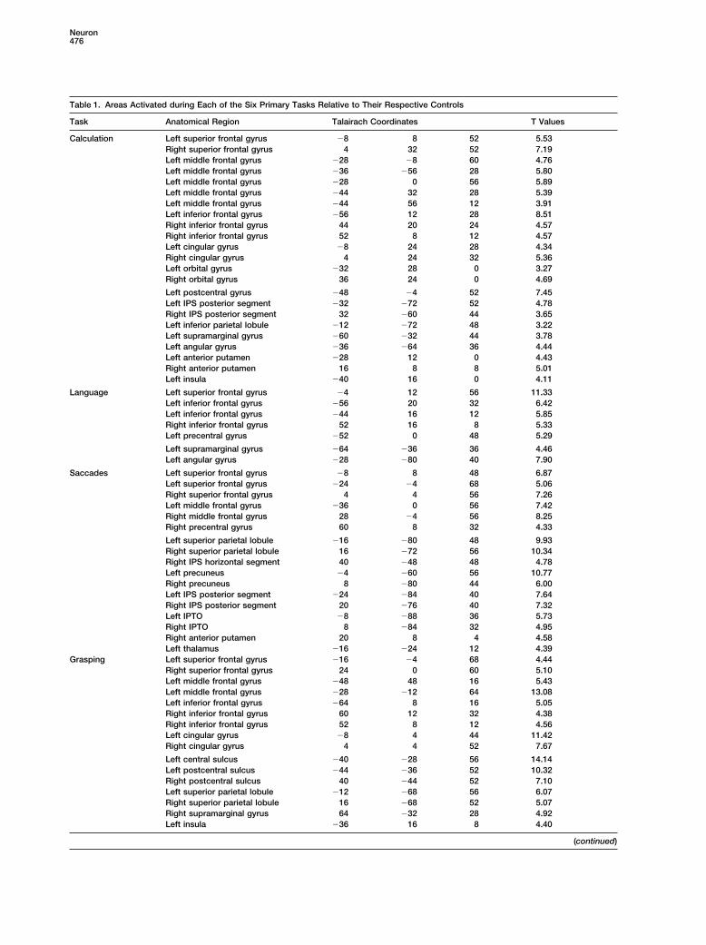
Table 1. Areas Activated during Each of the Six Primary Tasks Relative to Their Respective Controls
Task Anatomical Region Talairach Coordinates T Values
Calculation Left superior frontal gyrus �8 8 52 5.53Right superior frontal gyrus 4 32 52 7.19Left middle frontal gyrus �28 �8 60 4.76Left middle frontal gyrus �36 �56 28 5.80Left middle frontal gyrus �28 0 56 5.89Left middle frontal gyrus �44 32 28 5.39Left middle frontal gyrus �44 56 12 3.91Left inferior frontal gyrus �56 12 28 8.51Right inferior frontal gyrus 44 20 24 4.57Right inferior frontal gyrus 52 8 12 4.57Left cingular gyrus �8 24 28 4.34Right cingular gyrus 4 24 32 5.36Left orbital gyrus �32 28 0 3.27Right orbital gyrus 36 24 0 4.69
Left postcentral gyrus �48 �4 52 7.45Left IPS posterior segment �32 �72 52 4.78Right IPS posterior segment 32 �60 44 3.65Left inferior parietal lobule �12 �72 48 3.22Left supramarginal gyrus �60 �32 44 3.78Left angular gyrus �36 �64 36 4.44Left anterior putamen �28 12 0 4.43Right anterior putamen 16 8 8 5.01Left insula �40 16 0 4.11
Language Left superior frontal gyrus �4 12 56 11.33Left inferior frontal gyrus �56 20 32 6.42Left inferior frontal gyrus �44 16 12 5.85Right inferior frontal gyrus 52 16 8 5.33Left precentral gyrus �52 0 48 5.29
Left supramarginal gyrus �64 �36 36 4.46Left angular gyrus �28 �80 40 7.90
Saccades Left superior frontal gyrus �8 8 48 6.87Left superior frontal gyrus �24 �4 68 5.06Right superior frontal gyrus 4 4 56 7.26Left middle frontal gyrus �36 0 56 7.42Right middle frontal gyrus 28 �4 56 8.25Right precentral gyrus 60 8 32 4.33
Left superior parietal lobule �16 �80 48 9.93Right superior parietal lobule 16 �72 56 10.34Right IPS horizontal segment 40 �48 48 4.78Left precuneus �4 �60 56 10.77Right precuneus 8 �80 44 6.00Left IPS posterior segment �24 �84 40 7.64Right IPS posterior segment 20 �76 40 7.32Left IPTO �8 �88 36 5.73Right IPTO 8 �84 32 4.95Right anterior putamen 20 8 4 4.58Left thalamus �16 �24 12 4.39
Grasping Left superior frontal gyrus �16 �4 68 4.44Right superior frontal gyrus 24 0 60 5.10Left middle frontal gyrus �48 48 16 5.43Left middle frontal gyrus �28 �12 64 13.08Left inferior frontal gyrus �64 8 16 5.05Right inferior frontal gyrus 60 12 32 4.38Right inferior frontal gyrus 52 8 12 4.56Left cingular gyrus �8 4 44 11.42Right cingular gyrus 4 4 52 7.67
Left central sulcus �40 �28 56 14.14Left postcentral sulcus �44 �36 52 10.32Right postcentral sulcus 40 �44 52 7.10Left superior parietal lobule �12 �68 56 6.07Right superior parietal lobule 16 �68 52 5.07Right supramarginal gyrus 64 �32 28 4.92Left insula �36 16 8 4.40
(continued)

Topography of the Human Parietal Lobe477
Table 1. Continued.
Task Anatomical Region Talairach Coordinates T Value
Painting Left superior frontal gyrus �12 �8 60 8.82Right superior frontal gyrus 4 4 56 8.39Right middle frontal gyrus 24 0 56 7.32Left cingular gyrus �8 4 44 7.07Left precentral sulcus �32 �12 64 9.56Left central sulcus �40 �28 56 9.40
Left postcentral sulcus �36 �44 56 8.25Left superior parietal lobule �16 �76 48 11.37Right superior parietal lobule 16 �68 52 11.30Right superior parietal lobule 36 �48 56 5.29Right IPS horizontal segment 40 �40 60 4.89Left precuneus �8 �60 56 5.18Right IPS posterior segment 28 �76 32 4.76Left anterior putamen �24 12 4 5.07Right anterior putamen 24 4 4 4.45Left thalamus �12 �20 8 5.27
Attention Right superior frontal gyrus 4 4 60 3.79Left middle frontal gyrus �36 44 32 4.95Left middle frontal gyrus �28 �8 64 4.37Right middle frontal gyrus 24 64 28 4.83Right middle frontal gyrus 40 16 36 5.60Right inferior frontal gyrus 48 36 0 6.10
Right postcentral sulcus 44 �40 52 7.02Right superior parietal lobule 24 �64 60 6.91Left IPS horizontal segment �36 �60 60 3.35Right supramarginal gyrus 56 �44 32 4.08Left precuneus �4 �60 48 7.32Right precuneus 8 �64 52 7.27
The table shows the coordinates of the local maxima of significance within the Montreal Neurological Institute (MNI) coordinate system, witht values for the contrast between primary task and control task. IPTO, junction of intraparietal and tranverse occipital sulci.
ment of the intraparietal sulcus, and the posterior part parietal region. From front to back, the active areas werethe posterior superior parietal lobules and the horizontalof the superior parietal lobule. Finally, an activation was
seen in the left insula. and posterior segments of the intraparietal sulcus, ex-tending down to the transverse occipital sulcus. ThePointing
Inspection of the activation during the pointing task right putamen, right globus pallidus, and left thalamuswere also active.showed a considerable overlap with the grasping task,
including left central sulcus, left precentral and left post- AttentionThe attention task consistently yielded a lower degreecentral activation, bilateral mesial superior frontal gyrus
activation at the level of the supplementary motor area, of activation than the other tasks. Nevertheless, move-ments of visual attention activated areas partially similarand bilateral anterior cingulate activation. However, im-
portant differences with grasping were observed in the to those seen in the saccades task. The frontal networkshowed bilateral activations again lying at the intersec-parietal lobe. While pointing caused a bilateral activation
of the horizontal segment of the intraparietal sulcus, tion of the superior frontal, the middle frontal, and theprecentral gyrus, but with a predominance in the rightthere was little or no anterior supramarginal activity, but
there was a clear activation of the posterior part of the hemisphere where the activation extended downwardsinto the precentral sulcus. Bilateral activation was alsosuperior parietal lobule bilaterally, extending into the left
precuneus. Finally, we observed a symmetrical activa- observed in the lateral prefrontal cortex within the supe-rior frontal sulcus. A small cluster was observed in thetion of the anterior putamen and a left thalamic acti-
vation. right inferior frontal gyrus (BA 45). In the parietal lobe, abilateral activation was detected in the superior parietalSaccades
The saccades task yielded a distinct network of regions lobule, the precuneus, and the horizontal and superiorpart of the posterior segment of the IPS, with an anteriorwith three components. First, there was a bilateral clus-
ter lying at the intersection of the superior frontal, the extension to the posterior part of the postcentral sulcusin the right hemisphere.middle frontal, and the precentral gyrus, corresponding
to the known localization of the frontal eye fields (Cor- CalculationActivation was bilateral with a left hemisphere predomi-betta et al., 1998). Second, there was activation in the
mesial portion of the posterior superior frontal gyrus, nance. A large left cluster included the superior frontalgyrus, the precentral sulcus extending into dorsolateralplausibly corresponding with the supplementary eye
field area (Grosbras et al., 1999). Third, there was an prefrontal cortex, and the inferior prefrontal gyrus (BA44/45). Similar but smaller activation was observed inextended bilateral area of activation in the posterior
Neuron478
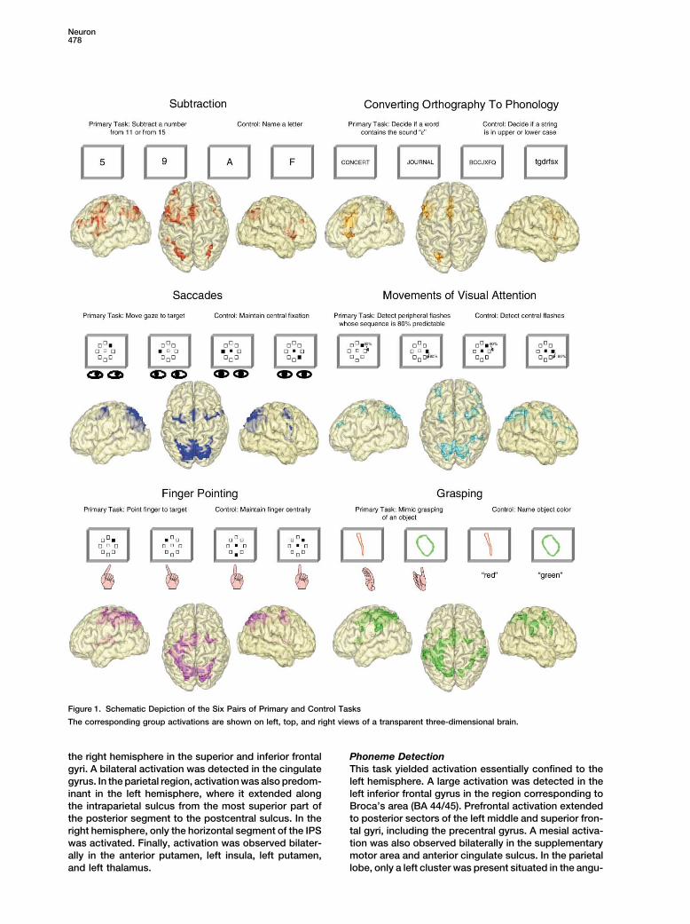
Figure 1. Schematic Depiction of the Six Pairs of Primary and Control Tasks
The corresponding group activations are shown on left, top, and right views of a transparent three-dimensional brain.
the right hemisphere in the superior and inferior frontal Phoneme DetectionThis task yielded activation essentially confined to thegyri. A bilateral activation was detected in the cingulate
gyrus. In the parietal region, activation was also predom- left hemisphere. A large activation was detected in theleft inferior frontal gyrus in the region corresponding toinant in the left hemisphere, where it extended along
the intraparietal sulcus from the most superior part of Broca’s area (BA 44/45). Prefrontal activation extendedto posterior sectors of the left middle and superior fron-the posterior segment to the postcentral sulcus. In the
right hemisphere, only the horizontal segment of the IPS tal gyri, including the precentral gyrus. A mesial activa-tion was also observed bilaterally in the supplementarywas activated. Finally, activation was observed bilater-
ally in the anterior putamen, left insula, left putamen, motor area and anterior cingulate sulcus. In the parietallobe, only a left cluster was present situated in the angu-and left thalamus.

Topography of the Human Parietal Lobe479
Table 2. Task-Specific Regions
Intersections Anatomical Regions Talairach Coordinates
Ca-Gr Ca-Po Ca-Sa Ca-At Ca-LgCalculation only Left IPS horizontal segment �36 �60 36 1.7 10�4 1.1 10�5 4.9 10�4 9.6 10�4 7.8 10�3
Gr-Po Gr-Ca Gr-Sa Cr-At Gr-LgGrasping only Left postcentral sulcus �28 �44 68 3.7 10�6 6.2 10�6 1.4 10�3 1.5 10�4 1.4 10�6
Left postcentral sulcus �44 �36 40 1.7 10�5 1.8 10�6 1.2 10�4 8.5 10�5 3.6 10�7
Left postcentral sulcus �60 �32 36 1.3 10�7 7.1 10�8 6 10�6 4.8 10�7 3.1 10�4
Left postcentral sulcus �48 �40 60 3.6 10�4 5.6 10�5 4.4 10�5 1.1 10�4 1.5 10�6
Right postcentral sulcus 36 �44 60 7.2 10�3 1.1 10�4 1.1 10�4 7.1 10�3 3.1 10�5
Right postcentral sulcus 64 �32 24 3.2 10�4 3.1 10�4 1.1 10�3 6.3 10�4 3.1 10�5
Sa-Gr Sa-Po Sa-Ca Sa-At Sa-LgSaccades only Left SPL posterior segment �8 �88 36 3.1 10�3 5 10�4 7.5 10�4 5.1 10�5 8 10�5
Left IPTO �24 �84 24 2.8 10�3 1.7 10�4 1.2 10�3 3.2 10�3 2 10�3
Right IPS posterior segment 28 �64 48 3.9 10�5 1.4 10�4 2.6 10�3 10�4 9.9 10�6
Right SPL posterior segment 8 �84 36 1.8 10�5 3.7 10�4 6 10�3 5.1 10�13 1.9 10�4
Right IPTO 28 �80 24 6.8 10�4 1.8 10�3 2.4 10�3 1.1 10�4 6.3 10�3
At-Gr At-Po At-Ca At-Sa At-LgAttention only Left precuneus �4 �52 52 6.6 10�6 9.8 10�3 2.9 10�5 2.4 10�3 1.8 10�4
The reported regions were significantly active (p � 0.01) during the indicated task, and not during the other five tasks. The columns at rightshow the outcome of all the relevant pairwise comparisons (Gr, grasping; Po, pointing; Sa, saccades; At, attention; Ca, calculation; and Lg,language). The p value indicates the uncorrected voxel-wise significance level at the local maximum closest to the given coordinates. IPS,intraparietal sulcus; SPL, superior parietal lobule.
lar gyrus and in the horizontal segment of the intraparie- language, but not relative to grasping, saccades, andattention.tal sulcus.Language OnlyNo parietal region passed all the criteria for a selective
Task-Specific Regions response during the phoneme detection task only.Grasping OnlyTwo clusters of voxels were activated only in graspingand significantly more during grasping than during any Task Intersections
Grasping and Pointingother task. They occupied symmetrical locations in theleft and right anterior supramarginal/postcentral re- A large cluster of activation selective for grasping and
pointing was observed in the left anterior IPS, extendinggions.Pointing Only into the superior parietal lobule and the postcentral sul-
cus, with a posterior extension into the horizontal seg-No parietal region passed all the criteria for a selectiveresponse during pointing only. ment of the IPS and an anterior extension into the central
sulcus. This activation was located posterior and dorsalSaccades OnlyWithin the parietal lobe, a large posterior cluster was to the bilateral activation specific to grasping only and
immediately dorsal to the activation observed duringobserved bilaterally during saccades only. As shownin Figure 2, this region included bilateral areas of the calculation only.
Four Visuospatial Taskssuperior parietal lobule as well as the posterior segmentof the intraparietal sulcus extending down to the inter- Two large clusters occupying symmetrical locations in
the left and right superior parietal lobules, dorsal to thesection between the intraparietal sulcus and the trans-verse occipital sulcus (area IPTO [Wojciulik and Kan- posterior horizontal segment of the IPS, were selectively
activated during the four visuospatial tasks (grasping,wisher, 1999]).Attention Only pointing, saccades, and attention).
Calculation, Language, and SaccadesA cluster of voxels situated in the left precuneus wasactivated during the attention task only. A similar cluster An area of overlap between the calculation, language,
and saccades tasks was detected in the left posteriorwas also observed at the symmetrical location in theright precuneus (coordinates 12, �60, 44). However, its segment of the IPS beneath the left angular gyrus. A
small cluster of activation common to the calculationactivation was greater during attention than during thecalculation, language, or grasping tasks, but not the and language tasks, but absent during the other four
tasks, was also observed just next to this activationsaccades or pointing tasks.Calculation Only (coordinates �28, �68, 36). However, this region failed
to show significantly greater activation during either theActivation induced by calculation only was present inthe horizontal segment of the left intraparietal sulcus. language or the calculation task than during the saccade
task.Another cluster was observed during calculation andno other task at the symmetrical location in the right The observation of an intersection between calcula-
tion, language, and saccades was unexpected. We rea-hemisphere (coordinates 40, �56, 52). However, it failedto pass all criteria for selectivity: activation was signifi- soned that intersubject averaging and smoothing could
have caused the intense saccade-related activations tocantly greater for calculation relative to pointing and
Neuron480
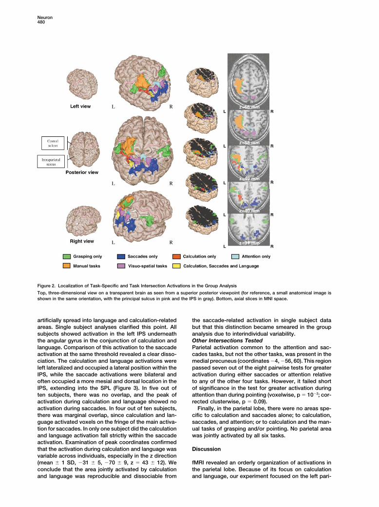
Figure 2. Localization of Task-Specific and Task Intersection Activations in the Group Analysis
Top, three-dimensional view on a transparent brain as seen from a superior posterior viewpoint (for reference, a small anatomical image isshown in the same orientation, with the principal sulcus in pink and the IPS in gray). Bottom, axial slices in MNI space.
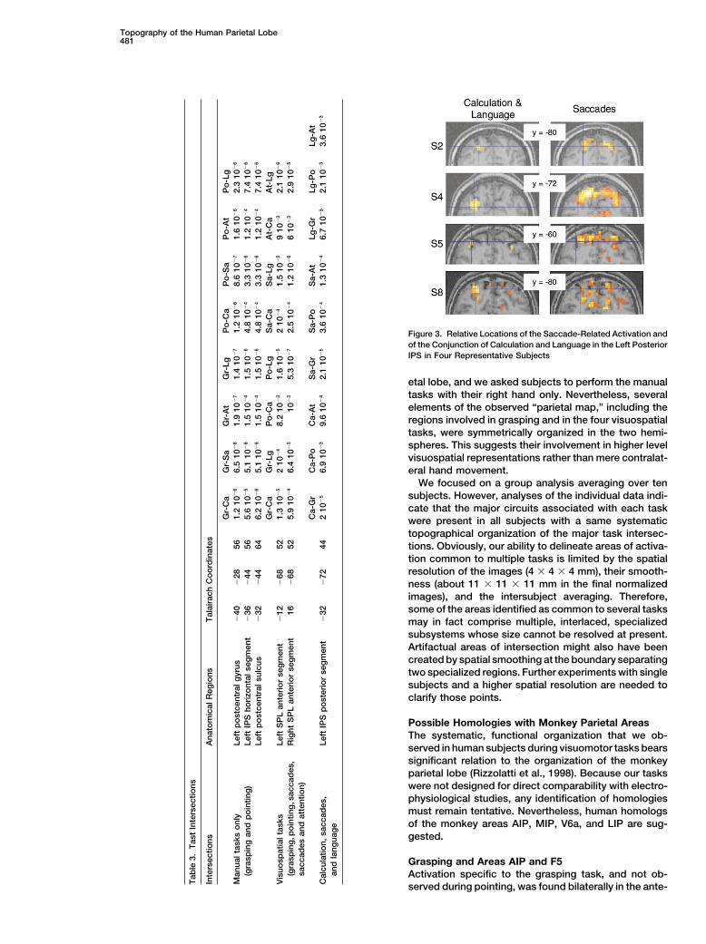
artificially spread into language and calculation-related the saccade-related activation in single subject databut that this distinction became smeared in the groupareas. Single subject analyses clarified this point. All
subjects showed activation in the left IPS underneath analysis due to interindividual variability.Other Intersections Testedthe angular gyrus in the conjunction of calculation and
language. Comparison of this activation to the saccade Parietal activation common to the attention and sac-cades tasks, but not the other tasks, was present in theactivation at the same threshold revealed a clear disso-
ciation. The calculation and language activations were medial precuneus (coordinates �4, �56, 60). This regionpassed seven out of the eight pairwise tests for greaterleft lateralized and occupied a lateral position within the
IPS, while the saccade activations were bilateral and activation during either saccades or attention relativeto any of the other four tasks. However, it failed shortoften occupied a more mesial and dorsal location in the
IPS, extending into the SPL (Figure 3). In five out of of significance in the test for greater activation duringattention than during pointing (voxelwise, p � 10�2; cor-ten subjects, there was no overlap, and the peak of
activation during calculation and language showed no rected clusterwise, p � 0.09).Finally, in the parietal lobe, there were no areas spe-activation during saccades. In four out of ten subjects,
there was marginal overlap, since calculation and lan- cific to calculation and saccades alone; to calculation,saccades, and attention; or to calculation and the man-guage activated voxels on the fringe of the main activa-
tion for saccades. In only one subject did the calculation ual tasks of grasping and/or pointing. No parietal areawas jointly activated by all six tasks.and language activation fall strictly within the saccade
activation. Examination of peak coordinates confirmedthat the activation during calculation and language was Discussionvariable across individuals, especially in the z direction(mean � 1 SD, �31 � 5, �70 � 9, z � 43 � 12). We fMRI revealed an orderly organization of activations in
the parietal lobe. Because of its focus on calculationconclude that the area jointly activated by calculationand language was reproducible and dissociable from and language, our experiment focused on the left pari-
Topography of the Human Parietal Lobe481
Figure 3. Relative Locations of the Saccade-Related Activation andof the Conjunction of Calculation and Language in the Left PosteriorIPS in Four Representative Subjects
etal lobe, and we asked subjects to perform the manualtasks with their right hand only. Nevertheless, severalelements of the observed “parietal map,” including theregions involved in grasping and in the four visuospatialtasks, were symmetrically organized in the two hemi-spheres. This suggests their involvement in higher levelvisuospatial representations rather than mere contralat-eral hand movement.
We focused on a group analysis averaging over tensubjects. However, analyses of the individual data indi-cate that the major circuits associated with each taskwere present in all subjects with a same systematictopographical organization of the major task intersec-tions. Obviously, our ability to delineate areas of activa-tion common to multiple tasks is limited by the spatialresolution of the images (4 � 4 � 4 mm), their smooth-ness (about 11 � 11 � 11 mm in the final normalizedimages), and the intersubject averaging. Therefore,some of the areas identified as common to several tasksmay in fact comprise multiple, interlaced, specializedsubsystems whose size cannot be resolved at present.Artifactual areas of intersection might also have beencreated by spatial smoothing at the boundary separatingtwo specialized regions. Further experiments with singlesubjects and a higher spatial resolution are needed toclarify those points.
Possible Homologies with Monkey Parietal AreasThe systematic, functional organization that we ob-served in human subjects during visuomotor tasks bearssignificant relation to the organization of the monkeyparietal lobe (Rizzolatti et al., 1998). Because our taskswere not designed for direct comparability with electro-physiological studies, any identification of homologiesmust remain tentative. Nevertheless, human homologsof the monkey areas AIP, MIP, V6a, and LIP are sug-gested.
Grasping and Areas AIP and F5Activation specific to the grasping task, and not ob-
Tab
le3.
Tas
tIn
ters
ectio
ns
Inte
rsec
tions
Ana
tom
ical
Reg
ions
Tal
aira
chC
oo
rdin
ates
Gr-
Ca
Gr-
Sa
Gr-
At
Gr-
LgP
o-C
aP
o-S
aP
o-A
tP
o-L
gM
anua
ltas
kso
nly
Left
po
stce
ntra
lgyr
us�
40�
2856
1.2
10�
66.
510
�8
1.9
10�
71.
410
�7
1.2
10�
68.
610
�7
1.6
10�
62.
310
�6
(gra
spin
gan
dp
oin
ting
)Le
ftIP
Sho
rizo
ntal
seg
men
t�
36�
4456
5.6
10�
55.
110
�6
1.5
10�
41.
510
�6
4.8
10�
43.
310
�6
1.2
10�
47.
410
�6
Left
po
stce
ntra
lsul
cus
�32
�44
646.
210
�6
5.1
10�
61.
510
�4
1.5
10�
64.
810
�4
3.3
10�
61.
210
�4
7.4
10�
6
Gr-
Ca
Gr-
LgP
o-C
aP
o-L
gS
a-C
aS
a-Lg
At-
Ca
At-
LgV
isuo
spat
ialt
asks
Left
SP
Lan
teri
or
seg
men
t�
12�
6852
1.3
10�
52
10�
48.
210
�3
1.6
10�
52
10�
41.
510
�5
910
�3
2.1
10�
6
(gra
spin
g,p
oin
ting
,sac
cad
es,
Rig
htS
PL
ante
rio
rse
gm
ent
16�
6852
5.9
10�
46.
410
�5
10�
35.
310
�7
2.5
10�
41.
210
�6
610
�3
2.9
10�
5
sacc
ades
and
atte
ntio
n)C
a-G
rC
a-P
oC
a-A
tS
a-G
rS
a-P
oS
a-A
tLg
-Gr
Lg-P
oLg
-At
Cal
cula
tion,
sacc
ades
,Le
ftIP
Sp
ost
erio
rse
gm
ent
�32
�72
442
10�
56.
910
�3
9.6
10�
42.
110
�5
3.6
10�
41.
310
�4
6.7
10�
52.
110
�3
3.6
10�
3
and
lang
uag
e
served during pointing, was found bilaterally in the ante-
Neuron482
rior part of the inferior parietal lobule within the banks that this area is specialized for visually guided handmovements (Kawashima et al., 1996). It may encompassof the inferior postcentral sulcus, which constitutes the
anterior and inferior extension of the IPS. Similar activa- the human homolog of the monkey area MIP (medialintraparietal; tentative coordinates in humans, �40,tion was observed during previous studies of actual or
imagined grasping (Binkofski et al., 1998; Decety et al., �40, 60). MIP neurons code for peripersonal space interms of its accessibility by the arm (Eskandar and As-1994; Faillenot et al., 1997; Grafton et al., 1996), as well
as during observation of grasping movements (Buccino sad, 1999) or by a tool (Iriki et al., 1996). They are selec-tively modulated by the direction of an upcoming handet al., 2001). Grafton et al. (1996) also observed anterior
inferior parietal activation during grasping but interpre- or arm movement even when the direction is distinctfrom the direction of visual motion (Eskandar and Assad,ted it as an engagement of the secondary somatosen-
sory area (SII). This interpretation can be rejected in the 1999). Through its connections with areas V6A, LIP, andthe premotor cortex, area MIP plays a critical role inpresent case because our task did not involve the actual
touch of an object. Furthermore, area SII is consistently spatially directed hand movements (Colby, 1998). Thisfunction was clearly required during the pointing tasklocalized in the upper bank of the Sylvian fissure immedi-
ately posterior to the central sulcus (Disbrow et al., 2000) but presumably also during the grasping task since itrequired programming finger movements appropriate toand thus inferior to the activation observed in the pres-
ent study. the location and orientation of visually presented ob-jects.Our grasping-specific area appears as a plausible an-
alog of monkey area AIP (anterior intraparietal), a region Our paradigm was not designed to separate area MIPfrom other regions that also contribute to motor action.involved in the visually guided shaping of the hand (Mu-
rata et al., 2000; Sakata et al., 1995). Its tentative coordi- Indeed, we also observed activation of the superior pari-etal lobule during both grasping and pointing. In mon-nates in humans, rounded to the nearest centimeter,
would then be �60, �30, 40. In humans, the lower extent keys, the convexity of the superior parietal lobule isformed by area PE, which is thought to be involved inof the postcentral sulcus, within which the grasping-
specific activation is found, lies in continuity with the the elaboration of proprioceptive information (Rizzolattiet al., 1998). Some PE neurons are activated by passiveintraparietal sulcus and hence constitutes a plausible
homolog of the anterior intraparietal sulcus in monkeys. joint rotation or active limb movements, while otherscombine tactile and joint information (Mountcastle etAIP neurons are active during grasping/manipulation
movements, presentation of visual objects, or both. Gal- al., 1975). Altogether, area PE may be involved in therepresentation of the body in space. Such a representa-lese et al. (1994) have shown that AIP inactivation results
in a deficit of hand shaping for grasping objects, with tion was clearly essential in the grasping and pointingtasks where subjects attended to their hand and fingerno deficit in the reaching component of the movement.
This is highly similar to human subjects in whom anterior orientation information in darkness.intraparietal lesions result in selective deficits of thecoordination of finger movements required for grasping Saccades, Attention, and Areas LIP, 7a,(Binkofski et al., 1998). V6, V6a, and PEc
The main frontal target of area AIP is the ventral pre- The saccades task and the attention task both lead tomotor area F5 (Luppino et al., 1999), which contains the activation of the bilateral dorsal parietal lobe. Manymirror neurons activated during both action execution of the active areas were common to attention and sac-and action recognition (Rizzolatti et al., 1998). We also cades. The area of intersection included the bilateralobserved a bilateral inferior prefrontal activation in Brod- dorsal superior parietal lobule, which was also activatedmann’s area 44, which is thought to be the human homo- during grasping and pointing, and a mesial region in thelog of area F5 (Iacoboni et al., 1999; Rizzolatti et al., precuneus, which also tended to be activated by the1998). Together, F5 and AIP may constitute a circuit pointing task. Attention and saccades also jointly acti-for the visual guidance of hand movements, which is vated the bilateral frontal eye fields and supplementarypresent in both humans and monkeys. eye fields. This replicates earlier results (Corbetta et al.,
1998; Nobre et al., 2000) and supports the hypothesisof a tight linkage between attention and eye movementsGoal-Directed Hand Movements
and Areas MIP and PE (Rizzolatti et al., 1987). A new outcome of the presentstudy, however, is that a major component of this circuit,We also observed hand movement-related activation in
a left-lateralized cluster of voxels ranging from the cen- located in the mesial and dorsal bank of the posteriorintraparietal sulcus and neighboring superior parietaltral sulcus to the postcentral sulcus, the superior parietal
lobule, and the horizontal segment of the IPS contralat- cortex, is also very strongly activated during graspingand pointing tasks in the absence of overt eye move-eral to the moving hand. It clearly encompassed the
primary motor and somatosensory hand areas. How- ments. Wojculik and Kanwisher (1999) also found thisarea to be activated during three different forms of atten-ever, its posterior extension into the left middle IPS re-
quires more discussion. This area was strongly activated tion: peripheral attention shifting, sustained attention toparafoveal locations, and temporal attention to featureduring both grasping and pointing movements but to-
tally inactive during eye or attention movements, calcu- conjunctions. They suggested that its function was notthe mere shifting of gaze and attention across locations,lation, and phoneme detection. A similar, though
smaller, focus was observed in the right middle IPS. but rather, the suppression of task-irrelevant distractors.It is unclear, however, why the grasping task wouldGiven that the pointing and saccade tasks involved
similar stimuli, the dissociation between them suggests require suppression since no distractors were pre-
Topography of the Human Parietal Lobe483
sented. The present data merely allow us to suggest tion (Galletti et al., 1995) (tentative coordinates inthat this region plays a general role in the selection of humans: �25, �80, 20). The human posterior intraparie-objects for pending actions, whether the actions are tal region active during saccades has been proposedpurely mental or involve actual object-directed hand or as a “posterior eye field” homologous to area LIP (lateraleye movements. intraparietal) (Muri et al., 1996). We observed bilateral
We also found some dissociations between the sac- activation during saccades in the posterior IPS, althoughcades and attention networks. Saccades yielded greater the left activation was partially masked by the languageactivation in the bilateral postero-mesial intraparietal and calculation activation in the group’s results, assulcus, close to its intersection with the transverse oc- noted above. Our tentative coordinates for this areacipital sulcus (region IPTO), in the bilateral posterior (�30, �60, 50) fit nicely with those recently obtainedsuperior parietal lobule, and in the right posterior IPS. using the method of retinotopic mapping (Sereno et al.,Previous studies have observed IPTO activation in hu- 2001). As noted by Van Essen et al. (2001), and furthermans during attention tasks without any overt saccades discussed below, this localization implies a consider-(Corbetta et al., 1998) or indeed without even any form able posterior displacement of area LIP in humans rela-of movement planning (Wojciulik and Kanwisher, 1999). tive to monkeys.Thus, it is unclear why our attention task, as well asanother study of covert attention orienting (Gitelman et Calculation and Language-Related Activationsal., 1999), failed to activate this area. A lack of statistical in the Human Inferior Parietal Lobulesensitivity cannot be excluded. Because only two short The main goal of our experiment was to localize calcula-fMRI sequences were available for each task, our images tion and language-related activations in the human pa-may underestimate the attention network. rietal lobe and to examine their relative location and
Conversely, greater activation for attention than for overlap with other visuospatial activations. During asaccades was observed in the precuneus, thus revealing subtraction task, activation was observed bilaterally ina double dissociation. Some, but not all, imaging studies the lateral bank of the intraparietal sulcus, replicatingof covert attention report precuneus activation during earlier work (Chochon et al., 1999; Dehaene et al., 1999;visual attentional tasks (Corbetta et al., 1998; Gitelman Lee, 2000; Stanescu-Cosson et al., 2000; Zago et al.,et al., 1999; Nobre et al., 2000). The origin of this discrep- 2001). This activation did not overlap with those ob-ancy is not understood. Note that the attention task served during grasping, pointing, saccades, or attentionused 80% predictable targets, while the saccade task movements. However, examination of the overlap withused unpredictable targets. The active anticipation of the phoneme detection task revealed a separation intoan upcoming target may lead to the activation of visual two regions. The middle portion of the IPS correspond-imagery processes with which the precuneus has been ing to its horizontal segment underneath the supramar-tentatively related (Fletcher et al., 1996). Active anticipa- ginal gyrus was activated bilaterally, though with greatertion may also explain the bilateral anterior dorsolateral intensity in the left hemisphere, solely during the sub-prefrontal activation, which, like the precuneus activa- traction task. However, a more posterior and strictly left-tion, was observed solely during the attention task (Fig-
lateralized region of IPS underneath the angular gyrusure 1).
was activated jointly by calculation and phoneme de-In terms of homologies to the monkey brain, the hu-
tection.man areas activated during saccades, attention, or both
The presence of two neighboring regions for calcula-are likely to contain homologs of the monkey areas 7ation within the left IPS, only one of which is shared(also called PG), LIP, V6, V6a, and PEc. All of thesewith phonological processing, fits with the theoreticalareas contain neurons that encode the location of visualhypothesis that number processing relies on the coordi-stimuli and modulate their activity as a function ofnation of two different representations of numbers: awhether the animal intends to make an eye movementnonverbal quantity code involving the bilateral intrapa-toward them (Colby et al., 1996; Rizzolatti et al., 1998;rietal sulci and a verbal code involving classical lan-Snyder et al., 1998). The area jointly activated by all fourguage areas including the left angular gyrus (Dehaenevisuospatial tasks (saccades, attention, pointing, andand Cohen, 1995, 1997; Dehaene et al., 1998). Activationgrasping) might correspond to area V6a (tentative coor-of the left angular gyrus was previously observed duringdinates in humans: �15, �70, 50). Recordings in V6aoperations that require access to a rote verbal memoryand the neighboring area PEc reveal a joint selectivityof arithmetic facts, such as single digit addition or multi-for movements of the hand and eye at the single neuronplication (Chochon et al., 1999; Lee, 2000; Zago et al.,level (Battaglia-Mayer et al., 2001). Furthermore, the2001). In particular, its coordinates match with those ofmonkey V6a is located on the just posterior and mesial tothe angular gyrus activation previously observed duringarea MIP, which fits well with our observed localization inexact arithmetic fact retrieval relative to calculation ap-the superior parietal lobule just dorsal to the horizontalproximation (Dehaene et al., 1999; Stanescu-Cosson etsegment of the IPS. The precuneus activation duringal., 2000). During fact retrieval, subjects are thoughtattention might then relate to the monkey area PEc (ten-to basically “read out” the result of visually presentedtative coordinates in humans: 0, �50, 50).problems from their verbal memory. Indeed, overlappingFinally, for the posterior saccade-related activations,activations were observed during rote calculation andtwo identifications have been proposed. The medial pa-during phoneme detection, not only in the left angularrieto-occipital sulcus has been proposed as the humangyrus, but also in the left inferior frontal gyrus extendinghomolog of the monkey V6 complex (Portin and Hari,toward the anterior insula, the left premotor cortex, and1999), a high-level visual region (Galletti et al., 1999)
where neural activity is strongly modulated by eye posi- the left supplementary motor area. Those areas consti-
Neuron484
tute a classical circuit for reading (Fiez and Petersen, Recently, Van Essen et al. (2001) attempted to estab-1998; Price, 1998). The precise contribution of the left lish a correspondence between macaque and humanangular gyrus within this language circuit remains to be brains by matching computerized surface-based atlasesspecified. According to Hickok and Poeppel (2000), the across species. In the posterior and superior parietalinferior parietal lobule serves as an auditory-motor inter- lobule, their warped map fits well with our tentative local-face in language processing, connecting the left tempo- ization for areas V6a/PEc (PO in their scheme), MIP,ral areas for phonological speech analysis to frontal and PE (MDP in their scheme). However, they note thatareas for articulatory speech production. Such a func- warping, even though it implies a 20-fold expansion oftion would be in line with the general role of the parietal the parietal surface, does not provide a satisfactory ac-lobe in sensorimotor conversion operations. count of the human IPS and inferior parietal lobule. In
Although some subtraction problems may be stored particular, the warping algorithm projects monkey areain verbal memory, many are not learned by rote and LIP onto the horizontal segment of the IPS, at a regiontherefore require novel quantity manipulations. Quantity where no saccadic activation is observed in humansmanipulation can be plausibly related to the calculation- (Corbetta et al., 1998). Saccade-related activity is foundspecific bilateral activation of the middle intraparietal more posteriorily in the human IPS than predicted byregion. This region was previously found to show greater mere warping. Our data suggest that, in the course ofactivation during approximation than during exact cal- hominid evolution, areas AIP and LIP have become in-culation (Dehaene et al., 1999; Stanescu-Cosson et al., creasingly separated in cortical space as a consequence2000) and during subtraction more than during multipli- of the expansion of the inferior parietal lobule wherecation (Chochon et al., 1999; Lee, 2000). Bilateral intra- activations related to uniquely human activities of calcu-parietal activation has also been observed during sim- lation and language are observed. Because some ani-pler quantity processing tasks that do not involve mals do exhibit rudimentary abilities for calculation (De-calculation, such as deciding which of two Arabic nu- haene et al., 1998), our results suggest that anmerals is the larger (Chochon et al., 1999; Pesenti et al., evolutionary precursor of number processing in pri-2000; Pinel et al., 2001). Using single event fMRI in a mates, if it exists, might be found in the anterior part ofparametric design, Pinel et al. (2001) recently demon- area LIP, the dorsal sectors of areas 7a (PF), 7b (PG),strated that the activation of this region during number or the intermediate area PFG.comparison was independent of whether the numberswere presented as words or as Arabic digits but was Experimental Proceduresinversely proportional to the semantic distance between
Subjectsthe numbers to be compared. For such reasons, thisTen neurologically normal right-handed volunteers (7 females; meanregion is thought to encode the proximity relationsage 25, range 22–34) gave informed consent to participate in the
among numerical quantities in a nonverbal format akin study, which was approved by the regional ethical committee. Fourto a mental “number line” (Dehaene et al., 1998). The additional subjects were tested and rejected. Two had excessiveinsertion of this region amid other visuospatial maps movement artifacts. Two others exceeded our criteria of mean re-
sponse time lower than two seconds and mean error rate lower thanfits with this hypothesis of an internal mental space for20%: one subject failed in the calculation test and the other in thenumbers. During number comparison and approxima-language test.tion tasks, the intraparietal region coactivates together
with the bilateral superior parietal lobule and precuneusProcedure
(Dehaene et al., 1999; Pinel et al., 2001; Stanescu-Cos- Cerebral activation was studied in each six conditions: subtraction,son et al., 2000) at sites coincident with those observed grasping, pointing, saccades, visuospatial attention, and phonemefor saccades, attention, and the four visuospatial tasks. detection. Each of these primary tasks was compared to a control
task matched for stimulus characteristics. During an fMRI scanningThis suggests that these number processing tasks callsequence, 6 blocks of 13 trials each (26 s), preceded by a 4 sfor generic processes of attention orientation and spatialinstruction period were presented, with an alternation of primarymanipulation that operate upon a “spatial map” of nu-task and control task blocks. Each subject performed two suchmerical quantities in the middle IPS.sequences for each of the six tasks, resulting in 12 fMRI sequences.Prior to scanning, subjects also participated in a training session
Concluding Remarks on Monkey-Human with a single run of each task.Homologies Stimuli were presented with an active matrix video projector on
a dark background at the rate of 2 s per trial, synchronized with theOur study identified two lateral intraparietal areas asso-acquisition of an fMRI volume, using expe6 software (Pallier et al.,ciated with functions particularly developed in the hu-1997). All stimuli were presented for 150 ms to prevent undesiredman species: calculation and phoneme detection. Thosesaccades. Subjects were instructed to avoid eye movement duringareas were surrounded by visuospatial areas plausiblyall tasks except the saccades task. No arm movement was allowed
homologous to the monkey areas AIP, MIP, V6A, and except during grasping and pointing tasks. The adequacy of eyeLIP. This organization of areas fits well with the cytoar- and hand movements was assessed visually during the trainingchitectonic parcellation of the human parietal lobe pro- period.posed by Eidelberg and Galaburda (1984). Both studies
Saccades Tasksuggest a considerable expansion and differentiation ofThis task isolated the circuit for visually guided eye movementsthe inferior parietal lobule in humans compared with(Muri et al., 1996). The visual display consisted of eight small redmonkeys. In particular, Eidelberg and Galaburda (1984)boxes regularly arranged on a circle at 6� eccentricity from a similar
identifiy a left-lateralized field PG in the human angular box centered on screen (see Figure 1). In the primary saccade task,gyrus, which may correspond to the strictly left-lateral- on each trial, a filled white square appeared within a randomly cho-ized region that we found active during phoneme de- sen box for 150 ms and was replaced by a fixation cross centered
in the box. Subjects had to move their eyes toward this box andtection.
Topography of the Human Parietal Lobe485
fixate it for 2000 ms until the next trial appeared. In the control were French concrete words, five to eight letters long, with fre-quency higher than one per million. Eighty percent of the wordsfixation task, subjects were asked to maintain fixation on a fixation
cross that stayed within the central box while distracting white contained the sound “e” or “�.” Subjects were instructed to mentallysound out the words and to detect stimuli containing the sound “e”squares appeared randomly in the periphery.or “�” by depressing a right hand button as quickly as possible.Each word was seen only once during the entire experiment. In thePointing Taskcontrol case-detection task, the stimuli were letter strings matchedThis task isolated the circuit for visually guided finger-pointingto the words in number of letters. Eighty percent of these stringsmovements. The stimulus was similar to the saccade task exceptwere in upper case. Subjects were instructed to detect uppercasethat the fixation cross remained in the central box throughout. Instimuli by pressing a right hand button. Subjects were significantlythe primary pointing task, subjects were asked to point their rightslower in phoneme detection than in case detection (644 versusindex finger in the direction of the box where a filled white square481 ms; t(9 df) � 6.70, p � 0.001), but the error rates did not differappeared while maintaining their gaze on the central box. In the(7.0% versus 5.2%; t(9 df) � 1.75, p � 0.10).control fixation task, subjects kept their finger pointed at the central
box, neglecting randomly appearing peripheral distractors. Notethat our subjects were asked to move only the index finger and to
Calculation Taskkeep their hand, wrist, and arm still. This “pointing” task differs fromThis task identified cortical areas associated with simple mentalthe classical “reaching” task where subjects extend their arm tocalculation (Chochon et al., 1999). In the primary calculation task,touch a target with the finger. Nevertheless, our activations duringstimuli were Arabic digits from 2 to 9. Subjects were instructed topointing were similar to published reports using reaching (Graftonsubtract each number from a fixed reference (11 in the first run, 15et al., 1996; Kawashima et al., 1996).in the second) and to mentally name the result. In the control namingtask, stimuli were uppercase letters between B and J, excludingAttention Taskletter I. Subjects were asked to mentally name each letter. BothThis task was inspired by Corbetta et al. (1998) and measured covertresponse times and error rates were significantly higher in the calcu-shifts of visual attention. Throughout the experiment, subjects werelation task than in the control task (RTs, 881 versus 553 ms; t(9 df) �asked to detect, with a right hand key press, the sudden appearance13.2, p � 0.001; errors, 5.0 versus 0%; t(9 df) � 2.65, p � 0.03).of an asterisk within the same boxes as in the above saccades and
pointing tasks. The onset of the stimuli was randomized relative totrial onset (flat distribution, 0–400 ms). The primary attention task Imaging Parameterswas designed to induce shifts of attention to peripheral locations Functional MR images were acquired on a Siemens Signa 1.5Twithin a visual field. Targets appeared at predictable locations on whole-body scanner. Sequence parameters were Gradient-echothe peripheral circle, in a clockwise order, with an 80% probability. EPI, TE � 60 ms, TR � 2 s, flip angle � 90�, slice thickness � 3.8On the remaining 20% of trials, another unpredictable location was mm, interslice gap � 0, image matrix � 64 � 64, FOV � 192 � 256probed. Subjects were informed of the partial predictability of the mm, and functional voxel size � 3.75 � 3.75 � 3.8 mm3 . 18 axialstimuli and were instructed to shift attention, in advance of each trial, slices were acquired covering the parietal lobe and the majority ofto the most probable location of the upcoming item. The efficacy the prefrontal cortex, excluding the orbitofrontal sector. Each taskof these attentional instructions was monitored by comparing the sequence consisted of 97 18-slice scans, the first 4 of which werereaction times (RTs) with predictable and unpredictable trials. A not analyzed. Three-dimensional high-resolution anatomical imagessignificant difference was found (303 versus 383 ms; t(9 df) � 4.83, were also acquired (TE � 2.2 ms, TR � 10 ms, flip angle � 10�,p � 0.001). The control task was intended to prevent orienting of inversion time (TI) � 600 ms, matrix � 256 � 192 mm, FOV � 220 �attention to peripheral locations by engaging subjects’ attention in 165 mm, 128 sagittal slices, and slice thickness � 1.5 or 1.2 mm).the center. Subjects responded to asterisks presented in the centralfixation box, while peripheral asterisks were also flashed simultane-ously at random peripheral locations. Response times were fast Statistical Analyses(295 ms) and no different from those observed on predictable periph- The data from each individual subject was processed with Statisticaleral trials (t(9 df) � 1.07). Parameter Mapping software (SPM99). Following motion correction,
Subjects were instructed to refrain from moving their eyes normalization to the Montreal Neurological Institute (MNI) template,throughout the experiment. During training, subjects made at most and minimal spatial smoothing (5 mm), the activation in each sessionone saccade during the entire experiment (mean � 0.26% saccades was modeled using a linear combination of two functions obtainedper trial). by convolving the known temporal profile of the primary and control
blocks with the standard hemodynamic function of SPM. SingleGrasping Task subject data were analyzed with simple contrasts for greater activa-This task identified cortical areas for visually guided hand-grasping tion during each primary task than during its control and with all 30movements. To simplify stimulus presentation in the fMRI scanner, pairwise contrasts for greater activation in one task than in anotherwe used a pantomime task without actual object grasping. Although (voxelwise, p � 0.001; cluster level, p � 0.05, corrected).there are important differences between pantomimed and actual Group analyses were performed with random effects analyses.grasping (Goodale et al., 1994), both require using a visuomotor We first examined, for each of the six primary tasks relative to theirtransformation, presumably implemented in parietal circuitry, to respective controls, the entire set of areas showing a significantshape and orient the hand in relation to a visual stimulus. Indeed, activation (voxelwise, p � 0.01; cluster level, p � 0.05, corrected).pantomimed grasping lead to parietal activations comparable to We then performed random effects analyses of all the 30 pairwisethose of earlier studies using actual grasping (Binkofski et al., 1998; comparisons between two tasks. For those pairwise comparisons,Faillenot et al., 1997; Grafton et al., 1996). The stimuli were eight the search was confined to voxels belonging to the parietal lobedifferent outline shapes appearing in ten different orientations and and which showed activation in at least one of the six tasks ofin four different colors (green, blue, red, or yellow). In the primary interest. The voxelwise threshold was again set at p � 0.01, andgrasping task, subjects had to move their right hand and wrist as the cluster extent threshold was set to p � 0.05, corrected forfast and precisely as possible in order to mimic grasping of the multiple comparisons across the parietal volume (using the smallobject. In the control task, subjects kept their hand still and mentally volume correction option of SPM99; minimal cluster size, 9 voxels).named the color of the objects. A complete table of those results is available from the authors. Here,
a region is reported only if (1) all of its voxels passed a thresholdfor significance of p � 0.01 for the tasks of interest; (2) all of itsPhoneme Detection Task
This task identified the cortical circuitry of word reading (Fiez and voxels failed to pass this threshold for the remaining tasks; (3) allof the appropriate pairwise comparisons, as described above, iden-Petersen, 1998), with particularly emphasis on the possible contribu-
tion of parietal cortex to the conversion from orthography to phonol- tified a significant region encompassing this location; and (4) thevolume of the region was at least 26 voxels.ogy (Price, 1998). In the primary phoneme detection task, the stimuli
Neuron486
Acknowledgments R.J. (1996). Brain activity during memory retrieval. The influence ofimagery and semantic cueing. Brain 119, 1587–1596.
We gratefully acknowledge the methodological contributions of E. Gallese, V., Murata, A., Kaseda, M., Niki, N., and Sakata, H. (1994).Giacomini, F. Hennel, J.B. Poline, and D. Riviere. This work was Deficit of hand preshaping after muscimol injection in monkey pari-supported by a McDonnell Foundation centennial fellowship to S.D. etal cortex. Neuroreport 5, 1525–1529.and a grant from the Fondation pour La Recherche Medicale to O.S.
Galletti, C., Battaglini, P.P., and Fattori, P. (1995). Eye position influ-ence on the parieto-occipital area PO (V6) of the macaque monkey.
Received July 27, 2001; revised December 17, 2001. Eur. J. Neurosci. 7, 2486–2501.
Galletti, C., Fattori, P., Gamberini, M., and Kutz, D.F. (1999). TheReferences cortical visual area V6: brain location and visual topography. Eur.
J. Neurosci. 11, 3922–3936.Battaglia-Mayer, A., Ferraina, S., Genovesio, A., Marconi, B., Gitelman, D.R., Nobre, A.C., Parrish, T.B., LaBar, K.S., Kim, Y.-H.,Squatrito, S., Molinari, M., Lacquaniti, F., and Caminiti, R. (2001). Meyer, J.R., and Mesulam, M.M. (1999). A large-scale distributedEye-hand coordination during reaching. II. An analysis of the rela- network for covert spatial attention. Further anatomical delineationtionships between visuomanual signals in parietal cortex and pa- based on stringent behavioural and cognitive controls. Brain 122,rieto-frontal association projections. Cereb. Cortex 11, 528–544. 1093–1106.Binkofski, F., Dohle, C., Posse, S., Stephan, K.M., Hefter, H., Seitz, Goodale, M.A., Jakobson, L.S., and Keillor, J.M. (1994). DifferencesR.J., and Freund, H.J. (1998). Human anterior intraparietal area sub- in the visual control of pantomimed and natural grasping move-serves prehension: a combined lesion and functional MRI activation ments. Neuropsychologia 32, 1159–1178.study. Neurology 50, 1253–1259.
Grafton, S.T., Fagg, A.H., Woods, R.P., and Arbib, M.A. (1996). Func-Buccino, G., Binkofski, R., Fink, G.R., Fadiga, L., Fogassi, L., Gallese, tional anatomy of pointing and grasping in humans. Cereb. CortexV., Seitz, R.J., Zilles, K., Rizzolatti, G., and Freund, H.-J. (2001). 6, 226–237.Action observation activates premotor and parietal areas in a soma-
Grosbras, M.H., Lobel, E., Van de Moortele, P.F., LeBihan, D., andtotopic manner: an fMRI study. Eur. J. Neurosci. 13, 400–404.
Berthoz, A. (1999). An anatomical landmark for the supplementaryChochon, F., Cohen, L., van de Moortele, P.F., and Dehaene, S. eye fields in human revealed with functional magnetic resonance(1999). Differential contributions of the left and right inferior parietal imaging. Cereb. Cortex 9, 705–711.lobules to number processing. J. Cogn. Neurosci. 11, 617–630. Hickok, G., and Poeppel, D. (2000). Towards a functional neuroanat-Colby, C.L. (1998). Action-oriented spatial reference frames in cor- omy of speech perception. Trends Cogn. Sci. 4, 131–138.tex. Neuron 20, 15–24. Iacoboni, M., Woods, R.P., Brass, M., Bekkering, H., Mazziotta, J.C.,Colby, C.l., Duhamel, J.R., and Goldberg, M.E. (1996). Visual, presac- and Rizzolatti, G. (1999). Cortical mechanisms of human imitation.cadic, and cognitive activation of single neurons in monkey lateral Science 286, 2526–2528.intraparietal area. J. Neurophysiol. 76, 2841–2852. Iriki, A., Tanaka, M., and Iwamura, Y. (1996). Coding of modifiedCorbetta, M., Akbudak, E., Conturo, T.E., Snyder, A.Z., Ollinger, J.M., body schema during tool use by macaque postcentral neurons.Drury, H.A., Linenweber, M.R., Petersen, S.E., Raichle, M.E., Van Neuroreport 7, 2325–2330.Essen, D.C., et al. (1998). A common network of functional areas Jonides, J., Schumacher, E.H., Smith, E.E., Koeppe, R.A., Awh, E.,for attention and eye movements. Neuron 21, 761–773. Reuter-Lorenz, P.A., Marshuetz, C., and Willis, C.R. (1998). The roleCulham, J.C., and Kanwisher, N.G. (2001). Neuroimaging of cognitive of parietal cortex in verbal working memory. J. Neurosci. 18, 5026–functions in human parietal cortex. Curr. Opin. Neurobiol. 11, 5034.157–163. Kawashima, R., Naitoh, E., Matsumura, M., Itoh, H., Ono, S., Satoh,
K., Gotoh, R., Koyama, M., Inoue, K., Yoshioka, S., et al. (1996).Decety, J., Perani, D., Jeannerod, M., Bettinardi, V., Tadary, B.,Topographic representation in human intraparietal sulcus of reach-Woods, R., Mazziotta, J.C., and Fazio, F. (1994). Mapping motoring and saccade. Neuroreport 7, 1253–1256.representations with PET. Nature 371, 600–602.
Lee, K.M. (2000). Cortical areas differentially involved in multiplica-Dehaene, S., and Cohen, L. (1995). Towards an anatomical andtion and subtraction: a functional magnetic resonance imaging studyfunctional model of number processing. Math. Cogn. 1, 83–120.and correlation with a case of selective acalculia. Ann. Neurol. 48,Dehaene, S., and Cohen, L. (1997). Cerebral pathways for calcula-657–661.tion: double dissociation between rote verbal and quantitativeLuppino, G., Murata, A., Govoni, P., and Matelli, M. (1999). Largelyknowledge of arithmetic. Cortex 33, 219–250.segregated parietofrontal connections linking rostral intraparietalDehaene, S., Dehaene-Lambertz, G., and Cohen, L. (1998). Abstractcortex (areas AIP and VIP) and the ventral premotor cortex (areasrepresentations of numbers in the animal and human brain. TrendsF5 and F4). Exp. Brain Res. 128, 181–187.Neurosci. 21, 355–361.Mountcastle, V.B., Lynch, J.C.G.A., Sakata, H., and Acuna, C. (1975).Dehaene, S., Spelke, E., Stanescu, R., Pinel, P., and Tsivkin, S.Posterior parietal association cortex of the monkey: command func-(1999). Sources of mathematical thinking: behavioral and brain-im-tions for operations within extrapersonal space. J. Neurophysiol.aging evidence. Science 284, 970–974.38, 871–908.
Disbrow, E., Roberts, T., and Krubitzer, L. (2000). Somatotopic orga-Murata, A., Gallese, V., Luppino, G., Kaseda, M., and Sakata, H.
nization of cortical fields in the lateral sulcus of Homo sapiens:(2000). Selectivity for the shape, size, and orientation of objects for
evidence for SII and PV. J. Comp. Neurol. 418, 1–21.grasping in neurons of monkey parietal area AIP. J. Neurophysiol.
Eidelberg, D., and Galaburda, A.M. (1984). Inferior parietal lobule. 83, 2580–2601.Divergent architectonic asymmetries in the human brain. Arch. Neu- Muri, R.M., Iba-Zizen, M.T., Derosier, C., Cabanis, E.A., and Pierrot-rol. 41, 843–852. Deseilligny, C. (1996). Location of the human posterior eye fieldEskandar, E.N., and Assad, J.A. (1999). Dissociation of visual, motor with functional magnetic resonance imaging. J. Neurol. Neurosurg.and predictive signals in parietal cortex during visual guidance. Nat. Psychiatry 60, 445–448.Neurosci. 2, 88–93. Nobre, A.C., Gitelman, D.R., Dias, E.C., and Mesulam, M.M. (2000).Faillenot, I., Toni, I., Decety, J., Gregoire, M.C., and Jeannerod, Covert visual spatial orienting and saccades: overlapping neuralM. (1997). Visual pathways for object-oriented action and object systems. Neuroimage 11, 210–216.recognition: functional anatomy with PET. Cereb. Cortex 7, 77–85. Pallier, C., Dupoux, E., and Jeannin, X. (1997). EXPE: an expandableFiez, J.A., and Petersen, S.E. (1998). Neuroimaging studies of word programming language for psychological experiments. Behav. Res.reading. Proc. Natl. Acad. Sci. USA 95, 914–921. Methods Instrum. Comput. 29, 322–327.
Pesenti, M., Thioux, M., Seron, X., and De Volder, A. (2000). Neuroan-Fletcher, P.C., Shallice, T., Frith, C.D., Frackowiak, R.S.J., and Dolan,
Topography of the Human Parietal Lobe487
atomical substrates of arabic number processing, numerical com-parison, and simple addition: a PET study. J. Cogn. Neurosci. 12,461–479.
Pinel, P., Riviere, D., Le Bihan, D., and Dehaene, S. (2001). Modula-tion of parietal activation by semantic distance in a number compari-son task. Neuroimage, 14, 1013–1026.
Portin, K., and Hari, R. (1999). Human parieto-occipital visual cortex:lack of retinotopy and foveal magnification. Proc. R. Soc. Lond. BBiol. Sci. 266, 981–985.
Price, C. (1998). The functional anatomy of word comprehensionand production. Trends Cog. Sci. 2, 281–288.
Rizzolatti, G., Riggio, L., Dascola, I., and Umilta, C. (1987). Reorient-ing attention across the horizontal and vertical meridians: evidencein favor of a premotor theory of attention. Neuropsychologia 25,31–40.
Rizzolatti, G., Luppino, G., and Matelli, M. (1998). The organizationof the cortical motor system: new concepts. Electroencephalogr.Clin. Neurophysiol. 106, 283–296.
Sakata, H., Taira, M., Murata, A., and Mine, S. (1995). Neural mecha-nisms of visual guidance of hand action in the parietal cortex of themonkey. Cereb. Cortex 5, 429–438.
Sereno, M.I., Pitzalis, S., and Martinez, A. (2001). Mapping of contra-lateral space in retinotopic coordinates by a parietal cortical areain humans. Science 294, 1350–1354.
Snyder, L.H., Grieve, K.L., Brotchie, P., and Andersen, R.A. (1998).Separate body- and world-referenced representations of visualspace in parietal cortex. Nature 394, 887–891.
Stanescu-Cosson, R., Pinel, P., van de Moortele, P.-F., Le Bihan,D., Cohen, L., and Dehaene, S. (2000). Cerebral bases of calculationprocesses: impact of number size on the cerebral circuits for exactand approximate calculation. Brain 123, 2240–2255.
Van Essen, D.C., Lewis, J.W., Drury, H.A., Hadjikhani, N., Tootell,R.B., Bakircioglu, M., and Miller, M.I. (2001). Mapping visual cortexin monkeys and humans using surface-based atlases. Vision Res.41, 1359–1378.
Wojciulik, E., and Kanwisher, N. (1999). The generality of parietalinvolvement in visual attention. Neuron 23, 747–764.
Zago, L., Pesenti, M., Mellet, E., Crivello, F., Mazoyer, B., andTzourio-Mazoyer, N. (2001). Neural correlates of simple and complexmental calculation. Neuroimage 13, 314–327.