toward a phylogenetic classification of the leotiomycetes ... pdfs/wang et al 2006... · toward a...
TRANSCRIPT

Toward a phylogenetic classification of the Leotiomycetes based on rDNA data
Zheng Wang1
Department of Biology, Clark University, 950 MainStreet, Worcester, Massachusetts 01610
Peter R. JohnstonHerbarium PDD, Landcare Research, Private bag92170, Auckland, New Zealand
Susumu TakamatsuFaculty of Bioresources, Mie University, 1515,Kamihama, Tsu 514-8507, Japan
Joseph W. SpataforaDepartment of Botany and Plant Pathology, OregonState University, Corvallis, Oregon 97331
David S. HibbettDepartment of Biology, Clark University, 950 MainStreet, Worcester, Massachusetts 01610
Abstract: Phylogenetic relationships of one of thelargest nonlichen-forming ascomycetous groups, theLeotiomycetes, were inferred from genes encodingthree rDNA regions (SSU+LSU+5.8S rDNA). Adataset was prepared with rDNA sequences data from108 isolates, among which we sampled 85 taxarepresenting four orders and 16 families in theLeotiomycetes. Equally weighted parsimony andBayesian analyses were performed. Bootstrap pro-portion and Bayesian posterior probability under theGTR+C+I model were estimated along the branches.Based on our results the Leotiomycetes is relativelywell defined as a class and it includes the Cyttariales,Erysiphales, Helotiales, Rhytismatales and two fami-lies of uncertain position, Myxotrichaceae and Pseu-deurotiaceae. The placements of the Thelebolalesand Ascocorticiaceae are not examined and areaccepted as tentative in the Leotiomycetes. Ourresults agree with previous studies to remove theGeoglossaceae, including Geoglossum, Trichoglossumand Sarcoleotia, from the Leotiomycetes. Positions ofthe Erysiphales and Rhytismatales in the Leotiomy-cetes are confirmed. The Helotiales and Myxotricha-ceae are paraphyletic. Close relationships are sup-ported strongly among the Hemiphacidiaceae,Rutstroemiaceae and Sclerotiniaceae, among Lora-mycetaceae, the northern hemisphere Vibrisseaceae,the Dark Septate Endophyte fungus Phialocephala
fortinii and Mollisia cinerea, and between species ofBulgaria and Phadidiopycnis. Sequence data of rDNAregions are not adequate to resolve the relationshipsamong major groups of the Leotiomycetes.
Key words: Ascomycetes, Geoglossaceae, Pseu-deurotiaceae
INTRODUCTION
The classification of the Ascomycota has been basedtraditionally on the morphology of the sporocarp(ascoma and apothecium) and ascus. The Leotiomy-cetes includes the nonlichenized fungi producinga generally small apothecium with an exposedhymenium and an inoperculate, unitunicate ascusthat has an apical perforation pore for releasingascospores. Recent molecular studies have shown thatsuch morphologically defined groups can be phylo-genetically misleading. Several groups of fungi withsimple, cleistothecial ascomata belong to the Leotio-mycetes, including the Erysiphales, Myxotrichaceaeand Thelebolales. Conversely other groups tradition-ally included in the class, such as the Geoglossaceaeand Orbiliaceae, have been shown to be phylogenet-ically distinct. Five orders, 21 families and about 510genera (115 with an uncertain position) currently areaccepted in the Leotiomycetes on the basis of bothtraditional classification and molecular phylogeneticstudies (Eriksson 2005, Kirk et al 2001).
Morphologically the Leotiomycetes is a highly di-verse group of the Pezizomycotina. The apotheciavary in their appearance. For example species ofCyttaria (Cyttariales) produce bright colored, globosefruiting bodies the size of ping-pong balls onhardwood trees, while apothecia of Lophodermiumspecies (Rhytismatales) often mature as small, darkdots on conifer needles. In addition to appearance,the texture of the ascoma can range from highlygelatinous as in Bulgaria or hairy and fleshy to fragileas in members of the Hyaloscyphaceae. Among theLeotiomycetes the fruiting bodies of the Erysiphalesand Thelebolales are exceptions, with tiny, closedascomata with one to many asci. Their position in theLeotiomycetes is based mainly on molecular data.Most fungi in the Leotiomycetes produce asci witheight ascospores, but more than 2000 spores can befound in a single ascus of Thelebolus stercoreus(Thelebolales) (de Hoog et al 2005). The ultrastruc-ture and histochemistry of asci and ascospores hasbeen used in some groups in the class (e.g. Baral1987, van Brummelen 1998, Verkley 1994) but lack of
Accepted for publication 13 June 2006.1 Corresponding author. E-mail: [email protected]. Currentaddress: Roy J. Carver Center for Comparative Genomics, De-partment of Biological Sciences, University of Iowa, Iowa City, IA52242-1324.
Mycologia, 98(6), 2006, pp. 1065–1075.# 2006 by The Mycological Society of America, Lawrence, KS 66044-8897
1065

widespread use of these characters in many groupssomewhat limits the broad utility of these techniquesin taxonomic or phylogenetic studies.
Characters relating to the ecology and biology ofthe Leotiomycetes have potential taxonomic value.Some orders, such as the Erysiphales, Cyttariales,Thelebolales and Rhytismatales, are associated withdistinct ecological characters and nutritional modesin addition to morphology almost unique for eachgroup. Members of the Helotiales, one of the largestnonlichen-forming ascomycetous groups, thrive invarious ecosystems and cover a broad range of niches,and helotialean fungi have been described as plantpathogens, endophytes, nematode-trapping fungi,mycorrhizae, ectomycorrhizal parasites, fungal para-sites, terrestrial saprobes, aquatic saprobes, rootsymbionts and wood rot fungi (Wang et al 2006).Further complicating systematics of the Helotiales arethe interconnections between anamorphs and tele-omorphs. Many helotialean fungi are known onlyfrom a teleomorphic stage, and their anamorphseither are undiscovered or it is assumed that they havebeen lost in the process of evolution. On the otherhand anamorphs in various environmental sampleshave been suggested as members of the Helotialeswithout any clear teleomorph connections (Raja andShearer http://fm5web.life.uiuc.edu/fungi/).
Most evolutionary studies of the Leotiomycetes arebased primarily on specimens from the temperatenorthern hemisphere, but many members within theLeotiomycetes are patchy in their broad geographicdistribution. For example there is no evidence thatSclerotiniaceae sensu Holst-Jensen et al (1997) occurnaturally in native ecosystems of Australasia and thesefungi might have evolved primarily in the northernhemisphere. However other genera such as Cyttariaand Chlorovibrissea are restricted to the southernhemisphere. Some genera such as Chlorociboriaappear to have an Asian/Australasian center ofdiversity (Johnston and Park 2005), while the basallineages of the Erysiphales seem restricted to narrowareas of South America and Asia (Takamatsu et al2005a, b). To properly represent the genetic diversityof the Leotiomycetes requires a broad geographicfocus to phylogenetic studies.
The historical classifications of the Leotiomycetesorders and families are based primarily on morpho-logical investigations (Dennis 1968, Kimbrough1970, Korf 1973, Nannfeldt 1932, Pfister and Kim-brough 2001). In contrast molecular phylogeneticanalysis of this group of fungi is comparatively young.Several studies have revealed polyphyletic assem-blages of some orders and families in the class(Gernandt et al 2001, Lutzoni et al 2004), but noneof these studies have focused at the class level with
sampling from all major groups in the Leotiomycetes.The aims of this study are to (i) reconstruct thephylogeny of the Leotiomycetes based on the mostinclusive rDNA dataset so far and (ii) briefly discussphylogenetic relationships at the family level based onrecognized clades in our tree and from previousstudies.
MATERIAL AND METHODS
Taxon sampling and preparation of molecular data.—Se-quence data of three nuclear ribosomal DNA (nuc-rDNA)regions, which were used in previous studies (Gernandt et al2001, Goodwin 2002, Holst-Jensen et al 1997, Lumbsch et al2005, Lutzoni et al 2004, Mori et al 2000, Rossman et al2004, Wang et al 2002, 2005, 2006), were downloaded fromGenBank and the AFTOL databases (SUPPLEMENTARY TABLE
I). Eighty-five taxa were sampled for 16 of 21 families in theLeotiomycetes accepted by Kirk et al (2001) and Eriksson(2005). A lack of adequate data excluded from our analysisthe Ascocorticiaceae and Phacidiaceae (Helotiales), theCryptomycetaceae and Ascodichaenaceae (Rhytismatales)and the Thelebolales. Species belonging to the Dothideo-mycetes, Lecanoromycetes, Eurotiomycetes, Orbiliomy-cetes, Pezizomycetes and of two budding yeasts also weresampled to address outgroup diversity. Because Neolectairregularis has been suggested having a basal position in theAscomycota (Landvik et al 2001, Liu and Hall 2004) it wasused to root the tree.
Phylogenetic analyses.—A nuc-rDNA dataset of 108 taxa wasprepared, which includes 1995 characters with 602 parsi-mony informative positions after the ambiguous or unalign-able positions in the LSU-rDNA region were excluded.Sequences were aligned by eye in the data editor of PAUP*4.0b (Swofford 1999), and the dataset was analyzed inPAUP* 4.0b and MrBayes 3.1.1 (Huelsenbeck and Ronquist2001) with gaps treated as missing data. Maximumparsimony analyses were based on heuristic searches of1000 replicates of random sequence addition. MAXREES wasset to auto-increase, and TBR branch swapping wasemployed. Bootstrap proportions (BP) were computed witha bootstrap analysis performed with 500 replicates, eachwith 20 random taxon addition sequences, MAXTREES was setto 1000, and TBR branch swapping was employed. Bayesianposterior probabilities were estimated with the metropolis-coupled Markov chain Monte Carlo method (MCMCMC)under the GTR+C+I model in MrBayes 3.1.1 by runningfour chains with 2 000 000 generations. Trees were sampledevery 100 generations. Likelihoods converged to a stablevalue after 150 000 generations, and all trees before theconvergence were discarded before computing a consensustree in PAUP*. Bayesian posterior probabilities (PP) wereobtained from the 50% majority rule consensus of theremaining trees. Clades receiving both BP $ 70% and PP $
95% were considered to be significantly supported (Lutzoniet al 2004). Alignments are available at TreeBASE (acces-sion number sn2880).
1066 MYCOLOGIA
RESULTS AND DISCUSSION
Relationships among major groups within the Leo-tiomycetes were investigated with three nuclear rDNAregions (LSU+SSU+5.8S) from 108 taxa. The com-bined genes had an aligned length of 2020 base pairswith 270 uninformative variable positions and 686parsimony informative positions, after 25 positionswere excluded from the analyses. Given the mostlyunresolved or unsupported backbone the conflicts intopology between the parsimony and Bayesian anal-yses were interpreted as insignificant and reflectedthe limitations of the data. The Leotiomycetes isa large and diverse ascomycetous group, but data inGenBank remain limited and biased in terms of taxonsampling and gene sampling. We present herephylogenetic analyses based on what is thus far themost complete rDNA dataset of the Leotiomycetes.
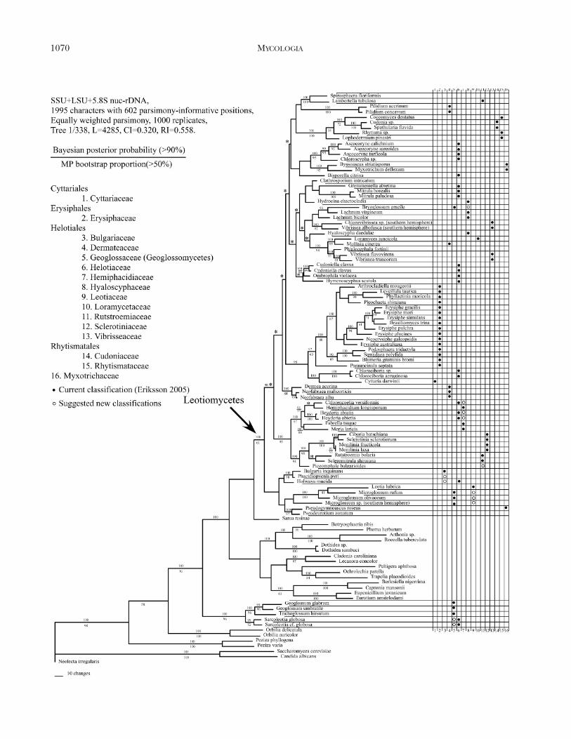
Parsimony and Bayesian analyses.—One of the 338equally parsimonious trees generated is shown andclades with BP $ 50% were marked under thebranches (FIG. 1). Bayesian posterior probabilities(PP) were estimated from the 50% majority ruleconsensus of the 18 500 trees, and clades with PP $
0.90 (90%) were marked above the branches (FIG. 1).The Leotiomycetes with the exclusion of the Geo-glossaceae, including the Cyttariales, Erysiphales,Helotiales, Rhytismatales, Myxotrichaceae and Pseu-deurotiaceae, was supported as a clade (BP 5 61%, PP5 100%). The Erysiphales (BP 5 63%, PP 5 97%)and Rhytismatales (BP 5 100%, PP 5 100%) weresupported as monophyletic groups. A notable excep-tion of support (PP 5 95%) of supraordinal relation-ships was the clade containing the Erysiphales, Cyttariadarwinii (Cyttariales) and two species of Chlorociboria(Helotiales). For the Erysiphales Parauncinula septatawas supported as the most ancestral lineage withother Erysiphales as the sister group, which wascomposed of two clades. The one included a subcladeof Podosphaera tridactyla and Sawadaea polyfida (BP 5
100%, PP 5 100%) with Blumeria graminis as themoderately supported sister group (BP 5 69%, PP 5
90%). The other clade (BP 5 87%, PP 5 100%)included two subclades that are without support.Leveillula taurica and Phyllactinia moricola wereclosely related (BP 5 99%, PP 5 100%). TheHelotiales is not resolved as monophyletic, andstrongly supported (BP . 70%, PP . 95%) terminalclades of two or more genera included the Leotia-Northern Hemisphere Microglossum clade (BP 5
100%, PP 5 100%), Bulgaria-Phacidiopycnis clade(BP 5 98%, PP 5 100%), Ciboria-Sclerotinia-Mon-ilinia-Rutstroemia-Scleromitrula clade (BP 5 95%, PP5 100%), Chlorencoelia-Hemiphacidium-Heyderia-Fabrella-Rhabdocline (teleomorph of Meria) clade
(BP 5 97%, PP 5 100%), Dermea-Neofabraea clade(BP 5 84%, PP 5 100%), Cudoniella-Ombrophilaclade (BP 5 88%, PP 5 100%), Phialocephala-northern hemisphere Vibrissea clade (BP 5 71%,PP 5 100%), Loramyces-Mollisia clade (BP 5 99%,PP 5 100%), Chlorovibrissea-Vibrissea albofuscaclade (BP 5 82%, PP 5 100%) and Ascocoryne-Chloroscypha clade (BP 5 95%, PP 5 100%). TheLoramyces-Mollisia clade was the sister group of thePhialocephala-northern hemisphere Vibrissea clade(BP 5 98%, PP 5 100%), and the Ciboria-Sclerotinia-Monilinia clade was grouped with species of Rutstroe-mia, Scleromitrula and Piceomphale (BP 5 94%, PP 5
100%), which was supported strongly (BP 5 95%, PP5 100%) as the sister group of the Chlorencoelia-Hemiphacidium-Heyderia-Fabrella-Rhabdocline clade.The Rhytismatales formed a well supported clade(BP 5 100%, PP 5 100%) that included theCudoniaceae and members of the Rhytismataceae(Coccomyces, Rhytisma and Lophodermium). TheMyxotrichaceae was not monophyletic in our treeand consisted of two distinct lineages. One lineageincluded Byssoascus striatisporus and Myxotrichumdeflexum (BP 5 92%, PP 5 100%). The second wasrepresented by Pseudogymnoascus roseus, which waspart of a larger clade that included Pseudeurotiumzonatum (Pseudeurotiaceae), Leotia (Leotiaceae)and Microglossum (Geoglossaceae).
Leotiomycetes.—The Leotiomycetes (Eriksson 2005),excluding the Geoglossaceae and including thePseudeurotiaceae, is supported in this study asa monophyletic group. Here we accept the Leotiomy-cetes to include the Cyttariales, Erysiphales, Helo-tiales, Rhytismatales, Thelebolales, Myxotrichaceaeand Pseudeurotiaceae. Two genera of the Geoglossa-ceae, Geoglossum and Trichoglossum, and genusSarcoleotia of the Helotiaceae are removed from theclass.
Cyttariales.—There is a single family, Cyttariaceae,with a single genus, Cyttaria, of about a dozenparasitic species on Nothofagus (southern beech) inthe southern hemisphere. The fungus produces largeand fleshy globose or pyriform stromata on galls onliving branches. The stromata sometimes form sper-matial pycnidia at early stages and later developnumerous separate, cup-shaped chambers containingasci and dark ascospores (Korf 1983, Pfister andKimbrough 2001). Different authors have usedmorphological evidence to place the genus variouslyin the Pezizales and the Helotiales (see discussion inGamundı 1991). Megoni (1986) showed that the ascihave a thin cell wall and an apical amyloid ring,characters shared by some helotialean fungi. Ourresults placing the Cyttariales in the Leotiomycetes
WANG ET AL: LEOTIOMYCETES 1067

agree with the Landvik and Eriksson (1994) SSU-rDNA study, and Bayesian analyses strongly support(PP 5 100%) a close relationship among species ofCyttaria and Chlorociboria (Helotiales) in a cladeincluding species of the Erysiphales. Species ofChlorociboria produce small disk-shaped apotheciaon dead wood, and their hyphae grow inside thewood and stain the substrate blue-green. Fifteenspecies, including 13 new species, were reported fromNew Zealand based on morphological characters andITS sequence data, and a possible Asian/Australasiancenter of diversity for Chlorociboria was suggested(Johnston and Park 2005).
Erysiphales.—The Erysiphales with one family of 15genera (Braun et al 2002, Takamatsu et al 2005a, b)are obligate plant pathogens that cause powderymildew diseases on about 10 000 plant species(Amano 1986). There are no direct morphologicalfeatures supporting inclusion of these fungi in theLeotiomycetes. The 16 sequences from the familygroup into a clade with 97% of posterior probabilityand 63% bootstrap values. Without exception allErysiphales have closed, nonostiolate fruit bodies(chasmothecium, Braun et al 2002) with persistentasci and basal hymenium. These characters mightfurther support monophyly of the Erysiphales, sug-gesting that the morphological characters as well asthe holobiotrophy were acquired in the commonancestor of the Erysiphales.
Evolutionary relationships of the Erysiphales havebeen based on morphological characteristics and hostplants for about 100 y. They are concerned withwhich characters are primitive and which derived,such as (i) numbers of asci in an ascoma, (ii) numberof ascospores in an ascus, (iii) morphology ofappendage, (iv) conidiogenesis (i.e. conidia pro-duced in chains [Euoidium type] or solitary [Pseu-doidium type]) and (v) ecto- or endoparasitism,ectotrophic or endotrophic. Of the 15 genera of theErysiphales, 12 are ectoparasitic and only the remain-ing three genera, Leveillula, Phyllactinia and Pleo-chaeta, are endoparasitic. Arnaud (1921) and Katu-moto (1973) regarded the endoparasitic genusLeveillula as primitive and all the ectoparasitic generaderivatives from it. On the other hand Raymond(1927), Blumer (1933) and Braun (1987) argued thatthe ectoparasitic habit of Leveillula was a xerophyticadaptation and not a primitive feature. The threeendoparasitic genera form a monophyletic clade inthis study, suggesting that the presence of internalmycelia is a synapomorphic character for thesegenera, which were derived from ectoparasitic generaby a single event. Morphology of ascomatal appen-dages has been important to define genera of the
Erysiphales. Mori et al (2000) performed phylogenet-ic analyses of the Erysiphales based on the combineddataset of sequences from the same three rDNAregions as used here. They reported that themycelioid appendage, which long has been regardedas a primitive character (e.g. Blumer 1933, Braun1981, 1987), is a derived character of recent origin.They suggested that the development of mycelioidappendages convergently occurred many times in therespective lineages along with host expansion of theErysiphales from trees to herbs. The basal position ofParauncinula septata on the present tree supports thereport of Mori et al (2000) that the uncinate-circinateappendage is the most primitive in the Erysiphales.
A phylogeny covering all known tribes of theErysiphales based on three rDNA regions by Taka-matsu et al (2005a, b) suggested that Caespitothecaforestalis and the species of Parauncinula septata arebasal in the order. Caespitotheca forestalis is restrictedto South America, while P. septata is found only ineastern Asia.
Helotiales.—Although the backbone of our tree is notresolved, the current concept of the Helotiales(Eriksson 2005) almost certainly includes nonmono-phyletic taxa. Although the Helotiales has beenreceiving more attention recently (e.g. Gernandt etal 2001, Lutzoni et al 2004, Wang et al 2005). Evenwith exclusion of groups such as the Cyttariales,Erysiphales, Myxotrichaceae, Pseudeurotiaceae, Geo-glossaceae and genetically widely divergent taxa suchas Chlorociboria and Neobulgaria species, the mono-phyly of the Helotiales is not strongly supported byrDNA data and relationships among major helotia-lean clades are not well resolved. No taxon representsthe Ascocorticiaceae or Phacidiaceae in our tree.
The family Bulgariaceae contains only two species,Bulgaria inquinans and B. nana, which producebrown-black to black, turbinate, highly gelatinousapothecia with brown-walled ascospores on bark ofliving hardwoods and might be weak plant pathogens(Doring and Triebel 1998). From our results B.inquinans shares a clade with a weak canker-causingpathogen, Phacidiopycnis pyri (teleomorph: Potebnia-myces pyri), that produces small, black, gelatinous disksfrom stromata beneath bark. Potebniamyces has beendoubtfully placed in the Rhytismatales by some authors(Eriksson 2005), with DiCosmo et al (1984) suggestinga possible relationship with either Phacidiaceae,Rhytismataceae or Dermateaceae. A LSU-rDNA se-quence is available for one Phacidium lacerum from theAFTOL database, and analyses including the sequencesuggests a sister relationship between Phacidiopycnispyri and Phacidium lacerum. This calls into doubt themorphological differences between Potebniamyces and
1068 MYCOLOGIA

Phacidium discussed by DiCosmo et al (1984). Anotherblack and slightly gelatinous fungus, Holwaya mucida,forms a sister group of the Bulgaria-Phacidiopycnisclade, although without support.
The Dermateaceae is a large, poorly studied, andheterogeneous family (Nauta and Spooner 1999,Pfister and Kimbrough 2001). Only four of the 77genera placed in the family (Eriksson 2005) aresampled in this study. Species of Dermea andNeofabraea form a strongly supported clade, whichwe consider to represent the Dermateaceae sensustricto. Previous studies using only the ITS regionshow that species of Pezicula and Ocellaria in theDermateaceae also belong to this clade (Abeln et al2000, de Jong et al 2001, Goodwin 2002, Verkley1999). Species in this family produce erumpent orsuperficial, fleshy and small apothecia on plants, withan excipulum consisting of rounded or angular cellswith dark walls. The hymenium frequently covered bya gelatinized ‘‘epithecium’’ and the thick walledascospores found in several genera raise a hypothesisabout their endophytic habits and the possible role ofinsect vectors (Wang et al unpubl). It is likely that thefamily will be restricted to endophytic speciesmorphologically similar to Dermea and Pezicula. Theother two genera sampled here traditionally placed inthe Dermateaceae fall outside the Dermateaceaeclade, Mollisia and Pilidium (teleomorph Discohaine-sia). Mollisia is a large, heterogeneous genus in-adequately represented by the single species in ourstudy. Crous et al (2003) discussed two phylogeneti-cally distinct groups of Mollisia characterized in partby different anamorphs, while Partel and Raitviir(2005) described two different types of ascus ultra-structure among Mollisia species.
Concepts of the Geoglossaceae have changed manytimes recently (Eriksson 2005, Kirk et al 2001, Pfisterand Kimbrough 2001, Platt 2000, Spooner 1987,Verkley 1994, Wang et al 2002, 2005). The separationof the Geoglossaceae from other helotialean fungi hasbeen suggested, but an alternative systematic positionof the family has not been proposed due to weakphylogenetic resolution (e.g. Platt 2000, Lutzoni et al2004). Club-shaped apothecia, dark colored paraph-yses and ascospores were considered as uniquecharacters defining this group, and some authorshave suggested that the hyaline-spored taxa beremoved from the family (Platt 2000, Pfister andKimbrough 2001). Species of Geoglossum and Tricho-glossum form a basal branch within the inoperculateascomycetes (Leotiomyceta) with the hyaline-sporedSarcoleotia species (Helotiaceae). Paraphyses (orhomologous structures) cover the stipe surface inGeoglossum and Trichoglossum and obscure theboundary of the fertile hymenium (Spooner 1987).
The pileate apothecia in Sarcoleotia globosa havea hymenium that is continuous with the stipe at anearly stage and then recedes from the stipe (Schu-macher and Sivertsen 1987). The genera, such asCudonia, Spathularia, and helotialean Microglossumand Bryoglossum, differ in that the hymenium is alwaysclearly demarcated from the stipe, even when nopileus is formed.
The family Helotiaceae is polyphyletic in our tree,and species of the family are found in eight clades.The first of these, the Cudoniella-Hymenoscyphusclade, although not strongly supported in our tree,could form the core of a monophyletic Helotiaceaesensu stricto, as suggested by Pfister and Kimbrough(2001). The genus Hymenoscyphus might includeparaphyletic lineages recognized as ericoid mycorrhi-zal fungi and saprobes (Monreal et al 1999, Vralstadet al 2002, Zhang and Zhuang 2004). The secondclade of the Helotiaceae, Ascocoryne-Chloroscypha,receives strong support, and species of Neobulgariaalso might group in this clade (Wang et al unpubl).Fungi in the Ascocoryne-Chloroscypha clade arecharacterized by a gelatinous apothecium and endo-phytic habits. The third clade is unsupported statis-tically and includes the aero-aquatic genera Mitrula,Hydrocina, Clathrosporium (anamorph) and the path-ogenic Gremmeniella abietina. An accurate phylogenyhere would provide insights into the evolution of theaquatic helotialean fungi. Two helotiaceous genera,Chlorencoelia and Heyderia, are nested within theHemiphacidiaceae. We suggest removal of thesegenera from the Helotiaceae to the Hemiphacidia-ceae (see notes below). The other genera in ouranalysis that traditionally have been included inHelotiaceae are Sarcoleotia (included here in theGeoglossaceae), Holwaya (forming an unsupportedclade that includes Bulgaria), Chlorociboria (seediscussion under Cyttariales) and Bisporella.
The families Hemiphacidiaceae, Rutstroemiaceaeand Sclerotiniaceae, and Piceomphale bulgarioides,Heyderia abietis and Chlorencoelia versiformis forma clade with high support; a new order could be anappropriate classification for this clade. The Hemi-phacidiaceae traditionally contains foliar pathogensproducing small, simple apothecia beneath the leafsurface, the erumpent apothecia pushing back thecovering host tissue (Korf 1962). In contrast species ofHeyderia and Chlorencoelia produce large apotheciawith a well developed excipulum. A PCR-based studyshows that Heyderia species colonize conifer needlesas endophytes and then produce apothecia on fallenleaves (Jean Berube pers comm). Chlorencoelia andHeyderia are examples of significant morphologicalchanges associated with the shift in habits fromobligate pathogen to endophyte/saprobe, also illus-
WANG ET AL: LEOTIOMYCETES 1069
1070 MYCOLOGIA

trated by the Cudoniaceae in the Rhytismatales. TheHemiphacidiaceae might need to be expanded toinclude such genera previously placed in the Helo-tiaceae. Except for the angiosperm-associated Chlor-encoelia species, fungi of the expanded Hemiphaci-diaceae are restricted to coniferous hosts. TheSclerotiniaceae and Rutstroemiaceae are among thebest studied in the Helotiales (e.g. Dumont and Korf1971, Holst-Jensen et al 1997, Kohn and Schumacher1984, Novak and Kohn 1991, Schumacher and Holst-Jensen 1997, Spooner 1987, Zhuang 1998). Piceom-phale bulgarioides, a spruce endophyte, is basal in thistree to the Sclerotiniaceae (sclerotial stromata pro-ducing) plus Rutstroemiaceae (substratal stromataproducing) clade. A recent study of a chestnutpathogen Sclerotinia pseudotuberosa (5 Ciboria batschi-ana) showed that the fungus could occur asymptom-atically in different tissues of the host (Vettraino et al2005). The ancestor of this ‘‘Sclerotiniales’’ clade wasprobably an endophyte living on conifers, withlineages in the Sclerotiniaceae and Rutstroemiaceaeshifting from coniferous hosts to angiospermoushosts and becoming opportunistic pathogens. Lam-bertella tuberosa has been placed in the Rutstroemia-ceae because of its pigmented ascospores. It has anaero-aquatic anamorph (Helicodendron tubulosum)and in this study it forms a clade with another aero-aquatic fungus, Spirosphaera floriformis, distinct fromthe Rutstroemiaceae. Only LSU-rDNA data wereavailable for species of Lambertella and Spriosphaerafor this study.
The Hyaloscyphaceae is poorly represented in thisstudy, including only three of the more than 60genera listed by Eriksson (2005). These fall in a poorlyresolved part of our tree. Phylogenies from studiesbased on ITS sequences (Cantrell and Hanlin 1997,Abeln et al 2000, Vralstad et al 2002) suggest theHyaloscyphaceae subfamilies Lachnioideae and Hya-loscyphoideae are phylogenetically distinct, and Rait-viir (2004) subsequently has treated these two taxa atthe family level. The analysis in this paper supportsremoving Bryoglossum from the Geoglossaceae byWang et al (2005), however the placement ofBryoglossum in the Hyaloscyphaceae require investi-gation with more inclusive taxa and gene sampling.
Habits of fungi in this family are basically unknown.Many species have been recorded as host-restrictedsaprobes, often an indication of an endophytic stagein their life cycle.
The Leotiaceae is represented by Leotia lubrica andthree species of Microglossum, and our results supportstudies (e.g. Landvik 1996, Liu and Hall 2004, Platt2000) that suggest a close relationship between Leotiaand Microglossum despite morphological differences.Both Leotia and Microglossum have worldwide distri-bution, but the southern and northern hemispherespecies of Microglossum are genetically distinct in ourtree. The Leotia-Microglossum clade is weakly sup-ported as forming a clade with Pseudeurotium,a finding however that is supported by analyses ofprotein coding data (Spatafora et al this volume).
The Loramycetaceae, Mollisia cinerea, Phialocephalafortinii (anamorphic dark septate endophyte), andtwo northern hemisphere species of the Vibrisseaceaeform a strongly supported clade. Ecological linkagesamong these fungi support molecular evidence ofevolutionary relationships between aquatic fungi androot endophytes that has been observed also withinaquatic hyphomycetes (e.g. Sati and Belwal 2005).The monotypic Loramycetaceae contains only twospecies that are aquatic and so adapted to the aquaticenvironment that they morphologically are hard toclassify in other families in the Helotiales. Dark cellspresent at the base of Loramyces apothecium and thehyphal structure of the apothecia are analogous tothose of the Dermateaceae (Digby and Goos 1987).Dark cells also can be found at the base of theapothecium of Vibrissea. Two southern hemispherespecies of the Vibrisseaceae, Chlorovibrissea sp. andVibrissea albofusca, are not included in this clade. TheVibrisseaceae is characterized by morphological fea-tures associated with an aquatic habit, and the originof the southern hemisphere vibrisseaceous fungicould be independent from the northern hemisphererepresentatives. One Mollisia species is reported asaquatic (Fisher and Webster 1983).
Rhytismatales.—A complete rDNA dataset is availablefor only five of the 73 genera listed under theRhytismatales by Eriksson (2005). Less completedatasets, or sequences from other genes, have been
r
FIG. 1. Phylogenetic relationships within the Leotiomycetes inferred from three rDNA regions using parsimony analysis.One of the 338 most parsimonious trees (Length 5 4285, CI 5 0.320, RI5 0.558). Bootstrap values greater than 50% areindicated under nodes, branches collapsed in the strict consensus tree are marked with an asterisk. Bayesian posteriorprobabilities also were estimated with Bayesian approaches under the GTR+C+I model. Group frequencies greater than 90%
are indicated above branches. New classifications suggested by molecular phylogeny are marked with open circles against soliddots as they are in current classifications.
WANG ET AL: LEOTIOMYCETES 1071

published for several other genera either currentlyplaced in the Rhytismatales or that have been referredto the order in the past, including Colpoma,Cyclaneusma, Elytroderma, Lirula, Meloderma, Naema-cyclus, Nematococcomyces, Phacidium, Rhabdocline,Rhytisma, Terriera, and Tryblidiopsis (Gernandt et al1997, Gernandt et al 2001, Hou et al 2004, Ortiz-Garcia et al 2003).
Many of the taxa placed in the Rhytismatales arepoorly understood, with both ordinal and familialrelationships of several genera remaining tentative.For most species the teleomorph comprises anapothecium with a reduced excipulum that developswithin a dark stroma, the stroma typically immersedwithin host tissue and sometimes covered by a clypeus(Johnston 2001). The hymenium becomes exposedafter the covering host and fungal tissue splits by oneor more elongate slits. The asci are typically thin-walled, undifferentiated at the apex, nonamyloid, theascospores typically with gelatinous sheaths or appen-dages. The anamorphs are mostly Leptostroma-like,the spermatial conidia developing on often percur-rently proliferating conidiogenous cells. Exceptionsto this typical morphology include a few genera withascospores lacking a gelatinous sheath or appendage(e.g. Discocainia [Reid and Funk 1966], Myriophaci-dium [Cannon and Minter 1986], Terriera [Johnston2001] and Therrya [Reid and Cain 1961, Yuan andMohammed 1997] within the Rhytismataceae, Asco-dichaena [Hawksworth and Sherwood 1982] withinthe Ascodichaenaceae and Ocotomyces [Evans andMinter 1985] incertae sedis within the Rhytismatales).The phylogenetic significance of an ascospore sheathis not yet understood, although the position ofTerriera within the Rhytismatales is supported by ITSsequences (Ortiz-Garcia et al 2003). Nothorhytisma isthe only genus in the order with an amyloid pore atthe apex (Minter et al 1998). The presence orabsence of an amyloid ascus pore has been found tobe of little phylogenetic significance at higher level indiscomycetes (e.g. Rhabdocline, see Gernandt et al1997) although the presence of such a pore inPhacidium was one of the factors leading DiCosmoet al (1984) to suggest this genus was more closelyrelated to the helotialean fungi than it was to theRhytismatales. The exclusion of Phacidium from theRhytismatales has been supported by molecular data(Gernandt et al 1997). Cyclaneusma and Naemacyclus,included in the Rhytismatales on the basis ofmorphology (Kirk et al 2001) and the molecularanalyses of Gernandt et al (1997, 2001), showed themto be phylogenetically distinct from the Rhytismatales.
Most genera of Rhytismatales form ascomataimmersed within the host tissue, but Cudonia andSpathularia, recently added to the Rhytismatales on
the basis of DNA data (Kirk et al 2001, Wang et al2002), are different. In both genera ascomata developon soil and form erect ascomata. The ascomata ofCudonia and Spathularia are macroscopically Helo-tiales-like, but careful anatomical studies have shownthe hymenium covered by a stromatic layer at earlystages of development (Landvik 1996, Wang et al2002). Other Rhytismatales-like features of Cudoniaand Spathularia are ascospores with gelatinoussheaths and simple asci lacking an amyloid pore(Wang et al 2002).
Available molecular data is insufficient to under-stand the morphological characteristics useful fordefining the order Rhytismatales. Sequence data fromgenera such as Lophodermium, Meloderma and Elytro-derma (Ortiz-Garcia et al 2003) suggest that genericlimits within the order are artificial, but samplingintensity is too low to suggest yet what might be moreappropriate generic or family limits.
Thelebolales.—A recent study of Thelebolus in Antarc-tica by de Hoog et al (2005) reveal the remarkableecology and biology of these fungi. Species ofThelebolus produce small, globose to disk-shapedascomata with sometimes polysporus asci, and thedevelopment of the ascomata varies from a cleistohy-menial type to a eugymnohymenial type (van Brum-melen 1967). The Thelebolaceae has been placed inthe Pezizales (Eckblad 1968, Kimbrough and Korf1967), and this placement has been studied from theapothecium macromorphology to ascus ultrastructure(e.g. Brummelen 1998). The placement of theThelebolales in the Leotiomycetes is based on SSU-rDNA sequence data, and many of the order still havenot been included in molecular phylogenetic studies(e.g. Gernandt et al 2001, Landvik et al 1998, Momolet al 1996). The order might not be monophyleticand its position in the Leotiomycetes should beregarded as a temporary treatment until sufficientproof becomes available.
Myxotrichaceae and Pseudeurotiaceae.—Myxotricha-ceae and Pseudeurotiaceae are two families ofcleistothecial ascomycetes, both of which containgenera linked to the Leotiomycetes in molecularstudies (Sogonov et al 2005, Sugiyama et al 1999, Suhand Blackwell 1999). Three genera of the Myxotri-chaceae are sampled in this study, and they resolve astwo widely separated lineages; one includes Byssoascusstriatisporus and Myxotrichum deflexum and the otherincludes Pseudogymnoascus roseus, along with Pseu-deurotium zonatum from the Pseudeurotiaceae. Theseparation of Pseudogymnascus from Myxotrichum andByssoascus supports earlier studies (Sugiyama et al1999, Sugiyama and Mikawa 2001). The link betweenPseudogymnascus and Pseudeurotium supports Sogo-
1072 MYCOLOGIA
nov et al (2005). The molecular-based phylogeniesare supported by electron microscope studies, withthe highly reduced cleistothecial ascomata of Myxo-trichum showing a striking similarity in morphogen-esis and gross morphology to typical helotialean fungisuch as Hymenoscyphus species (Tsuneda and Currah2004). Sequence data of Pseudogymnoascus andPseudeurotium are limited, and our inclusion ofPseudeurotiaceae in the Leotiomycetes is tentative.However analysis of protein coding data of Pseudeur-otium also supported its affinity to Leotia (Spatafora etal this volume), although Pseudogymnoascus was notincluded in those analyses.
Conclusion.—The Leotiomycetes represents a mor-phologically and ecologically diverse class of Pezizo-mycotina whose evolutionary history is only beginningto be unraveled through phylogenetic analyses ofmolecular data. Significant advances have been madeand include the classification of the Cudoniaceae inthe Rhytismatales, the inclusion of the Erysiphales,Cyttariales and Pseudeurotiaceae in the Leotiomy-cetes, and the exclusion of Geoglossaceae from theLeotiomycetes. Current taxon and character samplingis insufficient to address many of the internal nodesof the class, and future phylogenetic studies muststrive to significantly increase character sampling,especially that of protein-coding genes, from thediversity of species characterized as inoperculatediscomycetes.
ACKNOWLEDGMENTS
We thank RP Korf and two anonymous reviewers for readingthe manuscript, improvement of the English text andvaluable suggestions, KR Peterson at Harvard Universityfor rDNA sequences of Cyttaria. ZW thanks Drs DavidHibbett and Manfred Binder at Clark University for theirgreat help during his graduate research at Clark. This studywas supported by a grant from the National ScienceFoundation of the USA DEB-0228657 to DS Hibbett. Wealso acknowledge support form NSF0090301, ResearchCoordination Network: A Phylogeny for Kingdom Fungito M Blackwell, JW Spatafora and JW Taylor.
LITERATURE CITED
Abeln ECA, de Pagter MA, Verkley GJM. 2000. Phylogeny ofPezicula, Dermea and Neofabraea inferred from partialsequences of the nuclear ribosomal RNA gene cluster.Mycologia 92:685–693.
Amano K. 1986. Host range and geographical distributionof the powdery mildew fungi. Tokyo: Japan ScientificSocieties Press.
Arnaud G. 1921. Etude sur les champignons parasites. AnnEpiphyt 7:1–116.
Baral HO. 1987. Der Apikalapparat der Helotiales. Eine
lichtmikroskopische Studies uber Arten mit Amyloidr-ing. Zeitschrift Mykol 53:119–135.
Blumer S. 1933. Die Erysiphaceen Mitteleuropas unterbesonderer Berucksichtigung der Schweiz. Beitr Krypt-Fl Schweiz 7:1–483.
Braun U. 1981. Taxonomic studies in the genus Erysiphe I.Generic delimitation and position in the system of theErysiphaceae. Nov Hedwig 34:679–719.
———. 1987. A monograph of the Erysiphales (powderymildews). Beih Nov Hedwig 89:1–700.
———. 1998. Reconsideration of relationships within theThelebolaceae based on ascus ultrastructure. Persoonia16:425–469.
———, Cook RTA, Inman AJ, Shin HD. 2002. Thetaxonomy of the powdery mildew fungi. In: BelangerRR, Bushnell WR, Dik AJ, Carver TLW, eds. Thepowdery mildews: a comprehensive treatise. St Paul,Minnesota: APS Press.
Cannon PF, Minter DW. 1986. The Rhytismataceae of theIndian subcontinent. Mycol Papers 155:1–123.
Cantrell SA, Hanlin RT. 1997. Phylogenetic relationships inthe family Hyaloscyphaceae inferred from sequences ofITS regions, 5.8S ribosomla DNA and morphologicalcharacters. Mycologia 89:745–755.
Crous PW, Groenewald JZ, Gams W. 2003. Eyespot of cerealsrevisited: ITS phylogeny reveals new species relation-ships. Euro J Plant Pathol 109:841–850.
de Hoog GS, Gottlich E, Platas G, Genilloud O, Leotta G,van Brummelen J. 2005. Evolution, taxonomy andecology of the genus Thelebolus in Antarctica. StudMycol 51:33–76.
———, ———, ———, ———, ———, ———. 2005.Evolution, taxonomy and ecology of the genusThelebolus in Antarctica. Stud Mycol 51:33–76.
de Jong SN, Levesque CA, Verkley GJM, Abeln ECA, RaheJE, Braun PG. 2001. Phylogenetic relationships amongNeofabraea species causing tree cankers and bull’s eyerot of apple based on DNA sequencing of ITS nuclearrDNA, mitochondrial rDNA, and b-tubulin gene. MycolRes 93:508–512.
Dennis RWG. 1968. British Ascomycetes. Cramer, Lehre.DiCosmo F, Nag Raj TR, Kendrick WB. 1984. A revision of
the Phacidiaceae and related anamorphs. Mycotaxon21:1–234.
Digby S, Goos RD. 1987. Morphology, development andtaxonomy of Loramyces. Mycologia 79:821–831.
Doring H, Triebel D. 1998. Phylogenetic relationships ofBulgaria inferred by 18S sequence analysis. Cryptoga-mie Bryol Lichenol 19:123–136.
Dumont KP, Korf RP. 1971. Sclerotiniaceae I. Genericnomenclature. Mycologia 63:157–168.
Eckblad FE. 1968. The genera of the operculate discomy-cetes. A re-evaluation of their taxonomy, phylogeny andnomenclature. Norw J Bot 15:1–191.
Eriksson OE. 2005. Outline of Ascomycota 2005. Myconet11:1–113.
Evans HC, Minter DW. 1985. Two remarkable new fungi onpine from Central America. Trans Br Mycol Soc 84:57–78.
Fischer PJ, Webster J. 1983. The teleomorphs of Helicoden-
WANG ET AL: LEOTIOMYCETES 1073

dron giganteum and H. paradoxum. Trans Br Mycol Soc81:656–659.
Gamundı IJ. 1980. Subantarctic Geoglossaceae II. Sydowia32:86–98.
———. 1991. Review of recent advances in the knowledgeof the Cyttariales. Syst Ascomycet 10:69–77.
Gernandt DS, Camacho FJ, Stone JK. 1997. Meria laricis, ananamorph of Rhabdocline. Mycologia 89:735–744.
———, Platt JL, Spatafora JW, Holst-Jensen A, Hamelin RC,Kohn LM. 2001. Phylogenetics of Helotiales andRhytismatales based on partial small subunit nuclearribosomal DNA sequences. Mycologia 93:915–933.
Goodwin SB. 2002. The barley scald pathogen Rhynchospor-ium secalis is closely related to the discomycetes Tapesiaand Pyrenopeziza. Mycol Res 106:645–654.
Hawksworth DL, Sherwood MA. 1982. Two new families inthe Ascomycotina. Mycotaxon 16:262–264.
Holst-Jensen A, Kohn LM, Schumacher T. 1997. NuclearrDNA phylogeny of the Sclerotiniaceae. Mycologia 89:885–899.
Hou C-L, Piepenbring M, Oberwinkler F. 2004. Nematococ-comyces rhododendri, a new species in a new genus of theRhytismatales from China. Mycologia 96:1380–1385.
Huelsenbeck JP, Ronquist F. 2001. MrBayes: Bayesianinference of phylogenetic trees. Bioinformatics 17:754–755.
Johnston PR. 2001. Monograph of the monocotyledon-inhabiting species of Lophodermium. Mycol Papers 176:1–239.
———, Park D. 2005. Chlorociboria (Fungi, Helotiales) inNew Zealand. NZ J Bot 43:679–719.
Katumoto K. 1973. Notes on the genera Lanomyces Gaum.and Cystotheca Berk. et Curt Rep. Tottori Mycol Inst 10:437–446.
Kimbrough JW. 1970. Current trends in the classification ofDiscomycetes. Bot Rev 36:91–161.
———, Korf RP. 1967. A synopsis of the genera and speciesof the tribe Thelebolaceae (5 Pseudoascoboleae).Am J Bot 54:9–23.
Kirk PM, Cannon PF, David JC, Stalpers JA. 2001. Ainsworthand Bisby’s dictionary of the fungi. 9th edition.Wallingford, UK: CAB International.
Kohn LM, Schumacher T. 1984. Conserve Rutstroemia P.Karsten with R. firma as type. Taxon 33:508.
Korf RP. 1962. A synopsis of the Hemiphacidiaceae, a familyof the Helotiales (Discomycetes) causing needle blightsof conifers. Mycologia 54:12–33.
———. 1973. Discomycetes and Tuberales. In: AinsworthGC, Sparrow FK, Sussman AS, eds. The Fungi: anadvanced treatise. Vol IVA. New York: Academic Press.p 249–319.
———. 1983. Cyttaria (Cyttariales): coevolution withNothofagus, and evolutionary relationship to theBoedijinopezizeae (Pezizales, Sarcoscyphaceae).Aust J Bot Suppl Ser 10:77–87.
Landvik S. 1996. Phylogenetic rDNA studies of Discomy-cetes (Ascomycota) [Doctoral thesis]. Umea, Sweden:Department of Ecological Botany, Umea University.119 p.
———, Eriksson OE. 1994. Relationship of Tuber, Elapho-
myces, and Cyttaria (Ascomycotina), inferred from 18SrDNA studies. In: Hawksworth DL, ed. Ascomycetessystematics: problems and perspectives in the nineties.New York: Plenum Press. p 225–231.
———, ———, Berbee ML. 2001. Neolecta—a fungal di-nosaur? Evidence from beta-tubulin amino acid se-quences. Mycologia 93:1151–1163.
———, ———, Gargas A, Gustafsson P. 1993. Relationshipsof the genus Neolecta (Neolectales ordo. nov., Ascomy-cotina) inferred from 18S rDNA sequences. SysAscomyc 11:107–115.
———, Kristiansen R, Schumacher T. 1998. Phylogeneticand structural studies in the Thelebolaceae (Ascomy-cota). Mycoscience 39:49–56.
Liu YJ, Hall BD. 2004. Body plan evolution of ascomycetes,as inferred from an RNA polymerase II phylogeny. ProcNat Acad Sci USA 101:4507–4512.
Lumbsch HT, Schmitt I, Lindemuth R, Miller A, MangoldA, Fernandez F, Huhndorf S. 2005. Performance offour ribosomal DNA regions to infer higher-levelphylogenetic relationships of inoperculate euascomy-cetes (Leotiomyceta). Mol Phylogenet Evol 34:512–524.
Lutzoni F, Kauff F, Cox CJ, McLaughlin D, Celio G,Dentinger B, Padamsee M, Hibbett DS, James TY,Baloch E, Grube M, Reeb V, Hofstetter V, Schoch C,Arnold AE, Miadlikowska J, Spatafora J, Johnson D,Hambleton S, Crockett M, Schoemaker R, Sun GH,Lucking R, Lumbsch HT, O’Donnell K, Binder M,Diederich P, Ertz D, Gueidan C, Hall B, Hansen K,Harris RC, Hosaka K, Lim YW, Liu Y, Matheny B,Nishida H, Pfister D, Rogers J, Rossman A, Schmitt I,Sipman H, Stone J, Sugiyama J, Yahr R, Vilgalys R. 2004.Where are we in assembling the fungal tree of life,classifying the fungi, and understanding the evolutionof their subcellular traits? Am J Bot 91:1446–1480.
Megoni T. 1986. El aparato apical del asco de Cyttariaharioti (Ascomycetes-Cyttariales) con microscopia foto-nica y electonica. Bol Soc Arg Bot 24:393–401.
Minter DW, Cannon PF, Romero AI, Peredo HL. 1998. Anew member of the Rhytismatales from southern SouthAmerica. Syst Ascomycet 16:27–37.
Momol EA, Kimbrough JW, Eriksson OE. 1996. Phyloge-netic relationships indicated by 18S rDNA sequenceanalysis. Syst Ascomycet 14:91–100.
Monreal M, Berch SM, Berbee M. 1999. Molecular diversityof ericoid mycorrhizal fungi. Can J Bot 77:1580–1594.
Mori Y, Sato Y, Takamatsu S. 2000. Evolutionary analysis ofthe powdery mildew fungi using nucleotide sequencesof the nuclear ribosomal DNA. Mycologia 92:74–93.
Nannfeldt JA. 1932. Studien uber die Morphologie undSystematik der nichtlichenisierten inoperculaten Dis-comyceten. Nova Acta R Soc scient upsal 8:1–368.
Nauta MM, Spooner BM. 1999. British Dermateaceae 1.Introduction. Mycologist 13:3–6.
Novak LA, Kohn LM. 1991. Electrophoretic and immuno-logical comparisons of developmentally regulatedproteins in members of the Sclerotiniaceae and othersclerotial fungi. Appl Environm Microbiol 57:525–534.
Ortiz-Garcia S, Gernandt DS, Stone JK, Johnston PR,Chapela IH, Salas-Lizana R, Alvarez-Buylla ER. 2003.
1074 MYCOLOGIA

Phylogenetics of Lophodermium from pine. Mycologia95:846–859.
Partel K, Raitviir A. 2005. The ultrastructure of the ascusapical apparatus of some Dermateaceae (Helotiales).Mycol Progress 4:149–159.
Pfister DH, Kimbrough JW. 2001. Discomycetes. In:McLaughlin DJ, McLaughlin EG, Lemke PA, eds. TheMycota VII Part A. Systematics and Evolution. Berlin &Heidelberg: Springer-Verlag. p 257–281.
Platt JL. 2000. Lichens, earth tongues, and endophytes:evolutionary patterns inferred from phylogenetic anal-yses of multiple loci [Doctoral dissertation]. CorvalisOregon: Oregon State University. 178 p.
Raitviir A. 2004. Revised synopsis of the Hyaloscyphaceae.Scripta Mycol 20:1–132.
Raymond J. 1927. Le blanc du chene. Ann Epiphyt 13:94–129.
Reid J, Cain RF. 1961. The genus Therrya. Can J Bot 39:1117–1129.
———, Funk A. 1966. The genus Atropellis and a new genusof the Helotiales associated with branch cankers ofwestern hemlock. Mycologia 58:417–439.
Rossman AY, Aime MC, Farr DF, Castlebury LA, PetersonKR, Leahy R. 2004. The ceolomycetous generaChaetomella and Pilidium represent a newly discoveredlineage of inoperculate discomycetes. Mycol Prog 3:275–290.
Sati SC, Belwall M. 2005. Aquatic hyphomycetes as endo-phytes of riparian plant roots. Mycologia 97:45–49.
Schumacher T, Holst-Jensen A. 1997. A synopsis of thegenus Scleromitrula (5 Verpatinia) (Ascomycotina:Helotiales: Sclerotiniaceae). Mycoscience 38:55–69.
———, Sivertsen S. 1987. Sarcoleotia globosa (Som-merf. : Fr.) Korf, taxonomy, ecology and distribution.In: Larsen GA, Amirati JF, Redhead SA, eds. ArcticAlpine Mycol 2. New York & London: Plenum Press.p 163–176.
Sogonov MV, Schroers HJ, Gams W, Dijksterhuis J, Summer-bell RC. 2005. The hyphomycete Teberdinia hygrophilagen. nov. and related anamorphs of Pseudeurotiumspecies. Mycologia 97:695–709.
Spooner BM. 1987. Helotiales of Australasia: Geoglossaceae,Orbiliaceae, Sclerotiniaceae, Hyaloscyphaceae. BiblMycol 116:1–711.
Sugiyama M, Mikawa T. 2001. Phylogenetic analysis of thenon-pathogenic genus Spiromastix (Onygenaceae) andrelated onygenalean taxa based on large subunitribosomal DNA sequences. Mycoscience 42:413–421.
Suh SO, Blackwell M. 1999. Molecular phylogeny of thecleistothecial fungi placed in Cephalothecaceae andPseudeurotiaceae. Mycologia 91:836–848.
Swofford DL. 1999. PAUP*: phylogenetic analysis usingparsimony (*and other methods).Version 4. Sunder-land, Massachusetts: Sinauer Associates.
Tajamatsu S, Braun U, Limkaisang S. 2005a. Phylogeneticrelationships and generic affinity of Uncinula septateinferred from nuclear rDNA sequences. Mycoscience46:9–16.
———, Niinomi S, de Alvarez MGC, Alvarez RE, HavrylenkoM, Braun U. 2005. Caespitotheca gen. nov., an an-cestral genus in the Erysiphales. Mycol Res 109:903–911.
Tsuneda A, Currah RS. 2004. Ascomatal morphogenesis inMyxotrichum arcticum supports the derivation of theMyxotrichaceae from a discomycetous ancestor. Myco-logia 96:627–635.
van Brummelen J. 1967. A world-monograph of the generaAscobolus and Saccobolus (Ascomycetes, Pezizales).Persoonia (Suppl.) 1:1–260.
Verkley GJM. 1994. Ultrastructure of the apical apparatus inLeotia lubrica and some Geoglossaceae (Leotiales,Ascomycotina). Persoonia 15:405–430.
———. 1999. A monograph of Pezicula and its anamorphs.Stud Mycol 44:1–176.
Vettraino AM, Paolacci A, Vannini A. 2005. Endophyt-ism of Sclerotinia pseudotuberosa: PCR assay forspecific detection in chestnut tissues. Mycol Res 109:96–102.
Vralstad T, Schumacher T, Taylor FS. 2002. Mycorrhizalsynthesis between fungal strains of the Hymenoscyphusericae aggregate and potential ectomycorrhizal andericoid hosts. New Phytol 153:143–152.
Wang Z, Binder M, Hibbett DS. 2002. A new species ofCudonia based on morphological and molecular data.Mycologia 94:641–450.
———, ———, ———. 2005. Life history and systematics ofthe aquatic discomycetes Mitrula (Helotiales, Ascomy-cota) based on cultural, morphological, and molecularstudies. Am J Bot 92:1565–1574.
———, ———, Schoch CL, Johnston PR, Spatafora JW,Hibbett DS. 2006. Evolution of helotialean fungi(Leotiomycetes, Pezizomycotina): a nuclear rDNAphylogeny. Mol Phylogenet Evol 41:295–312.
Yuan ZQ, Mohammed C. 1997. Therrya eucalypti sp. nov. onstems of eucalypts from Tasmania, Australia. Myco-taxon 64:173–177.
Zhang YH, Zhuang WY. 2004. Phylogenetic relationships ofsome members in the genus Hymenoscyphus (Ascomy-cetes, Helotiales). Nov Hedwig 78:475–484.
Zhuang WY, ed. 1998. Flora Fungorum Sinicorum. Vol. 8.Sclerotiniaceae et Geoglossaceae. Beijing: SciencePress. p 1–135.
WANG ET AL: LEOTIOMYCETES 1075
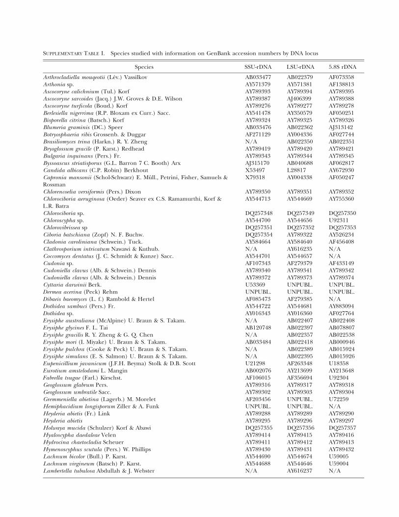
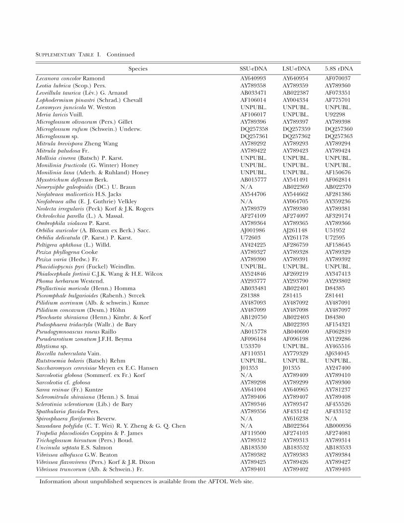
SUPPLEMENTARY TABLE I. Species studied with information on GenBank accession numbers by DNA locus
Species SSU-rDNA LSU-rDNA 5.8S rDNA
Arthrocladiella mouqeotii (Lev.) Vassilkov AB033477 AB022379 AF073358Arthonia sp. AY571379 AY571381 AF138813Ascocoryne calichnium (Tul.) Korf AY789393 AY789394 AY789395Ascocoryne sarcoides (Jacq.) J.W. Groves & D.E. Wilson AY789387 AJ406399 AY789388Ascocoryne turficola (Boud.) Korf AY789276 AY789277 AY789278Berlesiella nigerrima (R.P. Bloxam ex Curr.) Sacc. AY541478 AY350579 AF050251Bisporella citrina (Batsch.) Korf AY789324 AY789325 AY789326Blumeria graminis (DC.) Speer AB033476 AB022362 AJ313142Botryosphaeria ribis Grossenb. & Duggar AF271129 AY004336 AF027744Brasiliomyces trina (Harkn.) R. Y. Zheng N/A AB022350 AB022351Bryoglossum gracile (P. Karst.) Redhead AY789419 AY789420 AY789421Bulgaria inquinans (Pers.) Fr. AY789343 AY789344 AY789345Byssoascus striatisporus (G.L. Barron 7 C. Booth) Arx AJ315170 AB040688 AF062817Candida albicans (C.P. Robin) Berkhout X53497 L28817 AY672930Capronia mansonii (Schol-Schwarz) E. Mull., Petrini, Fisher, Samuels &Rossman
X79318 AY004338 AF050247
Chlorencoelia versiformis (Pers.) Dixon AY789350 AY789351 AY789352Chlorociboria aeruginosa (Oeder) Seaver ex C.S. Ramamurthi, Korf &L.R. Batra
AY544713 AY544669 AY755360
Chlorociboria sp. DQ257348 DQ257349 DQ257350Chloroscypha sp. AY544700 AY544656 U92311Chlorovibrissea sp DQ257351 DQ257352 DQ257353Ciboria batschiana (Zopf) N. F. Buchw. DQ257354 AY789322 AY526234Cladonia caroliniana (Schwein.) Tuck. AY584664 AY584640 AF456408Clathrosporium intricatum Nawawi & Kuthub. N/A AY616235 N/ACoccomyces dentatus (J. C. Schmidt & Kunze) Sacc. AY544701 AY544657 N/ACudonia sp. AF107343 AF279379 AF433149Cudoniella clavus (Alb. & Schwein.) Dennis AY789340 AY789341 AY789342Cudoniella clavus (Alb. & Schwein.) Dennis AY789372 AY789373 AY789374Cyttaria darwinii Berk. U53369 UNPUBL. UNPUBL.Dermea acerina (Peck) Rehm UNPUBL. UNPUBL. UNPUBL.Dibaeis baeomyces (L. f.) Rambold & Hertel AF085473 AF279385 N/ADothidea sambuci (Pers.) Fr. AY544722 AY544681 AY883094Dothidea sp. AY016343 AY016360 AF027764Erysiphe australiana (McAlpine) U. Braun & S. Takam. N/A AB022407 AB022408Erysiphe glycines F. L. Tai AB120748 AB022397 AB078807Erysiphe gracilis R. Y. Zheng & G. Q. Chen N/A AB022357 AB022538Erysiphe mori (I. Miyake) U. Braun & S. Takam. AB033484 AB022418 AB000946Erysiphe pulchra (Cooke & Peck) U. Braun & S. Takam. N/A AB022389 AB015924Erysiphe simulans (E. S. Salmon) U. Braun & S. Takam. N/A AB022395 AB015926Eupenicillium javanicum (J.F.H. Beyma) Stolk & D.B. Scott U21298 AF263348 U18358Eurotium amstelodami L. Mangin AB002076 AY213699 AY213648Fabrella tsugae (Farl.) Kirschst. AF106015 AF356694 U92304Geoglossum glabrum Pers. AY789316 AY789317 AY789318Geoglossum umbratile Sacc. AY789302 AY789303 AY789304Gremmeniella abietina (Lagerb.) M. Morelet AF203456 UNPUBL. U72259Hemiphacidium longisporum Ziller & A. Funk UNPUBL. UNPUBL. N/AHeyderia abietis (Fr.) Link AY789288 AY789289 AY789290Heyderia abietis AY789295 AY789296 AY789297Holwaya mucida (Schulzer) Korf & Abawi DQ257355 DQ257356 DQ257357Hyaloscypha daedaleae Velen AY789414 AY789415 AY789416Hydrocina chaetocladia Scheuer AY789411 AY789412 AY789413Hymenoscyphus scutula (Pers.) W. Phillips AY789430 AY789431 AY789432Lachnum bicolor (Bull.) P. Karst. AY544690 AY544674 U59005Lachnum virgineum (Batsch) P. Karst. AY544688 AY544646 U59004Lambertella tubulosa Abdullah & J. Webster N/A AY616237 N/A
Species SSU-rDNA LSU-rDNA 5.8S rDNA
Lecanora concolor Ramond AY640993 AY640954 AF070037Leotia lubrica (Scop.) Pers. AY789358 AY789359 AY789360Leveillula taurica (Lev.) G. Arnaud AB033471 AB022387 AF073351Lophodermium pinastri (Schrad.) Chevall AF106014 AY004334 AF775701Loramyces juncicola W. Weston UNPUBL. UNPUBL. UNPUBL.Meria laricis Vuill. AF106017 UNPUBL. U92298Microglossum olivaceum (Pers.) Gillet AY789396 AY789397 AY789398Microglossum rufum (Schwein.) Underw. DQ257358 DQ257359 DQ257360Microglossum sp. DQ257361 DQ257362 DQ257363Mitrula brevispora Zheng Wang AY789292 AY789293 AY789294Mitrula paludosa Fr. AY789422 AY789423 AY789424Mollisia cinerea (Batsch) P. Karst. UNPUBL. UNPUBL. UNPUBL.Monilinia fructicola (G. Winter) Honey UNPUBL. UNPUBL. UNPUBL.Monilinia laxa (Aderh. & Ruhland) Honey UNPUBL. UNPUBL. AF150676Myxotrichum deflexum Berk. AB015777 AY541491 AF062814Neoerysiphe galeopsidis (DC.) U. Braun N/A AB022369 AB022370Neofabraea malicorticis H.S. Jacks AY544706 AY544662 AF281386Neofabraea alba (E. J. Guthrie) Velkley N/A AY064705 AY359236Neolecta irregularis (Peck) Korf & J.K. Rogers AY789379 AY789380 AY789381Ochrolechia parella (L.) A. Massal. AF274109 AF274097 AF329174Ombrophila violacea P. Karst. AY789364 AY789365 AY789366Orbilia auricolor (A. Bloxam ex Berk.) Sacc. AJ001986 AJ261148 U51952Orbilia delicatula (P. Karst.) P. Karst. U72603 AY261178 U72595Peltigera aphthosa (L.) Willd. AY424225 AF286759 AF158645Peziza phyllogena Cooke AY789327 AY789328 AY789329Peziza varia (Hedw.) Fr. AY789390 AY789391 AY789392Phacidiopycnis pyri (Fuckel) Weindlm. UNPUBL. UNPUBL. UNPUBL.Phialocephala fortinii C.J.K. Wang & H.E. Wilcox AY524846 AF269219 AY347413Phoma herbarum Westend. AY293777 AY293790 AY293802Phyllactinia moricola (Henn.) Homma AB033481 AB022401 D84385Piceomphale bulgarioides (Rabenh.) Svrcek Z81388 Z81415 Z81441Pilidium acerinum (Alb. & schwein.) Kunze AY487093 AY487092 AY487091Pilidium concavum (Desm.) Hohn AY487099 AY487098 AY487097Pleochaeta shiraiana (Henn.) Kimbr. & Korf AB120750 AB022403 D84380Podosphaera tridactyla (Wallr.) de Bary N/A AB022393 AF154321Pseudogymnoascus roseus Raillo AB015778 AB040690 AF062819Pseudeurotium zonatum J.F.H. Beyma AF096184 AF096198 AY129286Rhytisma sp. U53370 UNPUBL. AY465516Roccella tuberculata Vain. AF110351 AY779329 AJ634045Rutstroemia bolaris (Batsch) Rehm UNPUBL. UNPUBL. UNPUBL.Saccharomyces cerevisiae Meyen ex E.C. Hansen J01353 J01355 AY247400Sarcoleotia globosa (Sommerf. ex Fr.) Korf N/A AY789409 AY789410Sarcoleotia cf. globosa AY789298 AY789299 AY789300Sarea resinae (Fr.) Kuntze AY641004 AY640965 AY781237Scleromitrula shiraiana (Henn.) S. Imai AY789406 AY789407 AY789408Sclerotinia sclerotiorum (Lib.) de Bary AY789346 AY789347 AF455526Spathularia flavida Pers. AY789356 AF433142 AF433152Spirosphaera floriformis Beverw. N/A AY616238 N/ASawadaea polyfida (C. T. Wei) R. Y. Zheng & G. Q. Chen N/A AB022364 AB000936Trapelia placodioides Coppins & P. James AF119500 AF274103 AF274081Trichoglossum hirsutum (Pers.) Boud. AY789312 AY789313 AY789314Uncinula septata E.S. Salmon AB183530 AB183532 AB183533Vibrissea albofusca G.W. Beaton AY789382 AY789383 AY789384Vibrissea flavovirens (Pers.) Korf & J.R. Dixon AY789425 AY789426 AY789427Vibrissea truncorum (Alb. & Schwein.) Fr. AY789401 AY789402 AY789403
Information about unpublished sequences is available from the AFTOL Web site.
SUPPLEMENTARY TABLE I. Continued