toxicological profile for dehp - california toxicological profile for di-(2-ethylhexyl) phthalate...
TRANSCRIPT

TOXICOLOGICAL PROFILE FOR DI-(2-ETHYLHEXYL) PHTHALATE (DEHP)
September 2009
Integrated Risk Assessment Branch Office of Environmental Health Hazard Assessment California Environmental Protection Agency


i
Toxicological Profile for
Di-(2-Ethylhexyl) Phthalate (DEHP)
September 2009
Prepared by
Office of Environmental Health Hazard Assessment
Prepared for
Ocean Protection Council
Under an Interagency Agreement, Number 07-055, with the
State Coastal Conservancy
LIST OF CONTRIBUTORS
Authors
Jim Carlisle, D.V.M., Ph.D., Senior Toxicologist, Integrated Risk Assessment Branch
Sarah Henkel, Ph.D., California Sea Grant Fellow, California Ocean Science Trust
Ling-Hong Li, Ph.D., Staff Toxicologist, Reproductive and Cancer Hazard Assessment Branch
Page Painter, M.D., Ph.D., Senior Toxicologist, Integrated Risk Assessment Branch
Dan Qiao, M.D., Ph.D. Staff Toxicologist, Integrated Risk Assessment Branch
Reviewers
David Siegel, Ph.D., Chief, Integrated Risk Assessment Branch

ii
Table of Contents
Executive Summary ....................................................................................................................... iv
Use and Exposure ...................................................................................................................... iv
Effects on Aquatic Life .............................................................................................................. iv
Health Hazard and Toxicity in Humans and Laboratory Animals ............................................ iv
Abbreviations ................................................................................................................................. vi
Introduction ..................................................................................................................................... 1
Properties and Uses ......................................................................................................................... 1
Freshwater and Marine Laboratory and Environmental Studies .................................................... 2
Environmental Contamination and Fate ..................................................................................... 2
Environmental Bio-uptake .......................................................................................................... 3
Toxicology: Marine and Other Aquatic Organisms .................................................................. 4
Fish .......................................................................................................................................... 4
Invertebrates ............................................................................................................................ 4
Environmental Criteria ............................................................................................................... 6
Summary ..................................................................................................................................... 6
Human and Laboratory Studies ...................................................................................................... 7
Reproductive and Developmental Toxicity ................................................................................ 7
Introduction ............................................................................................................................. 7
Male Reproductive Toxicity in Animals................................................................................. 7
Studies in Rats..................................................................................................................... 7
Studies in Other Species ................................................................................................... 10
Female Reproductive Toxicity in Animals ........................................................................... 13
Developmental Toxicity in Animals ..................................................................................... 14
Human Studies ...................................................................................................................... 15
Epidemiological Studies of Male Reproductive Endpoints .............................................. 17
Epidemiological Studies of Female Reproductive Endpoints .......................................... 19
Epidemiological Studies of Developmental Endpoints .................................................... 20
Summary ............................................................................................................................... 20
Cancer ....................................................................................................................................... 21
Introduction ........................................................................................................................... 21
Laboratory Rodent Studies ................................................................................................... 21
Effects on Cultured Mammalian Cells .................................................................................. 21
Summary ............................................................................................................................... 21
Obesity ...................................................................................................................................... 21
Thyroid ...................................................................................................................................... 22
Introduction ........................................................................................................................... 22
Laboratory Rodent Studies ................................................................................................... 22
Thyroid histopathology ..................................................................................................... 22
Thyroid function ............................................................................................................... 22
Human Studies ...................................................................................................................... 23
Summary ............................................................................................................................... 23
Immune System ........................................................................................................................ 23
Introduction ........................................................................................................................... 23
Laboratory Rodent Studies ................................................................................................... 24
In vitro Studies ...................................................................................................................... 24
iii
Human Studies ...................................................................................................................... 25
Summary ............................................................................................................................... 25
Nervous System ........................................................................................................................ 25
Introduction ........................................................................................................................... 25
Laboratory Rodent Studies ................................................................................................... 25
G protein-coupled receptors .................................................................................................. 26
The membrane Na+-K
+ ATPase ............................................................................................ 26
Intracellular calcium ............................................................................................................. 27
Protein kinase C (PKC) ......................................................................................................... 27
Brain aromatase enzyme ....................................................................................................... 27
Brain peroxisomes ................................................................................................................ 27
Summary ............................................................................................................................... 28
Liver and Kidney Effects .......................................................................................................... 28
Conclusions ................................................................................................................................... 28
Findings .................................................................................................................................... 28
Data Gaps .................................................................................................................................. 29
Recommendations ..................................................................................................................... 29
References ..................................................................................................................................... 31

iv
Executive Summary
This toxicological profile on di-(2-ethylhexyl) phthalate (DEHP) describes its effects on
freshwater and marine life, humans, and laboratory animals. Because of its prevalence in the
environment and the high likelihood of exposures to humans and other species, DEHP has been
the subject of considerable toxicological research.
Use and Exposure
DEHP is the most commonly used phthalate plasticizer. Plasticizers are used to make rigid
compounds such as polyvinyl chloride or PVC more flexible. PVC products containing DEHP
include packaging film and sheets, wall coverings, floor tiles, furniture upholstery, shower
curtains, garden hoses, swimming pool liners, rainwear, toys, shoes, sheathing for wire and
cable, and medical tubing.
Due to its high volume use, DEHP is a ubiquitous contaminant—found at low concentrations—
in air, water, soil and sediments. Since DEHP is not bound in plastic, it slowly leaches out of
plastic materials and into the environment. Examples of how humans can be exposed to DEHP
include ingestion of DEHP transferred to food from PVC films and plastic containers, and
transfer of DEHP in PVC plastic disposable medical equipment to patients (i.e., into their blood).
Effects on Aquatic Life
In water, DEHP predominantly attaches to suspended particles and sediments, but a small
amount remains dissolved in the water. DEHP in air binds to dust particles and is removed from
the atmosphere by settling of dry particles and by being washed out by rain and snow. The
toxicology of DEHP in aquatic animals has not been well studied. Reported toxic effects in
aquatic organisms in laboratory studies generally occur at exposure levels higher than the
concentration of DEHP that can dissolve in water (3 micrograms per liter). There is some
evidence of reproductive effects in salmon. Because fish can break down DEHP, it does not
accumulate in their tissues to any significant extent; invertebrates, however, are less able to break
down DEHP. Two studies report effects in invertebrates at lower concentrations. Further
environmental (field) and laboratory studies are needed to investigate the impacts of DEHP on
freshwater and marine life.
Health Hazard and Toxicity in Humans and Laboratory Animals
Reproductive and Developmental Effects. A major concern is DEHP’s effect on the
reproductive system. The male reproductive toxicity of DEHP is well established in
laboratory animals including rats, mice, hamsters, and ferrets. Depending on the dose,
duration of exposure, and age of animals, DEHP causes reduced fertility, decreased
weights of male reproductive organs, and histopathological changes in the testes of
juvenile and adult rats. Although it has not been studied as extensively, female
reproductive toxicity in laboratory animals has been reported. DEHP also has been found
to cause developmental toxicity, including intrauterine death, developmental delay, and
structural malformations and variations; neurological developmental effects have also
been reported. Evidence of DEHP’s reproductive toxicity in humans is less conclusive.

v
Cancer. According to U.S. EPA, there is sufficient evidence that DEHP causes cancer in
laboratory animals. However, the International Agency for Research on Cancer considers
the mechanism responsible for causing cancer in laboratory animals as unlikely to occur
in humans; there are some who disagree with this finding.
Neurological Effects. Alteration in brain function and behavioral changes has been
observed in rodent studies. The results thus indicated that DEHP might have potential
effects on neural development and behavior.
Other effects. There is some evidence in laboratory animals indicating that DEHP may
affect the thyroid and immune system. Little information is available on its effect on the
human thyroid and immune system.
Summary Table
This table provides some idea of the availability of information on the toxicology of BPA for the
endpoints and organisms identified. It also provides some sense of the evidence available in that
information can be used to determine if the endpoint effect does or does not occur. If there is no
information the evidence column will be marked with a ―--.‖
Health Effect Human Lab Animal Aquatic Life
Information Evidence Information Evidence Information Evidence
Reproductive S S
male S E Su Su
female S L Su Su
Cancer S E Su Su N --
Developmental L E Su Su N --
Neurological N -- S S
Immunological L L S S N --
Other Chronic effects S S
Thyroid L L S S
Other Sub-chronic
effects
S S
Acute S S
N = None S = Some
L = Little Su = Sufficient
E=Equivocal
These rating categories are qualitative in nature and designed to give the reader a general sense of the availability and
strength of the information.

vi
Abbreviations
ADHD attention-deficit hyperactivity disorder
AGD anogenital distance
ANOVA analysis of variance
ATPase adenosine triphosphatase
BBP butyl benzyl phthalate
BCF bioconcentration factor
CERHR Center for the Evaluation of Risks to Human Reproduction
CG chorionic gonadotropin
Con A concanavalin A
DBP Di-n-butyl phthalate
DEHP di-(2-ethylhexyl) phthalate
DEP diethyl phthalate
DINP di-isononyl phthalate
DNA deoxyribonucleic acid
DOP dioctyl phthalate
2-EH 2-ethylhexanol
ERL environmental risk limit
FSH follicle-stimulating hormone
GABA γ-aminobutyric acid
HPOA hypothalamic/preoptic area
IL-4 interleukin-4
Insl3 insulin-like hormone 3
IARC International Agency for Research on Cancer
LC50 lethal concentration to 50 percent of the population
LH luteinizing hormone
LOEC lowest observed concentration
LOELs lowest observed effect levels
MBP mono-n-butyl phthalate
MBzP mono-benzyl phthalate
MCSI Mitsubishi Chemical Safety Institute Ltd.
MEHP mono-ethylhexyl phthalate
MEP mono-ethyl phthalate
MiNP mono-isononyl phthalate
MIP-1α macrophage inflammatory protein-1α
MMP monomethyl phthalate
NIS sodium/iodide symporter
NOEC no observed effect concentration
NOELs no observed effect levels
OEHHA Office of Environmental Health Hazard Assessment
OPC California Ocean Protection Council
PA phthalic acid
PCBs polychlorinated biphenols
PKC Protein kinase C
PND postnatal day

vii
PPAR-γ peroxisome proliferators-activated receptor-γ
PVC polyvinyl chloride
RNA ribonucleic acid
SEB Staphylococcus enterotoxin B
SHBG sex hormone-binding globulin
T3 triiodothyronine
T4 thyroxine
TSH thyroid stimulating hormone
1
Introduction
On February 8, 2007, the California Ocean Protection Council (OPC) passed a resolution, ―On
Reducing and Preventing Marine Debris.‖ Research is underway to determine whether these
constituents leach out of plastic products in the marine environment, and as a result, present a
threat to the health of humans and wildlife. The OPC has asked the Office of Environmental
Health Hazard Assessment (OEHHA) to aid in this effort by preparing toxicity profiles
characterizing certain chemical constituents of plastics that are thought to be harmful to marine
life and humans. In preparing this profile, OEHHA reviewed reported information on the
adverse effects of exposure to DEHP in aquatic organisms in the laboratory and in the natural
environment, humans, and experimental laboratory animals.
Properties and Uses
Phthalate esters (esters of 1, 2-benzenedicarboxylic acid) are widely used as plasticizers to
increase the flexibility and workability of high-molecular-weight polymers. Phthalates constitute
up to 50 percent of the total weight of some plastics. Their low melting point and high boiling
point make them useful as heat transfer fluids and carriers. The world-wide production of
phthalates approximates 2.7 million metric tons a year, a quarter of which is di-(2-ethylhexyl)
phthalate (DEHP) (CAS number, 117-81-7) (van Wezel et al., 2000).
O
O
O
O
H3CCH
3
CH3H
3C
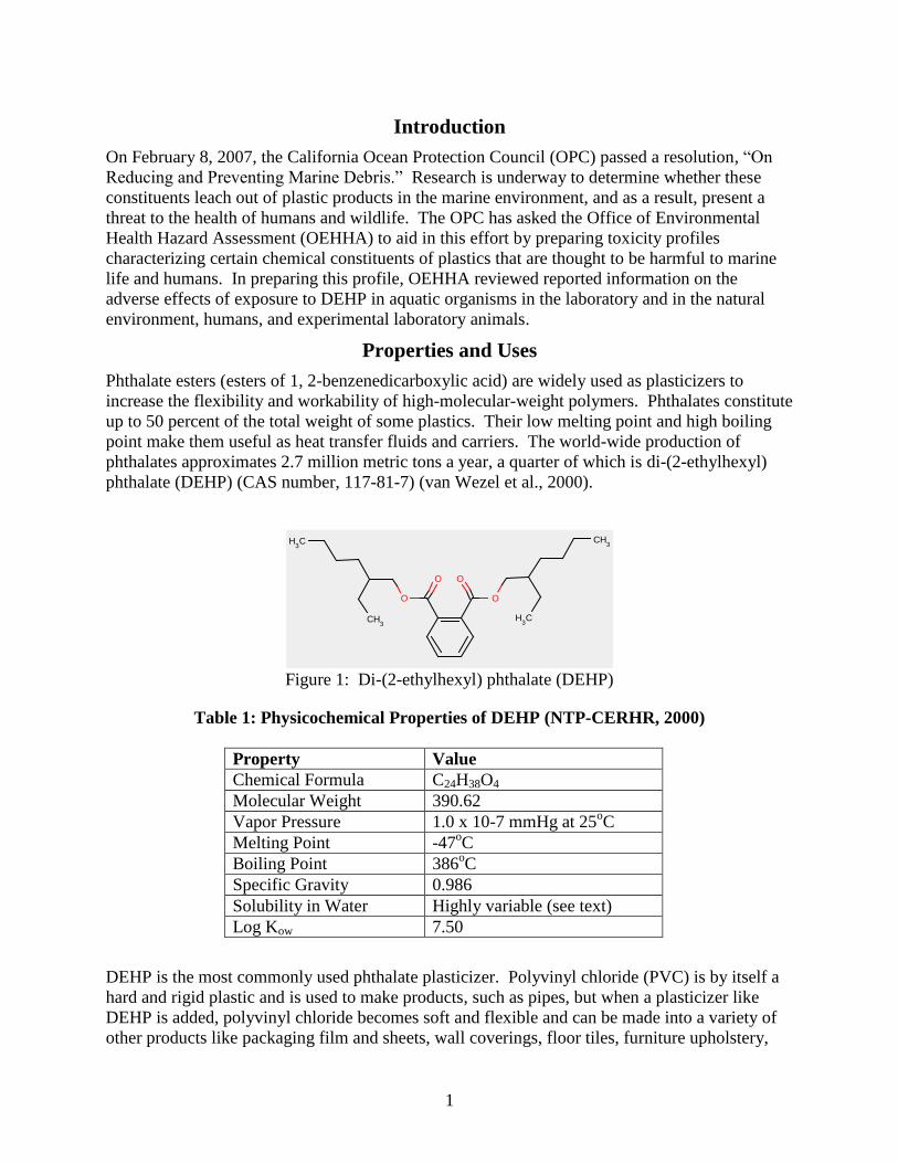
Figure 1: Di-(2-ethylhexyl) phthalate (DEHP)
Table 1: Physicochemical Properties of DEHP (NTP-CERHR, 2000)
Property Value
Chemical Formula C24H38O4
Molecular Weight 390.62
Vapor Pressure 1.0 x 10⁻⁷ mmHg at 25oC
Melting Point -47oC
Boiling Point 386oC
Specific Gravity 0.986
Solubility in Water Highly variable (see text)
Log Kow 7.50
DEHP is the most commonly used phthalate plasticizer. Polyvinyl chloride (PVC) is by itself a
hard and rigid plastic and is used to make products, such as pipes, but when a plasticizer like
DEHP is added, polyvinyl chloride becomes soft and flexible and can be made into a variety of
other products like packaging film and sheets, wall coverings, floor tiles, furniture upholstery,
2
shower curtains, garden hoses, swimming pool liners, rainwear, toys, shoes, sheathing for wire
and cable, and medical tubing. DEHP does not bind with the polymer in the plastic. It mixes
with the polymer and allows the polymer strands to move, providing the flexibility.
Since the DEHP is not bound in plastic, it slowly migrates or leaches out of the plastic and into
the environment. DEHP in the PVC films and plastic containers used to cover and store food can
transfer into the contents, thereby exposing people via ingestion of the food. DEHP is also used
in PVC plastic disposable medical equipment and has been found to transfer to patients.
DEHP is a ubiquitous contaminant in today’s environment. DEHP has been found in several
kinds of food; monitoring data indicate that DEHP residues are generally low in U.S. foods, but
the available data are in excess of 10 years old and might not be representative of current
conditions. Fish and other seafood have been reported to be contaminated with concentrations
ranging from 2 to 32,000 ppb. In the past and in addition to fish, DEHP has been detected in
foods such as milk, cheese, meat, margarine, eggs, cereal products, baby food, and infant
formula (ATSDR, 2002). In water, DEHP predominantly attaches to suspended particles and
sediments, but a small amount remains dissolved in the water. DEHP in air binds to dust
particles and is removed from the atmosphere by settling of dry particles and by being washed
out by rain and snow. In view of the considerable direct and expected indirect emission of
Freshwater and Marine Laboratory and Environmental Studies
Environmental Contamination and Fate
The solubility of DEHP in distilled water has been determined both experimentally and
theoretically and varied between 1.1 and 1,200 μg/L (Staples et al., 1997). The lowest
experimentally derived value for the solubility of DEHP in distilled water was 41 μg/L; however,
a value of 3 μg/L for the water solubility of DEHP has been recommended by Staples et al.
(1997) ―based on available evidence‖, rather than any one specific experimentally derived value
(ATSDR, 2002). Staples et al. note that most published values exceed true water solubilities due
to experimental difficulties associated with solubility determinations for these hydrophobic
organic liquids. Laboratory and field studies show that partitioning to suspended solids, soils,
sediments and aerosols increase as K~ increases and VP decreases. In seawater, Howard et al.
(1985) and Giam et al. (1980) report DEHP solubilities of 160 and 1160 μg/L, respectively,
using conventional techniques. Concentrations of DEHP in wastewater samples collected from
Oakland, California, urban sources ranged from 0.99 to 2700 μg/L (Jackson and Sutton, 2008).
DEHP concentrations in Japanese surface waters ranged from non-detect to58 μg/L (Naito et al.,
2006).
Huang et al. (2008) investigated phthalate compounds in sediments and fishes in 17 Taiwan
rivers and found DEHP in sediment at low-flow season was <0.05 to 46.5 mg/kg dry weight and
at high-flow season was <0.05 to 13.1 mg/kg dry weight; they report a water solubility of 300
μg/L. The DEHP sediment concentrations reported by Huang et al. (2008) are within the range
of those reported by other authors in other rivers (Fromme et al., 2002; Peijnenburg and Struijs,
2006; Sha et al., 2007; Tan, 1995). Naito et al. (2006) reported Japanese sediment
concentrations ranging from non-detect to 210,000 μg/kg dry weight. Adsorption of DEHP to
marine sediments might be greater than adsorption to freshwater sediments, due to reduced
solubility of DEHP in saltwater; levels of DEHP in a marine environment ranged from 0.1 to
0.7 ppb in the water and from 280 to 640 ppb in the suspended particles (ATSDR, 2002).
3
Phthalates have several degradation pathways and therefore are not considered to be persistent
chemicals. They are easily photodegraded in the atmosphere with predicted half-lives of
approximately 1 day, and they can be biodegraded by bacteria and actinomycetes, which is likely
the dominant loss mechanism in surface waters, soils and sediments (Staples et al., 1997).
Primary degradation half-lives in surface and marine waters range from <1 day to 2 weeks and in
soils from <1 week to several months; standardized aerobic biodegradation tests with sewage
sludge inocula show that phthalate esters undergo ~ 50% ultimate degradation within 28 days
(Staples et al., 1997).
Environmental Bio-uptake
DEHP experimentally has been shown to bioaccumulate in the aquatic plant Elodea canadensis,
the snail Physa sp., the water flea Daphnia magna and mosquito (Culex pipiens
quinquefasciatus) larvae and pupae (Metcalf et al., 1973), as well as in isolated rat liver (Jaeger
and Rubin, 1970). Furthermore, Taborsky (1967) isolated DEHP from bovine pineal glands, and
it has found it in mitochondria from the hearts of cattle, dogs, rabbit, and rat (Nazir et al., 1971).
Questions from these earlier studies remain whether the phthalates are of physiological origin or
are ingested by the animals from artificial sources and retained. Additionally, DEHP has been
found in human spleen, liver, lung, and abdominal fat in quantities ranging from 25 ppm (dry
weight) in spleen to 270 ppm in abdominal fat from patients who had received transfusions of
blood stored in plastic bags (Jaeger and Rubin, 1970). DEHP, fluxes may be high, explaining the
concentrations found in the environment and bio-uptake by organisms.
Despite its high tendency to accumulate in fatty tissues, fish do not extensively accumulate
DEHP. Norman et al (2007) found tissue concentrations in Atlantic salmon at 0.3 percent of the
dietary concentration. Experiments with rainbow trout fitted with an indwelling cannula showed
that the majority of [14C]
DEHP did not reach the systemic circulation of the fish, but DEHP
metabolites were present in the exposure water. Isolated perfused gill arches of the trout
metabolized DEHP in the exposure bath to monoethylhexyl phthalate, demonstrating the ability
of enzymes in the gill to break down DEHP, thus potentially limiting its entry into the fish
(Barron et al., 1989). Bioconcentration factors for fish are not strongly related to the organic
carbon absorption coefficient (Koc) of the pthalates; this is most likely due to metabolism.
Bioconcentrations in aquatic organisms other than fish, (e.g., algae, mollusks) exceeds that in
fish (Table 2), suggesting lower biotransformation capacities in these organisms.
Bioaccumulation in the food chain is limited because of biotransformation. An extra risk related
to accumulation in the food chain is therefore not expected (van Wezel et al., 2000).
Table 2: Bio-uptake and Bioconcentration
Compartment Water (µg/L) Tissue (µg/kg) Bioconcentration
Factor
Reference*
Rainbow trout ~100 42-113 (Barron et al., 1989)
Atlantic salmon 827 mg/kg diet 2551 (Norman et al.,
2007)
Daphnia magna 3.2
10
32
100
0.00053
0.00140
0.00835
0.02677
166
140
261
268
(Brown and
Thompson, 1982a)
Mytilus edulis 4 0.0097 2366 (Brown and
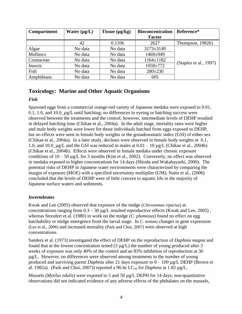
4
Compartment Water (µg/L) Tissue (µg/kg) Bioconcentration
Factor
Reference*
42 0.1106 2627 Thompson, 1982b)
Algae No data No data 3173±3149
(Staples et al., 1997)
Molluscs No data No data 1469±949
Crustacean No data No data 1164±1182
Insects No data No data 1058±772
Fish No data No data 280±230
Amphibians No data No data 605
Toxicology: Marine and Other Aquatic Organisms
Fish
Spawned eggs from a commercial orange-red variety of Japanese medaka were exposed to 0.01,
0.1, 1.0, and 10.0_µg/L until hatching; no differences in eyeing or hatching success were
observed between the treatments and the control; however, intermediate levels of DEHP resulted
in delayed hatching time (Chikae et al., 2004a). In the adult stage, mortality rates were higher
and male body weights were lower for those individuals hatched from eggs exposed to DEHP,
but no effects were seen in female body weights or the gonadosomatic index (GSI) of either sex
(Chikae et al., 2004a). In a later study, declines were observed in female body weights at 0.1,
1.0, and 10.0_µg/L and the GSI was reduced in males at 0.01 – 10 µg/L (Chikae et al., 2004b)
(Chikae et al., 2004b). Effects were observed in female medaka under chronic exposure
conditions of 10 – 50 µg/L for 3 months (Kim et al., 2002). Conversely, no effect was observed
in medaka exposed to higher concentrations for 14 days (Shioda and Wakabayashi, 2000). The
potential risks of DEHP in Japanese water environments were characterized by comparing the
margin of exposure (MOE) with a specified uncertainty multiplier (UM); Naito et al., (2006)
concluded that the levels of DEHP were of little concern to aquatic life in the majority of
Japanese surface waters and sediments.
Invertebrates
Kwak and Lee (2005) observed that exposure of the midge (Chironomus riparius) at
concentrations ranging from 0.3 – 30 µg/L resulted reproductive effects (Kwak and Lee, 2005) ,
whereas Streufert et al. (1980) in work on the midge (C. plumosus) found no effect on egg
hatchability or midge emergence from the larval stage. In C. tentans changes in gene expression
(Lee et al., 2006) and increased mortality (Park and Choi, 2007) were observed at high
concentrations.
Sanders et al. (1973) investigated the effect of DEHP on the reproduction of Daphnia magna and
found that at the lowest concentration tested (3 µg/L) the number of young produced after 3
weeks of exposure was only 40% of the control and an 83% inhibition of reproduction at 30
µg/L. However, no differences were observed among treatments in the number of young
produced and surviving parent Daphnia after 21 days exposure to 0 – 100 µg/L DEHP (Brown et
al. 1982a). (Park and Choi, 2007)) reported a 96-hr LC50 for Daphnia at 1.82 µg/L.
Mussels (Mytilus edulis) were exposed to 5 and 50 µg/L DEPH for 14 days; non-quantitative
observations did not indicated evidence of any adverse effects of the phthalates on the mussels,
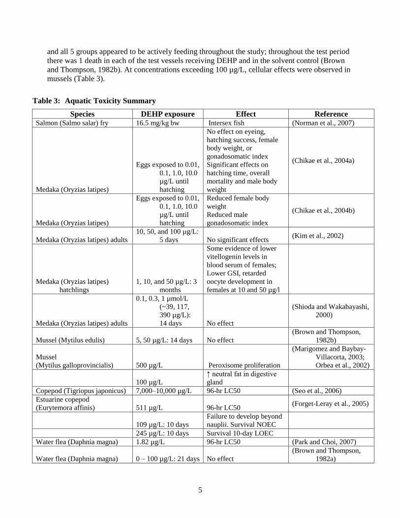
5
and all 5 groups appeared to be actively feeding throughout the study; throughout the test period
there was 1 death in each of the test vessels receiving DEHP and in the solvent control (Brown
and Thompson, 1982b). At concentrations exceeding 100 µg/L, cellular effects were observed in
mussels (Table 3).
Table 3: Aquatic Toxicity Summary
Species DEHP exposure Effect Reference Salmon (Salmo salar) fry 16.5 mg/kg bw Intersex fish (Norman et al., 2007)
Medaka (Oryzias latipes)
Eggs exposed to 0.01,
0.1, 1.0, 10.0
µg/L until
hatching
No effect on eyeing,
hatching success, female
body weight, or
gonadosomatic index
Significant effects on
hatching time, overall
mortality and male body
weight
(Chikae et al., 2004a)
Medaka (Oryzias latipes)
Eggs exposed to 0.01,
0.1, 1.0, 10.0
µg/L until
hatching
Reduced female body
weight
Reduced male
gonadosomatic index
(Chikae et al., 2004b)
Medaka (Oryzias latipes) adults
10, 50, and 100 µg/L:
5 days No significant effects (Kim et al., 2002)
Medaka (Oryzias latipes)
hatchlings
1, 10, and 50 µg/L: 3
months
Some evidence of lower
vitellogenin levels in
blood serum of females;
Lower GSI, retarded
oocyte development in
females at 10 and 50 µg/l
Medaka (Oryzias latipes) adults
0.1, 0.3, 1 µmol/L
(~39, 117,
390 µg/L):
14 days No effect
(Shioda and Wakabayashi,
2000)
Mussel (Mytilus edulis) 5, 50 µg/L: 14 days No effect
(Brown and Thompson,
1982b)
Mussel
(Mytilus galloprovincialis) 500 µg/L Peroxisome proliferation
(Marigomez and Baybay-
Villacorta, 2003;
Orbea et al., 2002)
100 µg/L
↑ neutral fat in digestive
gland
Copepod (Tigriopus japonicus) 7,000–10,000 µg/L 96-hr LC50 (Seo et al., 2006)
Estuarine copepod
(Eurytemora affinis) 511 µg/L 96-hr LC50 (Forget-Leray et al., 2005)
109 µg/L: 10 days
Failure to develop beyond
nauplii. Survival NOEC
245 µg/L: 10 days Survival 10-day LOEC
Water flea (Daphnia magna) 1.82 µg/L 96-hr LC50 (Park and Choi, 2007)
Water flea (Daphnia magna) 0 – 100 µg/L: 21 days No effect
(Brown and Thompson,
1982a)

6
Species DEHP exposure Effect Reference
Dragonfly (Latin name?) larva
587-623 mg/kg
sediment ↓ predation efficiency (Woin and Larsson, 1987)
Midge (Chironomous tentans) 500 – 5000 µg/L
↑ Heat-shock Protein gene
expression
↓ Hemoglobin gene
expression
(Lee et al., 2006)
Midge (Chironomous tentans) 1124 µg/L 96-hr LC50 (Park and Choi, 2007)
Midge (Chironomus riparius) >0.3 µg/L
↓ adult emergence (no
dose/response) (Kwak and Lee, 2005)
0.3, 30 µg/L Sex ratio Female > male
Environmental Criteria
The California State Water Resources Control Board does not have ambient water quality criteria
for the protection of freshwater or marine life. An environmental risk limit (ERL) is an
estimated ecosystem no-effect concentration used as a basis for environmental quality standards.
In order to derive a DEHP ERL for the Netherlands, van Wezel et al. (2000) reviewed the DEHP
literature. They considered data on chronic and acute toxicity of DEHP to aquatic species along
with effects on growth and reproduction. Their smallest aquatic lowest observed effect
concentration (LOEC) was 5 µg/L, based on survival and reproduction in a 15-week study in
rainbow trout. Because all identified aquatic toxicity no observed effect concentrations (NOEC)
exceeded the 3 µg/L water solubility of DEHP recommended by Staples et al. (1997), an ERL
based on aquatic toxicity was not developed. Instead, the ERL was based on sediment toxicity.
The lowest NOEC was 10 mg/kg sediment based on hatching success of the moor frog, Rana
arvalis. Based on equilibrium partitioning between solid and liquid phases, this sediment
concentration was equivalent to 1.9 µg/L; a 10-fold safety factor made the ERL 0.19 µg/L.
Summary
Toxicology data are summarized in Table 3. Arthropods appear to be the most sensitive, with
several reports of effects in the low or fractional µg/L range. Some fish, especially Medaka are
also sensitive, with effects reported in the low µg/L range. Mollusks appear to be less sensitive
with effects reported in the tens to hundreds of µg/L range. The relationship of the toxic
concentrations to the solubility limit is unclear, since published solubility limits for DEHP vary
widely. These disparate and conflicting findings indicate the need for further work to clarify the
risk of adverse effects on invertebrates at realistic environmental concentrations. More work
should focus on free-living organisms in marine and estuarine environments.

7
Human and Laboratory Studies
Reproductive and Developmental Toxicity
Introduction
Evidence on the developmental and reproductive toxicity of DEHP has been reviewed in several
comprehensive reports (e.g., the National Toxicology Program-Center for the Evaluation of Risk
to Human Reproduction (NTP-CERHR)(Kavlock et al., 2006; NTP-CERHR, 2000)). The
current review relies heavily on the experimental data summarized in those reports as well as on
major findings from numerous studies that were reported in the past few years. Because of the
general concordance between reproductive toxicity of chemicals in humans and wildlife, as
discussed in the endocrine disruption literature (Colborn, 1994; Hotchkiss et al., 2008), evidence
of toxicity in laboratory animals and humans should be taken into account in evaluating the
potential effects of exposure to DEHP on marine organisms.
Male Reproductive Toxicity in Animals
The male reproductive toxicity of DEHP is well established and has been studied in many
species including rats, mice, hamsters, ferrets, and non-human primates. Findings from the
majority of these studies have been well reviewed and summarized in many documents or review
reports (e.g., NTP-CERHR, 2000; U.S. FDA, 2001). Therefore, detailed findings from each
individual study are not discussed in this document. Instead, this document focuses on a number
of key studies that can be potentially identified as the most sensitive study of sufficient quality
for quantitative risk assessment.
Studies in Rats
The majority of studies on the male reproductive toxicity of DEHP were conducted in rats using
oral administration (gavage, feed, or drinking water). A few studies used non-oral routes of
exposure (e.g., intravenous injection or inhalation). The male reproductive toxicity of DEHP
following intravenous injection was similar to that following oral administration, though the
intravenous doses required to induce obvious testicular damage were higher than those by oral
treatment (Cammack et al., 2003). One inhalation study that investigated the testicular effects of
DEHP in prepubertal Wistar rats found that inhalation of DEHP at concentrations of 5 or
25 mg/m3, six hours per day for four or eight weeks caused significant increases in the plasma
level of testosterone and the weight of seminal vesicles. Although the underlying mechanism for
this DEHP-induced increase in testosterone and its long-term functional consequence remain to
be determined, these findings indicate that inhalation exposure to DEHP can alter testicular
function. The findings are also consistent with those reported by Akinbemi et al (2004; 2001)
who treated rats of similar ages by gavage.
Depending on the doses, dosing duration, age of animals, and endpoints included, it has been
shown that oral treatment with DEHP causes reduced fertility, decreased weights of male
reproductive organs, and histopathological changes in the testis of juvenile and adult rats (NTP-
CERHR, 2000; U.S. FDA, 2001). Characteristics of histopathological changes include
vacuolation and rarefaction of the cytoplasm, disruption of cytoskeletons, destruction of
intercellular specializations (e.g., ectoplasmic specialization, occluding junctions) in Sertoli cells,

8
followed by degeneration of spermatocytes by apoptosis and/or sloughing of germ cells into the
lumen of seminiferous tubules (e.g., Boekelheide, 2004; Park et al., 2002; Saitoh et al., 1997).
Different groups of germ cells in the testis of rats are organized in an orderly manner along the
length of seminiferous tubules. A defined group of germ cells is called a stage. Along the length
of a seminiferous tubule, there is a distinct ordering of stages, namely from stage I to XIV.
Sertoli cells undergo morphological and functional fluctuation from stages I to XIV. In the testis
of young rats (8-week-old), Sertoli cells and the spermatocytes associated with them in
seminiferous tubules at stages IX-XIV and I are most sensitive to the testicular effects of DEHP
(Saitoh et al., 1997).
Oral administration of DEHP to rats during the perinatal period results in severe permanent
abnormalities in the male reproductive system of male offspring (Arcadi et al., 1998; Gray et al.,
1999b; Moore et al., 2001; Schilling et al., 1999; Tandon et al., 1991). Neonatal or young rats
have been found to be more sensitive to the male reproductive effects of DEHP than are adults
(Akingbemi et al., 2004; 2001; Borch et al., 2004; Cammack et al., 2003; Dostal et al., 1988;
Gray and Butterworth, 1980; Li et al., 2000; NTP-CERHR, 2000; Sjoberg et al., 1986; 1985;
U.S. FDA, 2001). The testis at early developmental stages (late gestation and early days after
birth in rats) is more sensitive to DEHP than that of juvenile or adult animals (Gray et al., 1999a;
2000; Moore et al., 2001; NTP-CERHR, 2000). Thus, the no observed effect levels (NOELs)
and/or lowest observed effect levels (LOELs) for the male reproductive effects of DEHP
observed in studies that treated rats either perinatally or in the early weeks of the postnatal period
are in general lower than those observed in young or adult animals. It should be emphasized that
several recent studies have found that concurrent exposure to multiple phthalates can cause
cumulative effects on the male reproductive system in a dose-additive manner (e.g., Howdeshell
et al., 2007; 2008).
Table 4 summarizes a list of studies that observed relatively low values of LOELs and/or NOELs
in rats. The animals used in these studies received DEHP treatment either perinatally (Arcadi et
al., 1998) or as three-four week old juveniles (Akingbemi et al., 2004; 2001; David et al., 2000a;
Poon et al., 1997). Manifestation of DEHP-caused testicular damage takes different forms,
depending on the age of animals, dosing levels, and dosing durations. For example, as stated in
the document by the Center for the Evaluation of Risks to Human Reproduction (NTP-CERHR,
2000), ―during the time of Sertoli cell divisions (before pnd [post natal day] 15 in rats), phthalate
exposure apparently inhibits cell division. In animals older than pnd 15, toxicity is manifest as
vacuoles, followed by germ cell sloughing.‖ Therefore, when comparing different studies to
identify ―the most sensitive study,‖ OEHHA considered different endpoints used in different
studies and attempted to compare different studies based on the same or similar endpoints. In
addition, the clear difference in sensitivity to the testicular effects of DEHP between developing
and adult rats suggests that a NOEL observed in adult animals should be compared to those
observed in developing animals in order to determine if a NOEL in adult animals has no
observable effects in developing animals.
The study by Arcadi (1998) observed the lowest LOEL (32.5 l/L in drinking water) in rat dams
for the male reproductive effects of DEHP in male offspring exposed to DEHP from gestational
day 1 to postnatal day 21. The authors stated this dose was roughly equivalent to
3.0-3.5 mg/kg-day, but assumptions of body weights and water consumption for their estimate
were not reported. This study has some limitations. For example, DEHP is essentially insoluble
in water (3 g/L or approximately 0.003 l/L; (NTP-CERHR, 2000)). The concentrations of

9
DEHP used in the study were 32.5 and 325 l/L. The authors stated that ―the suspension was
prepared daily by adding DEHP to mineral water and then sonicating for 30 min.‖ However,
actual concentrations of DEHP in the drinking water were not verified. Daily water consumption
was not recorded. Maternal body weights were not reported. Therefore, although this study
provided important evidence on the adverse effects of DEHP on rat testicular development
during the perinatal period, this study report lacks detailed data that are critical for dose
estimation.
Among other studies listed in Table 4, the studies by Akingbemi et al. (2004; 2001) observed an
oral LOEL of 10 mg/kg-day, based on abnormal changes in testosterone production and altered
Leydig cell proliferation in the testes of prepubertal rats. This LOEL is markedly lower than
those based on histopathological changes in adult animals following long-term treatment with
DEHP (29 mg/kg-day as observed by David et al. (2000a) or 38 mg/kg-day by Poon et al.
(1997). It should be noted that the NOELs observed in adult animals by Poon et al. or David et
al., respectively, are lower than the LOEL of 10 mg/kg-day observed in juvenile animals by
Akingbemi et al. (2004; 2001). Therefore, based on endpoints indicative of morphological or
functional changes, there is no observed effect of DEHP on the testis at doses lower than
10 mg/kg-day following oral administration, regardless of the age of rats used in the studies. The
highest dose below 10 mg/kg-day used in the studies listed in Table 4 is the NOEL (5.8 mg/kg-
day) observed by David et al. (2000a). Thus, this NOEL (5.8 mg/kg-day) has no observable
testicular effects in rats of different ages.
Table 4. Oral studies that observed relatively low values of LOEL or NOEL for male
reproductive toxicity of DEHP in rats.
Study
Reference
Animals Treatment General
Toxicity
Male repro effects and
LOEL
NOEL
(Poon et al.,
1997)
Sprague-
Dawley rats,
about 6 wks
old at the
beginning, 10
rats per group.
Feed, 0, 5, 50,
500, 5,000 ppm
for 13 wks.
Increased liver
and kidney
weights,
histopathological
changes in the
liver at 5,000
ppm.
Sertoli cell vacuolation and
seminiferous tubular atrophy
at 5000 ppm. Minimal Sertoli
cell vacuolation in 7/10 rats at
500 ppm.
LOEL: 500 ppm (38 mg/kg-
day)
50 ppm (3.7
mg/kg-day).
(Arcadi et al.,
1998)
Long-Evans
rats, 12
pregnant rats
per group
Drinking water,
0, 32.5, 325 l/L
DEHP, from
gestational day 1
to postnatal day
(PND) 21. Pups
examined on
PND 21, 28, 35,
42, and 56.
No effects on
body weight
gains of dams or
pups. Changed
weights and
pathology in the
kidney and liver
of pups at both
doses.
Reduced testis weights and
histopathological changes in
the testes of male pups at both
doses.
LOEL: 32.5 l/L (3.0-3.5
mg/kg-day, estimated by the
study authors; water
consumption not reported)
Not observed.
(David et al.,
2000a)
Fischer 344
rats, about six-
wk-old at the
start, 55-80
rats per group.
Feed, 0, 100,
500, 2,500, or
12,500 ppm
DEHP for 104
wks.
Reduced survival
rates, reduced
body weights,
adverse effects in
the liver, kidney,
and pituitary at
2,500 ppm.
Significantly increased
incidence of aspermatogenesis
at 500 ppm at Week 105.
LOEL: 500 ppm (29 mg/kg-
day)
100 ppm (5.8
mg/kg-day)
(Akingbemi et
al., 2001) Male Long-
Evans rats,
21, 35, or 62
Gavage, 0, 1, 10,
100 or 200
mg/kg-day, PND
No obvious
general toxicity.
Decreased testosterone (T)
production by Leydig cells at
10 mg/kg-day at PND 21-34;
1 mg/kg-day
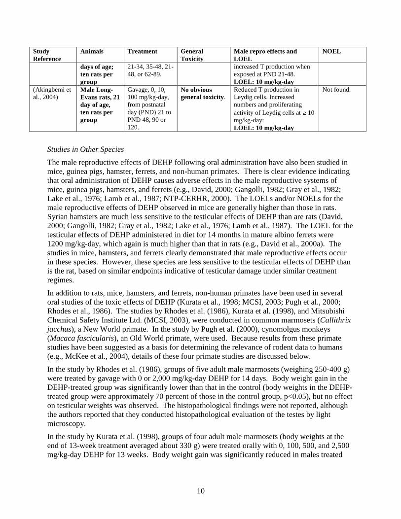
10
Study
Reference
Animals Treatment General
Toxicity
Male repro effects and
LOEL
NOEL
days of age;
ten rats per
group
21-34, 35-48, 21-
48, or 62-89.
increased T production when
exposed at PND 21-48.
LOEL: 10 mg/kg-day
(Akingbemi et
al., 2004) Male Long-
Evans rats, 21
day of age,
ten rats per
group
Gavage, 0, 10,
100 mg/kg-day,
from postnatal
day (PND) 21 to
PND 48, 90 or
120.
No obvious
general toxicity.
Reduced T production in
Leydig cells. Increased
numbers and proliferating
activity of Leydig cells at 10
mg/kg-day:
LOEL: 10 mg/kg-day
Not found.
Studies in Other Species
The male reproductive effects of DEHP following oral administration have also been studied in
mice, guinea pigs, hamster, ferrets, and non-human primates. There is clear evidence indicating
that oral administration of DEHP causes adverse effects in the male reproductive systems of
mice, guinea pigs, hamsters, and ferrets (e.g., David, 2000; Gangolli, 1982; Gray et al., 1982;
Lake et al., 1976; Lamb et al., 1987; NTP-CERHR, 2000). The LOELs and/or NOELs for the
male reproductive effects of DEHP observed in mice are generally higher than those in rats.
Syrian hamsters are much less sensitive to the testicular effects of DEHP than are rats (David,
2000; Gangolli, 1982; Gray et al., 1982; Lake et al., 1976; Lamb et al., 1987). The LOEL for the
testicular effects of DEHP administered in diet for 14 months in mature albino ferrets were
1200 mg/kg-day, which again is much higher than that in rats (e.g., David et al., 2000a). The
studies in mice, hamsters, and ferrets clearly demonstrated that male reproductive effects occur
in these species. However, these species are less sensitive to the testicular effects of DEHP than
is the rat, based on similar endpoints indicative of testicular damage under similar treatment
regimes.
In addition to rats, mice, hamsters, and ferrets, non-human primates have been used in several
oral studies of the toxic effects of DEHP (Kurata et al., 1998; MCSI, 2003; Pugh et al., 2000;
Rhodes et al., 1986). The studies by Rhodes et al. (1986), Kurata et al. (1998), and Mitsubishi
Chemical Safety Institute Ltd. (MCSI, 2003), were conducted in common marmosets (Callithrix
jacchus), a New World primate. In the study by Pugh et al. (2000), cynomolgus monkeys
(Macaca fascicularis), an Old World primate, were used. Because results from these primate
studies have been suggested as a basis for determining the relevance of rodent data to humans
(e.g., McKee et al., 2004), details of these four primate studies are discussed below.
In the study by Rhodes et al. (1986), groups of five adult male marmosets (weighing 250-400 g)
were treated by gavage with 0 or 2,000 mg/kg-day DEHP for 14 days. Body weight gain in the
DEHP-treated group was significantly lower than that in the control (body weights in the DEHP-
treated group were approximately 70 percent of those in the control group, p<0.05), but no effect
on testicular weights was observed. The histopathological findings were not reported, although
the authors reported that they conducted histopathological evaluation of the testes by light
microscopy.
In the study by Kurata et al. (1998), groups of four adult male marmosets (body weights at the
end of 13-week treatment averaged about 330 g) were treated orally with 0, 100, 500, and 2,500
mg/kg-day DEHP for 13 weeks. Body weight gain was significantly reduced in males treated
11
with 2,500 mg/kg-day, but no significant effect on blood testosterone levels, testis weights or
morphology at light and electron microscopic levels was observed.
In a recent study in juvenile marmosets, sponsored by the Japanese Plasticizer Industry
Association, conducted by Kurata et al. at the Mitsubishi Chemical Safety Institute Ltd. (MCSI,
2003), groups of male marmosets (8-10 animals per group) aged from 90 to 110 days were
treated by gavage for 65 weeks with 0, 100, 500, or 2,500 mg/kg-day DEHP. The authors stated
that there was no treatment-related effect on body weights or weights of reproductive organs
including testes and epididymides. No apparent histopathological changes in the testis were
observed in DEHP-treated animals. Epididymal sperm count in DEHP-treated animals was not
different from that in the control animals. There was no significant difference in mean levels of
blood testosterone in blood samples collected at intervals during the treatment between DEHP-
treated and control animals. No treatment-related changes in histochemical and biochemical
examinations for testicular functions were observed.
The findings from three studies conducted in common marmosets indicate that DEHP, even at
very high dose levels, does not cause testicular damage in this species. Because the seminiferous
epithelium in the testis of common marmoset is organized similarly to that in humans, some have
suggested the common marmoset to be a good model to predict the potential testicular effects of
chemicals in humans (Millar et al., 2000; Sharpe et al., 2000), while others have noted
fundamental species differences and have concluded otherwise (Li et al., 2005; Zuhlke and
Weinbauer, 2003). The testis of the common marmoset has some unique characteristics that are
dramatically different from other mammals including rats, cynomolgus macaques, and humans.
For example, sperm production and androgen synthesis in humans, macaque monkeys, and
rodents are regulated by hormones produced in the pituitary, such as FSH and LH. However, the
pituitary of the common marmoset does not produce LH. Instead, it produces chorionic
gonadotropin (CG), which is only produced in the placenta of humans or rodents (Muller et al.,
2004). Both CG and LH in mammals use the same receptor, the LH receptor. The gene for this
receptor in common marmoset is lacking one segment called exon 10. Lack of exon 10 in the
LH receptor causes androgen deficiency and hypogonadism in humans (Gromoll et al., 2000;
Zhang et al., 1998). Recent studies using transplanting techniques have also shown that the
conditions needed for initiation of spermatogenesis in the marmoset are remarkably different
from those present in most other mammals (e.g., Wistuba et al., 2004). Because of fundamental
differences in the testis between common marmosets and humans, it has been suggested that ―the
use of this animal model cannot be recommended for reproductive toxicology assessment‖
(Zuhlke and Weinbauer, 2003). In addition, vitamins C and E are protective against the
testicular effects of DEHP in rats or mice (Ablake et al., 2004; Ishihara et al., 2000). Common
marmosets require high levels of dietary vitamin C so regular diets for this species usually
contain high levels of vitamin C supplements (e.g., MCSI, 2003). Serum levels of vitamin C in
common marmosets are markedly higher (2.56 mg/100ml in average) than most other mammals
(0.63 mg/100 ml in average in humans; (Flurer and Zucker, 1987; 1989; Hampl et al., 2004)),
creating the possibility of reduced sensitivity to DEHP in this species.
In addition to the three studies in common marmosets discussed above, there is one study in
cynomolgus monkeys reported by Pugh et al. (2000). In this study, male monkeys of about two
years of age (weighing 1977-2921 g), four animals per group, were treated by gavage with 0, 500
mg/kg-day DEHP, 500 mg/kg-day di-isononyl phthalate (DINP), or 250 mg/kg-day clofibrate for
14 days. The overall objective of this study was to assess the effects of DEHP, DINP, and
12
clofibrate on peroxisome proliferation in the cynomolgus monkey. The initial body weights for
each group were not reported. The final body weight of monkeys in the DEHP-treated group
(2378 194 g; mean standard deviation) was lower than that of the control group (2590 138 g),
but the difference was not statistically significant (determined by analysis of variance (ANOVA)
followed by a Dunnet’s test, as reported by the authors). With regard to testicular effects of
DEHP, absolute testis or epididymis weights were not reported. Relative weight (percent) of
testes/epididymides in the DEHP group (0.069 0.005; mean standard deviation) was
approximately 83 percent of that of the control animals (0.083 0.018), indicating a 17 percent
decrease, but the difference is not statistically significant. It is unclear whether the relative
weight of testes/epididymides as reported was a combined weight of testes and epididymides or
testes only. The authors stated that there was no treatment-related histopathological change in
the testes, but detailed information on histopathological observations was not reported. No effect
on liver or kidney weight, hepatic peroxisomal beta-oxidation, or replicative DNA synthesis and
gap junctional intercellular communication in the liver was observed. The authors concluded
primates were unresponsive to the induction of DNA synthesis and peroxisomal beta-oxidation,
but did not make any conclusion regarding their observations on the possible testicular effects of
DEHP.
The study by Pugh et al. (2000) used four monkeys per group. The sample size is small and thus
has limited statistical power to reveal treatment-related effects among DEHP-treated animals.
Statistical power is the probability of detecting an effect if there really is one. It is highly
influenced by the size of a study (the number of subjects per group). A statistical power of 0.8 or
higher is generally used (Lenth, 2001; Schwetz, 2001). Based on reported means and standard
deviations of relative testis/epididymis weights, the sample size only provides a statistical power
of 0.2 – 0.3. Thus, the study by Pugh et al. (2000) has only approximately a 20-30 percent
chance to detect a difference in testicular weights between the control and DEHP-treated
monkeys if a real difference exists. In order to detect a statistically significant difference (at a
significance level of 0.05) in body weights or relative testis/epididymis weight with a statistical
power of 0.8 (i.e., an 80 percent likelihood of detecting the effect), at least 10-14 animals per
group are required (Stata Corporation, 2003). Thus, the study by Pugh et al. (2000) does not
have sufficient power to detect a statistically significant difference in the relative weight of
testis/epididymis in cynomolgus monkeys between the control and treated group under the
experimental designs used in the study.
Cynomolgus monkeys used in the Pugh et al. (2000) study were approximately two years of age
and weighing 1977-2921 g. The testis in two-three year old cynomolgus monkeys is immature
and relatively quiescent (e.g., (Cho et al., 1975; Kluin et al., 1983; Liang et al., 2001; Smedley et
al., 2002). Tightly-packed, small-diameter seminiferous cords consist of Sertoli cells with few
interspersed spermatogonia. There are no spermatocytes or spermatids since meiosis does not
occur until puberty around 3.5-4 years of age (Kluin et al., 1983; Smedley et al., 2002).
Therefore, degenerative changes in spermatocytes, which are seen in young or adult rat testis
following DEHP treatment, may not be expected in the testis of cynomolgus monkeys two-three
years of age. Sertoli cell proliferation remains at very low levels; with only approximately
0.3 percent of Sertoli cells in the S-phase of the cell cycle in cynomolgus monkeys two-three
years of age, as compared to approximately 10-20 percent in rats during the first two weeks after
birth (Kluin et al., 1983; Liang et al., 2001; Orth, 1982). This cellular event (i.e., Sertoli cell
proliferation) is critical for establishing normal testis size in the adult (e.g., Orth et al., 1988) and

13
has been shown to be targeted by DEHP in the developing testis (Li and Kim, 2003; Li et al.,
2000; 1998). Based on the physiological characteristics of the testis (e.g., slow growth in the
testis, low proliferating activity in Sertoli cells, low testosterone production in Leydig cells) in
two-to-three year old cynomolgus monkeys, it appears that the age of two to three years may
represent a window of relatively low sensitivity to the testicular effects of DEHP. Because
proliferative activity of Sertoli cells is low, any possible change in testis weight resulting from
inhibition of Sertoli cell proliferation by DEHP treatment as seen in neonatal rat testis may not
be dramatic in cynomolgus monkeys two or three years of age. Nevertheless, a decrease (by
approximately 17 percent) in relative weight of testes/epdidymides (assuming combined weights)
was observed in the DEHP-treated monkeys by Pugh et al. (2000).
Female Reproductive Toxicity in Animals
There are several studies showing sufficient evidence of female reproductive toxicity in
laboratory animals, though this toxicity endpoint has not been studied as extensively as has the
developmental or the male reproductive toxicity of DEHP. Critical findings in this area are
summarized below.
Schilling et al. (1999) reported a two-generation reproductive study of DEHP in Wistar rats.
Dietary exposure to DEHP at 9000 ppm (equivalent to approximately 1088 mg/kg-day) caused
reduction in the numbers of corpora lutea and growing follicles in parent (F0) and the first
generation female offspring (F1). Vaginal opening was also delayed in the female offspring,
indicating adverse effects on the development of the female reproductive system. In a recent
study by Grande et al. (2006), perinatal exposure to DEHP caused a significant delay in the age
of vaginal opening at doses of 15 mg/kg-day and above and a trend for a delay in the age at first
estrus at high doses. An increase in the number of ovarian atretic tertiary follicles was also
observed in adult female offspring exposed to DEHP during the perinatal period (Grande et al.,
2007).
Complete infertility in CD-1 female mice was observed in a National Toxicology Program
reproductive toxicity study with continuous breeding and cross-over mating design (Lamb et al.,
1987). Both male and female mice were treated with DEHP in feed at estimated doses from 14
to 425 mg/kg-day. Therefore, it is not clear if the complete infertility resulted from the
reproductive effects of DEHP in females or males alone, or both. However, significantly
reduced serum levels of estradiol and suppression of ovulation had been observed in female rats
treated with 2000 mg/kg-day of DEHP by gavage for 12 days, clearly indicating the female
reproductive toxicity of DEHP (Davis et al., 1994a). In addition, Davis et al. conducted a series
of in vitro studies exploring the mechanism(s) underlying the ovarian effects of DEHP (Davis et
al., 1994b; Lovekamp-Swan and Davis, 2003; Lovekamp-Swan et al., 2003; Lovekamp and
Davis, 2001). They found that MEHP, the active metabolite of DEHP, can reduce the production
of estradiol by suppressing transcription of the aromatase gene, which is mediated by activation
of peroxisome proliferator- activated receptors alpha and gamma in the granulose cells.
A chronic study in common marmosets had found that treatment with DEHP at 500 and 2,500
mg/kg-day from 90-115 days of age (juvenile) to 18 months of age (young adult) caused an
increase in serum 17-beta-estradiol, with a significant increase in ovarian and uterine weights,
indicative of early onset of puberty (MCSI, 2003). This line of data is in contrast to delayed
pubertal development following perinatal exposure, but is similar to the increased levels of

14
testosterone in juvenile male rats following prepubertal treatment with DEHP (Akingbemi et al.,
2004; Akingbemi et al., 2001).
Developmental Toxicity in Animals
The developmental toxicity of DEHP in laboratory animals has been extensively studied and the
results from numerous studies on the developmental toxicity of DEHP are remarkably consistent.
In traditional developmental toxicity studies, DEHP has been found to cause intrauterine death,
developmental delay, and structural malformations and variations (NTP-CERHR, 2000). The
pattern of malformations include morphological abnormalities of the axial skeleton (including
tail), cardiovascular system (heart and aortic arch), appendicular skeleton (missing bones, finger
abnormalities), eye (including open eye), and neural tube (exencephaly).
Based on the relevant data available, the CD-1 mouse appears to be the species most sensitive to
the developmental effects of DEHP following oral treatment. The lowest LOEL for the
developmental toxicity of DEHP via the oral route of exposure was 0.05 percent in feed as
observed in the studies reported by Tyl et al. (1988) and Price et al. (1988a). Major findings
from these two studies were presented in Table 5. The estimated doses, expressed as mg/kg-day,
of DEHP used in the study by Price et al. (1988a) were slightly higher (95 mg/kg-day for 0.05
percent; 48 mg/kg-day for 0.025 percent) than those in the study by Tyl et al., (1988); 91 mg/kg-
day for 0.05 percent; 44 mg/kg-day for 0.025 percent). The NOEL (48 mg/kg-day) for the
developmental toxicity of DEHP observed in the study by Price et al. (1988a) is slightly higher
than that (44 mg/kg-day) in the study by Tyl et al. (1988) and is lower than the LOEL from either
study (91 or 95 mg/kg-day). Therefore, endpoints for traditional developmental toxicity are not
as sensitive as those for the development and/or function of the male reproductive system in
rodents. Consequently, recent studies on the developmental toxicity of DEHP have focused on
the adverse effects of DEHP on the development of the reproductive systems of both sexes.
Table 5. Major findings in the studies by Tyl et al. (1984; 1988) and by Price et al. (1988)
Study
Reference
Animals Treatment Maternal
Toxicity
Developmental or male repro
effects and LOEL
NOEL
(Tyl et al.,
1988)
CD-1 mice,
30-31
pregnant
mice per
group.
Diet, 0, 0.025,
0.05, 0.10,
0.15% DEHP,
GD 0-17;
examined on
GD 17.
Reduced
maternal
body
weights at
0.10%.
Increased number and
percentage of resorptions;
reduced live litter size;
increased malformations.
LOEL: 0.05% (91 mg/kg-day)
0.025%
(44 mg/kg-
day)
(Price et al.,
1988a)
CD-1 mice,
28-29
pregnant
mice per
group.
Diet, 0, 0.01,
0.025, or 0.05%
DEHP, GD 0-
17. Examined
postnatally.
No obvious
maternal or
general
effects.
Increased prenatal mortality
and reduced live litter size at
0.05% on PND 1.
LOEL: 0.05% (95 mg/kg-day)
0.025%
(48 mg/kg-
day)
Findings from studies that investigated the effects of DEHP following gestational exposure on
development of the reproductive systems are summarized above. Briefly, it has been found that
DEHP administered during gestation causes adverse changes in testosterone production, Leydig
cell proliferation, prostate development, or expression of genes for insulin-like hormone 3 (Insl3)
in the testes of male fetuses or male offspring in rats (Akingbemi et al., 2004; 2001; Banerjee et
al., 2002; Borch et al., 2004; Wilson et al., 2004). Insl3 is considered to be a biomarker of
15
Leydig cell maturation in fetal and pubertal rats; disruption in this gene causes cryptorchidism in
mice (Ivell and Bathgate, 2002; Nef and Parada, 1999; Teerds et al., 1999). Thus, alteration in
expression of Insl3 gene by DEHP may represent one of the potential molecular pathways
underlying DEHP-caused damage in testicular development.
It should be noted that gestational exposure to DEHP at doses as low as 0.045 mg/kg-day can
cause alterations in the activity of brain aromatase in male pups on postnatal day (PND) 1
(Andrade et al., 2006b). Although it is unclear if or how changes in aromatase activity could
result in other neurological or reproductive damages in the offspring, this finding indicates that
the developmental effects of DEHP may occur at very low dose levels.
Human Studies
There are a number of epidemiological studies in the past few years that investigated the
potential developmental and reproductive effects of exposure to phthalates (including DEHP).
Major findings from these studies are briefly summarized in Tables 6 and 7.
Di-esters of phthalates (parent compounds) are quickly metabolized by hydrolysis into mono-
esters either in the gastrointestinal tract following oral exposure or in the circulation upon non-
oral exposure (e.g., Kavlock et al., 2006; NTP-CERHR, 2003). Therefore, concentrations of
mono-ethyl phthalate (MEP), mono-benzyl phthalate (MBzP), or mono-ethylhexyl phthalate
(MEHP) in human biological samples (blood, urine, semen, breast milk, or amniotic fluids)
indicate levels of human exposure to di-ethyl phthalate (DEP), benzylbutyl phthalate (BBP), or
DEHP, respectively (e.g., Blount et al., 2000; Hoppin et al., 2002; Kohn et al., 2000; Silva et al.,
2004b). In addition, oxidative metabolites of MEHP in biological samples have also been
measured to assess human exposure to DEHP (Koch et al., 2003). Most studies listed in Tables 6
and 7 measured levels of phthalate monoesters – metabolites of these diesters - in biological
samples for exposure evaluation. A few studies, for example, the studies by Reddy et al. (2006a;
2006b) or by Zang et al.(2006), analyzed concentrations of phthalate diesters (parent
compounds) in blood samples.
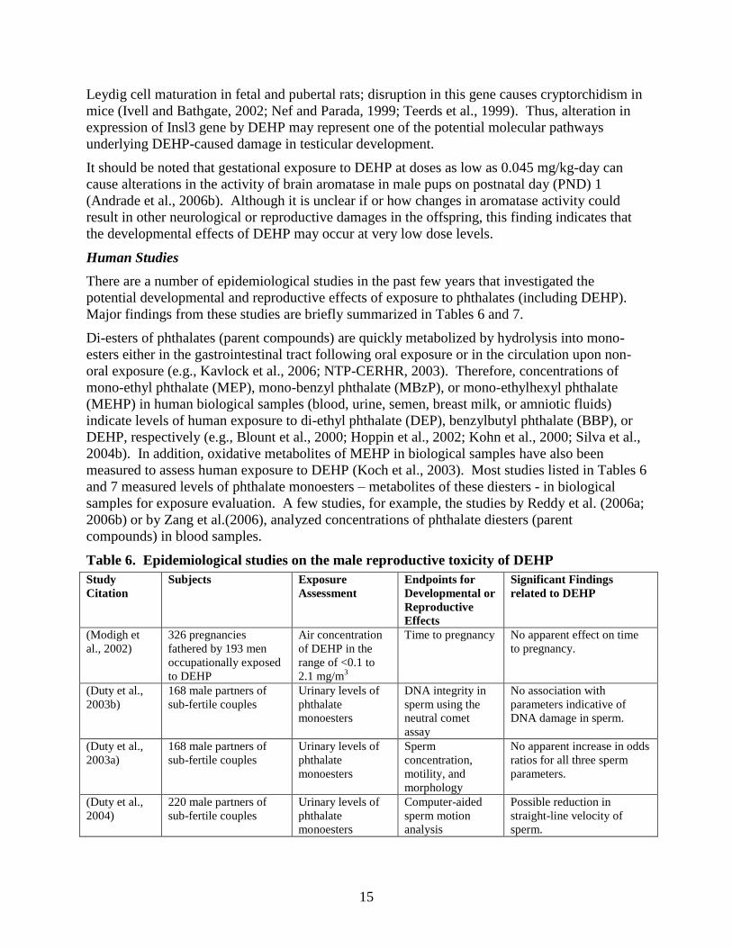
Table 6. Epidemiological studies on the male reproductive toxicity of DEHP
Study
Citation
Subjects Exposure
Assessment
Endpoints for
Developmental or
Reproductive
Effects
Significant Findings
related to DEHP
(Modigh et
al., 2002)
326 pregnancies
fathered by 193 men
occupationally exposed
to DEHP
Air concentration
of DEHP in the
range of <0.1 to
2.1 mg/m3
Time to pregnancy No apparent effect on time
to pregnancy.
(Duty et al.,
2003b)
168 male partners of
sub-fertile couples
Urinary levels of
phthalate
monoesters
DNA integrity in
sperm using the
neutral comet
assay
No association with
parameters indicative of
DNA damage in sperm.
(Duty et al.,
2003a)
168 male partners of
sub-fertile couples
Urinary levels of
phthalate
monoesters
Sperm
concentration,
motility, and
morphology
No apparent increase in odds
ratios for all three sperm
parameters.
(Duty et al.,
2004)
220 male partners of
sub-fertile couples
Urinary levels of
phthalate
monoesters
Computer-aided
sperm motion
analysis
Possible reduction in
straight-line velocity of
sperm.

16
Study
Citation
Subjects Exposure
Assessment
Endpoints for
Developmental or
Reproductive
Effects
Significant Findings
related to DEHP
(Duty et al.,
2005a)
295 male volunteers
(18-54 years of age)
from the general
population.
Urinary levels of
phthalate
monoesters
Levels of
reproductive
hormones in blood
samples
No relevant association with
blood hormone levels.
(Hauser et al.,
2006)
463 male partners of
sub-fertile couples (168
of them were subjects
reported by Duty et al.,
2003b).
Urinary levels of
phthalate
monoesters and
MEHP oxidative
metabolites
Semen quality
(sperm
concentration,
motility, and
morphology)
No apparent association
between levels of DEHP
metabolites and any sperm
parameter.
(Hauser et al.,
2007)
379 male partners of
sub-fertile couples
Urinary levels of
phthalate
monoesters &
MEHP oxidative
metabolites
DNA integrity in
sperm using the
neutral comet
assay
Significant association with
comet assay parameters
indicative of DNA damage
in sperm.
(Pan et al.,
2006)
74 male workers
occupationally exposed
to DBP and DEHP and
63 controls in China
Urinary levels of
MBP and MEHP
Blood levels FSH,
LH, testosterone,
and estradiol.
Decreased level of free
testosterone in the exposed
group.
(Jonsson et
al., 2005)
234 men 18-21 years of
age from military
recruits in Sweden
Urinary levels of
phthalate
monoesters
including MEH
Semen quality, and
blood levels of
reproductive
hormones
No clear pattern of
association with male
reproductive toxicity
endpoints evaluated.
(Main et al.,
2006)
Nursing mothers of 62
boys with
cryptorchidism and of
68 healthy boys as
controls.
Levels of
monoesters of
phthalates in breast
milks from nursing
mothers.
Blood levels of
testosterone, FSH,
LH, SHBG,
inhibin B from all
boys at about 3
months of age.
No apparent association of
MEHP level in breast milk
with any hormone endpoint.
(Zhang et al.,
2006)
52 male patients
attending a family
planning clinic in
Shanghai, China
Levels of DEP,
DBP, and DEHP
in seminal plasma.
Semen quality No apparent association
between DEHP levels and
semen parameters.
Table 7. Epidemiological studies on the developmental and female reproductive toxicity of
DEHP
Study Citation Subjects Exposure
Assessment
Endpoints for
Developmental or
Reproductive
Effects
Significant Findings
related to DEHP
(Colon et al.,
2000)
41 Puerto Rican girls
with premature breast
development and 35
controls
Levels of DBP,
DEP, BBP, DEHP,
and MEHP in
blood samples
Case-control study
to compare levels
of phthalates
between the two
groups.
High levels of phthalates
including DEHP and
MEHP in thelarche
patients
(Latini et al.,
2003)
82 pregnancies with
live newborns
DEHP and MEHP
concentrations in
the cord blood
samples
Birth weights,
gestational age
Significantly lower
gestational age among
MEHP-positive
newborns than that in
MEHP-negative ones.

17
Study Citation Subjects Exposure
Assessment
Endpoints for
Developmental or
Reproductive
Effects
Significant Findings
related to DEHP
(Reddy et al.,
2006b)
85 infertile female
patients with
endometriosis and
135 controls with
proven fertility
Levels of DBP,
BBP, DEHP, Di-
noctyl phthalate
and
polychlorinated
biphenyls (PCBs)
in blood samples
Case-control
study to compare
levels of phthalates
and PCBs between
the case and the
control group.
Significantly increased
levels of all phthalates
and PCBs in female
patients with
endometriosis
(Reddy et al.,
2006a)
49 infertile female
patients with
endometriosis as
cases; 38 infertile
female patients
without
endometriosis as
control group I and
21 with proven
fertility as control
group II
Levels of DBP,
BBP, DEHP,
DiOP in blood
samples
Case-control study
to compare levels
of phthalates
between the case
and control groups.
Significantly increased
levels of all phthalates
measured in the case
group as compared to the
control groups.
(Qiao et al.,
2007)
110 precocious and
100 normal girls
Concentrations of
DBP and DEHP in
blood samples
Case-control study
to compare levels
of phthalates,
volume of uterus
and ovaries
between the two
control groups.
Precocious girls have
higher concentrations of
DBP, DEHP, and larger
volumes of the uterus and
ovary.
(Swan et al.,
2005)
134 boys 2-36
months of age
Maternal urinary
levels of phthalate
metabolites
including MEHP
and its oxidative
metabolites during
pregnancy
Anogenital
distance measured
at 15.9 months of
age (mean)
No apparent association
between levels of DEHP
metabolites and
alterations in the
anogenital distance.
MMP: mono-methyl phthalate; MiNP: mono-iso-nonyl phthalate; DiOP: Di-iso-octyl phthalate; FSH: follicle-
stimulating hormone; SHBG: Sex hormone binding globulin; LH: Luteinizing hormone.
Epidemiological Studies of Male Reproductive Endpoints
A few studies listed in Table 6 indicate possible associations between elevated exposure to
DEHP and abnormal changes in endpoints indicative of male reproductive functions, though no
such association was observed in other studies.
Modigh et al. (2002) found no effect of DEHP at a mean exposure level of <0.5 mg/m3 on time
to pregnancy among partners of 193 men who were occupationally exposed to DEHP in air at
three plants either producing DEHP or processing polyvinyl chloride (PVC) plastic.
A series of studies reported by Duty et al. (2004; 2005b; 2003a; 2003b) and Hauser et al. (2006),
respectively, investigated the relationship between urinary levels of phthalate monoesters and
semen quality or blood sex hormone levels among male partners of subfertile couples presented
to an andrology laboratory the Massachusetts General Hospital in Boston. The authors found no
association between urinary levels of MEHP or other phthalate monoesters and sperm DNA

18
damage (Duty et al., 2003a) or blood sex hormone levels (Duty et al., 2005b). However, there
was a significant association between parameters indicative of sperm DNA damage and MEHP
or its oxidative metabolites in the recent study that included a larger study population and
measurement of multiple oxidative metabolites of DEHP (Hauser et al. 2007; 2008). A possible
reduction in the straight-line velocity of sperm among male partners with increased urinary level
of MEHP was observed in the 2004 study by Duty et al (2004). No other apparent changes in
parameters for semen quality or reproductive hormone levels were observed in the studies by
Duty et al. or Hauser et al.
A significantly decreased blood level of free testosterone has been observed in a cross-sectional
study among workers occupationally exposed to high levels of di-n-butyl phthalate (DBP) and
DEHP in China (Pan et al., 2006). In this study, urinary concentrations of mono-n-butyl
phthalate (MBP) and MEHP and levels of reproductive hormones were measured among 74 male
workers at a factory producing PVC flooring products (exposed group) and 63 workers working
in a construction company as controls. The two groups were matched to age (mean age 33.9
years), history of cigarette-smoking (60-62 percent smokers in both groups), and alcohol
consumption (54-64 percent consumers in both groups). The exposed group had one year (mean,
SD = 0.8) of employment time at the factory surveyed, suggesting that the exposed workers had
a limited time of exposure. Urinary concentrations of MBP and MEHP in exposed workers were
significantly higher than those in the control group (644.3 and 565.7 µg/g creatinine in the
exposed group vs. 129.6 and 5.7 µg/g creatinine in the control group for MBP and MEHP,
respectively, geometric means). The median concentrations of MBP and MEHP in the control
group were about two to seven fold higher than those in the general population in the U.S. (Silva
et al., 2004a), but were similar to those observed in German males (Koch et al., 2003). The
mean concentration of free testosterone in exposed workers was significantly lower than that of
the controls (8.4±1.5 vs. 9.7±1.4, log10-transformed blood levels of free testosterone: actual
concentrations of reproductive hormones were not reported). The authors reported no significant
difference in blood levels of other reproductive hormones, follicle-stimulating hormone (FSH),
luteinizing hormone (LH), and estradiol, between the control and exposed groups. Statistical
correlation and regression analysis conducted by the authors found that both MBP and MEHP
were responsible for the decreased level of free testosterone in blood. Thus, data from this study
clearly show that occupational exposure to DBP and DEHP is associated with decreased levels of
free testosterone, indicative of male reproductive toxicity of DBP and DEHP in men. However,
the data presented by the authors are not sufficient to quantitatively distinguish among the effects
attributable to DBP and those attributable to concurrent DEHP exposure, or to interaction
between the two phthalates.
Jonsson et al. (2005) found no association between urinary levels of MBP, MBzP, or MEHP and
parameters for semen quality or reproductive hormone levels among 234 Swedish men 18-21
years of age. All the subjects in this study were participants in the conscript examination before
military service in Sweden. Each subject received a comprehensive evaluation of parameters for
semen quality (e.g., semen volume, sperm count, motility, morphology, etc.). Serum levels of
reproductive hormones (FSH, LH, testosterone, sex hormone-binding globulin (SHBG),
estradiol) and urinary levels of phthalate metabolites (MEP, MBP, MBzP, MEHP and phthalic
acid) were analyzed. Urinary levels of MBP in this study were in the same order of magnitude
as those previously reported in the U.S. populations (e.g., Blount et al., 2000). The reason(s) for
the inconsistent findings between this study and those reported by Duty et al. (2003b) and Hauser
et al. (2006) is unclear. Jonsson et al. (2005) noted that the subjects in their study were from the

19
general population in Sweden and were healthy young (18-21 years old) males, whereas the
studies by Duty et al. (2003a; 2003b) or Hauser et al (2006) were based on male partners of
subfertile couples with a wide range of age (20-54 years). Thus, the U.S. studies may have been
more sensitive in design. Because of increased age and possible poor status of spermatogenesis,
subjects in the U.S. studies may have been more susceptible to the adverse effects of phthalates
than those who participated in the Swedish study.
Two epidemiological studies investigated potential male reproductive effects of phthalates in
boys who were exposed to phthalates through their pregnant or nursing mothers (Main et al.,
2006, respectively; Swan et al., 2005). Major findings from the study by Swan et al. (2005) are
discussed below in the subsections on developmental effects of DEHP. In the study by Main et
al. (2006), the authors evaluated the association between phthalate levels in breast milk of
nursing mothers and blood levels of reproductive hormones among 62 boys (29 from Denmark
and 33 from Finland) with cryptorchidism, and 68 healthy boys without cryptorchidism (36 from
Denmark and 32 from Finland) as controls. Aliquots of breast milk samples were collected and
pooled by each nursing mother between one and three months after birth. Blood samples from
all the boys were collected at 3 months of age. Concentrations of six monoesters of phthalates,
including monomethyl phthalate (MMP), MEP, MBP, MBzP, MEHP, and mono-isononyl
phthalate (MiNP) were measured in breast milk samples. Milk samples from Finland showed
significantly higher values for MBP, MBzP, and MEHP than were present in samples from
Denmark. The authors found no significant difference in any phthalate monoester concentration
in the mothers’ breast milk between children with or without cryptorchidism. However,
significant negative effects on milk quality, whether measured directly or reflected in impaired
development of young, is generally considered an adverse female reproductive effect, (e.g., U. S.
Environmental Protection Agency Guidelines for Reproductive Toxicity Risk Assessment (U.S.
EPA, 1996)). Thus, this study by Main et al. (2006) provided evidence on both the male and
female reproductive effects of DEHP in humans.
Epidemiological Studies of Female Reproductive Endpoints
In addition to the study by Main et al. (2006) as discussed above, there are a few studies
providing limited evidence on the female reproductive toxicity of DEHP in women.
Colon et al. (2000) found higher levels of phthalates, including DEHP and MEHP in 41 Puerto
Rican girls who had premature breast development (thelarche patients). Similar findings have
also been reported by Qiao et al. (2007) among 110 precocious Chinese girls in Shanghai.
Latini et al. (2003) investigated the possible association of concentrations of DEHP and MEHP,
in the cord blood of 84 newborns to birth outcomes including weight, gestational age, and other
endpoints. All 84 newborns were born at a general-practice hospital in Italy; there were 82
singleton births, one set of twins and 39 male and 45 female offspring. Eleven were preterm, and
three of them had very low birth weight. The maternal age range was from 18 to 24 years. The
authors found that DEHP or MEHP were present in 74 (88.1 percent) of the 84 examined cord
serum samples at mean concentrations of 1.19 1.15 μg/ml and 0.52 0.61 μg/ml (mean standard
deviation), respectively. MEHP-positive newborns (65 or 77.4 percent) had a significantly lower
gestational age (38.16 2.34 weeks) compared with MEHP-negative infants (39.35 1.35 weeks,
p<0.05). Logistic regression analysis also indicated a positive correlation between absence of
MEHP in cord blood and gestational age at delivery (odds ratio = 1.50, 95 percent confidence
interval = 1.013-2.21). No other statistically significant relationships were observed between

20
DEHP or MEHP concentrations and other birth outcomes (e.g., birth weight). The authors
concluded that phthalate exposure is significantly associated with shortened pregnancy duration.
Altered gestation length associated with maternal exposure to a chemical can be an indicator of
female reproductive toxicity (U.S. EPA, 1996).
Two study reports by Reddy et al. (2006a; 2006b) also reported possible associations between
exposure to DBP and female reproductive effects. In these two studies, Reddy et al. (2006a;
2006b) measured blood levels of polychlorinated biphenols (PCBs) and/or phthalate di-esters in
female patients with endometriosis (cases) or without endometriosis (controls) from a south
Indian region. It is unclear if there is an overlap between the subjects in two studies, which were
reported separately. The authors of both studies found that blood levels of all the compounds
measured in endometriosis patients, including four PCB congeners, DBP, BBP, DEHP, and
dioctyl phthalate (DOP), were significantly higher than those in controls (p<0.05). The authors
concluded that phthalates may have an etiological association with endometriosis in women.
However, it has been shown that exposure to PCBs is associated with endometriosis in
laboratory animals and in women (e.g., Arnold et al., 1996; Louis et al., 2005; Rier, 2002); high
concentrations of DBP have also been found in women with endometriosis (Cobellis et al.,
2003). Because the authors did not control for this confounding factor, the data from this study
are not sufficient to determine if or how much DEHP per se contributes to development of
endometriosis in women.
Epidemiological Studies of Developmental Endpoints
The study by Swan et al. is the only epidemiological study that provided relevant data on the
developmental effects of DEHP in humans. In this study, Swan et al. (2005) analyzed levels of
MEHP and eight other phthalate monoesters in urine samples from 85 pregnant women at a
mean gestational time of 28.3 weeks. The authors also performed genital examinations and
measured anogenital distance (AGD) among a total of 134 boys at 2-30 months of age. Data
from boys whose mother’s urine samples had been analyzed for phthalate levels were included in
the statistical regression analysis. The authors found that increased levels of MEP, MBP, MBzP,
and mono-isobutyl phthalate in prenatal urine samples in mothers was associated with decreased
AGD in boys after birth. However, there was no apparent association between MEHP levels and
the AGD.
Summary
The male reproductive toxicity of DEHP has been extensively studied in laboratory animals
including rats, mice, hamsters, and ferrets. Depending on the dose, duration of exposure, and
age of animals, DEHP causes reduced fertility, decreased weights of male reproductive organs,
and histopathological changes in the testes of juvenile and adult rats. Although it has not been
studied as extensively, female reproductive toxicity in laboratory animals has been reported.
DEHP also has been found to cause developmental toxicity, including intrauterine death,
developmental delay, and structural malformations and variations; neurological developmental
effects have also been reported. Evidence of DEHP’s reproductive toxicity in humans is less
conclusive. Concordant effects of exposure to DEHP may be anticipated in marine organisms,
particularly mammalian species.

21
Cancer
Introduction
In 2001, the International Agency for Research on Cancer (IARC) changed its classification of
DEHP from Group 2B (sufficient evidence for carcinogenicity to laboratory animals) to Group C
(not classifiable as to its carcinogenicity to humans). This change was based on agreement that
the mechanism of DEHP carcinogenesis in laboratory animals involves peroxisome proliferation
and that this mechanism is not likely to occur when humans are exposed to DEHP. Several
letters and articles expressing disagreement with the evaluation of IARC have been published
(Brody et al., 2003; Huff, 2003; Melnick, 2001).
Laboratory Rodent Studies
Chronic exposure of mice or rats to DEHP produces carcinomas of the liver and testicular tumors
(Fay et al., 1999). DEHP also is a promoter of liver carcinomas when given to rats following
exposure to the tumor initiator diethylnitrosamine (Sano et al., 1999).
Studies published since 2000 are consistent with earlier studies demonstrating that DEHP is
carcinogenic to rodents. Male Sprague-Dawley rats treated with 300 mg DEHP per kg body
weight per day for until death (up to 159 weeks) developed tumors of the liver and testes
(Toyosawa et al., 2001). DEHP exposure produced liver tumors in transgenic mice carrying the
human protooncogene c-Ha-ras (Toyosawa et al., 2001) in a 26 week carcinogenicity test.
Effects on Cultured Mammalian Cells
DEHP caused an increase in cell proliferation and a decrease in nuclear fragmentation, a sign of
apoptosis, in cultured mouse Sertoli cells (Kang et al., 2002). DEHP transformed mouse embryo
fibroblast T1 cells carrying bovine papillomavirus DNA (Kowalski et al., 2000).
Summary
There is sufficient evidence to conclude DEHP causes cancer in rats. The evidence suggest it is
caused by the formation of peroxisomes in the rat liver from exposure to DEHP. Since humans
are unlikely to form peroxisomes from DEHP exposure, it is unlikely humans will get cancer
from DEHP, although there is still debate on this point.
Obesity
Stemp-Morlock (2007) observed that the obesity rate has greatly increased over the past 20
years. An estimated one-third of U.S. adults are overweight and more than one-third of U.S.
children are overweight or at risk for being overweight. There is a strong association between
obesity and a number of health issues such as diabetes, coronary heart disease, hypertension, and
gall bladder disease. Traditionally, obesity has been viewed as a result of reduced physical
activities and increased caloric intake. Data from recent studies, however, suggest that exposure
to chemicals that perturb the critical pathways in adipogenesis, lipid metabolism or energy
balance could also initiate or exacerbate obesity. Phthalates were suggested as chemicals that
might possess obesogenic properties (Grun and Blumberg, 2007).
OEHHA’s literature search did not turn up specific information on DEHP with regard to obesity.
However, MEHP was shown to activate peroxisome proliferators-activated receptor-γ (PPAR-γ)
in adipocyte differentiation of 3T3-L1 preadipocytes (Feige et al., 2007). The role of PPAR-γ in
lipogenesis and adipose differentiation is well established (Kersten, 2002). Thus, DEHP via

22
metabolic activation could cause obesity. A low dose of DEHP (7.5 mg /kg) on alternate days
for 14 days caused a decrease in serum insulin and cortisol and an increase in serum glucose
(Gayathri et al., 2004). Incubation of Chang liver cells with 200 or 400 micromoles DEHP per
liter for 24 hours resulted in a decrease in insulin receptor activity and a decrease in glucose
oxidation (Rengarajan et al., 2007).
Other circumstantial evidence also suggests that additional research should be performed to
further characterize the obesogenic properties of DEHP. DEHP is also known to be anti-
androgenic. Androgen deprivation in prostate cancer therapy resulted in a higher incidence of
hyperlipidemia, hyperglycemia, and metabolic syndrome (Braga-Basaria et al., 2006; Sharifi et
al., 2005). Stahlhut et al. (2007) observed an association between urinary phthalate metabolites
to increased waist circumference and insulin resistance. This led them to suggest an obesogenic
outcome via the anti-androgenic pathway.
Thyroid
Introduction
A limited number of animal studies indicate that exposure to DEHP may be associated with
altered thyroid function. Thyroid hormones such as thyroxine (T4) and triiodothyronine (T3)
play a key role in many physiological events and are important for normal brain development.
Alterations in thyroid hormone levels and thyroid function may lead to adverse clinical
conditions. Environmental contaminants such as phthalates may bind to thyroid hormone
receptors and alter its signaling (Zoeller, 2005).
Laboratory Rodent Studies
Thyroid histopathology
DEHP-induced thyroid hyperactivity has been shown in some animal studies. Rats were given
DEHP in the diet over periods from 3 days to 9 months. DEHP induced hyperactivity and
changes in the colloid in thyroid. Rats treated with DEHP showed ultrastructural changes in the
thyroid, such as increase in the number and size of lysosomes, enlarged Golgi apparatus, dilation
of the rough endoplasmic reticulum, and damaged mitochondria (Hinton et al., 1986; Price et al.,
1988b). In a separate study, male rats were given DEHP in diet for 14 days. The thyroid of rats
receiving diet containing DEHP showed signs for hyperactivity, as indicated by the decrease in
follicular size and increase in the proportion of follicular cells with a columnar appearance
(Howarth et al., 2001). The subchronic oral toxicity of DEHP was also studied. DEHP was
given to rats in the diet for 13 weeks. DEHP again altered thyroid histopathology. Histological
changes in the thyroid include reduced follicle size and decreased colloid density (Poon et al.,
1997).
Thyroid function
DEHP was given to rats on alternate days for 14 days. Changes in the levels of T3, T4 and
thyroid stimulating hormone (TSH) in the serum were studied. Increases in serum T3 and T4
were seen in rats exposed to DEHP, while there was no change in TSH. Thus, the results
indicated that low levels of DEHP exposure in rats may alter thyroid function (Gayathri et al.,
2004). Iodide uptake is also important for the normal function of thyroid. Effects of DEHP on
iodide uptake were studied in vitro in the rat thyroid cell line FRTL-5. The results showed that
DEHP significantly enhanced iodide uptake at the concentrations that do not cause toxicity to the

23
cells. The key molecule in iodide transportation is the sodium/iodide symporter (NIS).
Additional experiments demonstrated that the increase in iodide uptake is due to NIS (Wenzel et
al., 2005). A separate study in a rat thyroid cell line PC C13 cells showed increased
transcriptional activity of NIS induced by DEHP. This led the authors to suggest that the
increase in iodide uptake in thyroid is a consequence of NIS transcriptional activation by DEHP
(Breous et al., 2005).
Human Studies
Human studies investigating the association between DEHP exposure and thyroid function are
limited. A decrease in serum levels of thyroid hormones such as T4 and T3 were observed in
human blood stored in DEHP plasticized blood bags (Gayathri et al., 2004). Meeker et al. (2007)
studied the potential association between DEHP exposure and serum levels of free T4, total T3,
and TSH in adult men. Samples of urine and blood were collected from 408 men. Meeker et al.
measured urinary concentrations of MEHP along with serum levels of T4, T3, and TSH. An
inverse association between MEHP urinary concentrations and free T4 and T3 serum levels were
observed. The results thus indicated that urinary MEHP concentrations may be associated with
altered T4 and/or T3 levels in adult men. The authors indicated in the paper that the associations
between MEHP and thyroid hormones observed in this human study are at certain levels
consistent with animal studies, and the urinary MEHP concentrations are comparable with those
found in the general human population in the United States (Meeker et al., 2007).
Summary
Thyroid as one of the largest endocrine glands in the body plays an important role in maintaining
the homeostasis and normal function of many organs and systems. Only a limited number of
studies are available to demonstrate the potential effects of DEHP on thyroid, but they do show
effects on the morphology and function of the gland. Considering the importance of the thyroid
in regulating the normal physiology of multiple organs and systems, the evidence should not be
ignored and further studies are necessary.
Immune System
Introduction
The prevalence of allergic diseases has been increasing dramatically for the past several decades.
The exposure to environmental contaminants may contribute to the increased sensitivity of the
human immune system, leading to elevated allergic responses. Alteration of immune function
induced by environmental contaminants may result in hypersensitivity, autoimmune diseases,
and changes in the ability to resist infectious diseases. Despite the fact that the number of people
who had been diagnosed with autoimmune or allergic diseases is on rise, the etiology still
remains unknown for most of these diseases. Both inherent and environmental factors may be
involved in the development of the disease. Foreign antigens may act as an immune modulator
to initiate autoimmune responses and cause allergy-related diseases such as asthma. There are
concerns that DEHP may compromise the immune system; however, limited studies are
available to elucidate the potential mechanism of the immunological effects of DEHP.
24
Laboratory Rodent Studies
The association between DEHP and immunological disorders was also studied in laboratory
animals. Mice were injected intraperitoneally with DEHP three times at 10-day intervals. High
levels of serum anti-DNA antibodies were developed in mice injected with DEHP in a dose-
dependent manner. This led the authors to suggest that DEHP might be involved in the induction
of autoimmune response (Lim and Ghosh, 2005). Takano et al. (2006) studies the effects of
DEHP exposure on the allergic reactions in mice to a dust mite allergen. Seven-week-old mice
were exposed to mite allergen and also received DEHP in olive oil by intraperitoneal injection.
DEHP enhances atopic dermatitis-like skin lesions induced by mite allergen. These changes
were consistent with the effects of DEHP on the protein expression of chemokines macrophage
inflammatory protein-1α (MIP-1α) and eotaxin (Takano et al., 2006). Male mice fed DEHP in
the diet had lower levels of lactoferrin, a protein with antimicrobial activity, in liver tissue than
did control mice (Hasmall et al., 2002). DEHP was given to 4-week-old mice in drinking water
for 2 weeks. DEHP has been shown to enhance the proliferative responses of thymus cells as
indicated by increased responses to mitogens concanavalin A (Con A) and Staphylococcus
enterotoxin B (SEB) (Yamashita et al., 2003). The thymus and spleen are important organs for
the normal function of immune system and they are also highly sensitive to xenobiotics. In a
separate study, atrophy of thymus and spleen was seen in mice exposed to DEHP. Yang et al.
(2000) treated adult male mice (8 weeks old) with DEHP in diet for 10 days. Decreased thymus
and spleen weight, decreased DNA content in thymus and spleen were observed in DEHP-treated
mice (Yang et al., 2000). The effects of DEHP on immune system were also studied in rats.
Young male albino rats were given DEHP by intraperitoneal injection for up to 90 days (long-
term exposure), or for 5 days (short-term exposure). In the long-term exposure groups, depletion
of cortical cells and hyperplasia of medulla in the thymus, cellular depletion of peripheral
follicles of white pulp in the spleen, and reduction in the number of follicles in cortical region of
lymph nodes were observed in DEHP-treated rats. In the short duration exposure groups, DEHP
caused dose-dependent reduction in the weight of lymphoid organs and their cellular population,
the spleen cell response to B-lymphocyte mitogen lipopolysaccharide is also reduced in DEHP-
treated rats, which led the authors to suggest that DEHP effect on the B-lymphocyte population
is how it modulates the immune system (Dogra et al., 1985). These animal studies indicate that
DEHP may have immunomodulating effects in vivo.
Inhalation of MEHP for 60 minutes by BALB/c mice resulted in an increase in the number of
pulmonary macrophages. The NOEL for this effect was estimated to be 1.7 mg per cubic meter
(Larsen et al., 2004). Inhalation exposure of BALB/c mice to a mixture of ovalbumin and DEHP
did not result in an increase in IgE (immunoglobulin E) production above the amount produced
in response to ovalbumin alone. However, DEHP exposure did increase the number of
macrophages in bronchi (Larsen et al., 2004).
In vitro Studies
The effects of DEHP on immune responses were also studied in vitro DEHP and MEHP
produced a significant increase in hydrogen peroxide production in isolated rat hepatocytes
(Kambia et al., 2004). MEHP reduced the production of inflammatory cytokines IL6 and IL8 in
A549 cells, a human epithelial cell line (Jepsen et al., 2004). DEHP has been shown to trigger
allergic responses by enhancement of cytokine interleukin-4 (IL-4) production in CD4+ T cells
in a concentration-dependent manner (Lee et al., 2004). In a separate study, blood leukocytes
from healthy volunteers were studied in vitro for the effects of DEHP and its metabolites MEHP,
25
2-ethylhexanol (2-EH), and phthalic acid (PA) on immune functions. The suppressed oxidative
respiratory metabolism and decreased superoxide (O2-) generation was observed in leukocytes
that were incubated with DEHP or its metabolites. The impairment of leukocyte oxidative
metabolism may imply the potential inhibition of leukocyte function by DEHP and its
metabolites (Fischer et al., 1998). The effects of DEHP on murine thymus and spleen cells were
also studied in vitro. DEHP has been shown to induce proliferative responses and enhance
cytokine (a group of inflammatory cell proteins) production in murine thymus and spleen cells
(Yamashita et al., 2002) (Yamashita et al., 2003). The findings of these studies raise concerns
regarding the possibility of DEHP to alter immune system and its potential role in
immunological disorders.
Human Studies
The potential link between DEHP and allergic diseases has been studied in humans. The use of
PVC plastics and other plasticizer-containing surface materials in the home was associated with
the development of bronchial obstruction in young children in Norway (Jaakkola et al., 1999).
The presence of plastic materials was associated with increased risks of asthma and pneumonia
and higher incidence of lower respiratory tract symptoms such as wheezing, cough, and phlegm
in children studied in Finland (Jaakkola et al., 2000). Another study from Finland also linked
plastic materials with the risk of asthma (Kavlock et al., 2006). The concentration of DEHP in
indoor dust was associated with asthma in children in Sweden (Bornehag et al., 2005). The
concentration of DEHP in indoor dust was also shown to be associated with allergic symptoms
such as wheezing in preschool children from children’s homes in Bulgaria (Kolarik et al., 2008).
Summary
Limited studies in humans and laboratory animals raised concerns regarding the potential link
between DEHP and immunological disorders. Despite the limitation of available studies,
attention should be made to the potential effects of DEHP on immune system and its
involvement in immunological disorders.
Nervous System
Introduction
The central nervous system and brain function has been demonstrated to be a target of many
environmental contaminants. Rodent animals as a model system have been heavily used for the
research on the nervous system. The following paragraphs will discuss the effects of DEHP on
the central nervous system based on currently available studies.
Laboratory Rodent Studies
The effects of DEHP on the central nervous system and brain function have been studied in
laboratory animals. Rodent studies showed behavioral changes caused by DEHP exposure
during development. For example, Tanaka (2002) studied neurobehavioral effects of DEHP in
male and female mice offspring following in utero and lactational DEHP exposures. DEHP was
given in the diet. The dose level used in the study did not produce significant adverse effects in
reproductive and neurobehavioral parameters examined. However, surface righting, an indicator
of the development of coordinated movement, was significantly depressed in both male and
female offspring during postnatal development (Tanaka, 2002). DEHP has also been shown to
cause motor hyperactivities in rats. Masuo et al. (2004a; 2004b) exposed 5-day-old rats with
26
DEHP in olive oil by intracisternal injection. The brain cell development such as differentiation
and synaptogenesis are still incomplete in rats at 5 days of age, which makes them more
vulnerable to environmental pollutants such as endocrine disrupters. The neonatal exposure is
thus useful in understanding the neurodevelopmental effects of DEHP. The results showed that
DEHP caused a significant increase in spontaneous motor activities at 4-5 weeks of age, a
juvenile stage. It is worth mentioning that the motor behavior was studied during a 24-hour
period under a 12-hour light (day) and 12-hour dark (night) condition. DEHP caused
hyperactivity in both the dark and light phases. This prompted the authors to suggest that
endocrine disrupters such as DEHP may cause deficits in sleep. These studies indicated that
DEHP can induce motor hyperactivity and other behavioral changes in laboratory animals.
Motor hyperactivity is associated with developmental disorders in humans, such as autism and
attention-deficit hyperactivity disorder (ADHD). To study the mechanisms underlying motor
hyperactivity, Masuo et al. also conducted gene-expression profiling using a cDNA
(complimentary DNA) membrane array in DEHP-treated rats. In order to understand the long-
lasting effects of neonatal exposure, the gene expression was evaluated at 8 weeks of age, long
after the cessation of DEHP exposure. The results demonstrated alteration of expression of
genes in the midbrain and striatum, which include those related to neuropeptides, growth factors,
and the transmission of important neurotransmitters such as dopamine, γ-aminobutyric acid
(GABA), and glutamate. These results suggest that further studies are necessary in order to
elucidate the mechanisms of the neurological effects of DEHP (Masuo et al., 2004a; 2004b).
G protein-coupled receptors
DEHP has been shown to alter gene expression of G-protein-coupled receptors in rat brain.
Ishido et al. (2005) conducted a study in rats. Single intracisternal administration of DEHP into
male Wistar rats at 5 days of age induced significant hyperactivity at 4-5 weeks of age. DNA
macroarray analyses were conducted in the midbrain at 8 weeks of age. DEHP changed the
levels of gene expression of G protein-coupled receptors that were important for the transmission
of dopamine and other neurotransmitters. For example, the gene expression of dopamine
receptor D1A was decreased by DEHP. The expression of galanin receptor 2 was also reduced by
DEHP. Dopamine and its transporters might be associated with the pathogenesis of ADHD.
Galanin, as a neuromodulator, plays an important role in cognition, learning and memory.
Galanin can also regulate midbrain dopamine activity (Ishido et al., 2005).
The membrane Na+-K
+ ATPase
The membrane Na+-K
+ adenosine triphosphatase (ATPase) of the brain plays an important role in
brain development and function, a decrease in this enzyme activity has been associated with a
number of neurodegenerative and psychiatric diseases. Dhanya et al. (2004; 2003) studied the
effects of DEHP on the activity of membrane Na+-K
+ ATPase in brain. DEHP was given to rats
by intraperitoneal injection, the histopathology of the brain and the activity of membrane Na+-K
+
ATPase in brain were studied. Histopathology examination showed neuronal degeneration in
rats exposed to DEHP. Significant inhibition of membrane Na+-K
+ ATPase was also observed in
the brain. Additional experiments demonstrated that the inhibition of membrane Na+-K
+ ATPase
is a direct effect of DEHP or its metabolites (Dhanya et al., 2004; Dhanya et al., 2003). The
authors indicated in the paper that Na+-K
+ ATPase is an important enzyme for cation
transportation and inhibition of this enzyme may lead to an increase in intracellular calcium, and
thus cause disruption of ion homeostasis. The increased intracellular calcium may elicit
apoptosis and cell death, while apoptosis may be involved in neuronal degeneration. A study on

27
cultured cortical neurons showed that the inhibition of Na+-K
+ ATPase resulted in neuronal cell
death. Electron microscopy confirmed the existence of apoptosis and necrosis in individual
cells. The effects were considered to be mediated by the disruption of ion homeostasis such as
the accumulation of intracellular calcium (Xiao et al., 2002).
Intracellular calcium
Intracellular calcium has been shown to play an important role in the neurotransmitter release,
modulation of excitability and gene expression of the nerve cells (Berridge, 1998). The
alteration in intracellular calcium level may, in part, be involved in the pathogenesis of a number
of neurological disorders. Studies have been conducted with a focus on the effects of DEHP on
intracellular calcium. DEHP has been shown to increase the intracellular calcium levels in
mammalian neurosecretory terminals and in cultured pheochromocytoma cells, a neuronotypic
cell line. Alterations in intracellular calcium in the nanomolar concentration range within
minutes of exposure led the authors to suggest the potential adverse effects in situations where
prolonged exposure occurs (Tully et al., 2000).
Protein kinase C (PKC)
Protein kinase C (PKC) is a family of enzymes that is considered to play a key role in signal
transduction in response to hormones or neurotransmitters. In vitro study showed that DEHP
inhibits the activity of PKC from rat brain in a concentration-dependent manner. The effect of
DEHP is noncompetitive and the mechanism of the inhibition may involve the interference with
the interaction between calcium ion and the regulatory domain of PKC (Shukla et al., 1989).
Brain aromatase enzyme
It has been suggested that DEHP may interfere with estrogen metabolism through inhibition of
aromatase enzyme activity. Brain aromatase enzyme controls the conversion of testosterone to
estradiol and plays an important role in the sexual differentiation of neural structures,
neuroendocrine functions, and sexual behaviors (Lephart, 1996). Andrade et al. (2006) studied
the effects of DEHP on brain aromatase activity of male and female rat offspring following in
utero and lactational DEHP exposures. Aromatase activity was examined in
hypothalamic/preoptic area (HPOA) from pups on two different developmental stages: perinatal
stage (postnatal day 1) and weanling stage (postnatal day 22). The results demonstrated that
DEHP alters brain aromatase activity in both male and female offspring following in utero and
lactational exposures, though males and females respond differently to DEHP (Andrade et al.,
2006a). Further study indicated that DEHP may exert its effects on aromatase at the
transcription level. A study showed that the decrease in aromatase mRNA (messenger RNA)
and protein can be induced by DEHP metabolite MEHP in cultured rat granulosa cells
(Lovekamp and Davis, 2001). Considering the importance of brain aromatase in the
development and function of the central nervous system, more attention should be given to the
alteration of aromatase induced by DEHP and its potential effect on neural development and
behavior.
Brain peroxisomes
Peroxisomes are cytoplasmic organelles that participate in the metabolism of fatty acids. In
mammals, over half of the peroxisomal enzymes are related to lipid metabolism. Peroxisomes
contain oxidative enzymes such as catalase, D-amino acid oxidase, etc. It may also play a role in
protecting cells from oxygen toxicity and peroxidation. The effects of DEHP on brain
28
peroxisomes were also studied. Female rats exposed to DEHP during lactation showed DEHP-
induced modifications of the peroxisomal enzymatic pattern in brain of both dams and
offsprings. Biochemical analysis showed alterations in the activities of catalase, D-amino acid
oxidase, palmitoyl-CoA oxidase. Electron microscopy showed high catalase-like
immunoreactivity in the cytosol of neurons (Cimini et al., 1994). In a separate study,
Peroxisomes were identified in the neurons of the cerebral cortex of lactating pups at 12 days of
age from DEHP fed mothers. Electron microscope study indicated that the diameters of
peroxisomes in the neurons and the number of neurons that contain peroxisomes are both
increased in brain of the DEHP-treated group. DEHP may therefore elicit peroxisomal
proliferation in developing neurons (Dabholkar, 1988).
Summary
Alterations in brain function and behavioral changes have been observed in rodent studies.
DEHP has also been shown to alter gene and protein expression in the brain. The results thus
indicated that DEHP might have potential effects on neural development and behavior.
However, further studies are necessary in order to elucidate the mechanisms of the neurological
effects of DEHP.
Liver and Kidney Effects
Administration of 10,000 mg per kg body weight by gavage to male Wistar rats for four weeks
resulted in an increase in the relative weight of the liver, kidney, brain and adrenal glands
compared with the relative weights of these organs in controls not given DEHP (Dalgaard et al.,
2000). Male and female Fischer rats given 12,500 ppm DEHP in the diet for 104 weeks had
increased weights of the liver and kidneys, increased concentrations of urea and albumin in
blood but decreased concentrations of erythrocytes and hemoglobin compared to control rats
(David et al., 2000b). Male Fischer 344 rats fed a diet containing DEHP for 360 days had
decreased concentrations of ceruloplasm in blood and had increased concentrations of copper
deposited in liver tissue (Eagon et al., 1999). Examination of livers of prepubertal rabbits given
an intravenous infusion of lipid emulsion for one week revealed peroxisome proliferation when
the infusion line was PVC that contained DEHP. Peroxisome proliferation was not seen when a
polyethylene infusion line that did not contain DEHP was used (Loff et al., 2007). No
histological effects and no effects on organ weights were seen in the liver, kidneys or testes of
adult male cynomolgus monkeys given 500 mg per kg DEHP for 14 days (Pugh et al., 2000).
Conclusions
Findings
This toxicological profile on DEHP describes its effects on freshwater and marine life, humans,
and laboratory animals.
Arthropods appear to be the most sensitive, with several reports of effects in the low or
fractional µg/L range. Some fish, especially Medaka are also sensitive, with effects
reported in the low µg/L range. Mollusks appear to be less sensitive with effects reported
in the tens to hundreds of µg/L range. The relationship of the toxic concentrations to the
solubility limit is unclear, since published solubility limits for DEHP vary widely. These
disparate and conflicting findings indicate the need for further work to clarify the risk of

29
adverse effects on invertebrates at realistic environmental concentrations. More work
should focus on free-living organisms in marine and estuarine environments.
The male reproductive toxicity of DEHP has been extensively studied in laboratory
animals including rats, mice, hamsters, and ferrets. DEHP causes reduced fertility,
decreased weights of male reproductive organs, and histopathological changes in the
testes of juvenile and adult rats. Female reproductive toxicity in laboratory animals has
been reported. DEHP also has been found to cause developmental toxicity. Evidence of
DEHP’s reproductive toxicity in humans is less conclusive
There is sufficient evidence to conclude DEHP causes cancer in rats and mice, but
evidence suggest that the same mechanism of action does not occur in humans.
However, there is still debate on whether the mechanism of action is the only important
one..
Only a limited number of studies are available to demonstrate the potential effects of
DEHP on thyroid, but they do show effects on the morphology and function of the gland.
Considering the importance of the thyroid in regulating the normal physiology of
multiple organs and systems, the evidence should not be ignored and further studies are
necessary.
Limited studies in humans and laboratory animals raised concerns regarding the potential
link between DEHP and immunological disorders.
Alterations in brain function and behavioral changes have been observed in rodent
studies. The findings indicated that DEHP might have potential effects on neural
development and behavior. Further studies are necessary in order to elucidate the
mechanisms of the neurological effects of DEHP.
Data Gaps
There are very few studies on DEHP toxicity to marine and freshwater organisms and
even fewer outside the laboratory.
Only two studies were found that showed toxicity in marine organisms at or below the
level of DEHP’s water solubility.
There is little information on freshwater and marine DEHP concentrations in the water
column and sediment.
The current studies in laboratory animals on DEHP’s adverse effects other than
reproduction and development raise concern but are very limited.
DEHP is only one of many phthalates used in large quantities in plastics and more
information is needed about the others.
Recommendations
Further study needs to be done on the potential toxicity of DEHP and other common
phthalates on aquatic organisms. These need to investigate if low concentrations cause
toxicity over subchronic and chronic timeframes.

30
Studies on environmental concentrations of DEHP and other phthlates in the water
column and sediment need to be conducted.
Because of DEHP and other phthalates widespread use, further work needs to be done on
the adverse effects to the edorine, immune and nervous systems.

31
References
Ablake M., Itoh M., Terayama H., Hayashi S., Shoji S., Naito M., Takahashi K., Suna S. and
Jitsunari F. (2004) Di-(2-ethylhexyl) phthalate induces severe aspermatogenesis in mice,
however, subsequent antioxidant vitamins supplementation accelerates regeneration of
the seminiferous epithelium. Int J Androl 27, 274-81.
Akingbemi B. T., Ge R., Klinefelter G. R., Zirkin B. R. and Hardy M. P. (2004) Phthalate-
induced Leydig cell hyperplasia is associated with multiple endocrine disturbances. Proc
Natl Acad Sci U S A 101, 775-80.
Akingbemi B. T., Youker R. T., Sottas C. M., Ge R., Katz E., Klinefelter G. R., Zirkin B. R. and
Hardy M. P. (2001) Modulation of rat Leydig cell steroidogenic function by di(2-
ethylhexyl)phthalate. Biol Reprod 65, 1252-9.
Andrade A. J., Grande S. W., Talsness C. E., Gericke C., Grote K., Golombiewski A., Sterner-
Kock A. and Chahoud I. (2006a) A dose response study following in utero and lactational
exposure to di-(2-ethylhexyl) phthalate (DEHP): reproductive effects on adult male
offspring rats. Toxicology 228, 85-97.
Andrade A. J., Grande S. W., Talsness C. E., Grote K. and Chahoud I. (2006b) A dose-response
study following in utero and lactational exposure to di-(2-ethylhexyl)-phthalate (DEHP):
Non-monotonic dose-response and low dose effects on rat brain aromatase activity.
Toxicology 227, 185-92.
Arcadi F. A., Costa C., Imperatore C., Marchese A., Rapisarda A., Salemi M., Trimarchi G. R.
and Costa G. (1998) Oral toxicity of bis(2-ethylhexyl) phthalate during pregnancy and
suckling in the Long-Evans rat. Food Chem Toxicol 36, 963-70.
Arnold D. L., Nera E. A., Stapley R., Tolnai G., Claman P., Hayward S., Tryphonas H. and
Bryce F. (1996) Prevalence of endometriosis in rhesus (Macaca mulatta) monkeys
ingesting PCB (Aroclor 1254): review and evaluation. Fundam Appl Toxicol 31, 42-55.
ATSDR. (2002) Agency for Toxic Substances and Disease Registry: Toxicological profile for
Di(2-ethylhexyl)phthalate (DEHP). U.S. Department of Health and Human Services,
Public Health Services, Alanta, GA.
Banerjee S., Thuillier R., Culty M., Papadopoulos V., Brown T. R. and Banerjee P. P. (2002) In
utero exposure to di(2-ethylhexyl) phthalate alters growth, tissue organization, and the
expression of androgen receptor protein of rat prostate. Biol Reprod 66(Suppl 1), 200.
Barron M. G., Schultz I. R. and Hayton W. L. (1989) Presystemic branchial metabolism limits
di-2-ethylhexyl phthalate accumulation in fish. Toxicol Appl Pharmacol 98, 49-57.
Berridge M. J. (1998) Neuronal calcium signaling. Neuron 21, 13-26.
Blount B. C., Silva M. J., Caudill S. P., Needham L. L., Pirkle J. L., Sampson E. J., Lucier G.
W., Jackson R. J. and Brock J. W. (2000) Levels of seven urinary phthalate metabolites
in a human reference population. Environ Health Perspect 108, 979-82.
Boekelheide K. (2004) Cracking the nut. Toxicol Sci 81, 1-2.
Borch J., Ladefoged O., Hass U. and Vinggaard A. M. (2004) Steroidogenesis in fetal male rats
is reduced by DEHP and DINP, but endocrine effects of DEHP are not modulated by
DEHA in fetal, prepubertal and adult male rats. Reprod Toxicol 18, 53-61.
Bornehag C. G., Sundell J., Weschler C. J. and Sigsgaard T. (2005) Correspondence: potential
selection biases. Environ Health Perspect 113, a152-3.

32
Braga-Basaria M., Dobs A. S., Muller D. C., Carducci M. A., John M., Egan J. and Basaria S.
(2006) Metabolic syndrome in men with prostate cancer undergoing long-term androgen-
deprivation therapy. J Clin Oncol 24, 3979-83.
Breous E., Wenzel A. and Loos U. (2005) The promoter of the human sodium/iodide symporter
responds to certain phthalate plasticisers. Mol Cell Endocrinol 244, 75-8.
Brody C., DiGangi J., Easthope T., Rossi M. and Schettler T. (2003) IARC downgrading of
DEHP. Int J Occup Environ Health 9, 399-400.
Brown D. and Thompson R. E. (1982a) Phthalates and the Aquatic Environment: Part I. The
effect of di-2-ethylhexyl phthalate and di-isodecyl phthalate on the reproduction of
Daphnia magna and observations on their bioconcentrations. Chemosphere 11, 417-426.
Brown D. and Thompson R. S. (1982b) Phthalates and the Aquatic Environment: Part II. The
bioconcentration and depuration of di-2-ethylhexyl phthalate (DEHP) and di-isodecyl
phthalate (DIDP) in mussels (Mytilus edulis). Chemosphere 11, 427-435.
Cammack J. N., White R. D., Gordon D., Gass J., Hecker L., Conine D., Bruen U. S., Friedman
M., Echols C., Yeh T. Y. and Wilson D. M. (2003) Evaluation of reproductive
development following intravenous and oral exposure to DEHP in male neonatal rats. Int
J Toxicol 22, 159-74.
Chikae M., Hatano Y., Ikeda R., Morita Y., Hasan H. and E. T. (2004a) Effects of bis(2-
ethylhexyl) phthalate and benzo[a]pyrene on the embryos of Japanese medaka (Oryzias
latipes). Environmental Toxicology and Pharmacology 16, 141-145.
Chikae M., Ikeda R., Hatano Y., Hasan Q., Morita Y. and Tamiya E. (2004b) Effects of bis(2-
ethylhexyl) phthalate, γ-hexachlorocyclohexane, and 17β-estradiol on the fry stage of
medaka (Oryzias latipes). Environmental Toxicology and Pharmacology. Environmental
Toxicology and Pharmacology 18, 9-12.
Cho F., Yabe M. and Honjo S. (1975) [The weight of the reproductive organs, hypophysis and
thyroid of male cynomolgus monkeys. (Macaca fascicularis) (author's transl)]. Jikken
Dobutsu 24, 173-5.
Cimini A. M., Sulli A., Stefanini S., Serafini B., Moreno S., Rossi L., Giorgi M. and Ceru M. P.
(1994) Effects of Di-(2-ethylhexyl)phthalate on peroxisomes of liver, kidney and brain of
lactating rats and their pups. Cell Mol Biol (Noisy-le-grand) 40, 1063-76.
Cobellis L., Latini G., De Felice C., Razzi S., Paris I., Ruggieri F., Mazzeo P. and Petraglia F.
(2003) High plasma concentrations of di-(2-ethylhexyl)-phthalate in women with
endometriosis. Hum Reprod 18, 1512-5.
Colborn T. (1994) The wildlife/human connection: modernizing risk decisions. Environ Health
Perspect 102 Suppl 12, 55-9.
Colon I., Caro D., Bourdony C. J. and Rosario O. (2000) Identification of phthalate esters in the
serum of young Puerto Rican girls with premature breast development. Environ Health
Perspect 108, 895-900.
Dabholkar A. S. (1988) Peroxisomes in the rat brain and the effects of di-(2-ethylhexyl)
phthalate during postnatal development. An electron-microscopic study. Acta Anat
(Basel) 131, 218-21.
Dalgaard M., Ostergaard G., Lam H. R., Hansen E. V. and Ladefoged O. (2000) Toxicity study
of di(2-ethylhexyl)phthalate (DEHP) in combination with acetone in rats. Pharmacol
Toxicol 86, 92-100.
David R. M. (2000) Exposure to phthalate esters. Environ Health Perspect 108, A440.
33
David R. M., Moore M. R., Finney D. C. and Guest D. (2000a) Chronic toxicity of di(2-
ethylhexyl)phthalate in mice. Toxicol Sci 58, 377-85.
David R. M., Moore M. R., Finney D. C. and Guest D. (2000b) Chronic toxicity of di(2-
ethylhexyl)phthalate in rats. Toxicol Sci 55, 433-43.
Davis B. J., Maronpot R. R. and Heindel J. J. (1994a) Di-(2-ethylhexyl) phthalate suppresses
estradiol and ovulation in cycling rats. Toxicol Appl Pharmacol 128, 216-23.
Davis B. J., Weaver R., Gaines L. J. and Heindel J. J. (1994b) Mono-(2-ethylhexyl) phthalate
suppresses estradiol production independent of FSH-cAMP stimulation in rat granulosa
cells. Toxicol Appl Pharmacol 128, 224-8.
Dhanya C. R., Gayathri N. S., Mithra K., Nair K. V. and Kurup P. A. (2004) Vitamin E prevents
deleterious effects of di (2-ethyl hexyl) phthalate, a plasticizer used in PVC blood storage
bags. Indian J Exp Biol 42, 871-5.
Dhanya C. R., Indu A. R., Deepadevi K. V. and Kurup P. A. (2003) Inhibition of membrane
Na(+)-K+ Atpase of the brain, liver and RBC in rats administered di(2-ethyl hexyl)
phthalate (DEHP) a plasticizer used in polyvinyl chloride (PVC) blood storage bags.
Indian J Exp Biol 41, 814-20.
Dogra R. K., Khanna S., Nagale S. L., Shukla L. J., Srivastava S. N., Bhatnagar M. C., Gupta P.
K. and Shanker R. (1985) Effect of dioctyl phthalate on immune system of rat. Indian J
Exp Biol 23, 315-9.
Dostal L. A., Chapin R. E., Stefanski S. A., Harris M. W. and Schwetz B. A. (1988) Testicular
toxicity and reduced Sertoli cell numbers in neonatal rats by di(2-ethylhexyl)phthalate
and the recovery of fertility as adults. Toxicol Appl Pharmacol 95, 104-21.
Duty S. M., Ackerman R. M., Calafat A. M. and Hauser R. (2005a) Personal care product use
predicts urinary concentrations of some phthalate monoesters. Environ Health Perspect
113, 1530-5.
Duty S. M., Calafat A. M., Silva M. J., Brock J. W., Ryan L., Chen Z., Overstreet J. and Hauser
R. (2004) The relationship between environmental exposure to phthalates and computer-
aided sperm analysis motion parameters. J Androl 25, 293-302.
Duty S. M., Calafat A. M., Silva M. J., Ryan L. and Hauser R. (2005b) Phthalate exposure and
reproductive hormones in adult men. Hum Reprod 20, 604-610.
Duty S. M., Silva M. J., Barr D. B., Brock J. W., Ryan L., Chen Z., Herrick R. F., Christiani D.
C. and Hauser R. (2003a) Phthalate exposure and human semen parameters.
Epidemiology 14, 269-77.
Duty S. M., Singh N. P., Silva M. J., Barr D. B., Brock J. W., Ryan L., Herrick R. F., Christiani
D. C. and Hauser R. (2003b) The relationship between environmental exposures to
phthalates and DNA damage in human sperm using the neutral comet assay. Environ
Health Perspect 111, 1164-9.
Eagon P. K., Teepe A. G., Elm M. S., Tadic S. D., Epley M. J., Beiler B. E., Shinozuka H. and
Rao K. N. (1999) Hepatic hyperplasia and cancer in rats: alterations in copper
metabolism. Carcinogenesis 20, 1091-6.
Fay M., Donohue J. M. and De Rosa C. (1999) ATSDR evaluation of health effects of chemicals.
VI. Di(2-ethylhexyl)phthalate. Agency for Toxic Substances and Disease Registry.
Toxicol Ind Health 15, 651-746.
Feige J. N., Gelman L., Rossi D., Zoete V., Metivier R., Tudor C., Anghel S. I., Grosdidier A.,
Lathion C., Engelborghs Y., Michielin O., Wahli W. and Desvergne B. (2007) The
Endocrine Disruptor Monoethyl-hexyl-phthalate Is a Selective Peroxisome Proliferator-

34
activated Receptor {gamma} Modulator That Promotes Adipogenesis. J Biol Chem 282,
19152-66.
Fischer F. P., Machleidt C., Rettenmeier A. W., Kuhlmann U. and Mettang T. (1998) Plasticizers
and inhibition of leukocyte function in vitro. Perit Dial Int 18, 620-5.
Flurer C. I. and Zucker H. (1987) Difference in serum ascorbate in two species of Callithricidae.
Int J Vitam Nutr Res 57, 297-8.
Flurer C. I. and Zucker H. (1989) Ascorbic acid in a New World monkey family: species
difference and influence of stressors on ascorbic acid metabolism. Z Ernahrungswiss 28,
49-55.
Forget-Leray J., Landriau I., Minier C. and Leboulenger F. (2005) Impact of endocrine toxicants
on survival, development, and reproduction of the estuarine copepod Eurytemora affinis
(Poppe). Ecotoxicol Environ Saf 60, 288-94.
Fromme H., Kuchler T., Otto T., Pilz K., Muller J. and Wenzel A. (2002) Occurrence of
phthalates and bisphenol A and F in the environment. Water Res 36, 1429-38.
Gangolli S. D. (1982) Testicular effects of phthalate esters. Environ Health Perspect 45, 77-84.
Gayathri N. S., Dhanya C. R., Indu A. R. and Kurup P. A. (2004) Changes in some hormones by
low doses of di (2-ethyl hexyl) phthalate (DEHP), a commonly used plasticizer in PVC
blood storage bags & medical tubing. Indian J Med Res 119, 139-44.
Giam C. S., Atlas E., Chart H. S. and Neff G. S. (1980) Phthalate Ester Plasticisers, PCB, DDT
Residues in the Gulf of Mexico Atmosphere. Atmos. Environ. 14, 65-69.
Grande S. W., Andrade A. J., Talsness C. E., Grote K. and Chahoud I. (2006) A dose-response
study following in utero and lactational exposure to di(2-ethylhexyl)phthalate: effects on
female rat reproductive development. Toxicol Sci 91, 247-54.
Grande S. W., Andrade A. J., Talsness C. E., Grote K., Golombiewski A., Sterner-Kock A. and
Chahoud I. (2007) A dose-response study following in utero and lactational exposure to
di-(2-ethylhexyl) phthalate (DEHP): reproductive effects on adult female offspring rats.
Toxicology 229, 114-22.
Gray L. E., Jr., Wolf C., Lambright C., Mann P., Price M., Cooper R. L. and Ostby J. (1999a)
Administration of potentially antiandrogenic pesticides (procymidone, linuron, iprodione,
chlozolinate, p,p'-DDE, and ketoconazole) and toxic substances (dibutyl- and
diethylhexyl phthalate, PCB 169, and ethane dimethane sulphonate) during sexual
differentiation produces diverse profiles of reproductive malformations in the male rat.
Toxicol Ind Health 15, 94-118.
Gray L. E. J., Ostby J., Furr J., Price M., Veeramachaneni D. N. and Parks L. (2000) Perinatal
exposure to the phthalates DEHP, BBP, and DINP, but not DEP, DMP, or DOTP, alters
sexual differentiation of the male rat. Toxicol Sci 58, 350-65.
Gray L. E. J., Price M., Lambright C., Wolf C., Hotchkiss A., Parks L. and Ostby J. (1999b)
Environmental antiandrogens: the malformation pattern varies with the mechanism of
antiandrogenic action. Biol Reprod ;60(Suppl 1):201.
Gray T. J. and Butterworth K. R. (1980) Testicular atrophy produced by phthalate esters. Arch
Toxicol Suppl 4, 452-5.
Gray T. J., Rowland I. R., Foster P. M. and Gangolli S. D. (1982) Species differences in the
testicular toxicity of phthalate esters. Toxicol Lett 11, 141-7.
Gromoll J., Eiholzer U., Nieschlag E. and Simoni M. (2000) Male hypogonadism caused by
homozygous deletion of exon 10 of the luteinizing hormone (LH) receptor: differential
action of human chorionic gonadotropin and LH. J Clin Endocrinol Metab 85, 2281-6.

35
Grun F. and Blumberg B. (2007) Perturbed nuclear receptor signaling by environmental
obesogens as emerging factors in the obesity crisis. Rev Endocr Metab Disord 8, 161-71.
Hampl J. S., Taylor C. A. and Johnston C. S. (2004) Vitamin C deficiency and depletion in the
United States: the Third National Health and Nutrition Examination Survey, 1988 to
1994. Am J Public Health 94, 870-5.
Hasmall S., Orphanides G., James N., Pennie W., Hedley K., Soames A., Kimber I. and Roberts
R. (2002) Downregulation of lactoferrin by PPARalpha ligands: role in perturbation of
hepatocyte proliferation and apoptosis. Toxicol Sci 68, 304-13.
Hauser R., Meeker J. D., Duty S., Silva M. J. and Calafat A. M. (2006) Altered Semen Quality in
Relation to Urinary Concentrations of Phthalate Monoester and Oxidative Metabolites.
Epidemiology 17, 682-91.
Hauser R., Meeker J. D., Singh N. P., Silva M. J., Ryan L., Duty S. and Calafat A. M. (2007)
DNA damage in human sperm is related to urinary levels of phthalate monoester and
oxidative metabolites. Hum Reprod 22, 688-95.
Hinton R. H., Mitchell F. E., Mann A., Chescoe D., Price S. C., Nunn A., Grasso P. and Bridges
J. W. (1986) Effects of phthalic acid esters on the liver and thyroid. Environ Health
Perspect 70, 195-210.
Hoppin J. A., Brock J. W., Davis B. J. and Baird D. D. (2002) Reproducibility of urinary
phthalate metabolites in first morning urine samples. Environ Health Perspect 110, 515-
8.
Hotchkiss A. K., Rider C. V., Blystone C. R., Wilson V. S., Hartig P. C., Ankley G. T., Foster P.
M., Gray C. L. and Gray L. E. (2008) Fifteen years after "Wingspread"--environmental
endocrine disrupters and human and wildlife health: where we are today and where we
need to go. Toxicol Sci 105, 235-59.
Howard P. H., Banerjee S. and Robillard K. H. (1985) Measirement of Water Solubilities,
Octanol/Water Coefficients and Vapor Pressures of Commercial Phthalate Esters.
Environ Tox. Chem. 4, 653-661.
Howarth J. A., Price S. C., Dobrota M., Kentish P. A. and Hinton R. H. (2001) Effects on male
rats of di-(2-ethylhexyl) phthalate and di-n-hexylphthalate administered alone or in
combination. Toxicol Lett 121, 35-43.
Howdeshell K. L., Furr J., Lambright C. R., Rider C. V., Wilson V. S. and Gray L. E. J. (2007)
Cumulative effects of dibutyl phthalate and diethylhexyl phthalate on male rat
reproductive tract development: altered fetal steroid hormones and genes. Toxicol Sci 99,
190-202.
Howdeshell K. L., Wilson V. S., Furr J., Lambright C. R., Rider C. V., Blystone C. R., Hotchkiss
A. K. and Gray L. E., Jr. (2008) A mixture of five phthalate esters inhibits fetal testicular
testosterone production in the Sprague Dawley rat in a cumulative, dose additive manner.
Toxicol Sci 105, 153-65.
Huang P. C., Tien C. J., Sun Y. M., Hsieh C. Y. and Lee C. C. (2008) Occurrence of phthalates
in sediment and biota: relationship to aquatic factors and the biota-sediment accumulation
factor. Chemosphere 73, 539-44.
Huff J. (2003) IARC and the DEHP quagmire. Int J Occup Environ Health 9, 402-4.
Ishido M., Morita M., Oka S. and Masuo Y. (2005) Alteration of gene expression of G protein-
coupled receptors in endocrine disruptors-caused hyperactive rats. Regul Pept 126, 145-
53.
36
Ishihara M., Itoh M., Miyamoto K., Suna S., Takeuchi Y., Takenaka I. and Jitsunari F. (2000)
Spermatogenic disturbance induced by di-(2-ethylhexyl) phthalate is significantly
prevented by treatment with antioxidant vitamins in the rat. Int J Androl 23, 85-94.
Ivell R. and Bathgate R. A. (2002) Reproductive biology of the relaxin-like factor (RLF/INSL3).
Biol Reprod 67, 699-705.
Jaakkola J. J., Oie L., Nafstad P., Botten G., Samuelsen S. O. and Magnus P. (1999) Interior
surface materials in the home and the development of bronchial obstruction in young
children in Oslo, Norway. Am J Public Health 89, 188-92.
Jaakkola J. J., Verkasalo P. K. and Jaakkola N. (2000) Plastic wall materials in the home and
respiratory health in young children. Am J Public Health 90, 797-9.
Jackson J. and Sutton R. (2008) Sources of endocrine-disrupting chemicals in urban wastewater,
Oakland, CA. Sci Total Environ 405, 153-60.
Jaeger R. J. and Rubin R. J. (1970) Plasticizers from plastic devices extraction, metabolism, and
accumulation by biological systems. Science 170, 460-2.
Jepsen K. F., Abildtrup A. and Larsen S. T. (2004) Monophthalates promote IL-6 and IL-8
production in the human epithelial cell line A549. Toxicol In Vitro 18, 265-9.
Jonsson B. A., Richthoff J., Rylander L., Giwercman A. and Hagmar L. (2005) Urinary phthalate
metabolites and biomarkers of reproductive function in young men. Epidemiology 16,
487-93.
Kambia K., Dine T., Gressier B., Dupin-Spriet T., Luyckx M. and Brunet C. (2004) Evaluation
of the direct toxicity of trioctyltrimellitate (TOTM), di(2-ethylhexyl) phthalate (DEHP)
and their hydrolysis products on isolated rat hepatocytes. Int J Artif Organs 27, 971-8.
Kang K. S., Lee Y. S., Kim H. S. and Kim S. H. (2002) DI-(2-ethylhexyl) phthalate-induced cell
proliferation is involved in the inhibition of gap junctional intercellular communication
and blockage of apoptosis in mouse Sertoli cells. J Toxicol Environ Health A 65, 447-59.
Kavlock R., Barr D., Boekelheide K., Breslin W., Breysse P., Chapin R., Gaido K., Hodgson E.,
Marcus M., Shea K. and Williams P. (2006) NTP-CERHR Expert Panel Update on the
Reproductive and Developmental Toxicity of di(2-ethylhexyl) phthalate. Reprod Toxicol
22, 291-399.
Kersten S. (2002) Peroxisome proliferator activated receptors and obesity. Eur J Pharmacol 440,
223-34.
Kim E. J., Kim J. W. and Lee S. K. (2002) Inhibition of oocyte development in Japanese medaka
(Oryzias latipes) exposed to di-2-ethylhexyl phthalate. Environ Int 28, 359-65.
Kluin P. M., Kramer M. F. and de Rooij D. G. (1983) Testicular development in Macaca irus
after birth. Int J Androl 6, 25-43.
Koch H. M., Rossbach B., Drexler H. and Angerer J. (2003) Internal exposure of the general
population to DEHP and other phthalates--determination of secondary and primary
phthalate monoester metabolites in urine. Environ Res 93, 177-85.
Kohn M. C., Parham F., Masten S. A., Portier C. J., Shelby M. D., Brock J. W. and Needham L.
L. (2000) Human exposure estimates for phthalates. Environ Health Perspect 108, A440-
2.
Kolarik B., Naydenov K., Larsson M., Bornehag C. G. and Sundell J. (2008) The association
between phthalates in dust and allergic diseases among Bulgarian children. Environ
Health Perspect 116, 98-103.
Kowalski L. A., Laitinen A. M., Mortazavi-Asl B., Wee R. K., Erb H. E., Assi K. P. and Madden
Z. (2000) In vitro determination of carcinogenicity of sixty-four compounds using a
37
bovine papillomavirus DNA-carrying C3H/10T(1/2) cell line. Environ Mol Mutagen 35,
300-11.
Kurata Y., Kidachi F., Yokoyama M., Toyota N., Tsuchitani M. and Katoh M. (1998)
Subchronic toxicity of Di(2-ethylhexyl)phthalate in common marmosets: lack of hepatic
peroxisome proliferation, testicular atrophy, or pancreatic acinar cell hyperplasia. Toxicol
Sci 42, 49-56.
Kwak I. S. and Lee W. (2005) Endpoint for DEHP exposure assessment in Chironomus riparius.
Bull Environ Contam Toxicol 74, 1179-85.
Lake B. G., Brantom P. G., Gangolli S. D., Butterworth K. R. and Grasso P. (1976) Studies on
the effects of orally administered Di-(2-ethylhexyl) phthalate in the ferret. Toxicology 6,
341-56.
Lamb J. C. t., Chapin R. E., Teague J., Lawton A. D. and Reel J. R. (1987) Reproductive effects
of four phthalic acid esters in the mouse. Toxicol Appl Pharmacol 88, 255-69.
Larsen S. T., Hansen J. S., Hammer M., Alarie Y. and Nielsen G. D. (2004) Effects of mono-2-
ethylhexyl phthalate on the respiratory tract in BALB/c mice. Hum Exp Toxicol 23, 537-
45.
Latini G., De Felice C., Presta G., Del Vecchio A., Paris I., Ruggieri F. and Mazzeo P. (2003) In
utero exposure to di-(2-ethylhexyl)phthalate and duration of human pregnancy. Environ
Health Perspect 111, 1783-5.
Lee M. H., Park J., Chung S. W., Kang B. Y., Kim S. H. and Kim T. S. (2004) Enhancement of
interleukin-4 production in activated CD4+ T cells by diphthalate plasticizers via
increased NF-AT binding activity. Int Arch Allergy Immunol 134, 213-22.
Lee S. M., Lee S. B., Park C. H. and Choi J. (2006) Expression of heat shock protein and
hemoglobin genes in Chironomus tentans (Diptera, chironomidae) larvae exposed to
various environmental pollutants: a potential biomarker of freshwater monitoring.
Chemosphere 65, 1074-81.
Lenth R. V. (2001) Some Practical Guidelines for Effective Sample Size Determination. Am
Statist 55, 187-193.
Lephart E. D. (1996) A review of brain aromatase cytochrome P450. Brain Res Brain Res Rev
22, 1-26.
Li H. and Kim K. H. (2003) Effects of mono-(2-ethylhexyl) phthalate on fetal and neonatal rat
testis organ cultures. Biol Reprod 69, 1964-72.
Li L. H., Donald J. M. and Golub M. S. (2005) Review on testicular development, structure,
function, and regulation in common marmoset. Birth Defects Res B Dev Reprod Toxicol
74, 450-69.
Li L. H., Jester W. F. J., Laslett A. L. and Orth J. M. (2000) A single dose of Di-(2-ethylhexyl)
phthalate in neonatal rats alters gonocytes, reduces sertoli cell proliferation, and
decreases cyclin D2 expression. Toxicol Appl Pharmacol 166, 222-9.
Li L. H., Jester W. F. J. and Orth J. M. (1998) Effects of relatively low levels of mono-(2-
ethylhexyl) phthalate on cocultured Sertoli cells and gonocytes from neonatal rats.
Toxicol Appl Pharmacol 153, 258-65.
Liang J. H., Sankai T., Yoshida T. and Yoshikawa Y. (2001) Immunolocalization of proliferating
cell nuclear antigen (PCNA) in cynomolgus monkey (Macaca fascicularis) testes during
postnatal development. J Med Primatol 30, 107-11.

38
Lim S. Y. and Ghosh S. K. (2005) Autoreactive responses to environmental factors: 3. Mouse
strain-specific differences in induction and regulation of anti-DNA antibody responses
due to phthalate-isomers. J Autoimmun 25, 33-45.
Loff P. D., Subotic U., Oulmi-Kagermann J., Kranzlin B., Reinecke M. F. and Staude C. (2007)
Diethylhexylphthalate extracted by typical newborn lipid emulsions from
polyvinylchloride infusion systems causes significant changes in histology of rabbit liver.
JPEN J Parenter Enteral Nutr 31, 188-93.
Louis G. M., Weiner J. M., Whitcomb B. W., Sperrazza R., Schisterman E. F., Lobdell D. T.,
Crickard K., Greizerstein H. and Kostyniak P. J. (2005) Environmental PCB exposure
and risk of endometriosis. Hum Reprod 20, 279-85.
Lovekamp-Swan T. and Davis B. J. (2003) Mechanisms of phthalate ester toxicity in the female
reproductive system. Environ Health Perspect 111, 139-45.
Lovekamp-Swan T., Jetten A. M. and Davis B. J. (2003) Dual activation of PPARalpha and
PPARgamma by mono-(2-ethylhexyl) phthalate in rat ovarian granulosa cells. Mol Cell
Endocrinol 201, 133-41.
Lovekamp T. N. and Davis B. J. (2001) Mono-(2-ethylhexyl) phthalate suppresses aromatase
transcript levels and estradiol production in cultured rat granulosa cells. Toxicol Appl
Pharmacol 172, 217-24.
Main K. M., Mortensen G. K., Kaleva M. M., Boisen K. A., Damgaard I. N., Chellakooty M.,
Schmidt I. M., Suomi A. M., Virtanen H. E., Petersen D. V., Andersson A. M., Toppari J.
and Skakkebaek N. E. (2006) Human breast milk contamination with phthalates and
alterations of endogenous reproductive hormones in infants three months of age. Environ
Health Perspect 114, 270-6.
Marigomez I. and Baybay-Villacorta L. (2003) Pollutant-specific and general lysosomal
responses in digestive cells of mussels exposed to model organic chemicals. Aquat
Toxicol 64, 235-57.
Masuo Y., Ishido M., Morita M. and Oka S. (2004a) Effects of neonatal treatment with 6-
hydroxydopamine and endocrine disruptors on motor activity and gene expression in rats.
Neural Plast 11, 59-76.
Masuo Y., Morita M., Oka S. and Ishido M. (2004b) Motor hyperactivity caused by a deficit in
dopaminergic neurons and the effects of endocrine disruptors: a study inspired by the
physiological roles of PACAP in the brain. Regul Pept 123, 225-34.
McKee R. H., Butala J. H., David R. M. and Gans G. (2004) NTP center for the evaluation of
risks to human reproduction reports on phthalates: addressing the data gaps. Reprod
Toxicol 18, 1-22.
MCSI. (2003) Sixty-five week repeated oral dose toxicity study of di(2-ethylhexyl) phthalate
(DEHP) in juvenile common marmosets. Mitsubishi Chemical Safety Institute Ltd.
Meeker J. D., Calafat A. M. and Hauser R. (2007) Di(2-ethylhexyl) phthalate metabolites may
alter thyroid hormone levels in men. Environ Health Perspect 115, 1029-34.
Melnick R. L. (2001) Is peroxisome proliferation an obligatory precursor step in the
carcinogenicity of di(2-ethylhexyl)phthalate (DEHP)? Environ Health Perspect 109, 437-
42.
Metcalf R. L., Booth G. M., Schuth C. K., Hansen D. J. and Lu P. Y. (1973) Uptake and fate of
Di-2-ethylhexyl phthalate in aquatic organisms and in a model ecosystem. Environ
Health Perspect 4, 27-34.

39
Millar M. R., Sharpe R. M., Weinbauer G. F., Fraser H. M. and Saunders P. T. (2000) Marmoset
spermatogenesis: organizational similarities to the human. Int J Androl 23, 266-77.
Modigh C. M., Bodin S. L., Lillienberg L., Dahlman-Hoglund A., Akesson B. and Axelsson G.
(2002) Time to pregnancy among partners of men exposed to di(2-ethylhexyl)phthalate.
Scand J Work Environ Health 28, 418-28.
Moore R. W., Rudy T. A., Lin T. M., Ko K. and Peterson R. E. (2001) Abnormalities of sexual
development in male rats with in utero and lactational exposure to the antiandrogenic
plasticizer Di(2-ethylhexyl) phthalate. Environ Health Perspect 109, 229-37.
Muller T., Simoni M., Pekel E., Luetjens C. M., Chandolia R., Amato F., Norman R. J. and
Gromoll J. (2004) Chorionic gonadotrophin beta subunit mRNA but not luteinising
hormone beta subunit mRNA is expressed in the pituitary of the common marmoset
(Callithrix jacchus). J Mol Endocrinol 32, 115-28.
Naito W., Gamo Y. and Yoshida K. (2006) Screening-level risk assessment of Di(2-
ethylhexyl)phthalate for aquatic organisms using monitoring data in Japan. Environ
Monit Assess 115, 451-71.
Nazir D. J., Alcaraz A. P., Bierl B. A., Beroza M. and Nair P. P. (1971) Isolation, identification,
and specific localization of di-2-ethylhexyl phthalate in bovine heart muscle
mitochondria. Biochemistry 10, 4228-32.
Nef S. and Parada L. F. (1999) Cryptorchidism in mice mutant for Insl3. Nat Genet 22, 295-9.
Norman A., Borjeson H., David F., Tienpont B. and Norrgren L. (2007) Studies of uptake,
elimination, and late effects in Atlantic salmon (Salmo salar) dietary exposed to Di-2-
ethylhexyl phthalate (DEHP) during early life. Arch Environ Contam Toxicol 52, 235-42.
NTP-CERHR. (2000) NTP-CERHR Expert Panel Report on Di(2-Ethylhexyl)Phthalate. In A
Report of the CERHR Phthalates Expert Panel, p. 115. Center for the Evaluation of Risks
to Human Reproduction/National Toxicology Program-National Institute of
Environmental Health Sciences-, Research Triangle Park, NC.
NTP-CERHR. (2003) NTP-CERHR Monograph on the Potential Human Reproductive and
Developmental Effects of Di-n-Butyl Phthalate (DBP). In Expert Panel Reports and
NTP-CERHR Monographs, p. 169. Center for the Evaluation of Risks to Human
Reproduction/National Toxicology Program-National Institute of Environmental Health
Sciences, Research Triangle Park, NC.
Orbea A., Ortiz-Zarragoitia M. and Cajaraville M. P. (2002) Interactive effects of
benzo(a)pyrene and cadmium and effects of di(2-ethylhexyl) phthalate on antioxidant and
peroxisomal enzymes and peroxisomal volume density in the digestive gland of mussel
Mytilus galloprovincialis Lmk. Biomarkers 7, 33-48.
Orth J. M. (1982) Proliferation of Sertoli cells in fetal and postnatal rats: a quantitative
autoradiographic study. Anat Rec 203, 485-92.
Orth J. M., Gunsalus G. L. and Lamperti A. A. (1988) Evidence from Sertoli cell-depleted rats
indicates that spermatid number in adults depends on numbers of Sertoli cells produced
during perinatal development. Endocrinology 122, 787-94.
Pan G., Hanaoka T., Yoshimura M., Zhang S., Wang P., Tsukino H., Inoue K., Nakazawa H.,
Tsugane S. and Takahashi K. (2006) Decreased serum free testosterone in workers
exposed to high levels of di-n-butyl phthalate (DBP) and di-2-ethylhexyl phthalate
(DEHP): a cross-sectional study in China. Environ Health Perspect 114, 1643-8.
Park J. D., Habeebu S. S. and Klaassen C. D. (2002) Testicular toxicity of di-(2-
ethylhexyl)phthalate in young Sprague-Dawley rats. Toxicology 171, 105-15.
40
Park S. Y. and Choi J. (2007) Cytotoxicity, genotoxicity and ecotoxicity assay using human cell
and environmental species for the screening of the risk from pollutant exposure. Environ
Int 33, 817-22.
Peijnenburg W. J. G. M. and Struijs J. (2006) Occurance of phthalate esters in the environment
of the Netherlands. Ecotoxicol. Environ. Safe. 63, 204-215.
Poon R., Lecavalier P., Mueller R., Valli V. E., Procter B. G. and Chu I. (1997) Subchronic oral
toxicity of di-n-octyl phthalate and di(2-Ethylhexyl) phthalate in the rat. Food Chem
Toxicol 35, 225-39.
Price C. J., Tyl R. W., Marr M. C., Myers C. B., Sadler B. M. and Kimmel C. A. (1988a)
REPRODUCTION AND FERTILITY EVALUATION OF DIETHYLHEXYL
PHTHALATE (CAS NO. 117-81-7) IN CD-1 MICE EXPOSED DURING
GESTATION. Report (Ntp 88-092):286 Pp,.
Price S. C., Chescoe D., Grasso P., Wright M. and Hinton R. H. (1988b) Alterations in the
thyroids of rats treated for long periods with di-(2-ethylhexyl) phthalate or with
hypolipidaemic agents. Toxicol Lett 40, 37-46.
Pugh G. J., Isenberg J. S., Kamendulis L. M., Ackley D. C., Clare L. J., Brown R., Lington A.
W., Smith J. H. and Klaunig J. E. (2000) Effects of di-isononyl phthalate, di-2-ethylhexyl
phthalate, and clofibrate in cynomolgus monkeys. Toxicol Sci 56, 181-8.
Qiao L., Zheng L. and Cai D. (2007) [Study on the di-n-butyl phthalate and di-2-ethylhexyl
phthalate level of girl serum related with precocious puberty in Shanghai]. Wei Sheng
Yan Jiu 36, 93-5.
Reddy B. S., Rozati R., Reddy B. V. and Raman N. V. (2006a) Association of phthalate esters
with endometriosis in Indian women. BJOG 113, 515-20.
Reddy B. S., Rozati R., Reddy S., Kodampur S., Reddy P. and Reddy R. (2006b) High plasma
concentrations of polychlorinated biphenyls and phthalate esters in women with
endometriosis: a prospective case control study. Fertil Steril 85, 775-9.
Rengarajan S., Parthasarathy C., Anitha M. and Balasubramanian K. (2007) Diethylhexyl
phthalate impairs insulin binding and glucose oxidation in Chang liver cells. Toxicol In
Vitro 21, 99-102.
Rhodes C., Orton T. C., Pratt I. S., Batten P. L., Bratt H., Jackson S. J. and Elcombe C. R. (1986)
Comparative pharmacokinetics and subacute toxicity of di(2-ethylhexyl) phthalate
(DEHP) in rats and marmosets: extrapolation of effects in rodents to man. Environ Health
Perspect 65, 299-307.
Rier S. E. (2002) The potential role of exposure to environmental toxicants in the
pathophysiology of endometriosis. Ann N Y Acad Sci 955, 201-12; discussion 230-2, 396-
406.
Saitoh Y., Usumi K., Nagata T., Marumo H., Imai K. and Katoh M. (1997) Early Changes in the
Rat Testis Induced by Di-(2-Ethylhexyl) Phthalate and 2,5-Hexanedione: Ultrastructure
and Lanthanum Trace Study. J Toxicol Pathol 10, 51-57.
Sanders H. O., Mayer F. L., Jr. and Walsh D. F. (1973) Toxicity, residue dynamics, and
reproductive effects of phthalate esters in aquatic invertebrates. Environ Res 6, 84-90.
Sano M., Hagiwara A., Tamano S., Hasegawa R., Imaida K., Ito N. and Shirai T. (1999) Dose-
dependent induction of carcinomas and glutathione S-transferase placental form negative
eosinophilic foci in the rat liver by di(2-ethylhexyl)phthalate after diethylnitrosamine
initiation. J Toxicol Sci 24, 177-86.
41
Schilling K., Gembardt C. and Hellwig J. (1999) Reproduction toxicity of di-2-ethylhexyl
phthalate (DEHP). Toxicologist 48, :147-8.
Schwetz B. A. (2001) From the Food and Drug Administration. JAMA 286, 2085.
Seo J. S., Park T. J., Lee Y. M., Park H. G., Yoon Y. D. and Lee J. S. (2006) Small heat shock
protein 20 gene (Hsp20) of the intertidal copepod Tigriopus japonicus as a possible
biomarker for exposure to endocrine disruptors. Bull Environ Contam Toxicol 76, 566-72.
Sha Y., Xia X., Yang Z. and Huang G. H. (2007) Distribution of PAEs in the middle and lower
reaches of the Yellow River, China. Environ Monit Assess 124, 277-87.
Sharifi N., Gulley J. L. and Dahut W. L. (2005) Androgen deprivation therapy for prostate
cancer. JAMA 294, 238-44.
Sharpe R. M., Walker M., Millar M. R., Atanassova N., Morris K., McKinnell C., Saunders P. T.
and Fraser H. M. (2000) Effect of neonatal gonadotropin-releasing hormone antagonist
administration on sertoli cell number and testicular development in the marmoset:
comparison with the rat. Biol Reprod 62, 1685-93.
Shioda T. and Wakabayashi M. (2000) Effect of certain chemicals on the reproduction of
medaka (Oryzias latipes). Chemosphere 40, 239-43.
Shukla R. R., Albro P. W., Corbett J. T. and Schroeder J. L. (1989) In vitro studies of the
inhibition of protein kinase C from rat brain by di-(2-ethylhexyl)phthalate. Chem Biol
Interact 69, 73-85.
Silva M. J., Barr D. B., Reidy J. A., Malek N. A., Hodge C. C., Caudill S. P., Brock J. W.,
Needham L. L. and Calafat A. M. (2004a) Urinary levels of seven phthalate metabolites
in the U.S. population from the National Health and Nutrition Examination Survey
(NHANES) 1999-2000. Environ Health Perspect 112, 331-8.
Silva M. J., Reidy J. A., Herbert A. R., Preau J. L. J., Needham L. L. and Calafat A. M. (2004b)
Detection of phthalate metabolites in human amniotic fluid. Bull Environ Contam Toxicol
72, 1226-31.
Sjoberg P., Bondesson U., Gray T. J. and Ploen L. (1986) Effects of di-(2-ethylhexyl) phthalate
and five of its metabolites on rat testis in vivo and in in vitro. Acta Pharmacol Toxicol
(Copenh) 58, 225-33.
Sjoberg P., Bondesson U., Kjellen L., Lindquist N. G., Montin G. and Ploen L. (1985) Kinetics
of di-(2-ethylhexyl) phthalate in immature and mature rats and effect on testis. Acta
Pharmacol Toxicol (Copenh) 56, 30-7.
Smedley J. V., Bailey S. A., Perry R. W. and O Rourke C. M. (2002) Methods for predicting
sexual maturity in male cynomolgus macaques on the basis of age, body weight, and
histologic evaluation of the testes. Contemp Top Lab Anim Sci 41, 18-20.
Stahlhut R. W., van Wijngaarden E., Dye T. D., Cook S. and Swan S. H. (2007) Concentrations
of urinary phthalate metabolites are associated with increased waist circumference and
insulin resistance in adult U.S. males. Environ Health Perspect 115, 876-82.
Staples C. A., Peterson D. R., F. P. T. and Adams W. J. (1997) THE ENVIRONMENTAL FATE
OF PHTHALATE ESTERS: A LITERATURE REVIEW. Chemosphere 35, 667-749.
Stata Corporation. (2003) Small Stata 8.0 for Windows. Stata Corporation, College Station, TX.
Stemp-Morlock G. (2007) Exploring developmental origins of obesity. Environ Health Perspect
115, A242.
Swan S. H., Main K. M., Liu F., Stewart S. L., Kruse R. L., Calafat A. M., Mao C. S., Redmon J.
B., Ternand C. L., Sullivan S. and Teague J. L. (2005) Decrease in anogenital distance

42
among male infants with prenatal phthalate exposure. Environ Health Perspect 113,
1056-61.
Taborsky R. G. (1967) Isolation studies on a lipidal portion of the bovine pinel gland. J. Agr.
Food. Chem. 15, 1073-1076.
Takano H., Yanagisawa R., Inoue K., Ichinose T., Sadakane K. and Yoshikawa T. (2006) Di-(2-
ethylhexyl) phthalate enhances atopic dermatitis-like skin lesions in mice. Environ
Health Perspect 114, 1266-9.
Tan G. H. (1995) Residue levels of phthalate esters in water and sediment samples from the
Klang River basin. Bull Environ Contam Toxicol 54, 171-6.
Tanaka T. (2002) Reproductive and neurobehavioural toxicity study of bis(2-ethylhexyl)
phthalate (DEHP) administered to mice in the diet. Food Chem Toxicol 40, 1499-506.
Tandon R., Seth P. K. and Srivastava S. P. (1991) Effect of in utero exposure to di(2-
ethylhexyl)phthalate on rat testes. Indian J Exp Biol 29, 1044-6.
Teerds K. J., de Boer-Brouwer M., Dorrington J. H., Balvers M. and Ivell R. (1999)
Identification of markers for precursor and leydig cell differentiation in the adult rat testis
following ethane dimethyl sulphonate administration. Biol Reprod 60, 1437-45.
Toyosawa K., Okimoto K., Kobayashi I., Kijima K., Kikawa E., Kohchi M., Koujitani T.,
Tanaka K. and Matsuoka N. (2001) Di(2-ethylhexyl)phthalate induces hepatocellular
adenoma in transgenic mice carrying a human prototype c-Ha-ras gene in a 26-week
carcinogenicity study. Toxicol Pathol 29, 458-66.
Tully K., Kupfer D., Dopico A. M. and Treistman S. N. (2000) A plasticizer released from IV
drip chambers elevates calcium levels in neurosecretory terminals. Toxicol Appl
Pharmacol 168, 183-8.
Tyl R. W., Price C. J., Marr M. C. and Kimmel C. A. (1988) Developmental toxicity evaluation
of dietary di(2-ethylhexyl)phthalate in Fischer 344 rats and CD-1 mice. Fundam Appl
Toxicol 10, 395-412.
U.S. EPA. (1996) Guidelines for Reproductive Toxicity Risk Assessment. In Risk Assessment
Forum, p. 132. United States Environmental Protection Agency, Office of Research and
Development, Washington, DC.
U.S. FDA. (2001) Safety Assessment of Di (2-ethylhexyl)phthalate (DEHP) Released from PVC
Medical Devices, p. 119. United States Food and Drug Administration, Centers for
Devices and Radiological Health, Rockville, MD.
van Wezel A. P., van Vlaardingen P., Posthumus R., Crommentuijn G. H. and Sijm D. T. (2000)
Environmental risk limits for two phthalates, with special emphasis on endocrine
disruptive properties. Ecotoxicol Environ Saf 46, 305-21.
Wenzel A., Franz C., Breous E. and Loos U. (2005) Modulation of iodide uptake by dialkyl
phthalate plasticisers in FRTL-5 rat thyroid follicular cells. Mol Cell Endocrinol 244, 63-
71.
Wilson V. S., Lambright C., Furr J., Ostby J., Wood C., Held G. and Gray L. E. J. (2004)
Phthalate ester-induced gubernacular lesions are associated with reduced insl3 gene
expression in the fetal rat testis. Toxicol Lett 146, 207-15.
Wistuba J., Mundry M., Luetjens C. M. and Schlatt S. (2004) Cografting of hamster (Phodopus
sungorus) and marmoset (Callithrix jacchus) testicular tissues into nude mice does not
overcome blockade of early spermatogenic differentiation in primate grafts. Biol Reprod
71, 2087-91.
43
Woin P. and Larsson P. (1987) Phthalate esters reduce predation efficiency of dragonfly larvae,
Odonata; Aeshna. Bull Environ Contam Toxicol 38, 220-5.
Xiao A. Y., Wei L., Xia S., Rothman S. and Yu S. P. (2002) Ionic mechanism of ouabain-
induced concurrent apoptosis and necrosis in individual cultured cortical neurons. J
Neurosci 22, 1350-62.
Yamashita U., Kuroda E., Yoshida Y. and Sugiura T. (2003) Effect of endocrine disrupters on
immune responses in vivo. J UOEH 25, 365-74.
Yamashita U., Sugiura T. and Kuroda E. (2002) Effect of endocrine disrupters on immune
responses in vitro. J UOEH 24, 1-10.
Yang Q., Xie Y. and Depierre J. W. (2000) Effects of peroxisome proliferators on the thymus
and spleen of mice. Clin Exp Immunol 122, 219-26.
Zhang F. P., Kero J. and Huhtaniemi I. (1998) The unique exon 10 of the human luteinizing
hormone receptor is necessary for expression of the receptor protein at the plasma
membrane in the human luteinizing hormone receptor, but deleterious when inserted into
the human follicle-stimulating hormone receptor. Mol Cell Endocrinol 142, 165-74.
Zhang Y. H., Zheng L. X. and Chen B. H. (2006) Phthalate exposure and human semen quality
in Shanghai: a cross-sectional study. Biomed Environ Sci 19, 205-9.
Zoeller R. T. (2005) Environmental chemicals as thyroid hormone analogues: new studies
indicate that thyroid hormone receptors are targets of industrial chemicals? Mol Cell
Endocrinol 242, 10-5.
Zuhlke U. and Weinbauer G. (2003) The common marmoset (Callithrix jacchus) as a model in
toxicology. Toxicol Pathol 31 Suppl, 123-7.