translational approach for automatic bcg - nus computingleowwk/papers/miccai2008-tutac.pdf ·...
TRANSCRIPT

Corresponding author : Adina Eunice Tutac, email: [email protected]
Translational Approach for Semi-Automatic Breast
Cancer Grading Using a Knowledge-Guided Semantic
Indexing of Histopathology Images
Adina Eunice Tutac1,4
, Daniel Racoceanu1,6
, Wee-Kheng Leow1,3
, Jean Romain Dalle1,3
,
Thomas Putti2, Wei Xiong
5, Vladimir Cretu
4
1Image Perception, Access & Language IPAL (UMI CNRS 2955,UJF,NUS,I2R) Singapore, 2National University Hospital, Singapore, 3National University of Singapore,
4Politehnica University of Timisoara Romania,
5Institute for Infocomm Research, Singapore
6University of Besançon France
Abstract – Within the last decade, histological grading has
become widely accepted as a powerful indicator of
prognosis in breast cancer. Currently, Breast Cancer
Grading (BCG) is achieved by pathologists using tedious
subjective visual examinations of hundred of slices day.
In order to eliminate these drawbacks, we propose a
semi-automatic grading system in a structured semantic
Content-Based Image Retrieval (CBIR) framework.
Although considered as an encouraging technology to
enhance the intrinsic functionality of managing medical
images, CBIR faces various issues with respect to clinical
applications. One of these problems is the content gap,
conceptually consisting of two major gaps: the semantic
gap, defined as the discrepancy between the low-level
visual features and high-level semantic concepts- and the
context gap, which refers to the limitation of CBIR usage
to a specific context. To tackle with these issues, this
paper introduces two approaches, related to the semi-
automatic breast cancer grading challenge: on one hand,
a medical knowledge guided paradigm for semantic
indexing of histopathology images, to overcome the
semantic gap, and on the other hand, we propose a semi-
automatic BCG approach, in order to improve
pathologists’ current manual procedures biased by
subjectivity and tedious factors. The key idea is to build a
Web Ontology Language standards compliant semi-
automatic translation framework, from the medical
concepts/rules related to the BCG, to computer vision
(CV) concepts/symbolic rules. The application is related
to a generic framework for BCG which narrows the
context gap. This approach was tested over six breast
cancer cases consisting of 7000 frames with domain
knowledge from experts from the Pathology Department
of Singapore National University Hospital. Our method
provides pathologists a consistent approach for BCG and
opens interesting perspectives for multi-scale image
processing and analysis, semantic retrieval and bona-fide
diagnosis/prognosis assistance.
1 INTRODUCTION
Worldwide, breast cancer is the second most common type
of cancer after lung cancer and the fifth most common cause
of cancer death. Breast cancer is by far the most common
cancer amongst women, with an increasing incidence rate.
Within the last decade, histological grading has become
widely accepted as a powerful indicator of the prognosis in
breast cancer. Currently, Breast Cancer Grading (BCG) is
achieved by visual examinations (hundreds of slides per day)
by the pathologists. Such a manual work is time-consuming
and subjective. Thus, developing a semi-automatic grading
system in a structured medical imaging framework represents
an important medical requirement.
This study aims to introduce a medical knowledge guided
paradigm for semantic indexing of histopathology images,
applied to BCG. Our method proposes to improve
pathologists’ current manual procedures consistency of the
diagnosis, by employing a semantic indexing technique, using
a case/image based reasoning approach related to Nottingham
BCG.
The paper is organized as follows. Section 2 introduces the
concept of CBIR, describing the main characteristics,
challenges, emphasizing our approach to overcome the
semantic gap. Section 3 provides a generic description of
BCG focusing on our solution to narrow the context gap and
initiate a semi-automatic BCG. Section 4 introduces medical
domain knowledge analysis and modeling by describing a
synthesis of the breast cancer grading standard system and
showing the importance of grading in breast cancer
prognosis, followed by a breast cancer grading ontology
model inspired from the medical concepts and rules and the
specific rule modeling adapted to a translational approach
between computer vision and pathologic rules. The semantic
indexing of image features to give the local and the global
grading is presented in section 6. Section 7 contains
experiments and results leading to understanding semantic
breast cancer image analysis, thus, to achieve the grading.
Finally, the results and approaches are analyzed and research
and clinical conclusion/perspectives are indicated.
2
2 CBIR IN MEDICAL APPLICATIONS
Content Based Image Retrieval is generally seen as a
technology to access pictures from image database by visual
content according to the users’ interest [1] [2]. Principally,
CBIR consists of three main phases: the indexing, the
retrieval and the relevance feedback, typically based on visual
similarity. The advantages of having CBIR systems oriented
on medical axis are illustrated in Table I, along with some
drawbacks related to specific techniques not yet used in
medical.
Table I. Advantages & Drawbacks of Medical CBIR
Medical
CBIR
Advantages Drawbacks
- increasing rate of
everyday image
production in
hospitals
- applications in
diagnosis
teaching &
research
- usual relevance feedback
doesn’t allow capitalizing
the contextual information
(the process needs to
restart from scratch for
every new query)
- user interfaces
- performance
- gaps
However, despite its promising characteristic to
innovatively exploit actual huge amount of digital data, the
clinical usage of CBIR is almost inexistent nowadays. One of
the reasons is the complexity of a medical application.
Another facet is related to the CBIR gaps. A comprehensive
overview of these gaps in medical CBIR is provided by [3].
In particular, in [4] the spotlight is set on semantic and
sensory gaps. An excellent review of Content-Based Medical
Image Retrieval (CBMIR), showing that the semantic and
sensory gaps inherently account for CBIR lack of significant
clinical usage is given in [5]. A compilation of all gaps is
presented in the Table II, with our own emphasis on
perception gap instead of aesthetic gap proposed by [2].
Table II. CBMIR gaps CBMIR gaps Characteristics
Content modeling & understanding
image/information vs. real
image/information
Features
computational numerical features vs.
real image/information
Performance
application, integration, indexing,
evaluation
Usability query, feedback, refinement
Perception
Visual information perception vs. real
image/information perception
Sensory
Information description vs. real
image/information
Content-based image indexing [6] has been a subject of
significant research in the context of medical imaging domain
[7], [8]. Bridging the semantic gap [9] between low-level
features and high-level semantic concepts [10] represents
cutting-edge research [11], [12], since it is influenced by the
versatility of image content and the lack of knowledge.
The research trend is to model ontologies and medical
diagnosis rules that can capture the essence of domain
knowledge and structure the information at the semantic
level.
Our work is focused on finding a solution to bridge the
semantic gap, by proposing a knowledge-guided semantic
indexing approach based on a novel Breast Cancer Grading
(BCG) ontology and rules base build in Protégé [13], a free,
open source ontology editor and knowledge-base framework
[14].
3 BREAST CANCER GRADING
Most of histological breast cancer grading systems [15]
combine criteria in nuclear pleomorphism, tubule formation
and mitotic counts. In general, each grading criteria is
evaluated by a score of 1 to 3 (the grade 3 being associated to
the most serious condition) and the score of all three
components are added together to give the global grade.
Breast Cancer Grading requires time and attention, dealing
with hundreds of cases by day, each of them having around
2000 frames. Currently, BCG is achieved by visual
examinations by pathologists. Such a manual work is time-
consuming and inconsistent, according even to the
pathologists’ opinion. The diagnosis made on the same slide
of the same patient by different pathologists or at different
time during the week can differ. This is mainly related to the
subjective manual scanning and evaluation of the mitosis,
tubule formations and the cells nuclei. Considering these
drawbacks, developing a semi-automatic grading system
could considerably improve the consistency of the diagnosis.
Several approaches have been developed considering only
individual parts of the BCG. Automated nuclear
pleomorphism score was proposed by [16], [17], [18], while
tubule formation score was addressed by [19] and mitosis
count by [20]. Yet, no attempt has been done to combine all
criteria in order to provide a complete automated BCG.
Therefore, we propose a solution to meet pathologist’s needs
for a novel semi-automatic BCG, thus alleviating the
shortcomings of the manual grading procedure.
Such a semi-automatic grading system should naturally be
able to semantically index the images according to their
content, in line with the medical domain knowledge. Beyond
this, we further model the BCG-related medical knowledge
(MK) as reasoning rules. These rules are embedded in the
semantic indexing approach.
The proposed method provides pathologists a robust and
consistent tool, as a second opinion for breast cancer grading,
using the Nottingham grading system [15]. The actual
precision of the proposed approach has been evaluated
considering six breast cancer cases consisting of 7000 frames
with domain knowledge from pathologist experts.
3
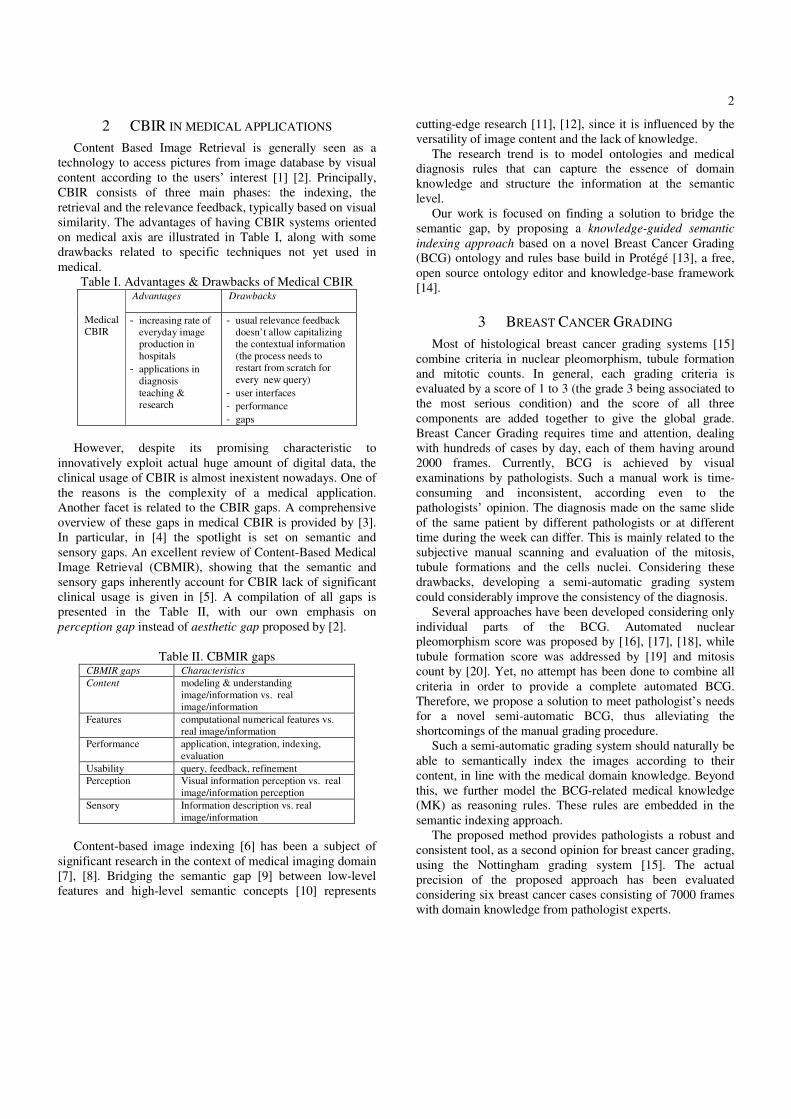
Our paradigm workflow depicted in Figure 1 respects the
following steps. The first step consists of digitizing the
histopathology slides analyzed under the microscope by the
pathologist. The slide consists of 2000 to 3000 frames that
will be processed and analyzed in the next step, by
integrating the medical knowledge. The subjective
knowledge coming from the pathologist as well as the
objective knowledge coming from the Nottingham Standard
Grading System are structured in a formal representation
based on Breast Cancer Ontology. Medical knowledge
concepts and rules are then translated into computer vision
(CV) concepts and rules (required for the image processing
and analysis step) thus providing the means for the semantic
indexing step. The output will be the Breast Cancer Grading.
Breast Cancer ontology has its implications in research
and teaching, knowledge-guided semantic indexing is of
high interest in research & teaching as well as in
diagnosis/prognosis assistance while BCG is nowadays the
most used procedure for prognosis of breast cancer.
4 MEDICAL DOMAIN KNOWLEDGE
4.1 Domain Knowledge analysis
To have a complete domain knowledge analysis, duality
of objective knowledge and subjective knowledge is required.
In our case, objective knowledge is provided by the
Nottingham Grading System (NGS) gold standard.
Identification of the regions of interest (ground truth) with
specific medical knowledge is given by the pathologists.
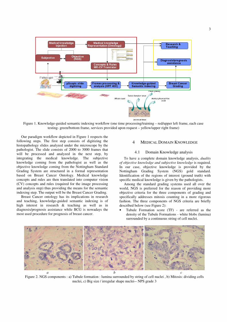
Among the standard grading systems used all over the
world, NGS is preferred for the reason of providing more
objective criteria for the three components of grading and
specifically addresses mitosis counting in a more rigorous
fashion. The three components of NGS criteria are briefly
described below (see Figure 2):
• Tubule Formation score (TF) - are referred as the
density of the Tubule Formations - white blobs (lumina)
surrounded by a continuous string of cell nuclei.
Figure 1. Knowledge-guided semantic indexing workflow (one time processing/training – red/upper left frame, each case
testing- green/bottom frame, services provided upon request – yellow/upper right frame)
a b c
Figure 2. NGS components : a) Tubule formation : lumina surrounded by string of cell nuclei , b) Mitosis: dividing cells
nuclei, c) Big size / irregular shape nuclei-- NPS grade 3
4
• Mitosis Count (MC) score represent the number of
Mitoses - diving cells nuclei. MC is assessed in the
peripheral areas of the neoplasm and it’s based on the
number of mitoses per 10 High Power Field’s (HPF’s) –
high resolution (usually 400X) frames obtained using
microscopic acquisition.
• Nuclear Pleomorphism Score (NPS) - categorizes cells
nuclei based on two features: size and shape.
The scores for the three separate parameters (tubules,
nuclei and mitoses) are summated and the overall grade of
the neoplasm is determined [1].
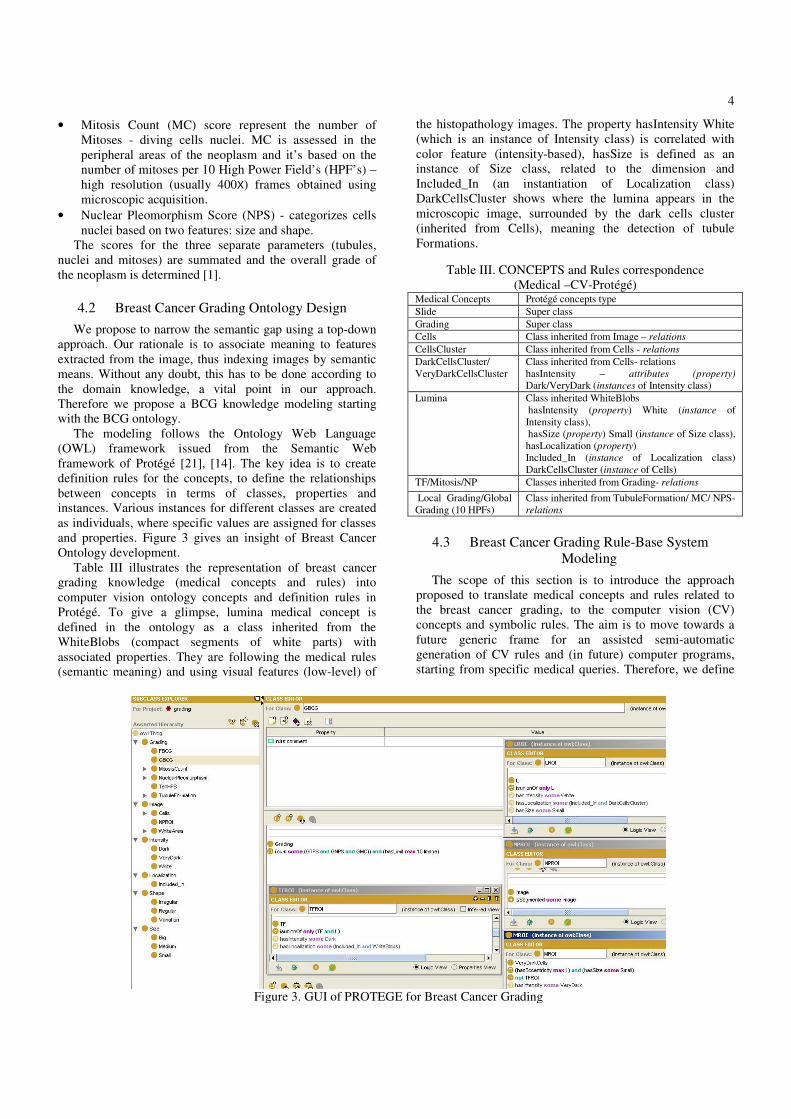
4.2 Breast Cancer Grading Ontology Design
We propose to narrow the semantic gap using a top-down
approach. Our rationale is to associate meaning to features
extracted from the image, thus indexing images by semantic
means. Without any doubt, this has to be done according to
the domain knowledge, a vital point in our approach.
Therefore we propose a BCG knowledge modeling starting
with the BCG ontology.
The modeling follows the Ontology Web Language
(OWL) framework issued from the Semantic Web
framework of Protégé [21], [14]. The key idea is to create
definition rules for the concepts, to define the relationships
between concepts in terms of classes, properties and
instances. Various instances for different classes are created
as individuals, where specific values are assigned for classes
and properties. Figure 3 gives an insight of Breast Cancer
Ontology development.
Table III illustrates the representation of breast cancer
grading knowledge (medical concepts and rules) into
computer vision ontology concepts and definition rules in
Protégé. To give a glimpse, lumina medical concept is
defined in the ontology as a class inherited from the
WhiteBlobs (compact segments of white parts) with
associated properties. They are following the medical rules
(semantic meaning) and using visual features (low-level) of
the histopathology images. The property hasIntensity White
(which is an instance of Intensity class) is correlated with
color feature (intensity-based), hasSize is defined as an
instance of Size class, related to the dimension and
Included_In (an instantiation of Localization class)
DarkCellsCluster shows where the lumina appears in the
microscopic image, surrounded by the dark cells cluster
(inherited from Cells), meaning the detection of tubule
Formations.
Table III. CONCEPTS and Rules correspondence
(Medical –CV-Protégé) Medical Concepts Protégé concepts type
Slide Super class
Grading Super class
Cells Class inherited from Image – relations
CellsCluster Class inherited from Cells - relations
DarkCellsCluster/
VeryDarkCellsCluster
Class inherited from Cells- relations
hasIntensity – attributes (property)
Dark/VeryDark (instances of Intensity class)
Lumina Class inherited WhiteBlobs
hasIntensity (property) White (instance of
Intensity class),
hasSize (property) Small (instance of Size class),
hasLocalization (property)
Included_In (instance of Localization class)
DarkCellsCluster (instance of Cells)
TF/Mitosis/NP Classes inherited from Grading- relations
Local Grading/Global
Grading (10 HPFs)
Class inherited from TubuleFormation/ MC/ NPS-
relations
4.3 Breast Cancer Grading Rule-Base System
Modeling
The scope of this section is to introduce the approach
proposed to translate medical concepts and rules related to
the breast cancer grading, to the computer vision (CV)
concepts and symbolic rules. The aim is to move towards a
future generic frame for an assisted semi-automatic
generation of CV rules and (in future) computer programs,
starting from specific medical queries. Therefore, we define
Figure 3. GUI of PROTEGE for Breast Cancer Grading

5
a Generic Translation Framework (GTF) to provide the
translation of medical concepts and rules into CV concepts
and rules.
Figure 4. Generic Translation Framework
As illustrated by Figure 4, the section is structured in
three parts, according to the main steps of the proposed
approach: development of the correspondence between
medical concepts and computer vision concepts (with respect
to the OWL standard); definition of intermediate CV rules
and generation of the final Symbolic Rules by fusion of the
CV concepts and intermediate CV symbolic rules. Note that
CV concepts are used as an input for the Rules Translator
together with the medical rules to generate the CV
preliminary symbolic rules.
4.3.1 Correspondance between the Medical concepts and
adequate Computer Vision concepts
According to the NGS synthesis, we proceed to the TF
extraction as the NPS and MC computation in order to create
the rule-based method able to automatically generate the
grading (see Table IV). Therefore, to clearly define the rules,
medical concepts are transformed into computer vision
concepts.
The MK-CV concept translator is based on a
classification of elements that need to be taken into
consideration to give the final grading.
• objects : Image, Cells, CellsCluster, Lumina, Tubule,
Mitosis, Nuclei Pleomorphism
• attributes: size, shape, intensity, localization
• values : small, medium, big, regular, variated,
irregular, white, dark, very dark, ecc
• operators :
An illustration of objects translation is given by Table IV.
Table IV. MK-CV objects of concept translator Medical Objects CV objects
Slide Image (digitized)
Cells Cells
CellsCluster Union of Cells
DarkCellsCluster/
VeryDarkCellsCluster
Union of Cells
Lumina White compact segments of the
Image included in the union of dark
cells
TF/Mitosis/NP Union of Cells/Diving Cells nuclei/
dimension & shape features of the
nuclei
Local Grading/Global
Grading
Grading Computation for TF.MC,
NP single frame/10 frames
4.3.2 Intermediate rules
To obtain the symbolic tubule formation rule, we create
intermediate rules for each domain concept used for this
criterion.
- DarkCellsCluster is defined as containing group of
adjacent cells with intensity property value setup between
VeryDark and White limits.
( )
( )
{ } 1,
{ ( ) |
}
i
i
c c C
DarkCellsClusters morphology adjacent Cell
VeryDark intensity Cell White
DarkCellsCluster =
=
< < =
=
∪
In terms of Protégé, this rule is defined as: Cells with
hasIntensity (property) some Dark, which is an instance of
Intensity class.
WhiteBlobs intermediate rule composes the Lumina (L)
rule as white blobs included in the existing DarkCellsCluster.
{ } 1,{ ( )} k k bWhiteBlobs morphWhiteArea WhiteBlob == =
{ | 1, , }ck k kL WhiteBlob c C DarkCellsCluster WhiteBlob= ∃ ∈ ⊃
- LROI intermediate CV rule is a union of all lumen detected
in the image.
1,ROI k
k l
L L=
= ∪
Figure 6. SEMANTIC Indexing in BCG Context

6
Following the same idea, intermediate rules are defined
for the mitosis count symbolic rule.
( )
{ }1,
{ | }j j
j j J
VeryDarkCells Cells intensity Cells VeryDark
VeryDarkCell=
= ≤ =
=
∪ -
( )jecc VeryDarkCell rule represents an eccentricity
deterministic operation computed for the VeryDarkCells.
- ( )jsize VeryDarkCell rule applies a size detection
threshold onto the VeryDarkCells. In practical image
processing/analysis, the detection of DarkCellsCluster,
VeryDarkCells or WhiteBlobs becomes a simple intensity-
based segmentation method.
For the nuclear pleomorphism rule definition, image
segmentation methods are performed to detect
( ), ( )i isize DarkCell shape DarkCell .
4.3.3 Generation of the final symbolic computer vision
rules
Considering the tubule formation criteria given by the
pathologist:
• Pathologist rule for Tubule = white lumina blobs
surrounded by string of dark cells nuclei.”
• Symbolic rule (used in our algorithm):
TF symbolic rule specifies that if there are WhiteBlobs
included in the DarkCellsCluster, the pathologic criterion is
satisfied. { | }c c ck
TF DarkCellsCluster WhiteBlob DarkCellsCluster= ∃ ⊂
where: TFROI (TF region of interest) is defined by:
= ROI cTF DarkCellsCluster{ }∪
The TFROI symbolic rule creates the union of all
DarkCellsCluster – with intensity and localization
dependence and L. The DarkCellsCluster∪ detection is
performed using morphologic operators.
The result of this operation is to index the medical image
by the TFROI. This is an important point of our approach,
since we are able to associate to each frame precise ROI
structure corresponding to the detected tubule formations.
• Pathologist Rule: Mitosis = very dark dividing cells
nuclei from the peripheral area of neoplasm
• Symbolic Rule:
MitosisROI:
{ | ( ),
( ), )}
ROI j j
j j ROI
M VeryDarkCell eccVeryDarkCell
sizeVeryDarkCell VeryDarkCell TF
=
⊄
∪
VeryDarkCells structures must not be contained in the tubule
formation area (TFROI), specified in the rule by the
⊄ operator. Thus, MROI rule is defined as a union of all
VeryDarkCells dependent of particular ecc, size and
localization values.
• Pathologist Rule for Nuclear Pleomorphism: Size
and Shape features of nuclei
• Symbolic Rule :
NucleiROI: { (Im)}ROI
NP segment= where
(Im) { ( ), ( )}i isegment size DarkCellsCluster shape DarkCellsCluster= A
n example of MK-CV rules translation, in the case of mitosis
detection, is given by:
5 SEMANTIC INDEXING APPROACH. BREAST
CANCER GRADING COMPUTATION
This approach intends to overcome the drawbacks of
classical indexing methods. The conceptual annotations are
rule-based defined in the grading model for every particular
frame and globally transmitted in a structure for the entire
case. Some prerequisites need to be considered. An important
role is played by the scale. The images have been scanned at
high-magnification (10X), thus the image processing and
analysis step is based on this scale. Another idea that was
conveyed in this approach is the computation of local
grading, at the first hand, in order to provide better results.
The local grading for each frame is used further for the global
grading, instead of computing directly the grading for the
whole slide.
The algorithm segments images and processes the object
recognition phase (feature extraction step) followed by the
semantic classification criteria rules modeling. Thus, it is
created a correspondence between the visual features and the
semantic image labeling, in terms of Mitosis, Nuclei and
Tubule Formation regions of interest - ROIs.
Image segmentation with gray scale conversion and
adaptive tresholding obtains a collection of such ROI,
meaningful for breast cancer grading and – more generally –
for breast cancer evolution diagnosis/prognosis. The region
selection is correlated with the model rules (see Figure 6).
Semantic indexing of concepts extracted from the image
gives us the means to create the rules for the computation of
local grading.
5.1 Local Grading Computation
The local grading computation process uses functions and
operators to define the required symbolic rules (see Table
V).
Table V. FUNCTIONS used in criteria score symbolic rules
Symbolic rules Description
1, 0.75
( ) 2, 0.10 0.75
3, 0.10
FTFS
x
f x x
x
> = < < <
the TF score as
reported in the
pathologist rule
1, 9
( ) 2,10 19
3, 19
FMC
x
f x x
x
< = < < >
the MC grade function
with the NGS values
f( ) + g( )Size Shape The pleomorphism
value off all nuclei

7
Frame Tubule Formation Score (FTFS) :
{ ( ( )/ ( ))}ROIFTFS f AreaTF AreaDarkCellsCluster=
Frame Mitosis Count (FMC): { ( ( ))}ROIFMC f count M=
Frame Nuclear Pleomorphism Score (FNPS):
count( )
i= 1
(f( ) + g( ))/count( )) ={round( }NPROI
ROISize Shape NPFNPS ∑
The local breast cancer grading (FBCG) rule
{ ( ), }FBCGi f FTFSi FMCi FNPSi i frameID= + + =
represents the sum of the three values computed for each
NGS criterion, over a single frame.
5.2 Global grading computation
The global breast cancer grading is computed similar
with each local score, but over 10 HPFs [1] (see Figure 6).
The 10 HPFs specification appears as the upper limit at each
computation of sum in the rules: 10
1
10
1
( )
(Im )
jROI
j
TF
j
j
Area TF
GTFS f
Area
=
=
=
∑
∑
10
1jMC ROI
j
GMC f count M=
= ∑
( )( )( )10
1 1
10
1
( )
ROIj
j
count NP
NP kj kjj k
ROIj
f f Size gShape
GNPS
count NP
= =
=
+ =
∑ ∑
∑
( ) { }{ , 1,...10 }j j j
GBCG f GTFS GMC GNPS j= + + =
6 EXPERIMENTS & RESULTS
The experimental part consists in analyzing and indexing
pathologic images of six breast core-biopsy cases stained with
H&E marker, consisting of 7000 frames scanned from the
tumor tissue slides and obtained from the Pathology
Department of National University Hospital of Singapore
(NUH). The database is composed by two sets: 1400 frames
used for the training algorithm phase and 5600 frames used
for the testing and validation phase. The slides were scanned
on a sequence of frames at 10X40 (400X) magnification with a
1080 X 1024 resolution.
The set of histopathology slides, labeled by our medical
partners, has been digitized into a number of hyperfields
(frames). Each frame is then analyzed and a local grading is
computed. According to this local grading, top ten images are
automatically retrieved to provide the slide global grading.
Table VI. PATHOLOGIC visual grading and configuration
of the training and testing database
Data type Case
ID
Tubule
score
Nuclear
score
Mitosis
count
BCG
(path)
Training
database
(1400
images)
1000 1 1 3 1
2000 1 2 1 1
4895 3 3 3 3
Testing 5020 2 3 3 3
database
(5600
images)
5042 3 3 2 3
5075 3 2 1 2
Table VII. Semi-AUTOMATIC grading results Data
type
Case
ID
Tubule
score
Nuclear
score
Mitosis
count
automatic
BCG
Training
database
1000 1 1 3 1
2000 2 2 1 1
4895 3 2 3 3
Testing
database
5020 3 2 3 3
5042 3 2 3 3
5075 3 2 1 2
Table VIII. COMPONENT scores and global grading errors
Data
base
Tubule
score
Nuclear
score
Mitosis
count
Compo
nent
scores
error
Global
BCG
error
Training
errors 11% 11% 0 7,33% 0%
Testing errors
11% 22% 0 11% 0%
We use Matlab programming environment to develop the method. The program is tuned to take into account the images’ scale [22] given by the microscope [23] in the automatic acquisition phase. Local errors were registered in the training base for the tubule score in case 2000 and for the nuclear score in case 4895. In the testing database, local errors were obtained at the tubule score and nuclear score for the case 5020 and only for the nuclear score in 5042 case. Note that for the mitosis count there was no registration in either training or testing database which gives us a good confidence degree in the detection of mitosis. (100% automated detection). The most interesting fact is that, when computing the BCG for training and testing database respectively, local errors (7.33%, 11%) are not propagated to the global level (0), computed by a simple formula of matches from the total items. The good results obtained on the global grading are promising and allow us to envisage interesting generic perspectives of this approach.
7 DISCUSSIONS, CONCLUSION AND
PERSPECTIVES
Despite being strongly related to a particular application
field and a specific medical domain, the presented semantic
indexing approach has a generic character. Indeed, in
association with the following ontology validation procedure:
• Ontology segmentation - extract a subset of existing thesauri (i.e. NCI thesaurus)
• Ontology structuring - according to the OWL standards
(related to Semantic Web standards); for this purpose we are
using the same Protégé framework.
• Automatic Ontology verification - use of the Protégé reasoner
in order to have a first evaluation of the ontology consistency
• Medical validation - consult the local medical collaborators
(important, even if somehow subjective and partial)
• Ontology official validation - submit the ontology to the OBO
web site (www.obofoundry.org/), agreeing that - once
validated by them - our ontology will be published on their
website.

8
this meaningful (medical domain relevant) semantic index
allows to design semantic query content-based medical image
retrieval systems, usable in translational approaches. These
types of CBIR systems will certainly replace in the near
future the actual query by example ones, based only on visual
features.
In the context of virtual microscope platform, automatic
semantic-query based visual positioning systems [24] present
also a high interest for the medical technicians and doctors in
terms of time efficiency. From the image processing and
analysis standpoint, we envisage that a multi-scale approach
will provide an improvement in terms of speed and time
consuming task. This reasoning follows even closer the
pathologist procedure and also helps in a better identification
of the invasive area (neoplasm) thus the grading will be
proceeded only on this specific part of the slide.
In addition, the purpose of generating computer vision
(CV) concepts and symbolic rules from medical
concepts/rules related to the breast cancer grading, with
respect to OWL and the Semantic Web is seen as future
generic perspectives for an assisted semi-automatic
generation of CV rules and computer programs, starting from
specific medical queries/rules. Obviously, a true maintenance
mechanism has to be included in the future in the existing
approach.
Apart coping with the semi-automatic breast cancer
grading challenge, our approach emphasizes medical imaging
vital importance for accurate “bona-fide” diagnosis
assistance. Indeed, a virtual microscope platform based on
our principle will allow pathologist to ensure a robust grading
procedure, by providing the opportunity to benefit from a
semi-automatic semantic annotation and further exploration
of the lesions’ neighborhood (region of interest-ROI) at
different scales.
ACKNOWLEDGMENT
This project is supported by the ONCO-MEDIA1 project.
REFERENCES
[1] F. Long, H. Zhang and D. Feng, “Fundamentals of Content-
Based Image Retrieval”, pp. 1-26, 2001
[2] R. Datta, D. Joshi, J. Li and J. Wang, “Image Retrieval: Ideas,
Influences, and Trends of New Age”, ACM Transactions on
Computing Surveys, pp.1-66, 2008
[3] T. Deserno, S. Antani, and R. Long, “Gaps in content-based
image retrieval”, pp.1-11, 2007
[4] A. Smeulders, M. Worring, S. Santini, A. Gupta and R.Jain,
“Content-Based Image Retrieval at the End of Early Years”,
IEEE Transactions on Pattern Analysis and Machine
Intelligence, vol.22, no.12, pp. 1349-1480, 2000
[5] H. Muller, N. Michoux, D. Bandon, and A. Geissbuhler, “A
Review of Content-Based Image Retrieval Systems in
Medical Application- Clinical Benefits and Future Directions”,
International Journal of Medical Informatics, vol. 73, pp. 1-
30, 2004
[6] G. Carneiro, A. Chan, P. Moreno, and N. Vasconcelos,
1 ONCO-MEDIA (ONtology and COntext related MEdical image
Distributed Intelligent Access) - ICT ASIA International Project –
www.onco-media.com
“Supervised Learning of Semantic Classes for Image
Annotation and Retrieval”, IEEE Transactions on Pattern
Analysis and Machine Intelligence, vol.29, no.3, 2007
[7] R. Zhao and W. Growski, “Bridging the Semantic Gap in
Image Retrieval, Distributed Multimedia Databases:
Techniques and Applications”, 2002
[8] N. Vasconcelos, “From Pixels to Semantic Spaces: Advances
in Content-Based Image Retrieval”, pp.20-26, 2007
[9] Y. KAlfoglou, S.Dasmahapatra, D.Dupplow, B.Hu, P.Lewis,
N. Shadbolt, “Living with the Semantic Gap: Experiences and
Remedies in the Context of Medical Imaging”, 1st
International Conference on Semantics and Digital Media
Technologies, 2006
[10] S. Little and J. Hunter, “Rules-By-Example- A Novel
Approach to Semantic Indexing and Querying of Images”,
International Semantic Web Conference ISWC, pp.534-548,
2004
[11] Y. Liu, N. Lazar, W. Rothfus, F. Dellaert, A. Moore,
J.Schneider, and T.Kanade, “Semantic - based Biomedical
Image Indexing and Retrieval”, Trends and Advances in
Content- Based Image and Video Retrieval”, Shapiro, Kriegel
and Veltkamp ed., pp. 1-20, in press, 2004
[12] H. Tang, R. Hanka, and H.Ip, “Histological Image Retrieval
Based on Semantic Content Analysis”, IEEE Transaction on
Information Technology Medicine, vol. 7, no. 1, 2003
[13] Tutac AE, Racoceanu D, Putti T, Xiong W, Leow W-K, Cretu
V., “Knowledge-Guided Semantic Indexing of Breast Cancer
Histopathology Images”, BioMedical Engineering and
Informatics: New Development and the Future, Proceedings
of the First International Conference on BioMedical
Engineering and Informatics, Editors: Yonghong Peng and
Yufeng Zhang, Published by IEEE Computer Society, 27 - 30
May 2008, Sanya, Hainan, China, pp. 107-112
[14] The Protégé Ontology Editor and Knowledge Acquisition
System - http://protege.stanford.edu/
[15] A. Tutac, “Histological Grading on Breast Cancer”, IPAL
internal report 2007, MIIRAD/IPAL – BCG, 2007
[16] C. Demir and B. Yener, “Automated cancer diagnosis based
on histopathological images: a systematic survey”, Tech Rep,
2005
[17] H. Jeong, T. Kim, H. Hwang and H-J. Choi, “Comparison of
thresholding methods for breast tumor cells segmentation”, in
Proc of 7th Int. Workshop on Enterprise networking and
Computing in Healthcare Industry, pp. 392-395, 2005
[18] M. Adawi, Z. Shehab, H. Keshk and M. Shourbagy, “A fast
algorithm for segmentation of microscopic cell images”, in
Proc. 4th Int. Conf. Inf. & Com. Tech, 2006
[19] S. Petushi, F. Garcia, M. Haber, C. Katsinis, and A. Tozeren,
“Large- Scale Computation on histology images reveal grade-
differentiating parameter for breast cancer”, pp. 1-11, 2006
[20] J.A. Beliën, J.P. Baak, P.J. van Diest and A.H. Ginkel,
“Counting mitosis by image processing in feulgen stained
breast cancer sections: the influence of resolution”, Cytometry,
vol.28 (2), pp.135-140, 1997
[21] D.L. McGuiness and F.van Harmelen, “OWL Web Ontology
Language W3C Overview”, pp. 1-26, 2004
[22] P. Van Osta, J.M. Geusebroek, K. Ver Donck, L. Bols, J.
Geysen, and B. Romeny, “The Principles of Scale Space
applied to structure and color in light microscopy”,
Proceedings RMS, vol. 37, no. 3, 2002
[23] I. Marandet, A. Tutac, “Smart Microscope User Guide”, IPAL
internal report 2006, MIIRAD/IPAL -µ-MediSearch, 2006
[24] G. Begelman, M. Lifshits, and E. Rivlin, “Visual Positioning
of Previously Defined ROIs on Microscopic Slides”, IEEE
Transactions on Information Technology in Biomedicine, vol.
10, no. 1, 2006