transposase-derived proteins fhy3/far1 interact with
TRANSCRIPT
Transposase-Derived Proteins FHY3/FAR1 Interact withPHYTOCHROME-INTERACTING FACTOR1 to RegulateChlorophyll Biosynthesis by Modulating HEMB1 duringDeetiolation in Arabidopsis W
Weijiang Tang,a,1 Wanqing Wang,a,b,1 Dongqin Chen,a Qiang Ji,a,b Yanjun Jing,a Haiyang Wang,c,d
and Rongcheng Lina,2
a Key Laboratory of Photobiology, Institute of Botany, Chinese Academy of Sciences, Beijing 100093, ChinabGraduate School of the Chinese Academy of Sciences, Beijing 100049, ChinacCollege of Life Sciences, Capital Normal University, Beijing 100048, ChinadDepartment of Molecular, Cellular, and Developmental Biology, Yale University, New Haven, Connecticut 06520
Successful chlorophyll biosynthesis during initial light exposure is critical for plant survival and growth, as excess accumulationof chlorophyll precursors in darkness can cause photooxidative damage to cells. Therefore, efficient mechanisms have evolvedto precisely regulate chlorophyll biosynthesis in plants. Here, we identify FAR-RED ELONGATED HYPOCOTYL3 (FHY3) and FAR-RED IMPAIRED RESPONSE1 (FAR1), two transposase-derived transcription factors, as positive regulators of chlorophyllbiosynthesis in Arabidopsis thaliana. We show that null mutations in FHY3 and FAR1 cause reduced protochlorophyllide (aprecursor of chlorophyll) levels in darkness and less photobleaching in the light. We find that FHY3 directly binds to thepromoter and activates expression of HEMB1, which encodes 5-aminolevulinic acid dehydratase in the chlorophyllbiosynthetic pathway. We reveal that PHYTOCHROME-INTERACTING FACTOR1 physically interacts with the DNA bindingdomain of FHY3, thereby partly repressing FHY3/FAR1-activated HEMB1 expression. Strikingly, FHY3 expression isupregulated by white light. In addition, our genetic data indicate that overexpression, severe reduction, or lack of HEMB1impairs plant growth and development. Together, our findings reveal a crucial role of FHY3/FAR1 in regulating chlorophyllbiosynthesis, thus uncovering a new layer of regulation by which light promotes plant dark–light transition in early seedlingdevelopment.
INTRODUCTION
As sessile organisms, plants respond to the surrounding envi-ronments by shaping their growth and development. Light is oneof the major environmental signals that influences plants through-out their life cycle, from seed germination to flowering. Underthe soil, germinating seedlings undergo etiolation (also calledskotomorphogenesis) with long hypocotyls and closed cotyle-dons lacking chlorophyll and functional chloroplasts. Uponemerging from the soil and reaching light, the etiolated seedlingsundergo deetiolation (also termed photomorphogenesis), includingcotyledon opening, chlorophyll biosynthesis, the development ofchloroplasts, and subsequently autotrophic growth (Von Arnimand Deng, 1996, Casal et al., 2004).
Phytochrome and cryptochrome photoreceptors are re-sponsible for perceiving and transducing light signals to regulatedistinct photomorphogenic responses. In Arabidopsis thaliana,
phytochrome A (phyA) to phyE form a small protein family thatpredominantly regulates various responses to red and far-red light,whereas the cryptochromes (cry1 and cry2) absorb the blue/UV-Alight (Whitelam et al., 1993; Neff et al., 2000; Lin 2002). phyA isthe primary photoreceptor for mediating far-red light signaling.FAR-RED ELONGATED HYPOCOTYL3 (FHY3) and FAR-RED-IMPAIRED RESPONSE1 (FAR1) are homologous proteins thatfunction as positive regulators and act early in phyA signaling(Hudson et al., 1999; Wang and Deng, 2002; Wang et al., 2002).They work together to modulate phyA nuclear accumulation andphyA responses through directly activating gene expression ofa pair of downstream targets, FHY1 and FHY1-LIKE (Lin et al.,2007, 2008). Studies also showed that FHY3 and FAR1 integratelight signals into the circadian clock and modulate chloroplastdivision by directly upregulating expression of EARLY FLOWER-ING4 and ACCUMULATION AND REPLICATION OF CHLORO-PLASTS5 (ARC5), respectively (Allen et al., 2006; Li et al., 2011;Ouyang, et al., 2011). We and others previously documented thatFHY3 and FAR1 define a type of transcription factors that werederived from ancient transposases during evolution and may playdiverse roles (Hudson et al., 2003; Lin et al., 2007). A recent studyfound that FHY3 has more than a thousand putative direct targetsin Arabidopsis (Ouyang et al., 2011), implicating it as having broadfunctions in plant growth and development, most of which, how-ever, are unknown.
1 These authors contributed equally to this work.2 Address correspondence to [email protected] author responsible for distribution of materials integral to the findingspresented in this article in accordance with the policy described in theInstructions for Authors (www.plantcell.org) is: Rongcheng Lin ([email protected]).WOnline version contains Web-only data.www.plantcell.org/cgi/doi/10.1105/tpc.112.097022
This article is a Plant Cell Advance Online Publication. The date of its first appearance online is the official date of publication. The article has been
edited and the authors have corrected proofs, but minor changes could be made before the final version is published. Posting this version online
reduces the time to publication by several weeks.
The Plant Cell Preview, www.aspb.org ã 2012 American Society of Plant Biologists. All rights reserved. 1 of 17

Chlorophyll formation is a hallmark of the photomorphogenicresponse, and chlorophylls serve as the major pigments inphotosynthesis by harvesting light energy and driving electrontransfer. Chlorophyll metabolism has been extensively studiedwith various organisms biochemically and genetically (Eckhardtet al., 2004; Tanaka and Tanaka, 2006, 2007). Chlorophyll bio-synthesis shares early steps from the first committed precursor5-aminolevulinic acid (ALA) to protoporphyrin IX with that ofheme, siroheme, and phytochromobilin in the tetrapyrrole bio-synthetic pathway (Battersby, 2000; Tanaka and Tanaka, 2007;Mochizuki et al., 2010). Two molecules of ALA are then con-densed to form a pyrrole molecule, porphobilinogen (PBG), byALA dehydratase (ALAD). After sequential enzymatic conversions,the pathway is divided by metal chelation reactions of pro-toporphyrin IX, thereby directing the formation of the endproducts chlorophyll and heme (Tanaka and Tanaka, 2007). Indarkness, the chlorophyll biosynthetic branch is blocked at theintermediate protochlorophyllide (Pchlide) because the conver-sion of Pchlide to chlorophyllide is catalyzed by the light-dependent enzyme NADPH:protochlorophyllide oxidoreductase(POR) in plants (Runge et al., 1996; Su et al., 2001; Heyes andHunter, 2005; Solymosi and Schoefs, 2010). However, the ac-cumulation of excess free Pchlide and/or other pyrrole inter-mediates in darkness may produce reactive oxygen species(ROS) upon light irradiation and thereby cause cotyledon pho-tobleaching or even cell death (Reinbothe et al., 1996; op denCamp et al., 2003; Wagner et al., 2004; Buhr et al., 2008).Therefore, chlorophyll biosynthesis is critical for plant survivaland must be properly regulated, particularly during the switchfrom skotomorphogenesis to photomorphogenesis. Plants haveevolved efficient mechanisms to regulate Pchlide content pre-cisely in the dark.
Accumulating evidence shows that the biosynthetic pathwayis primarily subject to transcriptional and posttranslational reg-ulation (Tanaka and Tanaka, 2007; Mochizuki et al., 2010). ALAformation is viewed as a control point for regulation of chloro-phyll supply. FLUORESCENT (FLU) was identified as an im-portant regulatory protein that represses ALA synthesis bybinding and thereby inhibiting the activity of glutamyl-tRNA re-ductase (GluTR) encoded by HEMA1 (Meskauskiene et al., 2001;Goslings et al., 2004). GluTRBP is the second GluTR bindingprotein that mediates spatial separation of ALA into heme bio-synthesis (Czarnecki et al., 2011). Another regulator, GENOMESUNCOUPLED4 (GUN4), interacts with Mg-chelatase and stim-ulates its activity by facilitating substrate binding and/or productrelease (Larkin et al., 2003; Adhikari et al., 2011). These proteinsare crucial for plant survival as a flu loss-of-function mutation islethal during deetiolation or when plants are grown in light-darkcycles, whereas lack of GluTRBP is lethal and absence of GUN4causes a complete absence of chlorophyll (Meskauskiene et al.,2001; Peter and Grimm, 2009).
Recently, several transcription factors have been found toplay important roles in regulating chlorophyll biosynthesis duringseedling deetiolation. The PHYTOCHROME-INTERACTINGFACTORs (including PIF1, PIF3, PIF4, and PIF5) are a subsetof the basic helix-loop-helix family of transcription factors andact as negative regulators of diverse phytochrome-mediatedsignaling responses (Leivar and Quail, 2011). In darkness, pif
mutants display constitutive photomorphogenic phenotypesand overaccumulate Pchlide. Their etiolated cotyledons areseverely photobleached after subsequent light illumination (Huqet al., 2004; Monte et al., 2004; Moon et al., 2008; Shin et al.,2009; Stephenson et al., 2009). It is believed that PIFs arenegative regulators of chlorophyll biosynthesis in the dark andthat light derepresses this response by triggering the protea-some-mediated degradation of PIFs (Al-Sady et al., 2006; Leivaret al., 2009; Shin et al., 2009; Shen et al., 2005). PIF1 directlyregulates chlorophyll biosynthesis by binding the promoter ofPORC and activating its gene expression (Moon et al., 2008),whereas PIF3 indirectly regulates a number of biosyntheticgenes, such as HEMA1 and GUN5 (Shin et al., 2009; Stephensonet al., 2009). It was also shown that ETHYLENE INSENSITIVE3(EIN3) and its homolog EIN3-LIKE1 cooperate with PIF1 to in-hibit Pchlide accumulation. EIN3 is able to upregulate PORA andPORB gene expression directly. Consequently, their loss-of-function mutants accumulate excess Pchlide in etiolated seed-lings and became photobleached when transferred to light(Zhong et al., 2009). A recent study demonstrated that DELLAproteins upregulate POR expression and limit the accumulationof ROS and photooxidative damage in PIF-dependent and -in-dependent manners during seedling deetiolation (Cheminantet al., 2011). These studies together reveal that POR is an im-portant target for transcriptional regulation in chlorophyll bio-synthesis, consistent with its function in conversion of Pchlideinto chlorophyllide during seedling deetiolation. However, themechanism for direct regulation of Pchlide levels is still notunderstood.In this study, we provide genetic, molecular and biochemical
evidence to demonstrate that FHY3 and FAR1 redundantlypromote chlorophyll biosynthesis by directly binding to andactivating the expression of HEMB1, which encodes ALAD, withFHY3 playing a predominant role. We show that FHY3 physicallyinteracts with the negative transcription regulator PIF1 to co-ordinate Pchlide synthesis and seedling greening. Furthermore,FHY3 expression is upregulated by white light during deetiola-tion. Our finding highlights FHY3 and FAR1 as positive and keytranscription factors in directly regulating chlorophyll biosynthesisfor seedling survival and provides insight into the functional di-vergence of these transposase-derived transcription factors inplants during evolution.
RESULTS
FHY3/FAR1 Promote Pchlide Accumulation inEtiolated Seedlings
A previous transcriptome analysis of Arabidopsis seedlings in-dicates that most of the genes involved in tetrapyrrole bio-synthesis are highly responsive to light (Matsumoto et al., 2004).We speculated that there might exist a direct positive regulator(s)of Pchlide biosynthesis in etiolated seedlings. To this end, wetested some known transcription factors that have been dem-onstrated to be key components in the light signaling pathway(Jiao et al., 2007). Five-day-old etiolated seedlings of variousmutants and the wild type were incubated with acetone over-night, and Pchlide levels were compared by scanning the
2 of 17 The Plant Cell
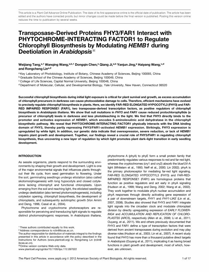
fluorescence emission in a fluorescence spectrophotometer(Huq et al., 2004). Consistent with previous studies, pif1 and pif3mutants accumulated extremely high levels of Pchlide com-pared with the wild type (Huq et al., 2004; Moon et al., 2008;Shin et al., 2009; see Supplemental Figure 1 online). The elon-gated hypocotyl5 homolog (hyh), long hypocotyl in far-red1(hfr1), and long after far-red light1 (laf1) mutants also had slightincreased amounts of Pchlide. By contrast, the Pchlide contentwas decreased in the fhy3 mutant and was slightly reduced inthe far1 and hy5 mutants compared with their correspondingwild-type seedlings (Figure 1A; see Supplemental Figure 1 on-line), suggesting that FHY3, FAR1, and HY5 may play positiveroles in regulating chlorophyll biosynthesis in the dark. In thisstudy, we focused on the roles of FHY3 and FAR1. We furtherfound that the fhy3 far1 double mutant had even less Pchlideaccumulation than their single mutant parents (Figure 1A),demonstrating a redundant function between these two pro-teins, with FHY3 playing a predominant role. In the followingexperiments, the function and mechanism of FHY3 will be in-vestigated in more detail. To confirm whether low Phclide in thefhy3-4 mutant is caused by the disruption of FHY3 protein,a FHY3p:FHY3 transgene in which the FHY3 open reading frame(ORF) is under the control of its own promoter was introducedinto fhy3-4 (Lin et al., 2008). The transgene was found to com-plement the fhy3-4 mutant phenotype (Figure 1B). In addition,dexamethasone (DEX; 1 µM) treatment greatly restored thePchlide level of fhy3-4/FHY3P:FHY3-GR transgenic plants (Linet al., 2007) compared with mock-treated plants (Figure 1C),suggesting that nuclear targeting of FHY3 is required for itsfunction. These results reveal that FHY3 and FAR1 promotePchlide accumulation and likely function as positive regulatorsof chlorophyll biosynthesis in the dark.
Because the tetrapyrrole biosynthetic pathway separates intotwo different branches to produce heme and chlorophyll, wethen tested if the heme branch was also affected by the fhy3 andfar1 mutations. Noncovalently bound heme was extracted from5-d-old etiolated seedlings and measured spectrophotometri-cally. However, no difference in heme production was observedbetween fhy3 far1, fhy3, and far1 mutants and the wild type (seeSupplemental Figure 2 online).
Loss of FHY3/FAR1 Enhances Seedling Greeningduring Deetiolation
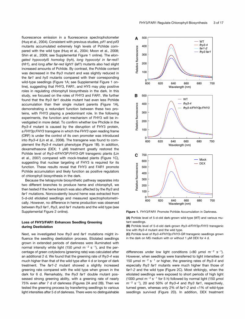
Next, we investigated how fhy3 and far1 mutations might in-fluence the seedling deetiolation process. Etiolated seedlingsgrown in extended periods of darkness were illuminated withnormal intensity white light (150 µmol m22 s21), and the per-centage of green cotyledons (greening rate) was calculated afteran additional 2 d. We found that the greening rate of fhy3-4 wasmuch higher than that of the wild type after 4 d or longer of darktreatment. The far1-2 mutant showed a slightly increasedgreening rate compared with the wild type when grown in thedark for 6 d. Remarkably, the fhy3 far1 double mutant pos-sessed strong greening ability, with a greening rate of nearly75% even after 7 d of darkness (Figures 2A and 2B). Then wetested the greening process by transferring seedlings to variouslight intensities after 5 d of darkness. There were no distinguishable
differences under low light conditions (<80 µmol m22 s21).However, when seedlings were transferred to light intensities of150 µmol m22 s21 or higher, the greening rates of fhy3-4 andespecially fhy3 far1 mutants were much higher than those offar1-2 and the wild type (Figure 2C). Most strikingly, when theetiolated seedlings were exposed to short periods of high light(1000 µmol m22 s21 for 5 h) followed by normal light (150 µmolm22 s21), 20 and 50% of fhy3-4 and fhy3 far1, respectively,turned green, whereas only 2% of far1-2 and <1% of wild-typeseedlings survived (Figure 2D). In addition, DEX treatment
Figure 1. FHY3/FAR1 Promote Pchlide Accumulation in Darkness.
(A) Pchlide level of 5-d-old dark-grown wild-type (WT) and various mu-tant seedlings.(B) Pchlide level of 5-d-old dark-grown fhy3-4/FHY3p:FHY3 transgenicline with fhy3-4 mutant and the wild type.(C) Pchlide level of fhy3-4/FHY3p:FHY3-GR transgenic seedlings grownin the dark on MS medium with or without 1 µM DEX for 4 d.
FHY3/FAR1 Regulate Chlorophyll Biosynthesis 3 of 17
greatly restored the greening phenotype of fhy3-4/FHY3p:FHY3-GR transgenic seedlings (Figure 2E). These data indicate thatFHY3 and FAR1 play an important role in seedling greeningduring deetiolation.
Upon light illumination, excess Pchlide may generate ROS orfree radicals, resulting in photobleaching or even cell death inthe cotyledons (Reinbothe et al., 1996; Buhr et al., 2008). To testwhether the increased greening rate of the mutants is the resultof less photobleaching, we investigated ROS production bydetecting 29,79-dichlorodihydrofluorescein diacetate (H2DCFDA)
fluorescence of the cotyledons (Zhong et al., 2009). We foundthat H2DCFDA fluorescence was remarkably lower in the fhy3-4 and fhy3 far1 mutants and slightly lower in far1-2 than that inthe wild type. However, chlorophyll autofluorescence wasobvious in fhy3-4 and fhy3 far1 (Figure 2F). Furthermore, whenstained with trypan blue (indicating dead cells), the cotyledonsof the wild type and far1 were stained blue, whereas fhy3 andfhy3 far1 seedlings were barely stained (Figure 2G). Therefore,disruption of FHY3 and FAR1 prevents photobleaching and celldeath.
Figure 2. Loss of FHY3/FAR1 Enhances Seedling Greening and Prevents Photobleaching during Deetiolation.
(A) Representative images of 6-d-old etiolated seedlings when exposed to light for 2 d. WT, the wild type.(B) Percentage of green cotyledons of seedlings grown in various time periods of darkness before being moved to 150 µmol m22 s21 white light for 2 d.(C) Greening rates of 5-d-old etiolated seedlings transferred to various intensities of white light for 2 d.(D) Greening rate of 5-d-old etiolated seedlings transferred to high light (1000 µmol m22 s21) for 5 h followed by 150 µmol m22 s21 for additional 2 d.(E) Greening rate of 5-d-old fhy3-4/FHY3-GR transgenic and fhy3-4 mutant seedlings grown in darkness in the absence (Mock) or presence of 1 µMDEX followed by 2 d of 150 µmol m22 s21 light exposure. Data in (B) to (E), mean 6 SD, n = 3.(F) Fluorescence microscope images of ROS (indicated by H2DCFDA fluorescence) and chlorophyll autofluorescence in the cotyledons of 5-d-oldetiolated seedlings followed by 2 d of 150 µmol m22 s21 light treatment.(G) Trypan blue staining of 6-d-old etiolated seedlings exposed to 150 µmol m22 s21 light for an additional 2 d. Bars in (F) and (G) = 200 µm.
4 of 17 The Plant Cell

FHY3 Activates HEMB1 Expression by Directly Binding toIts Promoter
The observed phenotypes prompted us to test whether theexpressions of tetrapyrrole biosynthetic genes were influencedby FHY3 and FAR1 (see Supplemental Figure 3A online). fhy3far1 double mutant and wild-type seedlings were grown indarkness for 5 d, and relative gene expression was analyzed byquantitative RT-PCR (qRT-PCR). We found that only a small setof genes, including HEMA3, HEMB1, FERROCHELATASE 2(FC2), HEME OXYGENASE 1 (HO1), HO3, and HO4, were eitherup- or downregulated more than 1.5-fold in the fhy3 far1 doublemutant compared with the wild type (see Supplemental Figure3B online). It was previously shown that FHY3 activates down-stream gene expression mainly through binding the FHY3/FAR1binding site (FBS) (CACGCGC) present in promoters of its tar-gets (Lin et al., 2007; Li et al., 2011). Therefore, we surveyed thepromoter sequences of these tetrapyrrole biosynthetic genesand found that only HEMB1 contains a putative FBS motif,which is located 385 bp upstream of the ATG start code in re-verse orientation (Figure 3A). To investigate whether FHY3 and
FAR1 could bind to the HEMB1 promoter through this putativeFBS motif, we first used a yeast one-hybrid system. GAD-FHY3(fused with GAL4 activation domain) and GAD-FAR1 proteinswere able to bind the wild-type HEMB1 oligonucleotide con-taining the FBS sequence (HEMB1wt:LacZ) and activate LacZreporter gene expression, but they did not bind the mutantoligonucleotide (HEMB1m:LacZ, in which GCGCGTG waschanged into GCttGTG) (Figure 3B). Next, we performed elec-trophoresis mobility shift assay (EMSA) to test whether FHY3binds the HEMB1 promoter fragment in vitro. Our data showedthat the FHY3 recombinant protein (N-terminal 250 amino acidsof FHY3 fused with glutathione S-transferase; GST-FHY3N)caused an upshift band with HEMB1 wild-type oligonucleotideslabeled with 32P, and this band was abolished by excess un-labeled wild-type oligonucleotides but not by excess unlabeledmutant oligonucleotides (Figure 3C). To investigate furtherwhether FHY3 binds HEMB1 DNA fragment in vivo, chromatinimmunoprecipitation (ChIP) was performed using 35S:GUS-FHY3 (GUS for b-glucuronidase) transgenic seedlings (Wangand Deng, 2002). The ChIP DNA was quantified by real-timePCR with three sets of primers spanning the upstream promoter,
Figure 3. FHY3/FAR1 Bind to the Promoter of HEMB1 and Activate Its Gene Expression.
(A) A schematic diagram of the HEMB1 gene. Black rectangles represent exons, and gray circle indicates the FBS motif. Wild-type (wt) and mutant (m)oligonucleotide sequences are shown below, and the FBS motif sequence is underlined. a, b, and c indicate fragments for ChIP-PCR.(B) Relative b-galactosidase activities of LacZ reporters (HEMB1wt:LacZ and HEMB1m:LacZ ) activated by GAD-fused effectors in the yeast one-hybridassay. Mean 6 SD, n = 6.(C) EMSA assay of GST-FHY3N or GST recombinant proteins incubated with 32P-labled wild-type oligonucleotides in the presence of a series of excessamounts of wild-type or mutant unlabeled competitors. Arrow indicates shifted bands of protein-DNA complexes.(D) ChIP assay of 5-d-old etiolated 35S:GUS-FHY3 transgenic seedlings. Samples were precipitated with anti-GUS antibody (GUS) or anti-FLAGantibody (negative control). ChIP DNA was quantified by real-time PCR with primers targeting fragments as shown in (A). Mean 6 SD, n = 3.(E) Transient activation assay of luciferase reporter gene driven by the wild type (HEMB1p:LUC ) or mutant (HEMB1pm:LUC, in which the FBS motif wasmutated) HEMB1 promoter in protoplasts. Protoplast transformation and incubation were conducted in weak light. Relative activity is expressed as theratio of LUC versus GUS internal control. Mean 6 SD, n = 3.(F) HEMB1 expression in 5-d-old dark-grown seedlings transferred to light or kept in darkness for 6 h. WT, the wild type.(G) HEMB1 expression in FHY3-GR transgenic seedlings treated without (Mock) or with 1 µM DEX. Relative expression levels are normalized to that ofUBQ. Mean 6 SD from three biological replicates in (F) and (G).
FHY3/FAR1 Regulate Chlorophyll Biosynthesis 5 of 17

the FBS motif, and the coding region, respectively. Our resultsshowed that only the “b” fragment containing FBS motif wassignificantly enriched in samples precipitated by anti-GUS an-tibody but not in samples pulled down by anti-FLAG negativecontrol (Figure 3D). Together, these results confirm that FHY3directly binds to the HEMB1 promoter in an FBS motif–dependentmanner.
A previous study demonstrated that FHY3 and FAR1 possessintrinsic transcriptional activation activity (Lin et al., 2007). Wethen examined how FHY3 regulates the downstream gene ex-pression by cotransforming a luciferase (LUC) reporter genedriven by the HEMB1 promoter (1.5 kb upstream of ATG) withvarious effectors into Arabidopsis protoplasts. Our transientexpression assay showed that FHY3 protein was able to acti-vate LUC reporter gene expression (Figure 3E). However, a pointmutation in the transposase domain of FHY3 (FHY3-G305R;deficient in transcriptional activation; Lin et al., 2007) failed toactivate LUC expression (Figure 3E). When LUC was driven bythe HEMB1 promoter with mutations in the FBS motif, the ex-pression level was drastically reduced and FHY3 was no longerable to activate it (Figure 3E). Next, using qRT-PCR analysis, wefound that HEMB1 expression was modestly decreased in far1-2and further dropped in fhy3-4, whereas it was significantly de-creased in fhy3 far1 (Figure 3F), suggesting that FHY3 and FAR1redundantly upregulate HEMB1 expression. In addition, DEXtreatment also promoted HEMB1 expression in the FHY3p:FHY3-GR transgenic plants compared with the mock treatment(Figure 3G). We thus conclude that FHY3 and FAR1 positivelyregulate HEMB1 gene expression in plant cells.
FHY3/FAR1 Promote ALAD Activity
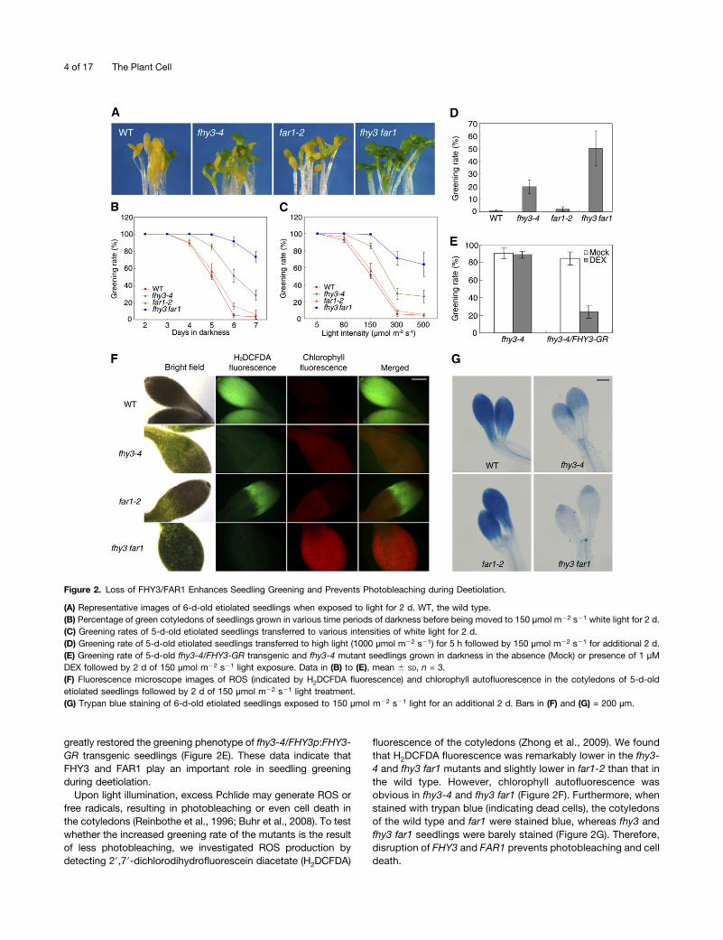
HEMB1 is one of two genes encoding ALAD, which catalyzesa reaction among the early steps of tetrapyrrole biosynthesis.We then examined how the ALAD protein level and enzymaticfunction were affected by fhy3 and far1 mutations. To this end,a polyclonal antibody against a peptide (amino acids 336 to 346)of ALAD was raised in rabbit. This antibody recognized theALAD recombinant protein at a size of 55 kD, which is close tothe predicted size of the ALAD mature protein, confirming thespecificity of this antibody (see Supplemental Figure 4A online).Compared with wild-type plants, the ALAD protein level in far1-2was slightly reduced, whereas it was clearly decreased in fhy3-4and further reduced in fhy3 far1 double mutant seedlings (seeSupplemental Figure 4B online), correlating well with the re-duced HEMB1 transcripts in these mutants (Figure 3F).
To determine how ALAD function was affected, an in vitroenzymatic activity assay was performed in a reaction systemcontaining both the plant protein extracts and ALA substrate,and PBG production was determined (Vajpayee et al., 2000). Wefound that less PBG was formed with proteins extracted fromfar1-2, and much less was formed from those of fhy3-4 and fhy3far1 compared with the wild type (Figure 4A). Next, 5-d-old eti-olated seedlings were fed exogenous 10 mM ALA for 12 h andthe ALAD conversion ability was tested in vivo. As shown inFigure 4B, significantly less PBG was detected in the fhy3-4 andfhy3 far1 mutants than in the wild-type seedlings. To assess thephysiological response further, we grew the plants with or without
ALA feeding for 3 d and measured the Pchlide content as well asgreening ability after light exposure. Without exogenous ALAtreatment, Pchlide levels and greening rates were indistinguish-able between fhy3 far1 and the wild type. However, when fedwith ALA, wild-type seedlings contained drastically increasedPchlide, whereas fhy3 far1 accumulated only half that of the wildtype (Figure 4C). Accordingly, almost all of the wild-type coty-ledons were severely photobleached, whereas ;75% of thefhy3 far1 mutant seedlings were still able to turn green after lightexposure (Figure 4D). These results firmly demonstrate that theability to convert ALA into PBG in fhy3 and particularly fhy3 far1mutants was reduced; therefore, FHY3 and FAR1 positivelyregulate ALAD function in plants.
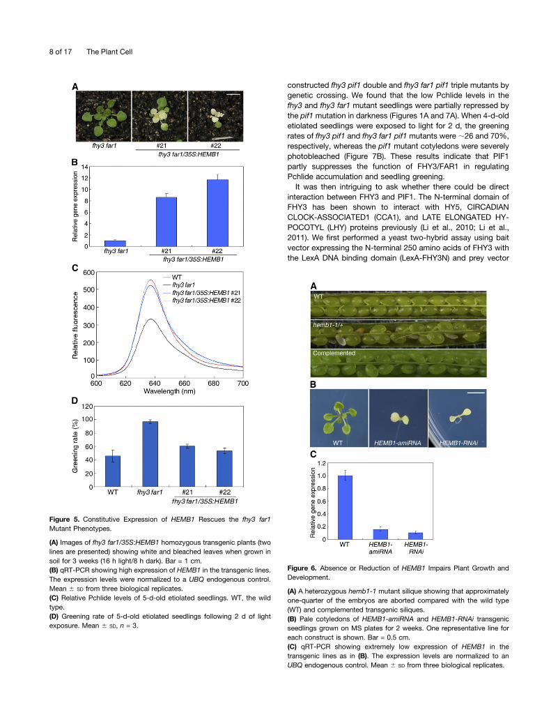
Constitutive Expression of HEMB1 Rescues the fhy3 far1Mutant Phenotypes
We then determined whether restoration of HEMB1 in the fhy3far1 mutant could rescue its phenotype by constitutively ex-pressing the HEMB1 ORF under the control of the cauliflowermosaic virus 35S promoter (35S:HEMB1). Multiple T1 resistanttransgenic lines were obtained. Unexpectedly, all T2 transgeniclines segregated with nearly one-fourth of plants developingpartially white leaves when grown in soil after 3 weeks (two lineswere shown; Figure 5A). The HEMB1 transcripts in the trans-genic lines were more than eightfold higher than those in themutant (Figure 5B). These plants were eventually died and wewere unable to obtain homozygous plants. Strikingly, we ob-served that the heterozygotes of HEMB1 overexpression plantsrescued the Pchlide accumulation and seedling greening phe-notypes of the fhy3 far1 mutant (Figures 5C and 5D), furtherconfirming that the function of FHY3 and FAR1 in Pchlidesynthesis and seedling greening is through the regulation ofHEMB1. Consistent with this, HEMB1 is highly expressed incotyledons during dark-to-light transition (see SupplementalFigure 5 online).
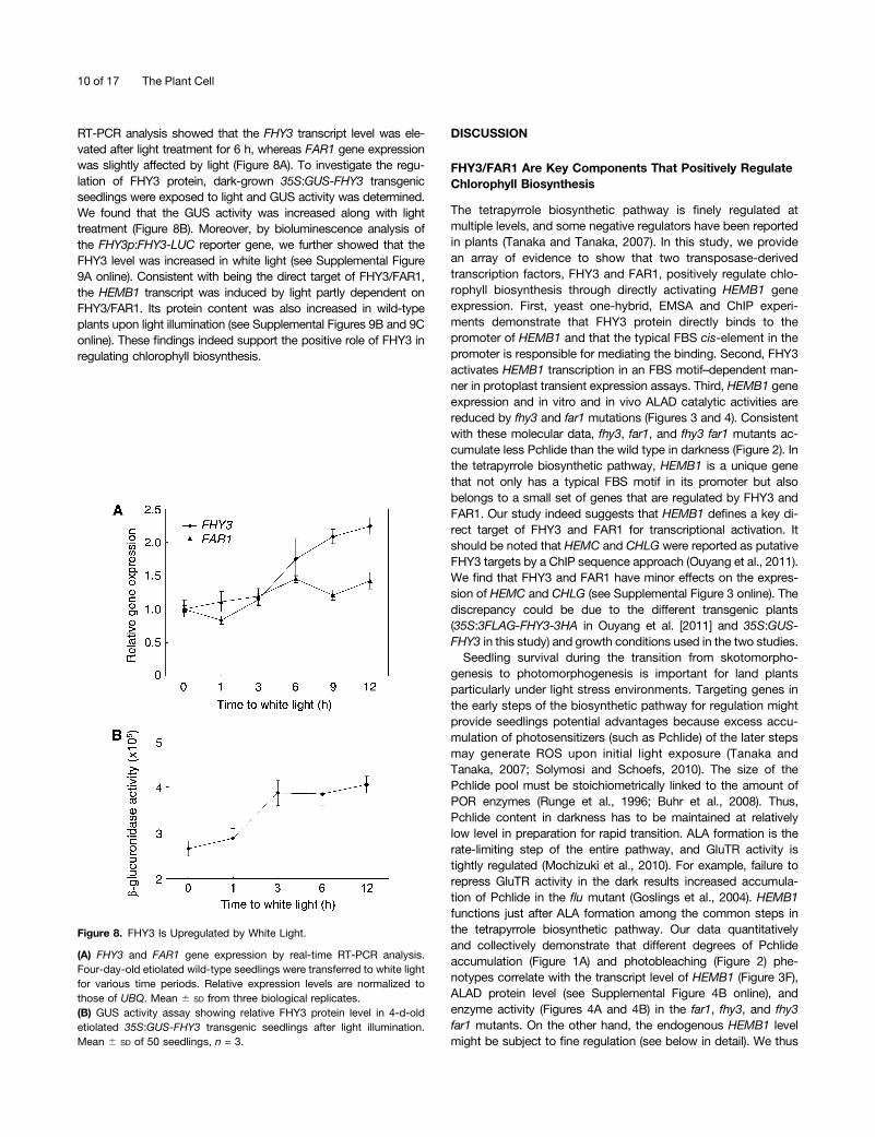
Overexpression, Absence, or Reduction of HEMB1 ImpairsPlant Growth and Development
Similar to what was observed in the fhy3 far1 mutant back-ground, the homozygous transgenic plants of 35S:HEMB1overexpression lines in the wild type displayed photobleachedleaves and did not survive (see Supplemental Figure 6 online).We speculated that HEMB1 might play essential role in regu-lating plant growth and development. To test this hypothesis, weobtained a T-DNA insertion mutant allele, Salk_016544, from theABRC. This allele contains a T-DNA inserted into the eighth in-tron of HEMB1, and the insertion site was sequence confirmed,thereafter it was designated hemb1-1 (see SupplementalFigures 7A and 7B online). When the heterozygous progeny ofhemb1-1 were grown on Murashige and Skoog (MS) platescontaining 50 mg/mL kanamycin, nearly two-thirds (494 out of747, P value > 0.75 by t test) of the seeds were resistant anddeveloped normal seedlings. We attempted to use PCR geno-typing to identify homozygotes from those kanamycin-resistantplants. However, all of the resistant plants were heterozygous,suggesting that the hemb1-1 mutation might be lethal in the
6 of 17 The Plant Cell
homozygous state. Then we dissected the developing siliquesand scored the seeds under a dissecting microscope. In wild-typesiliques, seeds were fully developed, whereas in 37 siliques fromindividual hemb1-1 heterozygous plants grown under the sameconditions as the wild-type plants, 359 out of 1351 ovules weresmall, shrunken, and aborted (Figure 6A). The ratio of nonabortedseeds to aborted seeds was ;3:1 (P value > 0.1). The lethality ofthe homozygous mutation was complemented by expressingHEMB1p:HEMB1 (Figure 6A). Taken together, we conclude thathemb1-1 is a recessive embryo lethal mutation. To dissect pre-cisely the stage of embryogenesis during which the hemb1 mu-tant arrests development, embryos within individual immaturesiliques from self-pollinated hemb1-1 plants were cleared andexamined under a microscope. The normal embryos underwenttypical developmental stages, ranging from preglobular, globular,heart, torpedo, and mature. However, the mutant embryos werearrested at the globular stage (see Supplemental Figure 7C on-line).
We then attempted to use RNA interference (RNAi) and arti-ficial microRNA (amiRNA) transgenic approaches to knock downthe endogenous HEMB1 expression in the Nossen wild-typebackground. When T1 seeds were germinated on 50 mg/L
hygromycin plates, surprisingly, all of the HEMB1-RNAi (159lines) and HEMB1-amiRNA (112 lines) resistant seedlings de-veloped white or pale cotyledons. These seedlings did not de-velop true leave even when grown in MS medium supplied with2% Suc (Figure 6B). We collected these resistant seedlings andtested the HEMB1 expression by qRT-PCR, finding that theendogenous HEMB1 transcripts in those HEMB1-RNAi orHEMB1-amiRNA lines were severely reduced to <20% of thosein the wild type (Figure 6C). These results revealed that severereduction of HEMB1 mRNA causes seedling lethality. Takentogether, our genetic data confirm that HEMB1 is critical forplant development and its transcript level has to be preciselymaintained.
FHY3 Physically Interacts with PIF1
It has been shown that etiolated pif1 mutant seedlings over-accumulate Pchlide and are sensitive to photobleaching (Huqet al., 2004; Moon et al., 2008). Here, we found that the fhy3 andfhy3 far1 mutants display opposite phenotypes to those of pif1.To investigate how the two types of proteins, FHY3/FAR1 andPIF1, antagonistically regulate chlorophyll biosynthesis, we
Figure 4. FHY3/FAR1 Promote ALAD Activity.
(A) In vitro ALAD enzymatic assay. Total proteins were extracted from 4-d-old etiolated seedlings and incubated with 100 µM ALA for 2.5 h. WT, the wildtype.(B) In vivo PBG formation of 4-d-old etiolated seedlings fed with 10 mM ALA for 12 h. FW, fresh weight.(C) and (D) Relative Pchlide fluorescence (C) and greening rate (D) of 3-d-old etiolated seedlings fed with or without 100 µM ALA. Seedlings wereexposed to white light for 2 d in (D).For (A), (B), and (D), mean 6 SD, n = 3.
FHY3/FAR1 Regulate Chlorophyll Biosynthesis 7 of 17
constructed fhy3 pif1 double and fhy3 far1 pif1 triple mutants bygenetic crossing. We found that the low Pchlide levels in thefhy3 and fhy3 far1 mutant seedlings were partially repressed bythe pif1 mutation in darkness (Figures 1A and 7A). When 4-d-oldetiolated seedlings were exposed to light for 2 d, the greeningrates of fhy3 pif1 and fhy3 far1 pif1 mutants were ;26 and 70%,respectively, whereas the pif1 mutant cotyledons were severelyphotobleached (Figure 7B). These results indicate that PIF1partly suppresses the function of FHY3/FAR1 in regulatingPchlide accumulation and seedling greening.It was then intriguing to ask whether there could be direct
interaction between FHY3 and PIF1. The N-terminal domain ofFHY3 has been shown to interact with HY5, CIRCADIANCLOCK-ASSOCIATED1 (CCA1), and LATE ELONGATED HY-POCOTYL (LHY) proteins previously (Li et al., 2010; Li et al.,2011). We first performed a yeast two-hybrid assay using baitvector expressing the N-terminal 250 amino acids of FHY3 withthe LexA DNA binding domain (LexA-FHY3N) and prey vector
Figure 5. Constitutive Expression of HEMB1 Rescues the fhy3 far1Mutant Phenotypes.
(A) Images of fhy3 far1/35S:HEMB1 homozygous transgenic plants (twolines are presented) showing white and bleached leaves when grown insoil for 3 weeks (16 h light/8 h dark). Bar = 1 cm.(B) qRT-PCR showing high expression of HEMB1 in the transgenic lines.The expression levels were normalized to a UBQ endogenous control.Mean 6 SD from three biological replicates.(C) Relative Pchlide levels of 5-d-old etiolated seedlings. WT, the wildtype.(D) Greening rate of 5-d-old etiolated seedlings following 2 d of lightexposure. Mean 6 SD, n = 3.
Figure 6. Absence or Reduction of HEMB1 Impairs Plant Growth andDevelopment.
(A) A heterozygous hemb1-1 mutant silique showing that approximatelyone-quarter of the embryos are aborted compared with the wild type(WT) and complemented transgenic siliques.(B) Pale cotyledons of HEMB1-amiRNA and HEMB1-RNAi transgenicseedlings grown on MS plates for 2 weeks. One representative line foreach construct is shown. Bar = 0.5 cm.(C) qRT-PCR showing extremely low expression of HEMB1 in thetransgenic lines as in (B). The expression levels are normalized to anUBQ endogenous control. Mean 6 SD from three biological replicates.
8 of 17 The Plant Cell

expressing full-length PIF1 with the GAL4 activation domain(GAD-PIF1). Our data showed that LexA-FHY3N indeed inter-acts with GAD-PIF1, and the N-terminal domain containing theC2H2 zinc finger motif is responsible for mediating the inte-raction (Figure 7C). We further performed a pull-down analysisbetween recombinant 63His-fused PIF1 (His-PIF1) and GST-tagged FHY3N (GST-FHY3N) and found that GST-FHY3N, butnot GST alone, was able to pull down PIF1 in vitro (Figure 7D).Next, we examined the in vivo interaction between FHY3 andPIF1. Transgenic plants expressing 35S:GUS-FHY3 togetherwith 35S:TAP-PIF1 were used for coimmunoprecipitation as-says. As shown in Figure 7E, GUS-FHY3 was able to precipitateTAP-PIF1 in planta. Furthermore, firefly LUC complementationimaging (LCI) assays (Chen et al., 2008) were conducted bytransiently expressing FHY3N-NLuc (or FHY3-NLuc) and CLuc-PIF1 fusions in Arabidopsis protoplasts. We found that coex-pression of FHY3N-NLuc and CLuc-PIF1 reconstituted strongLUC activity, but the controls did not (Figure 7F). In addition,cotransformation of FHY3-NLuc with CLuc-PIF1 also led to LUCactivity (see Supplemental Figure 8A online). Taken together,
these data indicate that FHY3 physically interacts with PIF1through its N-terminal domain.As shown earlier, FHY3 activates HEMB1 expression in plant
cells; we then ask whether FHY3-PIF1 interaction could influenceHEMB1 expression. To this end, we cotransformed FHY3 (35S:FHY3) and/or PIF1 (35S:PIF1) effectors together with HEMB1p:LUC reporter construct in Arabidopsis protoplasts. Our datashowed that PIF1 alone inhibited the transcription of the LUCreporter gene. Coexpression of PIF1 largely suppressed the ac-tivation activity of FHY3 on the HEMB1p:LUC reporter (Figure7G). Consistent with this, the HEMB1 transcript level in fhy3 far1mutant was largely derepressed by the pif1 mutation (seeSupplemental Figure 8B online). These results demonstrate thatPIF1 interferes with FHY3/FAR1-activated HEMB1 transcription.
FHY3 Is Upregulated by White Light
To assess how expressions of FHY3 and FAR1 themselves areregulated during deetiolation, 4-d-old etiolated wild-type seed-lings were transferred to white light in a time course. Real-time
Figure 7. FHY3 Directly Interacts with PIF1.
(A) Relative fluorescence indicating Pchlide accumulation in 4-d-old etiolated seedlings. WT, the wild type.(B) Percentage of seedlings with green cotyledons when 4-d-old etiolated seedlings were exposed to white light for 2 d. Mean 6 SD, n = 3.(C) A yeast two-hybrid assay for interaction between GAD-fused PIF1 and LexA-fused N-terminal fragment of FHY3 (1 to 250 amino acids, FHY3N).(D) In vitro pull-down assay between His-tagged PIF1 and GST-fused FHY3N. The His-PIF1 proteins were incubated with immobilized GST or GST-FHY3N, and immunoprecipitated fractions were probed with an anti-His or anti-GST antibody. IB, immunoblot; IP, immunoprecipitation.(E) Coimmunoprecipitation assay between GUS-FHY3 and TAP-PIF1 in vivo. Seedlings were grown in darkness for 4 d. After precipitation with anti-GUS antibody, proteins were immunoblotted with anti-GUS or anti-MYC antibodies. Arrow indicates GUS-FHY3 bands.(F) LCI assay between FHY3N and PIF1 fused with the N-terminal or C-terminal fragment of firefly luciferase, respectively. Relative LUC activity isnormalized to 35S:GUS internal control. Mean 6 SD, n = 3.(G) Relative HEMB1p:LUC reporter activity in Arabidopsis protoplasts cotransformed with the effector constructs. The relative LUC activities werenormalized to the 35S:GUS internal control. Mean6 SD, n = 3. Protoplast transformation, incubation, and protein extraction were performed in darkness([F] and [G]).
FHY3/FAR1 Regulate Chlorophyll Biosynthesis 9 of 17
RT-PCR analysis showed that the FHY3 transcript level was ele-vated after light treatment for 6 h, whereas FAR1 gene expressionwas slightly affected by light (Figure 8A). To investigate the regu-lation of FHY3 protein, dark-grown 35S:GUS-FHY3 transgenicseedlings were exposed to light and GUS activity was determined.We found that the GUS activity was increased along with lighttreatment (Figure 8B). Moreover, by bioluminescence analysis ofthe FHY3p:FHY3-LUC reporter gene, we further showed that theFHY3 level was increased in white light (see Supplemental Figure9A online). Consistent with being the direct target of FHY3/FAR1,the HEMB1 transcript was induced by light partly dependent onFHY3/FAR1. Its protein content was also increased in wild-typeplants upon light illumination (see Supplemental Figures 9B and 9Conline). These findings indeed support the positive role of FHY3 inregulating chlorophyll biosynthesis.
DISCUSSION
FHY3/FAR1 Are Key Components That Positively RegulateChlorophyll Biosynthesis
The tetrapyrrole biosynthetic pathway is finely regulated atmultiple levels, and some negative regulators have been reportedin plants (Tanaka and Tanaka, 2007). In this study, we providean array of evidence to show that two transposase-derivedtranscription factors, FHY3 and FAR1, positively regulate chlo-rophyll biosynthesis through directly activating HEMB1 geneexpression. First, yeast one-hybrid, EMSA and ChIP experi-ments demonstrate that FHY3 protein directly binds to thepromoter of HEMB1 and that the typical FBS cis-element in thepromoter is responsible for mediating the binding. Second, FHY3activates HEMB1 transcription in an FBS motif–dependent man-ner in protoplast transient expression assays. Third, HEMB1 geneexpression and in vitro and in vivo ALAD catalytic activities arereduced by fhy3 and far1 mutations (Figures 3 and 4). Consistentwith these molecular data, fhy3, far1, and fhy3 far1 mutants ac-cumulate less Pchlide than the wild type in darkness (Figure 2). Inthe tetrapyrrole biosynthetic pathway, HEMB1 is a unique genethat not only has a typical FBS motif in its promoter but alsobelongs to a small set of genes that are regulated by FHY3 andFAR1. Our study indeed suggests that HEMB1 defines a key di-rect target of FHY3 and FAR1 for transcriptional activation. Itshould be noted that HEMC and CHLG were reported as putativeFHY3 targets by a ChIP sequence approach (Ouyang et al., 2011).We find that FHY3 and FAR1 have minor effects on the expres-sion of HEMC and CHLG (see Supplemental Figure 3 online). Thediscrepancy could be due to the different transgenic plants(35S:3FLAG-FHY3-3HA in Ouyang et al. [2011] and 35S:GUS-FHY3 in this study) and growth conditions used in the two studies.Seedling survival during the transition from skotomorpho-
genesis to photomorphogenesis is important for land plantsparticularly under light stress environments. Targeting genes inthe early steps of the biosynthetic pathway for regulation mightprovide seedlings potential advantages because excess accu-mulation of photosensitizers (such as Pchlide) of the later stepsmay generate ROS upon initial light exposure (Tanaka andTanaka, 2007; Solymosi and Schoefs, 2010). The size of thePchlide pool must be stoichiometrically linked to the amount ofPOR enzymes (Runge et al., 1996; Buhr et al., 2008). Thus,Pchlide content in darkness has to be maintained at relativelylow level in preparation for rapid transition. ALA formation is therate-limiting step of the entire pathway, and GluTR activity istightly regulated (Mochizuki et al., 2010). For example, failure torepress GluTR activity in the dark results increased accumula-tion of Pchlide in the flu mutant (Goslings et al., 2004). HEMB1functions just after ALA formation among the common steps inthe tetrapyrrole biosynthetic pathway. Our data quantitativelyand collectively demonstrate that different degrees of Pchlideaccumulation (Figure 1A) and photobleaching (Figure 2) phe-notypes correlate with the transcript level of HEMB1 (Figure 3F),ALAD protein level (see Supplemental Figure 4B online), andenzyme activity (Figures 4A and 4B) in the far1, fhy3, and fhy3far1 mutants. On the other hand, the endogenous HEMB1 levelmight be subject to fine regulation (see below in detail). We thus
Figure 8. FHY3 Is Upregulated by White Light.
(A) FHY3 and FAR1 gene expression by real-time RT-PCR analysis.Four-day-old etiolated wild-type seedlings were transferred to white lightfor various time periods. Relative expression levels are normalized tothose of UBQ. Mean 6 SD from three biological replicates.(B) GUS activity assay showing relative FHY3 protein level in 4-d-oldetiolated 35S:GUS-FHY3 transgenic seedlings after light illumination.Mean 6 SD of 50 seedlings, n = 3.
10 of 17 The Plant Cell
believe that a low-fold gene expression change of HEMB1 islikely sufficient to trigger the drastic seedling greening phenotypein the fhy3 far1 mutant. In Arabidopsis, ALAD is encoded byHEMB1 and HEMB2. HEMB1 is greatly induced in cotyledonsduring dark-to-light transition and expressed in all tissues anddevelopmental stages examined. However, the expression ofHEMB2 is barely detected (see Supplemental Figure 5 online),suggesting that HEMB1 is the major contributor at this step.This notion is further supported by genetic study showing thatconstitutive expression of HEMB1 rescues the fhy3 far1 mutantphenotypes (Figure 5). A previous study showed that FHY3 andFAR1 regulate chloroplast development through activating ARC5expression (Ouyang et al., 2011). However, an arc5 loss-of-functionmutant did not show differences in Pchlide level or greeningability relative to Landsberg erecta wild type (see SupplementalFigure 10 online), indicating that ARC5 is not involved in theregulation of chlorophyll biosynthesis during deetiolation.
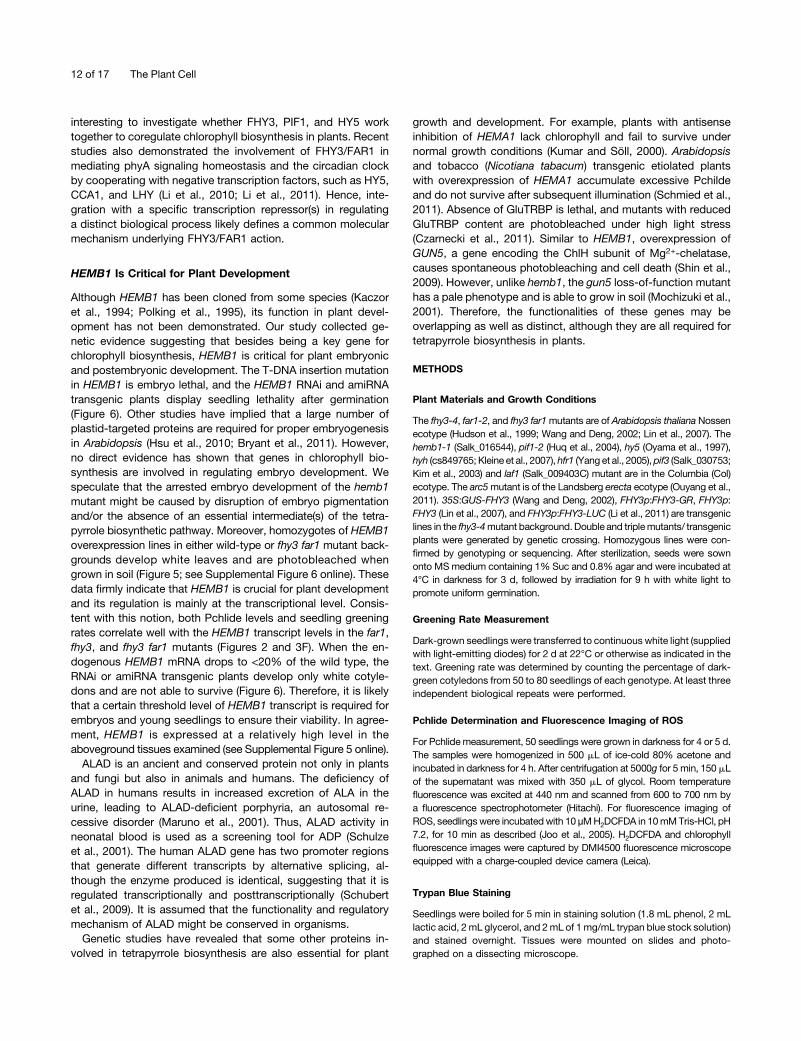
Most strikingly, FHY3 expression is repressed in darkness,and light induces its expression probably through activatingphytochrome signaling. Therefore, we propose a model inwhich, in darkness, less HEMB1 is induced by FHY3 (and FAR1),resulting in small pool of Pchlide. After light irradiation, in-creased FHY3 levels activate HEMB1 expression, thereby pro-moting the conversion of ALA to PBG and subsequent Pchlideaccumulation for chlorophyll synthesis and photoautotrophicgrowth (Figure 9).
When excess Pchlide accumulates in the dark-grown seed-lings, POR represents a critical regulatory layer for the control ofchlorophyll biosynthesis during seedling greening. In agreementwith this, ga1-3 and gai mutant seedlings showed increasedPOR expression and were more resistant to photooxidativedamage, despite a high accumulation of Pchlide in the dark(Cheminant et al., 2011). Moreover, overexpression of PORpromoted seedling greening (Sperling et al., 1997; Cheminantet al., 2011). By contrast, reduction of POR expression in the pif1and ein3mutants resulted in severe photobleaching during dark-to-light transition (Moon et al., 2008; Zhong et al., 2009).
Interestingly, we observed that mutations in fhy3 and far1 donot alter the nonbound heme content, probably through oppo-site regulation of FC2, HO1, HO3, and HO4 genes that act in theheme branch. Thus, it is postulated that FHY3 and FAR1 spe-cifically contribute to the chlorophyll biosynthetic pathway. Thisis in agreement with the idea that newly synthesized tetra-pyrroles need to be directed to the chlorophyll branch ratherthan the heme branch during initial seedling greening (Stephensonand Terry, 2008).
Integration of Two Distinct Types of Transcription Factors inChlorophyll Biosynthesis Regulation
The discovery of positive regulators is of utmost significance forunderstanding the exquisite regulation of the chlorophyll bio-synthetic pathway. In this study, we find that by means ofits N-terminal domain FHY3 physically interacts with PIF1,a phytochrome-interacting transcription factor (Figure 7). The pif1mutant accumulates extremely high levels of Pchlide in the dark,and PIF1 has been revealed as a negative regulator of chloro-phyll biosynthesis (Huq et al., 2004; Moon et al., 2008). Although
PIF1 may indirectly regulate GUN5 expression (Shin et al., 2009),the mechanism of PIF1 in regulating Pchlide synthesis is not wellunderstood. We show that PIF1 directly interacts with FHY3(and maybe FAR1 as well) and partly represses FHY3 activationactivity on HEMB1 gene expression. It is speculated that theregulation of PIF1 in Pchlide synthesis in the dark may be partlydependent on the function of FHY3/FAR1. Accordingly, the fhy3far1 double mutant is largely able to suppress the pif1 mutantphenotypes (Figure 7). Thus, two distinct types of transcriptionfactors in the phytochrome signaling pathway coordinate toregulate an important biological response. In agreement withthis finding, a recent analysis reported that FHY3 and PIF1 (PIL5)coregulate more than 100 genes (Ouyang et al., 2011). Theycould also be involved in mediating other diverse processesthrough their interaction. Our finding supports the hypothesisthat the expression of genes involved in chlorophyll biosynthesisis regulated by a tight and complex mechanism comprisingmultiple components (Tanaka and Tanaka, 2007). It also impliesthat both positive and negative players work coordinately tooptimize chlorophyll biosynthesis, thus enabling etiolated seedlingsto prepare properly for emerging at the soil surface (Figure 9).We also noticed that HY5 likely has a positive role in regu-
lating Pchlide synthesis (see Supplemental Figure 1 online). HY5acts as a key player in light signaling and interacts with FHY3and FAR1 (Oyama et al., 1997; Li et al., 2010). It will be
Figure 9. A Model for the Role of FHY3/FAR1 in Regulating ChlorophyllBiosynthesis.
FHY3 and FAR1 bind to the promoter and activate the expression ofHEMB1 (encoding the ALAD enzyme), with FHY3 playing a predominantrole. In darkness, FHY3 is maintained at a relatively low level, so that lessPchlide accumulates in cotyledons. Light promotes FHY3 expression,thereby increasing HEMB1 transcript and ALAD protein levels, allowingPchlide accumulation and subsequent chlorophyll formation. Meanwhile,PIF1 interferes with the activation activity of FHY3 (and FAR1) byphysically interacting with FHY3. Light releases this repression by pro-teasome-mediated degradation of PIF1. Arrows, positive regulation;bars, negative regulation. Arrows with dash lines indicate multiple steps.
FHY3/FAR1 Regulate Chlorophyll Biosynthesis 11 of 17
interesting to investigate whether FHY3, PIF1, and HY5 worktogether to coregulate chlorophyll biosynthesis in plants. Recentstudies also demonstrated the involvement of FHY3/FAR1 inmediating phyA signaling homeostasis and the circadian clockby cooperating with negative transcription factors, such as HY5,CCA1, and LHY (Li et al., 2010; Li et al., 2011). Hence, inte-gration with a specific transcription repressor(s) in regulatinga distinct biological process likely defines a common molecularmechanism underlying FHY3/FAR1 action.
HEMB1 Is Critical for Plant Development
Although HEMB1 has been cloned from some species (Kaczoret al., 1994; Polking et al., 1995), its function in plant devel-opment has not been demonstrated. Our study collected ge-netic evidence suggesting that besides being a key gene forchlorophyll biosynthesis, HEMB1 is critical for plant embryonicand postembryonic development. The T-DNA insertion mutationin HEMB1 is embryo lethal, and the HEMB1 RNAi and amiRNAtransgenic plants display seedling lethality after germination(Figure 6). Other studies have implied that a large number ofplastid-targeted proteins are required for proper embryogenesisin Arabidopsis (Hsu et al., 2010; Bryant et al., 2011). However,no direct evidence has shown that genes in chlorophyll bio-synthesis are involved in regulating embryo development. Wespeculate that the arrested embryo development of the hemb1mutant might be caused by disruption of embryo pigmentationand/or the absence of an essential intermediate(s) of the tetra-pyrrole biosynthetic pathway. Moreover, homozygotes of HEMB1overexpression lines in either wild-type or fhy3 far1 mutant back-grounds develop white leaves and are photobleached whengrown in soil (Figure 5; see Supplemental Figure 6 online). Thesedata firmly indicate that HEMB1 is crucial for plant developmentand its regulation is mainly at the transcriptional level. Consis-tent with this notion, both Pchlide levels and seedling greeningrates correlate well with the HEMB1 transcript levels in the far1,fhy3, and fhy3 far1 mutants (Figures 2 and 3F). When the en-dogenous HEMB1 mRNA drops to <20% of the wild type, theRNAi or amiRNA transgenic plants develop only white cotyle-dons and are not able to survive (Figure 6). Therefore, it is likelythat a certain threshold level of HEMB1 transcript is required forembryos and young seedlings to ensure their viability. In agree-ment, HEMB1 is expressed at a relatively high level in theaboveground tissues examined (see Supplemental Figure 5 online).
ALAD is an ancient and conserved protein not only in plantsand fungi but also in animals and humans. The deficiency ofALAD in humans results in increased excretion of ALA in theurine, leading to ALAD-deficient porphyria, an autosomal re-cessive disorder (Maruno et al., 2001). Thus, ALAD activity inneonatal blood is used as a screening tool for ADP (Schulzeet al., 2001). The human ALAD gene has two promoter regionsthat generate different transcripts by alternative splicing, al-though the enzyme produced is identical, suggesting that it isregulated transcriptionally and posttranscriptionally (Schubertet al., 2009). It is assumed that the functionality and regulatorymechanism of ALAD might be conserved in organisms.
Genetic studies have revealed that some other proteins in-volved in tetrapyrrole biosynthesis are also essential for plant
growth and development. For example, plants with antisenseinhibition of HEMA1 lack chlorophyll and fail to survive undernormal growth conditions (Kumar and Söll, 2000). Arabidopsisand tobacco (Nicotiana tabacum) transgenic etiolated plantswith overexpression of HEMA1 accumulate excessive Pchildeand do not survive after subsequent illumination (Schmied et al.,2011). Absence of GluTRBP is lethal, and mutants with reducedGluTRBP content are photobleached under high light stress(Czarnecki et al., 2011). Similar to HEMB1, overexpression ofGUN5, a gene encoding the ChlH subunit of Mg2+-chelatase,causes spontaneous photobleaching and cell death (Shin et al.,2009). However, unlike hemb1, the gun5 loss-of-function mutanthas a pale phenotype and is able to grow in soil (Mochizuki et al.,2001). Therefore, the functionalities of these genes may beoverlapping as well as distinct, although they are all required fortetrapyrrole biosynthesis in plants.
METHODS
Plant Materials and Growth Conditions
The fhy3-4, far1-2, and fhy3 far1mutants are of Arabidopsis thaliana Nossenecotype (Hudson et al., 1999; Wang and Deng, 2002; Lin et al., 2007). Thehemb1-1 (Salk_016544), pif1-2 (Huq et al., 2004), hy5 (Oyama et al., 1997),hyh (cs849765; Kleine et al., 2007), hfr1 (Yang et al., 2005), pif3 (Salk_030753;Kim et al., 2003) and laf1 (Salk_009403C) mutant are in the Columbia (Col)ecotype. The arc5mutant is of the Landsberg erecta ecotype (Ouyang et al.,2011). 35S:GUS-FHY3 (Wang and Deng, 2002), FHY3p:FHY3-GR, FHY3p:FHY3 (Lin et al., 2007), and FHY3p:FHY3-LUC (Li et al., 2011) are transgeniclines in the fhy3-4mutant background.Double and triplemutants/ transgenicplants were generated by genetic crossing. Homozygous lines were con-firmed by genotyping or sequencing. After sterilization, seeds were sownonto MSmedium containing 1% Suc and 0.8% agar and were incubated at4°C in darkness for 3 d, followed by irradiation for 9 h with white light topromote uniform germination.
Greening Rate Measurement
Dark-grown seedlings were transferred to continuous white light (suppliedwith light-emitting diodes) for 2 d at 22°C or otherwise as indicated in thetext. Greening rate was determined by counting the percentage of dark-green cotyledons from 50 to 80 seedlings of each genotype. At least threeindependent biological repeats were performed.
Pchlide Determination and Fluorescence Imaging of ROS
For Pchlidemeasurement, 50 seedlings were grown in darkness for 4 or 5 d.The samples were homogenized in 500 mL of ice-cold 80% acetone andincubated in darkness for 4 h. After centrifugation at 5000g for 5 min, 150 mLof the supernatant was mixed with 350 mL of glycol. Room temperaturefluorescence was excited at 440 nm and scanned from 600 to 700 nm bya fluorescence spectrophotometer (Hitachi). For fluorescence imaging ofROS, seedlingswere incubatedwith 10 µMH2DCFDA in 10mMTris-HCl, pH7.2, for 10 min as described (Joo et al., 2005). H2DCFDA and chlorophyllfluorescence images were captured by DMI4500 fluorescence microscopeequipped with a charge-coupled device camera (Leica).
Trypan Blue Staining
Seedlings were boiled for 5 min in staining solution (1.8 mL phenol, 2 mLlactic acid, 2 mL glycerol, and 2mL of 1mg/mL trypan blue stock solution)and stained overnight. Tissues were mounted on slides and photo-graphed on a dissecting microscope.
12 of 17 The Plant Cell
Phenotypic Analysis of Embryo Development
Embryos were excised from siliques at different developmental stagesand cleared with Herr’s solution (Herr, 1971) overnight at 37°C. Sampleswere mounted between a microscope slide and a cover slip with a drop ofHoyer’s solution (7.5 mL water, 1.3 g glycerol, 1.9 g gum arabic crystalsand 25 g chloral hydrate) and observed with a microscope (Olympus).
Plasmid Construction
To generate LacZ reporter genes driven by the HEMB1 promoter witha wild-type or mutant FBS motif, the 39-bp oligonucleotides were syn-thesized as two complementary primers (HEMB1WF and HEMB1WR forthe wild type, and HEMB1MF and HEMB1MR for mutant) with an EcoRIsite overhang at the 59 end and an XhoI site overhang at the 39 end,respectively. The annealed oligonucleotides were ligated into the EcoRI-XhoI sites of pLacZi2µ (Lin et al., 2007), resulting in HEMB1wt:LacZ andHEMB1m:LacZ, respectively.
To produce a LUC reporter gene driven by the HEMB1 promoter,a 1.8-kb fragment upstream of HEMB1 ATG translational start code wasPCR amplified with primers HEMB1P1 and HEMB1P2 from Col genomicDNA. The PCR fragment was inserted into the pGEM-T Easy (Promega)vector to produce pGEM-HEMB1p. To mutagenize the FBS motif(GCGCGTG) in the HEMB1 promoter, the fragment was amplified frompGEM-HEMB1p template using primers HEMB1pm1 and HEMB1pm2 inwhich the FBS motif site was changed into GCttGTG, giving rise topGEM-HEMB1pm. After sequencing confirmation, the wild-type andmutant fragments were released from pGEM-HEMB1p and pGEM-HEMB1pm cut with HindIII and BamHI and ligated into the HindIII-BamHI site of YY96 vector (Yamamoto et al., 1998) to produceHEMB1p:LUC and HEMB1pm:LUC, respectively.
To obtain the HEMB1 cDNA clone, the first-strand cDNA was reversetranscripted using oligo(dT)18 primer from total RNA extracted from Colwild-type seedlings. The ORF of HEMB1 gene was amplified with primersHEMB1F and HEMB1R by high-fidelity Pfu DNA polymerase (Invitrogen)and cloned into the pGEM-T Easy vector, resulting in pGEM-HEMB1.
To construct the HEMB1 overexpression binary vector, the HEMB1gene was released from pGEM-HEMB1 by digestion with NcoI and BglIIand ligated into the NcoI-BglII site of pCAMBIA1302 (http://www.cambia.org/daisy/cambia/585) to produce 35S:HEMB1. In addition, the HEMB1promoter was released from pGEM-HEMB1p cut by SalI and SacI andligated into the SacI-SalI site of 35S:HEMB1 to replace the 35S promoter,resulting in HEMB1p:HEMB1.
To generate an RNAi construct for HEMB1, a 540-bp conserved cDNAfragment of HEMB1 was PCR amplified with primers HEMB1R1 (con-taining SpeI and KpnI sites at the 59 end) and HEMB1R2 (containing SacIand BamHI sites at the 59 end) from pGEM-HEMB1 plasmid DNA. ThePCR fragment was cloned into pGEM-T Easy vector, resulting in pGEM-HEMB1R. The KpnI-BamHI fragment was released from pGEM-HEMB1Rand ligated into the KpnI-BamHI site of pDS1301 (Yuan et al., 2007) toproduce pDS1301-KB. Then, the SpeI-SacI fragment was released frompGEM-HEMB1R and ligated into the SpeI-SacI site of pDS1301-KB toproduce pDS1301-HEMB1-RNAi.
To make the HEMB1 amiRNA construct, the amiRNA target sequenceofHEMB1 (59-TAACGATACTGTTTACCCCAC-39) and primers HEMB1A1,HEMB1A2, HEMB1A3, and HEMB1A4 were designed using the WMD3Web microRNA Designer (http://wmd3.weigelworld.org/cgi-bin/webapp.cgi; Schwab et al., 2006). These primers were used to amplify the amiRNAprecursor by overlapping PCR from the pRS300 template to produce thefragment containing HEMB1 target amiRNA foldback. The amiRNAfoldback was released with KpnI and SpeI and then ligated to the KpnI-SpeI site of pDS1301 for constitutive expression under the control of thecauliflower mosaic virus 35S promoter, resulting in pDS1301-HEMB1-amiRNA.
To construct a HEMB1 bacterial expression vector, a 1.13-kb (fromamino acid 53 to 430, without the putative chloroplast transit signalpeptide) cDNA fragment of HEMB1 was PCR amplified with primersHEMB1B1 and HEMB1B2 from pGEM-HEMB1. The PCR fragment wasreleased with SacI and SalI and then ligated to the SacI-SalI site of pET28ato produce the pHEMB1-6His.
To construct PIF1 bacterial expression and yeast one-hybrid vectors,the PIF1 ORF was PCR amplified with primers PIF1-F and PIF1-R fromCol cDNA and cloned into pGEM-T Easy vector, resulting in pGEM-PIF1.The PCR fragment was released with EcoRI and SalI, then ligated to theEcoRI-SalI site of pET28a to produce p6His-PIF1, and ligated to theEcoRI-XhoI site of JG4-5 (Clontech) to produce GAD-PIF1, respectively.
The yeast vectors LexA-FHY3N, GAD-FHY3, and GAD-FAR1, therecombinant protein construct GST-FHY3N, and the transient expressionvectors pSPYCE-FHY3 and pSPYCE-FHY3-G305R were describedpreviously (Lin et al., 2007).
To construct LCI vectors for FHY3 and PIF1, full-length FHY3 andFHY3N were released from pGEM-FHY3 or pGEM-FHY3N by digestionwith BamHI and SalI and cloned into the BamHI-SalI site of 35S:NLuc(Chen et al., 2008) to produce FHY3-NLuc and FHY3N-NLuc, re-spectively. PIF1 was released from pGEM-PIF1 cut with KpnI and SalI andinserted into the KpnI-SalI site of 35S:CLuc to generate CLuc-PIF1.
All primer sequences are listed in Supplemental Table 1 online.
Yeast Assays
Yeast hybrid assays were performed as previously described (Lin et al.,2007). Briefly, for yeast one-hybrid assays, the GAD fusion constructs werecotransformed with the LacZ reporter plasmids. Transformants were grownon SD/-Trp-Ura dropout liquid media, and relative b-galactosidase activitywas quantified by a spectrophotometer. For yeast two-hybrid assays, therespective combinations of GAD and LexA fusionswere cotransformed intothe yeast strain EGY48, which contains the LexAop:LacZ reporter construct(Clontech). Transformants were grown on SD/-Trp-Ura-His dropout platescontaining 5-bromo-4-chloro-3-indolyl-b-D-galactopyranoside for blue colordevelopment.
Purification of Recombinant Protein
GST, GST-FHY3N, His-PIF1, and His-HEMB1 recombinant fusion pro-teins were induced by isopropyl b-D-1-thiogalactopyranoside and ex-pressed in the Escherichia coli BL21 (DE3) strain. The proteins were thenpurified by Glutathione Sepharose 4B beads (GE Healthcare; for GST andGST-FHY3N) or Ni-NTA Agarose (Qiagen; for His-PIF1 and His-HEMB1)following the manufacturer’s instructions.
EMSA
EMSA analysis was performed as previously described (Lin et al., 2007).Briefly, HEMB1 complementary oligonucleotides were labeled with[a-32P]dATP and incubated with GST-FHY3N or GST proteins in theabsence or presence of unlabeled oligonucleotides followed by sepa-ration on polyacrylamide gels. The DNA–protein binding signal was ex-posed to x-ray film and developed. The oligonucleotide sequences areshown in Supplemental Table 1 online.
Antibody Production and Immunoblotting
The peptide corresponding to amino acids 336 to 346 of ALAD (EAR-EDEAEGAD) conjugated with KLH was synthesized (Cali-Bio), andpolyclonal antibody was raised in rabbit. For immunoblotting, seedlingswere homogenized in extraction buffer containing 50 mM Tris-HCl, pH7.5, 150 mM NaCl, 10 mM MgCl2, 0.1% Tween 20, 1 mM PMSF, and 1
FHY3/FAR1 Regulate Chlorophyll Biosynthesis 13 of 17

complete protease inhibitor cocktail (Roche). The extracts were centrifugedat 14,000g twice at 4°C for 10 min each, and protein concentration wasdetermined by Bradford assay (Bio-Rad). Proteins were boiled in SDSloading buffer, separated by 10% SDS-PAGE gels, and blotted ontopolyvinylidene fluoride membranes (Pall). The proteins were then in-cubated with anti-ALAD (1:1000 dilution) or anti-His (Abcam) primaryantibodies and subsequently the horseradish peroxidase–conjugatedgoat-anti-rabbit secondary antibody (Abcam). The protein bands werevisualized by the standard ECL method.
Pull-Down Assay
About 2 µg of purified recombinant bait proteins (GST-FHY3N and GST)and 2 µg of prey proteins (His-PIF1) were incubated in binding buffer (50mM Tris-HCl, pH 7.5, 100mMNaCl, and 0.6% Triton X-100) for 2 h at 4°C.Glutathione Sepharose 4B beads were added and incubated for 1 h. Afterwashing with binding buffer, precipitated proteins were eluted in 23 SDSloading buffer. The proteins were then size fractioned on 10%SDS-PAGEand immunoblotted by anti-His or anti-GST antibodies (Abcam).
Coimmunoprecipitation
For coimmunoprecipitation assays, seedlings were grown in the dark fol-lowed by treatment with 50 mM MG132. Total proteins were extracted withextractionbuffer and incubatedwith 2mganti-GUS (Invitrogen) antibody for 2to 3 h at 4°C. Fiftymicroliters of proteinG-Sepharose (Roche)was added andincubated for another 2 to 3 h. The sepharose beads were washed threetimeswith coimmumoprecipitation buffer, and the precipitated proteins wereeluted in 23 SDS loading buffer by boiling for 10 min. The proteins wereseparated on 10% SDS-PAGE gel and detected by immunoblotting usinganti-MYC (Abcam) and anti-GUS antibodies.
ChIP
The 35S:GUS-FHY3 transgenic plants grown in darkness for 5 d wereused for ChIP assays following the procedure as described (Leibfriedet al., 2005). Briefly, the seedlings were cross-linked with 1% formal-dehyde and ground to powder under liquid nitrogen. After isolation andsonication, the chromatin complexes were incubated with anti-GUSantibody (Invitrogen) or anti-FLAG antibody (Abcam) as a negative control.The precipitated DNA fragments were recovered and quantified by real-time PCR with primers shown in Supplemental Table 1 online.
LUC Activity Assay
Protoplast isolation and transient expression assays were performed asdescribed previously (Lin et al., 2007). For transient expression assays, thereporter plasmids (HEMB1p:LUC or HEMB1pm:LUC), effector constructs(pSPYCE-FHY3 and pSPYCE-FHY3-G305R), and 35S:GUS internal controlwere cotransformed into protoplasts. For LCI assays, plastid combinationsof variousN- andC-terminal LUC fusionswere cotransformedwith 35S:GUSinternal control. The protoplasts were pelleted and resuspended in 13 cellculture lysis reagent (Promega). The GUS fluorescence was measured usinga Modulus luminometer/fluorometer with a UV fluorescence optical kit(Promega). The LUCactivitywas detectedwith a luminescence kit using LUCassay substrate (Promega). The relative reporter gene expression levels wereexpressed as the LUC/GUS ratios.
RNA Extraction and qRT-PCR
Seedlings were treated as indicated in the text, and plant total RNA wasextracted by RNA extraction kit (Tiangen). The first-strand cDNA was syn-thesized by reverse transcriptase (Invitrogen). Real-time PCRwas performed
with the SYBRPremix ExTaq kit (Takara) in a 15-mL reaction system followingthe manufacturer’s instructions. Three biological replicates were performedfor each sample, and the expression levels were normalized to those ofUBQ.All primers sequences are listed in Supplemental Table 1 online.
In Vivo ALA Feeding
For phenotype analysis, the seedlings were grown on MS plates con-taining 100 µM ALA (Sigma-Aldrich) in darkness for 4 d. For ALAD activityassay, the dark-grown seedlings were transferred to 10 mM ALA solution(5 mM MgCl2 and 10 mM NaH2PO4, pH 7.0) under green light and in-cubated for 12 h.
ALAD Enzyme Activity Determination and PBG Measurement
The in vitro ALAD activity was determined as Vajpayee et al. (2000) describedwith minor modifications. Briefly, tissues (;0.2 g) were homogenized with 1mL extraction buffer (50mMTris-HCl, pH 8.2, and 0.1mMDTT) in a prechilledmortar and pestle. The homogenatewas filtered through four layers of cheesecloth, and the filtrate was centrifuged at 10,000g for 1 h at 4°C. The su-pernatant was used for ALAD activity assay. One milliliter of the extract wasincubated with reaction buffer (0.27 mL of 1 mg/mL ALA, 1.35 mL of 50 mMTris-HCl, pH 8.2, 0.1 mMDTT, and 0.08mL of 0.2 MMgCl2) for 2.5 h at 37°C.The reaction was terminated by the addition of 0.3mL of 3.0M trichloroaceticacid. After cooling, sampleswere centrifuged at 2000g for 10min and used forPBG determination. The ALAD activity is expressed as nmol of PBG formed/mg protein/h at 37°C.
PBG content determination was performed as described (Mauzeralland Granick, 1956; Kayser et al., 2005). Briefly, samples were homog-enized and resuspended in 1 mL of 0.15 M cold trichloroacetic acid. Aftercentrifugation at 50,000g for 30 min at 0°C, the supernatants were ad-justed to a pH of;5.5 by addition of 1 N NaOH and 0.5 M sodium acetate.They were passed through a Dowex 138 column (200 to 400 mesh;Sigma-Aldrich) equilibrated at pH 4.6 to absorb PBG. The resins werewashed eight times with double distilled water, and PBG was eluted twotimes with 0.4mL of 1M acetic acid. The eluates weremixed with an equalvolume of Ehrlich reagent, and absorbance of themixture was determinedat 555 nm after 10 min on a spectrophotometer. PBG content of thesamples was calculated using a standard curve generated by commercialPBG (Sigma-Aldrich).
Heme Determination
The content of noncovalently bound heme was measured according tothemethod of Richter et al. (2010). Briefly, tissues were homogenized, andnoncovalently bound heme was extracted with 5mL of extraction solution(2 mL of dimethyl sulfoxide, 10 mL of acetone, and 0.5 mL of 37% HCl)followed by centrifugation at 16,000g for 10 min. Heme was transferred toether by addition of 3 mL of diethyl ether, 2 mL of saturated NaCl, and10 mL of water. The ether phase was mixed with ethanol and flowedthrough a DEAE-Sepharose CL-6B column (GE Healthcare). After se-quential washing with diethyl ether:ethanol (3:1, v/v), diethyl ether:ethanol(1:1, v/v), and ethanol, heme was eluted with ethanol:acetic acid:water(81:9:10, v/v/v) and quantified spectrophotometrically at 398 nm using theextinction coefficient of 144 mM21 cm21.
Arabidopsis Transformation
The plant binary expression vectors 35S:HEMB1, HEMB1p:HEMB1,pDS1301-HEMB1-RNAi, or pDS1301-HEMB1-amiRNA were electro-porated into the Agrobacterium tumefaciens strain GV3101 and thenintroduced into the wild type or fhy3 far1 via the floral dip method (Cloughand Bent, 1998). Transgenic plants were selected on MS plates in thepresence of 50 mg/L hygromycin.
14 of 17 The Plant Cell
Accession Numbers
Sequence data from this article can be found in the Arabidopsis GenomeInitiative or GenBank/EMBL data libraries under the following accessionnumbers: FHY3 (At3g22170), FAR1 (At4g15090), HEMB1 (At1g69740), PIF1(At2g20180), UBQ (At3g52590), HEMA1 (At1g58290), HEMA2 (At1g09940),HEMA3 (At2g31250), GSA1 (At5g63570), GSA2 (At3g48730), HEMB2(At1g44318), HEMC (At5g08280), HEMD (At2g26540), HEME1 (At3g14930),HEME2 (At2g40490), HEMF1 (At1g03475), HEMF2 (At4g03205), CPO3(At5g63290), HEMG1 (At4g01690), HEMG2 (At5g14220), FC1 (At5g26030),FC2 (At2g30390), HO1 (At2g26670), HO2 (At2g26550), HO3 (At1g69720),HO4 (At1g58300),HY2 (At3g09150),CHLD (At1g08520),CHLH (At5g13630),CHLI1 (At4g18480), CHLI2 (At5g45930), CHLM (At4g25080), CRD1(At3g56940), PORA (At5g54190), PORB (At4g27440), PORC (At1g03630),DVR (AT5G18660), CAO (At1g44446), and CHLG (At3g51820).
Supplemental Data
The following materials are available in the online version of this article.
Supplemental Figure 1. Relative Pchlide Fluorescence of VariousMutants and the Wild Type.
Supplemental Figure 2. Noncovalently Bound Heme Contents infhy3, far1, and fhy3 far1 Mutants and the Wild Type.
Supplemental Figure 3. Expression of Genes Involved in theTetrapyrrole Biosynthetic Pathway.
Supplemental Figure 4. Confirmation of ALAD Antibody and ProteinLevel.
Supplemental Figure 5. HEMB1 and HEMB2 Expression Patterns inDifferent Stages and Tissues.
Supplemental Figure 6. Phenotype of HEMB1 OverexpressionPlants.
Supplemental Figure 7. Identification and Characterization of hemb1Mutant.
Supplemental Figure 8. Interaction between FHY3 and PIF1.
Supplemental Figure 9. FHY3 and HEMB1 Are Induced by Light.
Supplemental Figure 10. Characterization of the arc5 Mutant Phe-notype.
Supplemental Table 1. List of Primers Used in This Study.
ACKNOWLEDGMENTS
We thank Hongwei Guo for his valuable comments on this article. Wealso thank Tingyun Kuang, Lixin Zhang, and Congming Lu for stimulatingdiscussions. We thank Enamul Huq for providing pif1-2 seeds, XingWang Deng for providing arc5 seeds, Shiping Wang for providingpDS1301 vector, and the ABRC for providing T-DNA seeds. This workwas supported by grants from the Chinese Academy of Sciences, theNational Natural Science Foundation of China (30970254 and 31170221),the Ministry of Agriculture of China (2011ZX08009-003), and the StateBasic Research Development Program (2009CB118500) to R.L.
AUTHOR CONTRIBUTIONS
W.T. and R.L. designed research. W.T., W.W., Q.J., D.C., and Y.Jperformed research. W.T., W.W., H.W., and R.L. analyzed data. R.L.wrote the article.
Received February 14, 2012; revised April 30, 2012; accepted May 10,2012; published May 25, 2012.
REFERENCES
Adhikari, N.D., Froehlich, J.E., Strand, D.D., Buck, S.M., Kramer,D.M., and Larkin, R.M. (2011). GUN4-porphyrin complexes bindthe ChlH/GUN5 subunit of Mg-Chelatase and promote chlorophyllbiosynthesis in Arabidopsis. Plant Cell 23: 1449–1467.
Allen, T., Koustenis, A., Theodorou, G., Somers, D.E., Kay, S.A.,Whitelam, G.C., and Devlin, P.F. (2006). Arabidopsis FHY3 spe-cifically gates phytochrome signaling to the circadian clock. PlantCell 18: 2506–2516.
Al-Sady, B., Ni, W., Kircher, S., Schäfer, E., and Quail, P.H. (2006).Photoactivated phytochrome induces rapid PIF3 phosphorylationprior to proteasome-mediated degradation. Mol. Cell 23: 439–446.
Battersby, A.R. (2000). Tetrapyrroles: The pigments of life. Nat. Prod.Rep. 17: 507–526.
Bryant, N., Lloyd, J., Sweeney, C., Myouga, F., and Meinke, D.(2011). Identification of nuclear genes encoding chloroplast-localizedproteins required for embryo development in Arabidopsis. Plant Physiol.155: 1678–1689.
Buhr, F., El Bakkouri, M., Valdez, O., Pollmann, S., Lebedev, N.,Reinbothe, S., and Reinbothe, C. (2008). Photoprotective role ofNADPH:protochlorophyllide oxidoreductase A. Proc. Natl. Acad.Sci. USA 105: 12629–12634.
Casal, J.J., Fankhauser, C., Coupland, G., and Blázquez, M.A.(2004). Signalling for developmental plasticity. Trends Plant Sci. 9:309–314.
Cheminant, S., Wild, M., Bouvier, F., Pelletier, S., Renou, J.P.,Erhardt, M., Hayes, S., Terry, M.J., Genschik, P., and Achard, P.(2011). DELLAs regulate chlorophyll and carotenoid biosynthesis toprevent photooxidative damage during seedling deetiolation inArabidopsis. Plant Cell 23: 1849–1860.
Chen, H.M., Zou, Y., Shang, Y.L., Lin, H.Q., Wang, Y.J., Cai, R.,Tang, X.Y., and Zhou, J.M. (2008). Firefly luciferase complemen-tation imaging assay for protein-protein interactions in plants. PlantPhysiol. 146: 368–376.
Clough, S.J., and Bent, A.F. (1998). Floral dip: A simplified method forAgrobacterium-mediated transformation of Arabidopsis thaliana.Plant J. 16: 735–743.
Czarnecki, O., Hedtke, B., Melzer, M., Rothbart, M., Richter, A.,Schröter, Y., Pfannschmidt, T., and Grimm, B. (2011). An Arabi-dopsis GluTR binding protein mediates spatial separation of5-aminolevulinic acid synthesis in chloroplasts. Plant Cell 23: 4476–4491.
Eckhardt, U., Grimm, B., and Hörtensteiner, S. (2004). Recent ad-vances in chlorophyll biosynthesis and breakdown in higher plants.Plant Mol. Biol. 56: 1–14.
Goslings, D., Meskauskiene, R., Kim, C., Lee, K.P., Nater, M., andApel, K. (2004). Concurrent interactions of heme and FLU with GlutRNA reductase (HEMA1), the target of metabolic feedback inhi-bition of tetrapyrrole biosynthesis, in dark- and light-grown Arabi-dopsis plants. Plant J. 40: 957–967.
Herr, J.M. Jr. (1971). A new clearing squash technique for the study ofovule development in angiosperms. Am. J. Bot. 58: 785–790.
Heyes, D.J., and Hunter, C.N. (2005). Making light work of enzymecatalysis: Protochlorophyllide oxidoreductase. Trends Biochem.Sci. 30: 642–649.
Hsu, S.-C., Belmonte, M.F., Harada, J.J., and Inoue, K. (2010). In-dispensable roles of plastids in Arabidopsis thaliana embryogene-sis. Curr. Genomics 11: 338–349.
FHY3/FAR1 Regulate Chlorophyll Biosynthesis 15 of 17

Hudson, M., Ringli, C., Boylan, M.T., and Quail, P.H. (1999). TheFAR1 locus encodes a novel nuclear protein specific to phyto-chrome A signaling. Genes Dev. 13: 2017–2027.
Hudson, M.E., Lisch, D.R., and Quail, P.H. (2003). The FHY3 andFAR1 genes encode transposase-related proteins involved in reg-ulation of gene expression by the phytochrome A-signaling path-way. Plant J. 34: 453–471.
Huq, E., Al-Sady, B., Hudson, M., Kim, C., Apel, K., and Quail, P.H.(2004). Phytochrome-interacting factor 1 is a critical bHLH regulatorof chlorophyll biosynthesis. Science 305: 1937–1941.
Jiao, Y., Lau, O.S., and Deng, X.W. (2007). Light-regulated tran-scriptional networks in higher plants. Nat. Rev. Genet. 8: 217–230.
Joo, J.H., Wang, S.Y., Chen, J.G., Jones, A.M., and Fedoroff, N.V.(2005). Different signaling and cell death roles of heterotrimeric Gprotein a and b subunits in the Arabidopsis oxidative stress re-sponse to ozone. Plant Cell 17: 957–970.
Kaczor, C.M., Smith, M.W., Sangwan, I., and O’Brian, M.R. (1994).Plant d-aminolevulinic acid dehydratase. Expression in soybeanroot nodules and evidence for a bacterial lineage of the Alad gene.Plant Physiol. 104: 1411–1417.
Kayser, H., Krull-Savage, U., and Rilk-van Gessel, R. (2005). De-velopmental profiles of 5-aminolevulinate, porphobilinogen and porpho-bilinogen synthase activity in Pieris brassicae related to the synthesis ofthe bilin-binding protein. Insect Biochem. Mol. Biol. 35: 165–174.
Kim, J., Yi, H., Choi, G., Shin, B., Song, P.-S., and Choi, G. (2003).Functional characterization of phytochrome interacting factor 3 inphytochrome-mediated light signal transduction. Plant Cell 15:2399–2407.
Kleine, T., Kindgren, P., Benedict, C., Hendrickson, L., and Strand,Å. (2007). Genome-wide gene expression analysis reveals a criticalrole for CRYPTOCHROME1 in the response of Arabidopsis to highirradiance. Plant Physiol. 144: 1391–1406.
Kumar, A.M., and Söll, D. (2000). Antisense HEMA1 RNA expressioninhibits heme and chlorophyll biosynthesis in Arabidopsis. PlantPhysiol. 122: 49–56.
Larkin, R.M., Alonso, J.M., Ecker, J.R., and Chory, J. (2003). GUN4,a regulator of chlorophyll synthesis and intracellular signaling.Science 299: 902–906.
Leibfried, A., To, J.P.C., Busch, W., Stehling, S., Kehle, A., Demar,M., Kieber, J.J., and Lohmann, J.U. (2005). WUSCHEL controlsmeristem function by direct regulation of cytokinin-inducible re-sponse regulators. Nature 438: 1172–1175.
Leivar, P., and Quail, P.H. (2011). PIFs: Pivotal components ina cellular signaling hub. Trends Plant Sci. 16: 19–28.
Leivar, P., Tepperman, J.M., Monte, E., Calderon, R.H., Liu, T.L.,and Quail, P.H. (2009). Definition of early transcriptional circuitryinvolved in light-induced reversal of PIF-imposed repression ofphotomorphogenesis in young Arabidopsis seedlings. Plant Cell 21:3535–3553.
Li, G. et al. (2011). Coordinated transcriptional regulation underlyingthe circadian clock in Arabidopsis. Nat. Cell Biol. 13: 616–622.
Li, J., Li, G., Gao, S., Martinez, C., He, G., Zhou, Z., Huang, X., Lee,J.-H., Zhang, H., Shen, Y., Wang, H., and Deng, X.W. (2010).Arabidopsis transcription factor ELONGATED HYPOCOTYL5 playsa role in the feedback regulation of phytochrome A signaling. PlantCell 22: 3634–3649.
Lin, C. (2002). Blue light receptors and signal transduction. Plant Cell14 (suppl.): S207–S225.
Lin, R., Ding, L., Casola, C., Ripoll, D.R., Feschotte, C., and Wang,H. (2007). Transposase-derived transcription factors regulate lightsignaling in Arabidopsis. Science 318: 1302–1305.
Lin, R., Teng, Y., Park, H.-J., Ding, L., Black, C., Fang, P., andWang, H. (2008). Discrete and essential roles of the multiple
domains of Arabidopsis FHY3 in mediating phytochrome A signaltransduction. Plant Physiol. 148: 981–992.
Maruno, M. et al. (2001). Highly heterogeneous nature of d-amino-levulinate dehydratase (ALAD) deficiencies in ALAD porphyria.Blood 97: 2972–2978.
Matsumoto, F., Obayashi, T., Sasaki-Sekimoto, Y., Ohta, H.,Takamiya, K., and Masuda, T. (2004). Gene expression profiling ofthe tetrapyrrole metabolic pathway in Arabidopsis with a mini-arraysystem. Plant Physiol. 135: 2379–2391.
Mauzerall, D., and Granick, S. (1956). The occurrence and de-termination of d-amino-levulinic acid and porphobilinogen in urine.J. Biol. Chem. 219: 435–446.
Meskauskiene, R., Nater, M., Goslings, D., Kessler, F., op denCamp, R., and Apel, K. (2001). FLU: A negative regulator of chlo-rophyll biosynthesis in Arabidopsis thaliana. Proc. Natl. Acad. Sci.USA 98: 12826–12831.
Mochizuki, N., Brusslan, J.A., Larkin, R., Nagatani, A., and Chory,J. (2001). Arabidopsis genomes uncoupled 5 (GUN5) mutant revealsthe involvement of Mg-chelatase H subunit in plastid-to-nucleussignal transduction. Proc. Natl. Acad. Sci. USA 98: 2053–2058.
Mochizuki, N., Tanaka, R., Grimm, B., Masuda, T., Moulin, M.,Smith, A.G., Tanaka, A., and Terry, M.J. (2010). The cell biology oftetrapyrroles: A life and death struggle. Trends Plant Sci. 15: 488–498.
Monte, E., Tepperman, J.M., Al-Sady, B., Kaczorowski, K.A.,Alonso, J.M., Ecker, J.R., Li, X., Zhang, Y.L., and Quail, P.H.(2004). The phytochrome-interacting transcription factor, PIF3, actsearly, selectively, and positively in light-induced chloroplast de-velopment. Proc. Natl. Acad. Sci. USA 101: 16091–16098.
Moon, J., Zhu, L., Shen, H., and Huq, E. (2008). PIF1 directly andindirectly regulates chlorophyll biosynthesis to optimize thegreening process in Arabidopsis. Proc. Natl. Acad. Sci. USA 105:9433–9438.
Neff, M.M., Fankhauser, C., and Chory, J. (2000). Light: An indicatorof time and place. Genes Dev. 14: 257–271.
op den Camp, R.G.L., Przybyla, D., Ochsenbein, C., Laloi, C., Kim,C., Danon, A., Wagner, D., Hideg, É., Göbel, C., Feussner, I.,Nater, M., and Apel, K. (2003). Rapid induction of distinct stressresponses after the release of singlet oxygen in Arabidopsis. PlantCell 15: 2320–2332.
Ouyang, X. et al. (2011). Genome-wide binding site analysis of FAR-RED ELONGATED HYPOCOTYL3 reveals its novel function in Arabi-dopsis development. Plant Cell 23: 2514–2535.
Oyama, T., Shimura, Y., and Okada, K. (1997). The Arabidopsis HY5gene encodes a bZIP protein that regulates stimulus-induced de-velopment of root and hypocotyl. Genes Dev. 11: 2983–2995.
Peter, E., and Grimm, B. (2009). GUN4 is required for post-translational control of plant tetrapyrrole biosynthesis. Mol. Plant 2:1198–1210.
Polking, G.F., Hannapel, D.J., and Gladon, R.J. (1995). A cDNAclone for 5-aminolevulinic acid dehydratase from tomato (Lyco-persicon esculentum Mill.). Plant Physiol. 107: 1033–1034.
Reinbothe, S., Reinbothe, C., Apel, K., and Lebedev, N. (1996).Evolution of chlorophyll biosynthesis—The challenge to survivephotooxidation. Cell 86: 703–705.
Richter, A., Peter, E., Pörs, Y., Lorenzen, S., Grimm, B., andCzarnecki, O. (2010). Rapid dark repression of 5-aminolevulinicacid synthesis in green barley leaves. Plant Cell Physiol. 51: 670–681.
Runge, S., Sperling, U., Frick, G., Apel, K., and Armstrong, G.A. (1996).Distinct roles for light-dependent NADPH:protochlorophyllide oxido-reductases (POR) A and B during greening in higher plants. Plant J. 9:513–523.
16 of 17 The Plant Cell
Schmied, J., Hedtke, B., and Grimm, B. (2011). Overexpression ofHEMA1 encoding glutamyl-tRNA reductase. J. Plant Physiol. 168:1372–1379.
Schubert, H.L., Erskine, P.T., and Cooper, J.B. (2009). 5-Amino-laevulinic acid dehydratase, porphobilinogen deaminase and ur-oporphyrinogen III synthase. In Tetrapyrroles: Birth, Life and Death,M.J. Warren and A.G. Smith, eds (Austin, TX: Landes Bioscienceand Springer Science and Business Media), pp. 43–73.
Schulze, A., Frommhold, D., Hoffmann, G.F., and Mayatepek, E.(2001). Spectrophotometric microassay for d-aminolevulinate de-hydratase in dried-blood spots as confirmation for hereditary ty-rosinemia type I. Clin. Chem. 47: 1424–1429.
Schwab, R., Ossowski, S., Riester, M., Warthmann, N., and Weigel,D. (2006). Highly specific gene silencing by artificial microRNAs inArabidopsis. Plant Cell 18: 1121–1133.
Shen, H., Moon, J., and Huq, E. (2005). PIF1 is regulated by light-mediated degradation through the ubiquitin-26S proteasome pathwayto optimize photomorphogenesis of seedlings in Arabidopsis. Plant J.44: 1023–1035.
Shin, J., Kim, K., Kang, H.J., Zulfugarov, I.S., Bae, G., Lee, C.H.,Lee, D., and Choi, G. (2009). Phytochromes promote seedling lightresponses by inhibiting four negatively-acting phytochrome-interactingfactors. Proc. Natl. Acad. Sci. USA 106: 7660–7665.
Solymosi, K., and Schoefs, B. (2010). Etioplast and etio-chloroplastformation under natural conditions: The dark side of chlorophyllbiosynthesis in angiosperms. Photosynth. Res. 105: 143–166.
Sperling, U., van Cleve, B., Frick, G., Apel, K., and Armstrong, G.A.(1997). Overexpression of light-dependent PORA or PORB in plantsdepleted of endogenous POR by far-red light enhances seedlingsurvival in white light and protects against photooxidative damage.Plant J. 12: 649–658.
Stephenson, P.G., Fankhauser, C., and Terry, M.J. (2009). PIF3 isa repressor of chloroplast development. Proc. Natl. Acad. Sci. USA106: 7654–7659.
Stephenson, P.G., and Terry, M.J. (2008). Light signalling pathwaysregulating the Mg-chelatase branchpoint of chlorophyll synthesisduring de-etiolation in Arabidopsis thaliana. Photochem. Photobiol.Sci. 7: 1243–1252.
Su, Q., Frick, G., Armstrong, G., and Apel, K. (2001). POR C of Arabi-dopsis thaliana: A third light- and NADPH-dependent protochlorophyllide
oxidoreductase that is differentially regulated by light. Plant Mol. Biol. 47:805–813.
Tanaka, R., and Tanaka, A. (2007). Tetrapyrrole biosynthesis inhigher plants. Annu. Rev. Plant Biol. 58: 321–346.
Tanaka, A., and Tanaka, R. (2006). Chlorophyll metabolism. Curr.Opin. Plant Biol. 9: 248–255.
Vajpayee, P., Tripathi, R.D., Rai, U.N., Ali, M.B., and Singh, S.N.(2000). Chromium (VI) accumulation reduces chlorophyll biosyn-thesis, nitrate reductase activity and protein content in Nymphaeaalba L. Chemosphere 41: 1075–1082.
Von Arnim, A., and Deng, X.W. (1996). Light control of seedling de-velopment. Annu. Rev. Plant Physiol. Plant Mol. Biol. 47: 215–243.
Wagner, D., Przybyla, D., Op den Camp, R., Kim, C., Landgraf, F.,Lee, K.P., Würsch, M., Laloi, C., Nater, M., Hideg, E., and Apel, K.(2004). The genetic basis of singlet oxygen-induced stress re-sponses of Arabidopsis thaliana. Science 306: 1183–1185.
Wang, H., and Deng, X.W. (2002). Arabidopsis FHY3 defines a keyphytochrome A signaling component directly interacting with itshomologous partner FAR1. EMBO J. 21: 1339–1349.
Wang, H., Ma, L., Habashi, J., Li, J.M., Zhao, H.Y., and Deng, X.W.(2002). Analysis of far-red light-regulated genome expression profiles ofphytochrome A pathway mutants in Arabidopsis. Plant J. 32: 723–733.
Whitelam, G.C., Johnson, E., Peng, J., Carol, P., Anderson, M.L.,Cowl, J.S., and Harberd, N.P. (1993). Phytochrome A null mutantsof Arabidopsis display a wild-type phenotype in white light. PlantCell 5: 757–768.
Yamamoto, Y.Y., Matsui, M., Ang, L.-H., and Deng, X.W. (1998).Role of a COP1 interactive protein in mediating light-regulated geneexpression in Arabidopsis. Plant Cell 10: 1083–1094.
Yang, J., Lin, R., Sullivan, J., Hoecker, U., Liu, B., Xu, L., Deng, X.W., and Wang, H. (2005). Light regulates COP1-mediated degra-dation of HFR1, a transcription factor essential for light signaling inArabidopsis. Plant Cell 17: 804–821.
Yuan, B., Shen, X., Li, X., Xu, C., and Wang, S. (2007). Mitogen-activated protein kinase OsMPK6 negatively regulates rice diseaseresistance to bacterial pathogens. Planta 226: 953–960.
Zhong, S.W., Zhao, M.T., Shi, T.Y., Shi, H., An, F.Y., Zhao, Q., andGuo, H.W. (2009). EIN3/EIL1 cooperate with PIF1 to prevent photo-oxidation and to promote greening of Arabidopsis seedlings. Proc.Natl. Acad. Sci. USA 106: 21431–21436.
FHY3/FAR1 Regulate Chlorophyll Biosynthesis 17 of 17
DOI 10.1105/tpc.112.097022; originally published online May 25, 2012;Plant Cell
LinWeijiang Tang, Wanqing Wang, Dongqin Chen, Qiang Ji, Yanjun Jing, Haiyang Wang and Rongcheng
Arabidopsis during Deetiolation in HEMB1FACTOR1 to Regulate Chlorophyll Biosynthesis by Modulating
Transposase-Derived Proteins FHY3/FAR1 Interact with PHYTOCHROME-INTERACTING
This information is current as of May 25, 2012
Supplemental Data http://www.plantcell.org/content/suppl/2012/05/22/tpc.112.097022.DC1.html
Permissions https://www.copyright.com/ccc/openurl.do?sid=pd_hw1532298X&issn=1532298X&WT.mc_id=pd_hw1532298X
eTOCs http://www.plantcell.org/cgi/alerts/ctmain
Sign up for eTOCs at:
CiteTrack Alerts http://www.plantcell.org/cgi/alerts/ctmain
Sign up for CiteTrack Alerts at:
Subscription Information http://www.aspb.org/publications/subscriptions.cfm
is available at:Plant Physiology and The Plant CellSubscription Information for
ADVANCING THE SCIENCE OF PLANT BIOLOGY © American Society of Plant Biologists