trauma services b.c. specialist trauma advisory …
TRANSCRIPT
TRAUMA SERVICES B.C.
SPECIALIST TRAUMA ADVISORY NETWORK
THORACO-ABDOMINAL TRAUMA
SPECIALIST ADVISORY GROUP
CLINICAL PRACTICE GUIDELINE FOR THE MANAGEMENT OF
BLUNT SPLENIC INJURY IN ADULTS 16 YEARS OF AGE OR OLDER
Version 2.2 February 2019
2
TABLE OF CONTENTS
BLUNT SPLENIC INJURY ............................................................................................................ 1
TABLE OF CONTENTS ............................................................................................................... 2
GUIDELINE DEVELOPMENT GROUP ............................................................................................ 3
KEY MANAGEMENT QUESTIONS ................................................................................................ 4
GUIDELINES REFERENCED ......................................................................................................... 6
DEFINITIONS ........................................................................................................................... 7
American Association for the Surgery of Trauma Spleen Injury Scale .......................................... 7
ALGORITHM ............................................................................................................................ 8
SUMMARY OF RECOMMENDATIONS ......................................................................................... 9
SCIENTIFIC DISCUSSION .......................................................................................................... 13
I. INITIAL ASSESSMENT AND MANAGEMENT .................................................................... 13
II. OPERATIVE MANAGEMENT ......................................................................................... 14
III. NON-OPERATIVE MANAGEMENT ................................................................................. 16
IV. ANGIOGRAPHY / ANGIOEMBOLIZATION ....................................................................... 19
V. TRANSFER TO HIGHER LEVEL OF CARE (HLOC) ............................................................... 23
VI. ACUTE HOSPITAL CARE ............................................................................................... 24
VII. VENOUS THROMBOEMBOLISM PROPHYLAXIS ........................................................... 27
VIII. OVERWHELMING POST-SPLENECTOMY INFECTION PROPHYLAXIS ................................ 29
IX. POST HOSPITAL CARE ................................................................................................. 32
REFERENCES ......................................................................................................................... 34
3
GUIDELINE DEVELOPMENT GROUP Thoracoabdominal Trauma Specialist Advisory Group membership:
Chair Morad Hameed, MD (Vancouver General Hospital, Vancouver Coastal Health) Members Sonia Butterworth, MD (BC Children’s Hospital, Providence Health) Michelle Goecke, MD (Royal Colombian Hospital, Fraser Health Authority) Alex Mihailovic, MD (Victoria general Hospital, Island Health) Nicole Robbins, MD (Caribou Memorial Hospital, Interior Health Authority) Heather Wilson, MD (Kelowna General Hospital, Interior Health Authority) Harvey Hawes, MD (Vancouver General Hospital, Vancouver Coastal Health) David Evans, MD (Trauma Services BC, Provincial Health Services Authority)
Clinical, administrative, and technical leadership: David Evans, MD (Trauma Services BC, Provincial Health Services Authority) Micheline Wiebe (Trauma Services BC, Provincial Health Services Authority) Beide Bekele (Trauma Services BC, Provincial Health Services Authority) Jaimini Thakore (Trauma Services BC, Provincial Health Services Authority) Viktoria Lichtenwald (Trauma Services BC, Provincial Health Services Authority) Helen Kang, PhD (Clearview Consultants) Lonne Clark (Clearview Consultants)
4
PURPOSE The purpose of this clinical practice guideline (CPG) is to review best evidence and generate expert consensus on recommendations for the management of isolated blunt splenic trauma in adult patients (age ≥16) in B.C.
KEY MANAGEMENT QUESTIONS
I. INITIAL ASSESSMENT AND MANAGEMENT
1. What are key considerations in the initial assessment and management of patients with suspected or confirmed blunt splenic injury?
II. OPERATIVE MANAGEMENT
2. What are the indications for operative management (OM) of blunt splenic injuries?
I. NON-OPERATIVE MANAGEMENT
3. What are the indications for non-operative management (NOM) in blunt splenic injuries?
II. ANGIOGRAPHY/ANGIOEMBOLIZATION
4. What are the indications for angiography/angioembolization (AG/AE) in blunt splenic injuries?
5. With regard to selective versus non-selective angioembolization, what is the preferred approach to angioembolization in splenic injuries?
III. TRANSFER TO HIGHER LEVEL OF CARE
6. What are the indications for transfer of patients with blunt splenic injuries to a higher-level trauma center?
IV. ACUTE HOSPITAL CARE
7. What type and duration of monitoring are necessary for patients with blunt splenic injuries?
8. When is supplementary imaging required in the hospitalized patient?
9. What activity restrictions should be imposed on patients with blunt splenic injuries, in hospital and post-discharge?
V. VENOUS THROMBOEMBOLISM (VTE) PROPHYLAXIS
5
10. What is the optimal timing for initiating deep vein thrombosis (DVT) prophylaxis in patients with blunt splenic injuries?
VI. OVERWHELMING POST SPLENECTOMY INFECTION (OPSI) PROPHYLAXIS
11. Which vaccinations should be administered and when in patients with blunt splenic injuries?
VII. POST HOSPITAL CARE
12. What is the optimal timing for repeat imaging after blunt splenic injury? Which imaging modality should be used to follow-up blunt splenic injury?
13. What is the preferred management of delayed pseudoaneurysm?
6
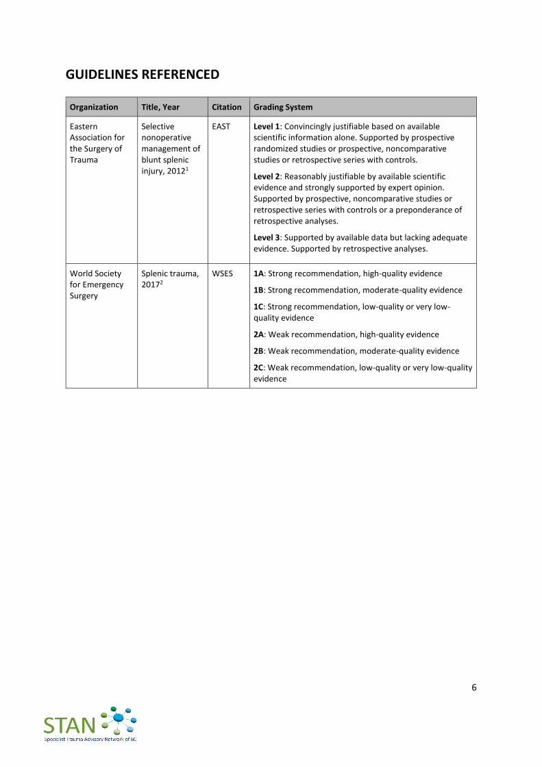
GUIDELINES REFERENCED
Organization Title, Year Citation Grading System
Eastern Association for the Surgery of Trauma
Selective nonoperative management of blunt splenic injury, 20121
EAST Level 1: Convincingly justifiable based on available scientific information alone. Supported by prospective randomized studies or prospective, noncomparative studies or retrospective series with controls.
Level 2: Reasonably justifiable by available scientific evidence and strongly supported by expert opinion. Supported by prospective, noncomparative studies or retrospective series with controls or a preponderance of retrospective analyses.
Level 3: Supported by available data but lacking adequate evidence. Supported by retrospective analyses.
World Society for Emergency Surgery
Splenic trauma, 20172
WSES 1A: Strong recommendation, high-quality evidence
1B: Strong recommendation, moderate-quality evidence
1C: Strong recommendation, low-quality or very low-quality evidence
2A: Weak recommendation, high-quality evidence
2B: Weak recommendation, moderate-quality evidence
2C: Weak recommendation, low-quality or very low-quality evidence
7
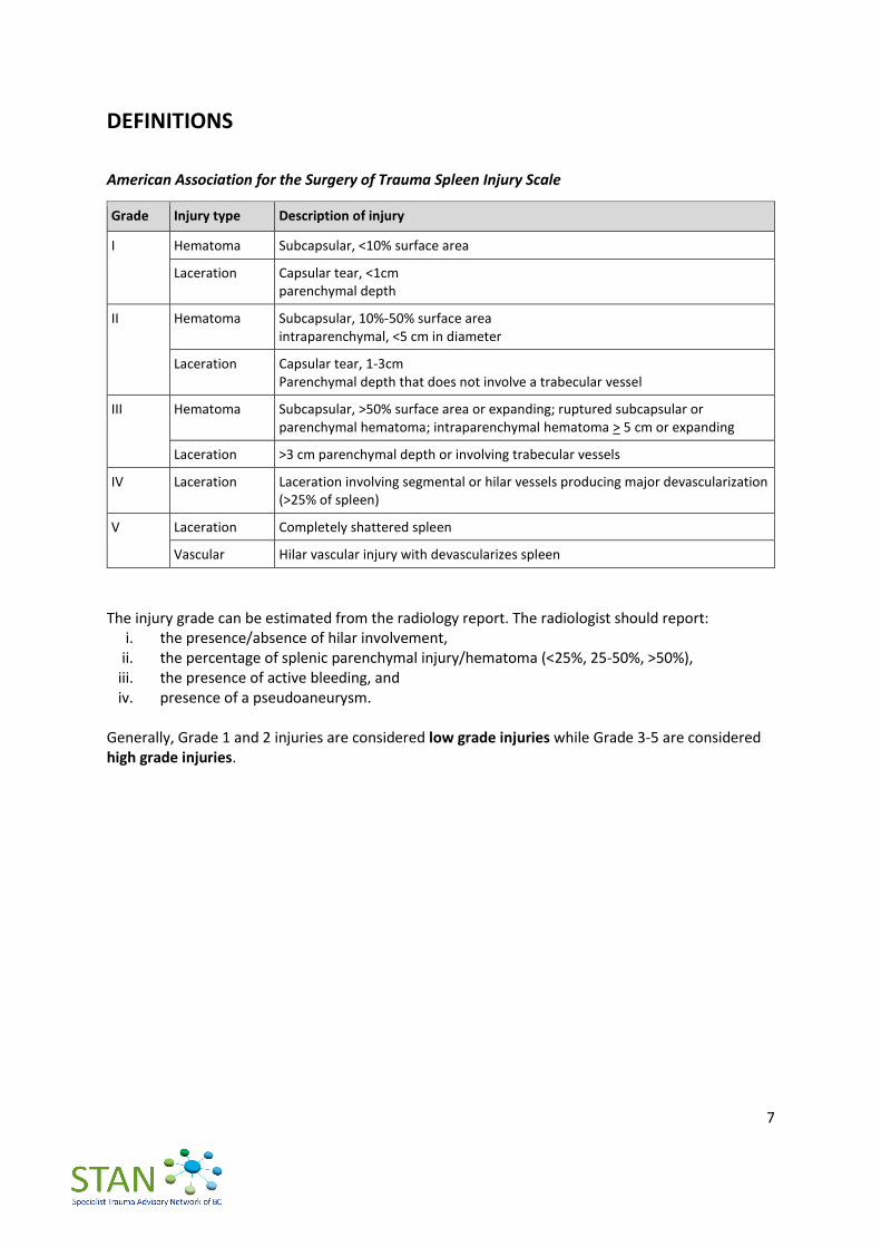
DEFINITIONS
American Association for the Surgery of Trauma Spleen Injury Scale
Grade Injury type Description of injury
I Hematoma Subcapsular, <10% surface area
Laceration Capsular tear, <1cm parenchymal depth
II Hematoma Subcapsular, 10%-50% surface area intraparenchymal, <5 cm in diameter
Laceration Capsular tear, 1-3cm Parenchymal depth that does not involve a trabecular vessel
III Hematoma Subcapsular, >50% surface area or expanding; ruptured subcapsular or parenchymal hematoma; intraparenchymal hematoma > 5 cm or expanding
Laceration >3 cm parenchymal depth or involving trabecular vessels
IV Laceration Laceration involving segmental or hilar vessels producing major devascularization (>25% of spleen)
V Laceration Completely shattered spleen
Vascular Hilar vascular injury with devascularizes spleen
The injury grade can be estimated from the radiology report. The radiologist should report:
i. the presence/absence of hilar involvement, ii. the percentage of splenic parenchymal injury/hematoma (<25%, 25-50%, >50%),
iii. the presence of active bleeding, and iv. presence of a pseudoaneurysm.
Generally, Grade 1 and 2 injuries are considered low grade injuries while Grade 3-5 are considered high grade injuries.

8
ALGORITHM

9
SUMMARY OF RECOMMENDATIONS All recommendations are newly drafted by the Thoraco-Abdominal SAG, unless indicated otherwise.
I. INITIAL ASSESSMENT AND MANAGEMENT
A. Initial resuscitation and management of the patient with blunt abdominal trauma should follow the Advanced Trauma Life Support® (ATLS®) principles.
B. In centres with surgical capability, the on-call general surgeon should be consulted promptly when a splenic injury is suspected or proven.
II. OPERATIVE MANAGEMENT
A. In centres with general surgical capability, urgent splenectomy should be performed for a hemodynamically unstable patient with a splenic injury who is not responding to appropriate resuscitation.
B. Grade or severity of splenic injury is not, in and of itself, an indication for surgical management of the injured spleen. The decision to proceed to splenectomy should be based on the clinical presentation of the patient and situational context, which includes the capabilities of the site, resources available, presence of other injuries, transport availability, and transfer related issues.
C. A general surgeon should be involved early in decision-making for suspected or proven splenic injury. Tele-conferencing through Patient Transfer Network (PTN) to discuss optimal management (transport vs. splenectomy) should be performed. The conference call should include the sending physician, the receiving general surgeon and the receiving Trauma Team Leader (TTL) at the higher level of care (HLOC) trauma referral centre.
III. NON-OPERATIVE MANAGEMENT
A. A trial of non-operative management (NOM) for splenic injury is indicated in patients with proven splenic injury who are hemodynamically stable after appropriate resuscitation. There are no absolute contraindications to a trial of NOM of known splenic injury in the hemodynamically stable or stabilized patient.
B. Hemodynamically stable patients with negligible risk* of ongoing or delayed hemorrhage may be safely managed, without higher level of care (HLOC) transfer, in a rural/remote facility provided at least 2 units of packed red blood cells are available. This management plan should be reviewed with a general surgeon and Trauma Team Leader (TTL) on call at the HLOC trauma referral centre in sites without surgical capabilities.
* CT-confirmed Grade 1-2 splenic injuries without evidence of active haemorrhage or pseudoaneurysm, anticoagulated patient, associated major injury, age ≥65 or limited physiologic reserve.
C. NOM of Grade 3-5 splenic injuries should only be considered in a hospital that has capabilities for physiologic monitoring and serial clinical evaluations by a general surgeon are possible. The hospital also needs 4 or more units of blood available, CT imaging, and 24-7 operating room access. Access to 24-7 interventional radiology for angiography/angioembolization is preferred but not essential. For transfer indications, see IV. TRANSFER TO HIGHER LEVEL OF CARE below.
10
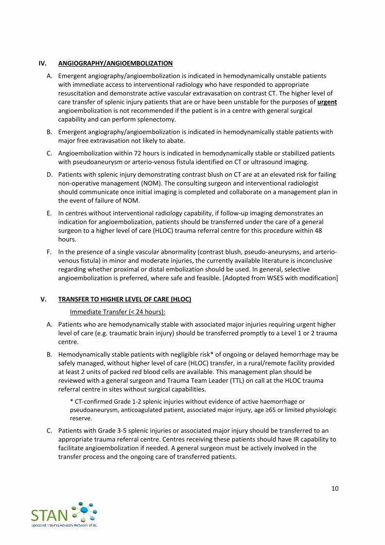
IV. ANGIOGRAPHY/ANGIOEMBOLIZATION
A. Emergent angiography/angioembolization is indicated in hemodynamically unstable patients with immediate access to interventional radiology who have responded to appropriate resuscitation and demonstrate active vascular extravasation on contrast CT. The higher level of care transfer of splenic injury patients that are or have been unstable for the purposes of urgent angioembolization is not recommended if the patient is in a centre with general surgical capability and can perform splenectomy.
B. Emergent angiography/angioembolization is indicated in hemodynamically stable patients with major free extravasation not likely to abate.
C. Angioembolization within 72 hours is indicated in hemodynamically stable or stabilized patients with pseudoaneurysm or arterio-venous fistula identified on CT or ultrasound imaging.
D. Patients with splenic injury demonstrating contrast blush on CT are at an elevated risk for failing non-operative management (NOM). The consulting surgeon and interventional radiologist should communicate once initial imaging is completed and collaborate on a management plan in the event of failure of NOM.
E. In centres without interventional radiology capability, if follow-up imaging demonstrates an indication for angioembolization, patients should be transferred under the care of a general surgeon to a higher level of care (HLOC) trauma referral centre for this procedure within 48 hours.
F. In the presence of a single vascular abnormality (contrast blush, pseudo-aneurysms, and arterio-venous fistula) in minor and moderate injuries, the currently available literature is inconclusive regarding whether proximal or distal embolization should be used. In general, selective angioembolization is preferred, where safe and feasible. [Adopted from WSES with modification]
V. TRANSFER TO HIGHER LEVEL OF CARE (HLOC)
Immediate Transfer (< 24 hours):
A. Patients who are hemodynamically stable with associated major injuries requiring urgent higher level of care (e.g. traumatic brain injury) should be transferred promptly to a Level 1 or 2 trauma centre.
B. Hemodynamically stable patients with negligible risk* of ongoing or delayed hemorrhage may be safely managed, without higher level of care (HLOC) transfer, in a rural/remote facility provided at least 2 units of packed red blood cells are available. This management plan should be reviewed with a general surgeon and Trauma Team Leader (TTL) on call at the HLOC trauma referral centre in sites without surgical capabilities.
* CT-confirmed Grade 1-2 splenic injuries without evidence of active haemorrhage or pseudoaneurysm, anticoagulated patient, associated major injury, age ≥65 or limited physiologic reserve.
C. Patients with Grade 3-5 splenic injuries or associated major injury should be transferred to an appropriate trauma referral centre. Centres receiving these patients should have IR capability to facilitate angioembolization if needed. A general surgeon must be actively involved in the transfer process and the ongoing care of transferred patients.
11
D. The HLOC transfer of splenic injury patients that are or have been unstable for the purposes of urgent angioembolization is not recommended if the patient is in a centre with general surgical capability and can perform splenectomy.
E. For patients undergoing emergent splenectomy prior to HLOC transfer, arrangements for transfer through Patient Transfer Network (PTN) should be made as early as possible, preferably pre-operatively or intraoperatively to avoid delay.
Delayed Transfer (> 24 hours):
F. In centres without interventional radiology capability, if follow-up imaging demonstrates an indication for angioembolization, patients should be transferred under the care of a general surgeon to a HLOC trauma referral centre for this procedure within 48 hours.
VI. ACUTE HOSPITAL CARE
A. Patients with Grade 1-2 splenic injuries can be monitored in a general surgery ward. The patient should have good IV access and assessed frequently for vital signs.
B. Patients with Grade 3-5 splenic injuries undergoing non-operative management (NOM) should be observed initially in a monitored intermediate care unit or intensive care unit (ICU). Appropriate initial monitoring includes the capacity to provide hourly vital signs as well as cardiac, oxygen saturation and urine output monitoring. Serial examination by a general surgeon is essential.
C. Hemoglobin should be monitored at regular intervals until stabilized.
D. It is recommended that therapeutic anticoagulation be reversed promptly in patients with high risk splenic injury, unless the risk of reversal is considered higher than the risk of splenic hemorrhage.
E. Repeat CT imaging in hemodynamically stable patients should be obtained within 72 hours post-injury for Grade 3-5 splenic injuries. Any changes in clinical status should prompt urgent reassessment, including laboratory investigations and/or CT as appropriate.
F. There is no need to restrict mobilization in patients with splenic injury and early mobilization is encouraged. Patients with high risk injuries* should remain supervised until assessed as safe to ambulate independently off unit.
*CT-confirmed Grade 3-5 splenic injuries, particularly with evidence of active haemorrhage or pseudoaneurysm, anticoagulated patient, associated major injury, age ≥65 or limited physiologic reserve.
G. Post-discharge, patients with Grade 3-5 splenic injuries should avoid contact sports or vigorous activities for at least 8 weeks. Patients with Grade 3-5 splenic injuries should be re-imaged prior to resuming high-risk activities.
VII. VENOUS THROMBOEMBOLISM (VTE) PROPHYLAXIS
A. Pharmacologic prophylaxis to prevent venous thromboembolism (VTE) can be used for patients with isolated blunt splenic injuries without increasing the failure rate of non-operative management. Although the optimal timing of safe initiation has not been determined, deep vein thrombosis (DVT) prophylaxis may be started as soon as possible after trauma and within 12 hours for every Grade of splenic injury (e.g. 36 hours for Grade 3 injury) or sooner if hemoglobin is stable. [Adopted from EAST and WSES with modification]
12
B. Mechanical prophylaxis should be used in all patients with absolute contraindication to pharmacologic prophylaxis, except in patients with lower extremity trauma in which case mechanical prophylaxis is not efficacious. [Adopted from WSES with modification]
VIII. OVERWHELMING POST SPLENECTOMY INFECTION (OPSI) PROPHYLAXIS
A. Patients should receive immunization against the encapsulated bacteria (S. pneumoniae, H. influenzae, and N. meningitidis) post-splenectomy or post-proximal angioembolization. Refer to national guidelines for vaccine dosage. [Adopted from WSES with modification]
B. Revaccination against pneumococcus is recommended every 10 years.
C. Vaccination should be administered >14 days post-splenectomy/embolization. For patients where follow-up is a concern, vaccination prior to discharge is recommended. [Adopted from EAST and WSES]
D. Regarding infection prophylaxis in asplenic and hyposplenic adult patients:
immunization against seasonal flu is recommended;
malaria prophylaxis is strongly recommended for travellers;
antibiotic therapy should be strongly considered in the event of any sudden onset of unexplained fever, malaise, chills or other constitutional symptoms, especially when medical review is not readily accessible; and
primary care providers should be aware of the splenectomy/angioembolization. [Adopted from WSES]
IX. POST HOSPITAL CARE
A. Post-discharge outpatient follow-up with imaging is recommended within 12 weeks. Patients with Grade 1-2 injuries should avoid contact sports or vigorous activities for at least 8 weeks. Grade 3-5 splenic injuries should be re-imaged at 8 weeks if the patient plans to resume high-risk activities to rule out pseudoaneurysm, subcapsular hematoma, etc.
B. Abdominal CT can be used for follow-up imaging and may allow for earlier return to sports activities. [Adapted from WSES]
C. If a new pseudoaneurysm is noted on follow-up imaging, discussion with general surgery is recommended to determine best management, e.g. serial imaging vs. embolization.
13
SCIENTIFIC DISCUSSION All recommendations are newly drafted by the Thoraco-Abdominal SAG, unless indicated otherwise.
I. INITIAL ASSESSMENT AND MANAGEMENT KMQ-1. What are key considerations in the initial assessment and management patients with
suspected or confirmed blunt splenic injury? RECOMMENDATIONS
A. Initial resuscitation and management of the patient with blunt abdominal trauma should follow the Advanced Trauma Life Support® (ATLS®) principles.
B. In centres with surgical capability, the on-call general surgeon should be consulted promptly when a splenic injury is suspected or proven.
KNOWLEDGE SYNTHESIS
External Recommendations SAG’s Rationale
None Developed new recommendations based on expert opinion of the SAG and the BC trauma system.
14
II. OPERATIVE MANAGEMENT KMQ-2. What are the indications for operative management (OM) of blunt splenic injuries? RECOMMENDATIONS
A. In centres with general surgical capability, urgent splenectomy should be performed for a hemodynamically unstable patient with a splenic injury who is not responding to appropriate resuscitation.
B. Grade or severity of splenic injury is not, in and of itself, an indication for surgical management of the injured spleen. The decision to proceed to splenectomy should be based on the clinical presentation of the patient and situational context, which includes the capabilities of the site, resources available, presence of other injuries, transport availability, and transfer related issues.
C. A general surgeon should be involved early in decision-making for suspected or proven splenic injury. Tele-conferencing through Patient Transfer Network (PTN) to discuss optimal management (transport vs. splenectomy) should be performed. The conference call should include the sending physician, the receiving general surgeon and the receiving Trauma Team Leader (TTL) at the higher level of care (HLOC) trauma referral centre.
KNOWLEDGE SYNTHESIS
External Recommendation SAG’s Rationale
Patients who have diffuse peritonitis or who are hemodynamically unstable after blunt abdominal trauma should be taken urgently for laparotomy. [EAST: Level 1]
OM should be performed in patients with hemodynamic instability and/or with associated lesions like peritonitis or bowel evisceration or impalement requiring surgical exploration. [WSES: 2A]
Accepted hemodynamic instability as an indicator of OM but rejected diffuse peritonitis and bowel evisceration (A).
Splenectomy should be performed when NOM with AG/AE failed, and patient remains hemodynamically unstable or shows a significant drop in hematocrit levels or continuous transfusion are required. [WSES: 2A]
Accepted continued hemodynamic instability as an indicator of OM (A). Emphasized a balance of clinical presentation and other situational contexts, including site-specific resources and feasibility of transfer/transport to reflect the BC trauma system (B).
OM should be performed in moderate and severe lesions even in stable patients in centers where intensive monitoring cannot be performed and/or when AG/AE is not rapidly available. [WSES: 2A]
Accepted the concept of resource requirements for OM. Emphasized early consult with general surgery and initiation of PTN call to discuss transport versus onsite

15
External Recommendation SAG’s Rationale
splenectomy and to encourage site-to-site communication.
16
III. NON-OPERATIVE MANAGEMENT KMQ-3. What are the indications for non-operative management (NOM) in blunt splenic
injuries? RECOMMENDATIONS
A. A trial of non-operative management (NOM) for splenic injury is indicated in patients with proven splenic injury who are hemodynamically stable after appropriate resuscitation. There are no absolute contraindications to a trial of NOM of known splenic injury in the hemodynamically stable or stabilized patient.
B. Hemodynamically stable patients with negligible risk* of ongoing or delayed hemorrhage may be safely managed, without higher level of care (HLOC) transfer, in a rural/remote facility provided at least 2 units of packed red blood cells are available. This management plan should be reviewed with a general surgeon and Trauma Team Leader (TTL) on call at the HLOC trauma referral centre in sites without surgical capabilities.
* CT-confirmed Grade 1-2 splenic injuries without evidence of active haemorrhage or pseudoaneurysm, anticoagulated patient, associated major injury, age ≥65 or limited physiologic reserve.
C. NOM of Grade 3-5 splenic injuries should only be considered in a hospital that has capabilities for physiologic monitoring and serial clinical evaluations by a general surgeon are possible. The hospital also needs 4 or more units of blood available, CT imaging, and 24-7 operating room access. Access to 24-7 interventional radiology for angiography/angioembolization is preferred but not essential. For transfer indications, see IV. TRANSFER TO HIGHER LEVEL OF CARE below.
KNOWLEDGE SYNTHESIS
External Recommendations SAG’s Rationale
Indications for NOM
A routine laparotomy is not indicated in the hemodynamically stable patient without peritonitis presenting with an isolated splenic injury. [EAST: Level 2]
NOM in splenic injuries is contraindicated in the setting of unresponsive hemodynamic instability or other indicators for laparotomy (peritonitis, hollow organ injuries, bowel evisceration, impalement). [WSES: 1A]
Adapted EAST and WSES recommendations to create a new recommendation (A) indicating a trial of NOM in patients who are hemodynamically stable after resuscitation.
Non-contraindications for a trial of NOM
The severity of splenic injury (as suggested by CT grade or degree of hemoperitoneum), neurologic status, age >55 and/or the presence of associated injuries are not contraindications to a trial of non-operative management in a hemodynamically stable patient. [EAST: Level 2]
Consolidated the external recommendations into a single statement (A) regarding the absence of absolute contraindications to a trial of NOM in the hemodynamically stable or stabilized patient.

17
External Recommendations SAG’s Rationale
Age above 55 years old alone, large hemoperitoneum alone, hypotension before resuscitation, GCS < 12 and low-hematocrit level at the admission, associated abdominal injuries, blush at CT scan, anticoagulation drugs, HIV disease, drug addiction, cirrhosis and need for blood transfusions should be taken into account, but they are not absolute contraindications for NOM. [WSES: 2B]
Patients with hemodynamic stability and absence of other abdominal organ injuries requiring surgery should undergo an initial attempt of NOM irrespective of injury grade. [WSES: 2A]
Other considerations: Monitoring and OR availability
Nonoperative management of splenic injuries should only be considered in an environment that provides capabilities for monitoring, serial clinical evaluations, and an operating room available for urgent laparotomy. [EAST: Level 2]
NOM of moderate or severe spleen injuries should be considered only in an environment that provides capability for patient intensive monitoring, AG/AE, an immediately available OR and immediate access to blood and blood product or alternatively in the presence of a rapid centralization system and only in patients with stable or stabilized hemodynamic and absence of other internal injuries requiring surgery. [WSES: 2A]
Strong evidence exists that age above 55 years old, high ISS, and moderate to severe splenic injuries are prognostic factors for NOM failure. These patients require more intensive monitoring and higher index of suspicion. [WSES: 2B]
Incorporated concepts from EAST and WSES statements. The concept of negligible risk of ongoing or delayed hemorrhage was introduced to indicate the types of splenic injuries that can be safely managed in a rural/remote facility with consult with a HLOC centre (B). Adopted WSES statement regarding NOM of moderate to severe splenic injuries and added site-specific requirements, such as access to radiology, interventional radiology and surgical capabilities. Provincial communication pathways for trauma were outlined and emphasized. (C)
Additional Literature Support What is the success rate of non-operative management of blunt splenic injuries?
Overall reported success rate of observational management (without angiography) is 92-96 %.3,4,5,6 Success rate of observational management of blunt splenic injury by injury grade
Source (lead author, year)
Grade 1 Grade 2 Grade 3 Grade 4 Grade 5*
Brillantino 20165 100 % 95.4 % 95 % 90.9 % 83.3 %
Brault-Noble 20123 100 % 98 % 84 % 79 % 78 %
Bhullar 20127 99 % 98 % 94 % 77 % 37 %
McCray 20084 100 % 99 % 94 % 84 % 100 %
* Grade 5 blunt splenic injuries are rare, resulting in a greater variability in success rate reported in studies
18
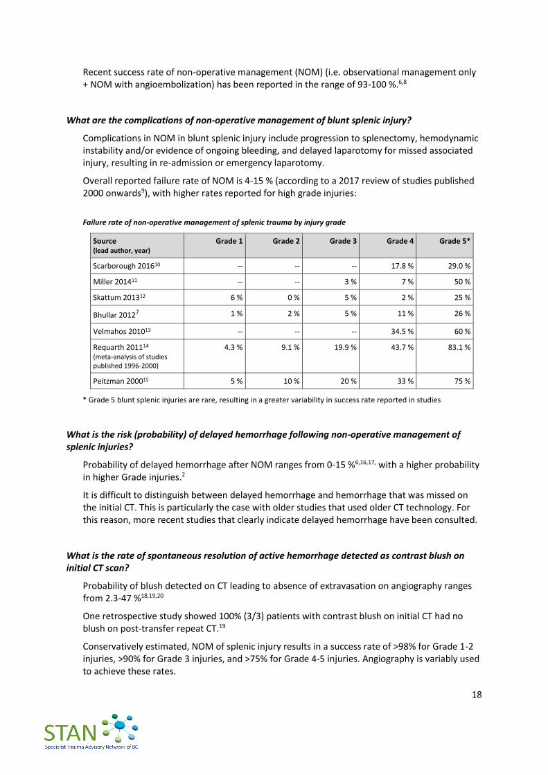
Recent success rate of non-operative management (NOM) (i.e. observational management only + NOM with angioembolization) has been reported in the range of 93-100 %.6,8
What are the complications of non-operative management of blunt splenic injury?
Complications in NOM in blunt splenic injury include progression to splenectomy, hemodynamic instability and/or evidence of ongoing bleeding, and delayed laparotomy for missed associated injury, resulting in re-admission or emergency laparotomy.
Overall reported failure rate of NOM is 4-15 % (according to a 2017 review of studies published 2000 onwards9), with higher rates reported for high grade injuries:
Failure rate of non-operative management of splenic trauma by injury grade
Source (lead author, year)
Grade 1 Grade 2 Grade 3 Grade 4 Grade 5*
Scarborough 201610 -- -- -- 17.8 % 29.0 %
Miller 201411 -- -- 3 % 7 % 50 %
Skattum 201312 6 % 0 % 5 % 2 % 25 %
Bhullar 20127 1 % 2 % 5 % 11 % 26 %
Velmahos 201013 -- -- -- 34.5 % 60 %
Requarth 201114
(meta-analysis of studies published 1996-2000)
4.3 % 9.1 % 19.9 % 43.7 % 83.1 %
Peitzman 200015 5 % 10 % 20 % 33 % 75 %
* Grade 5 blunt splenic injuries are rare, resulting in a greater variability in success rate reported in studies
What is the risk (probability) of delayed hemorrhage following non-operative management of splenic injuries?
Probability of delayed hemorrhage after NOM ranges from 0-15 %6,16,17, with a higher probability in higher Grade injuries.2
It is difficult to distinguish between delayed hemorrhage and hemorrhage that was missed on the initial CT. This is particularly the case with older studies that used older CT technology. For this reason, more recent studies that clearly indicate delayed hemorrhage have been consulted.
What is the rate of spontaneous resolution of active hemorrhage detected as contrast blush on initial CT scan?
Probability of blush detected on CT leading to absence of extravasation on angiography ranges from 2.3-47 %18,19,20
One retrospective study showed 100% (3/3) patients with contrast blush on initial CT had no blush on post-transfer repeat CT.19
Conservatively estimated, NOM of splenic injury results in a success rate of >98% for Grade 1-2 injuries, >90% for Grade 3 injuries, and >75% for Grade 4-5 injuries. Angiography is variably used to achieve these rates.
19
IV. ANGIOGRAPHY / ANGIOEMBOLIZATION KMQ-4. What are the indications for angiography/angioembolization (AG/AE) in blunt splenic
injuries? RECOMMENDATIONS
A. Emergent angiography/angioembolization is indicated in hemodynamically unstable patients with immediate access to interventional radiology who have responded to appropriate resuscitation and demonstrate active vascular extravasation on contrast CT. The higher level of care transfer of splenic injury patients that are or have been unstable for the purposes of urgent angioembolization is not recommended if the patient is in a centre with general surgical capability and can perform splenectomy.
B. Emergent angiography/angioembolization is indicated in hemodynamically stable patients with major free extravasation not likely to abate.
C. Angioembolization within 72 hours is indicated in hemodynamically stable or stabilized patients with pseudoaneurysm or arterio-venous fistula identified on CT or ultrasound imaging.
D. Patients with splenic injury demonstrating contrast blush on CT are at an elevated risk for failing non-operative management (NOM). The consulting surgeon and interventional radiologist should communicate once initial imaging is completed and collaborate on a management plan in the event of failure of NOM.
E. In centres without interventional radiology capability, if follow-up imaging demonstrates an indication for angioembolization, patients should be transferred under the care of a general surgeon to a higher level of care (HLOC) trauma referral centre for this procedure within 48 hours.
KNOWLEDGE SYNTHESIS
External Recommendations SAG’s Rationale
Indications
Angiography should be considered for patients with American Association for the Surgery of Trauma (AAST) grade of greater than III injuries, presence of a contrast blush, moderate hemoperitoneum, or evidence of ongoing splenic bleeding. [EAST: Level 2]
AG/AE may be considered the first-line intervention in patients with hemodynamic stability and arterial blush on CT scan irrespective from injury grade. [WSES: 2B]
AG/AE may be performed in hemodynamically stable and rapid responder patients with moderate and severe lesions and in those with vascular injuries at CT scan (contrast blush, pseudo-aneurysms and arterio-venous fistula). [WSES: 2A]
Accepted hemodynamic stability (including after resuscitation and not likely to abate) and diagnostic imaging abnormalities (i.e. active vascular extravasation, pseudoaneurysm, and arterio-venous fistula) as indicators for IR consult for AG/AE (A, B, C).
Contrast blush on CT emphasized as an elevated risk for NOM failure.
Emphasized interdisciplinary collaboration between consulting surgeon and interventional radiologist (D).
20
External Recommendations SAG’s Rationale
AG/AE should be considered in all hemodynamically stable patients with WSES grade III lesions, regardless with the presence of CT blush. [WSES: 1B]
AG/AE could be considered in patients undergoing to NOM, hemodynamically stable with signs of persistent hemorrhage regardless of the presence of CT blush once excluded extrasplenic source of bleeding. [WSES: 1C]
Contraindications
Contrast blush on CT scan alone is not an absolute indication for an operation or angiographic intervention. Factors such as patient age, grade of injury, and presence of hypotension need to be considered in the clinical management of these patients. [EAST: Level 3]
Hemodynamically stable patients with WSES grade II lesions without blush should not undergo routine AG/AE but may be considered for prophylactic proximal embolization in presence of risk factors for NOM failure. [WSES: 2B]
Emphasized interdisciplinary collaboration between consulting surgeon and interventional radiologist in the clinical decision-making (D).
Management Pathway
In patients with bleeding vascular injuries and in those with intraperitoneal blush, AG/AE should be performed as part of NOM only in centers where AG/AE is rapidly available. In other centers and in case of rapid hemodynamic deterioration, OM should be considered. [WSES: 2B]
Outlined transfer requirements to HLOC and emphasized inter-facility communication. (E)
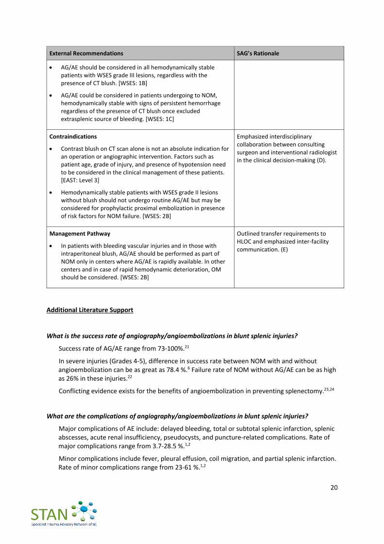
Additional Literature Support What is the success rate of angiography/angioembolizations in blunt splenic injuries?
Success rate of AG/AE range from 73-100%.21
In severe injuries (Grades 4-5), difference in success rate between NOM with and without angioembolization can be as great as 78.4 %.6 Failure rate of NOM without AG/AE can be as high as 26% in these injuries.22
Conflicting evidence exists for the benefits of angioembolization in preventing splenectomy.23,24
What are the complications of angiography/angioembolizations in blunt splenic injuries?
Major complications of AE include: delayed bleeding, total or subtotal splenic infarction, splenic abscesses, acute renal insufficiency, pseudocysts, and puncture-related complications. Rate of major complications range from 3.7-28.5 %.1,2
Minor complications include fever, pleural effusion, coil migration, and partial splenic infarction. Rate of minor complications range from 23-61 %.1,2
21
No randomized control trials exist comparing morbidity related to AG/AE and NOM without AG/AE.
A large prospective study found AG/AE-related morbidity of 47% compared to morbidity of 10% in NOM without AG/AE.6
A large study of post-discharge complications in patients who received NOM found higher rate of thirty-day readmission among patients who received NOM with AE than patients who did not receive AE (12.8% vs. 7.4%, p=0.002).25
KMQ-5. With regard to selective versus non-selective angioembolization, what is the preferred approach to angioembolization in splenic injuries?
RECOMMENDATIONS
F. In the presence of a single vascular abnormality (contrast blush, pseudo-aneurysms, and arterio-venous fistula) in minor and moderate injuries, the currently available literature is inconclusive regarding whether proximal or distal embolization should be used. In general, selective angioembolization is preferred, where safe and feasible. [Adopted from WSES with modification]
KNOWLEDGE SYNTHESIS
External Recommendations SAG’s Rationale
In the presence of a single vascular abnormality (contrast blush, pseudo-aneurysms, and artero-venous fistula) in minor and moderate injuries, the currently available literature is inconclusive regarding whether proximal or distal embolization should be used. In the presence of multiple splenic vascular abnormalities or in the presence of a severe lesion, proximal or combined AG/AE should be used, after confirming the presence of a permissive pancreatic vascular anatomy. [WSES: 1C]
Adopted first sentence. Replaced second sentence with preference for selective (i.e. proximal) angioembolization due to fewer minor complications reported in retrospective cohort studies (see below).
Additional Literature Support What is the effectiveness of selective versus non-selective angioembolization? What are the complications?
No prospective studies or randomized controlled trials available on the subject.
No significant difference observed in overall failure rate between distal and proximal embolization.
No significant difference has been observed between proximal and distal embolization for incidence of major infarctions, infections or re-bleeding.
Higher rate of minor complications has been reported in distal than in proximal embolization (see table below). Proximal embolization is also protective in high grade injuries.26

22
Complications in proximal vs. distal splenic embolization
Complication Proximal Embolization Distal Embolization
Minor infarction 0.0-8.4 %27 14.3-19.8 %27
Re-bleeding 2.2-2.8 %27 1.6-4.5 %27

23
V. TRANSFER TO HIGHER LEVEL OF CARE (HLOC) KMQ-6. What are the indications for transfer of patients with blunt splenic injuries to a higher-
level trauma center? RECOMMENDATIONS
Immediate Transfer (< 24 hours):
A. Patients who are hemodynamically stable with associated major injuries requiring urgent higher level of care (e.g. traumatic brain injury) should be transferred promptly to a Level 1 or 2 trauma centre.
B. Hemodynamically stable patients with negligible risk* of ongoing or delayed hemorrhage may be safely managed, without higher level of care (HLOC) transfer, in a rural/remote facility provided at least 2 units of packed red blood cells are available. This management plan should be reviewed with a general surgeon and Trauma Team Leader (TTL) on call at the HLOC trauma referral centre in sites without surgical capabilities.
* CT-confirmed Grade 1-2 splenic injuries without evidence of active haemorrhage or pseudoaneurysm, anticoagulated patient, associated major injury, age ≥65 or limited physiologic reserve.
C. Patients with Grade 3-5 splenic injuries or associated major injury should be transferred to an appropriate trauma referral centre. Centres receiving these patients should have IR capability to facilitate angioembolization if needed. A general surgeon must be actively involved in the transfer process and the ongoing care of transferred patients.
D. The HLOC transfer of splenic injury patients that are or have been unstable for the purposes of urgent angioembolization is not recommended if the patient is in a centre with general surgical capability and can perform splenectomy.
E. For patients undergoing emergent splenectomy prior to HLOC transfer, arrangements for transfer through Patient Transfer Network (PTN) should be made as early as possible, preferably pre-operatively or intraoperatively to avoid delay.
Delayed Transfer (> 24 hours):
F. In centres without interventional radiology capability, if follow-up imaging demonstrates an indication for angioembolization, patients should be transferred under the care of a general surgeon to a HLOC trauma referral centre for this procedure within 48 hours.
KNOWLEDGE SYNTHESIS
External Recommendations SAG’s Rationale
None Recommendations regarding transfer to higher level of care were drafted, based on provincial realities and the expert opinion of the SAG.
24
VI. ACUTE HOSPITAL CARE KMQ-7. What type and duration of monitoring are necessary for patients with blunt splenic
injuries? RECOMMENDATIONS
A. Patients with Grade 1-2 splenic injuries can be monitored in a general surgery ward. The patient should have good IV access and assessed frequently for vital signs.
B. Patients with Grade 3-5 splenic injuries undergoing non-operative management (NOM) should be observed initially in a monitored intermediate care unit or intensive care unit (ICU). Appropriate initial monitoring includes the capacity to provide hourly vital signs as well as cardiac, oxygen saturation and urine output monitoring. Serial examination by a general surgeon is essential.
C. Hemoglobin should be monitored at regular intervals until stabilized.
D. It is recommended that therapeutic anticoagulation be reversed promptly in patients with high risk splenic injury, unless the risk of reversal is considered higher than the risk of splenic hemorrhage.
KNOWLEDGE SYNTHESIS
External Recommendations SAG’s Rationale
Clinical and laboratory observation associated [with] bed rest in moderate and severe lesions is the cornerstone in the first 48-72 hour follow-up. [WSES: 1C]
The only external recommendation for monitoring pertains to the first 48-72 hours. Created new recommendations outlining monitoring requirements based on expert opinion.
KMQ-8. When is supplementary imaging required in the hospitalized patient?
RECOMMENDATIONS
E. Repeat CT imaging in hemodynamically stable patients should be obtained within 72 hours post-injury for Grade 3-5 splenic injuries. Any changes in clinical status should prompt urgent reassessment, including laboratory investigations and/or CT as appropriate.
KNOWLEDGE SYNTHESIS
External Recommendations SAG’s Rationale
25
After blunt splenic injury, clinical factors such as a persistent systemic inflammatory response, increasing/persistent abdominal pain, or an otherwise unexplained drop in hemoglobin should dictate the frequency of and need for follow-up imaging for a patient with blunt splenic injury. [EAST: Level 3]
CT scan repetition during the admission should be considered in patients with moderate and severe lesions or in decreasing hematocrit, in presence of vascular anomalies or underlying splenic pathology or coagulopathy, and in neurologically impaired patients. [WSES: 2A]
Developed umbrella phrase “any changes in clinical status” as potential indicator of repeat imaging or other investigations. Accepted WSES indication for repeat CT in higher grade injuries and added time frame within which to obtain the repeat scan based on evidence of delayed splenic pseudoaneurysm formation as early as 48 hours (see below) and on logistical realities of provincial trauma centres.
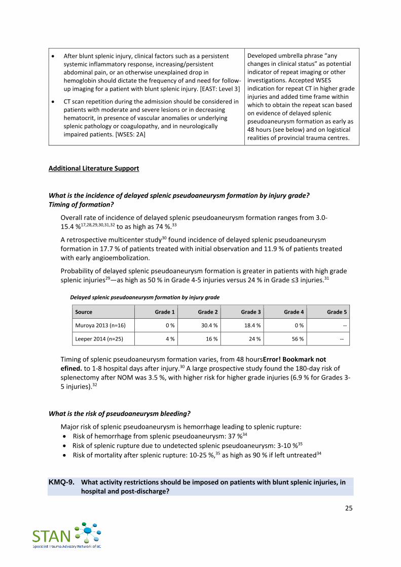
Additional Literature Support What is the incidence of delayed splenic pseudoaneurysm formation by injury grade? Timing of formation?
Overall rate of incidence of delayed splenic pseudoaneurysm formation ranges from 3.0-15.4 %17,28,29,30,31,32 to as high as 74 %.33
A retrospective multicenter study30 found incidence of delayed splenic pseudoaneurysm formation in 17.7 % of patients treated with initial observation and 11.9 % of patients treated with early angioembolization.
Probability of delayed splenic pseudoaneurysm formation is greater in patients with high grade splenic injuries29—as high as 50 % in Grade 4-5 injuries versus 24 % in Grade ≤3 injuries.31
Delayed splenic pseudoaneurysm formation by injury grade
Source Grade 1 Grade 2 Grade 3 Grade 4 Grade 5
Muroya 2013 (n=16) 0 % 30.4 % 18.4 % 0 % --
Leeper 2014 (n=25) 4 % 16 % 24 % 56 % --
Timing of splenic pseudoaneurysm formation varies, from 48 hoursError! Bookmark not efined. to 1-8 hospital days after injury.30 A large prospective study found the 180-day risk of splenectomy after NOM was 3.5 %, with higher risk for higher grade injuries (6.9 % for Grades 3-5 injuries).32
What is the risk of pseudoaneurysm bleeding?
Major risk of splenic pseudoaneurysm is hemorrhage leading to splenic rupture:
Risk of hemorrhage from splenic pseudoaneurysm: 37 %34
Risk of splenic rupture due to undetected splenic pseudoaneurysm: 3-10 %35
Risk of mortality after splenic rupture: 10-25 %,35 as high as 90 % if left untreated34 KMQ-9. What activity restrictions should be imposed on patients with blunt splenic injuries, in
hospital and post-discharge?
26
RECOMMENDATIONS
F. There is no need to restrict mobilization in patients with splenic injury and early mobilization is encouraged. Patients with high risk injuries* should remain supervised until assessed as safe to ambulate independently off unit.
*CT-confirmed Grade 3-5 splenic injuries, particularly with evidence of active haemorrhage or pseudoaneurysm, anticoagulated patient, associated major injury, age ≥65 or limited physiologic reserve.
G. Post-discharge, patients with Grade 3-5 splenic injuries should avoid contact sports or vigorous activities for at least 8 weeks. Patients with Grade 3-5 splenic injuries should be re-imaged prior to resuming high-risk activities.
KNOWLEDGE SYNTHESIS
External Recommendations SAG’s Rationale
Activity restriction may be suggested for 4-6 weeks in minor injuries and up to 2-4 months in moderate and severe injuries. [WSES: 2C]
New recommendation has been created, based on recent evidence (see below) and expert opinion of the SAG.
Additional Literature Support What is the risk of delayed hemorrhage in blunt splenic patients without activity restrictions?
Several recent studies have shown no association between early mobilization with minimal bed rest and delayed splenic hemorrhage both in adult16,36,37,38 and pediatric39,40,41 patients with blunt splenic injuries managed via NOM.

27
VII. VENOUS THROMBOEMBOLISM PROPHYLAXIS
KMQ-10. What is the optimal timing for initiating deep vein thrombosis (DVT) prophylaxis in
patients with blunt splenic injuries? RECOMMENDATIONS
A. Pharmacologic prophylaxis to prevent venous thromboembolism (VTE) can be used for patients with isolated blunt splenic injuries without increasing the failure rate of non-operative management. Although the optimal timing of safe initiation has not been determined, deep vein thrombosis (DVT) prophylaxis may be started as soon as possible after trauma and within 12 hours for every Grade of splenic injury (e.g. 36 hours for Grade 3 injury) or sooner if hemoglobin is stable. [Adopted from EAST and WSES with modification]
B. Mechanical prophylaxis should be used in all patients with absolute contraindication to pharmacologic prophylaxis, except in patients with lower extremity trauma in which case mechanical prophylaxis is not efficacious. [Adopted from WSES with modification]
KNOWLEDGE SYNTHESIS
External Recommendations SAG’s Rationale
Chemical prophylaxis
Pharmacologic prophylaxis to prevent venous thromboembolism can be used for patients with isolated blunt splenic injuries without increasing the failure rate of non-operative management, although the optimal timing of safe initiation has not been determined. [EAST: Level 3]
Spleen trauma without ongoing bleeding is not an absolute contraindication to LMWH-based prophylactic anticoagulation. [WSES: 2A]
LMWH-based prophylactic anticoagulation should be started as soon as possible from trauma and may be safe in selected patients with blunt splenic injury undergoing NOM. [WSES: 2B]
In patients with oral anticoagulants the risk-benefit balance of reversal should be individualized. [WSES: 2B]
Consolidated external recommendations. Added timing of VTE prophylaxis (i.e. within 12 hours for every injury grade) based on the expert opinion of the SAG.
Mechanical prophylaxis
Mechanical prophylaxis is safe and should be considered in all patients without absolute contraindication to its use. [WSES: 2A]
Added a contraindication for the use of mechanical prophylaxis: patients with lower extremity trauma.
28
Additional Literature Support What is the risk of developing thrombosis VTE prophylaxis after blunt splenic injuries?
A prospective study (n=147) found 5 % risk of developing VTE after trauma-related splenectomy.42
A large retrospective study (n=6,162) found 1.97 times greater risk of VTE in splenic injury than in control, with a rate of 10.08 per 10,000 person-years (8.46 no splenectomy, 11.81 splenectomy).43
A large prospective study (n=675) found increased risk for VTE with splenectomy (AOR 2.6, 95% CI 1.2 to 5.9).44
What is the incidence of hemorrhage in splenic patients with/without VTE prophylaxis?
Several retrospective studies indicate low-molecular weight heparin (LMWH) administration does not increase the failure rate of NOM45,46 or increase the risk of bleeding events.47
29
VIII. OVERWHELMING POST-SPLENECTOMY INFECTION PROPHYLAXIS KMQ-11. Which vaccinations should be administered and when in patients with blunt splenic
injuries? RECOMMENDATIONS
A. Patients should receive immunization against the encapsulated bacteria (S. pneumoniae, H. influenzae, and N. meningitidis) post-splenectomy or post-proximal angioembolization. Refer to national guidelines for vaccine dosage. [Adopted from WSES with modification]
B. Revaccination against pneumococcus is recommended every 10 years.
C. Vaccination should be administered >14 days post-splenectomy/embolization. For patients where follow-up is a concern, vaccination prior to discharge is recommended. [Adopted from EAST and WSES]
D. Regarding infection prophylaxis in asplenic and hyposplenic adult patients:
immunization against seasonal flu is recommended;
malaria prophylaxis is strongly recommended for travellers;
antibiotic therapy should be strongly considered in the event of any sudden onset of unexplained fever, malaise, chills or other constitutional symptoms, especially when medical review is not readily accessible; and
primary care providers should be aware of the splenectomy/angioembolization. [Adopted from WSES]
KNOWLEDGE SYNTHESIS
External Recommendations SAG’s Rationale
Vaccination type
Patients should receive immunization against the encapsulated bacteria (S. pneumoniae, H. influenzae, and N. meningitidis). [WSES: 1A]
Adopted and added “post-splenectomy or post-proximal angioembolization” for clarity in clinical management.
Vaccination schedule/timing
Vaccination programs should be started no sooner than 14 days after splenectomy or spleen total vascular exclusion. [WSES: 2C]
In patients discharged before 15 days after splenectomy or angioembolization, where the risk to miss vaccination is deemed high, the best choice is to vaccinate before discharge. [WSES: 1B]
Adopted and combined the two statements into one recommendation.
Other vaccination indications Adopted and combined the four statements into one recommendation for easier reading.
30
External Recommendations SAG’s Rationale
Regarding infections prophylaxis in asplenic and hyposplenic adult and pediatric patients, immunization against seasonal flu is recommended for patients over 6 months of age. [WSES: 1C]
Regarding infections prophylaxis in asplenic and hyposplenic adult and pediatric patients, Malaria prophylaxis is strongly recommended for travelers. [WSES: 2C]
Regarding infections prophylaxis in asplenic and hyposplenic adult and pediatric patients, antibiotic therapy should be strongly considered in the event of any sudden onset of unexplained fever, malaise, chills or other constitutional symptoms, especially when medical review is not readily accessible. [WSES: 2A]
Regarding infections prophylaxis in asplenic and hyposplenic adult and pediatric patients, primary care providers should be aware of the splenectomy/ angioembolization. [WSES: 2C]
Additional Literature Support What is the risk of overwhelming post-splenectomy infection (OPSI) with splenectomy or splenic embolization after splenic injury?
Risk of overwhelming post-splenectomy infection (OPSI) with splenectomy or splenic embolization ranges from 0.05-23 %,2,48 with the majority of infections occurring more than 2 years following the procedure.49
A large retrospective study (n= 4,360) of blunt splenic trauma patients in California reported short- and long-term infectious complications by procedure50:
Procedure Admission 30 days after injury 1 year after injury
Splenic angioembolization 1.59 % 5.18 % 9.16 %
Splenectomy 1.76 % 4.85 % 8.85 %
A larger retrospective study of over 4000 patients with Grade 4-5 splenic injuries reported infectious complications in 11.7 % in the angioembolization group and 23.1 % in the splenectomy group.48
Risk of mortality due to OPSI is 30-70 %, most deaths occurring within first 24 hours.2
What is the optimal timing of vaccination?
All vaccines are best administered 2 weeks after surgery. If the patient is discharged earlier and there is concern that they might not return for follow-up, vaccines should be administered prior to discharge.51
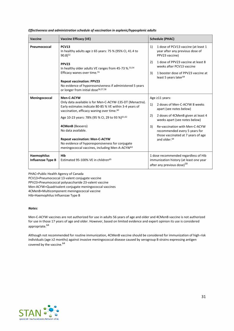
What is the effectiveness of vaccination? What is the effectiveness of repeat vaccination?
31
Effectiveness and administration schedule of vaccination in asplenic/hyposplenic adults
Vaccine Vaccine Efficacy (VE) Schedule (PHAC)
Pneumococcal PCV13 In healthy adults age ≥ 65 years: 75 % (95% CI, 41.4 to 90.8)52 PPV23 In healthy older adults VE ranges from 45-73 %.53,54 Efficacy wanes over time.55 Repeat vaccination: PPV23 No evidence of hyporesonsiveness if administered 5 years or longer from initial dose56,57,58
1) 1 dose of PCV13 vaccine (at least 1 year after any previous dose of PPV23 vaccine)
2) 1 dose of PPV23 vaccine at least 8 weeks after PCV13 vaccine
3) 1 booster dose of PPV23 vaccine at least 5 years later59
Meningococcal Men-C-ACYW Only data available is for Men-C-ACYW-135-DT (Menactra). Early estimates indicate 80-85 % VE within 3-4 years of vaccination, efficacy waning over time.60
Age 10-23 years: 78% (95 % CI, 29 to 93 %)61,62
4CMenB (Bexsero) No data available. Repeat vaccination: Men-C-ACYW No evidence of hyporesponsiveness for conjugate meningococcal vaccines, including Men-A-ACYW63
Age ≥11 years:
1) 2 doses of Men-C-ACYW 8 weeks apart (see notes below)
2) 2 doses of 4CMenB given at least 4 weeks apart (see notes below)
3) Re-vaccination with Men-C-ACYW recommended every 5 years for those vaccinated at 7 years of age and older.64
Haemophilus Influenzae Type B
Hib Estimated 95-100% VE in children65
1 dose recommended regardless of Hib immunization history (at least one year
after any previous dose)59
PHAC=Public Health Agency of Canada PCV13=Pneumococcal 13-valent conjugate vaccine PPV23=Pneumococcal polysaccharide 23-valent vaccine Men-ACYW=Quadrivalent conjugate meningococcal vaccines 4CMenB=Multicomponent meningococcal vaccine Hib=Haemophilus Influenzae Type B Notes: Men-C-ACYW vaccines are not authorized for use in adults 56 years of age and older and 4CMenB vaccine is not authorized for use in those 17 years of age and older. However, based on limited evidence and expert opinion its use is considered
appropriate.64 Although not recommended for routine immunization, 4CMenB vaccine should be considered for immunization of high-risk individuals (age ≥2 months) against invasive meningococcal disease caused by serogroup B strains expressing antigen
covered by the vaccine.64
32
IX. POST HOSPITAL CARE KMQ-12. What is the optimal timing for repeat imaging after blunt splenic injury? Which imaging
modality should be used to follow-up blunt splenic injury? RECOMMENDATIONS
A. Post-discharge outpatient follow-up with imaging is recommended within 12 weeks. Patients with Grade 1-2 injuries should avoid contact sports or vigorous activities for at least 8 weeks. Grade 3-5 splenic injuries should be re-imaged at 8 weeks if the patient plans to resume high-risk activities to rule out pseudoaneurysm, subcapsular hematoma, etc..
B. Abdominal CT can be used for follow-up imaging and may allow for earlier return to sports activities. [Adapted from WSES]
KNOWLEDGE SYNTHESIS
External Recommendations SAG’s Rationale
Doppler US and contrast-enhanced US are useful to evaluate splenic vascularization and in follow-up. [WSES: 1B]
SAG agreed with WSES recommendation to use Doppler ultrasound for follow-up imaging. Added further recommendation for follow-up more broadly, including timeline, imaging and return to work/sports evaluations, to offer guidance in clinical judgment based on the expert opinion of the SAG.
Additional Literature Support For risk of delayed hemorrhage after non-operative management of blunt splenic injury, see page 17. For the risk and timing of pseudoaneurysm formation after non-operative management of blunt splenic injury, see p. 24. KMQ-13. What is the preferred management of delayed pseudoaneurysm? RECOMMENDATIONS
C. If a new pseudoaneurysm is noted on follow-up imaging, discussion with general surgery is recommended to determine best management, e.g. serial imaging vs. embolization.

33
KNOWLEDGE SYNTHESIS
External Recommendations SAG’s Rationale
None available With lack of scientific evidence or external clinical guidance on the management of delayed splenic pseudoaneurysms, a new recommendation was developed based on the SAG’s expert opinion.

34
REFERENCES
1 Stassen NA, Bhullar I, Cheng JD, Crandall ML, Friese RS, Guillamondegui OD, et al. Selective nonoperative management of blunt splenic injury: An Eastern Association for the Surgery of Trauma practice management guideline. Journal of Trauma and Acute Care Surgery. 2012 Nov;73(5):S294.
2 Coccolini F, Montori G, Catena F, Kluger Y, Biffl W, Moore EE, et al. Splenic trauma: WSES classification and guidelines for adult and pediatric patients. World Journal of Emergency Surgery. 2017 Aug 18;12:40
3 Brault-Noble G, Charbit J, Chardon P, Barral L, Guillon F, Taourel P, et al. Age should be considered in the decision making of prophylactic splenic angioembolization in nonoperative management of blunt splenic trauma: a study of 208 consecutive civilian trauma patients. J Trauma Acute Care Surg. 2012 Nov;73(5):1213–20.
4 McCray VW, Davis JW, Lemaster D, Parks SN. Observation for nonoperative management of the spleen: how long is long enough? J Trauma. 2008 Dec;65(6):1354–8.
5 Brillantino A, Iacobellis F, Robustelli U, Villamaina E, Maglione F, Colletti O, et al. Non operative management of blunt splenic trauma: a prospective evaluation of a standardized treatment protocol. Eur J Trauma Emerg Surg. 2016 Oct;42(5):593–8.
6 Chastang L, Bège T, Prudhomme M, Simonnet AC, Herrero A, Guillon F, et al. Is non-operative management of severe blunt splenic injury safer than embolization or surgery? Results from a French prospective multicenter study. J Visc Surg. 2015 Apr;152(2):85–91.
7 Bhullar IS, Frykberg ER, Siragusa D, Chesire D, Paul J, Tepas JJ, et al. Selective angiographic embolization of blunt splenic traumatic injuries in adults decreases failure rate of nonoperative management. J Trauma Acute Care Surg. 2012 May;72(5):1127–34.
8 Dehli T, Skattum J, Christensen B, Vinjevoll O-P, Rolandsen B-Å, Gaarder C, et al. Treatment of splenic trauma in Norway: a retrospective cohort study. Scand J Trauma Resusc Emerg Med [Internet]. 2017 Nov 23 [cited 2018 Jul 23];25. Available from: https://www.ncbi.nlm.nih.gov/pmc/articles/PMC5701344/
9 Coccolini F, Montori G, Catena F, Kluger Y, Biffl W, Moore EE, et al. Splenic trauma: WSES classification and guidelines for adult and pediatric patients. World J Emerg Surg. 2017;12:40.
10 Scarborough JE, Ingraham AM, Liepert AE, Jung HS, O’Rourke AP, Agarwal SK. Nonoperative Management Is as Effective as Immediate Splenectomy for Adult Patients with High-Grade Blunt Splenic Injury. J Am Coll Surg. 2016 Aug;223(2):249–58.
11 Miller PR, Chang MC, Hoth JJ, Mowery NT, Hildreth AN, Martin RS, et al. Prospective trial of angiography and embolization for all grade III to V blunt splenic injuries: nonoperative management success rate is significantly improved. J Am Coll Surg. 2014 Apr;218(4):644–8.
12 Skattum J, Naess PA, Eken T, Gaarder C. Refining the role of splenic angiographic embolization in high-grade splenic injuries. J Trauma Acute Care Surg. 2013 Jan;74(1):100–3; discussion 103-104.
13 Velmahos GC, Zacharias N, Emhoff TA, Feeney JM, Hurst JM, Crookes BA, et al. Management of the most severely injured spleen: a multicenter study of the Research Consortium of New England Centers for Trauma (ReCONECT). Arch Surg. 2010 May;145(5):456–60.
14 Requarth JA, D’Agostino RBJ, Miller PR. Nonoperative Management of Adult Blunt Splenic Injury With and Without Splenic Artery Embolotherapy: A Meta-Analysis. Journal of Trauma and Acute Care Surgery. 2011 Oct;71(4):898.
15 Peitzman AB, Heil B, Rivera L, Federle MB, Harbrecht BG, Clancy KD, et al. Blunt splenic injury in adults: Multi-institutional Study of the Eastern Association for the Surgery of Trauma. J Trauma. 2000 Aug;49(2):177–87; discussion 187-189.
16 London JA, Parry L, Galante J, Battistella F. Safety of early mobilization of patients with blunt solid organ injuries. Arch Surg. 2008 Oct;143(10):972–6; discussion 977.
17 Leeper WR, Leeper TJ, Ouellette D, Moffat B, Sivakumaran T, Charyk-Stewart T, et al. Delayed hemorrhagic complications in the nonoperative management of blunt splenic trauma: Early screening leads to a decrease in failure rate. Journal of Trauma and Acute Care Surgery. 2014 Jun;76(6):1349.
18 Alarhayem AQ, Myers JG, Dent D, Lamus D, Lopera J, Liao L, et al. “Blush at first sight”: significance of computed tomographic and angiographic discrepancy in patients with blunt abdominal trauma. The American Journal of Surgery. 2015 Dec 1;210(6):1104–11.
35
19 Bhullar IS, Frykberg ER, Tepas JJ, Siragusa D, Loper T, Kerwin AJ. At first blush: absence of computed tomography contrast
extravasation in Grade IV or V adult blunt splenic trauma should not preclude angioembolization. J Trauma Acute Care Surg. 2013 Jan;74(1):105–11; discussion 111-112.
20 Shanmuganathan K, Mirvis SE, Boyd-Kranis R, Takada T, Scalea TM. Nonsurgical Management of Blunt Splenic Injury: Use of CT Criteria to Select Patients for Splenic Arteriography and Potential Endovascular Therapy. Radiology. 2000 Oct 1;217(1):75–82.
21 Cirocchi R, Boselli C, Corsi A, Farinella E, Listorti C, Trastulli S, et al. Is non-operative management safe and effective for all splenic blunt trauma? A systematic review. Critical Care. 2013 Sep 3;17:R185.
22 Rowell SE, Biffl WL, Brasel K, Moore EE, Albrecht RA, DeMoya M, et al. Western Trauma Association Critical Decisions in Trauma: Management of adult blunt splenic trauma—2016 updates. Journal of Trauma and Acute Care Surgery. 2017 Apr;82(4):787.
23 Muroya T, Ogura H, Shimizu K, Tasaki O, Kuwagata Y, Fuse T, et al. Delayed formation of splenic pseudoaneurysm following nonoperative management in blunt splenic injury: Multi-institutional study in Osaka, Japan. Journal of Trauma and Acute Care Surgery. 2013 Sep;75(3):417
24 Zarzaur BL, Kozar R, Myers JG, Claridge JA, Scalea TM, Neideen TA, et al. The splenic injury outcomes trial: An American Association for the Surgery of Trauma multi-institutional study. Journal of Trauma and Acute Care Surgery. 2015 Sep;79(3):335.
25 Freitas G, Olufajo OA, Hammouda K, Lin E, Cooper Z, Havens JM, et al. Postdischarge complications following nonoperative management of blunt splenic injury. Am J Surg. 2016 Apr;211(4):744–749.e1.
26 Frandon J, Rodière M, Arvieux C, Michoud M, Vendrell A, Broux C, et al. Blunt splenic injury: Outcomes of proximal versus distal and combined splenic artery embolization. Diagnostic and Interventional Imaging. 2014 Sep 1;95(9):825–31.
27 Schnüriger B, Inaba K, Konstantinidis A, Lustenberger T, Chan LS, Demetriades D. Outcomes of proximal versus distal splenic artery embolization after trauma: a systematic review and meta-analysis. J Trauma. 2011 Jan;70(1):252–60.
28 Savage SA, Zarzaur BL, Magnotti LJ, Weinberg JA, Maish GO, Bee TK, et al. The Evolution of Blunt Splenic Injury: Resolution and Progression. Journal of Trauma and Acute Care Surgery. 2008 Apr;64(4):1085.
29 Morrison CA, Gross BW, Kauffman M, Rittenhouse KJ, Rogers FB. Overview of Nonoperative Blunt Splenic Injury Management with Associated Splenic Artery Pseudoaneurysm. The American Surgeon. 2017 Jun 1;83(6):554–8.
30 Muroya T, Ogura H, Shimizu K, Tasaki O, Kuwagata Y, Fuse T, et al. Delayed formation of splenic pseudoaneurysm following nonoperative management in blunt splenic injury: Multi-institutional study in Osaka, Japan. Journal of Trauma and Acute Care Surgery. 2013 Sep;75(3):417.
31 Weinberg JA, Lockhart ME, Parmar AD, Griffin RL, Melton SM, Vandromme MJ, et al. Computed Tomography Identification of Latent Pseudoaneurysm After Blunt Splenic Injury: Pathology or Technology? Journal of Trauma and Acute Care Surgery. 2010 May;68(5):1112.
32 Zarzaur BL, Kozar R, Myers JG, Claridge JA, Scalea TM, Neideen TA, et al. The splenic injury outcomes trial: An American Association for the Surgery of Trauma multi-institutional study. Journal of Trauma and Acute Care Surgery. 2015 Sep;79(3):335.
33 Soffer D, Wiesel O, Schulman CI, Ben Haim M, Klausner JM, Kessler A. Doppler ultrasound for the assessment of conservatively treated blunt splenic injuries: a prospective study. Eur J Trauma Emerg Surg. 2011 Apr;37(2):197–202.
34 Agrawal GA, Johnson PT, Fishman EK. Splenic Artery Aneurysms and Pseudoaneurysms: Clinical Distinctions and CT Appearances. American Journal of Roentgenology. 2007 Apr 1;188(4):992–9.
35 Huang I-H, Zuckerman DA, Matthews JB. Occlusion of a giant splenic artery pseudoaneurysm with percutaneous thrombin-collagen injection. J Vasc Surg. 2004 Sep;40(3):574–7.
36 Wang E, Inaba K, Byerly S, Mendelsberg R, Sava J, Benjamin E, et al. Safety of early ambulation following blunt abdominal solid organ injury: A prospective observational study. Am J Surg. 2017 Sep;214(3):402–6.
37 Teichman A, Scantling D, McCracken B, Eakins J. Early mobilization of patients with non-operative liver and spleen injuries is safe and cost effective. Eur J Trauma Emerg Surg. 2017 Dec 5;
38 Kittaka H, Yagi Y, Zushi R, Hazui H, Akimoto H. The Investigation of Posttraumatic Pseudoaneurysms in Patients Treated with Nonoperative Management for Blunt Abdominal Solid Organ Injuries. PLOS ONE. 2015 Mar 17;10(3):e0121078.

36
39 St Peter SD, Aguayo P, Juang D, Sharp SW, Snyder CL, Holcomb GW, et al. Follow up of prospective validation of an
abbreviated bedrest protocol in the management of blunt spleen and liver injury in children. J Pediatr Surg. 2013 Dec;48(12):2437–41.
40 St Peter SD, Sharp SW, Snyder CL, Sharp RJ, Andrews WS, Murphy JP, et al. Prospective validation of an abbreviated bedrest protocol in the management of blunt spleen and liver injury in children. J Pediatr Surg. 2011 Jan;46(1):173–7.
41 Notrica DM, Eubanks JW, Tuggle DW, Maxson RT, Letton RW, Garcia NM, et al. Nonoperative management of blunt liver and spleen injury in children: Evaluation of the ATOMAC guideline using GRADE. J Trauma Acute Care Surg. 2015 Oct;79(4):683–93.
42 Stamou KM, Toutouzas KG, Kekis PB, Nakos S, Gafou A, Manouras A, et al. Prospective Study of the Incidence and Risk Factors of Postsplenectomy Thrombosis of the Portal, Mesenteric, and Splenic Veins. Arch Surg. 2006 Jul 1;141(7):663–9.
43 Lin J-N, Chen H-J, Lin M-C, Lai C-H, Lin H-H, Yang C-H, et al. Risk of venous thromboembolism in patients with splenic injury and splenectomy. A nationwide cohort study. Thromb Haemost. 2016 Jan;115(1):176–83.
44 Lee DH, Barmparas G, Fierro N, Sun BJ, Ashrafian S, Li T, et al. Splenectomy is associated with a higher risk for venous thromboembolism: A prospective cohort study. International Journal of Surgery. 2015 Dec 1;24:27–32.
45 Eberle BM, Schnüriger B, Inaba K, Cestero R, Kobayashi L, Barmparas G, Oliver M, Demetriades D. Thromboembolic prophylaxis with low-molecular-weight heparin in patients with blunt solid abdominal organ injuries undergoing non-operative management: current practice and outcomes. J Trauma. 2011; 70: 141–147.
46 Rostas JW, Manley J, Gonzalez RP, Brevard SB, Ahmed N, Frotan MA, et al. The safety of low molecular-weight heparin after blunt liver and spleen injuries. Am J Surg. 2015 Jul;210(1):31–4.
47 Alejandro KV, Acosta JA, Rodríguez PA. Bleeding manifestations after early use of low-molecular-weight heparins in blunt splenic injuries. Am Surg. 2003; 69: 1006–1009.
48 Aiolfi A, Inaba K, Strumwasser A, Matsushima K, Grabo D, Benjamin E, et al. Splenic artery embolization versus splenectomy: Analysis for early in-hospital infectious complications and outcomes. Journal of Trauma and Acute Care Surgery. 2017 Sep;83(3):356.
49 Barmparas G, Lamb AW, Lee D, Nguyen B, Eng J, Bloom MB, et al. Postoperative infection risk after splenectomy: A prospective cohort study. Int J Surg. 2015 May;17:10–4.
50 Olufajo OA, Rios-Diaz A, Peetz AB, Williams KJ, Havens JM, Cooper ZR, et al. Comparing Readmissions and Infectious Complications of Blunt Splenic Injuries Using a Statewide Database. Surg Infect (Larchmt). 2016 Apr;17(2):191–7.
51 Public Health Agency of Canada. Canadian Immunization Guide: Part 3 - Vaccination of Specific Populations [Internet]. Government of Canada; 2015 Jul. Available from: https://www.canada.ca/en/public-health/services/publications/healthy-living/canadian-immunization-guide-part-3-vaccination-specific-populations/page-7-immunization-persons-with-chronic-diseases.html#p3c6a2
52 Bonten MJM, Huijts SM, Bolkenbaas M, Webber C, Patterson S, Gault S, et al. Polysaccharide Conjugate Vaccine against Pneumococcal Pneumonia in Adults. New England Journal of Medicine. 2015 Mar 19;372(12):1114–25.
53 Kraicer-Melamed H, O’Donnell S, Quach C. The effectiveness of pneumococcal polysaccharide vaccine 23 (PPV23) in the general population of 50 years of age and older: A systematic review and meta-analysis. Vaccine. 2016 Mar 18;34(13):1540–50.
54 Falkenhorst G, Remschmidt C, Harder T, Hummers-Pradier E, Wichmann O, Bogdan C. Effectiveness of the 23-Valent Pneumococcal Polysaccharide Vaccine (PPV23) against Pneumococcal Disease in the Elderly: Systematic Review and Meta-Analysis. PLOS ONE. 2017 Jan 6;12(1):e0169368.
55 Shapiro ED, Berg AT, Austrian R, Schroeder D, Parcells V, Margolis A, et al. The protective efficacy of polyvalent pneumococcal polysaccharide vaccine. N Engl J Med. 1991 Nov 21;325(21):1453–60.
56 Hammitt LL, Bulkow LR, Singleton RJ, Nuorti JP, Hummel KB, Miernyk KM, et al. Repeat revaccination with 23-valent pneumococcal polysaccharide vaccine among adults aged 55–74 years living in Alaska: No evidence of hyporesponsiveness. Vaccine. 2011 Mar 9;29(12):2287–95.
57 Musher DM, Manoff SB, McFetridge RD, Liss CL, Marchese RD, Raab J, et al. Antibody persistence ten years after first and second doses of 23-valent pneumococcal polysaccharide vaccine, and immunogenicity and safety of second and third doses in older adults. Hum Vaccin. 2011 Sep;7(9):919–28.
58 Manoff SB, Liss C, Caulfield MJ, Marchese RD, Silber J, Boslego J, et al. Revaccination with a 23-valent pneumococcal polysaccharide vaccine induces elevated and persistent functional antibody responses in adults aged 65 > or = years. J Infect Dis. 2010 Feb 15;201(4):525–33.
37
59 Public Health Agency of Canada. Canadian Immunization Guide: Part 4 - Active Vaccines [Internet]. Government of
Canada; 2016 Oct. Available from: https://www.canada.ca/en/public-health/services/publications/healthy-living/canadian-immunization-guide-part-4-active-vaccines/page-16-pneumococcal-vaccine.html
60 MacNeil JR, Cohn AC, Zell ER, Schmink S, Miller E, Clark T, et al. Early Estimate of the Effectiveness of Quadrivalent Meningococcal Conjugate Vaccine. The Pediatric Infectious Disease Journal. 2011 Jun;30(6):451.
61 Advisory Committee on Immunization Practices (ACIP). Updated Recommendations for Use of Meningococcal Conjugate Vaccines, Morbidity and Mortality Weekly Report (MMWR) [Internet]. Center for Disease Control and Prevention; 2010. Available from: https://www.cdc.gov/mmwr/preview/mmwrhtml/mm6003a3.htm
62 National Advisory Committee on Immunization (NACI). Update on the Use of Quadravalent Conjugate Meningococcal Vaccines [Internet]. Public Health Agency of Canada; 2013 Jan. Available from: http://www.phac-aspc.gc.ca/publicat/ccdr-rmtc/13vol39/acs-dcc-1/assets/pdf/acs-dcc-4-eng.pdf
63 Keyserling H, Papa T, Koranyi K, Ryall R, Bassily E, Bybel MJ, et al. Safety, Immunogenicity, and Immune Memory of a Novel Meningococcal (Groups A, C, Y, and W-135) Polysaccharide Diphtheria Toxoid Conjugate Vaccine (MCV-4) in Healthy Adolescents. Arch Pediatr Adolesc Med. 2005 Oct 1;159(10):907–13.
64 Public Health Agency of Canada. Meningococcal Vaccine: Canadian Immunization Guide [Internet]. Government of Canada; 2015 May. Available from: https://www.canada.ca/en/public-health/services/publications/healthy-living/canadian-immunization-guide-part-4-active-vaccines/page-13-meningococcal-vaccine.html
65 Centers for Disease Control and Prevention. Epidemiology and Prevention of Vaccine-Preventable Diseases. Hamborsky J, Kroger A, Wolfe S, eds. 13th ed. Washington D.C. Public Health Foundation, 2015.