treatment of abdominal aortic aneurysms: the role … of abdominal aortic aneurysms: the role of...
TRANSCRIPT

CONTINUING EDUCATION
Treatment of AbdominalAortic Aneurysms: The Roleof Endovascular Repair
PHYLLIS A. GORDON,MSN, APRN, ACNS-BC; BOULOS TOURSARKISSIAN,MD, FACS 3.2www.aorn.org/CE
Continuing Education Contact Hoursindicates that continuing education (CE) contact hours
are available for this activity. Earn the CE contact hours by
reading this article, reviewing the purpose/goal and objectives,
and completing the online Examination and Learner Evalua-
tion at http://www.aorn.org/CE. A score of 70% correct on the
examination is required for credit. Participants receive feed-
back on incorrect answers. Each applicant who successfully
completes this program can immediately print a certificate of
completion.
Event: #14534
Session: #0001
Fee: Members $25.60, Nonmembers $51.20
The CE contact hours for this article expire September 30,
2017. Pricing is subject to change.
Purpose/GoalTo provide the learner with knowledge specific to caring for
the patient with an abdominal aortic aneurysm (AAA) under-
going endovascular aortic repair (EVAR).
Objectives
1. Describe aortic aneurysms.
2. Explain methods for diagnosing AAAs.
3. Discuss risk factors associated with the development of
AAAs.
4. Identify intervention parameters for the patient with an
AAA.
5. Discuss perioperative nursing care of the patient under-
going an EVAR procedure.
6. Identify postoperative complications of the EVAR
procedure.
http://dx.doi.org/10.1016/j.aorn.2014.01.025
� AORN, Inc, 2014
AccreditationAORN is accredited as a provider of continuing nursing
education by the American Nurses Credentialing Center’s
Commission on Accreditation.
ApprovalsThis program meets criteria for CNOR and CRNFA recertifi-
cation, as well as other CE requirements.
AORN is provider-approved by the California Board of
Registered Nursing, Provider Number CEP 13019. Check with
your state board of nursing for acceptance of this activity for
relicensure.
Conflict of Interest DisclosuresPhyllis A. Gordon, MSN, APRN, ACNS-BC, and Boulos
Toursarkissian, MD, FACS, have no declared affiliations that
could be perceived as posing potential conflicts of interest in
the publication of this article.
The behavioral objectives for this program were created
by Rebecca Holm, MSN, RN, CNOR, clinical editor, with
consultation from Susan Bakewell, MS, RN-BC, director,
Perioperative Education. Ms Holm and Ms Bakewell have no
declared affiliations that could be perceived as posing potential
conflicts of interest in the publication of this article.
Sponsorship or Commercial SupportNo sponsorship or commercial support was received for this
article.
DisclaimerAORN recognizes these activities as CE for RNs. This rec-
ognition does not imply that AORN or the American Nurses
Credentialing Center approves or endorses products mentioned
in the activity.
September 2014 Vol 100 No 3 � AORN Journal j 241

Treatment of Abdominal
Aortic Aneurysms: The Roleof Endovascular RepairPHYLLIS A. GORDON, MSN, APRN, ACNS-BC; BOULOS TOURSARKISSIAN, MD, FACS 3.2www.aorn.org/CE
ABSTRACT
Rupture of an abdominal aortic aneurysm (AAA) is a significant cause of mortality
in the United States. Often asymptomatic, AAA is considered a silent killer because
it frequently remains undiagnosed until the time of rupture or the patient’s death.
Major risk factors, such as smoking, age, sex, race, and family history of aortic
aneurysm, affect the formation of AAAs. National screening recommendations and
advancements in treatment modalities during the past 20 years have improved
morbidity and mortality, especially with the introduction of stent grafts for endo-
vascular repair of the aorta. Endovascular aneurysm repair is less invasive than open
surgical repair. This article describes the major risk factors, pathophysiology, and
diagnosis of AAA; patient selection for endovascular repair; common adverse events
and complications; and perioperative implications for the patient undergoing
endovascular repair of an AAA. Knowing the treatment options for patients with
AAA who are at high risk for rupture should allow clinicians to determine the
best course of immediate and long-term care. Patients who undergo endovascular
repair of an AAA should receive lifelong monitoring for complications, especially
endoleaks. AORN J 100 (September 2014) 242-256. � AORN, Inc, 2014.
http://dx.doi.org/10.1016/j.aorn.2014.01.025
Key words: aorta, aortic aneurysm, abdominal aortic aneurysm, rupture, endo-
vascular aneurysm repair, EVAR, endovascular, stent graft, endoleak, open repair.
An aortic aneurysm is a dilation of the wall
of the aorta. Dilation can occur at any
point along the aorta from the ascending
aorta to the bifurcation at the level of or including
the iliac arteries. The location of the aortic aneu-
rysm is an important aspect of determining treat-
ment options. Thus, aortic aneurysms commonly
are described by their location:
242 j AORN Journal � September 2014 Vol 100 No 3
n thoracic aortic aneurysms,
n abdominal aortic aneurysms (AAAs),
n thoracoabdominal aortic aneurysms, or
n aortoiliac aneurysms (ie, aneurysmal involve-
ment that includes the iliac arteries).
An AAA is a localized dilation of the part of the
aorta that is in the abdomen. Such aneurysms are
http://dx.doi.org/10.1016/j.aorn.2014.01.025
� AORN, Inc, 2014
AAA AND EVAR www.aornjournal.org
further classified by their relationship to the renal
arteries, and dilation may occur in the aortic wall in
these locations:
n above the renal arteries (ie, suprarenal),
n at the level of the renal arteries (ie, pararenal),
or
n below the renal arteries (ie, infrarenal).
An AAA is a serious condition because of its
asymptomatic development and because it frequently
is not diagnosed until the aneurysm ruptures. With-
out repair, a large AAA is nearly always fatal, which
is why this condition is considered a silent killer.
According to studies, ruptured AAAs carry a mor-
tality rate of nearly 80%.1,2 National screening
recommendations and advancements in treatment
modalities during the past 20 years have improved
morbidity and mortality, especially with the intro-
duction of stent grafts for endovascular aortic
repair.3 Endovascular repair of AAAs is a less in-
vasive alternative to open surgical repair because it
involves insertion of a stent graft into the aorta
without making a large incision into the aorta. The
stent graft acts as an artificial lumen and minimizes
the risk of rupture. Advancements in imaging studies
have contributed to an increase in endovascular repair
as a treatment option.
This article presents considerations and factors
associated with providing care to patients with an
AAA. Understanding the features of an AAA should
allow surgical team members to distinguish it from
other aortic aneurysms and determine treatment.
PATHOPHYSIOLOGY AND ETIOLOGY OFAAAS
By definition, an aneurysm is a dilation of the ves-
sel diameter of more than 150% of the diameter just
proximal to it.4 Normal infrarenal aortic diameter is
approximately 2 cm but may be smaller in women.
In general, the diagnosis of AAA is usually made if
the infrarenal aortic diameter is 3 cm or greater.4
Aneurysms are further described as being saccular
(ie, ballooning of a focal area of the aorta) or
fusiform (ie, circumferential dilation of the aorta).5
The etiology of AAAs may be secondary to
atherosclerotic changes, inflammatory conditions
(eg, large vessel vasculitis), infectious diseases (ie,
bacterial infection of the arterial wall resulting in
a mycotic aneurysm), trauma, or genetic collagen
disorders (eg, Marfan syndrome, Ehlers-Danlos
syndrome). Most aneurysms are considered de-
generative in nature; although frequently seen in
the setting of atherosclerosis, aneurysms are not
necessarily atherosclerotic in etiology and may
be independent of each other.6 The role of athero-
sclerosis in aneurysmal development is not well
understood but may be related to the inflammatory
response of atherosclerosis and degradation of the
extracellular matrix at the vessel wall. The degen-
eration of the arterial wall is the result of proteol-
ysis and cytokine-induced breakdown in elastin,
which can affect the metabolism of collagen, an
essential component of the extracellular matrix.
Degeneration leads to weakening of the aortic
wall, which results in aortic dilation and aneurysm
formation.6-8
RISK FACTORS
Rupture of an AAA is a significant cause of mor-
tality that accounts for approximately 9,000 deaths
in the United States annually,9 a figure that prob-
ably underestimates the mortality rate given the
lack of routine autopsies for unexpected deaths.
Abdominal aortic aneurysms primarily affect peo-
ple 65 years of age or older and is more common in
white people, especially those of Northern Euro-
pean descent.10,11 Table 1 lists the risk factors
associated with development of AAAs. The most
important risk factor for formation of AAAs is
smoking (ie, the relative risk in smokers is five
times higher than in nonsmokers), and smoking
also may increase the rate of aneurysmal expansion
and risk of rupture.12-14 Men are 5.6 times more
likely to experience an AAA than women.12-14
Although AAAs present less frequently in women,
the prognosis is more serious and the risk of rupture
is higher.15,16 White people of Northern European
AORN Journal j 243

TABLE 1. Risk Factors Associated With Development of Abdominal Aortic Aneurysms1-5
Major risk factors
n History of smoking (ie, at least 100 cigarettes in a lifetime)n Men 65 years of age and oldern Women older than 55 yearsn Sex (men > women)n Atherosclerosisn Hypertensionn White race, especially those of Northern European descentn Family history of abdominal aortic aneurysm (ie, first-degree relative with an abdominal aortic aneurysm)n Genetic conditions (eg, Marfan syndrome, Ehlers-Danlos syndrome)
Negative risk factors
n Diabetes mellitus
1. Lederle FA, Johnson GR, Wilson SE, et al. Prevalence and associations of abdominal aortic aneurysm detected through screening. Aneurysm Detectionand Management (ADAM) Veterans Affairs Cooperative Study Group. Ann Intern Med. 1997;126(6):441-449.
2. Lederle FA, Wilson SE, Johnson GR, et al. Immediate repair compared with surveillance of small abdominal aortic aneurysms. N Engl J Med.2002;346(19):1437-1444.
3. Forsdahl SH, Singh K, Solberg S, Jacobsen BK. Risk factors for abdominal aortic aneurysms: a 7-year prospective study: the Tromsø Study, 1994-2001.Circulation. 2009;119(16):2202-2208.
4. US Preventive Services Task Force. Screening for abdominal aortic aneurysm: recommendation statement. Ann Intern Med. 2005;142(3):198-202.5. Aortic aneurysm fact sheet. Centers for Disease Control and Prevention Division for Heart Disease and Stroke Prevention. http://www.cdc.gov/dhdsp/
data_statistics/fact_sheets/fs_aortic_aneurysm.htm. Accessed March 31, 2014.
September 2014 Vol 100 No 3 GORDONdTOURSARKISSIAN
descent are twice as likely to experience a ruptured
AAA compared with multiracial people. Other
risk factors include advanced age and family his-
tory of a first-degree relative who had an AAA.12-14
According to Torsney et al,15 diabetes mellitus is
negatively correlated with development of AAAs.
Stackelberg et al17 investigated the risk of AAA
and excess abdominal adipose tissue (ie, visceral
fat). Results from their study of 63,655 men and
women ages 46 to 84 years demonstrated that the
risk of AAA increased by 15% for each 5-cm
increment in waist circumference; however, re-
searchers found no association between body mass
index and increased risk of AAA.17
SCREENING AND DIAGNOSTICCONSIDERATIONS
Results from multiple randomized controlled trials
have shown that screening for AAA is effective in
reducing AAA-related mortality.12,14,18 Screening,
however, should be suggested for people with a
high risk of developing an AAA. The US Preven-
tive Services Task Force (USPSTF)19 recommends
244 j AORN Journal
screening for men aged 65 to 74 years who have
smoked at least 100 cigarettes at any time in their
life. All major risk factors (ie, advanced age, sex,
race or ethnicity, family history of AAA) also
should be considered when health care providers
determine which patients ought to be screened.
With the advancements in imaging studies, na-
tional screening recommendations, and treatment
options, the age-adjusted mortality rate of all aortic
aneurysms has now declined from a steady rate
of seven per 100,000 in 1990 to four per 100,000
in 2010.10,11 Screening for aortic aneurysms has
demonstrated an average of 42% reduction in aortic
aneurysmerelated mortality.18,20
Physical examination has a poor ability to detect
AAAs, especially in patients with protuberant ab-
domens. Plain radiographic films are notoriously
unreliable for sizing AAAs because they show the
dimension in only one plane. The most appropriate
screening study, therefore, is abdominal ultrasound,
which has a sensitivity of 95% and specificity
near 100% when performed in a quality setting.19
Ultrasound is limited, however, in its ability to

AAA AND EVAR www.aornjournal.org
identify suprarenal or iliac components. If the ul-
trasound findings are uncertain, a computed tomo-
graphy (CT) scan may be indicated. An IV contrast
CT scan should be performed with no more than
3-mm slice thickness to accurately size aneurysms,
a step that is vital in determining whether the patient
is a candidate for endovascular aneurysm repair
(EVAR). If a patient is a candidate for EVAR,
the IV contrast CT scan also should be used for
determining the correct size of the stent graft for
the patient.5
The Centers for Medicare & Medicaid Services21
has instituted a provision for a single ultrasound
screening for AAA, provided that the ultrasound is
performed within the patient’s first year of enroll-
ment in Medicare. Subsequent testing cannot be
performed for screening purposes only but should
be performed based on clinical data (eg, abnormal
pulsation, aortic bruit, pain).21
PRESENTING SYMPTOMS
Most patients with AAAs are asymptomatic, which
is why AAAs often are found incidentally when
an imaging study is obtained for unrelated reasons.
When symptoms are present, they are usually re-
lated to the anatomic location of the aneurysm and
to a mass effect of the expanding aneurysm related
to pressure on surrounding organs (eg, early satiety;
abdominal pain; pressure in the groin, back, or
flank). The most frequently presenting symptom is
pain in the back, flank, or abdomen, although it
may be referred to various areas. The patient may
describe a pulse in his or her abdomen and may
actually feel a pulsatile mass. Many AAAs also can
present with thromboembolic symptoms because
aneurysms are lined by laminated thrombus (ie,
thrombus formed gradually by clotting of the blood
in successive layers22). Embolization is rarely spon-
taneous but may occur when an aneurysm is ma-
nipulated or crossed by a catheter or guide wire
during unrelated procedures, such as cardiac cath-
eterization.23 Sudden, severe pain; symptoms of
dizziness, nausea, or vomiting; cold, clammy skin;
and a rapid heart rate when standing up should raise
concerns about the possibility of a rapidly expanding
aneurysm and impending rupture. These symp-
toms can indicate the urgent need for evaluation.
INTERVENTION PARAMETERS
The most concerning outcome of an aneurysm
is rupture, and the risk of rupture increases with
the size of the aneurysm.24 Thus, after a patient
has been diagnosed as having an AAA, the primary
care provider or a vascular specialist should mon-
itor the patient on a regular basis for increases in
the aneurysm diameter. The annual risk of rupture
is low in patients who have an AAA that is less
than 5.5 cm in diameter and size and has been
stable (ie, minimal changes between imaging pe-
riods); thus, intervention is not recommended.25
However, patients who have an AAA that is greater
than 5.5 cm in diameter or has an annual expansion
rate of 1 cm or greater in a year (or 0.5 cm in
6 months) are at high risk for rupture26,27 and are
candidates for aneurysm repair.14,28,29 Progressive
expansion of an AAA from the patient’s diagnosed
baseline diameter is most strongly associated with
patients who continue to smoke.19 An AAA that is
less than 4 cm can be monitored safely every one
to two years25; however, because of the increased
risk of rupture,14,30 an AAA that is greater than
4 cm in diameter should be monitored every six to
12 months.25,31 National guidelines from the So-
ciety for Vascular Surgery and the International
Society for Cardiovascular Surgery recommend
intervening in a patient with an AAA if one or more
of the following criteria are present:
n clinicians suspect or have documentation of a
rupture;
n the patient is experiencing symptoms of an AAA;
n the AAA is rapidly expanding in diameter (ie,
> 1.0 cm per year or 0.5 cm per 6 months);
n the AAA is 5.5 cm in diameter or larger;
n clinicians suspect a complicated aneurysm (eg,
embolism, thrombosis); or
n clinicians suspect an atypical AAA (ie, dissec-
ting, mycotic, saccular shape).28
AORN Journal j 245

September 2014 Vol 100 No 3 GORDONdTOURSARKISSIAN
TREATMENT OPTIONS FOR ELECTIVEREPAIR
Treatment of AAAs to prevent aneurysm rupture
can be achieved through elective repair. Repair
options are an open surgical repair or an EVAR
procedure. These procedures vary in terms of risks
and optimal outcomes.
Open surgical repair is performed in the OR and
requires general anesthesia, longer surgical time
(ie, four to five hours longer or more), and a large
surgical incision to expose the abdominal aorta.
The vascular surgeon clamps the aorta above and
below the aneurysm, opens the aneurysmal aorta,
removes the thrombus, and attaches a synthetic
tube graft made of polyethylene terephthalate or
polytetrafluoroethylene in place of the aneurysmal
section of the aorta. Open repair requires a longer
recovery, including several days in the intensive
care unit (ICU), and has an increased risk of com-
plications, such as
n pulmonary insufficiency requiring mechanical
ventilation, especially if chronic obstructive
pulmonary disease is present;
n myocardiac dysfunction, arrhythmias, and in-
farction, especially if heart failure or coronary
artery disease was present preoperatively;
n the occurrence of significant fluid shifts as a
result of the larger area of surgical intervention,
which can lead to inflammation and increased
vascular permeability;
n prolonged ileus and return of gastrointestinal
function;
n microthromboembolism to multiple organs and
extremities;
n stroke;
n spinal cord ischemia, which can result in paralysis;
n renal ischemia, insufficiency, and failure; and
n infection and bleeding.
Some surgeons are performing minimally in-
vasive (ie, laparoscopic) surgical aortic repair in
which surgery is accomplished through a smaller
abdominal incision, thus decreasing some of the
complications related to open repair. For an open
246 j AORN Journal
repair, Beck et al32 report a 30-day mortality rate
of 2.3% and a one-year mortality rate of 5.8%.
Repairing an abdominal aneurysm via EVAR
has become increasingly popular because of the
higher perioperative morbidity and mortality linked
to open repair. The EVAR procedure is considered
minimally invasive and can be performed in a
surgical suite with fluoroscopic capabilities, a car-
diovascular catheter laboratory, or an interventional
radiology room. It can be performed under general,
regional (ie, epidural, spinal), or local anesthesia.
Hospital length of stay is reduced significantly to
one to two days. Although the perioperative 30-day
mortality rate for EVAR is 0.5%,32 which is sig-
nificantly lower than the rate for open repair, the
one-year mortality rate is similar at 5.7%. Health
care providers and patients need to consider the
potential complications of the EVAR approach.
These complications include
n vessel injury, resulting in groin pseudoaneur-
ysm and hematoma;
n groin infection;
n spinal cord ischemia;
n limb ischemia, secondary to thromboembolism;
n renal ischemia and insufficiency; and
n endoleaks from the stent graft.
INCLUSION AND EXCLUSION CRITERIAFOR EVAR
The type of EVAR stent graft used is dependent on
a number of factors, such as the patient’s anatomy,
location of the AAA (ie, suprarenal, pararenal, in-
frarenal), and whether the aneurysm involves the
iliac arteries. Currently, a variety of endovascular
graft systems are available. For further detail on
aortic stent grafts, more information is available in
the article by Buckley and Buckley33 in this issue
of the AORN Journal.
Currently, not all patients with AAA who are
candidates for elective repair are candidates for
EVAR. Patients may be excluded for a number of
reasons, including aortic neck anatomy and size of
access vessels to allow safe delivery of the stent

AAA AND EVAR www.aornjournal.org
graft. The aortic neck (ie, the section of the aorta
above the aneurysm and below the renal arteries)
is the area in which the stent graft is placed during
Preoperatively, clinicians should endeavor tooptimize any patient conditions that affect thepulmonary, cardiac, vascular, or renal systems.
EVAR. To meet in-
clusion criteria and so
the stent graft can be
anchored to the aorta,
the aortic neck must
have adequate length
and diameter, be free
of circumferential thrombus or calcifications, and
not have acute angulation relative to the suprarenal
aorta. What constitutes an adequate neck varies
by the brand of stent graft. Most stent graft in-
structions for use require a neck length of 15 mm
and a neck diameter of less than 32 mm, although
some stent graft manufactures can accommodate
neck lengths of 10 mm and 5 mm. Another brand
on the market allows for a 5-mm neck and has
scallops to accommodate flow in the renal arteries.
Most stent graft manufacturers also require neck
angulation of less than 60 degrees, although a brand
released in 2013 has US Food and Drug Adminis-
tration approval for treatment of patients with neck
angulation up to 90 degrees.5
In the past, the size of the iliac vessels has been
an anatomic exclusion factor for EVAR, requiring
treatment using an open method. Earlier EVAR
devices were characterized by large-diameter de-
livery systems ranging from 18 Fr to 24 Fr. Current
EVAR technology, however, has reduced the dia-
meter of some delivery systems to as small as 12
Fr, depending on the manufacturer. Thus, advances
in EVAR device technology have widened the
availability of EVAR to include most small iliac
vessels. Nevertheless, because each stent graft
has a unique combination of features, advances in
EVAR technology should not suggest that all pa-
tients can be accommodated. The treating physician
must have experience using a variety of stent graft
devices and skill at working with small delivery
systems to treat a range of patient anatomies.
There are currently no branched endografts on
the market in the United States, unlike in Europe
and Australia. They are available under research
protocols in a limited number of academic centers
in the United States. These grafts have branches for
the renal and mesen-
teric arteries, thereby
allowing treatment of
suprarenal and thor-
acoabdominal aneu-
rysms. They will
likely be released in
the United States in a few years.5
PREOPERATIVE CONSIDERATIONS FOREVAR
Most patients with an AAA who are preparing to
undergo EVAR have multiple comorbid condi-
tions that need to be optimized before the pro-
cedure. Preoperatively, clinicians should endeavor
to optimize any patient conditions that affect the
pulmonary, cardiac, vascular, or renal systems.
Patients with pulmonary problems, especially
those who have a history of smoking and chronic
obstructive pulmonary disease, are at increased
risk for rupture at any AAA diameter size.20 To be
beneficial, preoperative smoking cessation needs to
occur two months before the planned date
of surgery.34
Significant cardiac comorbidities are present in
a large number of patients with an AAA regardless
of symptoms.35,36 It remains a matter of debate
regarding how extensive a cardiac workup is re-
quired before elective AAA repair. That discussion
is beyond the scope of this article; however, cardiac
disease is very prevalent in patients with an AAA
and can be asymptomatic.35,36 The cardiac stress of
undergoing AAA surgery is much less with EVAR
compared with open repair, however.35,36
Many patients with an AAA have associated
aneurysms in other vessels, such as the femoral
and popliteal arteries.37,38 Frequently, diagnostic
screening for popliteal aneurysms also is accom-
plished with ultrasound. If the peripheral pulses are
abnormal, the surgeon may order a lower-extremity
arterial Doppler ultrasound scan. A carotid artery
AORN Journal j 247

TABLE 2. Nursing Care Plan for a Patient Undergoing Endovascular Aortic AneurysmRepair
Diagnosis Nursing interventions
Decreased cardiac output n Identifies baseline cardiac status.n Identifies physiological status.n Identifies baseline tissue perfusion.n Reports the presence of implantable cardiac devices.n Assesses factors related to risk of ineffective tissue perfusion.n Reports deviation in diagnostic study results.n Monitors physiological parameters.n Monitors changes in cardiac status.n Uses monitoring equipment to assess cardiac status.n Performs ongoing evaluation of cardiac status.n Evaluates tissue perfusion.n Evaluates progress of wound healing.
Acute pain n Assesses pain control.n Identifies cultural and value components related to pain.n Implements pain guidelines.n Implements alternative methods of pain control.n Collaborates in initiating patient-controlled analgesia.n Evaluates response to pain management interventions.
Risk of infection n Assesses susceptibility for infection.n Classifies surgical wound.n Implements aseptic technique.n Protects from cross-contamination.n Initiates traffic control.n Administers prescribed prophylactic treatments.n Administers prescribed medications.n Administers prescribed antibiotic therapy as ordered.n Performs skin preparations.n Monitors for signs and symptoms of infection.n Minimizes the length of the invasive procedure by planning care.n Maintains continuous surveillance.n Administers care to wound sites.n Administers care to invasive device sites.n Encourages deep breathing and coughing exercises.n Evaluates factors associated with increased risk of postoperative
infection at the completion of the procedure.n Evaluates progress of wound healing.n Evaluates for signs and symptoms of infection through the 30 days
following the operative or invasive procedure.
September 2014 Vol 100 No 3 GORDONdTOURSARKISSIAN
duplex scan should be performed as well if the
patient has cervical bruits or a history of transient
ischemic events or strokes.
Given the use of contrast for angiographic imaging
during EVAR procedures, it is critical that the clini-
cian assess each patient for impaired renal function.
In the past, the criterion for renal impairment was a
248 j AORN Journal
serum creatinine level of 1.5 mg/dL or greater;
however, this is now considered to be an insensi-
tive measure, missing 40% of patients at risk for
contrast-induced nephropathy (CIN).39 Current
recommendations to determine renal insufficiency
include using a creatinine clearance or glomerular
filtration rate (GFR). Results from one study

Interim outcome statement Outcome statement
n The patient’s vital signs are stable at the time of dischargefrom the OR and PACU.
n The patient’s hemodynamic status is within the expectedrange at transfer to the postoperative unit.
n The patient’s peripheral pulses are palpable bilaterally andof good quality.
n The patient’s skin is warm, dry, and free from edema.n The capillary refill and SaO2 show adequate tissue
perfusion.
n The patient’s cardiovascular status is maintained at orimproved from baseline levels.
n The patient verbalizes control of pain.n The patient’s vital signs at discharge from the OR are
equal to or improved from preoperative values.
n The patient demonstrates and/or reports adequate paincontrol.
n The patient’s wound is free from signs or symptoms ofinfection and pain, redness, swelling, drainage, or delayedhealing at the time of discharge.
n The patient is free from signs and symptoms of infection.
TABLE 2. (continued) Nursing Care Plan for a Patient Undergoing Endovascular Aortic AneurysmRepair
AAA AND EVAR www.aornjournal.org
demonstrated that patients with a GFR of less
than 60 mL/min/1.73 m2 have a 30% to 40%
increased risk of CIN.40 Oral hydration one
to two days before the procedure may be ade-
quate for these patients. However, according
to Goldfarb et al,41 if the GFR is less than
40 mL/min/1.73 m2, IV hydration with normal
saline solution is recommended for several
hours (and possibly up to 12 hours) before and
after the procedure. Other recommendations to
minimize nephrotoxicity may include use of
N-acetylcysteine 600 mg orally two times a day
starting the day before and on the day of the pro-
cedure for four total doses. Patients with diabetes
AORN Journal j 249

September 2014 Vol 100 No 3 GORDONdTOURSARKISSIAN
who have been taking metformin or a metformin
product should not take the medication for 24 hours
before and 48 hours after the procedure.41,42
Preoperative Assessment
During the preoperative assessment, the clinician
should identify the patient’s allergies and, in par-
ticular, should ascertain whether the patient is
allergic or hypersensitive to contrast dye, iodine, or
shellfish. If present, a protocol of prednisone and
diphenhydramine should be started 12 to 13 hours
before the procedure, another dose at 6 to 7 hours
before, and the last dose administered one hour
before surgery.
The preoperative assessment also should include
an assessment of the patient’s bleeding or clotting
history. The patient can continue platelet inhibitors,
such as aspirin, perioperatively. Clopidogrel is likely
acceptable as well, although some surgeons would
rather avoid it because of a slightly increased risk of
bleeding. The clinician should instruct the patient to
discontinue other more potent agents such as pra-
sugrel and ticagrelor as well as anticoagulants (eg,
warfarin, dabigatran etexilate, rivaroxaban, apix-
aban) for a period of time depending on the specific
agent. The clinician should have any discontinued
medications restarted very soon postoperatively. In
general, warfarin should be held for several days
before the procedure, and the clinician should
prescribe a bridge of a rapid-acting anticoagulant,
with the latter held the day of the procedure.
A bowel prep is not mandatory preoperatively.
However, EVAR is radiation intensive and bowel
gases make visualization of the abdomen more
difficult. Therefore, clinicians may consider imple-
menting measures to reduce flatulence, such as use of
simethicone for a day before the EVAR procedure.
Preoperative instructions should include routine
teaching about postoperative activities to prevent
complications. The clinician should explain respi-
ratory exercises and use of incentive spirometry.
The clinician also should explain how to perform
calf muscle exercises for prevention of venous
stasis and deep vein thrombosis.
250 j AORN Journal
Intraoperative Considerations for EVARGraft Placement
Most EVAR procedures are performed in an OR
setting with a high-quality fluoroscopy unit (C-arm)
or in a special hybrid suite with combined cathe-
terization laboratory and OR capabilities in a sterile
environment. After interviewing the patient in
the preoperative area, the RN circulator should
develop a care plan specific to the patient (Table 2).
Based on the care plan, the RN circulator may need
to obtain standard equipment, such as items need-
ed for protection from C-arm radiation (eg, lead
aprons, thyroid shields, mobile shields, glasses).
The RN circulator also may need to ensure the
availability of a number of specific pieces of
interventional equipment, such as equipment for
rapid injection of the radiopaque dye, if the pro-
cedure is not being performed in a combined
catheterization laboratory and OR. An equipped
OR or hybrid suite would need to have appropriate
surgical equipment and supplies in preparation
for the possibility of the procedure suddenly re-
quiring conversion into an open repair. Although
the RN circulator should be prepared to insert
an indwelling urinary catheter, if ordered, surgical
wound drains are rarely required or used.
The RN circulator may need to ensure the avail-
ability of prophylactic antibiotics to be administered
preoperatively and postoperatively to cover skin
flora and to ensure that antimicrobial levels in the
tissue are adequate and maintained for the duration
of the procedure. According to the National Surgical
Infection Prevention Project, “infusion of the first
antimicrobial dose should begin within 60 minutes
before the initial surgical incision and . . . prophy-
lactic antimicrobials should be discontinued within
24 hours after the end of surgery.”43(p1706)
For the procedure, the perioperative team should
place the patient in a supine position on the OR
bed. The RN circulator extends one of the patient’s
arms outward in case brachial access is needed;
however, it is important not to impede C-arm
movement. Typically, the anesthesia professional
inserts an arterial line, usually via radial artery
PATIENT EDUCATION
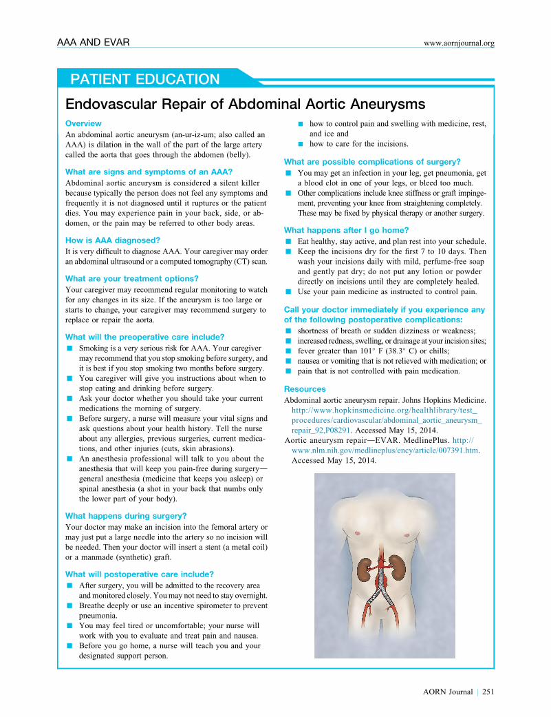
Endovascular Repair of Abdominal Aortic AneurysmsOverview
An abdominal aortic aneurysm (an-ur-iz-um; also called an
AAA) is dilation in the wall of the part of the large artery
called the aorta that goes through the abdomen (belly).
What are signs and symptoms of an AAA?
Abdominal aortic aneurysm is considered a silent killer
because typically the person does not feel any symptoms and
frequently it is not diagnosed until it ruptures or the patient
dies. You may experience pain in your back, side, or ab-
domen, or the pain may be referred to other body areas.
How is AAA diagnosed?
It is very difficult to diagnose AAA. Your caregiver may order
an abdominal ultrasound or a computed tomography (CT) scan.
What are your treatment options?
Your caregiver may recommend regular monitoring to watch
for any changes in its size. If the aneurysm is too large or
starts to change, your caregiver may recommend surgery to
replace or repair the aorta.
What will the preoperative care include?
n Smoking is a very serious risk for AAA. Your caregiver
may recommend that you stop smoking before surgery, and
it is best if you stop smoking two months before surgery.
n You caregiver will give you instructions about when to
stop eating and drinking before surgery.
n Ask your doctor whether you should take your current
medications the morning of surgery.
n Before surgery, a nurse will measure your vital signs and
ask questions about your health history. Tell the nurse
about any allergies, previous surgeries, current medica-
tions, and other injuries (cuts, skin abrasions).
n An anesthesia professional will talk to you about the
anesthesia that will keep you pain-free during surgerydgeneral anesthesia (medicine that keeps you asleep) or
spinal anesthesia (a shot in your back that numbs only
the lower part of your body).
What happens during surgery?
Your doctor may make an incision into the femoral artery or
may just put a large needle into the artery so no incision will
be needed. Then your doctor will insert a stent (a metal coil)
or a manmade (synthetic) graft.
What will postoperative care include?
n After surgery, you will be admitted to the recovery area
andmonitored closely. Youmay not need to stay overnight.
n Breathe deeply or use an incentive spirometer to prevent
pneumonia.
n You may feel tired or uncomfortable; your nurse will
work with you to evaluate and treat pain and nausea.
n Before you go home, a nurse will teach you and your
designated support person.
n how to control pain and swelling with medicine, rest,
and ice and
n how to care for the incisions.
What are possible complications of surgery?
n You may get an infection in your leg, get pneumonia, get
a blood clot in one of your legs, or bleed too much.
n Other complications include knee stiffness or graft impinge-
ment, preventing your knee from straightening completely.
These may be fixed by physical therapy or another surgery.
What happens after I go home?
n Eat healthy, stay active, and plan rest into your schedule.
n Keep the incisions dry for the first 7 to 10 days. Then
wash your incisions daily with mild, perfume-free soap
and gently pat dry; do not put any lotion or powder
directly on incisions until they are completely healed.
n Use your pain medicine as instructed to control pain.
Call your doctor immediately if you experience anyof the following postoperative complications:
n shortness of breath or sudden dizziness or weakness;
n increased redness, swelling, or drainage at your incision sites;
n fever greater than 101� F (38.3� C) or chills;
n nausea or vomiting that is not relieved with medication; or
n pain that is not controlled with pain medication.
Resources
Abdominal aortic aneurysm repair. Johns Hopkins Medicine.
http://www.hopkinsmedicine.org/healthlibrary/test_
procedures/cardiovascular/abdominal_aortic_aneurysm_
repair_92,P08291. Accessed May 15, 2014.
Aortic aneurysm repairdEVAR. MedlinePlus. http://
www.nlm.nih.gov/medlineplus/ency/article/007391.htm.
Accessed May 15, 2014.
AORN Journal j 251
AAA AND EVAR www.aornjournal.org

September 2014 Vol 100 No 3 GORDONdTOURSARKISSIAN
access. The anesthesia professional also may place
a central line, although this is not mandatory.
Choice of anesthesia (eg, general, regional, local)
is dependent on the surgeon’s and anesthesia pro-
fessional’s preference, as well as the patient’s
physiology and ability to cooperate.
Many surgeons perform a groin cutdown on the
femoral artery, although a percutaneous approach is
increasingly popular, especially with the availabil-
ity of lower profile devices. When that is the case,
the surgeon may use a vascular device to control
the opening in the femoral artery, which requires
the postanesthesia care unit (PACU) nurse to
closely monitor the patient postprocedure for
bleeding or thrombosis.
The surgeon may use a large sheath for access,
which he or she removes in the procedure room
immediately after stent graft delivery. The surgeon
may use contrast material to help visualize the
anatomy, although the amount can be decreased
quite a bit by the judicious use of IV ultrasound.
The surgeon determines the location of the plan-
ned placement area with fluoroscopy and intravas-
cular ultrasound. The surgeon achieves endovascular
access via small, bilateral femoral incisions. The
surgeon exposes the femoral artery and inserts the
catheters and stent graft in the aneurysmal section of
the aorta. The duration of the EVAR procedure can
vary. It can be as short as 90 minutes for a routine,
uncomplicated procedure and up to several hours for
a more complicated procedure, such as one requiring
adjunctive stenting of renal arteries.
Postoperative Considerations andComplications
In the immediate postoperative period, the PACU
nurse monitors the patient for signs of hemody-
namic insufficiency. The PACU nurse assesses
the patient’s hemodynamic status by measuring
vital signs (eg, blood pressure, heart rate, pulse
oximetry, respiratory rate, pain), urine output, level
of consciousness, and general skin appearance.
The nurse also should monitor the patient’s groin
for hematoma formation, especially if the surgeon
252 j AORN Journal
delivered the stent via percutaneous access.42
Rupture immediately after stent graft insertion is
rare but has been reported.31 Rupture of one of the
iliac vessels used for access also may be a source
of hemorrhage and hemodynamic instability. Fluid
shifting may occur postoperatively and can lead to
hypovolemia; however, fluid shifting is less sub-
stantial in EVAR than in open repair.
Monitoring for thromboembolic events.
Thrombotic complications are possible, either in the
femoral vessel or in one of the limbs of the graft.42
Graft limb thrombosis may be caused by either a kink
or unrecognized dissection just distal to the graft.38
Therefore, the PACU nurse should palpate the pa-
tient’s pulses frequently. Performing a Doppler ultra-
sound should be part of the postoperative routine.42
Postoperatively, the nurse should closely observe for
evidence of a thromboembolic event as indicated by
n distal petechiae in the lower extremities;
n focal areas of cyanosis, especially to one of the
toes; or
n a cold, pale, pulseless extremity.42
Monitoring for ischemic bowel. Complications
related to bowel ischemia may result from thrombus
or atheroembolization, especially if unrecognized
mesenteric arterial occlusive disease is present or the
aneurysm extends suprarenal where the celiac and
superior mesenteric arteries arise from the aorta;
thus, thrombus can be dislodged during stent graft
placement. Often, the origin of the inferior mesen-
teric artery arises from the aneurysmal area of the
infrarenal aorta; therefore, placement of the stent
graft inadvertently can occlude the artery, resulting
in ischemic bowel. Indications of postoperative
ischemic bowel includes abdominal pain, distention,
nausea or vomiting, or prolonged ileus.42
Monitoring renal function. Renal failure can
develop from
n CIN,41
n the stent graft inadvertently covering the renal
artery orifice,

AAA AND EVAR www.aornjournal.org
n failure of a branched graft in cases of branched
endograft placement, or possibly
n severe atheroembolization during wire manip-
ulation of the aorta during stent graft placement.
The PACU nurse should monitor the patient’s serum
chemistries, especially the serum creatinine and blood
urea nitrogen levels, for elevations.28,29 The nurse
also should monitor the patient’s urine output for
the amount and presence of overt hematuria.42
Monitoring for spinal cord ischemia. Spinal
cord ischemia may occur if the spinal arteries are
occludedby the stentgraft orbymicrothromboemboli.44
The ICU or step-down unit nurse should focus onmonitoring thepatient’s vital signs andperipheralpulses frequently, assessing the patient forbleeding, and maintaining the patient’s bloodpressure at a normotensive level.
This complication is
more likely to occur if
the stent graft place-
ment is suprarenal,
including the area
where the visceral
and some of the lum-
bar arteries exit the
aorta.44 The PACU
nurse should assess for neuromotor deficits by
evaluating the patient’s extremity strength and
movement together with the patient’s quality of
bladder control.42
After the immediate postoperative recovery
period in the PACU, most patients recover for the
first 24 hours in a monitored bed setting, either an
ICU or a step-down unit. The ICU or step-down
unit nurse should focus on
n monitoring the patient’s vital signs and periph-
eral pulses frequently,
n assessing the patient for bleeding, and
n maintaining the patient’s blood pressure at a
normotensive level (ie, preventing hypotension
or hypertension) to decrease the risk of rupture
or other complications.
In general, additional postoperative management
considerations for the ICU or step-down unit nurse
include fluid administration, diet, line removal,
and activities of daily living. Patients who have
undergone an EVAR procedure do not usually
require the large amounts of IV fluids seen with
patients who have undergone open AAA proce-
dures. However, fluids usually are administered to
offset the contrast load often used in endovascular
procedures, which can lead to diuresis. Fluids also
are administered to decrease the risk of CIN.
Typically, the nurse resumes the patient’s diet on
the evening after surgery for a routine EVAR
procedure performed that morning. Often, the
nurse can remove invasive lines and monitors on
the first postoperative day. The patient may sit up
in bed as early as a couple of hours after the
procedure if he or she underwent a vascular
cutdown for cannula
passage. Supine bed
rest likely will be
longer, however, if the
patient underwent the
percutaneous approach
to the EVAR proce-
dure to decrease the
risk of bleeding at the
stent graft access site. The patient can be ambu-
lated on postoperative day one, and many are dis-
charged on postoperative day two.
On discharge, the nurse should instruct the patient
to avoid strenuous physical activities for at least
one week. The actual duration of restrictions
depends on the approach (ie, open versus percu-
taneous) andwhether any postoperative complica-
tions developed. The nurse should instruct the
patient not todrive until he or she is free of pain
and not taking pain medications. The nurse provides
the patient with routine oral and written discharge in-
structions for incision care. The nurse also instructs
the patient on signs of infection and bleeding or healing
problems that should be reported to the surgeon.
LONG-TERM FOLLOW-UP CARE
Complications unique to EVAR are related to endo-
leaks (ie, blood flow outside the stent graft yet inside
the aneurysm sac),24 which are classified as types
1 through 4 (Table 3). The most common endoleak
AORN Journal j 253

TABLE 3. Endoleak Complications After Stent Graft Insertion1-3
Types ofendoleaks Description
Risk of abdominalaortic rupture
Treatmentrecommendations
Type 1 Blood flow around the site of thestent graft device caused bypoor graft attachment eitherproximally or distally
Very high Should be corrected if possible
Type 2 Blood flow back into the aneu-rysmal sac from either the infe-rior mesenteric artery orthe lumbar arteries
Low risk, unless sac growthaccompanies this complication
May require more frequent sur-veillance for continued increasein size because of the associ-ated increased risk of rupture
Type 3 Blood flows directly into theaneurysmal sac because ofstructural failure of the stentgraft in which there is a sepa-ration of stent graft devicecomponents
High Requires surgical repair as soonas possible
Type 4 Transient graft porosity phenom-enon (ie, blood flows throughthe graft itself as a result ofabnormally high graft porosity)is seen for a few days afterimplantation with some graftbrandsa
Low Usually resolves without interven-tion; observing the endoleakand aortic size with CT scans isacceptable
a Usually a transient phenomenon but may be of relevance in stent brand selection if the stent graft implantation is for an acute abdominal aortic aneurysmrupture.
1. Eliason JL, Rasmussen T. Complications after endovascular ruptured abdominal aortic aneurysm repair. In: Upchurch GR Jr, Criado E, eds. Contem-porary Cardiology Aortic Aneurysms Pathogenesis and Treatment. New York, NY: Humana Press, Springer ScienceþBusiness Media: 2009:207-216.
2. Toursarkissian B. Abdominal aortic aneurysm. In: Cohn SM, Brower ST, eds. Surgery: Evidence-based Practice. Shelton, CT: People’s Medical Pub-lishing House-USA; 2012:661-666.
3. White SB, Stayropoulos SW. Management of endoleaks following endovascular aneurysm repair. Semin Intervent Radiol. 2009;26(1):33-38.
September 2014 Vol 100 No 3 GORDONdTOURSARKISSIAN
is a type 2, which 40% of patients may experience
after undergoing an EVAR procedure.24,45
Endoleaks are one of the reasons why stent
grafts for AAAs require lifelong imaging surveil-
lance.24 In addition to identifying endoleaks, ul-
trasound or CT scanning is used to detect other
postoperative complications, such as graft migra-
tion, kinking, or aortic neck enlargement. Ultra-
sound requires an experienced observer for
endoleak detection. A CT scan ideally is performed
with and without contrast and with delayed imaging
to detect type 2 endoleaks.46,47 If the initial scan
is free of anomalies, the patient usually is asked
to return in one year for another scan unless there
are reasons for earlier surveillance. Other potential
long-term complications are limb occlusion, graft
254 j AORN Journal
infection, and development of aneurysms in other
locations, such as the iliac and popliteal arteries.46
CONCLUSION
Often clinicians diagnose AAAs incidentally be-
cause they are asymptomatic until a rupture occurs.
The seriousness of the diagnosis is related to the
fact that rupture carries a very high mortality rate.
This rate has decreased with the initiation of
screening programs based on known AAA risk
factors, especially for men 65 to 74 years of age
with any previous smoking history. An elective
repair should be considered for AAAs that are
5.5 cm in diameter or larger or have an increase
in size of 1 cm in the previous year.14,26-29 In an
elective repair, preoperative evaluation for cardiac

AAA AND EVAR www.aornjournal.org
and pulmonary risks should be considered along
with the patient’s candidacy for an endovascular
versus open repair. For the patient who undergoes
EVAR, ongoing monitoring for complications,
especially endoleaks, is an essential part of the
patient’s care.
References1. Adam DJ, Mohan IV, Stuart WP, Bain M, Bradbury AW.
Community and hospital outcome from ruptured abdom-
inal aortic aneurysm within the catchment area of a
regional vascular surgical service. J Vasc Surg. 1999;
30(5):922-928.
2. Lindholt JS, Sogaard R, Laustsen J. Prognosis of ruptured
abdominal aortic aneurysms in Denmark from 1994-2008.
Clin Epidemiol. 2012;4:111-113.
3. Parodi JC, Palmaz JC, Barone HD. Transfemoral intra-
luminal graft implantation for abdominal aortic aneu-
rysms. Ann Vasc Surg. 1991;5(6):491-499.
4. Upchurch GR, Schaub TA. Abdominal aortic aneurysm.
Am Fam Physician. 2006;73(7):1198-1204.
5. Krishnamurthy VN, Rectenwald JE. Patient selection
criteria for endovascular aortic aneurysm repair. In:
Upchurch GR Jr, Criado E, eds. Contemporary Cardiol-
ogy Aortic Aneurysms Pathogenesis and Treatment. New
York, NY: Humana Press, Springer ScienceþBusiness
Media; 2009:207-216.
6. Golledge J, Norman PE. Atherosclerosis and abdominal
aortic aneurysm: cause, response, or common risk factors?
Arterioscler Thromb Vasc Biol. 2010;30(6):1075-1077.
7. Ince H, Nienaber CA. Etiology, pathogenesis and man-
agement of thoracic aortic aneurysm. Nat Clin Pract
Cardiovasc Med. 2007;4(8):418-427.
8. Wassef M, Baxter T, Chisholm RL, et al. Pathogenesis
of abdominal aortic aneurysms: a multidisciplinary
research program supported by the National Heart, Lung,
and Blood Institute. J Vasc Surg. 2001;34(4):730-738.
9. Creager MA, Halperin JL, Whittemore AD. Aneurysmal
disease of the aorta and its branches. In: Loscalzo J,
Creager MA, Dzau VJ, eds. Vascular Medicine. New
York, NY: Little, Brown; 1996:901.
10. Compressed Mortality File 1999e2006. CDC WONDER
On-line Database, compiled from Compressed Mortality
File 1999-2006 Series 20 No. 2L, 2009. Centers for
Disease Control and Prevention, National Center for
Health Statistics. http://wonder.cdc.gov/cmf-icd10.html.
Accessed March 31, 2014.
11. Compressed Mortality File 1979e1998. CDC WONDER
On-line Database, compiled from Compressed Mortality
File CMF 1968e1988, Series 20, No. 2A, 2000 and
CMF 1989e1998, Series 20, No. 2E, 2003. Centers for
Disease Control and Prevention, National Center for
Health Statistics. http://wonder.cdc.gov/cmf-icd9.html.
Accessed March 31, 2014.
12. Lederle FA, Johnson GR, Wilson SE, et al. Prevalence
and associations of abdominal aortic aneurysm detected
through screening. Aneurysm Detection And Manage-
ment (ADAM) Veterans Affairs Cooperative Study Group.
Ann Intern Med. 1997;126(6):441-449.
13. United Kingdom Small Aneurysm Trial Participants.
Long-term outcomes of immediate repair compared with
surveillance of small abdominal aortic aneurysms. N Engl
J Med. 2002;346(19):1445-1452.
14. Lederle FA, Wilson SE, Johnson GR, et al. Immediate
repair compared with surveillance of small abdominal
aortic aneurysms. N Engl J Med. 2002;346(19):
1437-1444.
15. Torsney E, Pirianov G, Cockerill GW. Diabetes as a
negative risk factor for abdominal aortic aneurysm edoes the disease aetiology or the treatment provide the
mechanism of protection? Curr Vasc Pharmacol. 2013;
11(3):293-298.
16. DeRango P, Cao P, Cieri E, et al; Comparison of Sur-
veillance vs. Aortic Endografting for Small Aneurysm
Repair (CAESAR) Investigators Group. Effects of dia-
betes on small aortic aneurysms under surveillance ac-
cording to a subgroup analysis from a randomized trial.
J Vasc Surg. 2012;56(6):1555-1563.
17. Stackelberg O, Bj€orck M, Sadr-Azodi O, Larsson SC,
Orsini N, Wolk A. Obesity and abdominal aortic aneu-
rysm. Br J Surg. 2013;100(3):360-366.
18. Thompson SG, Ashton HA, Gao L, Buxton MJ, Scott RA;
Multicentre Aneurysm Screening Study (MASS). Final
follow-up of the Multicentre Aneurysm Screening Study
(MASS) randomized trial of abdominal aortic aneurysm
screening. Br J Surg. 2012;99(12):1649-1656.
19. US Preventive Services Task Force. Screening for ab-
dominal aortic aneurysm: recommendation statement.
Ann Intern Med. 2005;142(3):198-202.
20. Fleming C, Whitlock EP, Beil T, Lederle F. Primary Care
Screening for Abdominal Aortic Aneurysm. Evidence Syn-
thesis No. 35 (Prepared by the Oregon Evidence-based
Practice Center under Contract No. 290-02-0024.)
Rockville, MD: Agency for Healthcare Research and
Quality. February 2005. http://www.ncbi.nlm.nih.gov/
books/NBK42895. Accessed March 31, 2014.
21. Implementation of a One-Time Only Ultrasound Screening
for Abdominal Aortic Aneurysms (AAA), Resulting from
a Referral from an Initial Preventive Physical Examination.
2006. MLN Matters Number: MM5235 Related Change
Request (CR) #:5235. Centers for Medicare & Medicaid
Services. http://www.cms.gov/outreach-and-education/
medicare-learning-network-mln/mlnmattersarticles/
downloads/MM5235.pdf. Accessed March 31, 2014.
22. Laminated thrombus. mediLexicon. http://www.medi
lexicon.com/medicaldictionary.php?t¼91879. Accessed
May 20, 2014.
23. Pearce WH, Rowe VL, Annambhotla S. Abdominal
aortic aneurysm. Medscape. http://emedicine.medscape
.com/article/1979501-clinical. Updated October 28, 2013.
Accessed March 31, 2014.
24. Eliason JL, Rasmussen T. Complications after endovas-
cular ruptured abdominal aortic aneurysm repair. In:
Upchurch GR Jr, Criado E, eds. Contemporary Cardiol-
ogy Aortic Aneurysms Pathogenesis and Treatment. New
York, NY: Humana Press, Springer ScienceþBusiness
Media; 2009:207-216.
25. Anderson JL, Halperin JL, Albert NM, et al. Management
of patients with peripheral artery disease (Compilation
of 2005 and 2011 ACCF/AHA Guideline Recommenda-
tions): a report of the American College of Cardiology
AORN Journal j 255

September 2014 Vol 100 No 3 GORDONdTOURSARKISSIAN
Foundation/American Heart Association Task Force on
practice guidelines. Circulation. 2013;127:1425-1437.
26. Greenhalgh RM, Powell JT. Endovascular repair of ab-
dominal aortic aneurysm. N Engl J Med. 2008;358(5):
494-501.
27. Robinson D, Barend M, Verhagen H, Chuen J. Aortic
aneurysmsdscreening, surveillance and referral. Aust
Fam Physician. 2013;42(6):364-369.
28. Hollier LL, Taylor LM, Ochsner J. Recommended in-
dications for operative treatment of abdominal aortic-
aneurysms. Report of a subcommittee of the Joint Council
of the Society for Vascular Surgery and the North Amer-
ican Chapter of the International Society for Cardiovas-
cular Surgery. J Vasc Surg. 1992;15(6):1046-1056.
29. Brady AR, Thompson SG, Fowkes FG, Greenhalgh RM,
Powell JT; UK Small Aneurysm Trial Participants. Abdom-
inal aortic aneurysm expansion: risk factors and time intervals
for surveillance. Circulation. 2004;110(1):16-21.
30. Powell JJ, Brady AR, Brown LC, et al. Mortality results
for randomised controlled trial of early elective surgery
or ultrasonographic surveillance for small abdominal
aortic aneurysms. Lancet. 1998;352(9141):1649-1655.
31. Brown LL, Powell JT. Risk factors for aneurysm rupture
in patients kept under ultrasound surveillance. Ann Surg.
1999;230(3):289-296.
32. Beck AW, Goodney PP, Nolan BW, Likosky DS, Eldrup-
Jorgensen J, Cronenwett JL; Vascular Study Group of
Northern New England. Predicting 1-year mortality after
elective abdominal aortic aneurysm repair. J Vasc Surg.
2009;49(4):838-843.
33. Buckley SD, Buckley CJ. Advances in endovascular
repair of aortoiliac aneurysmal disease: device design
and nursing implications. AORN J. 2014;100(3):
271-279.
34. Warner MA, Offord KP, Warner ME, Lennon RL,
Conover MA, Jansson-Schumacher U. Role of preoperative
cessation of smoking and other factors in postoperative
pulmonary complications. Mayo Clin Proc. 1989;
64(6):609-616.
35. Calling S, Ji J, Sundquist J, Sundquist K, Z€oller B.Shared and non-shared familial susceptibility of
coronary heart disease, ischemic stroke, peripheral artery
disease and aortic disease. Int J Cardiol. 2013;168(1):
2844-2850.
36. Brady AR, Fowkes FG, Thompson SG, Powell JT. Aortic
aneurysm diameter and risk of cardiovascular mor-
tality. Arterioscler Thromb Vasc Biol. 2001;21(7):
1203-1207.
37. Claridge M, Hobbs S, Quick C, Adam D, Bradbury A,
Wilmink T. Screening for popliteal aneurysms should
not be a routine part of a community-based aneurysm
screening program. Vasc Health Risk Manag. 2006;2(2):
189-191.
38. DiwanA,SarkarR,Stanley JC,ZelenockGB,WakefieldTW.
Incidence of femoral and popliteal artery aneurysms in pa-
tients with abdominal aortic aneurysms. J Vasc Surg. 2000;
31(5):863-869.
39. Band RA, Gaieski DF, Mills AM, et al. Discordance
between serum creatinine and creatinine clearance for
identification of ED patients with abdominal pain at risk
for contrast-induced nephropathy. Am J Emerg Med.
2007;25(3):268-272.
256 j AORN Journal
40. Lameire N, Adam A, Becker CR, et al; CIN Consensus
Working Panel. Baseline renal function screening. Am
J Cardiol. 2006;98(6A):21K-26K.
41. Goldfarb S, McCullough PA, McDermott J, Spencer BG.
Contrast-induced acute kidney injury: specialty-specific
protocols for interventional radiology, diagnostic com-
puted tomography radiology, and interventional cardiol-
ogy. Mayo Clin Proc. 2006;84(2):170-179.
42. Smith D, DeVeaux T, Dillard C, et al; Society for Vascular
Nursing Task Force for Clinical Practice Guidelines.
2009 clinical practice guideline for patients undergoing
endovascular repair of abdominal aortic aneurysms
(AAA). J Vasc Nurs. 2009;27:48-63.
43. Bratzler DW, Houck PM; Surgical Infection Prevention
Guidelines Writers Workgroup; et al. Antimicrobial
prophylaxis for surgery: an advisory statement from
the National Surgical Infection Prevention Project. Clin
Infect Dis. 2004;38(12):1706-1715.
44. Zipfel B, Buz S, RedlinM,HullmeineD,Hammerschmidt R,
Hetzer R. Spinal cord ischemia after thoracic stent-grafting:
causes apart from intercostal artery coverage. Ann Thorac
Surg. 2013;96(1):31-38.
45. Parent FN, Meier GH, Godziachvili V, et al. The inci-
dence and natural history of type I and II endoleak:
a 5-year follow-up assessment with color duplex ul-
trasound scan. J Vasc Surg. 2002;35(3):474-481.
46. Toursarkissian B. Abdominal aortic aneurysm. In:
Cohn SM, Brower ST, eds. Surgery: Evidence-based
Practice. Shelton, CT: People’s Medical Publishing
House-USA; 2012:661-666.
47. White SB, Stayropoulos SW. Management of endoleaks
following endovascular aneurysm repair. Semin Intervent
Radiol. 2009;26(1):33-38.
Phyllis A. Gordon, MSN, APRN, ACNS-BC,
is a clinical nurse specialist in the Vascular
Surgery Division of the Department of Sur-
gery, School of Medicine, and a clinical as-
sistant professor in the School of Nursing at
the University of Texas Health Science Center
at San Antonio. Ms Gordon has no declared
affiliation that could be perceived as posing a
potential conflict of interest in the publication
of this article.
Boulos Toursarkissian, MD, FACS, is a pro-
fessor and the chief of the Vascular Surgery
Division of the Department of Surgery, School
of Medicine, at the University of Texas Health
Science Center at San Antonio. Dr Toursarkissian
has no declared affiliation that could be per-
ceived as posing a potential conflict of interest
in the publication of this article.
EXAMINATION
CONTINUING EDUCATION3.2
www.aorn.org/CETreatment of Abdominal AorticAneurysms: The Role of EndovascularRepair
PURPOSE/GOAL
�
To provide the learner with knowledge specific to caring for the patient with an
abdominal aortic aneurysm (AAA) undergoing endovascular aneurysm repair
(EVAR).
OBJECTIVES
1. Describe aortic aneurysms.
2. Explain methods for diagnosing AAAs.
3. Discuss risk factors associated with the development of AAAs.
4. Identify intervention parameters for the patient with an AAA.
5. Discuss perioperative nursing care of the patient undergoing an EVAR procedure.
6. Identify postoperative complications of the EVAR procedure.
The Examination and Learner Evaluation are printed here for your conve-
nience. To receive continuing education credit, you must complete the online
Examination and Learner Evaluation at http://www.aorn.org/CE.
QUESTIONS
1. Aortic aneurysms commonly are described by
their location, which includes
1. abdominal.
2. aortoiliac.
3. thoracic.
4. thoracoabdominal.
a. 1 and 2 b. 2 and 3
AORN, Inc, 2014
c. 1, 2, and 3 d. 1, 2, 3, and 4
2. Diagnosis of an AAA is usually made if the
infrarenal aortic diameter is 3 cm or greater.
a. true b. false
3. Risk factors for formation of an AAA are
1. body mass index.
2. diabetes mellitus.
3. excess visceral fat.
4. sex.
5. race or ethnicity.
6. smoking.
a. 1, 2, and 3 b. 3, 4, 5, and 6
September 2014 Vol
c. 1, 2, 4, 5, and 6 d. 1, 2, 3, 4, 5, and 6
4. The most appropriate screening study of AAAs,
with a sensitivity of 95% and specificity near
100% when performed in a quality setting, is
a. abdominal ultrasound.
100 No 3 � AORN Journal j 257
September 2014 Vol 100 No 3 CE EXAMINATION
b. computed tomography.
c. physical examination.
d. plain radiographic films.
5. National guidelines from the Society for Vascular
Surgery and the International Society for Car-
diovascular Surgery recommend intervening in a
patient with an AAA if
1. it is 5.5 cm in diameter or larger.
2. it is suspected to be an atypical AAA.
3. it is suspected to be a complicated aneurysm.
4. it is rapidly expanding in diameter.
5. clinicians suspect or have documentation of a
rupture.
6. the patient is experiencing symptomsof anAAA.
a. 1, 3, and 5 b. 2, 4, and 6
258 j AORN Journal
c. 2, 3, 5, and 6 d. 1, 2, 3, 4, 5, and 6
6. For preoperative smoking cessation to be
beneficial, the patient must quit smoking
____________ before the planned date of
surgery.
a. two weeks b. four weeks
c. two months d. six months
7. A preoperative bowel preparation is mandatory
because EVAR is radiation intensive and bowel
gases make visualization more difficult.
a. true b. false
8. For an EVAR procedure, the RN circulator may
need to obtain equipment and supplies including
1. an indwelling urinary catheter.
2. appropriate surgical equipment and supplies
to manage an open repair.
3. equipment for rapid injection of the
radiopaque dye.
4. lead aprons, thyroid shields, mobile shields,
and lead glasses.
5. prophylactic antibiotics for preoperative and
postoperative administration.
6. surgical wound drains in a variety of sizes.
a. 1, 3, and 5 b. 2, 4, and 6
c. 1, 2, 3, 4, and 5 d. 1, 2, 3, 4, 5, and 6
9. The nurse should closely observe the patient
for evidence of a postoperative thromboembolic
event as indicated by
1. distal petechiae in the lower extremities.
2. focal areas of cyanosis, especially to one of
the toes.
3. heat and warmth over the calf.
4. a cold, pale, pulseless extremity.
a. 1 and 3 b. 2 and 4
c. 1, 2, and 4 d. 1, 2, 3, and 4
10. Potential postoperative complications after an
EVAR procedure include
1. aneurysmal development in other locations.
2. endoleaks.
3. graft infection.
4. graft kinking or migration.
5. aortic neck enlargement.
6. limb occlusion.
a. 1, 3, and 5 b. 2, 4, and 6
c. 2, 3, 5, and 6 d. 1, 2, 3, 4, 5, and 6

LEARNER EVALUATION
CONTINUING EDUCATION PROGRAM3.2
www.aorn.org/CETreatment of Abdominal AorticAneurysms: The Role of EndovascularRepair
This evaluation is used to determine the extent to
which this continuing education program met
your learning needs. The evaluation is printed
here for your convenience. To receive continuing
education credit, you must complete the online
Examination and Learner Evaluation at http://www.aorn.org/CE. Rate the items as described below.
OBJECTIVES
To what extent were the following objectives of this
continuing education program achieved?
1. Describe aortic aneurysms.
Low 1. 2. 3. 4. 5. High
2. Explain methods for diagnosing AAAs.
Low 1. 2. 3. 4. 5. High
3. Discuss risk factors associated with the development
of AAAs. Low 1. 2. 3. 4. 5. High
4. Identify intervention parameters for the patient with
an AAA. Low 1. 2. 3. 4. 5. High
5. Discuss perioperative nursing care of the patient
undergoing an EVAR procedure.
Low 1. 2. 3. 4. 5. High
6. Identify postoperative complications of the endo-
vascular aneurysm repair (EVAR) procedure.
Low 1. 2. 3. 4. 5. High
CONTENT
7. To what extent did this article increase your
knowledge of the subject matter?
Low 1. 2. 3. 4. 5. High
8. To what extent were your individual objectives met?
Low 1. 2. 3. 4. 5. High
� AORN, Inc, 2014
9. Will you be able to use the information from this
article in your work setting? 1. Yes 2. No
10. Will you change your practice as a result of
reading this article? (If yes, answer question
#10A. If no, answer question #10B.)
10A. How will you change your practice? (Select all
that apply)
1. I will provide education to my team
regarding why change is needed.
2. I will work with management to change/
implement a policy and procedure.
3. I will plan an informational meeting with
physicians to seek their input and acceptance
of the need for change.
4. I will implement change and evaluate the
effect of the change at regular intervals until
the change is incorporated as best practice.
5. Other: ______________________________
10B. If you will not change your practice as a result of
reading this article, why? (Select all that apply)
1. The content of the article is not relevant to
my practice.
2. I do not have enough time to teach others
about the purpose of the needed change.
3. I do not have management support to make a
change.
4. Other: ______________________________
11. Our accrediting body requires that we verify
the time you needed to complete the 3.2 con-
tinuing education contact hour (192-minute)
program:________________________________
September 2014 Vol 100 No 3 � AORN Journal j 259