endovascular repair of thoracic and abdominal aortic aneurysms
TRANSCRIPT
Endovascular Repair of Thoracic and Abdominal Aortic Aneurysms / Dissections
Review Article
INTRODUCTION
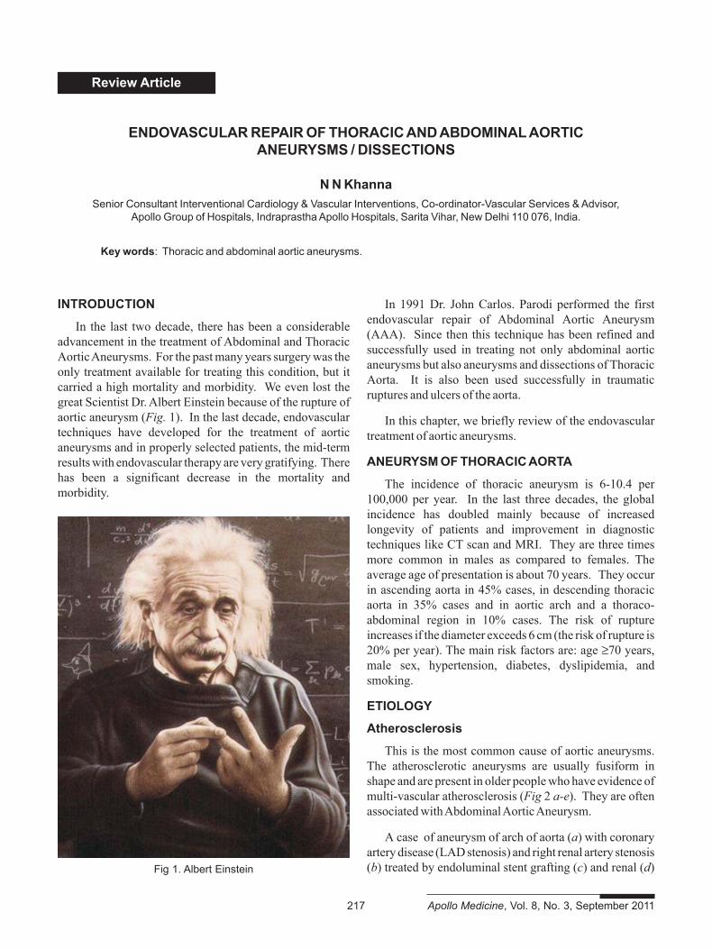
In the last two decade, there has been a considerableadvancement in the treatment of Abdominal and ThoracicAortic Aneurysms. For the past many years surgery was theonly treatment available for treating this condition, but itcarried a high mortality and morbidity. We even lost thegreat Scientist Dr. Albert Einstein because of the rupture ofaortic aneurysm (Fig. 1). In the last decade, endovasculartechniques have developed for the treatment of aorticaneurysms and in properly selected patients, the mid-termresults with endovascular therapy are very gratifying. Therehas been a significant decrease in the mortality andmorbidity.
ENDOVASCULAR REPAIR OF THORACIC AND ABDOMINAL AORTICANEURYSMS / DISSECTIONS
N N KhannaSenior Consultant Interventional Cardiology & Vascular Interventions, Co-ordinator-Vascular Services & Advisor,
Apollo Group of Hospitals, Indraprastha Apollo Hospitals, Sarita Vihar, New Delhi 110 076, India.
Key words: Thoracic and abdominal aortic aneurysms.
In 1991 Dr. John Carlos. Parodi performed the firstendovascular repair of Abdominal Aortic Aneurysm(AAA). Since then this technique has been refined andsuccessfully used in treating not only abdominal aorticaneurysms but also aneurysms and dissections of ThoracicAorta. It is also been used successfully in traumaticruptures and ulcers of the aorta.
In this chapter, we briefly review of the endovasculartreatment of aortic aneurysms.
ANEURYSM OF THORACIC AORTA
The incidence of thoracic aneurysm is 6-10.4 per100,000 per year. In the last three decades, the globalincidence has doubled mainly because of increasedlongevity of patients and improvement in diagnostictechniques like CT scan and MRI. They are three timesmore common in males as compared to females. Theaverage age of presentation is about 70 years. They occurin ascending aorta in 45% cases, in descending thoracicaorta in 35% cases and in aortic arch and a thoraco-abdominal region in 10% cases. The risk of ruptureincreases if the diameter exceeds 6 cm (the risk of rupture is20% per year). The main risk factors are: age ≥70 years,male sex, hypertension, diabetes, dyslipidemia, andsmoking.
ETIOLOGY
Atherosclerosis
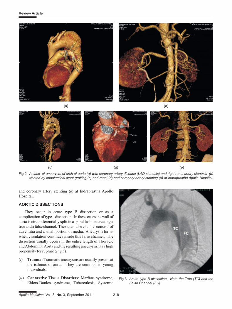
This is the most common cause of aortic aneurysms.The atherosclerotic aneurysms are usually fusiform inshape and are present in older people who have evidence ofmulti-vascular atherosclerosis (Fig 2 a-e). They are oftenassociated with Abdominal Aortic Aneurysm.
A case of aneurysm of arch of aorta (a) with coronaryartery disease (LAD stenosis) and right renal artery stenosis(b) treated by endoluminal stent grafting (c) and renal (d)
217 Apollo Medicine, Vol. 8, No. 3, September 2011
Fig 1. Albert Einstein
Review Article
Apollo Medicine, Vol. 8, No. 3, September 2011 218
(a) (b)
(c) (d) (e)
and coronary artery stenting (e) at Indraprastha ApolloHospital.
AORTIC DISSECTIONS
They occur in acute type B dissection or as acomplication of type a dissection. In these cases the wall ofaorta is circumferentially split in a spiral fashion creating atrue and a false channel. The outer false channel consists ofadventitia and a small portion of media. Aneurysm formswhen circulation continues inside this false channel. Thedissection usually occurs in the entire length of Thoracicand Abdominal Aorta and the resulting aneurysm has a highpropensity for rupture (Fig 3).
(i) Trauma: Traumatic aneurysms are usually present atthe isthmus of aorta. They are common in youngindividuals.
(ii) Connective Tissue Disorders: Marfans syndrome,Ehlers-Danlos syndrome, Tuberculosis, Systemic
Fig 2. A case of aneurysm of arch of aorta (a) with coronary artery disease (LAD stenosis) and right renal artery stenosis (b)treated by endoluminal stent grafting (c) and renal (d) and coronary artery stenting (e) at Indraprastha Apollo Hospital.
Fig 3 Acute type B dissection. Note the True (TC) and theFalse Channel (FC)
Review Article
219 Apollo Medicine, Vol. 8, No. 3, September 2011
Lupus Erythromatosis etc are associated with aorticaneurysms, commonly in thoracic aorta.
(iii) Autoimmune Arteritis - Takayasu Arteritis, Behcet’ssyndrome, Giant Cell Arteritis.
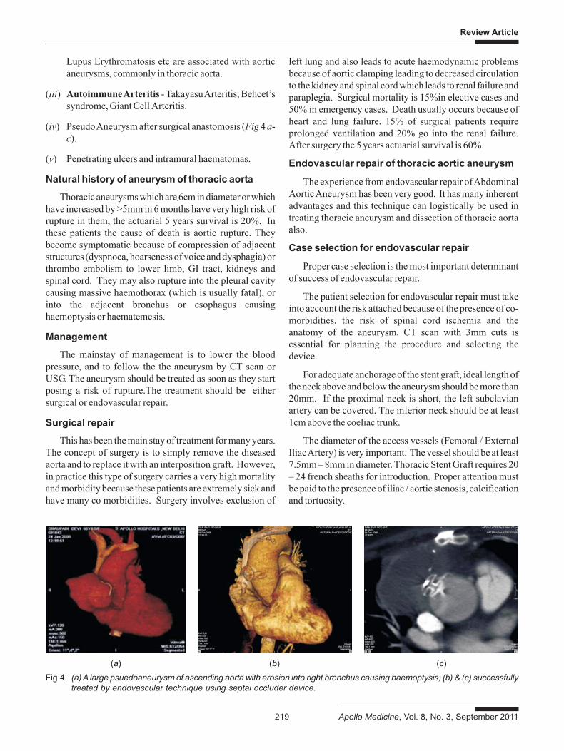
(iv) Pseudo Aneurysm after surgical anastomosis (Fig 4 a-c).
(v) Penetrating ulcers and intramural haematomas.
Natural history of aneurysm of thoracic aorta
Thoracic aneurysms which are 6cm in diameter or whichhave increased by >5mm in 6 months have very high risk ofrupture in them, the actuarial 5 years survival is 20%. Inthese patients the cause of death is aortic rupture. Theybecome symptomatic because of compression of adjacentstructures (dyspnoea, hoarseness of voice and dysphagia) orthrombo embolism to lower limb, GI tract, kidneys andspinal cord. They may also rupture into the pleural cavitycausing massive haemothorax (which is usually fatal), orinto the adjacent bronchus or esophagus causinghaemoptysis or haematemesis.
Management
The mainstay of management is to lower the bloodpressure, and to follow the the aneurysm by CT scan orUSG. The aneurysm should be treated as soon as they startposing a risk of rupture.The treatment should be eithersurgical or endovascular repair.
Surgical repair
This has been the main stay of treatment for many years.The concept of surgery is to simply remove the diseasedaorta and to replace it with an interposition graft. However,in practice this type of surgery carries a very high mortalityand morbidity because these patients are extremely sick andhave many co morbidities. Surgery involves exclusion of
(a) (b) (c)
Fig 4. (a) A large psuedoaneurysm of ascending aorta with erosion into right bronchus causing haemoptysis; (b) & (c) successfullytreated by endovascular technique using septal occluder device.
left lung and also leads to acute haemodynamic problemsbecause of aortic clamping leading to decreased circulationto the kidney and spinal cord which leads to renal failure andparaplegia. Surgical mortality is 15%in elective cases and50% in emergency cases. Death usually occurs because ofheart and lung failure. 15% of surgical patients requireprolonged ventilation and 20% go into the renal failure.After surgery the 5 years actuarial survival is 60%.
Endovascular repair of thoracic aortic aneurysm
The experience from endovascular repair of AbdominalAortic Aneurysm has been very good. It has many inherentadvantages and this technique can logistically be used intreating thoracic aneurysm and dissection of thoracic aortaalso.
Case selection for endovascular repair
Proper case selection is the most important determinantof success of endovascular repair.
The patient selection for endovascular repair must takeinto account the risk attached because of the presence of co-morbidities, the risk of spinal cord ischemia and theanatomy of the aneurysm. CT scan with 3mm cuts isessential for planning the procedure and selecting thedevice.
For adequate anchorage of the stent graft, ideal length ofthe neck above and below the aneurysm should be more than20mm. If the proximal neck is short, the left subclavianartery can be covered. The inferior neck should be at least1cm above the coeliac trunk.
The diameter of the access vessels (Femoral / ExternalIliac Artery) is very important. The vessel should be at least7.5mm – 8mm in diameter. Thoracic Stent Graft requires 20– 24 french sheaths for introduction. Proper attention mustbe paid to the presence of iliac / aortic stenosis, calcificationand tortuosity.
Review Article
Apollo Medicine, Vol. 8, No. 3, September 2011 220
The diameter of the stent graft should be at least 15% -20% more that the diameter of the normal aorta proximalto the aneurysm. The length of the graft is usually 15 cm-20 cm. It can be extended by overlapping with additionalstent grafts. The recommended overlap between the twoshould be 2cm.
Technique of device implantation
The procedure is undertaken in the cardiaccatheterization suite with facilities of digital substractionangiography (DSA) and road mapping. It is important tofix landmarks after the control angiogram and not to movethe table during the procedure. The LAO view is usuallyused to profile these aneurysm and the arch vessels.
Arterial access is gained by surgical exposure of thefemoral artery. Ideally, the common femoral artery shouldbe ≥8 mm in diameter. If the common femoral artery issmall, it is prudent to expose the external iliac artery byextending the incision. Sometimes common iliac artery isexposed by retroperitoneal approach and the graft conduitis anastomosed to it for easy delivery of the stent graft.
After the femoral artery exposure, arterial puncture ismade and a 0.035" guide wire (super stiff amplatz guidewire / Backup Mier wire) is introduced all the way up toascending aorta. The stent graft is then advanced on theguide wire so that the covered portion of the stent is placedat least 20 mm proximal to the origin of aneurysm/dissection. Right brachial access is often very useful and
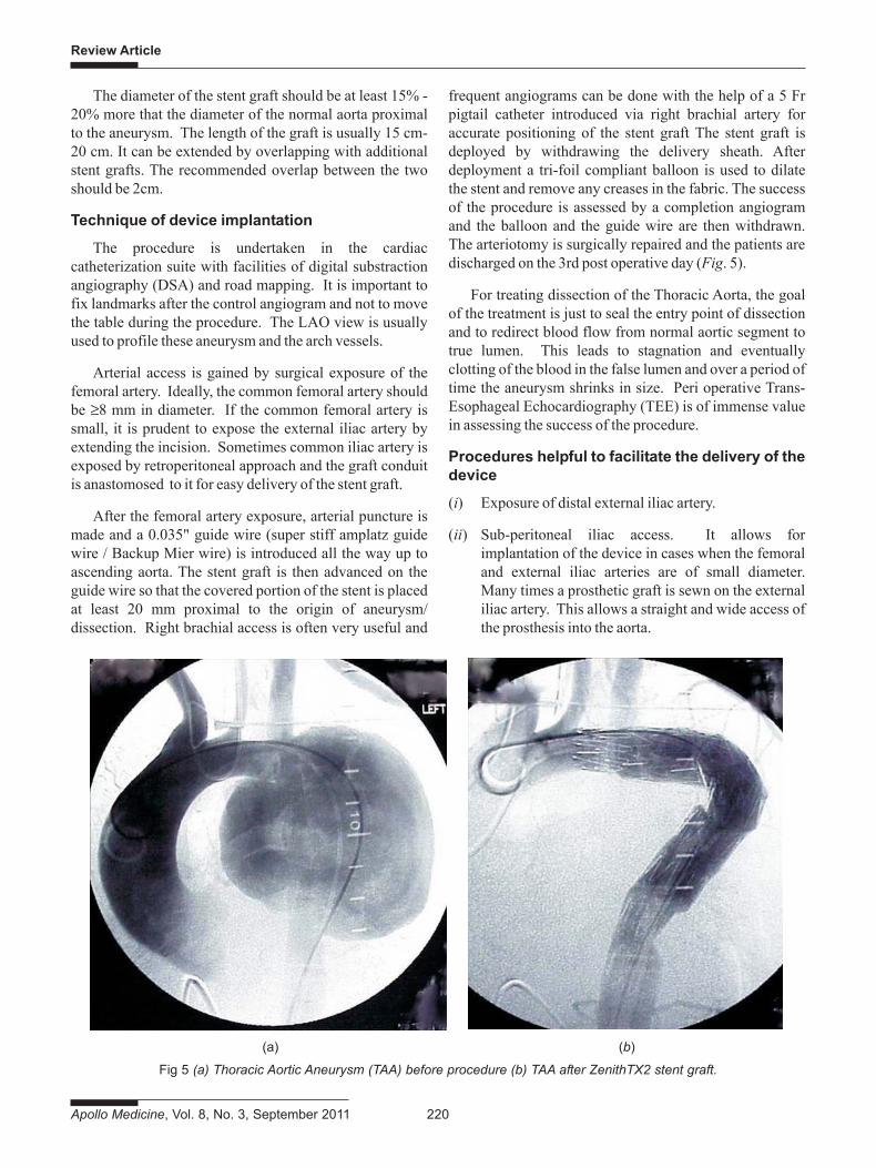
frequent angiograms can be done with the help of a 5 Frpigtail catheter introduced via right brachial artery foraccurate positioning of the stent graft The stent graft isdeployed by withdrawing the delivery sheath. Afterdeployment a tri-foil compliant balloon is used to dilatethe stent and remove any creases in the fabric. The successof the procedure is assessed by a completion angiogramand the balloon and the guide wire are then withdrawn.The arteriotomy is surgically repaired and the patients aredischarged on the 3rd post operative day (Fig. 5).
For treating dissection of the Thoracic Aorta, the goalof the treatment is just to seal the entry point of dissectionand to redirect blood flow from normal aortic segment totrue lumen. This leads to stagnation and eventuallyclotting of the blood in the false lumen and over a period oftime the aneurysm shrinks in size. Peri operative Trans-Esophageal Echocardiography (TEE) is of immense valuein assessing the success of the procedure.
Procedures helpful to facilitate the delivery of thedevice
(i) Exposure of distal external iliac artery.
(ii) Sub-peritoneal iliac access. It allows forimplantation of the device in cases when the femoraland external iliac arteries are of small diameter.Many times a prosthetic graft is sewn on the externaliliac artery. This allows a straight and wide access ofthe prosthesis into the aorta.
Fig 5 (a) Thoracic Aortic Aneurysm (TAA) before procedure (b) TAA after ZenithTX2 stent graft.
(a) (b)
Review Article
221 Apollo Medicine, Vol. 8, No. 3, September 2011
(iii) Femoral and brachial access: In this we have afemoral access and a contra lateral brachial accessand a super stiff guide wire is passed from thebrachial artery into the descending thoracic aorta andabdominal aorta and is snared out of the contralateral femoral artery. This straightens the axis ofimplantation and facilitates the delivery of thedevice.
Treatment of visceral ischemia in aorticdissection
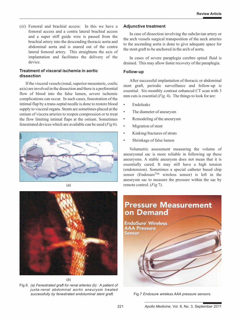
If the visceral vessels (renal, superior mesenteric, coelicaxis) are involved in the dissection and there is a preferentialflow of blood into the false lumen, severe ischemiccomplications can occur. In such cases, fenestration of theintimal flap by a trans-septal needle is done to restore bloodsupply to visceral organs. Stents are sometimes placed at theostium of viscera arteries to reopen compression or to treatthe flow limiting intimal flaps at the ostium. Sometimesfenestrated devices which are available can be used (Fig 6).
Adjunctive treatment
In case of dissection involving the subclavian artery orthe arch vessels surgical transposition of the neck arteriesto the ascending aorta is done to give adequate space forthe stent graft to be anchored in the arch of aorta.
In cases of severe paraplegia cerebro spinal fluid isdrained. This may allow faster recovery of the paraplegia.
Follow-up
After successful implantation of thoracic or abdominalstent graft, periodic surveillance and follow-up isessential. Six-monthly contrast enhanced CT scan with 3mm cuts is essential (Fig. 4). The things to look for are:
• Endoleaks
• The diameter of aneurysm
• Remodeling of the aneurysm
• Migration of stent
• Kinking/fractures of struts
• Shrinkage of false lumen
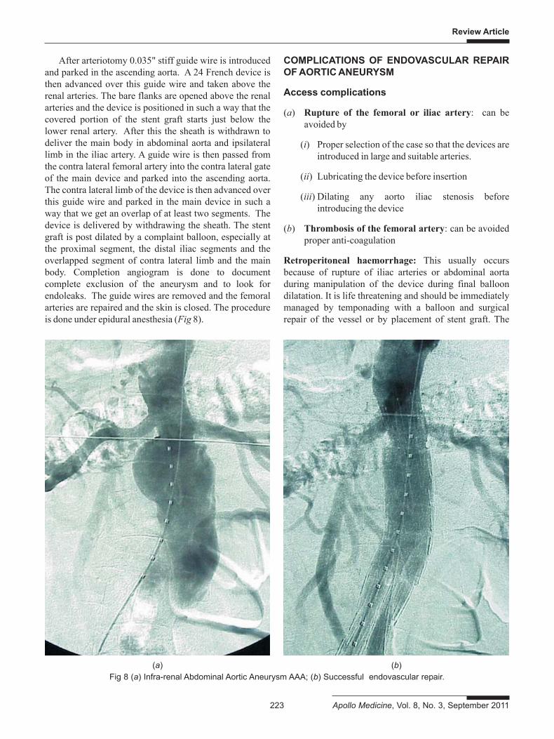
Volumetric assessment measuring the volume ofaneurysmal sac is more reliable in following up theseaneurysms. A stable aneurysm does not mean that it isessentially cured. It may still have a high tension(endotension). Sometimes a special catheter based chipsensor (EndosureTm wireless sensor) is left in theaneurysm sac to measure the pressure within the sac byremote control. (Fig 7).(a)
(b)Fig 6. (a) Fenestrated graft for renal arteries (b) : A patient of
juxta-renal abdominal aortic aneurysm treatedsuccessfully by fenestrated endoluminal stent graft. Fig 7 Endosure wireless AAA pressure sensors.

Review Article
Apollo Medicine, Vol. 8, No. 3, September 2011 222
If the aneurysmal sac is increasing in size, it means thatthe tension within the aneurysm is high either because ofthe endoleak or because of endotension. Secondaryprocedures like placement of additional stent grafts to sealtype I endoleaks may be done. The feeders responsible forType II endoleaks should be closed by interventionalradiological techniques. In case we cannot seal theseendoleaks, we should resort to open surgical treatment.
Results of endovascular treatment of thoracicaneurysms
The results of endovascular treatment of TAA areshown in the following table (Table 1). The meanactuarial survival of these patients is 85% over 18-24months.
Abdominal aortic aneurysm
In 1991, John Carlos Parodi implanted the firstendoluminal stent graft to exclude Abdominal AorticAneurysm (AAA). This technique has emerged as analternative to surgical treatment of AAA. Initial and mid-term results of ELG (Endoluminal stent grafting) are veryencouraging.
Case selection for endovascular repair
The principles for case selection for endovascularrepair of Abdominal Aortic Aneurysm are the same to thatof the Thoracic Aneurysms. Following parameters areessential for predicting the success of endovascular repair.
(i) Infra renal neck of > 2 cm
(ii) Angulation of the neck < 45º
(iii) Neck <30 mm in diameter
(iv) Fusiform aneurysm without history of rupture
(v) Minimal or no tortuosity of iliac arteries
(vi) Absence of aneurysmal iliac arteries
(vii) Femoral access vessel >7.5 mm
(viii) Absence of stenosis in iliac vessels
(ix) Absence of calcification and stenosis of aorticbifurcation
(x) Healthy femoral vessels on both sides
(xi) Absence of stenosis of ≥2 mesenteric arteries
Endovascular repair
The technique of deployment of the stent graft forabdominal aortic aneurysm is similar to that described forThoracic Aneurysms. In Abdominal Aortic Aneurysm, weneed to have bilateral femoral cut-down as the device ismodular in design and is reconstructed inside theaneurysmal abdominal aorta.
Essentially, these grafts consists of a nitinol selfexpanding stent covered with fabric either on the innersurface or the outer surface. The fabric is either Dacron(polyethylene terephthalate Dacron) or PTFE (PolyTetraFluro Ethylene). In modular designs there is a main bodyand an ipsilateral limb of the device and a contra laterallimb which docks into the contra lateral gate of the maindevice. Aorto Uni-iliac stent graft is used when the one ofthe iliac arteries is extremely tortuous and calcified. Herewe have a tapered stent graft from the infra renalabdominal aorta to one of the iliac arteries. The contralateral iliac artery is occluded endovascularly byimplanting an occluder. This is followed by a fem-femcross over graft to maintain a perfusion in the oppositelower limb.
Table 1: Results of thoracic aortic aneurysms
Author (Year) Aortic stent- No Early Conversion Paraplegia Long-termgrafting TAA Mortality (%) (%) (%) survival (%)
Mitchell R. Dake M. 1999 103 103 9 4.8 2.9 73+5actuarial 2 yrs.Greenberg R. 2000 25 25 20 12 12 NABuffolo E. 2002 191 61 10.4 9.8 0 87.4+29(actuarial)Criado F. 2002 47 31 2.1 0 0 87.2 FU:18 mthsHeijmen R. Moll F.,2002 28 28 0 3.6 0 36.4 FU:18 mthsHerold U2002 34 7 2.9 0 0 91.2FU:18 mthsNajibi S. Lumsden A. 2002 19 19 5.3 5.3 0 94.712 MOrend K. Sunder-Plassmann L. 2003 74 40 9.5 (30 days) 8 0 91.7FU:18 mths
Review Article
223 Apollo Medicine, Vol. 8, No. 3, September 2011
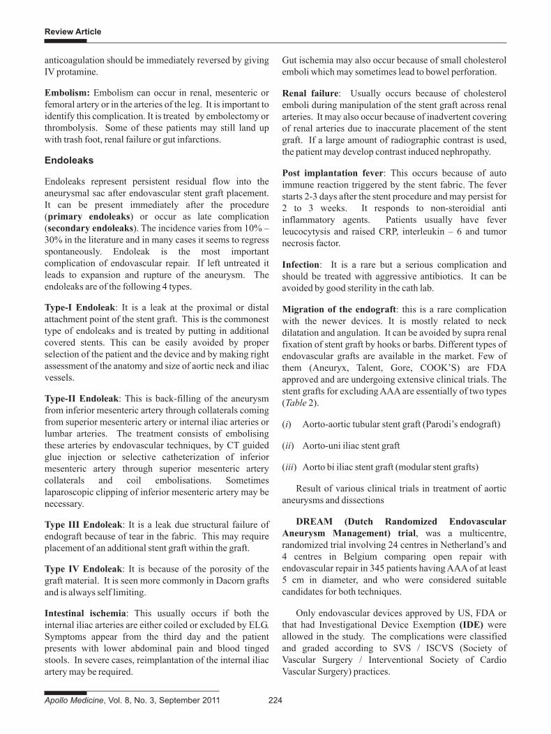
After arteriotomy 0.035" stiff guide wire is introducedand parked in the ascending aorta. A 24 French device isthen advanced over this guide wire and taken above therenal arteries. The bare flanks are opened above the renalarteries and the device is positioned in such a way that thecovered portion of the stent graft starts just below thelower renal artery. After this the sheath is withdrawn todeliver the main body in abdominal aorta and ipsilaterallimb in the iliac artery. A guide wire is then passed fromthe contra lateral femoral artery into the contra lateral gateof the main device and parked into the ascending aorta.The contra lateral limb of the device is then advanced overthis guide wire and parked in the main device in such away that we get an overlap of at least two segments. Thedevice is delivered by withdrawing the sheath. The stentgraft is post dilated by a complaint balloon, especially atthe proximal segment, the distal iliac segments and theoverlapped segment of contra lateral limb and the mainbody. Completion angiogram is done to documentcomplete exclusion of the aneurysm and to look forendoleaks. The guide wires are removed and the femoralarteries are repaired and the skin is closed. The procedureis done under epidural anesthesia (Fig 8).
COMPLICATIONS OF ENDOVASCULAR REPAIROF AORTIC ANEURYSM
Access complications
(a) Rupture of the femoral or iliac artery: can beavoided by
(i) Proper selection of the case so that the devices areintroduced in large and suitable arteries.
(ii) Lubricating the device before insertion
(iii) Dilating any aorto iliac stenosis beforeintroducing the device
(b) Thrombosis of the femoral artery: can be avoidedproper anti-coagulation
Retroperitoneal haemorrhage: This usually occursbecause of rupture of iliac arteries or abdominal aortaduring manipulation of the device during final balloondilatation. It is life threatening and should be immediatelymanaged by temponading with a balloon and surgicalrepair of the vessel or by placement of stent graft. The
Fig 8 (a) Infra-renal Abdominal Aortic Aneurysm AAA; (b) Successful endovascular repair.(a) (b)
Review Article
Apollo Medicine, Vol. 8, No. 3, September 2011 224
anticoagulation should be immediately reversed by givingIV protamine.
Embolism: Embolism can occur in renal, mesenteric orfemoral artery or in the arteries of the leg. It is important toidentify this complication. It is treated by embolectomy orthrombolysis. Some of these patients may still land upwith trash foot, renal failure or gut infarctions.
Endoleaks
Endoleaks represent persistent residual flow into theaneurysmal sac after endovascular stent graft placement.It can be present immediately after the procedure(primary endoleaks) or occur as late complication(secondary endoleaks). The incidence varies from 10% –30% in the literature and in many cases it seems to regressspontaneously. Endoleak is the most importantcomplication of endovascular repair. If left untreated itleads to expansion and rupture of the aneurysm. Theendoleaks are of the following 4 types.
Type-I Endoleak: It is a leak at the proximal or distalattachment point of the stent graft. This is the commonesttype of endoleaks and is treated by putting in additionalcovered stents. This can be easily avoided by properselection of the patient and the device and by making rightassessment of the anatomy and size of aortic neck and iliacvessels.
Type-II Endoleak: This is back-filling of the aneurysmfrom inferior mesenteric artery through collaterals comingfrom superior mesenteric artery or internal iliac arteries orlumbar arteries. The treatment consists of embolisingthese arteries by endovascular techniques, by CT guidedglue injection or selective catheterization of inferiormesenteric artery through superior mesenteric arterycollaterals and coil embolisations. Sometimeslaparoscopic clipping of inferior mesenteric artery may benecessary.
Type III Endoleak: It is a leak due structural failure ofendograft because of tear in the fabric. This may requireplacement of an additional stent graft within the graft.
Type IV Endoleak: It is because of the porosity of thegraft material. It is seen more commonly in Dacorn graftsand is always self limiting.
Intestinal ischemia: This usually occurs if both theinternal iliac arteries are either coiled or excluded by ELG.Symptoms appear from the third day and the patientpresents with lower abdominal pain and blood tingedstools. In severe cases, reimplantation of the internal iliacartery may be required.
Gut ischemia may also occur because of small cholesterolemboli which may sometimes lead to bowel perforation.
Renal failure: Usually occurs because of cholesterolemboli during manipulation of the stent graft across renalarteries. It may also occur because of inadvertent coveringof renal arteries due to inaccurate placement of the stentgraft. If a large amount of radiographic contrast is used,the patient may develop contrast induced nephropathy.
Post implantation fever: This occurs because of autoimmune reaction triggered by the stent fabric. The feverstarts 2-3 days after the stent procedure and may persist for2 to 3 weeks. It responds to non-steroidial antiinflammatory agents. Patients usually have feverleucocytysis and raised CRP, interleukin – 6 and tumornecrosis factor.
Infection: It is a rare but a serious complication andshould be treated with aggressive antibiotics. It can beavoided by good sterility in the cath lab.
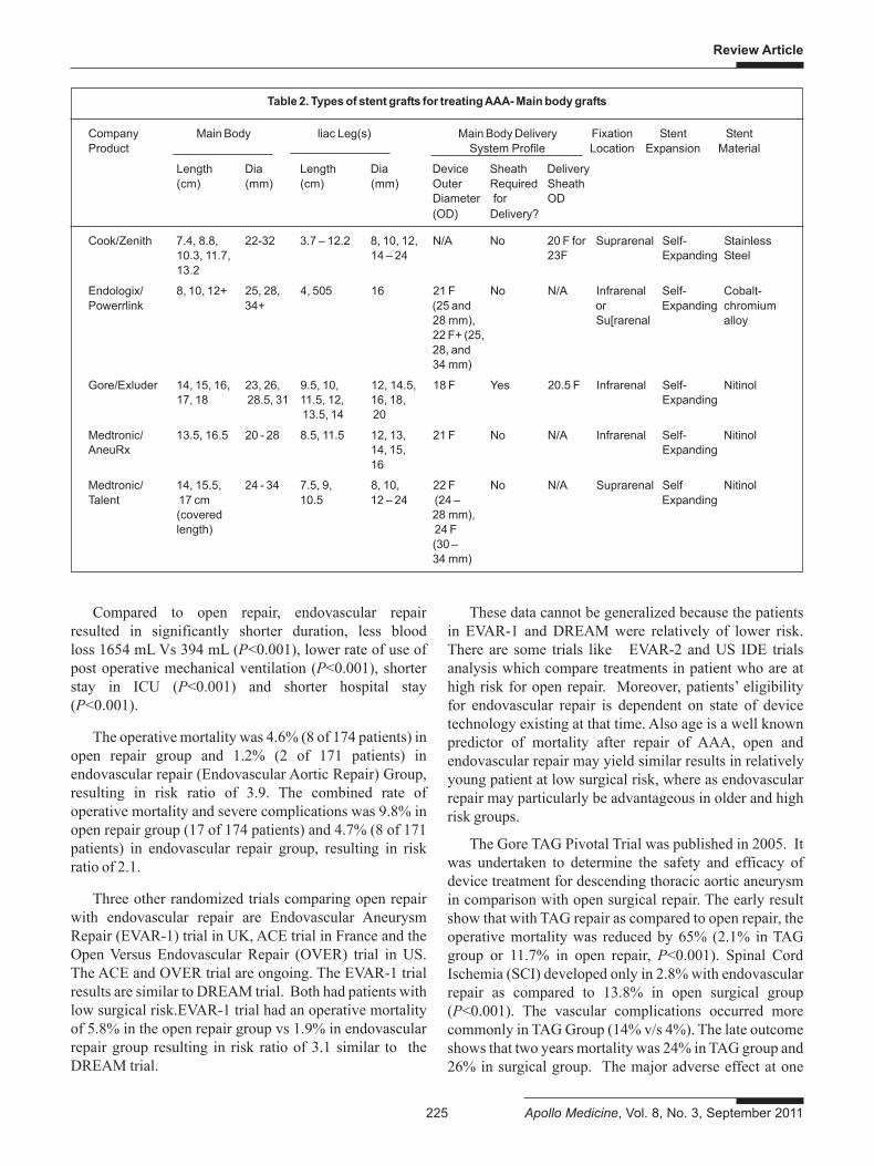
Migration of the endograft: this is a rare complicationwith the newer devices. It is mostly related to neckdilatation and angulation. It can be avoided by supra renalfixation of stent graft by hooks or barbs. Different types ofendovascular grafts are available in the market. Few ofthem (Aneuryx, Talent, Gore, COOK’S) are FDAapproved and are undergoing extensive clinical trials. Thestent grafts for excluding AAA are essentially of two types(Table 2).
(i) Aorto-aortic tubular stent graft (Parodi’s endograft)
(ii) Aorto-uni iliac stent graft
(iii) Aorto bi iliac stent graft (modular stent grafts)
Result of various clinical trials in treatment of aorticaneurysms and dissections
DREAM (Dutch Randomized EndovascularAneurysm Management) trial, was a multicentre,randomized trial involving 24 centres in Netherland’s and4 centres in Belgium comparing open repair withendovascular repair in 345 patients having AAA of at least5 cm in diameter, and who were considered suitablecandidates for both techniques.
Only endovascular devices approved by US, FDA orthat had Investigational Device Exemption (IDE) wereallowed in the study. The complications were classifiedand graded according to SVS / ISCVS (Society ofVascular Surgery / Interventional Society of CardioVascular Surgery) practices.
Review Article
225 Apollo Medicine, Vol. 8, No. 3, September 2011
Compared to open repair, endovascular repairresulted in significantly shorter duration, less bloodloss 1654 mL Vs 394 mL (P<0.001), lower rate of use ofpost operative mechanical ventilation (P<0.001), shorterstay in ICU (P<0.001) and shorter hospital stay(P<0.001).
The operative mortality was 4.6% (8 of 174 patients) inopen repair group and 1.2% (2 of 171 patients) inendovascular repair (Endovascular Aortic Repair) Group,resulting in risk ratio of 3.9. The combined rate ofoperative mortality and severe complications was 9.8% inopen repair group (17 of 174 patients) and 4.7% (8 of 171patients) in endovascular repair group, resulting in riskratio of 2.1.
Three other randomized trials comparing open repairwith endovascular repair are Endovascular AneurysmRepair (EVAR-1) trial in UK, ACE trial in France and theOpen Versus Endovascular Repair (OVER) trial in US.The ACE and OVER trial are ongoing. The EVAR-1 trialresults are similar to DREAM trial. Both had patients withlow surgical risk.EVAR-1 trial had an operative mortalityof 5.8% in the open repair group vs 1.9% in endovascularrepair group resulting in risk ratio of 3.1 similar to theDREAM trial.
These data cannot be generalized because the patientsin EVAR-1 and DREAM were relatively of lower risk.There are some trials like EVAR-2 and US IDE trialsanalysis which compare treatments in patient who are athigh risk for open repair. Moreover, patients’ eligibilityfor endovascular repair is dependent on state of devicetechnology existing at that time. Also age is a well knownpredictor of mortality after repair of AAA, open andendovascular repair may yield similar results in relativelyyoung patient at low surgical risk, where as endovascularrepair may particularly be advantageous in older and highrisk groups.
The Gore TAG Pivotal Trial was published in 2005. Itwas undertaken to determine the safety and efficacy ofdevice treatment for descending thoracic aortic aneurysmin comparison with open surgical repair. The early resultshow that with TAG repair as compared to open repair, theoperative mortality was reduced by 65% (2.1% in TAGgroup or 11.7% in open repair, P<0.001). Spinal CordIschemia (SCI) developed only in 2.8% with endovascularrepair as compared to 13.8% in open surgical group(P<0.001). The vascular complications occurred morecommonly in TAG Group (14% v/s 4%). The late outcomeshows that two years mortality was 24% in TAG group and26% in surgical group. The major adverse effect at one
Table 2. Types of stent grafts for treating AAA- Main body grafts
Company Main Body liac Leg(s) Main Body Delivery Fixation Stent StentProduct System Profile Location Expansion Material
Length Dia Length Dia Device Sheath Delivery(cm) (mm) (cm) (mm) Outer Required Sheath
Diameter for OD(OD) Delivery?
Cook/Zenith 7.4, 8.8, 22-32 3.7 – 12.2 8, 10, 12, N/A No 20 F for Suprarenal Self- Stainless10.3, 11.7, 14 – 24 23F Expanding Steel13.2
Endologix/ 8, 10, 12+ 25, 28, 4, 505 16 21 F No N/A Infrarenal Self- Cobalt-Powerrlink 34+ (25 and or Expanding chromium
28 mm), Su[rarenal alloy22 F+ (25,28, and34 mm)
Gore/Exluder 14, 15, 16, 23, 26, 9.5, 10, 12, 14.5, 18 F Yes 20.5 F Infrarenal Self- Nitinol17, 18 28.5, 31 11.5, 12, 16, 18, Expanding
13.5, 14 20
Medtronic/ 13.5, 16.5 20 - 28 8.5, 11.5 12, 13, 21 F No N/A Infrarenal Self- NitinolAneuRx 14, 15, Expanding
16
Medtronic/ 14, 15.5, 24 - 34 7.5, 9, 8, 10, 22 F No N/A Suprarenal Self NitinolTalent 17 cm 10.5 12 – 24 (24 – Expanding
(covered 28 mm),length) 24 F
(30 –34 mm)
Review Article
Apollo Medicine, Vol. 8, No. 3, September 2011 226
year was significantly lower after TAG repair (42%) thenafter open repair (77%). The same trend was observedthroughout the three year follow-up. No device relateddeaths were noted in the three years follow up and it wasconcluded that endovascular repair with the Gore TAGendovascular graft has shown safety and efficacy withimproved mid-term result compared to open surgicalrepair. Long term effects are essential to ensure the bestoutcome.
Recently EVAR-2 trial (2005) was published as afollow up of 338 patient aged ≥60 years who had AAAof at least 5.5 cm, in diameter and who meet highsurgical risk criteria for open repair due to cardiac,pulmonary in renal co-morbidities. The patients wererandomized to Endovascular repair (EVAR) or notreatment. The 30 days mortality for EVAR inEVAR-2 trial was 9% and at 4 years was 14% for EVARand 19% for no intervention (P=NS). Overall 4 yearssurvival was only 34% in EVAR and 39% in nointervention group (P=NS). EVAR-2 trial suggested thatendovascular repair is not a safe procedure in high riskpatients.
This result was challenged in Sixteenth AnnualMeeting of the Society for Vascular Surgery atPhiladelphia in June 2006 as the US IDE trialsAnalysis which was a meta analysis of five trials forInterventional Device Exemption (IDE) and was a OpenSurgical Versus Endovascular Repair long term outcomemeasures in patient who are high risk for open surgery.The primary outcome was AAA related deaths, all causedeath and aneurysm rupture, secondary outcomes wereendoleaks, AAA sac enlargement and stent-graftmigration.
Taken together DREAM trial and EVAR-1withlow risk subsets and US IDE trials analysis with highrisk groups indicates that endovascular repair canbe undertaken in a wide spectrum of patients withAAA with equal or even better short (DREAMtrial EVAR-1 trial) and long term (US IDE analysis)outcome.
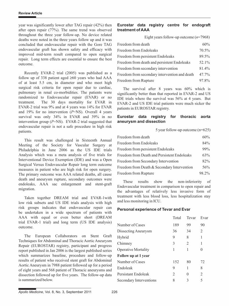
The European Collaborators on Stent GraftTechniques for Abdominal and Thoracic Aortic AneurysmRepair (EUROSTAR) registry, participant and progressreport published in Jan 2006 is the largest published serieswhich summarizes baseline, procedure and follow-upresults of patient who received stent graft for AbdominalAortic Aneurysm in 7988 patient followed up for a periodof eight years and 568 patient of Thoracic aneurysms anddissection followed up for five years. The follow-up datais summarized below.
Eurostar data registry centre for endografttreatment of AAA
Eight years follow-up outcome (n=7968)
Freedom from death 60%Freedom from Endoleaks 70.5%Freedom from persistent Endoleaks 89.5%Freedom from death and persistent Endoleaks 52.1%Freedom from secondary intervention 81.4%Freedom from secondary intervention and death 47.7%Freedom from Rupture 97.8%
The survival after 8 years was 60% which issignificantly better than that reported in EVAR-2 and USIDE trials where the survival was 56% at 4 years. ButEVAR-2 and US IDE trial patients were much sicker thepatients in EUROSTAR registry.
Eurostar data registry for thoracic aortaaneurysm and dissection
5 year follow-up outcome (n=625)
Freedom from death 60%Freedom from Endoleaks 84%Freedom from persistent Endoleaks 99%Freedom from Death and Persistent Endoleaks 63%Freedom from Secondary Intervention 82%Freedom from Death & Secondary Intervention 56%Freedom from Rupture 98.2%
These results show the non-inferiority ofEndovascular treatment in comparison to open repair andthe advantages of relatively less invasive form oftreatment with less blood loss, less hospitalization stayand less monitoring in ICU.
Personal experience of Tevar and Evar
Total Tevar Evar
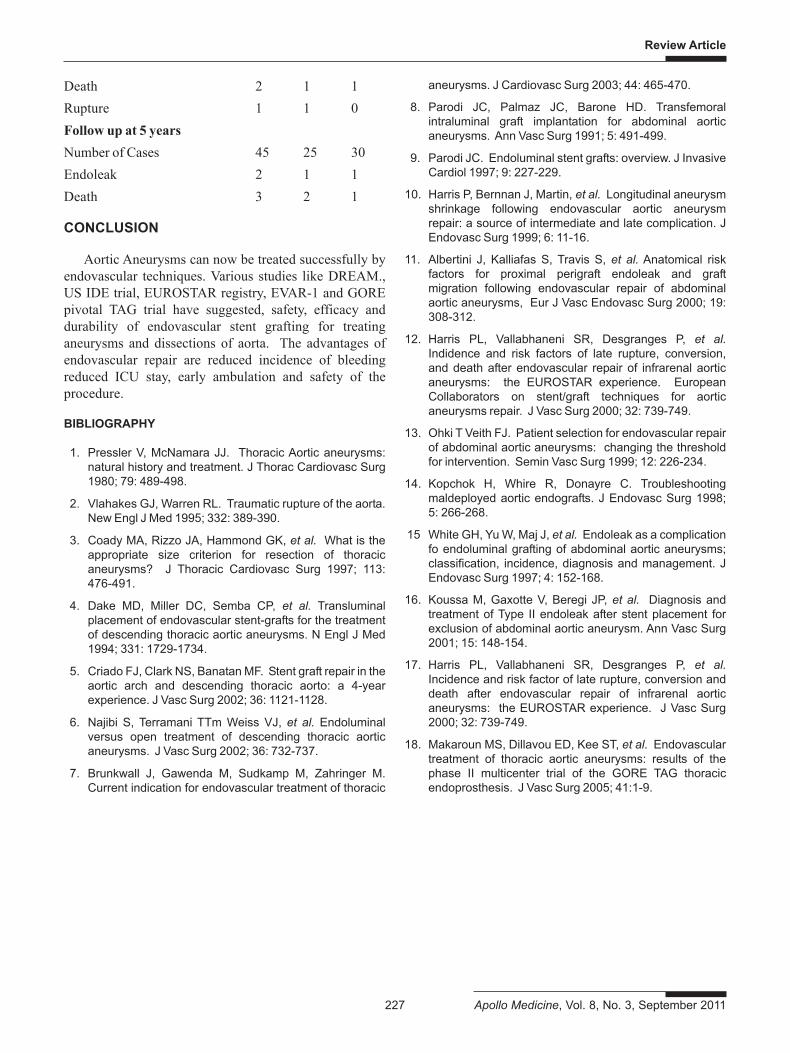
Number of Cases 189 99 90Dissecting Aneurysm 36 34 2Hybrid 9 8 1Chimney 3 2 1Operative Mortality 1 1 0Follow up at 1 yearNumber of Cases 152 80 72Endoleak 9 1 8Persistant Endoleak 2 0 2Secondary Interventions 8 3 5
Review Article
227 Apollo Medicine, Vol. 8, No. 3, September 2011
Death 2 1 1Rupture 1 1 0Follow up at 5 yearsNumber of Cases 45 25 30Endoleak 2 1 1Death 3 2 1
CONCLUSION
Aortic Aneurysms can now be treated successfully byendovascular techniques. Various studies like DREAM.,US IDE trial, EUROSTAR registry, EVAR-1 and GOREpivotal TAG trial have suggested, safety, efficacy anddurability of endovascular stent grafting for treatinganeurysms and dissections of aorta. The advantages ofendovascular repair are reduced incidence of bleedingreduced ICU stay, early ambulation and safety of theprocedure.
BIBLIOGRAPHY
1. Pressler V, McNamara JJ. Thoracic Aortic aneurysms:natural history and treatment. J Thorac Cardiovasc Surg1980; 79: 489-498.
2. Vlahakes GJ, Warren RL. Traumatic rupture of the aorta.New Engl J Med 1995; 332: 389-390.
3. Coady MA, Rizzo JA, Hammond GK, et al. What is theappropriate size criterion for resection of thoracicaneurysms? J Thoracic Cardiovasc Surg 1997; 113:476-491.
4. Dake MD, Miller DC, Semba CP, et al. Transluminalplacement of endovascular stent-grafts for the treatmentof descending thoracic aortic aneurysms. N Engl J Med1994; 331: 1729-1734.
5. Criado FJ, Clark NS, Banatan MF. Stent graft repair in theaortic arch and descending thoracic aorto: a 4-yearexperience. J Vasc Surg 2002; 36: 1121-1128.
6. Najibi S, Terramani TTm Weiss VJ, et al. Endoluminalversus open treatment of descending thoracic aorticaneurysms. J Vasc Surg 2002; 36: 732-737.
7. Brunkwall J, Gawenda M, Sudkamp M, Zahringer M.Current indication for endovascular treatment of thoracic
aneurysms. J Cardiovasc Surg 2003; 44: 465-470.
8. Parodi JC, Palmaz JC, Barone HD. Transfemoralintraluminal graft implantation for abdominal aorticaneurysms. Ann Vasc Surg 1991; 5: 491-499.
9. Parodi JC. Endoluminal stent grafts: overview. J InvasiveCardiol 1997; 9: 227-229.
10. Harris P, Bernnan J, Martin, et al. Longitudinal aneurysmshrinkage following endovascular aortic aneurysmrepair: a source of intermediate and late complication. JEndovasc Surg 1999; 6: 11-16.
11. Albertini J, Kalliafas S, Travis S, et al. Anatomical riskfactors for proximal perigraft endoleak and graftmigration following endovascular repair of abdominalaortic aneurysms, Eur J Vasc Endovasc Surg 2000; 19:308-312.
12. Harris PL, Vallabhaneni SR, Desgranges P, et al.Indidence and risk factors of late rupture, conversion,and death after endovascular repair of infrarenal aorticaneurysms: the EUROSTAR experience. EuropeanCollaborators on stent/graft techniques for aorticaneurysms repair. J Vasc Surg 2000; 32: 739-749.
13. Ohki T Veith FJ. Patient selection for endovascular repairof abdominal aortic aneurysms: changing the thresholdfor intervention. Semin Vasc Surg 1999; 12: 226-234.
14. Kopchok H, Whire R, Donayre C. Troubleshootingmaldeployed aortic endografts. J Endovasc Surg 1998;5: 266-268.
15 White GH, Yu W, Maj J, et al. Endoleak as a complicationfo endoluminal grafting of abdominal aortic aneurysms;classification, incidence, diagnosis and management. JEndovasc Surg 1997; 4: 152-168.
16. Koussa M, Gaxotte V, Beregi JP, et al. Diagnosis andtreatment of Type II endoleak after stent placement forexclusion of abdominal aortic aneurysm. Ann Vasc Surg2001; 15: 148-154.
17. Harris PL, Vallabhaneni SR, Desgranges P, et al.Incidence and risk factor of late rupture, conversion anddeath after endovascular repair of infrarenal aorticaneurysms: the EUROSTAR experience. J Vasc Surg2000; 32: 739-749.
18. Makaroun MS, Dillavou ED, Kee ST, et al. Endovasculartreatment of thoracic aortic aneurysms: results of thephase II multicenter trial of the GORE TAG thoracicendoprosthesis. J Vasc Surg 2005; 41:1-9.

Apollo hospitals: http://www.apollohospitals.com/Twitter: https://twitter.com/HospitalsApolloYoutube: http://www.youtube.com/apollohospitalsindiaFacebook: http://www.facebook.com/TheApolloHospitalsSlideshare: http://www.slideshare.net/Apollo_HospitalsLinkedin: http://www.linkedin.com/company/apollo-hospitalsBlog:Blog: http://www.letstalkhealth.in/