truncus arteriosus cicu lecture january 7, 2011 chrissy sarlone
TRANSCRIPT
Truncus Arteriosus
CICU lectureJanuary 7, 2011Chrissy Sarlone
Features
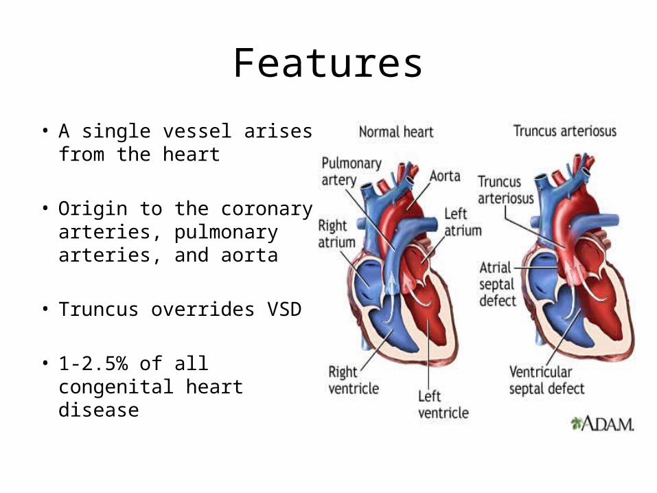
• A single vessel arises from the heart
• Origin to the coronary arteries, pulmonary arteries, and aorta
• Truncus overrides VSD
• 1-2.5% of all congenital heart disease
Features
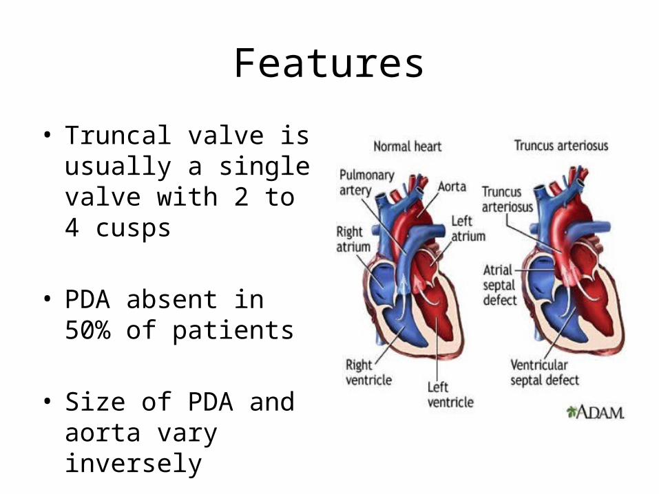
• Truncal valve is usually a single valve with 2 to 4 cusps
• PDA absent in 50% of patients
• Size of PDA and aorta vary inversely
Embryology
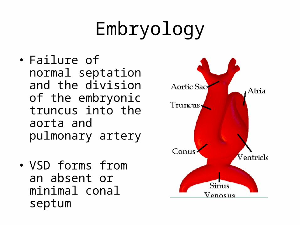
• Failure of normal septation and the division of the embryonic truncus into the aorta and pulmonary artery
• VSD forms from an absent or minimal conal septum
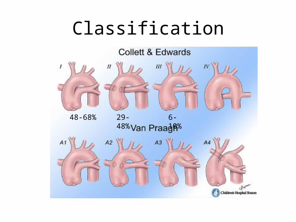
Classification
48-68% 29-48% 6-10%
Risk Factors
• Genetics: Recurrence rates vary from 1.2% to 13.6% with complex disease(Allen et al, 2000 )
• IDM (12-13 fold increase in Baltimore Washington Infant Study)– Ferencz, Teratology, 1990
• Fetal exposure to retinoic acid
Associated Defects
• Truncal Valve Insufficiency/Stenosis-detected by 24 weeks gestation
• Right sided aortic arch- 30%• Interrupted aortic arch- 11-20%
– Type B• Coronary Artery Anomalies
22q11 Microdeletion Syndrome
• 1/3 of infants
• Higher risk if associated aortic arch anomaly
• Important to assess for hypocalcemia and immunodeficiency as these complicate the course
Physiology
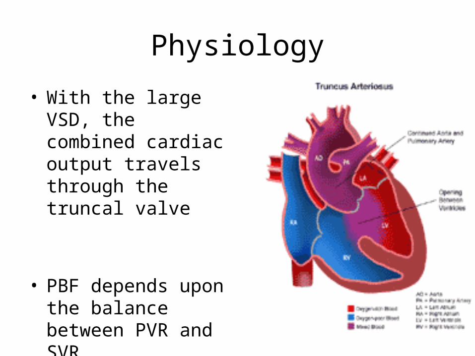
• With the large VSD, the combined cardiac output travels through the truncal valve
• PBF depends upon the balance between PVR and SVR
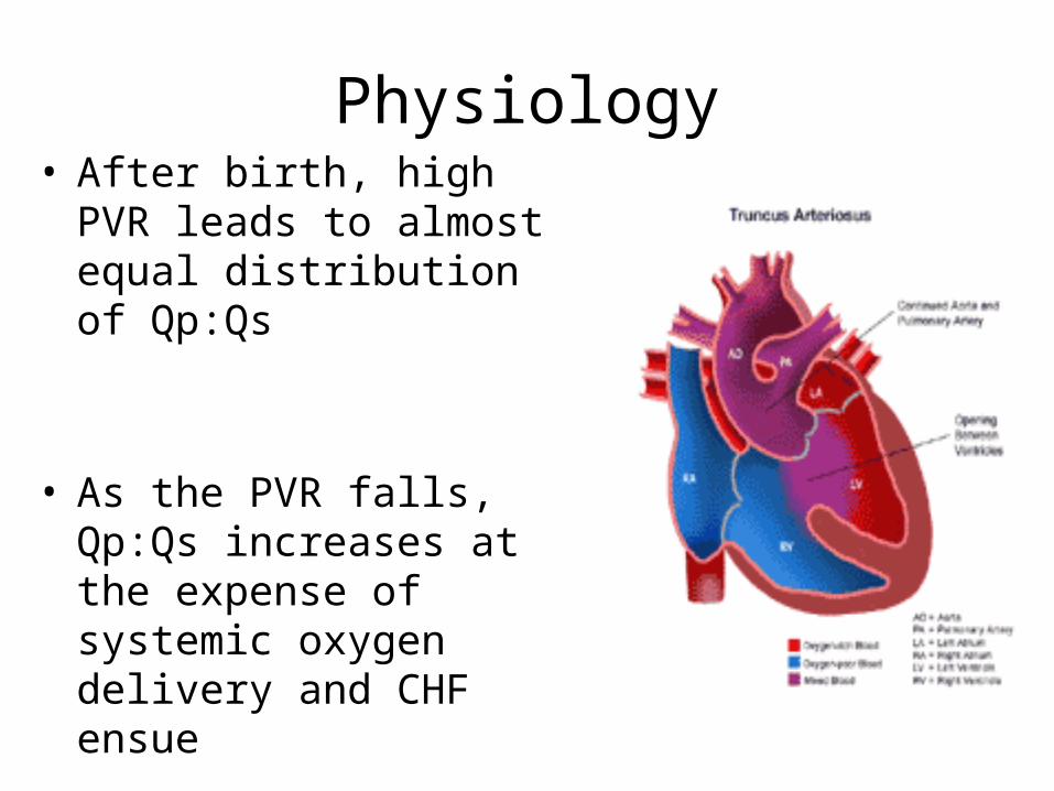
Physiology• After birth, high PVR leads to
almost equal distribution of Qp:Qs
• As the PVR falls, Qp:Qs increases at the expense of systemic oxygen delivery and CHF ensue
Delivery Room Management• Usually no intervention needed in the DR
• Several Factors to Consider:– Truncal valve insufficiency– Pulmonary artery hypoplasia
• If moderate or greater truncal valve insufficiency, the infant will be gray at birth because of low Mvsat and decreased systemic oxygen delivery
“The Unknown” Presentation• If not prenatally diagnosed and no truncal valve
insufficiency or PA hypoplasia, there may be mild cyanosis due to complete mixing
• Once PVR falls, sats will range 90% and higher, no visible cyanosis (increase Qp:Qs ratio)
• Over several weeks, the infant will become symptomatic with signs of CHF due to increased PBF and as a result decreased systemic oxygen delivery– Tachypnea, retractions– Poor feeding and growth– Excessive sweating
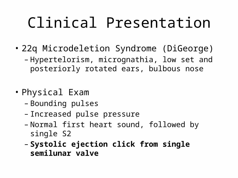
Clinical Presentation
• 22q Microdeletion Syndrome (DiGeorge)– Hypertelorism, micrognathia, low set and
posteriorly rotated ears, bulbous nose
• Physical Exam– Bounding pulses– Increased pulse pressure– Normal first heart sound, followed by single S2– Systolic ejection click from single semilunar valve
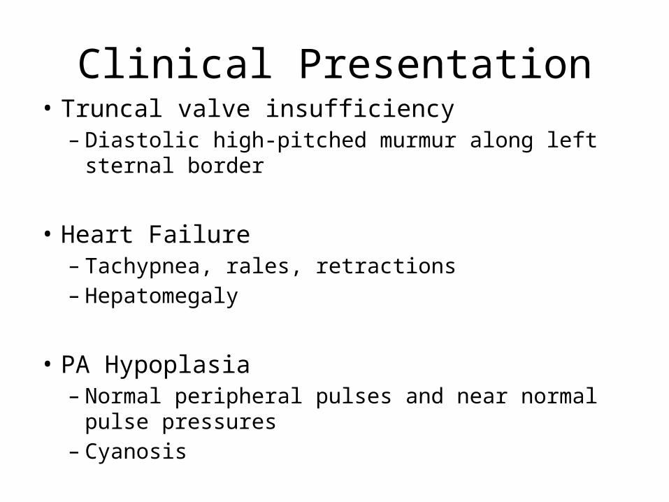
Clinical Presentation• Truncal valve insufficiency
– Diastolic high-pitched murmur along left sternal border
• Heart Failure– Tachypnea, rales, retractions– Hepatomegaly
• PA Hypoplasia– Normal peripheral pulses and near normal pulse
pressures– Cyanosis
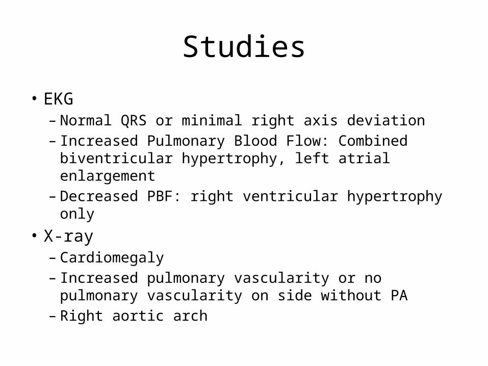
Studies
• EKG– Normal QRS or minimal right axis deviation– Increased Pulmonary Blood Flow: Combined
biventricular hypertrophy, left atrial enlargement– Decreased PBF: right ventricular hypertrophy only
• X-ray– Cardiomegaly– Increased pulmonary vascularity or no pulmonary
vascularity on side without PA– Right aortic arch
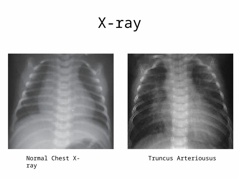
X-ray
Normal Chest X-ray Truncus Arteriousus
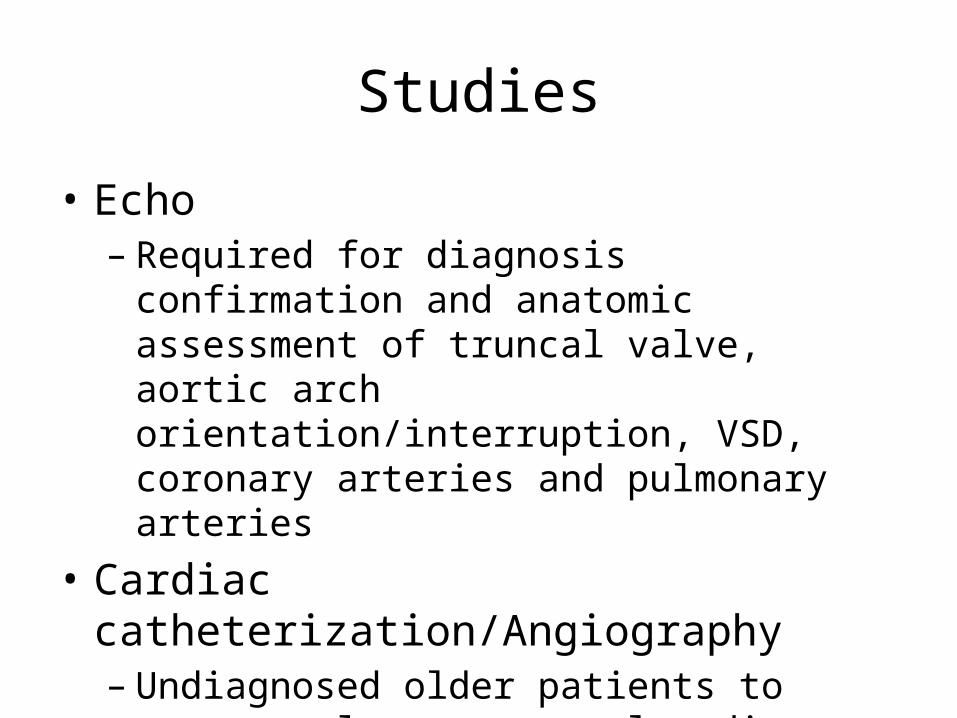
Studies
• Echo– Required for diagnosis confirmation and anatomic
assessment of truncal valve, aortic arch orientation/interruption, VSD, coronary arteries and pulmonary arteries
• Cardiac catheterization/Angiography– Undiagnosed older patients to assess pulmonary
vascular disease– Coronary artery and pulmonary artery mapping
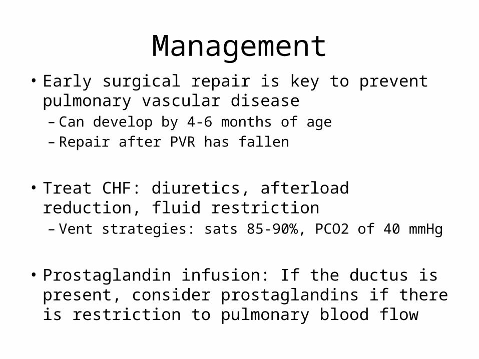
Management• Early surgical repair is key to prevent pulmonary
vascular disease– Can develop by 4-6 months of age– Repair after PVR has fallen
• Treat CHF: diuretics, afterload reduction, fluid restriction– Vent strategies: sats 85-90%, PCO2 of 40 mmHg
• Prostaglandin infusion: If the ductus is present, consider prostaglandins if there is restriction to pulmonary blood flow
Surgical Repair Goals
• Detach pulmonary arteries from truncus and if separated (Type II or III) create anastomosis
• Patch Closure of the aorta
Surgical Repair Goals• Closure of the VSD via right ventriculotomy
(via patch baffle) with LVOT blood flow directed to truncal valve
• Creation of RV to PA continuity via homograft conduit usually, rarely direct connection of MPA to Anterior RV
• Leave PFO for “pop-off” elevated RV end-diastolic pressures
Other Surgical Factors
• Truncal Valve Insufficiency– Manage conservatively if mild to moderate– Suture prolapsing leaflet to other leaflets making
tricuspid or bicuspid valve– May need to replace valve if severe TV insufficiency with
homograft valve, mechanical valve in older children• Interrupted Aortic Arch
– Repaired at the same time– May require homograft to connect ascending aorta and
aortic arch
Post-Op Complications
• Decreased LV output (Qs) due to RV systolic (RV infundibulotomy), RV diastolic dysfunction, and increased PVR – Decreased RV filling– Decreased RV output– Decreased preload to the LV– PFO extremely beneficial (Shunting Right to Left)
Better to be “blue” than “gray”
Post-Op Complications
• Pulmonary Hypertension– Can be present post-op despite no disease pre-op– High Qp:Qs pre-op with excessive flow
• Postop hemorrhage– Tamponade
• AV block– Conduction injury from VSD repair– May require cardiac pacing
Long-Term Outcomes
• Likely to need replacement of RV to PA conduit as child grows
• Potential replacement of truncal valve• Progressive Left Ventricular Dysfunction if
there is moderate to severe truncal valve insufficiency
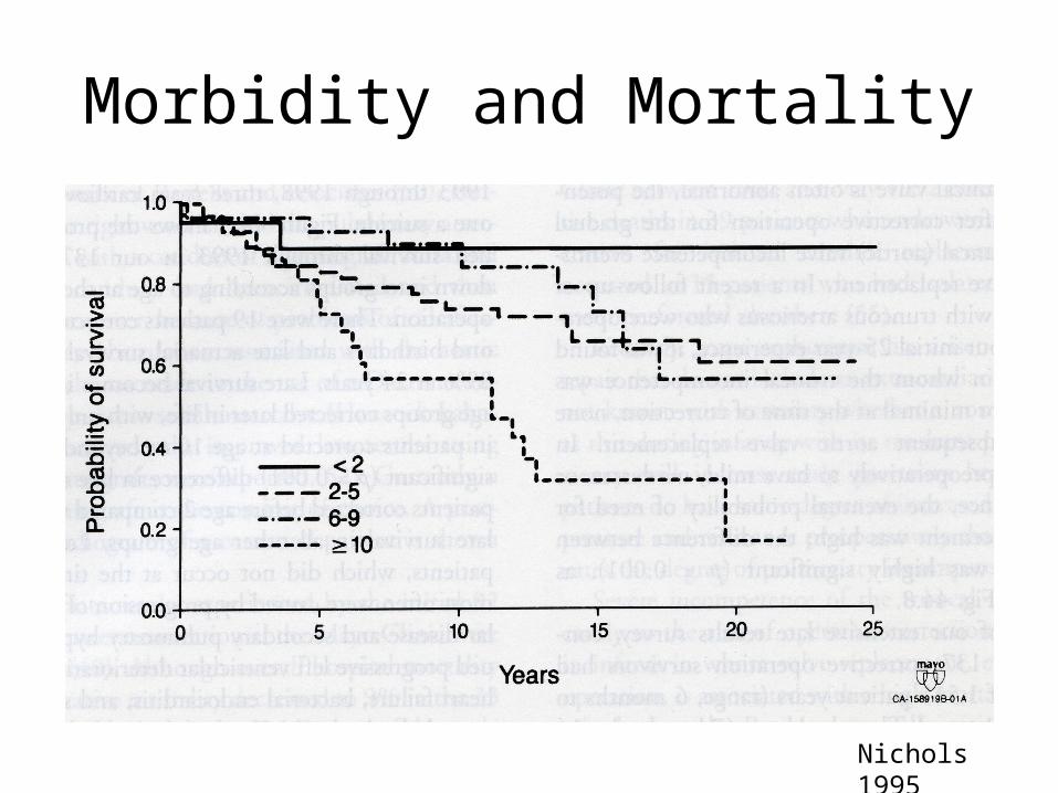
Morbidity and Mortality
Nichols 1995
Morbidity & Mortality
• Decreased with earlier repair over the years• 90% survival at CCHMC
• Eur J Cardiothorac Surg 2010– 83 patients in Prague– Post-op mortality of 46% between 1981-1997 and
4% between 1997-2008, – 54% with Type I, 12% with severe truncal valve
insufficiency, 17% with IAA– 75% patients required conduit replacements
within 5 years, 13% with balloon dilation or stent placement in conduit
Truncal Valve Replacement
• Ann Thorac Surg, 2010: Utah– 1995 to 2008, 27% of patients with truncus
arteriosus underwent truncal valve repair/replacement initially
– Repeated repair or replacement indicated when severe regurgitation with symptoms, LV dilatation
– 30% underwent second truncal valve operation (mean age 47 +/- 33 months) for severe regurgitation
– 70% required no intervention after 5 years, 50% after 7 years
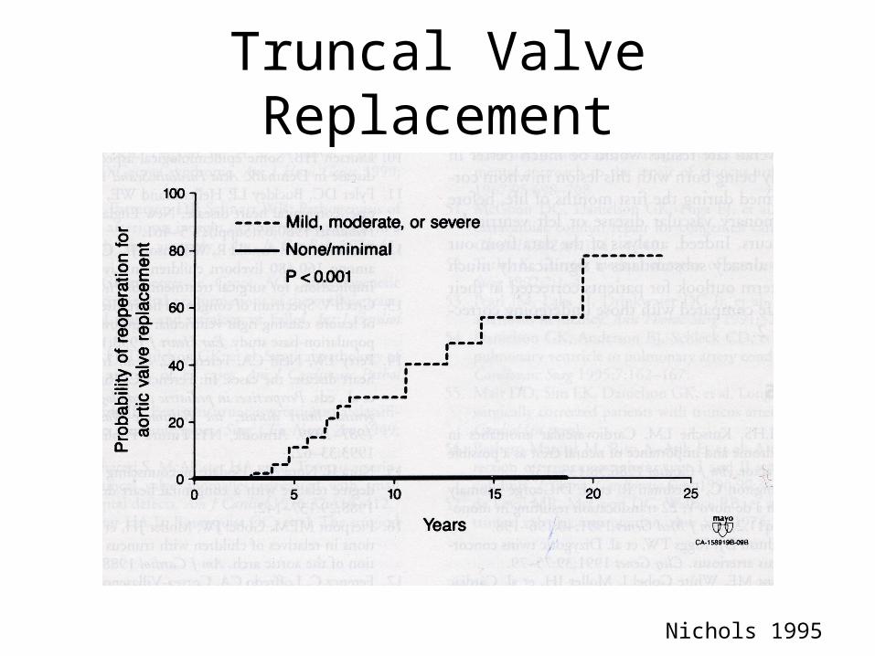
Truncal Valve Replacement
Nichols 1995
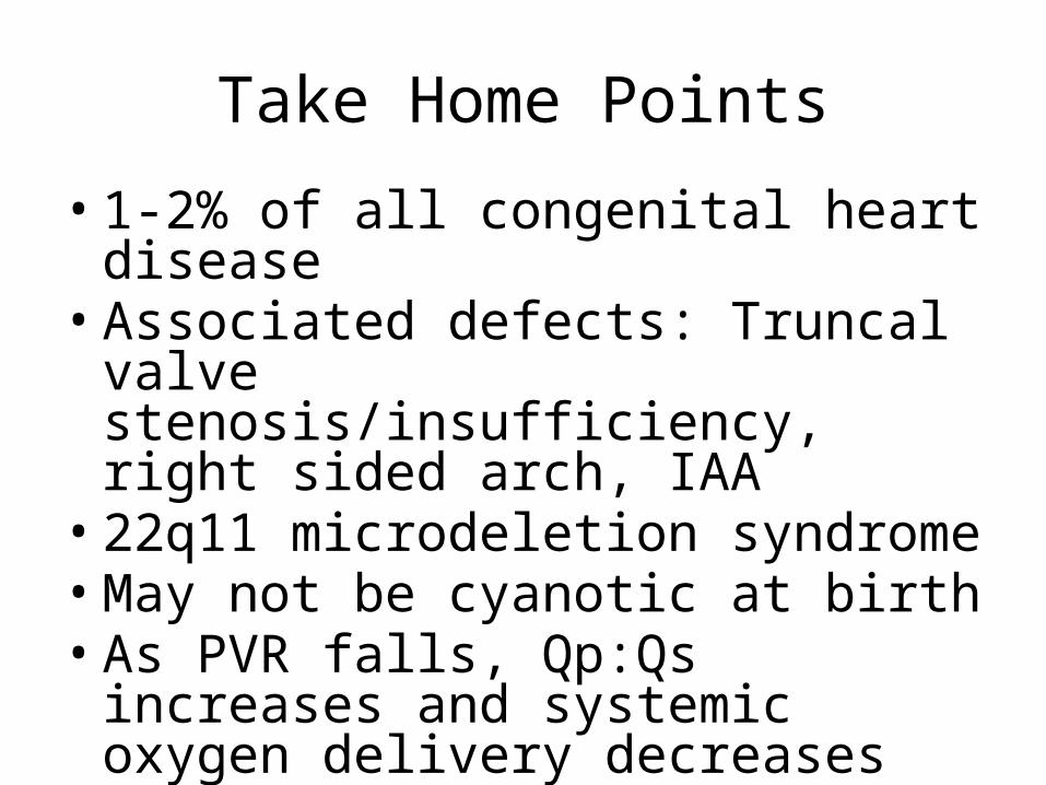
Take Home Points
• 1-2% of all congenital heart disease• Associated defects: Truncal valve
stenosis/insufficiency, right sided arch, IAA
• 22q11 microdeletion syndrome• May not be cyanotic at birth• As PVR falls, Qp:Qs increases and
systemic oxygen delivery decreases
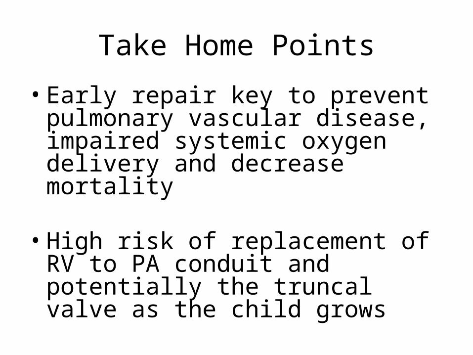
Take Home Points
• Early repair key to prevent pulmonary vascular disease, impaired systemic oxygen delivery and decrease mortality
• High risk of replacement of RV to PA conduit and potentially the truncal valve as the child grows
References• Allen, H. D., Adams, F. H., & Moss, A. J. (2000). Moss and adams' heart disease in infants,
children, and adolescents : Including the fetus and young adult (6th ed.). Philadelphia, PA: Lippincott Williams and Wilkins.
• Artman, M., Mahony, L., & Teitel, D. F. (2002). Neonatal cardiology. New York: McGraw-Hill, Medical Pub. Division.
• Henaine, R., Azarnoush, K., Belli, E., Capderou, A., Roussin, R., Planche, C., et al. (2008). Fate of the truncal valve in truncus arteriosus. The Annals of Thoracic Surgery, 85(1), 172-178.
• Kaza, A. K., Burch, P. T., Pinto, N., Minich, L. L., Tani, L. Y., & Hawkins, J. A. (2010). Durability of truncal valve repair. The Annals of Thoracic Surgery, 90(4), 1307-12; discussion 1312.
• Nichols, D. G. (1995). Critical heart disease in infants and children. St. Louis: Mosby. • Stark, J., De Leval, M., & Tsang, V. T. (2006). Surgery for congenital heart defects (3rd ed.).
Chichester ; Hoboken, NJ: J. Wiley & Sons. • Tlaskal, T., Chaloupecky, V., Hucin, B., Gebauer, R., Krupickova, S., Reich, O., et al. (2010).
Long-term results after correction of persistent truncus arteriosus in 83 patients. European Journal of Cardio-Thoracic Surgery : Official Journal of the European Association for Cardio-Thoracic Surgery, 37(6), 1278-1284.