type-specific hpv e6/e7 mrna detection by real-time pcr ... · 3 cervical lesions. assays detecting...
TRANSCRIPT
1
Type-specific HPV E6/E7 mRNA detection by real-time PCR improves 1
identification of cervical neoplasia. 2
3
Elin Andersson*1, Cecilia Kärrberg2, Thomas Rådberg2, Lennart Blomqvist3, Britt-Marie 4
Zetterqvist4, Walter Ryd5, Magnus Lindh1, Peter Horal1. 5
Departments of 1Clinical Virology, 2Obstetrics and Gynecology and 5Pathology, 6
Sahlgrenska Academy, University of Gothenburg, Sweden. 3Department of Gynecology and 7
Obstetrics, Southern Älvsborg Hospital, Borås, Sweden. 4Division of Obstetrics and 8
Gynecology, NU Hospital Organisation, Trollhättan, Sweden. 9
10
11
12
*Corresponding author: 13
Elin Andersson, Department of Clinical Virology, Guldhedsgatan 10B, 413 46 Gothenburg, 14
Sweden 15
Tel +46 31 3424663 16
Mobile +46 733 940160 17
Fax +46 31 827032 18
e-mail: [email protected] 19
20
21
Copyright © 2011, American Society for Microbiology and/or the Listed Authors/Institutions. All Rights Reserved.J. Clin. Microbiol. doi:10.1128/JCM.00549-11 JCM Accepts, published online ahead of print on 21 September 2011
on June 9, 2019 by guesthttp://jcm
.asm.org/
Dow
nloaded from
2
Abstract 1
DNA-based HPV assays show high sensitivity but poor specificity in detecting high-grade 2
cervical lesions. Assays detecting mRNA of oncogenic E6/E7 show higher specificity, but 3
lack either detection of all high-risk HPV genotypes or the capacity to specify the detected 4
genotypes. Therefore, a real-time PCR assay detecting type-specific E6/E7 mRNA was 5
developed and the clinical performance evaluated. 210 cervical LBC (liquid based cytology) 6
samples from 204 women were analysed for HPV DNA and mRNA with the in house real-7
time PCR as well as PreTect HPV-Proofer. The sensitivity of real-time PCR mRNA-detection 8
to detect histologically confirmed CIN2+ (cervical intraepithelial neoplasia grade 2 or higher) 9
were 0.91, compared to 0.95 for DNA-analysis. The specificity was 0.68 compared to 0.38, 10
and the positive predictive value (PPV) was higher for mRNA (0.67 vs 0.52) without any loss 11
in negative predictive value (NPV). The sensitivity of the real-time PCR mRNA-test was 12
somewhat higher than for PreTect HPV-Proofer (0.83 vs 0.75), when analysing for the same 13
genotypes. The specificity was similar (0.76 vs 0.77). When analysing for mRNA of the eight 14
most common genotypes in cervical cancer (HPV16, 18, 31, 33, 35, 45, 52, 58), the 15
sensitivity to detect CIN2+ lesions was 0.87 and the specificity 0.74, with a PPV of 0.70. In 16
conclusion, real-time PCR for detection of HPV E6/E7 mRNA transcripts can be a sensitive 17
and specific tool in screening and investigation of cervical neoplasia. The composition of 18
HPV-types in mRNA-testing needs to be further investigated to optimize sensitivity and 19
specificity. 20
21
22
23
24
25
26
on June 9, 2019 by guesthttp://jcm
.asm.org/
Dow
nloaded from
3
Introduction 1
Cervical cancer is closely associated with infection of human papillomaviruses (HPV), but 2
only a small proportion of these infections cause cancer. There are at least 12 oncogenic 3
genotypes (HPV16, 18, 31, 33, 35, 39, 45, 51, 52, 56, 58, 59) associated with high risk of 4
cervical cancer (HR-HPV) and a number of genotypes that probably also have oncogenic 5
properties (18). The viral proteins E6 and E7 are considered to be responsible for 6
transformation of the infected epithelial cell, as well as the maintenance of the malignant 7
phenotype. The proteins can affect many cellular proteins, such as the tumour suppressor 8
proteins pRB and p53 (reviewed by Ghittoni et al (11) and McLaughlin-Drubin and Munger 9
(16)) in a manner that leads to extension of cellular life span (including resistance to 10
apoptosis), DNA synthesis, genomic instability and interference with antiviral and antitumour 11
immune responses. The mechanisms that regulate if an HPV infection will be cleared by the 12
immune system or become persistent and cause transformation are not well understood. 13
However, integration of the viral genome into the cellular genome seems to be an important 14
event. Usually, the viral gene coding for E2, a regulator of E6/E7 transcription, is lost during 15
integration. Thus, integration typically leads to overexpression of E6/E7, which may facilitate 16
tumour progression (19). Moreover, common fragile sites are frequently targeted for viral 17
integration, possibly causing genomic instability (25). 18
There are numerous commercial tests available for HR-HPV DNA detection, but only a few 19
based on detection of oncogenic mRNA. DNA detection tests are highly sensitive for 20
detection of high-grade cervical intraepithelial neoplasia (CIN), and have been shown to be a 21
valuable tool in triage of Atypical Squamous Cells of Uncertain Significance (ASCUS) and 22
follow-up after treatment (5). Furthermore, the use of HPV DNA tests in primary screening 23
have in several studies been shown to be more sensitive than conventional cytology in 24
detecting cervical cancer and severe pre-cancerous lesions, and may serve to prolong the 25
screening interval (1, 7, 17). However, the specificities of HPV DNA tests for identification 26
on June 9, 2019 by guesthttp://jcm
.asm.org/
Dow
nloaded from
4
of cervical neoplasia are lower than for cytology especially among younger women (24). 1
Therefore, HPV-positive women need to be triaged before referral for further investigations, 2
such as colposcopy, but the preferable triage is yet to be established. Cytology could be an 3
alternative, as well as detection of HPV E6/E7 mRNA or cellular tumour markers such as p16 4
(4). One commercially available mRNA-test is the PreTect HPV-Proofer (NorChip, 5
Klokkarstua, Norway), also called NucliSENS EasyQ (Biomérieux, Marcy l’Etoile, France), 6
which detects mRNA of the five most common HPV-types, HPV16, 18, 31, 33 and 45, based 7
on nucleic acid sequence based amplification (NASBA) technique. The specificity of the test 8
is higher than for DNA-tests (21, 24), but the sensitivity is lower, and mainly due to the fact 9
that it doesn’t detect all HR-HPV, it can never be as sensitive as a DNA-test. The other 10
commercially available mRNA-test for HPV is APTIMA (Gen-Probe, San Diego, CA), which 11
detects mRNA of 12 HR-HPV as well as mRNA of HPV66 and HPV68, based on 12
transcription-mediated amplification (TMA). However, APTIMA does not specify the 13
individual detected HPV types. The test has similar sensitive as a DNA-test, but with higher 14
specificity for detection of dysplasia (8, 20, 24). There is a difference in specificity between 15
PreTect HPV-Proofer and APTIMA, the former being more specific. The reason for the 16
higher sensitivity and lower specificity than observed with PreTect HPV-Proofer could be 17
that APTIMA detects mRNA of more genotypes, some more common in low-grade lesions. 18
However, APTIMA also detects HPV DNA, even though it is more sensitive for mRNA (10). 19
Similarly, there have been reports that the NASBA-technique can detect DNA (3, 22), causing 20
false positive results. 21
We have previously developed a real-time PCR test based on amplification of E6/E7 DNA of 22
12 HR-HPV and 2 LR-HPV (15). The performance of the method has been validated by 23
showing agreement with the Linear Array assay (Roche) (15) and by a 100% proficiency 24
when participating in the WHO LabNet proficiency panel study 2009 (9). We have adapted 25
this assay to detect only mRNA by adding a DNase-digesting step and a reverse transcription 26
on June 9, 2019 by guesthttp://jcm
.asm.org/
Dow
nloaded from

5
step. In this study we evaluate the clinical performance of this type-specific HPV mRNA test, 1
and correlate the results with the HPV DNA analysis (using the same primers and probes) and 2
the mRNA-test PreTect HPV-Proofer. 3
4
Materials and methods 5
Samples 6
Liquid based cytology (LBC) samples collected in PreservCyt media (Cytyc, Marlborough, 7
MA, USA) from 204 women who were attending gynaecological screening (n=51, 26 8
pregnant) or had been admitted to a referral center for investigation because of abnormalities 9
in cervical cytology (n=153, 25 pregnant) were included. The age of the women ranged 10
between 21 and 79 with a median and mean age of 32 and 34 years, respectively. Five of the 11
women were sampled two or three times, resulting in 210 samples. All women received 12
information of the study design and provided written consent. Approval was obtained from 13
the local ethics committee. Neoplasias were evaluated by colposcopy-directed biopsies and/or 14
total excitional biopsies (conisation) and subsequent histological examination. An expert 15
pathologist re-evaluated all histological samples. If the second diagnosis differed from the 16
original diagnosis by more than one level of severity, the pathologist confirmed the diagnosis 17
with another pathologist. 18
19
DNA and RNA extraction 20
DNA or total NA was extracted using a MagNA Pure LC instrument (Roche). For DNA 21
analysis 250-500 µL of the LBC sample was used for extraction with the DNA I protocol. For 22
mRNA analysis 3-5 mL of the sample was briefly centrifuged and pelleted cells were 23
resuspended in 1 mL of RLT lysis buffer (Qiagen, Hilden, Germany) for extraction with the 24
totNA LV protocol. To assure the quality of mRNA, the LBC samples were not allowed a 25
storage period longer than 30 days before resuspension in lysis buffer and total NA extraction. 26
on June 9, 2019 by guesthttp://jcm
.asm.org/
Dow
nloaded from
6
After lysis treatment, some samples were stored in -70°C before extraction. Prior to analysis, 1
extractions were stored in -70°C. 2
Real-time PCR 3
The Taqman real-time PCR assay targets 12 high-risk (16, 18, 31, 33, 35, 39, 45, 51, 52, 56, 4
58, 59) and two low-risk (6 and 11) types using E6/E7 region primers and probes in a duplex 5
format (15). Detection of the human gene betaglobin serves as a control of sample 6
sufficiency. Briefly, 10 µL of extracted DNA was added to master mix of 25 µL Universal 7
PCR master mix (Roche diagnostics, Branchburg, NJ) with 0.3 µM primers and 0.2 µM 8
probes, supplemented with nuclease-free water to a final volume of 40 µL. After uracil DNA 9
glycosylase activation at 50°C for 2 min and initial denaturation at 95°C for 10 min, the PCR 10
for DNA detection was run for 45 cycles (15 s at 95°C, 60 s at 58°C) on an ABI 7300 11
instrument (Applied Biosystems, Carlsbad, CA). The threshold cycle, Ct, value for each 12
reaction was recorded (a low Ct-value indicates high amount of target). Only samples yielding 13
a Ct value for betaglobin below 36 were included in analysis. The modified method used for 14
HPV mRNA detection included a DNase digestion step, using the Ambion TURBO DNA-15
free kit (Applied Biosystems). Ten µL of the DNase-treated sample was added to a one-step 16
RT-PCR mastermix containing 1 µL Ribonuclase inhibitor (RNase OUT) and 1 µL 17
SuperScript (all Invitrogen, Carlsbad, CA), supplemented with nuclease-free water to a final 18
reaction volume of 50 µl, including 0.3 µM primers and 0.2 µM probes. The PCR program 19
was identical to that for DNA except for an initiating step of reverse transcription at 48°C for 20
30 minutes. To ascertain that no remaining HPV DNA was present, the DNase-treated 21
samples were also run with the DNA detection protocol, i.e. without the RT step. Any mRNA 22
detection was only accepted if the corresponding DNA was not detected or detected with a Ct 23
value more than10 cycles above the Ct value for mRNA. The E6/E7 gene is transcribed into 24
one full-length mRNA-transcript coding for both proteins, but is also spliced to an E7-25
encoding transcript (HPV16 is the only genotype transcribed into two spliced transcripts as 26
on June 9, 2019 by guesthttp://jcm
.asm.org/
Dow
nloaded from
7
well as a full-length) (23). Our real-time PCR detects all transcripts, both full-length and 1
spliced, except for genotypes 33, 52, 58 and 59 where only the full-length transcripts are 2
detected. For HPV16, the shorter of the two spliced transcripts is not detected. 3
4
PreTect HPV-Proofer 5
Detection of E6/E7 mRNA of genotypes 16, 18, 31, 33 and 45 was performed using the 6
PreTect HPV-Proofer kit according to the manufacturer’s guidelines. Briefly, the analysis is 7
based on NASBA technique with isothermal amplification of mRNA in a duplex format, 8
measured in real-time. Five µL of total NA-extract was added to 10 µL of a master mix with 9
primers, molecular beacon probes and KCl. After incubation for 2 min at 65°C and 2 min at 10
41°C, 5 µL of enzyme was added and spinned down before amplification at 41°C. Analysis of 11
the cellular U1A transcript was included in the test to determine the validity of the results. 12
13
Statistics 14
The sensitivity, specificity, positive predictive value and negative predictive value of each test 15
algorithm were calculated with histologically confirmed CIN2+ as gold standard, but 16
calculations were also made for CIN3+. Calculations of 95% confidence intervals (95% CI) 17
were based on the normal approximation to the binomial distribution as suggested by Harper 18
and Reeves (12). 19
20
Results 21
Cytological and histological diagnoses 22
For 155 (74%) of the 210 LBC samples, histological evaluations from biopsies and/or total 23
excised specimens taken at the same time were available (Table 1). For 53 samples with 24
benign cytology (51 of them from women in screening) and two samples with ASCUS in 25
cytology, no histological data was available. The histological diagnosis “benign” (n=32) 26
on June 9, 2019 by guesthttp://jcm
.asm.org/
Dow
nloaded from
8
includes inflammation (n=11), metaplasia (n=2), ulcus (n=1) and HPV-infection without signs 1
of CIN (n=5). The diagnosis “CIN3” (n=31) includes adenocarcinoma in situ (n=3). 2
Histological data were available for 50 cytologically benign samples, showing CIN1 or worse 3
in 24 cases (48%) including six cases of CIN3 and four cases of cancer. These 24 represent 4
16% of the women in this material undergoing investigation for dysplasia, mostly due to 5
earlier atypical cytology. Overall, histological examination tended to upgrade the cytological 6
diagnoses. 7
8
HPV DNA and mRNA type distribution 9
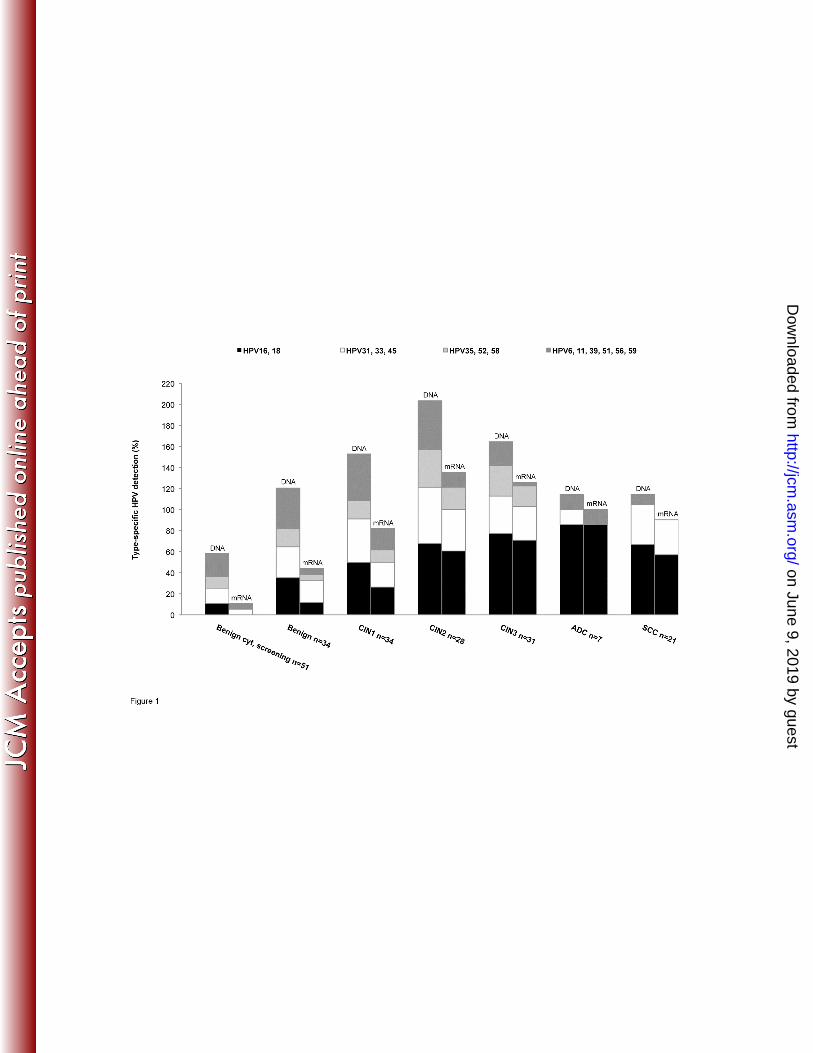
Type-specific detection rates of HPV DNA and mRNA according to histology are shown in 10
Figure 1 (one sample with glandular dysplasia has been included in the group of CIN1). 11
HPV16 was the most prevalent type in the 87 samples histologically classified as CIN2+ 12
(n=45, 52%), followed by HPV18 (n=18, 21%), HPV31 (n=17, 20%), HPV33 (n=13, 15%), 13
HPV52 (n=12, 14%), HPV39 (n=6, 7%) and HPV45, HPV51 and HPV56 (n=5, 6%). The five 14
most common HPV-types in the 28 samples with cancer were HPV16, HPV18, HPV33, 15
HPV45 and HPV31, in that order. 16
When analysing for HPV mRNA, the picture was similar. The most common genotype in 17
CIN2+ samples expressing E6/E7 mRNA was HPV16 (41, 47%), followed by HPV18 (16, 18
18%), HPV31 (15, 17%), HPV33 (8, 9%), HPV52 (7, 8%) and HPV45 (5, 6%). 19
Consequently, 40% of HPV56-infections, 33% of HPV39-infections and 0% of HPV59-20
infections in CIN2+ lesions showed expression of E6/E7 mRNA, in comparison to HPV45 21
(100%), HPV35 (100%), HPV16 (91%), HPV18 (89%) and HPV31 (88%). In 24% (28/118) 22
of mRNA-positive samples (68% of them CIN2+), mRNA of multiple genotypes was present. 23
The samples expressing mRNA of two or more genotypes represent 22% of all CIN2+ 24
samples. 25
on June 9, 2019 by guesthttp://jcm
.asm.org/
Dow
nloaded from
9
In four CIN3+ samples (three cancers), no HR-HPV mRNA could be found. In two of these 1
samples (both cancers), HR-HPV DNA was undetectable. (The samples tested positive for 2
HPV68 or HPV70, respectively, with other methods). The two DNA-positive CIN3+ samples 3
with undetectable mRNA were two single infections with HPV16 or HPV33, respectively. 4
Out of 34 samples with benign histology, 24 (71%) were HPV-positive and 12 (35%) 5
expressed E6/E7 mRNA. However, all these women had a history of dysplasia. When looking 6
at a screening-cohort of 51 women (median age 31) with benign cytology (no histology 7
available), the prevalence of HPV-infection was 43% (22) of which 9.8% (5) showed 8
expression of E6/E7 mRNA. 9
Overall, there was a good agreement between DNA and mRNA testing, and of all 258 10
detected HPV types 118 were identified by both DNA and mRNA testing (46%). As expected 11
the DNA analysis had a higher detection rate, and identified 140 HPV types that were not 12
detected by mRNA testing. Conversely, mRNA was detected in 5 samples in which the same 13
genotype was not detected by the DNA assay. 14
15
Sensitivity, specificity, PPV and NPV 16
The sensitivity, specificity, positive predictive value (PPV) and negative predictive value 17
(NPV) of detecting CIN3+ or CIN2+ lesions were calculated for analysis of HPV DNA, 18
mRNA and with PreTect HPV-Proofer. Furthermore, calculations were made for the two in 19
house tests (DNA and mRNA) when including only the five most common HPV-types in 20
cervical cancer (HPV16, 18, 31, 33, 45 (2) which are the five genotypes included in the 21
PreTect HPV-Proofer assay). However, recent data suggest that in different parts of the world, 22
the most common HPV-types in cervical cancer may vary (6, 14). HPV16 and 18 are the most 23
common world wide, and with a few exceptions, the most common genotypes after HPV16 24
and 18 are HPV31, 33, 35, 45, 52 and 58, in varying order. We therefore made calculations 25
for sensitivity, specificity, PPV and NPV also for these eight genotypes. Since it was not 26
on June 9, 2019 by guesthttp://jcm
.asm.org/
Dow
nloaded from
10
possible to differentiate between low and moderate grades of neoplasia in glandular cells, one 1
sample with glandular neoplasia not reaching the level of adenocarcinoma in situ was 2
included as CIN1 in the calculations tabulated in table 2. The sensitivity and specificity 3
results are illustrated in Figure 2. 4
5
Discussion 6
This study aimed to evaluate the clinical performance of a real-time PCR assay that detects 7
mRNA transcripts coding for the oncogenic proteins E6 and E7 of 12 high-risk HPV and 2 8
low-risk, using the same primers and probes as described previously for HPV DNA. For 210 9
LBC samples with various grades of cervical neoplasia, there was good agreement between 10
HPV mRNA and HPV DNA results although the detection rate was higher with the DNA 11
assay, as expected. 12
Our assay for mRNA detection, which includes a step that verifies that the mRNA signal is 13
not due to detection of DNA, had a sensitivity of detection of CIN2+ and CIN3+ that was 14
only slightly lower than for DNA-detection (0.91 vs 0.95 and 0.93 vs 0.97, respectively), but 15
the NPV did not decrease compared to the DNA-test. Importantly, the specificity was higher 16
for mRNA than for DNA detection (0.68 vs 0.38 for CIN2+ lesions and 0.58 vs 0.32 for 17
CIN3+ lesions). These results may be compared with the observations by Szarewski et al in 18
which several HPV DNA and mRNA tests were compared (24). The sensitivity and 19
specificity for CIN2+ using the PreTect HPV-Proofer assay (detecting five HR-HPV) was 20
0.74 and 0.73 in their study as compared with 0.75 and 0.77 in our evaluation. 21
When comparing our assay for mRNA typing with PreTect HPV-Proofer, the sensitivity of 22
real-time PCR was somewhat higher (0.83 vs 0.75 for CIN2+ lesions and 0.86 vs 0.81 for 23
CIN3+ lesions, when analysing for the same five genotypes). This may reflect a higher 24
analytical sensitivity by real-time PCR compared with NASBA (because samples negative 25
with PreTect HPV-Proofer but positive with real-time PCR in general contained low amounts 26
on June 9, 2019 by guesthttp://jcm
.asm.org/
Dow
nloaded from
11
of virus, as indicated by high Ct values, data not shown). The genotype most commonly 1
detected by real-time PCR but not PreTect HPV-Proofer was HPV31, and this is in agreement 2
with calculations of analytical sensitivity of the NucliSENS EasyQ assay (based on the same 3
platform as PreTect HPV-Proofer) showing that the sensitivity of detection of HPV31 mRNA 4
is 10-100 times lower than for the other types (13). Moreover, our in house real-time PCR 5
detects not only full-length mRNA, but also spliced mRNA transcripts of most genotypes, in 6
contrast to PreTect HPV-Proofer (13), which may increase the sensitivity of the real-time 7
PCR assay. A high analytical sensitivity might confer a risk of detecting low amounts of HPV 8
mRNA not significant for disease, but the CIN2+ specificity of the real-time PCR was equal 9
to that for PreTect HPV-Proofer when analysing for the same genotypes. 10
There have been suggestions that the high specificity of PreTect HPV-Proofer is mainly due 11
to the fact that it analyzes the five most common genotypes, and that a DNA-test analysing 12
these five genotypes might be just as specific (3). However, this speculation is contradicted 13
by our data, since specificity calculated for these five genotypes was higher for both CIN2+ 14
and CIN3+ lesions using the mRNA as compared with the DNA version of our real-time PCR 15
(0.74 vs 0.57 and 0.64 vs 0.50, respectively), suggesting that presence of E6/E7 transcripts is 16
important for disease. The high specificity by mRNA testing was illustrated by the finding 17
that in a screening cohort of 51 women (median age 31) with normal cytology (but with no 18
histology available), HPV mRNA was detected in 9.8% and HPV DNA in 43%. The PPV of 19
detection of both CIN2+ and CIN3+ was therefore higher for mRNA-detection compared to 20
DNA-detection (0.67 vs 0.52 and 0.47 vs 0.36, respectively), suggesting that mRNA testing 21
may be a useful tool not only in triage, but also in primary screening of cervical neoplasias. 22
One should bear in mind that not all CIN2+ lesions will progress to cancer, and a hypothetical 23
perfect test identifying only truly precancerous lesions would rate poorly in sensitivity with 24
CIN2+ in histology used as a golden standard (as in this and most other studies). 25
26
on June 9, 2019 by guesthttp://jcm
.asm.org/
Dow
nloaded from
12
The five most common genotypes present in CIN2+ and CIN3+ lesions were HPV16, 1
followed by HPV18, 31, 33 and 52 (in that order). In LBC samples from our patients with 2
cancer however, the five most common genotypes were HPV16, 18, 33, 45 and 31. This may 3
reflect that the oncogenic properties of the genotypes vary. This idea was supported by the 4
observation that some genotypes expressed E6/E7 mRNA more often than others. The eight 5
genotypes most prone to express mRNA in CIN2+ lesions were, in descending order, HPV45, 6
35, 16, 18, 31, 33, 51, 52 and 58, the same genotypes (except for HPV51) most commonly 7
found in cervical cancer worldwide (6, 14). Possibly, the association of these eight genotypes 8
with cancers may be a consequence of their potential to express oncogenic mRNA. Our 9
finding encourages further and larger studies comparing mRNA and DNA detection rates for 10
different HPV types. 11
We specifically evaluated the performance of the real-time PCR detection of mRNA for only 12
the above mentioned eight genotypes that are most commonly observed in cancer. With this 13
limitation, the sensitivity of the assay increased somewhat compared to analysis of five 14
genotypes, but the specificity did not substantially decrease and the PPV remained, 15
suggesting that these eight might constitute a good balance between sensitivity and 16
specificity. This was relevant also for HPV DNA testing, since analysing eight as compared 17
to all genotypes resulted in a significant increase in specificity at the expense of only a small 18
loss in sensitivity, however without decreasing the high NPV. 19
Our data suggest that mRNA-testing with real-time PCR may be a useful tool in investigation 20
of as well as in primary screening for cervical neoplasias, and there might be an idea to 21
consider which genotypes to include in further investigations to optimize sensitivity and 22
specificity, especially in a post vaccine era when it may be necessary to reconsider HPV 23
testing strategies. 24
25
Acknowledgments 26
on June 9, 2019 by guesthttp://jcm
.asm.org/
Dow
nloaded from
13
We thank Monika Dohsé for technical assistance. This study was supported by grants from 1
the Western region R&D Fund, ALF-funds, Capio Research foundation and Assar 2
Gabrielsson foundation. 3
4
Legends to figures 5
Figure 1. Type-specific detection (%) of HPV DNA and mRNA, distributed by histology. 6
Each HPV type in a multiple infection is counted, which may result in an accumulative 7
percentage of more than 100. ADC: Adenocarcinoma, SCC: Squamous cervical carcinoma 8
9
Figure 2. Clinical sensitivity and specificity (with 95% confidence intervals) of the different 10
test algorithms for detection of A) CIN3+ lesions and B) CIN2+ lesions. 11
12
13
References 14
1. Anttila, A., L. Kotaniemi-Talonen, M. Leinonen, M. Hakama, P. Laurila, J. 15
Tarkkanen, N. Malila, and P. Nieminen. 2010. Rate of cervical cancer, severe 16
intraepithelial neoplasia, and adenocarcinoma in situ in primary HPV DNA screening 17
with cytology triage: randomised study within organised screening programme. BMJ 18
340:c1804. 19
2. Bosch, F. X., A. N. Burchell, M. Schiffman, A. R. Giuliano, S. de Sanjose, L. 20
Bruni, G. Tortolero-Luna, S. K. Kjaer, and N. Munoz. 2008. Epidemiology and 21
natural history of human papillomavirus infections and type-specific implications in 22
cervical neoplasia. Vaccine 26 Suppl 10:K1-16. 23
3. Boulet, G. A., I. M. Micalessi, C. A. Horvath, I. H. Benoy, C. E. Depuydt, and J. 24
J. Bogers. Nucleic acid-sequence based amplification assay for HPV mRNA detection 25
and typing: evidence for DNA amplification. J Clin Microbiol. 26
on June 9, 2019 by guesthttp://jcm
.asm.org/
Dow
nloaded from
14
4. Cuschieri, K., and N. Wentzensen. 2008. Human papillomavirus mRNA and p16 1
detection as biomarkers for the improved diagnosis of cervical neoplasia. Cancer 2
Epidemiol Biomarkers Prev 17:2536-45. 3
5. Cuzick, J., M. Arbyn, R. Sankaranarayanan, V. Tsu, G. Ronco, M. H. Mayrand, 4
J. Dillner, and C. J. Meijer. 2008. Overview of human papillomavirus-based and 5
other novel options for cervical cancer screening in developed and developing 6
countries. Vaccine 26 Suppl 10:K29-41. 7
6. de Sanjose, S., W. G. Quint, L. Alemany, D. T. Geraets, J. E. Klaustermeier, B. 8
Lloveras, S. Tous, A. Felix, L. E. Bravo, H. R. Shin, C. S. Vallejos, P. A. de Ruiz, 9
M. A. Lima, N. Guimera, O. Clavero, M. Alejo, A. Llombart-Bosch, C. Cheng-10
Yang, S. A. Tatti, E. Kasamatsu, E. Iljazovic, M. Odida, R. Prado, M. Seoud, M. 11
Grce, A. Usubutun, A. Jain, G. A. Suarez, L. E. Lombardi, A. Banjo, C. 12
Menendez, E. J. Domingo, J. Velasco, A. Nessa, S. C. Chichareon, Y. L. Qiao, E. 13
Lerma, S. M. Garland, T. Sasagawa, A. Ferrera, D. Hammouda, L. Mariani, A. 14
Pelayo, I. Steiner, E. Oliva, C. J. Meijer, W. F. Al-Jassar, E. Cruz, T. C. Wright, 15
A. Puras, C. L. Llave, M. Tzardi, T. Agorastos, V. Garcia-Barriola, C. Clavel, J. 16
Ordi, M. Andujar, X. Castellsague, G. I. Sanchez, A. M. Nowakowski, J. 17
Bornstein, N. Munoz, and F. X. Bosch. 2010. Human papillomavirus genotype 18
attribution in invasive cervical cancer: a retrospective cross-sectional worldwide 19
study. Lancet Oncol. 20
7. Dillner, J., M. Rebolj, P. Birembaut, K. U. Petry, A. Szarewski, C. Munk, S. de 21
Sanjose, P. Naucler, B. Lloveras, S. Kjaer, J. Cuzick, M. van Ballegooijen, C. 22
Clavel, and T. Iftner. 2008. Long term predictive values of cytology and human 23
papillomavirus testing in cervical cancer screening: joint European cohort study. BMJ 24
337:a1754. 25
on June 9, 2019 by guesthttp://jcm
.asm.org/
Dow
nloaded from
15
8. Dockter, J., A. Schroder, C. Hill, L. Guzenski, J. Monsonego, and C. Giachetti. 1
2009. Clinical performance of the APTIMA HPV Assay for the detection of high-risk 2
HPV and high-grade cervical lesions. J Clin Virol 45 Suppl 1:S55-61. 3
9. Eklund, C., T. Zhou, and J. Dillner. 2010. Global proficiency study of human 4
papillomavirus genotyping. J Clin Microbiol 48:4147-55. 5
10. Getman, D., A. Aiyer, J. Dockter, C. Giachetti, F. Zhang, and C. C. Ginocchio. 6
2009. Efficiency of the APTIMA HPV Assay for detection of HPV RNA and DNA 7
targets. J Clin Virol 45 Suppl 1:S49-54. 8
11. Ghittoni, R., R. Accardi, U. Hasan, T. Gheit, B. Sylla, and M. Tommasino. 2010. 9
The biological properties of E6 and E7 oncoproteins from human papillomaviruses. 10
Virus Genes 40:1-13. 11
12. Harper, R., and B. Reeves. 1999. Reporting of precision of estimates for diagnostic 12
accuracy: a review. BMJ 318:1322-3. 13
13. Jeantet, D., F. Schwarzmann, J. Tromp, W. J. Melchers, A. A. van der Wurff, T. 14
Oosterlaken, M. Jacobs, and A. Troesch. 2009. NucliSENS EasyQ HPV v1 test - 15
Testing for oncogenic activity of human papillomaviruses. J Clin Virol 45 Suppl 16
1:S29-37. 17
14. Li, N., S. Franceschi, R. Howell-Jones, P. J. Snijders, and G. M. Clifford. 2010. 18
Human papillomavirus type distribution in 30,848 invasive cervical cancers 19
worldwide: Variation by geographical region, histological type and year of 20
publication. Int J Cancer. 21
15. Lindh, M., S. Gorander, E. Andersson, P. Horal, I. Mattsby-Balzer, and W. Ryd. 22
2007. Real-time Taqman PCR targeting 14 human papilloma virus types. J Clin Virol 23
40:321-4. 24
16. McLaughlin-Drubin, M. E., and K. Munger. 2009. Oncogenic activities of human 25
papillomaviruses. Virus Res 143:195-208. 26
on June 9, 2019 by guesthttp://jcm
.asm.org/
Dow
nloaded from

16
17. Mesher, D., A. Szarewski, L. Cadman, H. Cubie, H. Kitchener, D. Luesley, U. 1
Menon, G. Hulman, M. Desai, L. Ho, G. Terry, A. Williams, P. Sasieni, and J. 2
Cuzick. 2010. Long-term follow-up of cervical disease in women screened by 3
cytology and HPV testing: results from the HART study. Br J Cancer 102:1405-10. 4
18. Munoz, N., X. Castellsague, A. B. de Gonzalez, and L. Gissmann. 2006. Chapter 1: 5
HPV in the etiology of human cancer. Vaccine 24 Suppl 3:S3/1-10. 6
19. Pett, M., and N. Coleman. 2007. Integration of high-risk human papillomavirus: a 7
key event in cervical carcinogenesis? J Pathol 212:356-67. 8
20. Ratnam, S., F. Coutlee, D. Fontaine, J. Bentley, N. Escott, P. Ghatage, V. Gadag, 9
G. Holloway, E. Bartellas, N. Kum, C. Giede, and A. Lear. 2011. Aptima HPV 10
E6/E7 mRNA test is as sensitive as Hybrid Capture 2 Assay but more specific at 11
detecting cervical precancer and cancer. J Clin Microbiol 49:557-64. 12
21. Ratnam, S., F. Coutlee, D. Fontaine, J. Bentley, N. Escott, P. Ghatage, V. Gadag, 13
G. Holloway, E. Bartellas, N. Kum, C. Giede, and A. Lear. Clinical performance of 14
the PreTect HPV-Proofer E6/E7 mRNA assay in comparison with that of the Hybrid 15
Capture 2 test for identification of women at risk of cervical cancer. J Clin Microbiol 16
48:2779-85. 17
22. Rodriguez-Lazaro, D., J. Lloyd, J. Ikonomopoulos, M. Pla, and N. Cook. 2004. 18
Unexpected detection of DNA by nucleic acid sequence-based amplification 19
technique. Mol Cell Probes 18:251-3. 20
23. Sotlar, K., A. Stubner, D. Diemer, S. Menton, M. Menton, K. Dietz, D. 21
Wallwiener, R. Kandolf, and B. Bultmann. 2004. Detection of high-risk human 22
papillomavirus E6 and E7 oncogene transcripts in cervical scrapes by nested RT-23
polymerase chain reaction. J Med Virol 74:107-16. 24
24. Szarewski, A., L. Ambroisine, L. Cadman, J. Austin, L. Ho, G. Terry, S. Liddle, 25
R. Dina, J. McCarthy, H. Buckley, C. Bergeron, P. Soutter, D. Lyons, and J. 26
on June 9, 2019 by guesthttp://jcm
.asm.org/
Dow
nloaded from

17
Cuzick. 2008. Comparison of predictors for high-grade cervical intraepithelial 1
neoplasia in women with abnormal smears. Cancer Epidemiol Biomarkers Prev 2
17:3033-42. 3
25. Thorland, E. C., S. L. Myers, B. S. Gostout, and D. I. Smith. 2003. Common 4
fragile sites are preferential targets for HPV16 integrations in cervical tumors. 5
Oncogene 22:1225-37. 6
7
8
on June 9, 2019 by guesthttp://jcm
.asm.org/
Dow
nloaded from
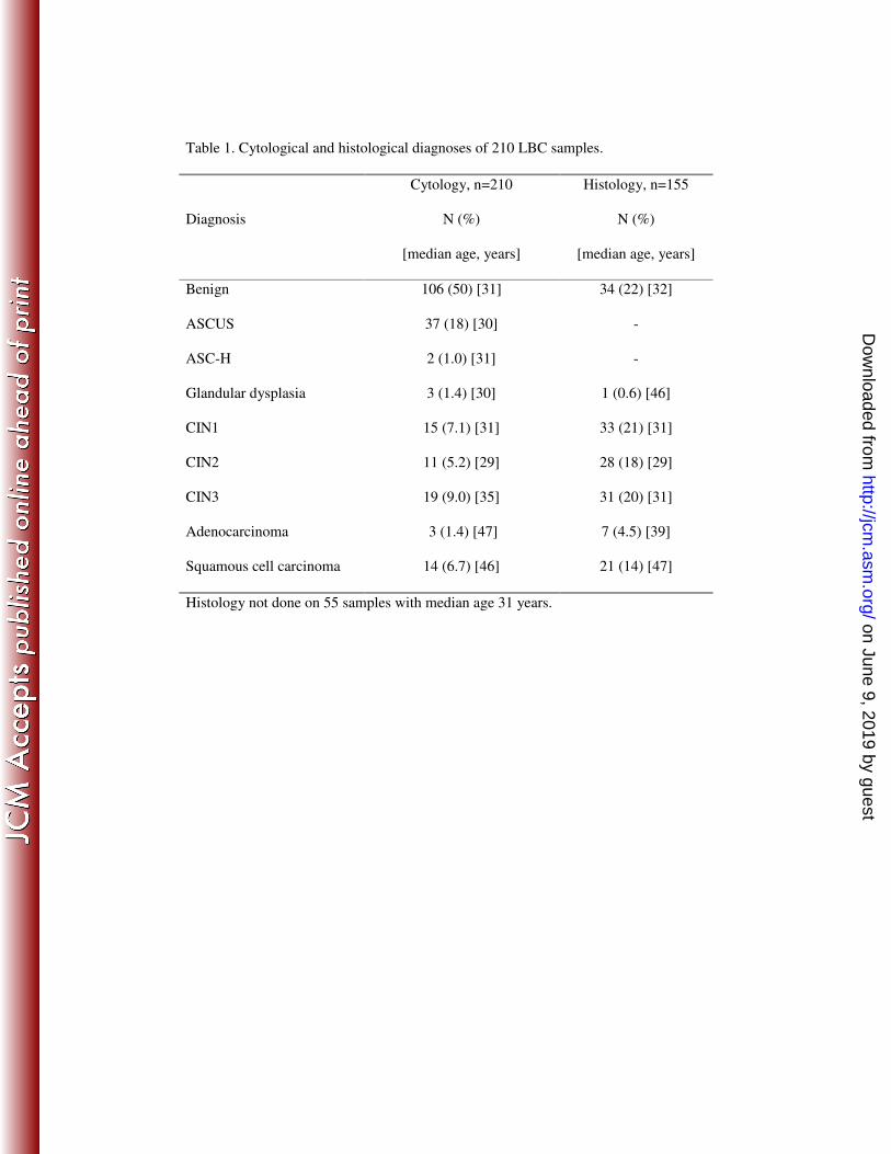
Table 1. Cytological and histological diagnoses of 210 LBC samples.
Diagnosis
Cytology, n=210
N (%)
[median age, years]
Histology, n=155
N (%)
[median age, years]
Benign 106 (50) [31] 34 (22) [32]
ASCUS 37 (18) [30] -
ASC-H 2 (1.0) [31] -
Glandular dysplasia 3 (1.4) [30] 1 (0.6) [46]
CIN1 15 (7.1) [31] 33 (21) [31]
CIN2 11 (5.2) [29] 28 (18) [29]
CIN3 19 (9.0) [35] 31 (20) [31]
Adenocarcinoma 3 (1.4) [47] 7 (4.5) [39]
Squamous cell carcinoma 14 (6.7) [46] 21 (14) [47]
Histology not done on 55 samples with median age 31 years.
on June 9, 2019 by guesthttp://jcm
.asm.org/
Dow
nloaded from
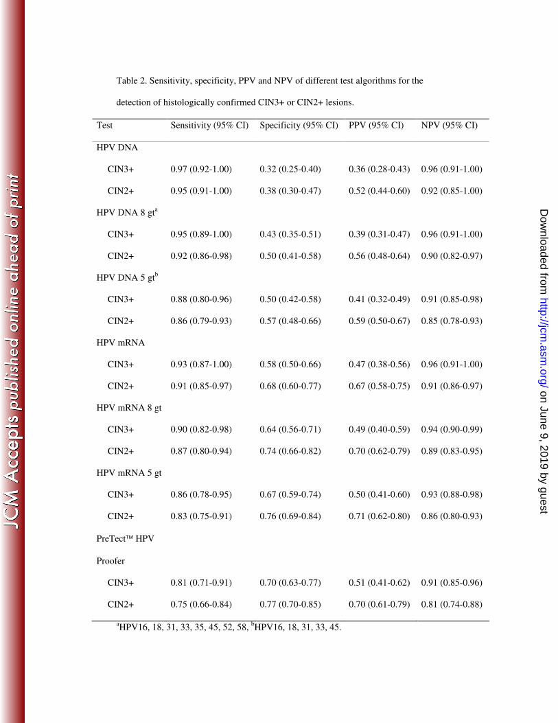
Table 2. Sensitivity, specificity, PPV and NPV of different test algorithms for the
detection of histologically confirmed CIN3+ or CIN2+ lesions.
Test Sensitivity (95% CI) Specificity (95% CI) PPV (95% CI) NPV (95% CI)
HPV DNA
CIN3+ 0.97 (0.92-1.00) 0.32 (0.25-0.40) 0.36 (0.28-0.43) 0.96 (0.91-1.00)
CIN2+ 0.95 (0.91-1.00) 0.38 (0.30-0.47) 0.52 (0.44-0.60) 0.92 (0.85-1.00)
HPV DNA 8 gta
CIN3+ 0.95 (0.89-1.00) 0.43 (0.35-0.51) 0.39 (0.31-0.47) 0.96 (0.91-1.00)
CIN2+ 0.92 (0.86-0.98) 0.50 (0.41-0.58) 0.56 (0.48-0.64) 0.90 (0.82-0.97)
HPV DNA 5 gtb
CIN3+ 0.88 (0.80-0.96) 0.50 (0.42-0.58) 0.41 (0.32-0.49) 0.91 (0.85-0.98)
CIN2+ 0.86 (0.79-0.93) 0.57 (0.48-0.66) 0.59 (0.50-0.67) 0.85 (0.78-0.93)
HPV mRNA
CIN3+ 0.93 (0.87-1.00) 0.58 (0.50-0.66) 0.47 (0.38-0.56) 0.96 (0.91-1.00)
CIN2+ 0.91 (0.85-0.97) 0.68 (0.60-0.77) 0.67 (0.58-0.75) 0.91 (0.86-0.97)
HPV mRNA 8 gt
CIN3+ 0.90 (0.82-0.98) 0.64 (0.56-0.71) 0.49 (0.40-0.59) 0.94 (0.90-0.99)
CIN2+ 0.87 (0.80-0.94) 0.74 (0.66-0.82) 0.70 (0.62-0.79) 0.89 (0.83-0.95)
HPV mRNA 5 gt
CIN3+ 0.86 (0.78-0.95) 0.67 (0.59-0.74) 0.50 (0.41-0.60) 0.93 (0.88-0.98)
CIN2+ 0.83 (0.75-0.91) 0.76 (0.69-0.84) 0.71 (0.62-0.80) 0.86 (0.80-0.93)
PreTect HPV
Proofer
CIN3+ 0.81 (0.71-0.91) 0.70 (0.63-0.77) 0.51 (0.41-0.62) 0.91 (0.85-0.96)
CIN2+ 0.75 (0.66-0.84) 0.77 (0.70-0.85) 0.70 (0.61-0.79) 0.81 (0.74-0.88)
aHPV16, 18, 31, 33, 35, 45, 52, 58,
bHPV16, 18, 31, 33, 45.
on June 9, 2019 by guesthttp://jcm
.asm.org/
Dow
nloaded from