u-plex biomarker group 1 human multiplex product insert/media/files/product...for research use only....
TRANSCRIPT

FOR RESEARCH USE ONLY. NOT FOR USE IN DIAGNOSTIC OR THERAPEUTIC PROCEDURES.
17379-v1-2007Mar
U-PLEX® Biomarker Group 1 (Human)
Multiplex Assays

18158-v5-2018May| 2
MSD U-PLEX Platform
U-PLEX Biomarker Group 1 (Human)Multiplex Assays
For use with serum, EDTA plasma, and cell culture supernatants.
For use with U-PLEX Biomarker Group 1 Human 71-Plex (catalog numbers K15081K-1, K15081K-2, K15081K-4), U-PLEX Group 1 Human Combos, and U-PLEX Biomarker Group 1 Human assays (K15067L-1, K15067L-2, K15067L-4). Catalog numbers for U-PLEX Biomarker Group 1 (hu) multiplex Combos are provided in Table 11 on page 18.
FOR RESEARCH USE ONLY.
NOT FOR USE IN DIAGNOSTIC PROCEDURES.
MESO SCALE DISCOVERYP
®
A division of Meso Scale Diagnostics, LLC. 1601 Research Blvd. Rockville, MD 20850 USA www.mesoscale.com
MESO SCALE DISCOVERY, MESO SCALE DIAGNOSTICS, MSD, MSD GOLD, DISCOVERY WORKBENCH, MULTI-ARRAY, MULTI-SPOT, QUICKPLEX, SECTOR, SECTOR PR, SECTOR HTS, SULFO-TAG, R-PLEX, S-PLEX, T-PLEX, U-PLEX, V-PLEX, STREPTAVIDIN GOLD, MESO, www.mesoscale.com, SMALL SPOT (design), 96 WELL 1, 4, 7, 9, & 10-SPOT (designs), 384 WELL 1 & 4-SPOT (designs), MSD (design), R-PLEX (design), S-PLEX (design), T-PLEX (design), U-PLEX (design), V-PLEX (design), It’s All About U, and SPOT THE DIFFERENCE are trademarks and/or service marks of Meso ScaleDiagnostics, LLC. ©2015-2018 Meso Scale Diagnostics, LLC. All rights reserved.

18158-v5-2018May| 3
Table of Contents Introduction .................................................................................................................................................................. 4 Principle of the Assay ................................................................................................................................................... 5 Components ................................................................................................................................................................. 6 Additional Materials and Equipment ............................................................................................................................. 10 Safety ......................................................................................................................................................................... 10 Best Practices ............................................................................................................................................................. 11 Reagent Preparation .................................................................................................................................................... 12 Assay Protocol ............................................................................................................................................................ 16 Assay Performance ...................................................................................................................................................... 17 Specificity .................................................................................................................................................................. 17 Appendix .................................................................................................................................................................... 18 Summary Protocol ...................................................................................................................................................... 22 Plate Diagram ............................................................................................................................................................. 23
Contact Information MSD Customer Service Phone: 1-240-314-2795 Fax: 1-301-990-2776 Email: [email protected]
MSD Scientific Support Phone: 1-240-314-2798 Fax: 1-240-632-2219 Attn: Scientific Support Email: [email protected]

18158-v5-2018May| 4
Introduction U-PLEX technology allows for the creation of custom multiplex assays for any combination of analytes by using two simple tools: a 10-spot U-PLEX plate and unique Linkers (Figure 1). The U-PLEX platform combines high sensitivity, up to 5 logs of linear dynamic range, a read time of less than 2 minutes, and the flexibility to create your own personalized multiplex assays.
The U-PLEX assay menu is organized into groups, which include a broad menu of analytes assembled by species and analytical compatibility. As many as 10 U-PLEX assays from an assay group may be multiplexed on each plate for simultaneous quantification. For ultimate flexibility, custom combinations can be created from a selection of MSD U-PLEX assays, R-PLEX™ Antibody Sets, your own antibodies, or a combination.
The U-PLEX Biomarker Group 1 (hu) contains 71 analytes (Table 1) that are important in many biological processes. These assays can detect secreted biomarkers in a variety of body fluids and cell culture supernatants where over- or under-expression may indicate a shift in the biological equilibrium.
A representative data set for each of the assays in U-PLEX Biomarker Group 1 (hu) is presented in the product-specific datasheets available at www.mesoscale.com/U-PLEX-documents.
CTACK (CCL27) IL-2 IL-17C M-CSF
ENA-78 (CXCL5) IL-2Rα IL-17D MDC (CCL22)
Eotaxin (CCL11) IL-3 IL-17E/IL-25 MIF
Eotaxin-2 (CCL24) IL-4 IL-17F MIP-1α (CCL3)
Eotaxin-3 (CCL26) IL-5 IL-18 MIP-1β (CCL4)
EPO IL-6 IL-21 MIP-3α (CCL20)
FLT3L IL-7 IL-22 MIP-3β (CCL19)
Fractalkine (CX3CL1) IL-8 (CXCL8) IL-23 MIP-5 (CCL15)
G-CSF IL-9 IL-27 SDF-1α (CXCL12)
GM-CSF IL-10 IL-29/IFN-λ1 TARC (CCL17)
GRO-α (CXCL1) IL-12/IL-23p40 IL-31 TNF-α
I-309 (CCL1) IL-12p70 IL-33 TNF-β
IFN-α2a IL-13 IP-10 (CXCL10) TPO
IFN-β IL-15 I-TAC (CXCL11) TRAIL
IFN-γ IL-16 MCP-1 (CCL2) TSLP
IL-1α IL-17A MCP-2 (CCL8) VEGF-A
IL-1β IL-17A/F MCP-3 (CCL7) YKL-40
IL-1RA IL-17B MCP-4 (CCL13)
Notes:
1. The IL-12/IL-23p40, IL-12p70, and IL-23 analytes contain the p40 subunit. Due to cross-reactivity at the IL-12/IL-23p40 spot, we do not recommend multiplexing the assays for IL-12p70 or IL-23 on the same plate as IL-12/IL-23p40.
2. IL-17A and IL-17F analytes cross-react with the IL-17A/F heterodimer. We do not recommend multiplexing IL-17A with IL-17A/F on the same plate. Do not multiplex IL-17A with IL-17F if IL-17A/F may be present in the samples.
3. Eotaxin (Calibrator 2) nonspecifically binds MCP-2 (2.8%) and MCP-3 (3.7%) detection antibodies. We do not recommend multiplexing MCP-2 or MCP-3 with Eotaxin on the same plate.
Table 1. Assays included in U-PLEX Biomarker Group 1 (hu)

18158-v5-2018May| 5
Principle of the Assay Biotinylated capture antibodies are coupled to U-PLEX Linkers, which self-assemble onto unique spots on the U-PLEX plate. Analytes in the sample bind to the capture reagents; detection antibodies conjugated with electrochemiluminescent labels (MSD GOLD™ SULFO-TAG) bind to the analytes to complete the sandwich immunoassay (Figure 1). Once the sandwich immunoassay is complete, the U-PLEX plate is loaded into an MSD instrument where a voltage applied to the plate electrodes causes the captured labels to emit light. The instrument measures the intensity of emitted light (which is proportional to the amount of analyte present in the sample) and provides a quantitative measure of each analyte in the sample.
Figure 1. U-PLEX Immunoassay on a U-PLEX 10-Assay Plate

18158-v5-2018May| 6
Components The following tables list the components provided with U-PLEX Biomarker Group 1 (hu) assays. You will only receive components relevant to the assays that you order.
Reagents Supplied With All U-PLEX Multiplex Assays
Reagent Storage Catalog # Size Quantity Supplied
1 Plate 5 Plates 25 Plates Description
Diluent 43 ≤-10°C R50AG-1 10 mL 1 bottle
Diluent for samples and Calibrators; contains serum, blockers, and preservatives. R50AG-2 50 mL
1 bottle 5 bottles
Diluent 3 ≤-10°C R50AP-1 8 mL 1 bottle
Diluent for detection antibody; contains protein, blockers, and preservatives. R50AP-2 40 mL 1 bottle 5 bottles
Stop Solution 2–8°C R50AO-1 40 mL 1 bottle 1 bottle 5 bottles Biotin-containing buffer to stop Linker-antibody coupling reaction.
Read Buffer T (4X) RT R92TC-3 50 mL 1 bottle 1 bottle 5 bottles
Buffer to catalyze the electro-chemiluminescence reaction. Dilute to 2X and use at room temperature.
Note: If combining U-PLEX with R-PLEX analytes, guidance on diluents is provided in the U-PLEX Development Pack product insert available at www.mesoscale.com/U-PLEX-documents.
Assay-Specific Reagents
10-spot 96-well U-PLEX Plates
U-PLEX assays use MSD 96-Well 10-Spot plates, provided in a sealed foil pouch with desiccant. The spots correspond to 10 unique U-PLEX Linkers. The number and layout of the active spots on the plate depends on the number of assays to be multiplexed (Figure 2). For example, if 5 assays are being multiplexed, the U-PLEX 5-Assay Plate will be provided.
Table 2. Reagents that are supplied with all U-PLEX Biomarker Group 1 (hu) assays
Figure 2. Spot Map of the different U-PLEX multiplex plates showing the placement of Linkers within a well. The colored spots represent the active U-PLEX binding spots. The numbering convention for the different spots is maintained in the software visualization tools, on the plate packaging, and in the data files.
Note: In cases where there is a remainder of one assay, you will receive a U-PLEX 2-Assay plate. This will hold true for orders of 11, 21, 31 assays, and so on.

18158-v5-2018May| 7
Table 3. Part numbers and pack sizes for U-PLEX multiplex plates
SECTOR® Plates Part # Storage Number of Plates Supplied 1 Plate 5 Plates 25 Plates
U-PLEX 2-Assay N05227A-1/-2/-4 2–8°C 1 5 25
U-PLEX 3-Assay N05228A-1/-2/-4 2–8°C 1 5 25
U-PLEX 4-Assay N05229A-1/-2/-4 2–8°C 1 5 25
U-PLEX 5-Assay N05230A-1/-2/-4 2–8°C 1 5 25
U-PLEX 6-Assay N05231A-1/-2/-4 2–8°C 1 5 25
U-PLEX 7-Assay N05232A-1/-2/-4 2–8°C 1 5 25
U-PLEX 8-Assay N05233A-1/-2/-4 2–8°C 1 5 25
U-PLEX 9-Assay N05234A-1/-2/-4 2–8°C 1 5 25
U-PLEX 10-Assay N05235A-1/-2/-4 2–8°C 1 5 25
Linkers
Based upon the number of assays you select for multiplexing, you will receive the corresponding number of unique Linkers (Table 5). Each Linker has a biotin-binding domain that couples to the biotinylated capture antibody, as well as a domain that binds to its matching spot on the U-PLEX plate. The Linkers are color coded and numbered with the spot to which they attach on the plate. 1-Plate packs include 300 µL of each Linker; 5-Plate packs include 1.8 mL of each Linker. 25-Plate packs include 5 vials of 1.8 mL of each Linker.
Figure 3. Unique color coded Linkers (10), Antibody Sets (10), and Calibrator vials (5) as shipped in a U-PLEX box

18158-v5-2018May| 8
Table 4. U-PLEX Linker color-coding, storage conditions, part numbers, and size
Name Color-coding Storage Part # Size Quantity Supplied 1 Plate 5 Plates 25 Plates
U-PLEX Linker 1
2–8°C E2226-2 0.3 mL 1 vial
E2226-3 1.8 mL 1 vial 5 vials
U-PLEX Linker 2
2–8°C E2227-2 0.3 mL 1 vial E2227-3 1.8 mL 1 vial 5 vials
U-PLEX Linker 3
2–8°C E2228-2 0.3 mL 1 vial E2228-3 1.8 mL
1 vial 5 vials
U-PLEX Linker 4
2–8°C E2229-2 0.3 mL 1 vial
E2229-3 1.8 mL 1 vial 5 vials
U-PLEX Linker 5
2–8°C E2230-2 0.3 mL 1 vial E2230-3 1.8 mL 1 vial 5 vials
U-PLEX Linker 6
2–8°C E2231-2 0.3 mL 1 vial E2231-3 1.8 mL
1 vial 5 vials
U-PLEX Linker 7
2–8°C E2232-2 0.3 mL 1 vial
E2232-3 1.8 mL 1 vial 5 vials
U-PLEX Linker 8
2–8°C E2233-2 0.3 mL 1 vial E2233-3 1.8 mL 1 vial 5 vials
U-PLEX Linker 9
2–8°C E2234-2 0.3 mL 1 vial E2234-3 1.8 mL
1 vial 5 vials
U-PLEX Linker 10
2–8°C E2235-2 0.3 mL 1 vial
E2235-3 1.8 mL 1 vial 5 vials
Table 5. U-PLEX Linkers supplied with each pack
Pack Name Linker 1 Linker 2 Linker 3 Linker 4 Linker 5 Linker 6 Linker 7 Linker 8 Linker 9 Linker 10
U-PLEX 1-Assay U-PLEX 2-Assay
U-PLEX 3-Assay
U-PLEX 4-Assay
U-PLEX 5-Assay
U-PLEX 6-Assay
U-PLEX 7-Assay
U-PLEX 8-Assay
U-PLEX 9-Assay
U-PLEX 10-Assay
We recommend recording which antibody is coupled to each Linker when performing the coupling step (as described in the Reagent Preparation section).

18158-v5-2018May| 9
U-PLEX Antibody Sets
Based upon the analytes selected, you will receive U-PLEX Antibody Sets containing the biotinylated capture antibody and the SULFO-TAG™ conjugated detection antibody. The biotinylated capture antibody is provided at a ready-to-use concentration, and the SULFO-TAG conjugated detection antibody is provided at a 100X concentration. A complete list of all Antibody Sets available for U-PLEX Biomarker Group 1 (hu) and their respective catalog numbers is provided in the Appendix (Table 12).
Name Storage Size Quantity Supplied 1 Plate 5 Plates 25 Plates Description
U-PLEX Human Analyte-Specific Antibody Set
2–8°C 1 Plate 1
Set containing biotinylated capture antibody and SULFO-TAG conjugated detection antibody 5 Plate 1 5
Calibrators
Calibrators are multi-analyte blends, each containing multiple recombinant human proteins lyophilized in a buffered diluent. Individual analyte concentrations are provided in the lot-specific certificates of analysis (COA). Depending on the specific assays requested, one or more of the following multi-analyte Calibrators will be provided.
Name Storage Catalog # Size Quantity Supplied 1 Plate 5 Plates 25 Plates Analytes
Calibrator 1 2–8°C C0060-2 1 vial 1 vial 5 vials 25 vials GM-CSF, IFN-γ, IL-1β, IL-2, IL-4, IL-5, IL-6, IL-8, IL-10, IL-12p70, IL-13, IL-17A, TNF-α, VEGF-A
Calibrator 2 2–8°C C0061-2 1 vial 1 vial 5 vials 25 vials Eotaxin, Eotaxin-3, IP-10, MCP-1, MCP-4, MDC, MIP-1α, MIP-1β, TARC
Calibrator 3 2–8°C C0062-2 1 vial 1 vial 5 vials 25 vials G-CSF, IFN-α2a, IL-1α, IL-7, IL-12/IL-23p40, IL-15, IL-16, IL-18, TNF-β, TPO
Calibrator 4 2–8°C C0063-2 1 vial 1 vial 5 vials 25 vials CTACK, ENA-78, Fractalkine, I-TAC, MIP-3α, MIP-3β, SDF-1α
Calibrator 6 2–8°C C0072-2 1 vial 1 vial 5 vials 25 vials
IL-17A/F, IL-17E/IL-25, IL-17F, IL-21, IL-22, IL-23, IL-27, IL-29/IFN-λ1, IL-31, IL-33, TSLP
Calibrator 9 2–8°C C0090-2 1 vial 1 vial 5 vials 25 vials EPO, FLT3L, IFN-β, IL-1RA, IL-2Rα, IL-3, IL-9, IL-17B, IL-17C, IL-17D
Calibrator 10 2–8°C C0091-2 1 vial 1 vial 5 vials 25 vials Eotaxin-2, GRO-α, I-309, MCP-2, MCP-3, M-CSF, MIF, MIP-5, TRAIL, YKL-40
Important Notes: 1. Do not combine Calibrators 1 or 6 with Calibrator 3 if measuring IL-12/IL-23p40.
2. Do not combine Calibrator 6 with Calibrator 1 if measuring IL-17A or IL-17A/F.
3. Do not combine Calibrator 2 with Calibrator 10 if measuring MCP-2 or MCP-3.
Table 6. Contents of U-PLEX Antibody Set
Table 7. Analytes included in the Calibrator blends available for U-PLEX Biomarker Group 1 (hu)

18158-v5-2018May| 10
Additional Materials and Equipment Appropriately sized tubes for reagent preparation
Polypropylene microcentrifuge tubes for preparing dilutions
Liquid handling equipment suitable for dispensing 10 to 150 µL/well into a 96-well microtiter plate
Plate washing equipment: automated plate washer or multichannel pipette
Microtiter plate shaker (rotary) capable of shaking at 500–1,000 rpm
MSD Wash Buffer (20X, 100 mL, catalog # R61AA-1) or Phosphate-buffered saline (PBS) plus 0.05% Tween-20 (PBS-T) for plate washing
• This size of MSD Wash Buffer is sufficient for washing 4 plates manually or for washing 2 plates with an automated plate washer
• Prepare a 1X working solution. For one plate, combine 15 mL of MSD Wash Buffer (20X) with 285 mL of deionized water. 1X MSD Wash Buffer can be stored at room temperature for up to two weeks.
Adhesive plate seals
Deionized water
Vortex mixer
Safety Use safe laboratory practices: wear gloves, safety glasses, and lab coats when handling assay components. Handle and dispose of all hazardous samples properly in accordance with local, state, and federal guidelines.
Additional product-specific safety information is available in the safety data sheet (SDS), which can be obtained from MSD Customer Service or at www.mesoscale.com.

18158-v5-2018May| 11
Best Practices • Bring frozen diluent to room temperature in a 22-25°C water bath.
• Avoid cross-contamination between Linkers by following the techniques below:
o Pulse centrifuge the vials to get all of the contents to the bottom of the vial. o Open one U-PLEX Linker vial at a time. Close the cap after use. o Each Linker vial is color coded; ensure that each cap and tube have matching colors when opening and closing
vials. o Use filtered pipette tips. o Use a fresh pipette tip after each reagent addition.
• For long-term studies using multiple plates of the same assay, it is recommended that the same Linker be coupled with the same antibody for the duration of the study.
• While most lyophilized material is located at the bottom of the vial, some may be on the sides or cap. To ensure that all lyophilized powder is reconstituted, it is recommended to vortex the vial in 3 short pulses (upright, inverted, upright) after the solution sits at room temperature for 15-30 minutes.
• Prepare Calibrator Standards and samples in polypropylene microcentrifuge tubes. Use a fresh pipette tip for each dilution and mix by vortexing after each dilution.
• Use reverse pipetting when necessary to avoid the introduction of bubbles. For empty wells, pipette gently to the bottom corner.
• Avoid prolonged exposure of the detection antibody (stock or diluted) to light. During the antibody incubation step, plates do not need to be shielded from light (except for direct sunlight).
• Avoid bubbles in wells during all pipetting steps as they may lead to variable results. Bubbles introduced when adding Read Buffer T may interfere with signal detection.
• Plate shaking should be vigorous, with a rotary motion between 500 and 1,000 rpm. Binding reactions may reach equilibrium sooner if you use shaking at the middle of this range (~700 rpm) or above.
• Reversing plate orientation between wash cycles may improve assay precision.
• Gently tap the plate on a paper towel to remove residual fluid after washing.
• If you plan to coat U-PLEX plates for later use, keep each plate pouch and the desiccant that came with the plate. After the plates are incubated with the coating solution, wash them with MSD Wash Buffer or PBS-T, then return each plate to its original packaging with the desiccant, and seal.
• If an incubation step needs to be extended, leave the sample or detection antibody solution in the plate to keep the plate from drying out.
• Remove the plate seal prior to reading the plate.
• Make sure that the Read Buffer T is at room temperature when added to a plate.
• Do not shake the plate after adding Read Buffer T.
• To improve inter-plate precision, keep time intervals consistent between adding Read Buffer T and reading the plate. Unless otherwise directed, read the plate as soon as possible after adding Read Buffer T.
• If the sample results are above the top of the calibration curve, dilute the samples and repeat the assay.

18158-v5-2018May| 12
• When running a partial plate, seal the unused sectors to avoid contaminating unused wells. Remove all seals before reading. The uncoated wells of a partially used plate may be sealed and stored up to 30 days at 2–8°C in the original foil pouch with desiccant. You may adjust volumes proportionally when preparing reagents.
• The components for assays that are larger than 10-plex or are otherwise provided in more than one box are specifically assembled so that each box contains fully compatible assay components. Each box includes one 10-spot U-PLEX plate (with the appropriate number of activated spots), Linkers, antibody pairs, and Calibrators that run optimally together. Components should not be mixed between boxes with the exception of Stop Solution, Diluents, and Read Buffer. For an example of a multi-plate assay, see the U-PLEX Biomarker Group 1 (hu) 71-Plex Assay layout on page 21.
Reagent Preparation Bring all reagents to room temperature and refer to the Best Practices section (page 11) before beginning the protocol.
Important: Upon first thaw, aliquot Diluent 3 and Diluent 43 into suitable volumes before refreezing.
To prepare MSD Wash Buffer and other supplemental reagents, please refer to the Additional Materials and Equipment section (page 10).
Prepare U-PLEX Plate
The preparation of a U-PLEX plate involves coating the provided plate with Linker-coupled capture antibodies. A U-PLEX 4-Assay plate is shown below as an example. This kit includes a plate with four activated spots at locations 1, 3, 8, and 10. Assign each antibody to a unique Linker and record the antibody identity next to the assigned Linker, as shown in the example below.
Figure 4. A U-PLEX 4-Assay Plate, with recorded antibodies and assigned Linkers
The protocol in this section describes the preparation of a multiplex coating solution for one 96-well plate. The volumes can be adjusted depending on the number of plates or wells, but the ratios of the reagents should remain the same (Table 8).
STEP 1: Create Individual U-PLEX Linker-Coupled Antibody Solutions
A different Linker must be used for each unique biotinylated antibody. Below are the steps to complete the coupling reactions for the above example of a 1-plate 4-plex assay.
Couple each biotinylated capture antibody to a unique Linker and record the antibody identity next to the Linker number on the Spot Map (blank Spot Map is provided on page 22).
18158-v5-2018May| 13
Add 200 µL of each biotinylated antibody to 300 µL of the assigned Linker in the Linker vial. Mix by vortexing. Incubate at room temperature for 30 minutes. Do not shake. If using a 5-plate Linker vial, combine each Linker with the biotinylated antibody in a new clean tube.
Notes: • Each Linker vial has a matching colored cap and label. • To remove liquid from the cap, briefly centrifuge the Linker vial and open the cap gently. • Open one Linker at a time and close its cap as soon as you are done using it. Take precautions to avoid reagent
contamination. • For studies using multiple plates of the same assay, it is recommended that the same Linker be coupled with the
same antibody for the duration of the study.
Add 200 µL of Stop Solution. Mix by vortexing. Incubate at room temperature for 30 minutes.
Note: At the end of Step 1, each individual U-PLEX Linker-coupled antibody solution is at 10X the coating concentration and can be stored at 2-8°C. Do not store for more than 7 days. Adjust the volumes for multiple plates (see Table 8). The volumetric ratio of Linker: antibody: Stop Solution is 3:2:2.
STEP 2: Prepare the Multiplex Coating Solution
Combine 600 µL of each U-PLEX Linker-coupled antibody solution (10X) into a single tube and vortex. Up to 10 U-PLEX Linker-coupled antibodies can be pooled. Do not combine U-PLEX Linker-coupled antibody solutions that share the same Linker.
When combining fewer than 10 antibodies, bring the solution up to 6 mL with Stop Solution. This will result in a final 1X concentration. Mix by vortexing. For example, for a 4-assay coating solution, add 3.6 mL of Stop Solution to the 2.4 mL of combined antibodies.
Note: At the end of Step 2, the U-PLEX multiplex coating solution is at 1X and can be stored at 2-8°C. Do not store for more than 7 days.
STEP 3: Coat the U-PLEX Plate
Add 50 µL of multiplex coating solution to each well. Seal the plate with an adhesive plate seal and incubate with shaking at room temperature for 1 hour. The plate may also be shaken overnight, while incubating at 2-8°C.
Wash the plate 3 times with at least 150 µL/well of 1X MSD Wash Buffer or PBS-T (PBS plus 0.05% Tween-20).
The plate is now coated and ready for use. Coated plates may be stored in the original pouch with desiccant and sealed, for up to 7 days at 2-8°C.
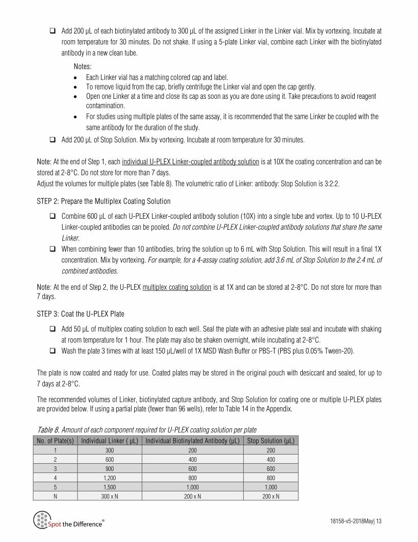
The recommended volumes of Linker, biotinylated capture antibody, and Stop Solution for coating one or multiple U-PLEX plates are provided below. If using a partial plate (fewer than 96 wells), refer to Table 14 in the Appendix.
No. of Plate(s) Individual Linker ( µL) Individual Biotinylated Antibody (µL) Stop Solution (µL) 1 300 200 200 2 600 400 400 3 900 600 600 4 1,200 800 800 5 1,500 1,000 1,000 N 300 x N 200 x N 200 x N
Table 8. Amount of each component required for U-PLEX coating solution per plate

18158-v5-2018May| 14
Prepare Calibrator Standards Depending on the assays ordered, you may receive one or more multi-analyte Calibrator vials with your order. The following instructions will enable you to prepare eight Calibrator Standards (7 calibrator solutions plus a zero calibrator) for up to six replicates.
• Bring the Calibrator vial(s) to room temperature. Reconstitute each vial of Calibrator by adding 250 µL of Diluent 43 to the glass vial. This will result in a 5X concentrated stock of each Calibrator, which will need to be diluted 5-fold (per the instructions given below) to generate the highest point in the standard curve (i.e., Calibrator Standard 1).
• Invert the reconstituted Calibrator at least 3 times. Do not vortex. Let the solution equilibrate at room temperature for 15-30 minutes and then vortex briefly. This will result in a 5X concentrated stock of the Calibrator.
• Dilute the 5X Calibrator stock solution.
o For single Calibrator standard curves, add 50 µL of the 5X stock to 200 µL of Diluent 43 to generate the highest point in the standard curve (i.e., Calibrator Standard 1). The Calibrator is now ready for use.
o To blend multiple Calibrators, follow Table 9, and mix by vortexing briefly. Keep dilutions at room temperature.
Note: We recommend that reconstituted Calibrators are used immediately. If storage is necessary, divide into 60 µL aliquots and store immediately at ≤-70°C.
# of Calibrator Blends Provided Volume of Reconstituted Calibrator (µL) Diluent 43 (µL) Total volume (µL)
1 50 200 250
2 50 each 150 250
3 50 each 100 250
4 50 each 50 250
5 50 each 0 250
Prepare the subsequent 6 dilutions for the curve (4-fold serial dilutions) in Diluent 43 (see Table 10 below for an example). Use Diluent 43 for the Calibrator Standard 8 (zero Calibrator/blank). Mix by vortexing the tubes between each serial dilution.
Calibrator Standard #
Tube # Source of Calibrator Volume of Reconstituted Calibrator (µL)
Assay Diluent (µL)
Total volume (µL)
1 1 Calibrator Standard 1
(top of curve) See Table 9
2 2 From tube 1 75 225 300
3 3 From tube 2 75 225 300
4 4 From tube 3 75 225 300
5 5 From tube 4 75 225 300
6 6 From tube 5 75 225 300
7 7 From tube 6 75 225 300
8 (zero Calibrator) 8 - 0 300 300
Note: In certain situations 5-fold serial dilutions may be desired and can be made by transferring 60 µL of Reconstituted Calibrator into 240 µL of Assay Diluent instead of the volumes noted in Table 10.
Table 9. Combining Calibrators to generate the Calibrator Standard 1 (top of the curve) level
Table 10. Serial dilution to generate the standard curve

18158-v5-2018May| 15
Figure 5. Dilution schema for preparation of Calibrator Standards for U-PLEX Biomarker Group 1 (hu) assays Alternate Calibrator handling procedures If an assay needs more than 5 Calibrators blended together, reconstitute each Calibrator with 125 µL of Diluent 43. This will result in a 10X concentrated stock of the Calibrator. Take extra care that all of the lyophilized material is reconstituted. Follow the instructions in Table 9 (page 14), but blend 25 µL of each Calibrator (rather than 50 µL) and add enough Diluent 43 to get a final volume of 250 µL. Dilute Samples Depending on the sample set under investigation, a dilution may be necessary. Diluent 43 may be used for sample dilution. The dilution factor for the given sample type needs to be optimized.
Note: For MIF, MIP-5, and YKL-40, in-house data indicate that a large sample dilution is required to generate optimal results. Refer to the product-specific datasheets for additional information.
Prepare Detection Antibody Solution
The detection antibody is provided as a 100X stock solution. The working solution is 1X. Prepare the detection antibody solution immediately prior to use. Adjust the volumes proportionally for partial plates.
For one plate, combine:
60 µL of each 100X detection antibody
Diluent 3 to bring the final volume to 6 mL

18158-v5-2018May| 16
Prepare Read Buffer T
MSD provides Read Buffer T as a 4X stock solution. The working solution is 2X.
For one plate, combine:
10 mL of Read Buffer T (4X) 10 mL of deionized water
You may keep excess diluted Read Buffer T in a tightly sealed container at room temperature for up to one month.
Assay Protocol Note: Follow Reagent Preparation before beginning this assay protocol.
STEP 1: Add Samples and Calibrators
Add 25 µL of Diluent 43 to each well. Tap the plate gently on all sides. Add 25 µL of the prepared Calibrator Standard or sample to each well. Seal the plate with an adhesive plate seal. Incubate
at room temperature with shaking for 1 hour.
STEP 2: Wash and Add Detection Antibody Solution
Wash plate 3 times with at least 150 µL/well of 1X MSD Wash Buffer or PBS-T. Add 50 µL of detection antibody solution to each well. Seal the plate with an adhesive plate seal. Incubate at room
temperature with shaking for 1 hour.
STEP 3: Wash and Read
Wash the plate 3 times with at least 150 µL/well of 1X MSD Wash Buffer or PBS-T. Add 150 µL of 2X Read Buffer T to each well. Analyze the plate on an MSD instrument. Incubation in Read Buffer T is not
required before reading the plate.
Alternate Protocols
The suggestions below may be useful for simplifying the protocol.
Alternate Protocol 1, Extended Incubation: Incubating samples overnight at 2–8°C with shaking may improve sensitivity for some assays.
Alternate Protocol 2, Reduced Wash: For cell culture supernatants, you may simplify the protocol by eliminating one of the wash steps. After incubating the Calibrator Standard or sample, add detection antibody solution to the plate without decanting or washing the plate.

18158-v5-2018May| 17
Assay Performance A representative data set for each assay is presented in the product-specific datasheets available at www.mesoscale.com/U-PLEX-documents. The data represent performance of the assay tested in multiplex format on U-PLEX plates. The data were generated during the development of the assay and do not represent the product specifications. Under your experimental conditions and with your specific multiplex, the assay may perform differently than the representative data shown.
Specificity To assess specificity, the Antibody Set for each assay in the group was tested individually against a larger panel of antibodies and recombinant human analytes for nonspecific binding (CTACK, ENA-78, Eotaxin, Eotaxin-2, Eotaxin-3, EPO, FLT3L, Fractalkine, G-CSF, GM-CSF, GRO-α, I-309, IFN-α2a, IFN-β, IFN-γ, IL-1α, IL-1β, IL-1RA, IL-2, IL-2Rα, IL-3, IL-4, IL-5, IL-6, IL-7, IL-8, IL-9, IL-10, IL-12/IL-23p40, IL-12p70, IL-13, IL-15, IL-16, IL-17A, IL-17A/F, IL-17B, IL-17C, IL-17D, IL-17E/IL-25, IL-17F, IL-18, IL-21, IL-22, IL-23, IL-27, IL-29/IFN-λ1, IL-31, IL-33, IP-10, I-TAC, MCP-1, MCP-2, MCP-3, MCP-4, M-CSF, MDC, MIF, MIP-1α, MIP-1β, MIP-3α, MIP-3β, MIP-5, SDF-1α, TARC, TNF-α, TNF-β, TPO, TRAIL, TSLP, VEGF-A, and YKL-40).
Nonspecific binding was less than 0.5% for all assays included in the U-PLEX Biomarker Group 1 (hu) using the following calculation.
% 𝑁𝑁𝑁𝑁𝑁𝑁𝑁𝑁𝑁𝑁𝑁𝑁𝑁𝑁𝑁𝑁𝑁𝑁𝑁𝑁𝑁𝑁𝑁𝑁𝑁𝑁𝑁𝑁 =𝑁𝑁𝑁𝑁𝑁𝑁𝑁𝑁𝑁𝑁𝑁𝑁𝑁𝑁𝑁𝑁𝑁𝑁𝑁𝑁𝑁𝑁 𝑁𝑁𝑁𝑁𝑠𝑠𝑁𝑁𝑠𝑠𝑠𝑠𝑁𝑁𝑁𝑁𝑁𝑁𝑁𝑁𝑁𝑁𝑁𝑁𝑁𝑁𝑁𝑁 𝑁𝑁𝑁𝑁𝑠𝑠𝑁𝑁𝑠𝑠𝑠𝑠
∗ 100
Exceptions are noted below:
1. IL-12p70 and IL-23 contain the p40 subunit and will cross-react with the IL-12/IL-23p40 assay.
2. IL-17A cross-reacts with the IL-17A/F assay. IL-17A/F cross-reacts with the IL-17A assay.
3. Eotaxin (Calibrator 2) nonspecifically binds MCP-2 (2.8%) and MCP-3 (3.7%) detection antibodies.

18158-v5-2018May| 18
Appendix U-PLEX Combinations U-PLEX Combinations include U-PLEX Plates, Linkers, Antibody Sets, Calibrators, Stop Solution, diluents, and Read Buffer T.
Product Analytes Catalog Numbers
(-1/-5/-25 Plate Size)
U-PLEX Biomarker Group 1 (hu) 71-Plex, SECTOR*
CTACK, ENA-78, Eotaxin, Eotaxin-2, Eotaxin-3, EPO, FLT3L, Fractalkine, G-CSF, GM-CSF, GRO-α, I-309, IFN-α2a, IFN-β, IFN-γ, IL-1α, IL-1β, IL-1RA, IL-2, IL-2Rα, IL-3, IL-4, IL-5, IL-6, IL‑7, IL-8, IL-9, IL-10, IL-12/IL-23p40, IL-12p70, IL-13, IL-15, IL-16, IL-17A, IL-17A/F, IL-17B, IL-17C, IL-17D, IL-17E/IL-25, IL-17F, IL-18, IL-21, IL-22, IL-23, IL-27, IL-29/IFN-λ1, IL-31, IL-33, IP-10, I‑TAC, MCP-1, MCP-2, MCP-3, MCP-4, M-CSF, MDC, MIF, MIP-1α, MIP-1β, MIP‑3α, MIP-3β, MIP-5, SDF-1α, TARC, TNF-α, TNF-β, TPO, TRAIL, TSLP, VEGF-A, YKL-40
K15081K-1/-2/-4
U-PLEX TH1/TH2 Combo (hu) SECTOR IFN-γ, IL-1β, IL-2, IL-4, IL-5, IL-8, IL-10, IL-12p70, IL-13, TNF-α K15010K-1/-2/-4
U-PLEX TH17 Combo 1 (hu) SECTOR
IL-17A, IL-17E/IL-25, IL-17F, IL-21, IL-22, IL-23, IL-27, IL-31, IL-33 K15075K-1/-2/-4
U-PLEX TH17 Combo 2 (hu) SECTOR IFN-γ, IL-1β, IL-6, IL-10, IL-17A, IL-17E/IL-25, IL-17F, IL-21, IL-22, TNF-α K15076K-1/-2/-4
U-PLEX Proinflam Combo 1 (hu) SECTOR IFN-γ, IL-1β, IL-2, IL-4, IL-6, IL-8, IL-10, IL-12p70, IL-13, TNF-α K15049K-1/-2/-4
U-PLEX Proinflam Combo 2 (hu) SECTOR GM-CSF, IFN-γ, IL-1β, IL-2, IL-4, IL-6, IL-8, IL-10, IL-12p70 K15066K-1/-2/-4
U-PLEX Proinflam Combo 3 (hu) SECTOR IFN-γ, IL-1β, IL-6, TNF-α K15025K-1/-2/-4
U-PLEX Proinflam Combo 4 (hu) SECTOR IL-1β, IL-6, IL-8, TNF-α K15072K-1/-2/-4
U-PLEX Cytokine Combo 1 (hu) SECTOR
GM-CSF, IL-1α, IL-5, IL-7, IL-12/IL-23p40, IL-15, IL-16, IL-17A, TNF-β, VEGF-A
K15045K-1/-2/-4
U-PLEX Chemokine Combo 1 (hu) SECTOR
Eotaxin, Eotaxin-2, Eotaxin-3, IL-8, IP-10, MCP-1, MCP-2, MCP-3, MCP-4, MDC, MIP-1α, MIP-1β, TARC
K15047K-1/-2/-4
U-PLEX Chemokine Combo 2 (hu) SECTOR
CTACK, ENA-78, Fractalkine, GRO-α, I-309, I-TAC, MIF, MIP-3α, MIP-3β, MIP-5, SDF-1α
K15046K-1/-2/-4
U-PLEX T-Cell Profiling Combo (hu) SECTOR
GM-CSF, IFN-γ, IL-2, IL-4, IL-9, IL-10, IL-13, IL-17A, IL-17E/IL-25, IL-17F, IL-21, IL-22, MIP-3α, TNF-α
K15093K-1/-2/-4
U-PLEX Interferon Combo (hu) SECTOR IFN-α2a, IFN-β, IFN-γ, IL-29/IFN-λ1 K15094K-1/-2/-4
U-PLEX TGF-β Combo (hu) SECTOR**
TGF-β1, TGF-β2, TGF-β3 K15241K-1/-2/-4
*See additional instructions on page 21 to run this assay. **The U-PLEX TGF-β Combo (hu) assay is a Group 2 Combo, and cannot be combined with Group 1 assays.
Table 11. Catalog numbers of U-PLEX Biomarker (hu) multiplex combinations

18158-v5-2018May| 19
U-PLEX Antibody Sets Antibody Sets include a biotinylated capture antibody and SULFO-TAG conjugated detection antibody.
Product Catalog Numbers
(-1/-5 Plate Size) Product Catalog Numbers
(-1/-5 Plate Size) U-PLEX Human CTACK Antibody Set B21VD-2/-3 U-PLEX Human IL-17C Antibody Set B21WJ-2/-3
U-PLEX Human ENA-78 Antibody Set B21VE-2/-3 U-PLEX Human IL-17D Antibody Set B21XO-2/-3
U-PLEX Human Eotaxin Antibody Set B21UD-2/-3 U-PLEX Human IL-17E/IL-25 Antibody Set B21VZ-2/-3
U-PLEX Human Eotaxin-2 Antibody Set B21XQ-2/-3 U-PLEX Human IL-17F Antibody Set B21WA-2/-3
U-PLEX Human Eotaxin-3 Antibody Set B21UE-2/-3 U-PLEX Human IL-18 Antibody Set B21VJ-2/-3
U-PLEX Human EPO Antibody Set B21VX-2/-3 U-PLEX Human IL-21 Antibody Set B21WB-2/-3
U-PLEX Human FLT3L Antibody Set B21XF-2/-3 U-PLEX Human IL-22 Antibody Set B21WI-2/-3
U-PLEX Human Fractalkine Antibody Set B21VC-2/-3 U-PLEX Human IL-23 Antibody Set B21WG-2/-3
U-PLEX Human G-CSF Antibody Set B21VG-2/-3 U-PLEX Human IL-27 Antibody Set B21WC-2/-3
U-PLEX Human GM-CSF Antibody Set B21UM-2/-3 U-PLEX Human IL-29/IFN-λ1 Antibody Set B21WD-2/-3
U-PLEX Human GRO-α Antibody Set B21UX-2/-3 U-PLEX Human IL-31 Antibody Set B21WE-2/-3
U-PLEX Human I-309 Antibody Set B21UY-2/-3 U-PLEX Human IL-33 Antibody Set B21WF-2/-3
U-PLEX Human IFN-α2a Antibody Set B21VH-2/-3 U-PLEX Human IP-10 Antibody Set B21UF-2/-3
U-PLEX Human IFN-β Antibody Set B21VI-2/-3 U-PLEX Human I-TAC Antibody Set B21UW-2/-3
U-PLEX Human IFN-γ Antibody Set B21TT-2/-3 U-PLEX Human MCP-1 Antibody Set B21UG-2/-3
U-PLEX Human IL-1α Antibody Set B21UN-2/-3 U-PLEX Human MCP-2 Antibody Set B21XH-2/-3
U-PLEX Human IL-1β Antibody Set B21TU-2/-3 U-PLEX Human MCP-3 Antibody Set B21XI-2/-3
U-PLEX Human IL-1RA Antibody Set B21XP-2/-3 U-PLEX Human MCP-4 Antibody Set B21UH-2/-3
U-PLEX Human IL-2 Antibody Set B21TV-2/-3 U-PLEX Human M-CSF Antibody Set B21XR-2/-3
U-PLEX Human IL-2Rα Antibody Set B21XG-2/-3 U-PLEX Human MDC Antibody Set B21UI-2/-3
U-PLEX Human IL-3 Antibody Set B21XM-2/-3 U-PLEX Human MIF Antibody Set B21XJ-2/-3
U-PLEX Human IL-4 Antibody Set B21TW-2/-3 U-PLEX Human MIP-1α Antibody Set B21UJ-2/-3
U-PLEX Human IL-5 Antibody Set B21UO-2/-3 U-PLEX Human MIP-1β Antibody Set B21UK-2/-3
U-PLEX Human IL-6 Antibody Set B21TX-2/-3 U-PLEX Human MIP-3α Antibody Set B21UZ-2/-3
U-PLEX Human IL-7 Antibody Set B21UP-2/-3 U-PLEX Human MIP-3β Antibody Set B21VA-2/-3
U-PLEX Human IL-8 Antibody Set B21TY-2/-3 U-PLEX Human MIP-5 Antibody Set B21XS-2/-3
U-PLEX Human IL-9 Antibody Set B21XK-2/-3 U-PLEX Human SDF-1α Antibody Set B21VB-2/-3
U-PLEX Human IL-10 Antibody Set B21TZ-2/-3 U-PLEX Human TARC Antibody Set B21UL-2/-3
U-PLEX Human IL-12/IL-23p40 Antibody Set B21UQ-2/-3 U-PLEX Human TNF-α Antibody Set B21UC-2/-3
U-PLEX Human IL-12p70 Antibody Set B21UA-2/-3 U-PLEX Human TNF-β Antibody Set B21UU-2/-3
U-PLEX Human IL-13 Antibody Set B21UB-2/-3 U-PLEX Human TPO Antibody Set B21VK-2/-3
U-PLEX Human IL-15 Antibody Set B21UR-2/-3 U-PLEX Human TRAIL Antibody Set B21XT-2/-3
U-PLEX Human IL-16 Antibody Set B21US-2/-3 U-PLEX Human TSLP Antibody Set B21WH-2/-3
U-PLEX Human IL-17A Antibody Set B21UT-2/-3 U-PLEX Human VEGF-A Antibody Set B21UV-2/-3
U-PLEX Human IL-17A/F Antibody Set B21VY-2/-3 U-PLEX Human YKL-40 Antibody Set B21VL-2/-3
U-PLEX Human IL-17B Antibody Set B21XN-2/-3
Table 13. Catalog numbers of Antibody Sets available for U-PLEX Biomarker Group 2
Product Catalog Numbers (-1/-5 Plate Size)
U-PLEX TGF-β1 Antibody Set B20XW-2/-3
U-PLEX TGF-β2 Antibody Set B20XU-2/-3
U-PLEX TGF-β3 Antibody Set B20XV-2/-3
Table 12. Catalog numbers of Antibody Sets available for U-PLEX Biomarker Group 1 (hu)
18158-v5-2018May| 20
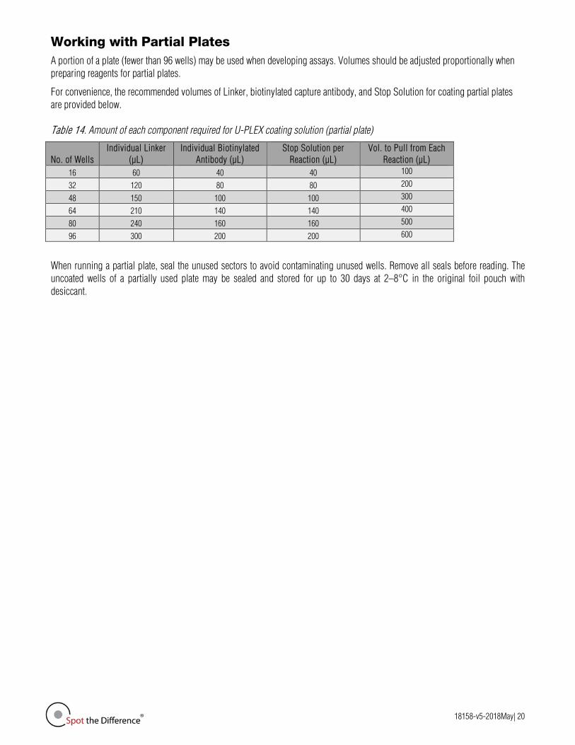
Working with Partial Plates
A portion of a plate (fewer than 96 wells) may be used when developing assays. Volumes should be adjusted proportionally when preparing reagents for partial plates.
For convenience, the recommended volumes of Linker, biotinylated capture antibody, and Stop Solution for coating partial plates are provided below. Table 14. Amount of each component required for U-PLEX coating solution (partial plate)
When running a partial plate, seal the unused sectors to avoid contaminating unused wells. Remove all seals before reading. The uncoated wells of a partially used plate may be sealed and stored for up to 30 days at 2–8°C in the original foil pouch with desiccant.
No. of Wells Individual Linker
(µL) Individual Biotinylated
Antibody (µL) Stop Solution per
Reaction (µL) Vol. to Pull from Each
Reaction (µL) 16 60 40 40 100
32 120 80 80 200
48 150 100 100 300
64 210 140 140 400
80 240 160 160 500
96 300 200 200 600

18158-v5-2018May| 21
Multi-plate Assays
Multiplex U-PLEX assays can occupy more than one plate, depending on the number and compatibility of the selected assays. Pre-configured multiplexes, including Combos and Biomarker Groups, are arranged in optimal layouts, with compatible assays and Calibrators packaged together along with adequate linkers and an activated U-PLEX plate. Components of a multi-plate assay should not be mixed between boxes, with the exception of Stop Solution, Diluents, and Read Buffer.
An example of a multi-plate U-PLEX assay is the U-PLEX Biomarker Group 1 (hu) 71-Plex (K15081K). The assay is supplied in eight separate U-PLEX boxes. Each box includes one 10-spot U-PLEX plate (with the appropriate number of activated spots), Linkers, antibody pairs, and Calibrators that run optimally together. Components should not be mixed between boxes, with the exception of Stop Solution, Diluents, and Read Buffer.
To perform the U-PLEX Biomarker Group 1 (hu) 71-Plex assay, we recommend that you position the eight boxes as shown in the table below. When multiple Calibrators are in one box, they should be blended as instructed in this product insert. Do not combine with any other Calibrators from another box. There will be a unique Calibrator curve for each box. Table 15. Human 71-Plex Layout
Box 1 10-Activated Spots Plate
Box 2 10-Activated Spots Plate
Box 3 10-Activated Spots Plate
Box 4 10-Activated Spots Plate
Box 5 10-Activated Spots Plate
Box 6 10-Activated Spots Plate
Box 7 8-Activated Spots Plate
Box 8* 3-Activated Spot Plate
IL-17A/F TSLP G-CSF EPO GM-CSF IL-17A Eotaxin-2 MIF
IL-17E Eotaxin IFN-α2a FLT3L IFN-γ CTACK GRO-α MIP-5
IL-17F Eotaxin-3 IL-1α IFN-β IL-1β ENA-78 I-309 YKL-40
IL-21 IP-10 IL-7 IL-3 IL-2 Fractalkine IL-13
IL-22 MCP-1 IL-12/IL-23p40 IL-9 IL-4 I-TAC M-CSF
IL-23 MCP-4 IL-15 IL-17B IL-5 MIP-3α MCP-2
IL-27 MDC IL-16 IL-17C IL-6 MIP-3β MCP-3
IL-29/IFN-λ1 MIP-1α IL-18 IL-17D IL-8 SDF-1α TRAIL
IL-31 MIP-1β TNF-β IL-1RA IL-10 TNF-α IL-33 TARC TPO IL-2Rα IL-12p70 VEGF-A
Calibrator 6 Calibrators 2 and 6 Calibrator 3 Calibrator 9 Calibrator 1 Calibrators 1 and 4 Calibrators 1 and 10 Calibrator 10
*These analytes are expressed at much higher levels than the other analytes in the samples. They are separated to allow samples to be diluted specifically for these analytes. Internal testing suggests that normal serum and plasma should be diluted 100-fold.
Open Spots
The U-PLEX platform allows users to add other analytes besides those that are available in the U-PLEX assay menu, such as R-PLEX antibody sets or your own antibodies, to a U-PLEX assay. This is enabled when open spots are included in a U-PLEX assay order. For more information about diluents when combining U-PLEX assays and R-PLEX Antibody Sets, refer to the U-PLEX Development Pack product insert or the R-PLEX Multiplex Assays product insert available at www.mesoscale.com.

18158-v5-2018May| 22
Summary Protocol Prepare U-PLEX Plates
STEP 1: Create Individual U-PLEX Linker-Coupled Antibody Solutions
Couple an individual biotinylated antibody to a unique Linker, and record the antibody identity next to the Linker number on the Spot Map below.
Add 200 µL of each biotinylated antibody to 300 µL of the assigned Linker. Refer to the U-PLEX plate Spot Map to determine which Linkers can be combined. A different Linker must be used for each unique biotinylated antibody. Mix by vortexing. Incubate at room temperature for 30 minutes.
Add 200 µL of Stop Solution. Mix by vortexing. Incubate at room temperature for 30 minutes.
STEP 2: Prepare the Multiplex Coating Solution
Combine 600 µL of each U-PLEX Linker-coupled antibody solution into a single tube and mix by vortexing. Up to 10 U-PLEX Linker-coupled antibodies can be pooled. Do not combine U-PLEX Linker-coupled antibody solutions that share the same Linker.
When combining fewer than 10 antibodies, bring the solution up to 6 mL by mixing with Stop Solution to result in a final 1X concentration. Mix by vortexing.
STEP 3: Coat the U-PLEX Plates
Add 50 µL of multiplex coating solution to each well. Seal the plate with an adhesive plate seal and shake for 1 hour at room temperature. Alternatively, you can shake the plate overnight while incubating at 2-8°C.
Wash the plate 3 times with at least 150 µL/well of 1X Wash Buffer or PBS-T. The plate is now coated and ready for use.
Spot Map
Figure 6. Spot map

18158-v5-2018May| 23
Assay Protocol
STEP 1: Add Sample and Calibrator Standards
Add 25 µL of Diluent 43 to each well. Tap the plate gently on all sides. Add 25 µL of the prepared Calibrator Standard or sample to each well. Seal the plate with an adhesive plate seal. Incubate
at room temperature with shaking for 1 hour.
STEP 2: Wash and Add Detection Antibody Solution
Wash plate 3 times with at least 150 µL/well of 1X MSD Wash Buffer or PBS-T. Add 50 µL of detection antibody solution to each well. Seal the plate with an adhesive plate seal. Incubate at room
temperature with shaking for 1 hour.
STEP 3: Wash and Read
Wash the plate 3 times with at least 150 µL/well of 1X MSD Wash Buffer or PBS-T. Add 150 µL of 2X Read Buffer T to each well. Analyze the plate on an MSD instrument. Incubation in Read Buffer T is not
required before reading the plate.
Plate Diagram
Figure 7. Plate diagram. A similar plate layout can be created in Excel and easily imported into DISCOVERY WORKBENCH® software.