ultrasonographic abnormalities in canine urinary and ...€¦ · this study, clinical signs were...
TRANSCRIPT

NE USAcademic Publishers
Research Journal for Veterinary Practitioners
July 2015 | Volume 3 | Issue 4 | Page 93
INTRODUCTION
In veterinary practice, ultrasound imaging possesses plentiful advantages and with minimal biological haz-
ards recorded even with repeated utilization making ultra-sound safe diagnostic tool for both patients and operating personals (Preston and Shaw, 2001).
Ultrasonographic assessment of the urinary tract has be-come a routine practice in veterinary medicine (Nyland and Matton, 2014). Urinary ultrasonography offers out-standing visualization and monitoring of organs, particu-larly when conventional radiographic assessment fails to reach final diagnosis (Larson, 2009).
Prostatic affections are more frequent in dogs compared to other domestic animals. Canine prostatic affections often give overlapping signs making final diagnosis much more difficult to achieve based only on symptoms (Smith, 2008).
Prostatic hyperplasia, inflammation, cysts and neoplasia are the most common affections recorded in dogs; benign hyperplasia is a disease of both man and dogs. Clinical findings even accompanied with digital rectal findings can provide presumptive diagnosis, however, the precise and final diagnosis could be reached only via ultrasonographic assessment (Nicola, 2006; Paclikova et al., 2006; Kiber et al., 2012; Boucif et al 2015).
Consequently, the current study was accomplished to in-vestigate selected different urinary and prostatic affections in dogs.
MATERIAL AND METHODS
Thirty-two dogs (N=32) of different ages, sexes, and breeds were involved in this study; the dogs were referred to small animal medicine-teaching hospital, faculty of veterinary medicine, Cairo University, Egypt.
Research Article
Abstract | Ultrasonography is one the most superior modalities for diagnosis of numerous urinary and prostatic affec-tions. The current study aimed to diagnose and stipulate different cases of dogs with urinary and prostatic affections on basis of clinical and ultrasonographic findings. Thirty-two dogs of different ages, sexes, and breeds were involved in this study, clinical signs were recorded at time of admission and the definitive diagnosis was upon ultrasound exami-nation. The most consistent clinical signs observed at time of admission were dysuria, hematuria, pollakuria, polydipsia and polyuria. Diagnosed urinary bladder affections (No=18) included cystitis, cystic calculi, cystic neoplasms, and bladder hemorrhage. Kidney affections (No=8) included renal cyst, hydronephrosis, nephrolithiasis, chronic interstitial nephritis and end-stage kidney failure. Prostatic affections (No=6) involved benign prostatic hyperplasia, prostatitis and prostatic cysts. Clinical examination and ultrasonography were the main diagnostic aids in all cases. Clinical examination was unsuccessful to establish a definitive diagnosis and ultrasonography was preferable to conventional tools of this specification.
Keywords | Dogs, Clinical signs, Urinary, Prostatic affections, Ultrasound
Shimaa Ghanem Yehia*, noha YouSef Salem
Ultrasonographic Abnormalities in Canine Urinary and Prostatic Affections
Editor | Muhammad Abubakar, National Veterinary Laboratories, Islamabad, Pakistan.Received | December 10, 2015; Revised | January 10, 2016; Accepted | January 13, 2016; Published | February 12, 2016 *Correspondence | Shimaa G Yehia, Cairo University, Egypt, Email: [email protected] Citation | Yehia SG, Salem NY (2015). Ultrasonographic abnormalities in canine urinary and prostatic affections. Res. J. Vet. Pract. 4(3): 93-98.DOI | http://dx.doi.org/10.14737/journal.rjvp/2015/3.4.93.98ISSN | 2308-2798
Copyright © 2015 Yehia and Slem . This is an open access article distributed under the Creative Commons Attribution License, which permits unrestricted use, distribution, and reproduction in any medium, provided the original work is properly cited.
Department of Internal Medicine and Infectious Diseases, Faculty of Veterinary Medicine, Cairo University, Egypt.

NE USAcademic Publishers
Research Journal for Veterinary Practitioners
July 2015 | Volume 3 | Issue 4 | Page 94
Clinical examination findings and clinical signs were re-corded at the time of admission. The urinary bladder was scanned at pubic area and wall thickness was assessed ac-cording to method described by Dennis et al. (2010).
For examination of kidney, ventral approach with animal held in dorsal recumbency was applied using 5 MHZ con-vex probe to obtain transverse and sagittal planes. Kidneys scanned over the last two intercostal spaces on the right and just caudal to the last rib on the left for visualization of right and left kidneys. Standard transverse and sagittal planes of the kidney were performed according to Barr and Gaschen (2011).
The prostate was scanned while the animals in lateral, ven-trodorsal recumbency or in the standing position in large dogs. Transcutaneous sagittal and transverse scans were achieved using 5 MHz probe. The bladder was first rec-ognized as described before (Atalan et al., 1998) and the transducer then moved caudally to the neck of the bladder and then to the prostate. A moderately full bladder aided localization of the prostate (Leroy et al., 2013).
Table 1: The most consistent clinical signs recorded in diseased dogs with different urinary and prostatic affections
Affection No of cases Clinical signsCystitis 8 Frequent attempts to mictur-
ate, with the passage of small amounts of cloudy or bloody urine.
Cystic calculi 5 Bloody urine , pollakuria and dysuria
Cystic neo-plasms
4 Hematuria is the common pre-senting sign, with frequent and/ or dysuria.
Bladder hemor-rhage
1 Hematuria and history of trauma.
Renal cysts 1 Abdominal distention, vomiting, anorexia, olydipsia, polyuria and weight loss
Hydronephrosis 1 Acute abdomen, hematuria.
End-stage renal failure
3 Off-food, lethargy, vomiting
Nephrolithasis 1 Off-food, abdominal pain ,dysuria
Chronic inter-stitial nephritis
2 Anorexia, lethargy, polyuria
Benign prostat-ic hyperplasia
3 constipation and tenesmus
Prostatic cyst 2 constipation and tenesmus
Prostatitis 1 dysuria, stranguriaPyuria, and a discharge from the penis.
RESULTS
The most consistent clinical signs observed at time of ad-mission were recorded in Table 1. The affections were clas-sified into urinary affections (26/32) and prostatic affec-tions (6/32). The final diagnosis was completed based on the ultrasonographic findings.
Figure 1: Ultrasonographic findings in renal affectionsa: Sagittal scan of kidney in 8 –years- old Griffon male dog with history of abdominal distention, vomiting, anorexia, polydipsia and polyuria showing anechoic, smooth-margined, round defect in the renal tissue measuring (1.7x1.6 cm) with distal acoustic enhancement. Diagnosis: Renal cyst, b: Sagittal scan of kidney in 10-years- old Griffon bitch with history of acute abdomen and hematuria showing dilated renal pelvis containing anechoic fluid and measuring (1.15 cm) with increased echogenicity of renal cortex (0.657cm). Diagnosis: Hydronephrosis, c: Sagittal scan of kidney in 8-years- old Great Dane male dog with history of Off-food, lethargy, vomiting showing small and irregular kidney measuring (4x2.9cm), with increased cortical echogenicity and poor corticomedullary junction. Diagnosis: End-stage renal failure; d: Sagittal scan of kidney in 5-years- old German shepherd dog with history of off-food, abdominal pain, dysuria showing hyperechoic, discrete foci which cause marked acoustic shadowing. Diagnosis: Nephrolithiasis; e: Sagittal scan of kidney in 7-years- old Labrador Reteriever dog with history of anorexia, lethargy, polyuria showing poor corticomedullary junction, diffuse hyperechoic cortical infiltration, increased cortical (0.48 cm) to medullar (0.32cm) diameter. Diagnosis: Chronic interstitial nephritis; f: Sagittal scan of kidney in 11-years- old Griffon dog with history of anorexia, lethargy, polyuria showing poor corticomedullary junction, diffuse hyperechoic cortex, increased cortical (0.95 cm) to medullar (0.49cm) diameter. Diagnosis: Chronic interstitial nephritis.
NE USAcademic Publishers
Research Journal for Veterinary Practitioners
July 2015 | Volume 3 | Issue 4 | Page 95
In the present study, urinary bladder affections (18/ 56.25%), included cystitis (8/32, 25%), cystic calculi (5/32, 15.6%), cystic neoplasms (4/32, 12.5 %) and bladder hem-orrhage (1/32, 3.12%). Kidney affections (8/32, 25%) in-volved renal cyst (1/32, 3.12%), hydronephrosis (1/32, 3.12%), nephrolithiasis (1/32, 3.12%), chronic interstitial nephritis (2/32, 6.25%) and end-stage kidney failure (3/32, 8.5%). In the present investigation, prostatic affections constituted (6/32, 18.75%) of urinary cases.
The renal affections diagnosed via ultrasound examina-tion included renal cyst in which the presence of anechoic, smooth-margined, round defect in the renal tissue with distal acoustic enhancement was recognized (Figure 1a). Hydronephrosis identified as dilatation of the renal pelvis with anechoic region on ultrasonographic scan (Figure 1b).
In case of end-stage renal failure, ultrasound scan revealed small and irregular outline kidney, with increased cortical echogenicity and poor corticomedullary junction (Figure 1c). Nephrolithiasis diagnosed as hyperechoic, discrete foci that cause marked acoustic shadowing within the renal tis-sue (Figure 1d). In case of chronic interstitial nephritis, the ultrasonographic examination revealed poor corticomed-ullary junction and diffuse hyperechoic cortical infiltration (Figure 1e and 1f ).
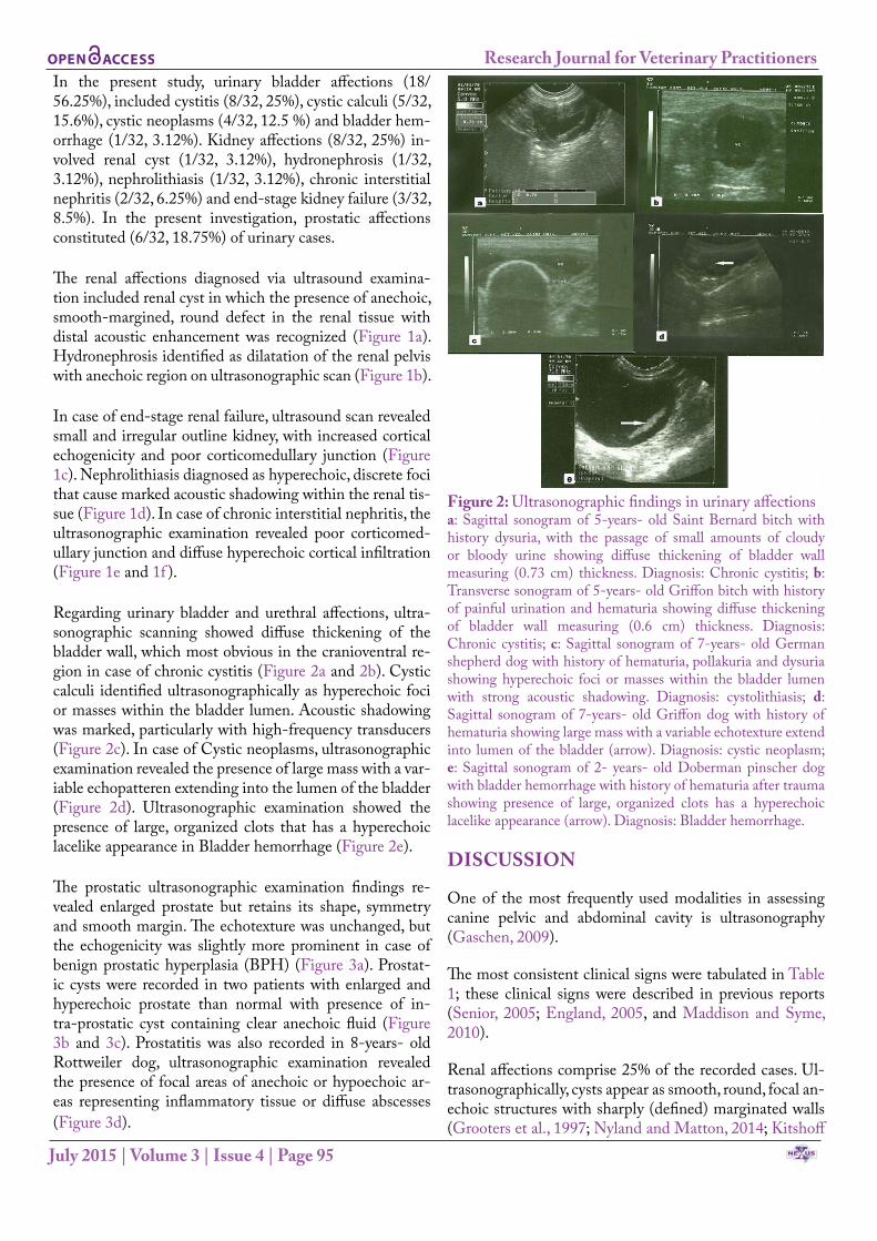
Regarding urinary bladder and urethral affections, ultra-sonographic scanning showed diffuse thickening of the bladder wall, which most obvious in the cranioventral re-gion in case of chronic cystitis (Figure 2a and 2b). Cystic calculi identified ultrasonographically as hyperechoic foci or masses within the bladder lumen. Acoustic shadowing was marked, particularly with high-frequency transducers (Figure 2c). In case of Cystic neoplasms, ultrasonographic examination revealed the presence of large mass with a var-iable echopatteren extending into the lumen of the bladder (Figure 2d). Ultrasonographic examination showed the presence of large, organized clots that has a hyperechoic lacelike appearance in Bladder hemorrhage (Figure 2e).
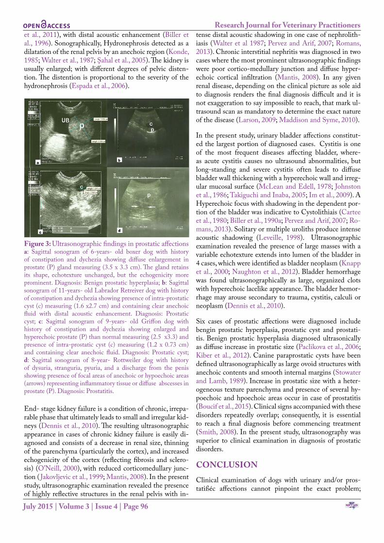
The prostatic ultrasonographic examination findings re-vealed enlarged prostate but retains its shape, symmetry and smooth margin. The echotexture was unchanged, but the echogenicity was slightly more prominent in case of benign prostatic hyperplasia (BPH) (Figure 3a). Prostat-ic cysts were recorded in two patients with enlarged and hyperechoic prostate than normal with presence of in-tra-prostatic cyst containing clear anechoic fluid (Figure 3b and 3c). Prostatitis was also recorded in 8-years- old Rottweiler dog, ultrasonographic examination revealed the presence of focal areas of anechoic or hypoechoic ar-eas representing inflammatory tissue or diffuse abscesses (Figure 3d).
Figure 2: Ultrasonographic findings in urinary affectionsa: Sagittal sonogram of 5-years- old Saint Bernard bitch with history dysuria, with the passage of small amounts of cloudy or bloody urine showing diffuse thickening of bladder wall measuring (0.73 cm) thickness. Diagnosis: Chronic cystitis; b: Transverse sonogram of 5-years- old Griffon bitch with history of painful urination and hematuria showing diffuse thickening of bladder wall measuring (0.6 cm) thickness. Diagnosis: Chronic cystitis; c: Sagittal sonogram of 7-years- old German shepherd dog with history of hematuria, pollakuria and dysuria showing hyperechoic foci or masses within the bladder lumen with strong acoustic shadowing. Diagnosis: cystolithiasis; d: Sagittal sonogram of 7-years- old Griffon dog with history of hematuria showing large mass with a variable echotexture extend into lumen of the bladder (arrow). Diagnosis: cystic neoplasm; e: Sagittal sonogram of 2- years- old Doberman pinscher dog with bladder hemorrhage with history of hematuria after trauma showing presence of large, organized clots has a hyperechoic lacelike appearance (arrow). Diagnosis: Bladder hemorrhage.
DISCUSSION
One of the most frequently used modalities in assessing canine pelvic and abdominal cavity is ultrasonography (Gaschen, 2009).
The most consistent clinical signs were tabulated in Table 1; these clinical signs were described in previous reports (Senior, 2005; England, 2005, and Maddison and Syme, 2010).
Renal affections comprise 25% of the recorded cases. Ul-trasonographically, cysts appear as smooth, round, focal an-echoic structures with sharply (defined) marginated walls (Grooters et al., 1997; Nyland and Matton, 2014; Kitshoff
NE USAcademic Publishers
Research Journal for Veterinary Practitioners
July 2015 | Volume 3 | Issue 4 | Page 96
et al., 2011), with distal acoustic enhancement (Biller et al., 1996). Sonographically, Hydronephrosis detected as a dilatation of the renal pelvis by an anechoic region (Konde, 1985; Walter et al., 1987; Şahal et al., 2005). The kidney is usually enlarged; with different degrees of pelvic disten-tion. The distention is proportional to the severity of the hydronephrosis (Espada et al., 2006).
Figure 3: Ultrasonographic findings in prostatic affectionsa: Sagittal sonogram of 6-years- old boxer dog with history of constipation and dychezia showing diffuse enlargement in prostate (P) gland measuring (3.5 x 3.3 cm). The gland retains its shape, echotexture unchanged, but the echogenicity more prominent. Diagnosis: Benign prostatic hyperplasia; b: Sagittal sonogram of 11-years- old Labrador Retreiver dog with history of constipation and dychezia showing presence of intra-prostatic cyst (c) measuring (1.6 x2.7 cm) and containing clear anechoic fluid with distal acoustic enhancement. Diagnosis: Prostatic cyst; c: Sagittal sonogram of 9-years- old Griffon dog with history of constipation and dychezia showing enlarged and hyperechoic prostate (P) than normal measuring (2.5 x3.3) and presence of intra-prostatic cyst (c) measuring (1.2 x 0.73 cm) and containing clear anechoic fluid. Diagnosis: Prostatic cyst; d: Sagittal sonogram of 8-year- Rottweiler dog with history of dysuria, stranguria, pyuria, and a discharge from the penis showing presence of focal areas of anechoic or hypoechoic areas (arrows) representing inflammatory tissue or diffuse abscesses in prostate (P). Diagnosis: Prostatitis.
End- stage kidney failure is a condition of chronic, irrepa-rable phase that ultimately leads to small and irregular kid-neys (Dennis et al., 2010). The resulting ultrasonographic appearance in cases of chronic kidney failure is easily di-agnosed and consists of a decrease in renal size, thinning of the parenchyma (particularly the cortex), and increased echogenicity of the cortex (reflecting fibrosis and sclero-sis) (O’Neill, 2000), with reduced corticomedullary junc-tion ( Jakovljevic et al., 1999; Mantis, 2008). In the present study, ultrasonographic examination revealed the presence of highly reflective structures in the renal pelvis with in-
tense distal acoustic shadowing in one case of nephrolith-iasis (Walter et al 1987; Pervez and Arif, 2007; Romans, 2013). Chronic interstitial nephritis was diagnosed in two cases where the most prominent ultrasonographic findings were poor cortico-medullary junction and diffuse hyper-echoic cortical infiltration (Mantis, 2008). In any given renal disease, depending on the clinical picture as sole aid to diagnosis renders the final diagnosis difficult and it is not exaggeration to say impossible to reach, that mark ul-trasound scan as mandatory to determine the exact nature of the disease (Larson, 2009; Maddison and Syme, 2010).
In the present study, urinary bladder affections constitut-ed the largest portion of diagnosed cases. Cystitis is one of the most frequent diseases affecting bladder, where-as acute cystitis causes no ultrasound abnormalities, but long-standing and severe cystitis often leads to diffuse bladder wall thickening with a hyperechoic wall and irreg-ular mucosal surface (McLean and Edell, 1978; Johnston et al., 1986; Takiguchi and Inaba, 2005; Im et al., 2009). A Hyperechoic focus with shadowing in the dependent por-tion of the bladder was indicative to Cystolithiais (Cartee et al., 1980; Biller et al., 1990a; Pervez and Arif, 2007; Ro-mans, 2013). Solitary or multiple uroliths produce intense acoustic shadowing (Leveille, 1998). Ultrasonographic examination revealed the presence of large masses with a variable echotexture extends into lumen of the bladder in 4 cases, which were identified as bladder neoplasm (Knapp et al., 2000; Naughton et al., 2012). Bladder hemorrhage was found ultrasonographically as large, organized clots with hyperechoic lacelike appearance. The bladder hemor-rhage may arouse secondary to trauma, cystitis, calculi or neoplasm (Dennis et al., 2010).
Six cases of prostatic affections were diagnosed include bengin prostatic hyperplasia, prostatic cyst and prostati-tis. Benign prostatic hyperplasia diagnosed ultrasonically as diffuse increase in prostatic size (Paclikova et al., 2006; Kiber et al., 2012). Canine paraprostatic cysts have been defined ultrasonographically as large ovoid structures with anechoic contents and smooth internal margins (Stowater and Lamb, 1989). Increase in prostatic size with a heter-ogeneous texture parenchyma and presence of several hy-poechoic and hpoechoic areas occur in case of prostatitis (Boucif et al., 2015). Clinical signs accompanied with these disorders repeatedly overlap; consequently, it is essential to reach a final diagnosis before commencing treatment (Smith, 2008). In the present study, ultrasonography was superior to clinical examination in diagnosis of prostatic disorders.
CONCLUSION
Clinical examination of dogs with urinary and/or pros-tatißéc affections cannot pinpoint the exact problem;

NE USAcademic Publishers
Research Journal for Veterinary Practitioners
July 2015 | Volume 3 | Issue 4 | Page 97
therefore, ultrasonography is compulsory for this specifica-tion to reach the most reliable diagnosis.
Ultrasonography doesn’t only detect superficial lesions due to sectioning of organs and hence detect deep seated le-sions which not accessible by routine examinations.
CONFLICT OF INTEREST
There exist no conflict of interest.
AUTHOR’S CONTRIBUTION
Both authors of this manuscript contributed equally.
ACKNOWLEDGMENT
None
REFERENCES
• Atalan G, Barr FJ, Holt PE (1998). The assessment of bladder volume by means of linear ultrasonographic measurements. Am. J. Vet. Res. 59: 10–15.
• Barr F, Gaschen L (2011). BSAVA Manual of Canine and Feline. Ultrasonography (BSAVA British Small Animal Veterinary Association).
• Biller DS, Di Bartola SP, Eaton KA, Pflueger S, Wellman ML, Radin MJ (1996). Inheritance of polycystic kidney disease in Persian cats. J. Hered. 87(1): 1-5. http://dx.doi.org/10.1093/oxfordjournals.jhered.a022945
• Boucif A, Madani K, Boulkaboul A, Slimani K (2015). Chronic prostatitis (CP) in Atlas shepherd dog: A case-control study. Clin. Microbiol. 4: 2.
• Cartee RE, Selcer BA, Patton CS (1980). Ultrasonographic diagnosis of renal disease in small animals. J. Am. Vet. Med. Assoc. 176(5): 426-30.
• Dennis R, Kirberger RM, Barr F, Wrigley RH (2010). Handbook of small animal radiology and ultrasound: Techniques and differential diagnoses. Second edition. Elsevier. Pp. 305–308.
• England GCW (2005). Fever and hypothermia. In: Dunn, J.K.: Textbook of. Small Animal Medicine, W.B. Saunders, Philadelphia.
• Espada Y, Novellas R, Ruiz de Gopegui R (2006). Renal ultrasound in dogs and cats. Vet. Res. Commun. 30(1): 133–137. http://dx.doi.org/10.1007/s11259-006-0026-8
• Gaschen L (2009). Update on hepatobiliary imaging. Vet. Clin. North Am. Small Anim. Pract. 39(3): 439-67. http://dx.doi.org/10.1016/j.cvsm.2009.02.005
• Grooters AM, Cuypers MD, Partington BP, Williams J, Pechman RD (1997). Renomegaly in dogs and cats, part II diagnostic approach. Compend. Contin. Educ. Pract. Vet. 19: 1213-1229.
• Im E, Kang S, Jung J, Jeon J, Kim J (2009). A case of polypoid cystitis in a dog. Korean J. Vet. Res. 49(2): 163-166.
• Jakovljevic S, Rivers WJ, Chun R, King VL, Han CM (1999). Results of renal ultrasonography performed before, during and after administration of saline (0.9 NaCl) solution to induce diuresis in dogs without evidence of renal disease.
Am. J. Vet. Res. 60(4):405-409.• Johnston GR, Walter PA, Feeney DA (1986). Radiographic and
ultrasonic features of uroliths and other urinary tract filling defects. Vet clin North Am Small Anim Pract. 16:261-292. http://dx.doi.org/10.1016/S0195-5616(86)50030-9
• Kibar, M., Oltu, K., Kalin, R., Atalan, G. (2012). Investigation of prostatic diseases by radiolographical, ultrasonographical, and laboratory examinations in Geriatric dogs. Journal of Health Sciences, 21(1): 30-37
• Kitshoff AM, McClure V, Lim C K, Kirberger RM (2011). Bilateral multiple cystic kidney disease and renal cortical abscess in a Boerboel. J. S. Afr. Vet. Assoc. 82(2):120-124. http://dx.doi.org/10.4102/jsava.v82i2.45
• Konde LJ (1985). Sonography of the kidney.Vet Clin North Am Small Anim Pract, 15(6):1149-1158. http://dx.doi.org/10.1016/S0195-5616(85)50362-9
• Knapp DW, Glickman NW, De Nicola DB, Bonney PL, Lin TL, Glickman LT (2000). Naturally-occurring canine transitional cell carcinoma of the urinary bladder, a relevant model of human invasive bladder cancer. Urol Oncol. 5(2):47-59. http://dx.doi.org/10.1016/S1078-1439(99)00006-X
• Larson MM (2009). The kidney and ureter. In: BSAVA Manual of Canine and Feline Abdominal Imaging. In: O’Brien, R. and Barr, F. (Eds.), Wiely- Inter science. http://dx.doi.org/10.1053/j.ajkd.2008.09.013
• Leroy C, Conchou F, Layssol-Lamour C, Deviers A, Sautet J, Concordet D, Mogicato G (2013). Normal canine prostate gland: Repeatability, reproducibility, observer-dependent variability of ultrasonographic measurements of the prostate in healthy intact beagles. Anat. Histol. Embryol. 42(5): 355-61. http://dx.doi.org/10.1111/ahe.12022
• Maddison J, Syme H (2010). Chronic kidney disease in dogs and cats: pathophysiology and diagnosis. Irish V. J. 63 (1): 44-48.
• Mantis P (2008). Ultrasonography of the urinary and genital system of the dog and cat. Iranian J. Vet. Surg. Suppl for the 2nd ISVS and 7th ISVAR: 63-71.
• McLean GK, Edell, SL (1978). Determination of bladder volume by gray scale ultrasonography. Radiology.128:181-182. http://dx.doi.org/10.1148/128.1.181
• Naughton JF, WidmerWR, Constable D, Knapp DW (2012). Accuracy of three-dimensional and two-dimensional ultrasonography in measuring tumor volume in dogs with transitional cell carcinoma of the urinarybladder. Am. J. Vet. Res. 73: 1919-1924. http://dx.doi.org/10.2460/ajvr.73.12.1919
• Nicola MAP (2006). Non-inflammatory diseases of the canine prostate gland. UK Vet. (Small Animal Laboratory). 11(7): 1-5.
• Nyland TG, Mattoon JS (2014). Chapter 16: ‘Urinary Tract.’ In: Small Animal Diagnostic Ultrasound, 3rd edition, pp.557-607. W. B. Saunders Co., Philadelphia.
• Leveille R (1998). Ultrasonography of urinary bladder disorders. Vet. Clin. North Am. Small Anim. Pract. 28(4): 799–821. http://dx.doi.org/10.1016/S0195-5616(98)50079-4
• O’Neill WC (2000). Sonographic evaluation of renal failure. Am. J. Kidney Dis. 35(6): 1021-1038. http://dx.doi.org/10.1016/S0272-6386(00)70036-9
• Osborne CA, Unger LK, Lulich JP (1995). Canine and feline nephroliths. In: Kirk RW, Bonagura JD (eds): Current Veterinary Therapy XII: Small Animal Practice. Philadelphia, WB Saunders. Pp. 981-985.
• Paclikova K, Kohout P, Vlasin M (2006). Diagnostic possibilities

NE USAcademic Publishers
Research Journal for Veterinary Practitioners
July 2015 | Volume 3 | Issue 4 | Page 98
in the management of canine prostatic disorders. Veterinarni Medicina. 51: 1-13.
• Pervez A, Arif A (2007). Role of ultrasound in evaluation of renal colic and assessment of risk factor for renal calculi. Gomal J. Med. Sci. 5(1): 22-27.
• Preston RC, Shaw A (2001). Recommended Ultrasound Field Safety. Classification for Medical Diagnostic Devices 1–16. National Physics Laboratory, Middlesex.
• Romans L (2013). Urinary tract calculi. ECEI Enterprises for Continuing Education Inc. Journal. Online access. Cited at: www.cewebsource.com
• Şahal M, Haziroglu R, Özkanlar Y, Beyaz L (2005). Bilateral hydronephrosis and hydroureter in a German shepherd dog. Ankara Üniv. Vet. Fak. Derg. 52: 193-196.
• Senior DF (2005). Diseases of urinary System. In: Dunn, J.K.: Textbook of Small Animal Medicine, W.B. Saunders,
Philadelphia.• Smith J (2008). Canine prostatic disease: A review
of anatomy, pathology, diagnosis, and treatment. Theriogenology. 70: 375–383. http://dx.doi.org/10.1016/j.theriogenology.2008.04.039
• Stowater JL, Lamb CR (1989). Ultrasonographic features of paraprostatic cysts in nine dogs. Vet. Radiol. 30(5): 232-239. http://dx.doi.org/10.1111/j.1740-8261.1989.tb00780.x
• Takiguchi M, Inaba M (2005). Diagnostic ultrasound of polypoid cystitis in dogs. J. Vet. Med. Sci. 67(1): 57-61. http://dx.doi.org/10.1292/jvms.67.57
• Walter PA, Johnston GR, Feeney DA, O’Leary TP (1987). Ultrasonographic evaluation of renal parenchymal disease in dogs: 32 cases (1981-1986). J. Am. Vet. Med. Assoc. 191(8): 999-1007.