ultrasonographic assessment of diaphragm function...
TRANSCRIPT
Ultrasonographic Assessment of Diaphragm Function in CriticallyIll Subjects
Michele Umbrello MD and Paolo Formenti MD
IntroductionDiaphragm PhysiologyVentilator-Induced Diaphragm DysfunctionAssessment of Respiratory Muscle Function and StrengthClinical AssessmentImagingAirway Pressure and FlowEsophageal and Transdiaphragmatic PressuresElectrical Activity of the DiaphragmBedside Ultrasonography in Critically Ill PatientsMeasurement of Diaphragm ThicknessB-ModeM-ModeDiaphragm DisplacementConclusions
The majority of patients admitted to the ICU require mechanical ventilation as a part of theirprocess of care. However, mechanical ventilation itself or the underlying disease can lead to dys-function of the diaphragm, a condition that may contribute to the failure of weaning from me-chanical ventilation. However, extended time on the ventilator increases health-care costs andgreatly increases patient morbidity and mortality. Nevertheless, symptoms and signs of muscledisease in a bedridden (or bed rest-only) ICU patient are often difficult to assess because ofconcomitant confounding factors. Conventional assessment of diaphragm function lacks specific,noninvasive, time-saving, and easily performed bedside tools or requires patient cooperation. Re-cently, the use of ultrasound has raised great interest as a simple, noninvasive method of quanti-fication of diaphragm contractile activity. In this review, we discuss the physiology and the relevantpathophysiology of diaphragm function, and we summarize the recent findings concerning theevaluation of its (dys)function in critically ill patients, with a special focus on the role of ultrasounds.We describe how to assess diaphragm excursion and diaphragm thickening during breathing andthe meaning of these measurements under spontaneous or mechanical ventilation as well as thereference values in health and disease. The spread of ultrasonographic assessment of diaphragmfunction may possibly result in timely identification of patients with diaphragm dysfunction and toa potential improvement in the assessment of recovery from diaphragm weakness. Key words:critical illness; diaphragm function; ultrasound; weaning; ventilator support. [Respir Care2016;61(4):542–555. © 2016 Daedalus Enterprises]
542 RESPIRATORY CARE • APRIL 2016 VOL 61 NO 4
Introduction
More than 40% of ICU patients require mechanical ven-tilation support as a part of their process of care.1 More-over, it is well known that prolonged mechanical ventila-tion may lead to contractile dysfunction of respiratorymuscles,2 in particular the diaphragm, causing a so-calledventilator-induced diaphragm dysfunction. The latter is de-fined as a loss of diaphragm force-generating capacityspecifically related to the use of mechanical ventilation.3,4
The major clinical implication of ventilator-induced dia-phragm dysfunction is that even when used for relativelyshort periods, mechanical ventilation can lead to substan-tial diaphragm weakness and could delay the process ofweaning from the ventilator.5
Diseases that interfere with diaphragm innervations, con-tractile properties, or mechanical coupling to the chestwall can result in diaphragm dysfunction.6,7 Such dysfunc-tion, in turn, can lead to dyspnea, decreased exercise per-formance, sleep-disordered breathing, constitutional symp-toms, hypersomnia, reduced quality of life, atelectasis, andrespiratory failure. Diaphragm dysfunction can be con-firmed by a number of invasive and noninvasive tests.8
However, most of the studies available have been con-ducted on healthy volunteers or subjects breathing spon-taneously, and only a few recent papers have focused at-tention on the function of respiratory muscles during theweaning process in mechanically ventilated subjects.
Moreover, the pathophysiology of weaning failure iscomplex, and it involves interaction between cardiopul-monary reserve, autonomic function, and musculoskeletalcapacity. Thus, it may be hard to assess the interplay be-tween those factors based on a single or a few predictors.
Indeed, monitoring respiratory muscle function in ICUpatients is still an uncommon practice. One of the majorchallenges still lies in how to evaluate diaphragm functionwith a specific, noninvasive, time-saving, and easily per-formed bedside technique. Many tools are available, evenif most of them require patient collaboration and are notspecific for diaphragm dysfunction. Nevertheless, in re-cent times, the use of ultrasonographic evaluation of thediaphragm has raised great interest. The aim of this reviewis to describe the physiology and the relevant pathophys-iology of diaphragm function and to summarize the recent
literature concerning the evaluation of its (dys)function incritically ill patients.
Diaphragm Physiology
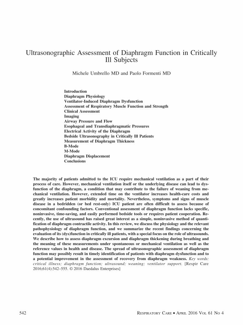
The rib cage, diaphragm, abdominal wall, and lungs arecharacterized by nonlinear stress-strain characteristics thatdepend on muscle activity.9 The positions, volumes, forces,and pressures for each set of initial conditions are deter-mined by mechanical equilibrium. The geometrical com-plexity of the chest wall and its associated muscles ofrespiration make rigorous analysis of the mechanics of therespiratory system difficult. In fact, the diaphragm is adome-shaped upward-curved structure of muscle and fi-brous tissue that separates the thoracic cavity from theabdomen. The superior surface of the dome forms thebottom of the thoracic cavity, and the inferior surface formsthe top of the abdominal cavity.10 As a dome, the dia-phragm has peripheral attachments to structures that makeup the abdominal and chest walls. Its peripheral part con-sists of muscular fibers that take origin from the circum-ference of the inferior thoracic opening and converge toinsert into a central tendon. Since thoracic pressure de-creases upon inspiration and draws the caval blood upwardtoward the right atrium, each contraction allows more bloodto return to the heart, maximizing the efficacy of loweredthoracic pressure to venous return.11 The diaphragm isprimarily innervated by the phrenic nerve; whereas thecentral portion sends sensory afferents via the phrenic nerve,the peripheral portions send sensory afferents via the in-tercostal (T5–T11) and subcostal nerves (T12).12 The areaof contact between the diaphragm and the rib cage is re-ferred to as the zone of apposition, and it is of great im-portance for proper diaphragm function (Fig. 1). The forceof contraction of the zone of apposition is controlled bythe abdominal muscles and significantly affects diaphragmtension. Diaphragm efficiency, in fact, largely dependsupon its position and anatomical relationship with the lowerrib cage.
In a seminal study, Goldman and Mead13 showed howthe superior diaphragm surface is in straight relation withthe entire chest wall. As consequence, the lower rib cagebehaves during tidal breathing as if it is driven by trans-abdominal rather than trans-thoracic pressure.14 During in-spiration, diaphragm muscle fibers shorten, and the dia-phragm as a whole moves caudally in piston-like fashion;the dome of the diaphragm normally changes size or shapevery little during tidal breathing, and the changes in mus-cle length are accommodated mainly by increasing anddecreasing the area of apposition.12 As the diaphragm con-tracts, it lowers pleural pressure and increases abdominalpressure. The reduction in pleural pressure produces aninflationary effect on the lungs, but, if no other force wereactive, its effect on the rib cage would be deflationary. The
The authors are affiliated with Unita Operativa di Anestesia e Rianima-zione, Azienda Ospedaliera San Paolo-Polo Universitario, Milano, Italy.
The authors have disclosed no conflicts of interest.
Correspondence: Michele Umbrello MD, UO Anestesia e Rianimazione,A.O. San Paolo, Polo Universitario, Via A Di Rudinì 8, 20142 Milano,Italy. E-mail: [email protected].
DOI: 10.4187/respcare.04412
ULTRASONOGRAPHIC ASSESSMENT OF DIAPHRAGM FUNCTION
RESPIRATORY CARE • APRIL 2016 VOL 61 NO 4 543
accompanying effects of increasing abdominal pressure,which tends to expand the rib cage, however, more thancounteract this.
It should, however, be considered that respiratory mus-cles comprise not only the diaphragm, but also intercostaland accessory muscles as well as muscles of the abdomen.Since all 3 groups share inspiratory and expiratory func-tions and work together in intricate ways, this must beaccurately taken into account during weaning phases. Infact, the intercostal muscles and the accessory musclesprimarily serve an inspiratory function that can be ob-served in the external intercostals in the upper few inter-costal spaces during quiet breathing, with the lower inter-costals becoming active with increased ventilation. Theabdominal respiratory muscles are regarded as expiratorymuscles that augment the passive recoil of the lungs. Afew studies13,15-17 have demonstrated how the accessoryrespiratory muscles have the primary role to stabilize thechest wall and convert diaphragm contraction into intratho-racic pressure and volume changes. For these reasons, itshould always be taken into account that the evaluation ofsingle diaphragm function could be insufficient in the eval-uation of the weaning process.
During spontaneous breathing, the inspiratory pressuregenerated by respiratory muscles (Pmus) is dissipated tocompel the elastic and resistive forces, as described by the
motion law, Pmus � (RRS � V) � (ERS � V), where RRS
is resistance of the respiratory system, V is the air flow,
ERS is respiratory system elastance, and V is the volumeabove the functional residual capacity. Instead, during con-trolled mechanical ventilation, Pmus is equal to 0, and theinspiratory force is generated by the ventilator (Pvent) only,
as described by the equation, Pvent � (RRS � V) �(ERS � V).
During assisted mechanical ventilation, the inspiratorymuscles and the ventilator interact to generate the requiredpressure, with the motion law that can be described as
follows: Pappl � Pvent � Pmus � (RRS � V) � (ERS � V),where Pappl is the total respiratory pressure. As suggestedby Younes,18 the last equation could be followed by its
rearrangement to isolate Pmus, Pmus � (RRS � V � V) �Pvent, so that the relative contribution of respiratory mus-cles can be determined.
Ventilator-Induced Diaphragm Dysfunction
Evidence supporting the occurrence of diaphragm dys-function in critically ill patients is scarce, although it iscommon experience that most mechanically ventilated pa-tients display profound diaphragm weakness. This maydepend on the presence of confounding factors, such as theunderlying disease state, different modes of mechanicalventilation, medications, and newly acquired complica-tions, which can all impair diaphragm function.19 Never-theless, some data exist to actually support the presence of
Fig. 1. Schematic view of the zone of apposition. On the right is depicted the relationship between the rib cage, right lung, and upperabdominal content in the zone of apposition. On the left, anatomical structures are magnified so that the anatomic relationship between theparietal pleura, the diaphragm, and the parietal peritoneum is highlighted. Arrows represent the forces acting on the different anatomicregions of the area. During inspiration, the diaphragm fibers shorten and the diaphragm as a whole moves caudally (1). As the diaphragmcontracts, it lowers the pleural pressure and increases the abdominal pressure. The reduction in pleural pressure produces an inflationaryeffect on the lungs (2). The accompanying effects of increasing abdominal pressure tend to expand the rib cage (3).
ULTRASONOGRAPHIC ASSESSMENT OF DIAPHRAGM FUNCTION
544 RESPIRATORY CARE • APRIL 2016 VOL 61 NO 4
ventilator-induced diaphragm dysfunction in subjects.20-22
The decrease in diaphragm contractility is time-dependent,and it worsens as mechanical ventilation is prolonged. Theeffects of critical illness on respiratory muscle function areoften part of a more generalized phenomenon, known asICU-acquired weakness. In the last decade, the understand-ing of the molecular and cellular mechanisms underlyingrespiratory muscle weakness in the critically ill has beenthe subject of extensive research.23 Muscle atrophy canresult from decreased protein synthesis, increased proteindegradation, or both21; fiber remodeling with change fromslow to fast fibers may reduce the endurance of the dia-phragm, because fewer slow, fatigue-resistant fibers areavailable.24 Moreover, oxidative stress and structural in-jury are implicated as potential mechanisms of ventilator-induced diaphragm dysfunction. Jaber et al25 reported thefunctional consequences of critical illness on respiratorymuscles, showing an approximately 30% reduction in twitchairway pressure induced by magnetic phrenic nerve stim-ulation in the first days of invasive mechanical ventilation.
Experimental and clinical studies that have investigatedventilator-induced diaphragm dysfunction have mainlyused controlled mechanical ventilation. One study showedthat the contractile response of rabbit diaphragm to tetanicstimulation was decreased by almost a half after 3 d ofcontrolled mechanical ventilation.26 In clinical practice,very few patients are ventilated with controlled mechani-cal ventilation, whereas modes like pressure support ven-tilation are widely used with the aim of unloading therespiratory muscles while avoiding muscle atrophy2 afterresolution of the acute phase of illness. In such a mode, avariable amount of work is generated by the patient’s in-spiratory muscles, whereas the remainder is provided bythe ventilator.27 However, an inappropriate level of pres-sure support may lead to fatigue and discomfort (if it is toolow) or patient-ventilator asynchrony (if it is too high),28
then possibly promoting the onset of ventilator-induceddiaphragm dysfunction.
The decrease in diaphragm force-generating capacitythat occurs during controlled mechanical ventilation is at-tenuated during assisted modes of ventilation.29 Whetherthe decrease in diaphragm contractility observed duringcontrolled ventilation contributes to failure to wean fromthe ventilator is difficult to determine. In fact, weaningfailure patients have reasons other than ventilator-induceddiaphragm dysfunction for respiratory muscle weakness.However, until further data are available, it seems prudentto reduce as much as possible the use of controlledmechanical ventilation in patients with acute respiratoryfailure.
To summarize, diaphragm function is a major determi-nant of the ability to successfully wean patients from me-chanical ventilation.30 Concern has been raised thatmechanical ventilation may itself be harmful to the
diaphragm.31 In animals,6,32-36 diaphragm inactivity asso-ciated with mechanical ventilation leads to muscle fiberatrophy in the diaphragm and a reduction in its force-generating capacity.37 Importantly, the impact of suchchanges on diaphragm contractile function and the rapiditywith which diaphragm atrophy develops during mechani-cal ventilation in humans remain unknown.
Assessment of Respiratory Muscle Functionand Strength
Global respiratory muscle weakness and severe isolatedweakness of the diaphragm or bilateral diaphragm paral-ysis cause dyspnea38 when the patient is in the supineposition, with the abdominal paradox that occurs when thestrength of the diaphragm is reduced to approximately onefourth of normal.39 In mechanically ventilated patients,these signs can be masked by ventilator support. More-over, a severe respiratory muscle weakness may itself causerespiratory failure.
Clinical Assessment
Despite the tremendous increase in the number and so-phistication of diagnostic tests, the most important elementin the evaluation of patients with a suspected muscle weak-ness remains the physical examination. Unfortunately,symptoms and signs of muscle disease in a bed rest-onlyICU patient are insufficient and difficult to assess becauseof concomitant confounding factors, such as analgesia andsedation, that are widely present. However, respiratorymuscle weakness is often advanced before clinical symp-toms occur. This follows from the relatively low respira-tory muscle force that is required to overcome most respi-ratory tasks. Symptoms are thus often referred to as negativecomplaints, such as weakness, minimum exercise intoler-ance (such as raising arms), fatigue, and muscle atrophy.In neuromuscular diseases, close attention should be paidto the involvement of both inspiratory and expiratory mus-cles. In lung diseases, such as cystic fibrosis and COPD,inspiratory muscle weakness is often present.40 Less ob-vious is the detection of respiratory muscle weakness inpatients with heart failure,41 cancer, and systemic diseases.In patients with asthma,42,43 respiratory muscle weaknesscan contribute to the sensation of dyspnea, and the assess-ment of respiratory muscle function may be helpful insolving the diagnostic dilemma of the unexplained dys-pnea. When patients are treated with drugs that may in-duce myopathy, it seems prudent to assess respiratory mus-cle strength before initiating treatment, and properfollow-up is advised.
The most characteristic physical sign of diaphragm dys-function is the abdominal paradox, which is the paradox-ical inward motion of the abdomen as the rib cage expands
ULTRASONOGRAPHIC ASSESSMENT OF DIAPHRAGM FUNCTION
RESPIRATORY CARE • APRIL 2016 VOL 61 NO 4 545

during inspiration. This disordered breathing pattern re-sults from compensatory use of the accessory inspiratorymuscles of the rib cage and neck. When these musclescontract and lower pleural pressure, the weakened or flac-cid diaphragm moves in a cephalad direction, and the ab-dominal wall moves inward. This paradoxical breathingpattern rarely occurs in unilateral diaphragm paralysis.When this is present in unilateral diaphragm paralysis, itsuggests generalized weakness of the respiratory muscles.44
Accessory respiratory muscle recruitment, especially thesternocleidomastoid muscle, may be apparent by palpationin patients when inspiratory load exceeds the capacity ofthe diaphragm.45 Contraction of the abdominal musclesduring expiration and subsequent relaxation as an assis-tance to inspiration may give the appearance of outwardmotion of the anterior abdominal wall during inspiration.Thus, activation of the abdominal muscles during expira-tion could also be regarded as a sign of respiratory muscledysfunction.
When respiratory muscle strength is moderately to se-verely reduced, discrete clinical symptoms may occur, andthis may prompt assessment of muscle function to help inthe diagnostic process. The cardinal symptom of respira-tory muscle weakness is dyspnea. In the case of severeexpiratory muscle weakness, reduced cough efficiency maybecome an important handicap, and patients may becomeventilator-dependent. In severe respiratory muscle dysfunc-tion, vital capacity is generally reduced as a consequenceof the respiratory muscle weakness, and it may become abetter predictor of morbidity than measurements of respi-ratory muscle strength.6
Traditionally, several tools have been available to eval-uate diaphragm function. However, most of them are noteasily used at the bedside in the ICU and have reducedspecificity and sensibility.
Imaging
Chest radiographs may reveal elevated hemidiaphragms(sensitivity of 90%), even if the elevation of both hemi-diaphragms is common in mechanically ventilated patientsand has low specificity for diagnosing diaphragm dysfunc-tion (specificity of 44%).46
Fluoroscopy of the diaphragm was extensively used inthe past, but it is no longer considered a helpful test indiagnosing diaphragm paralysis. With unilateral diaphragmparalysis, a paradoxical movement of the paralyzed hemi-diaphragm can be shown, but false negative results (�6%)occur with active contraction of the abdominal musclesduring expiration.47 This technique can still be useful inthe case of unilateral diaphragm palsy, especially if it isperformed during a sniff maneuver. In fact, during thesniff maneuver, the paradoxical movement of the para-lyzed hemidiaphragm, cephalad with inspiration, in con-
trast with the rapid caudal movement of the unaffectedmuscle, can be easily detected.48 Computed tomographyand magnetic resonance imaging have been proposed toevaluate diaphragm function, but they are not suitable formonitoring mechanically ventilated patients.49,50
Airway Pressure and Flow
The most widely used tests of global inspiratory andexpiratory muscle strength are the static maximum pres-sures measured at the mouth (PImax and PEmax).51-53 Bothcan be measured either when the patient is connected tothe ventilator or during a brief disconnection using a hand-held pressure monitoring device. Indeed, such voluntarymaneuvers require patient cooperation and are influencedby sedation level, anxiety, and pain. These tests have theadvantage of being noninvasive and easily performed atthe bedside. A high PImax or PEmax can exclude clinicallyimportant inspiratory or expiratory weakness. PImax andPEmax are influenced by age, sex, posture, lung volume,and the type of mouthpiece.54 To obtain more reliablemeasurements of PImax in ventilated and sedated patients,a 20-s end-expiratory occlusion period can be performed,55
using a one-way valve that permits exhalation. The samebreath-stacking method can be used to assess inspiratorycapacity in uncooperative patients.56 PImax and PEmax canbe used as a global measure for respiratory muscle func-tion and possibly to monitor the response of respiratorymuscle training. Quality control of the measurements canonly be obtained from inspection of the pressure-timecurves. The peak pressure should be obtained in the verybeginning of the maneuver. A pressure maintained for �1s is generally reported as the PImax or PEmax (plateau pres-sure).57 However, one study58 has challenged the use ofthe plateau pressure, concluding that the peak pressuremay be easier to obtain and equally reliable when subjectsare well instructed.
In absolute numbers, PEmax is roughly double PImax whena rigid mouthpiece is used. It is uncommon to find PEmax
inferior to PImax even if in some neurological diseases,such as cervical spinal cord injury,59 PEmax is typicallymore reduced than PImax. Different studies reported vary-ing normal values for PImax and PEmax that are lower infemales and fall with advancing age with large SD valuesof the data, which mean that lower limits of normality areapproximately 50% of predicted normal values.60 Never-theless, impressive differences are observed between thenormal values reported in the literature.52,53,61-66 In all mod-els of maximal inspiratory and expiratory pressures, theexplained variance is low, reflecting large inter-individualdifferences even when age, sex, and anthropometric valuesare taken into account. Therefore, a low PImax should al-ways be interpreted with caution. A normal PImax, how-ever, generally excludes clinically relevant inspiratory mus-
ULTRASONOGRAPHIC ASSESSMENT OF DIAPHRAGM FUNCTION
546 RESPIRATORY CARE • APRIL 2016 VOL 61 NO 4
cle pathology. Moreover, the effect of starting lung volumeshould be considered when assessing inspiratory/expira-tory pressures.54
Esophageal and Transdiaphragmatic Pressures
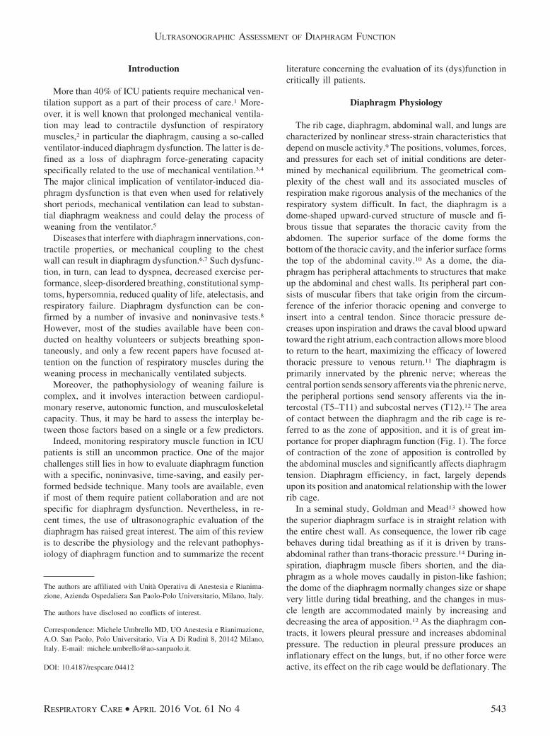
The differential measurement of gastric and esophageal(Pes) pressures determines the pressure developed acrossthe diaphragm (transdiaphragmatic pressure [Pdi]) and pro-vides quantitative information concerning the mechanicalbehavior of this muscle in relative isolation from intercos-tals, accessory muscles, and elastic recoil of the chest wall.67
Measurement of Pdi has been suggested to be used in allpatients with suspected diaphragm weakness47 increasinglyperformed in the evaluation of patients suspected of hav-ing compromised diaphragm function, even if most of thenormal values that have been published were obtainedfrom laboratory studies.
At variance from PImax, Pdi is a specific measure ofdiaphragm muscle strength. Voluntary measurements ofmaximum Pdi can be obtained by having the patient inspireas forcefully as possible against a closed airway or byhaving the patient perform a sniff maneuver.67 The higherthe value, the greater the contribution of the diaphragm tototal inspiratory effort. However, it should be kept in mindthat Pdi measurement varies widely among individuals,and the range of Pdi is nearly independent of body size.68
Moreover, the volume at which the maximal Pdi maneuveris initiated is very important, because the diaphragm short-ens progressively as lung volume increases and is able togenerate less force as it shortens. Maximum pressure gen-eration occurs at residual volume, although it is commonpractice to measure maximum Pdi at functional residualcapacity. The normal range for Pdi depends on size, sex,body position, and the initial volume of the respiratorysystem during the maneuver, but a normal Pdi for an adultis around 100 cm H2O.69 To estimate the energy expendi-ture of the diaphragm, the tension-time index and pres-sure-time product can be calculated using Pdi.70
Indeed, isolated Pes measurements have also enhancedour understanding of not only the pathophysiology of acutelung injury, but also the patient-ventilator interaction andweaning failure.71 By providing a practical means of quan-tifying respiratory effort, Pes measurements may make itfeasible to individualize the level of muscle unloadingduring mechanical ventilation. In fact, Pmus, the standardreference for the measurement of the pressure developedby the respiratory muscles, is based on Pes measurement.69
In particular, it is generally computed as the differencebetween Pes and the chest wall elastic recoil curve, calcu-lated as the instant product of volume signal times chestwall elastance.
Gastric and esophageal balloons are currently exten-sively used for research purposes. However, their use is
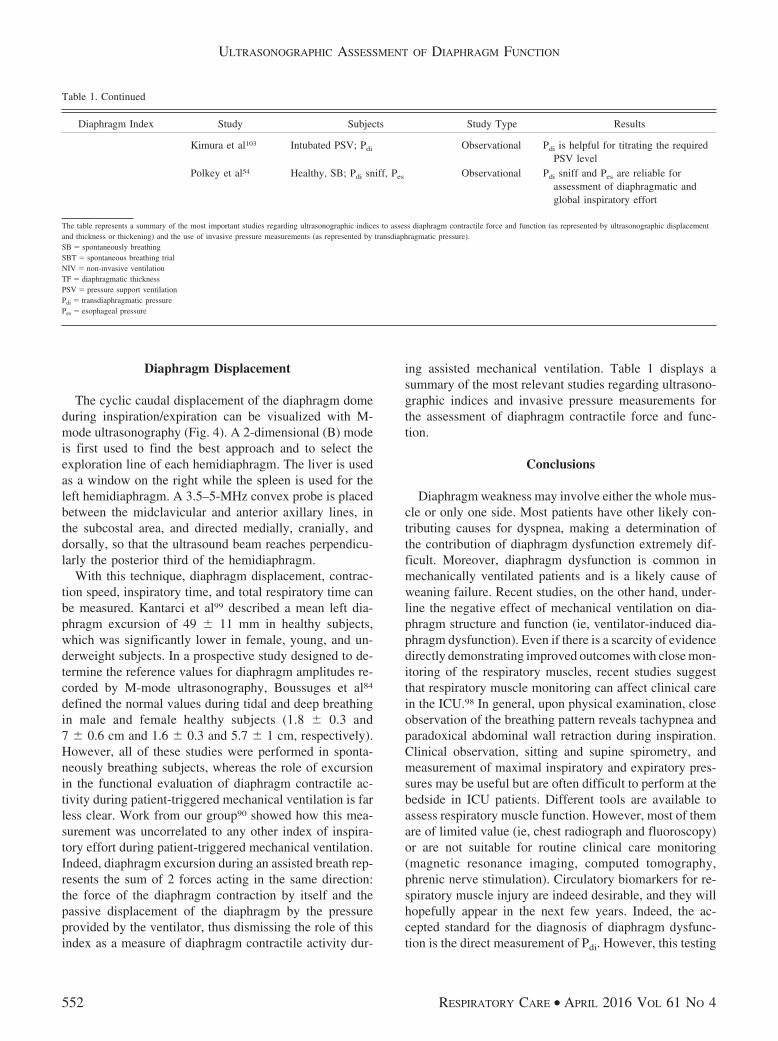
still uncommon in clinical care. Some of the newer ven-tilators have auxiliary ports to measure esophageal pres-sure, which is a step forward toward implementing mea-surement of esophageal pressure in clinical care, yet oneshould keep in mind that Pdi is variably influenced bypositive pressure of the mechanical ventilator, and ideallyit should be measured during a trial of spontaneous breath-ing (Fig. 2).
One study72 showed that a signal generated noninva-sively from flow, volume, and airway pressure using im-provised resistance and elastance values can be used formonitoring patient-ventilator interaction and for providinginformation to optimize ventilator settings. This applica-tion makes it possible to non-invasively identify the onsetand end of inspiratory efforts with reasonable accuracy.Such a signal may be used to synchronize the ventilatorwith the patient or simply to provide information on pa-tient-ventilator interaction that may be helpful in makingappropriate ventilator adjustments and in evaluating thepatient. Moreover, the use of improvised values of ERS andRRS can be used to non-invasively derive a Pmus estimatefrom the equation of motion so that monitoring of patientrespiratory effort can be extended into clinical practice.72
Another approach to provide noninvasive estimates ofrespiratory effort has been proposed.73 The authors showedhow a noninvasive Pmus can be derived from the electricalactivity of the diaphragm (Edi) signal displayed with neu-rally adjusted ventilatory assist (NAVA) ventilation, ex-tending from the use of bedside estimates of commonlyaccepted standard indexes.
Electrical Activity of the Diaphragm
NAVA is a recently developed mode of mechanicalventilation, which provides proportional pressure supportbased on measurements of the Edi, which serves as a proxyfor the neuronal output of the respiratory center.
With an expressly designed nasogastric tube, the Edi iscaptured, fed to the ventilator, and used to assist the pa-tient’s breathing in synchrony with and in proportion tohis/her own efforts, regardless of patient category or size.74
Because the work of the ventilator and the diaphragm iscontrolled by the same signal, coupling between the dia-phragm and the ventilator is synchronized simultaneously.
With NAVA, the ventilator delivers a pressure propor-tional to the integral of Edi
75 and therefore proportional tothe neural output of the patient’s central respiratory com-mand. The level of pressure delivered is determined by thepatient’s respiratory center neural output; the ventilator istriggered and cycled off based on the Edi value, whichdirectly reflects the activity of the neural respiratory com-mand. The assist levels are adjusted by changing the pro-portionality between the Edi and delivered pressure. Step-wise increases in the NAVA level cause a gradual reduction
ULTRASONOGRAPHIC ASSESSMENT OF DIAPHRAGM FUNCTION
RESPIRATORY CARE • APRIL 2016 VOL 61 NO 4 547
in respiratory drive, and therefore the expected increase inpressure is not necessarily achieved. Due to this physio-logical down-regulation of the Edi signal, airway pressureand tidal volume plateau at adequate levels of unloading.76
Indeed, NAVA delivers proportional assistance: the levelof pressure support varies from one cycle to the next cycleand is proportional to the Edi signal, which is proportionalto the intensity of the diaphragm contraction. The strongerthe diaphragm contraction, the greater the level of supportdelivered by the ventilator. If diaphragm contraction isinsufficient, positive feedback will cause a more powerfulEdi signal and thus more support. This assistance allowsproportional support to limit the periods of over- or un-derassistance and provides the patient with more adapta-tion to physiological breathing.77 A recent prospective studyfocusing on patient-ventilator asynchronies in NAVA andpressure support ventilation showed how NAVA reducedthe risk of over- and undersupport while providing morephysiological ventilation with tidal volume variability thanpressure support ventilation.78 The recent commercialspread of NAVA has given the opportunity to obtain sim-ple and minimally invasive monitoring of Edi, so that thisindex can be used to monitor patient inspiratory activityduring any mode of ventilation,79 based on the tight pro-portionality between Edi and Pmus.80 One study73 proposedthe combined use of Pmus and Edi, previously termed neuro-mechanical efficiency, which is supposed to indicate theamount of pressure that respiratory muscles are generatingfor a unit of electrical activity. The authors found in sub-jects with acute respiratory failure that this index is highly
variable between subjects but relatively stable in a singleindividual, even across different levels of assistance andventilator modalities.
Bedside Ultrasonography in Critically Ill Patients
The relative contribution of patient effort during as-sisted breathing is difficult to measure in clinical condi-tions. Moreover, the diaphragm is inaccessible to directclinical assessment. Bedside ultrasonography, which is al-ready crucial in several aspects of critically illness,81,82 hasrecently been proposed as a simple, noninvasive method ofquantification of diaphragm contractile activity.4 Ultra-sound can be used to determine diaphragm excursion,83,84
which may help to identify patients with diaphragm dys-function.85
Ultrasonographic examination can also allow for thedirect visualization of diaphragm thickness in its zone ofapposition.86 Thickening during active breathing has beenproposed to reflect the magnitude of diaphragm effort,similar to an ejection fraction of the heart.87
A number of recent studies have employed ultrasoundto measure diaphragm thickness and inspiratory thicken-ing in ventilated subjects. Some of them focused on thefeasibility and reproducibility of the technique,88,89 whereasanother90 showed how with increasing levels of pressuresupport ventilation, parallel reductions were found betweendiaphragm thickening and both the diaphragm and esoph-ageal pressure-time product, suggesting that diaphragmthickening is a reliable indicator of respiratory effort.
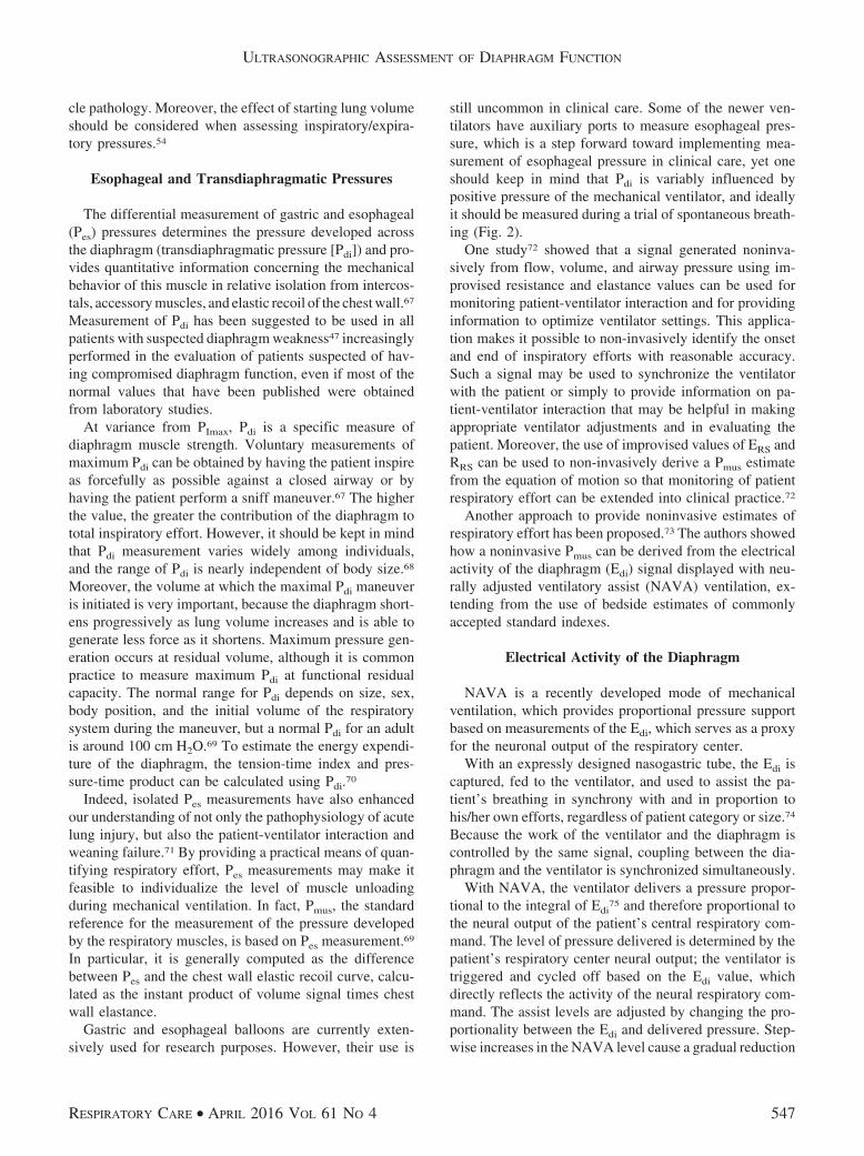
Fig. 2. Example of invasive measurement of respiratory pressures. Shown is the recording of a respiratory trace during spontaneousbreathing with CPAP in a subject in whom an esophageal and a gastric balloon catheter are inserted. Pes � esophageal pressure (a proxyfor pleural pressure); Pga � gastric pressure (a proxy for abdominal pressure); Paw � airway pressure; Pdi � transdiaphragmatic pressure.White areas represent inspiration, whereas gray shaded areas depict expiration. The horizontal line represents zero flow.
ULTRASONOGRAPHIC ASSESSMENT OF DIAPHRAGM FUNCTION
548 RESPIRATORY CARE • APRIL 2016 VOL 61 NO 4
Measurement of Diaphragm Thickness
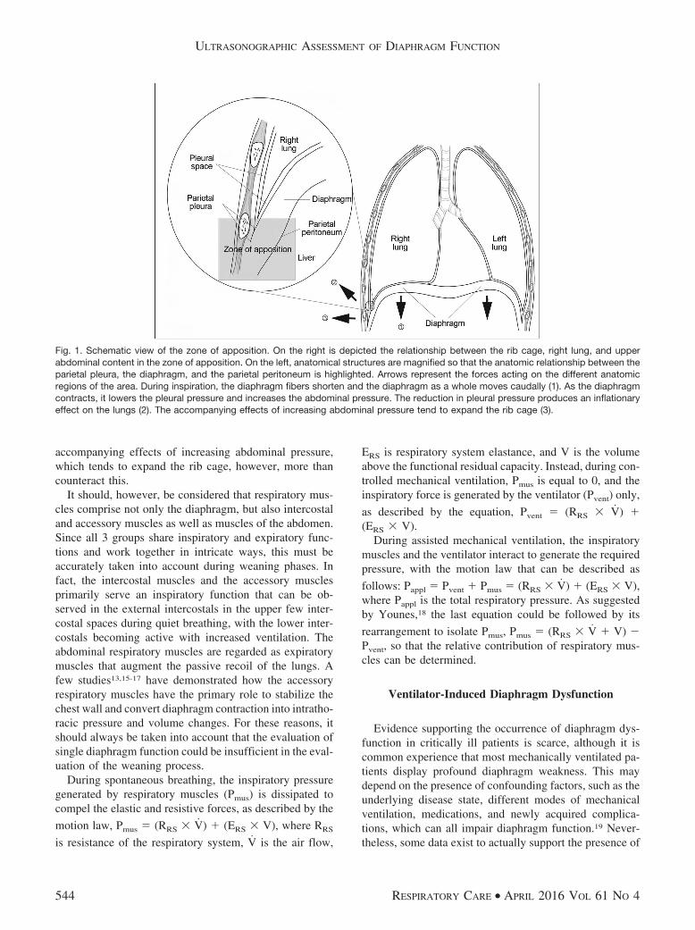
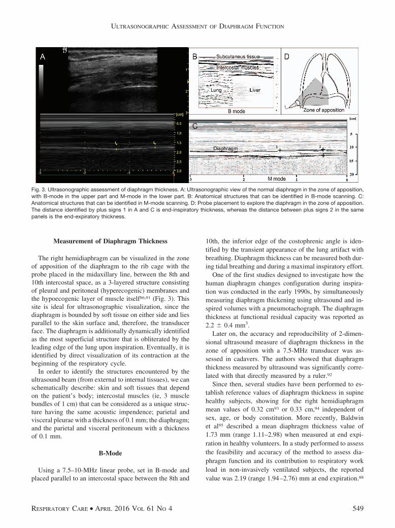
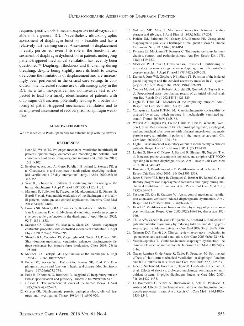
The right hemidiaphragm can be visualized in the zoneof apposition of the diaphragm to the rib cage with theprobe placed in the midaxillary line, between the 8th and10th intercostal space, as a 3-layered structure consistingof pleural and peritoneal (hyperecogenic) membranes andthe hypoecogenic layer of muscle itself86,91 (Fig. 3). Thissite is ideal for ultrasonographic visualization, since thediaphragm is bounded by soft tissue on either side and liesparallel to the skin surface and, therefore, the transducerface. The diaphragm is additionally dynamically identifiedas the most superficial structure that is obliterated by theleading edge of the lung upon inspiration. Eventually, it isidentified by direct visualization of its contraction at thebeginning of the respiratory cycle.
In order to identify the structures encountered by theultrasound beam (from external to internal tissues), we canschematically describe: skin and soft tissues that dependon the patient’s body; intercostal muscles (ie, 3 musclebundles of 1 cm) that can be considered as a unique struc-ture having the same acoustic impendence; parietal andvisceral pleurae with a thickness of 0.1 mm; the diaphragm;and the parietal and visceral peritoneum with a thicknessof 0.1 mm.
B-Mode
Using a 7.5–10-MHz linear probe, set in B-mode andplaced parallel to an intercostal space between the 8th and
10th, the inferior edge of the costophrenic angle is iden-tified by the transient appearance of the lung artifact withbreathing. Diaphragm thickness can be measured both dur-ing tidal breathing and during a maximal inspiratory effort.
One of the first studies designed to investigate how thehuman diaphragm changes configuration during inspira-tion was conducted in the early 1990s, by simultaneouslymeasuring diaphragm thickening using ultrasound and in-spired volumes with a pneumotachograph. The diaphragmthickness at functional residual capacity was reported as2.2 � 0.4 mm3.
Later on, the accuracy and reproducibility of 2-dimen-sional ultrasound measure of diaphragm thickness in thezone of apposition with a 7.5-MHz transducer was as-sessed in cadavers. The authors showed that diaphragmthickness measured by ultrasound was significantly corre-lated with that directly measured by a ruler.92
Since then, several studies have been performed to es-tablish reference values of diaphragm thickness in supinehealthy subjects, showing for the right hemidiaphragmmean values of 0.32 cm93 or 0.33 cm,94 independent ofsex, age, or body constitution. More recently, Baldwinet al95 described a mean diaphragm thickness value of1.73 mm (range 1.11–2.98) when measured at end expi-ration in healthy volunteers. In a study performed to assessthe feasibility and accuracy of the method to assess dia-phragm function and its contribution to respiratory workload in non-invasively ventilated subjects, the reportedvalue was 2.19 (range 1.94–2.76) mm at end expiration.88
Fig. 3. Ultrasonographic assessment of diaphragm thickness. A: Ultrasonographic view of the normal diaphragm in the zone of apposition,with B-mode in the upper part and M-mode in the lower part. B: Anatomical structures that can be identified in B-mode scanning. C:Anatomical structures that can be identified in M-mode scanning. D: Probe placement to explore the diaphragm in the zone of apposition.The distance identified by plus signs 1 in A and C is end-inspiratory thickness, whereas the distance between plus signs 2 in the samepanels is the end-expiratory thickness.
ULTRASONOGRAPHIC ASSESSMENT OF DIAPHRAGM FUNCTION
RESPIRATORY CARE • APRIL 2016 VOL 61 NO 4 549
Thickening during active breathing has been proposedto reflect the magnitude of diaphragm effort. To betterdescribe this phenomenon, the use of thickening fractionhas been proposed, calculated as the difference betweenthickness at end inspiration and end expiration: thickeningfraction � (end-inspiratory thickness � end-expiratorythickness)/end-expiratory thickness � 100.
Harper et al96 described a mean thickening fraction of20% during tidal breathing, without significant differencebetween right or left hemidiaphragm in mechanically ven-tilated subjects. Other authors reported how left hemidia-phragm measurements could not be consistently obtained,whereas right hemidiaphragm thickness measurements arehighly reproducible, particularly after marking the locationof the probe.87 Some authors evaluated the variation ofthickness at different lung volumes from RV to TLC innormal subjects,90-93 suggesting the use of this maximalcontractile capacity of the diaphragm. Another study mea-sured diaphragm thickness in subjects with diaphragm pa-ralysis to monitor recovery of the muscle over time.97
Interestingly, in this latter study, no thickening was ob-served by ultrasound in subjects who did not recover fromparalysis, thus providing useful information for both diag-nosing diaphragm paralysis and indicating recovery.
Moreover, diaphragm thickness was recently studied asa weaning index.98 All subjects were ventilated in pressuresupport and underwent a spontaneous breathing trial whilethe right hemidiaphragm was visualized in the zone ofapposition. The results showed how the assessment of di-aphragm thickness by diaphragm ultrasound may performsimilarly to other weaning indexes.
The increment of lung volume is associated with anincrement in thickening fraction of about 50%. Whereasinspiratory volume and muscle pressure generation are en-tirely collinear in spontaneously breathing patients, theyare variably dissociated during mechanical ventilation. Atrelatively low inspiratory volumes (�50% of inspiratorycapacity), thickening of the diaphragm during inspirationarises from muscle contraction rather than from increasingthoracic volume.89 Accordingly, a given inspiratory thick-ening fraction value does not imply the same transdia-phragmatic pressure swing across different patients, thuslimiting the utility of this measurement for precise inter-patient comparison. Considering the wide variability ofdiaphragm thickness and thickening fraction values, thesemeasurements are therefore best suited for qualitative com-parisons of inspiratory muscle activity between patientsand for quantitative comparisons of changes in inspiratorymuscle activity within patients.
M-Mode
The probe is placed in the same position described pre-viously for B-mode with the aim of identifying pleural andperitoneal membranes around the diaphragm. Diaphragmthickness is measured at end expiration and peak inspira-tion as the distance between the diaphragmatic pleura andthe peritoneum using M-mode (Fig. 3). Goligher et al89
have suggested the measurement of end expiration andpeak inspiration diaphragm thickness on 2 breaths visual-ized in a single M-mode image and demonstrated the highreproducibility of this technique in mechanically venti-lated subjects.
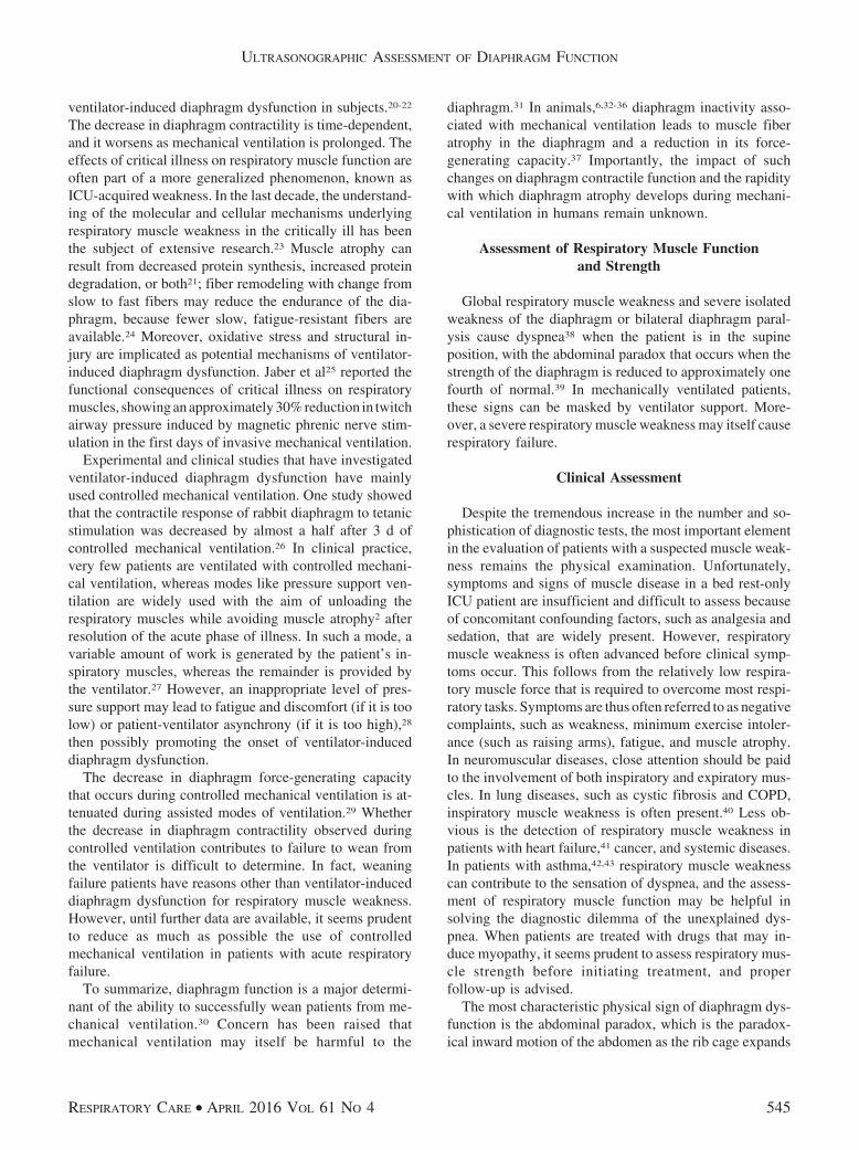
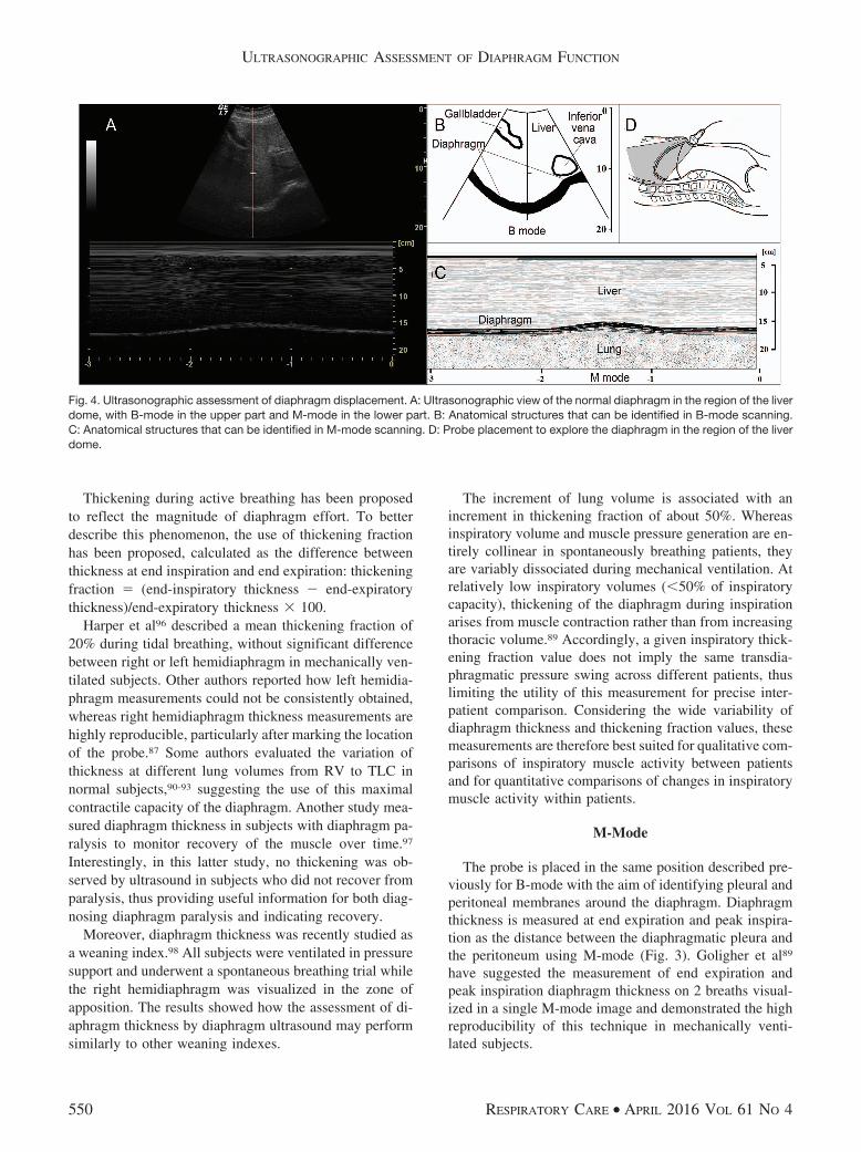
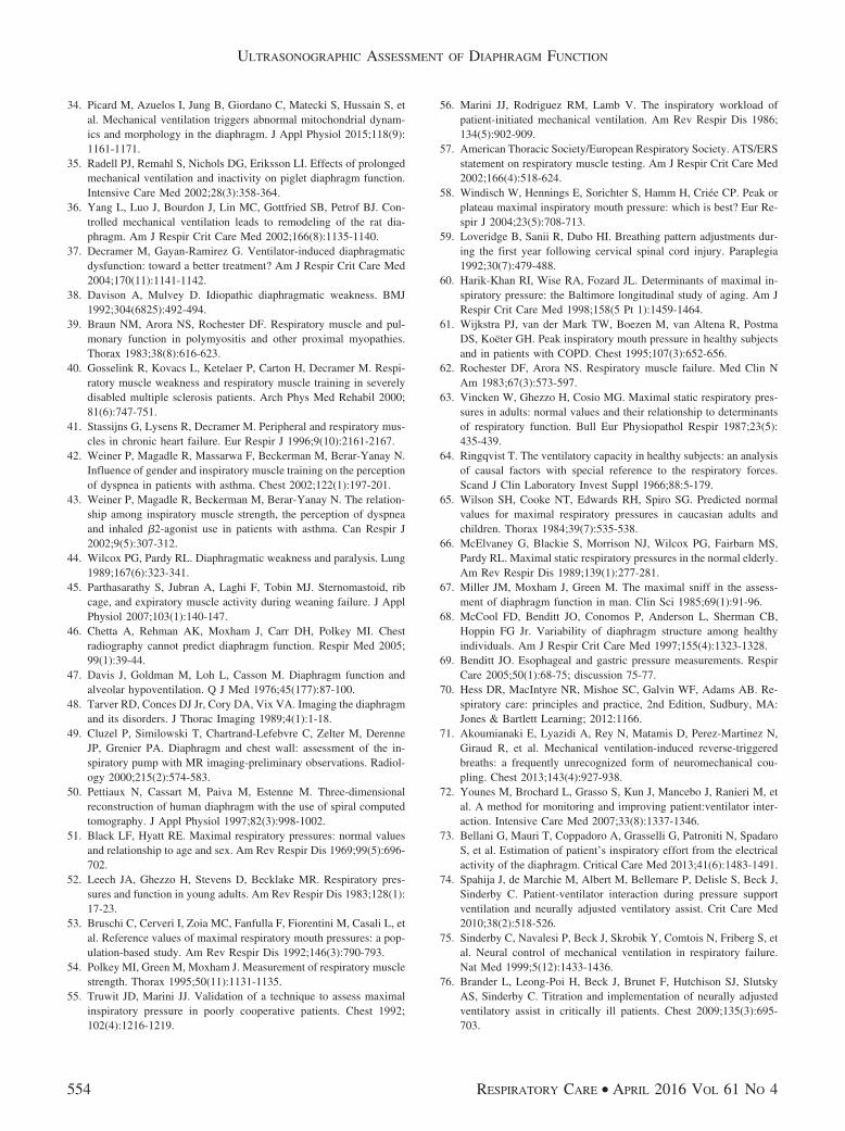
Fig. 4. Ultrasonographic assessment of diaphragm displacement. A: Ultrasonographic view of the normal diaphragm in the region of the liverdome, with B-mode in the upper part and M-mode in the lower part. B: Anatomical structures that can be identified in B-mode scanning.C: Anatomical structures that can be identified in M-mode scanning. D: Probe placement to explore the diaphragm in the region of the liverdome.
ULTRASONOGRAPHIC ASSESSMENT OF DIAPHRAGM FUNCTION
550 RESPIRATORY CARE • APRIL 2016 VOL 61 NO 4
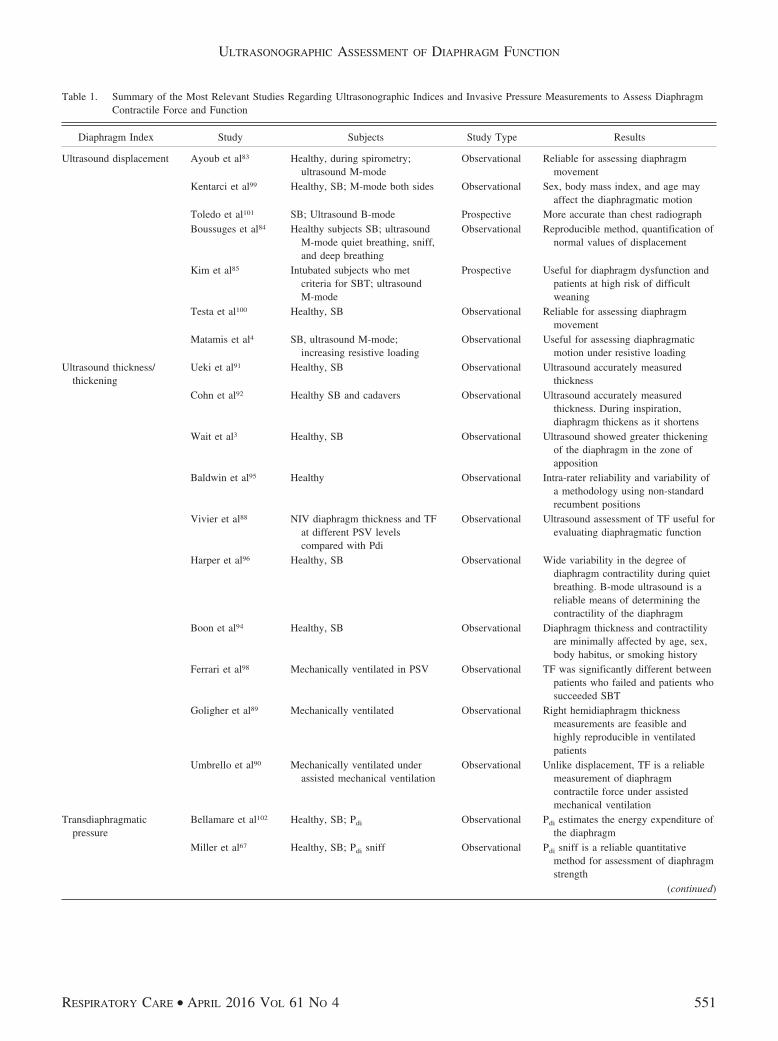
Table 1. Summary of the Most Relevant Studies Regarding Ultrasonographic Indices and Invasive Pressure Measurements to Assess DiaphragmContractile Force and Function
Diaphragm Index Study Subjects Study Type Results
Ultrasound displacement Ayoub et al83 Healthy, during spirometry;ultrasound M-mode
Observational Reliable for assessing diaphragmmovement
Kentarci et al99 Healthy, SB; M-mode both sides Observational Sex, body mass index, and age mayaffect the diaphragmatic motion
Toledo et al101 SB; Ultrasound B-mode Prospective More accurate than chest radiographBoussuges et al84 Healthy subjects SB; ultrasound
M-mode quiet breathing, sniff,and deep breathing
Observational Reproducible method, quantification ofnormal values of displacement
Kim et al85 Intubated subjects who metcriteria for SBT; ultrasoundM-mode
Prospective Useful for diaphragm dysfunction andpatients at high risk of difficultweaning
Testa et al100 Healthy, SB Observational Reliable for assessing diaphragmmovement
Matamis et al4 SB, ultrasound M-mode;increasing resistive loading
Observational Useful for assessing diaphragmaticmotion under resistive loading
Ultrasound thickness/thickening
Ueki et al91 Healthy, SB Observational Ultrasound accurately measuredthickness
Cohn et al92 Healthy SB and cadavers Observational Ultrasound accurately measuredthickness. During inspiration,diaphragm thickens as it shortens
Wait et al3 Healthy, SB Observational Ultrasound showed greater thickeningof the diaphragm in the zone ofapposition
Baldwin et al95 Healthy Observational Intra-rater reliability and variability ofa methodology using non-standardrecumbent positions
Vivier et al88 NIV diaphragm thickness and TFat different PSV levelscompared with Pdi
Observational Ultrasound assessment of TF useful forevaluating diaphragmatic function
Harper et al96 Healthy, SB Observational Wide variability in the degree ofdiaphragm contractility during quietbreathing. B-mode ultrasound is areliable means of determining thecontractility of the diaphragm
Boon et al94 Healthy, SB Observational Diaphragm thickness and contractilityare minimally affected by age, sex,body habitus, or smoking history
Ferrari et al98 Mechanically ventilated in PSV Observational TF was significantly different betweenpatients who failed and patients whosucceeded SBT
Goligher et al89 Mechanically ventilated Observational Right hemidiaphragm thicknessmeasurements are feasible andhighly reproducible in ventilatedpatients
Umbrello et al90 Mechanically ventilated underassisted mechanical ventilation
Observational Unlike displacement, TF is a reliablemeasurement of diaphragmcontractile force under assistedmechanical ventilation
Transdiaphragmaticpressure
Bellamare et al102 Healthy, SB; Pdi Observational Pdi estimates the energy expenditure ofthe diaphragm
Miller et al67 Healthy, SB; Pdi sniff Observational Pdi sniff is a reliable quantitativemethod for assessment of diaphragmstrength
(continued)
ULTRASONOGRAPHIC ASSESSMENT OF DIAPHRAGM FUNCTION
RESPIRATORY CARE • APRIL 2016 VOL 61 NO 4 551
Diaphragm Displacement
The cyclic caudal displacement of the diaphragm domeduring inspiration/expiration can be visualized with M-mode ultrasonography (Fig. 4). A 2-dimensional (B) modeis first used to find the best approach and to select theexploration line of each hemidiaphragm. The liver is usedas a window on the right while the spleen is used for theleft hemidiaphragm. A 3.5–5-MHz convex probe is placedbetween the midclavicular and anterior axillary lines, inthe subcostal area, and directed medially, cranially, anddorsally, so that the ultrasound beam reaches perpendicu-larly the posterior third of the hemidiaphragm.
With this technique, diaphragm displacement, contrac-tion speed, inspiratory time, and total respiratory time canbe measured. Kantarci et al99 described a mean left dia-phragm excursion of 49 � 11 mm in healthy subjects,which was significantly lower in female, young, and un-derweight subjects. In a prospective study designed to de-termine the reference values for diaphragm amplitudes re-corded by M-mode ultrasonography, Boussuges et al84
defined the normal values during tidal and deep breathingin male and female healthy subjects (1.8 � 0.3 and7 � 0.6 cm and 1.6 � 0.3 and 5.7 � 1 cm, respectively).However, all of these studies were performed in sponta-neously breathing subjects, whereas the role of excursionin the functional evaluation of diaphragm contractile ac-tivity during patient-triggered mechanical ventilation is farless clear. Work from our group90 showed how this mea-surement was uncorrelated to any other index of inspira-tory effort during patient-triggered mechanical ventilation.Indeed, diaphragm excursion during an assisted breath rep-resents the sum of 2 forces acting in the same direction:the force of the diaphragm contraction by itself and thepassive displacement of the diaphragm by the pressureprovided by the ventilator, thus dismissing the role of thisindex as a measure of diaphragm contractile activity dur-
ing assisted mechanical ventilation. Table 1 displays asummary of the most relevant studies regarding ultrasono-graphic indices and invasive pressure measurements forthe assessment of diaphragm contractile force and func-tion.
Conclusions
Diaphragm weakness may involve either the whole mus-cle or only one side. Most patients have other likely con-tributing causes for dyspnea, making a determination ofthe contribution of diaphragm dysfunction extremely dif-ficult. Moreover, diaphragm dysfunction is common inmechanically ventilated patients and is a likely cause ofweaning failure. Recent studies, on the other hand, under-line the negative effect of mechanical ventilation on dia-phragm structure and function (ie, ventilator-induced dia-phragm dysfunction). Even if there is a scarcity of evidencedirectly demonstrating improved outcomes with close mon-itoring of the respiratory muscles, recent studies suggestthat respiratory muscle monitoring can affect clinical carein the ICU.98 In general, upon physical examination, closeobservation of the breathing pattern reveals tachypnea andparadoxical abdominal wall retraction during inspiration.Clinical observation, sitting and supine spirometry, andmeasurement of maximal inspiratory and expiratory pres-sures may be useful but are often difficult to perform at thebedside in ICU patients. Different tools are available toassess respiratory muscle function. However, most of themare of limited value (ie, chest radiograph and fluoroscopy)or are not suitable for routine clinical care monitoring(magnetic resonance imaging, computed tomography,phrenic nerve stimulation). Circulatory biomarkers for re-spiratory muscle injury are indeed desirable, and they willhopefully appear in the next few years. Indeed, the ac-cepted standard for the diagnosis of diaphragm dysfunc-tion is the direct measurement of Pdi. However, this testing
Table 1. Continued
Diaphragm Index Study Subjects Study Type Results
Kimura et al103 Intubated PSV; Pdi Observational Pdi is helpful for titrating the requiredPSV level
Polkey et al54 Healthy, SB; Pdi sniff, Pes Observational Pdi sniff and Pes are reliable forassessment of diaphragmatic andglobal inspiratory effort
The table represents a summary of the most important studies regarding ultrasonographic indices to assess diaphragm contractile force and function (as represented by ultrasonographic displacementand thickness or thickening) and the use of invasive pressure measurements (as represented by transdiaphragmatic pressure).SB � spontaneously breathingSBT � spontaneous breathing trialNIV � non-invasive ventilationTF � diaphragmatic thicknessPSV � pressure support ventilationPdi � transdiaphragmatic pressurePes � esophageal pressure
ULTRASONOGRAPHIC ASSESSMENT OF DIAPHRAGM FUNCTION
552 RESPIRATORY CARE • APRIL 2016 VOL 61 NO 4
requires specific tools, time, and expertise not always avail-able in the general ICU. Nevertheless, ultrasonographicassessment of diaphragm function is feasible and has arelatively fast learning curve. Assessment of displacementis easily performed, even if its role in the functional as-sessment of diaphragm dysfunction in patients undergoingpatient-triggered mechanical ventilation has recently beenquestioned.90 Diaphragm thickness and thickening duringbreathing, despite being slightly more difficult to assess,overcome the limitations of displacement and are increas-ingly been performed in the critical care setting. In con-clusion, the increased routine use of ultrasonography in theICU as a fast, inexpensive, and noninvasive test is ex-pected to lead to a timely identification of patients withdiaphragm dysfunction, potentially leading to a better tai-loring of patient-triggered mechanical ventilation and toan improved assessment of recovery from diaphragm weak-ness.
ACKNOWLEDGMENTS
We are indebted to Paolo Spanu MD for valuable help with the artwork.
REFERENCES
1. Lone NI, Walsh TS. Prolonged mechanical ventilation in critically illpatients: epidemiology, outcomes and modelling the potential costconsequences of establishing a regional weaning unit. Crit Care 2011;15(2):R102.
2. Esteban A, Anzueto A, Frutos F, Alıa I, Brochard L, Stewart TE, etal. Characteristics and outcomes in adult patients receiving mechan-ical ventilation: a 28-day international study. JAMA 2002;287(3):345-355.
3. Wait JL, Johnson RL. Patterns of shortening and thickening of thehuman diaphragm. J Appl Physiol 1997;83(4):1123-1132.
4. Matamis D, Soilemezi E, Tsagourias M, Akoumianaki E, Dimassi S,Boroli F, et al. Sonographic evaluation of the diaphragm in criticallyill patients: technique and clinical applications. Intensive Care Med2013;39(5):801-810.
5. Powers SK, Shanely RA, Coombes JS, Koesterer TJ, McKenzie M,Van Gammeren D, et al. Mechanical ventilation results in progres-sive contractile dysfunction in the diaphragm. J Appl Physiol 2002;92(5):1851-1858.
6. Sassoon CS, Caiozzo VJ, Manka A, Sieck GC. Altered diaphragmcontractile properties with controlled mechanical ventilation. J ApplPhysiol 2002;92(6):2585-2595.
7. Shanely RA, Coombes JS, Zergeroglu AM, Webb AI, Powers SK.Short-duration mechanical ventilation enhances diaphragmatic fa-tigue resistance but impairs force production. Chest 2003;123(1):195-201.
8. McCool FD, Tzelepis GE. Dysfunction of the diaphragm. N EnglJ Med 2012;366(10):932-942.
9. Poole DC, Sexton WL, Farkas GA, Powers SK, Reid MB. Dia-phragm structure and function in health and disease. Med Sci SportsExerc 1997;29(6):738-754.
10. Polla B, D’Antona G, Bottinelli R, Reggiani C. Respiratory musclefibres: specialisation and plasticity. Thorax 2004;59(9):808-817.
11. Briscoe C. The interchondral joints of the human thorax. J Anat1925;59(Pt 4):432-437.
12. Gibson GJ. Diaphragmatic paresis: pathophysiology, clinical fea-tures, and investigation. Thorax 1989;44(11):960-970.
13. Goldman MD, Mead J. Mechanical interaction between the dia-phragm and rib cage. J Appl Physiol 1973;35(2):197-204.
14. Piehler JM, Pairolero PC, Gracey DR, Bernatz PE. Unexplaineddiaphragmatic paralysis: a harbinger of malignant disease? J ThoracCardiovasc Surg 1982;84(6):861-864.
15. Derenne JP, Macklem PT, Roussos C. The respiratory muscles: me-chanics, control, and pathophysiology. Am Rev Respir Dis 1978;118(1):119-133.
16. Macklem PT, Gross D, Grassino GA, Roussos C. Partitioning ofinspiratory pressure swings between diaphragm and intercostal/ac-cessory muscles. J Appl Physiol 1978;44(2):200-208.
17. Danon J, Druz WS, Goldberg NB, Sharp JT. Function of the isolatedpaced diaphragm and the cervical accessory muscles in C1 quadri-plegics. Am Rev Respir Dis 1979;119(6):909-919.
18. Younes M, Puddy A, Roberts D, Light RB, Quesada A, Taylor K, etal. Proportional assist ventilation: results of an initial clinical trial.Am Rev Respir Dis 1992;145(1):121-129.
19. Laghi F, Tobin MJ. Disorders of the respiratory muscles. Am JRespir Crit Care Med 2003;168(1):10-48.
20. Cattapan SE, Laghi F, Tobin MJ. Can diaphragmatic contractility beassessed by airway twitch pressure in mechanically ventilated pa-tients? Thorax 2003;58(1):58-62.
21. Watson AC, Hughes PD, Louise Harris M, Hart N, Ware RJ, Wen-don J, et al. Measurement of twitch transdiaphragmatic, esophageal,and endotracheal tube pressure with bilateral anterolateral magneticphrenic nerve stimulation in patients in the intensive care unit. CritCare Med 2001;29(7):1325-1331.
22. Laghi F. Assessment of respiratory output in mechanically ventilatedpatients. Respir Care Clin N Am 2005;11(2):173-199.
23. Levine S, Biswas C, Dierov J, Barsotti R, Shrager JB, Nguyen T, etal. Increased proteolysis, myosin depletion, and atrophic AKT-FOXOsignaling in human diaphragm disuse. Am J Respir Crit Care Med2011;183(4):483-490.
24. Hussain SN, Vassilakopoulos T. Ventilator-induced cachexia. Am JRespir Crit Care Med 2002;166(10):1307-1308.
25. Jaber S, Petrof BJ, Jung B, Chanques G, Berthet JP, Rabuel C, et al.Rapidly progressive diaphragmatic weakness and injury during me-chanical ventilation in humans. Am J Respir Crit Care Med 2011;183(3):364-371.
26. Sassoon CS, Zhu E, Caiozzo VJ. Assist-control mechanical ventila-tion attenuates ventilator-induced diaphragmatic dysfunction. Am JRespir Crit Care Med 2004;170(6):626-632.
27. Hess DR. Ventilator waveforms and the physiology of pressure sup-port ventilation. Respir Care 2005;50(2):166-186; discussion 183-166.
28. Thille AW, Cabello B, Galia F, Lyazidi A, Brochard L. Reduction ofpatient-ventilator asynchrony by reducing tidal volume during pres-sure-support ventilation. Intensive Care Med 2008;34(8):1477-1486.
29. Grinnan DC, Truwit JD. Clinical review: respiratory mechanics inspontaneous and assisted ventilation. Crit Care 2005;9(5):472-484.
30. Vassilakopoulos T. Ventilator-induced diaphragm dysfunction: theclinical relevance of animal models. Intensive Care Med 2008;34(1):7-16.
31. Gayan-Ramirez G, de Paepe K, Cadot P, Decramer M. Detrimentaleffects of short-term mechanical ventilation on diaphragm functionand IGF-I mRNA in rats. Intensive Care Med 2003;29(5):825-833.
32. Jaber S, Sebbane M, Koechlin C, Hayot M, Capdevila X, Eledjam JJ,et al. Effects of short vs. prolonged mechanical ventilation on anti-oxidant systems in piglet diaphragm. Intensive Care Med 2005;31(10):1427-1433.
33. Le Bourdelles G, Viires N, Boczkowski J, Seta N, Pavlovic D,Aubier M. Effects of mechanical ventilation on diaphragmatic con-tractile properties in rats. Am J Respir Crit Care Med 1994;149(6):1539-1544.
ULTRASONOGRAPHIC ASSESSMENT OF DIAPHRAGM FUNCTION
RESPIRATORY CARE • APRIL 2016 VOL 61 NO 4 553
34. Picard M, Azuelos I, Jung B, Giordano C, Matecki S, Hussain S, etal. Mechanical ventilation triggers abnormal mitochondrial dynam-ics and morphology in the diaphragm. J Appl Physiol 2015;118(9):1161-1171.
35. Radell PJ, Remahl S, Nichols DG, Eriksson LI. Effects of prolongedmechanical ventilation and inactivity on piglet diaphragm function.Intensive Care Med 2002;28(3):358-364.
36. Yang L, Luo J, Bourdon J, Lin MC, Gottfried SB, Petrof BJ. Con-trolled mechanical ventilation leads to remodeling of the rat dia-phragm. Am J Respir Crit Care Med 2002;166(8):1135-1140.
37. Decramer M, Gayan-Ramirez G. Ventilator-induced diaphragmaticdysfunction: toward a better treatment? Am J Respir Crit Care Med2004;170(11):1141-1142.
38. Davison A, Mulvey D. Idiopathic diaphragmatic weakness. BMJ1992;304(6825):492-494.
39. Braun NM, Arora NS, Rochester DF. Respiratory muscle and pul-monary function in polymyositis and other proximal myopathies.Thorax 1983;38(8):616-623.
40. Gosselink R, Kovacs L, Ketelaer P, Carton H, Decramer M. Respi-ratory muscle weakness and respiratory muscle training in severelydisabled multiple sclerosis patients. Arch Phys Med Rehabil 2000;81(6):747-751.
41. Stassijns G, Lysens R, Decramer M. Peripheral and respiratory mus-cles in chronic heart failure. Eur Respir J 1996;9(10):2161-2167.
42. Weiner P, Magadle R, Massarwa F, Beckerman M, Berar-Yanay N.Influence of gender and inspiratory muscle training on the perceptionof dyspnea in patients with asthma. Chest 2002;122(1):197-201.
43. Weiner P, Magadle R, Beckerman M, Berar-Yanay N. The relation-ship among inspiratory muscle strength, the perception of dyspneaand inhaled �2-agonist use in patients with asthma. Can Respir J2002;9(5):307-312.
44. Wilcox PG, Pardy RL. Diaphragmatic weakness and paralysis. Lung1989;167(6):323-341.
45. Parthasarathy S, Jubran A, Laghi F, Tobin MJ. Sternomastoid, ribcage, and expiratory muscle activity during weaning failure. J ApplPhysiol 2007;103(1):140-147.
46. Chetta A, Rehman AK, Moxham J, Carr DH, Polkey MI. Chestradiography cannot predict diaphragm function. Respir Med 2005;99(1):39-44.
47. Davis J, Goldman M, Loh L, Casson M. Diaphragm function andalveolar hypoventilation. Q J Med 1976;45(177):87-100.
48. Tarver RD, Conces DJ Jr, Cory DA, Vix VA. Imaging the diaphragmand its disorders. J Thorac Imaging 1989;4(1):1-18.
49. Cluzel P, Similowski T, Chartrand-Lefebvre C, Zelter M, DerenneJP, Grenier PA. Diaphragm and chest wall: assessment of the in-spiratory pump with MR imaging-preliminary observations. Radiol-ogy 2000;215(2):574-583.
50. Pettiaux N, Cassart M, Paiva M, Estenne M. Three-dimensionalreconstruction of human diaphragm with the use of spiral computedtomography. J Appl Physiol 1997;82(3):998-1002.
51. Black LF, Hyatt RE. Maximal respiratory pressures: normal valuesand relationship to age and sex. Am Rev Respir Dis 1969;99(5):696-702.
52. Leech JA, Ghezzo H, Stevens D, Becklake MR. Respiratory pres-sures and function in young adults. Am Rev Respir Dis 1983;128(1):17-23.
53. Bruschi C, Cerveri I, Zoia MC, Fanfulla F, Fiorentini M, Casali L, etal. Reference values of maximal respiratory mouth pressures: a pop-ulation-based study. Am Rev Respir Dis 1992;146(3):790-793.
54. Polkey MI, Green M, Moxham J. Measurement of respiratory musclestrength. Thorax 1995;50(11):1131-1135.
55. Truwit JD, Marini JJ. Validation of a technique to assess maximalinspiratory pressure in poorly cooperative patients. Chest 1992;102(4):1216-1219.
56. Marini JJ, Rodriguez RM, Lamb V. The inspiratory workload ofpatient-initiated mechanical ventilation. Am Rev Respir Dis 1986;134(5):902-909.
57. American Thoracic Society/European Respiratory Society. ATS/ERSstatement on respiratory muscle testing. Am J Respir Crit Care Med2002;166(4):518-624.
58. Windisch W, Hennings E, Sorichter S, Hamm H, Criee CP. Peak orplateau maximal inspiratory mouth pressure: which is best? Eur Re-spir J 2004;23(5):708-713.
59. Loveridge B, Sanii R, Dubo HI. Breathing pattern adjustments dur-ing the first year following cervical spinal cord injury. Paraplegia1992;30(7):479-488.
60. Harik-Khan RI, Wise RA, Fozard JL. Determinants of maximal in-spiratory pressure: the Baltimore longitudinal study of aging. Am JRespir Crit Care Med 1998;158(5 Pt 1):1459-1464.
61. Wijkstra PJ, van der Mark TW, Boezen M, van Altena R, PostmaDS, Koeter GH. Peak inspiratory mouth pressure in healthy subjectsand in patients with COPD. Chest 1995;107(3):652-656.
62. Rochester DF, Arora NS. Respiratory muscle failure. Med Clin NAm 1983;67(3):573-597.
63. Vincken W, Ghezzo H, Cosio MG. Maximal static respiratory pres-sures in adults: normal values and their relationship to determinantsof respiratory function. Bull Eur Physiopathol Respir 1987;23(5):435-439.
64. Ringqvist T. The ventilatory capacity in healthy subjects: an analysisof causal factors with special reference to the respiratory forces.Scand J Clin Laboratory Invest Suppl 1966;88:5-179.
65. Wilson SH, Cooke NT, Edwards RH, Spiro SG. Predicted normalvalues for maximal respiratory pressures in caucasian adults andchildren. Thorax 1984;39(7):535-538.
66. McElvaney G, Blackie S, Morrison NJ, Wilcox PG, Fairbarn MS,Pardy RL. Maximal static respiratory pressures in the normal elderly.Am Rev Respir Dis 1989;139(1):277-281.
67. Miller JM, Moxham J, Green M. The maximal sniff in the assess-ment of diaphragm function in man. Clin Sci 1985;69(1):91-96.
68. McCool FD, Benditt JO, Conomos P, Anderson L, Sherman CB,Hoppin FG Jr. Variability of diaphragm structure among healthyindividuals. Am J Respir Crit Care Med 1997;155(4):1323-1328.
69. Benditt JO. Esophageal and gastric pressure measurements. RespirCare 2005;50(1):68-75; discussion 75-77.
70. Hess DR, MacIntyre NR, Mishoe SC, Galvin WF, Adams AB. Re-spiratory care: principles and practice, 2nd Edition, Sudbury, MA:Jones & Bartlett Learning; 2012:1166.
71. Akoumianaki E, Lyazidi A, Rey N, Matamis D, Perez-Martinez N,Giraud R, et al. Mechanical ventilation-induced reverse-triggeredbreaths: a frequently unrecognized form of neuromechanical cou-pling. Chest 2013;143(4):927-938.
72. Younes M, Brochard L, Grasso S, Kun J, Mancebo J, Ranieri M, etal. A method for monitoring and improving patient:ventilator inter-action. Intensive Care Med 2007;33(8):1337-1346.
73. Bellani G, Mauri T, Coppadoro A, Grasselli G, Patroniti N, SpadaroS, et al. Estimation of patient’s inspiratory effort from the electricalactivity of the diaphragm. Critical Care Med 2013;41(6):1483-1491.
74. Spahija J, de Marchie M, Albert M, Bellemare P, Delisle S, Beck J,Sinderby C. Patient-ventilator interaction during pressure supportventilation and neurally adjusted ventilatory assist. Crit Care Med2010;38(2):518-526.
75. Sinderby C, Navalesi P, Beck J, Skrobik Y, Comtois N, Friberg S, etal. Neural control of mechanical ventilation in respiratory failure.Nat Med 1999;5(12):1433-1436.
76. Brander L, Leong-Poi H, Beck J, Brunet F, Hutchison SJ, SlutskyAS, Sinderby C. Titration and implementation of neurally adjustedventilatory assist in critically ill patients. Chest 2009;135(3):695-703.
ULTRASONOGRAPHIC ASSESSMENT OF DIAPHRAGM FUNCTION
554 RESPIRATORY CARE • APRIL 2016 VOL 61 NO 4
77. Schmidt M, Demoule A, Cracco C, Gharbi A, Fiamma MN, StrausC, et al. Neurally adjusted ventilatory assist increases respiratoryvariability and complexity in acute respiratory failure. Anesthesiol-ogy 2010;112(3):670-681.
78. Yonis H, Crognier L, Conil JM, Serres I, Rouget A, Virtos M, et al.Patient-ventilator synchrony in neurally adjusted ventilatory assist(NAVA) and pressure support ventilation (PSV): a prospective ob-servational study. BMC Anesthesiol 2015;15:117.
79. Colombo D, Cammarota G, Alemani M, Carenzo L, Barra FL,Vaschetto R, et al. Efficacy of ventilator waveforms observation indetecting patient-ventilator asynchrony. Crit Care Med 2011;39(11):2452-2457.
80. Beck KC. Influence of vascular distending pressure on regional flowsin isolated perfused dog lungs. J Appl Physiol 1990;69(5):1869-1874.
81. Beaulieu Y, Marik PE. Bedside ultrasonography in the ICU: part 2.Chest 2005;128(3):1766-1781.
82. Beaulieu Y, Marik PE. Bedside ultrasonography in the ICU: part 1.Chest 2005;128(2):881-895.
83. Ayoub J, Milane J, Targhetta R, Prioux J, Chamari K, Arbeille P, etal. Diaphragm kinetics during pneumatic belt respiratory assistance:a sonographic study in Duchenne muscular dystrophy. NeuromusculDisord 2002;12(6):569-575.
84. Boussuges A, Gole Y, Blanc P. Diaphragmatic motion studied byM-mode ultrasonography: methods, reproducibility, and normal val-ues. Chest 2009;135(2):391-400.
85. Kim WY, Suh HJ, Hong SB, Koh Y, Lim CM. Diaphragm dysfunc-tion assessed by ultrasonography: influence on weaning from me-chanical ventilation. Crit Care Med 2011;39(12):2627-2630.
86. Wait JL, Nahormek PA, Yost WT, Rochester DP. Diaphragmaticthickness-lung volume relationship in vivo. J Appl Physiol 1989;67(4):1560-1568.
87. DiNino E, Gartman EJ, Sethi JM, McCool FD. Diaphragm ultra-sound as a predictor of successful extubation from mechanical ven-tilation. Thorax 2014;69(5):423-427.
88. Vivier E, Mekontso Dessap A, Dimassi S, Vargas F, Lyazidi A,Thille AW, Brochard L. Diaphragm ultrasonography to estimate thework of breathing during non-invasive ventilation. Intensive CareMed 2012;38(5):796-803.
89. Goligher EC, Laghi F, Detsky ME, Farias P, Murray A, Brace D, etal. Measuring diaphragm thickness with ultrasound in mechanicallyventilated patients: feasibility, reproducibility and validity. IntensiveCare Med 2015;41(4):734.
90. Umbrello M, Formenti P, Longhi D, Galimberti A, Piva I, Pezzi A,et al. Diaphragm ultrasound as indicator of respiratory effort in crit-ically ill patients undergoing assisted mechanical ventilation: a pilotclinical study. Crit Care 2015;19:161.
91. Ueki J, De Bruin PF, Pride NB. In vivo assessment of diaphragmcontraction by ultrasound in normal subjects. Thorax 1995;50(11):1157-1161.
92. Cohn D, Benditt JO, Eveloff S, McCool FD. Diaphragm thickeningduring inspiration. J Appl Physiol 1997;83(1):291-296.
93. Baria MR, Shahgholi L, Sorenson EJ, Harper CJ, Lim KG, Strom-men JA, et al. B-mode ultrasound assessment of diaphragm struc-ture and function in patients with COPD. Chest 2014;146(3):680-685.
94. Boon AJ, Harper CJ, Ghahfarokhi LS, Strommen JA, Watson JC,Sorenson EJ. Two-dimensional ultrasound imaging of the dia-phragm: quantitative values in normal subjects. Muscle Nerve 2013;47(6):884-889.
95. Baldwin CE, Paratz JD, Bersten AD. Diaphragm and peripheralmuscle thickness on ultrasound: intra-rater reliability and variabil-ity of a methodology using non-standard recumbent positions. Re-spirology 2011;16(7):1136-1143.
96. Harper CJ, Shahgholi L, Cieslak K, Hellyer NJ, Strommen JA,Boon AJ. Variability in diaphragm motion during normal breathing,assessed with B-mode ultrasound. J Orthop Sports Phys Ther 2013;43(12):927-931.
97. Summerhill EM, El-Sameed YA, Glidden TJ, McCool FD. Moni-toring recovery from diaphragm paralysis with ultrasound. Chest2008;133(3):737-743.
98. Ferrari G, De Filippi G, Elia F, Panero F, Volpicelli G, Apra F.Diaphragm ultrasound as a new index of discontinuation from me-chanical ventilation. Crit Ultrasound J 2014;6(1):8.
99. Kantarci F, Mihmanli I, Demirel MK, Harmanci K, Akman C,Aydogan F, et al. Normal diaphragmatic motion and the effects ofbody composition: determination with M-mode sonography. J Ul-trasound Med 2004;23(2):255-260.
100. Testa A, Soldati G, Giannuzzi R, Berardi S, Portale G, GentiloniSilveri N. Ultrasound M-mode assessment of diaphragmatic kinet-ics by anterior transverse scanning in healthy subjects. UltrasoundMed Biol 2011;37(1):44-52.
101. Toledo NS, Kodaira SK, Massarollo PC, Pereira OI, Dalmas JC,Cerri GG, et al. Left hemidiaphragmatic mobility: assessment withultrasonographic measurement of the craniocaudal displacement ofthe splenic hilum and the inferior pole of the spleen. J UltrasoundMed 2006;25(1):41-49.
102. Bellemare F, Grassino A. Effect of pressure and timing of contrac-tion on human diaphragm fatigue. J Appl Physiol Respir EnvironExerc Physiol 1982;53(5):1190-1195.
103. Kimura T, Takezawa J, Nishiwaki K, Shimada Y. Determination ofthe optimal pressure support level evaluated by measuring trans-diaphragmatic pressure. Chest 1991;100(1):112-117.
ULTRASONOGRAPHIC ASSESSMENT OF DIAPHRAGM FUNCTION
RESPIRATORY CARE • APRIL 2016 VOL 61 NO 4 555