ultrasonographic diagnosis of thoracic outlet syndrome ... · diagnostics article ultrasonographic...
TRANSCRIPT
diagnostics
Article
Ultrasonographic Diagnosis of Thoracic OutletSyndrome Secondary to Brachial PlexusPiercing Variation
Vanessa Leonhard 1, Gregory Caldwell 1, Mei Goh 2, Sean Reeder 1 and Heather F. Smith 3,4,*1 Department of Osteopathic Manipulative Medicine, Arizona College of Osteopathic Medicine,
Midwestern University, Glendale, AZ 85308, USA; [email protected] (V.L.);[email protected] (G.C.); [email protected] (S.R.)
2 Arizona College of Osteopathic Medicine, Midwestern University, Glendale, AZ 85308, USA;[email protected]
3 Department of Anatomy, Midwestern University, Glendale, AZ 85308, USA4 School of Human Evolution and Social Change, Arizona State University, Tempe, AZ 85287, USA* Correspondence: [email protected]; Tel.: +1-623-572-3726; Fax: +1-623-572-3679
Received: 28 April 2017; Accepted: 29 June 2017; Published: 4 July 2017
Abstract: Structural variations of the thoracic outlet create a unique risk for neurogenic thoracicoutlet syndrome (nTOS) that is difficult to diagnose clinically. Common anatomical variations inbrachial plexus (BP) branching were recently discovered in which portions of the proximal plexuspierce the anterior scalene. This results in possible impingement of BP nerves within the musclebelly and, therefore, predisposition for nTOS. We hypothesized that some cases of disputed nTOSresult from these BP branching variants. We tested the association between BP piercing and nTOSsymptoms, and evaluated the capability of ultrasonographic identification of patients with clinicallyrelevant variations. Eighty-two cadaveric necks were first dissected to assess BP variation frequency.In 62.1%, C5, superior trunk, or superior + middle trunks pierced the anterior scalene. Subsequently,22 student subjects underwent screening with detailed questionnaires, provocative tests, and BPultrasonography. Twenty-one percent demonstrated atypical BP branching anatomy on ultrasound;of these, 50% reported symptoms consistent with nTOS, significantly higher than subjects with classicBP anatomy (14%). This group, categorized as a typical TOS, would be missed by provocative testingalone. The addition of ultrasonography to nTOS diagnosis, especially for patients with BP branchingvariation, would allow clinicians to visualize and identify atypical patient anatomy.
Keywords: anatomical variation; brachial plexus; superior trunk; middle trunk; anterior scalenemuscle; neurogenic thoracic outlet syndrome; ultrasound; provocative testing
1. Introduction
Neurogenic thoracic outlet syndrome (nTOS) is a neurologic impingement syndrome that isnotoriously difficult to diagnose in the clinical setting [1,2]. There are vascular and neurogenic formsof thoracic outlet syndrome (TOS), with nTOS being the most common and comprising over 90%of cases [3]. The arterial type, affecting the subclavian artery, is more concretely diagnosable bytraditional provocative tests [1], as these directly evaluate the radial pulse. Adson’s [4], Wright’s,and Costoclavicular tests utilize the classic relationship of the subclavian artery and the branchesof the brachial plexus to identify specific sites of neurovascular impingement (Table 1). These testsdiagnose compression at three distinct sites: within the interscalene space, deep to the pectoralis minortendon, and between the first rib and clavicle. Adson’s test evaluates the passage of the brachial plexustrunks and subclavian artery as they pass through the interscalene space between the anterior and
Diagnostics 2017, 7, 40; doi:10.3390/diagnostics7030040 www.mdpi.com/journal/diagnostics
Diagnostics 2017, 7, 40 2 of 13
middle scalene muscles and relies on change in radial pulse due to compression of the subclavianartery between those muscles [4].
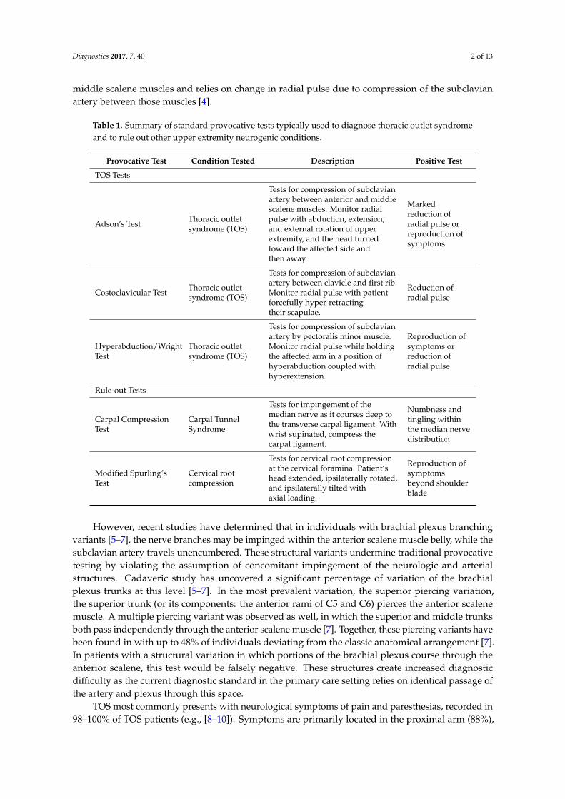
Table 1. Summary of standard provocative tests typically used to diagnose thoracic outlet syndromeand to rule out other upper extremity neurogenic conditions.
Provocative Test Condition Tested Description Positive Test
TOS Tests
Adson’s Test Thoracic outletsyndrome (TOS)
Tests for compression of subclavianartery between anterior and middlescalene muscles. Monitor radialpulse with abduction, extension,and external rotation of upperextremity, and the head turnedtoward the affected side andthen away.
Markedreduction ofradial pulse orreproduction ofsymptoms
Costoclavicular Test Thoracic outletsyndrome (TOS)
Tests for compression of subclavianartery between clavicle and first rib.Monitor radial pulse with patientforcefully hyper-retractingtheir scapulae.
Reduction ofradial pulse
Hyperabduction/WrightTest
Thoracic outletsyndrome (TOS)
Tests for compression of subclavianartery by pectoralis minor muscle.Monitor radial pulse while holdingthe affected arm in a position ofhyperabduction coupled withhyperextension.
Reproduction ofsymptoms orreduction ofradial pulse
Rule-out Tests
Carpal CompressionTest
Carpal TunnelSyndrome
Tests for impingement of themedian nerve as it courses deep tothe transverse carpal ligament. Withwrist supinated, compress thecarpal ligament.
Numbness andtingling withinthe median nervedistribution
Modified Spurling’sTest
Cervical rootcompression
Tests for cervical root compressionat the cervical foramina. Patient’shead extended, ipsilaterally rotated,and ipsilaterally tilted withaxial loading.
Reproduction ofsymptomsbeyond shoulderblade
However, recent studies have determined that in individuals with brachial plexus branchingvariants [5–7], the nerve branches may be impinged within the anterior scalene muscle belly, while thesubclavian artery travels unencumbered. These structural variants undermine traditional provocativetesting by violating the assumption of concomitant impingement of the neurologic and arterialstructures. Cadaveric study has uncovered a significant percentage of variation of the brachialplexus trunks at this level [5–7]. In the most prevalent variation, the superior piercing variation,the superior trunk (or its components: the anterior rami of C5 and C6) pierces the anterior scalenemuscle. A multiple piercing variant was observed as well, in which the superior and middle trunksboth pass independently through the anterior scalene muscle [7]. Together, these piercing variants havebeen found in with up to 48% of individuals deviating from the classic anatomical arrangement [7].In patients with a structural variation in which portions of the brachial plexus course through theanterior scalene, this test would be falsely negative. These structures create increased diagnosticdifficulty as the current diagnostic standard in the primary care setting relies on identical passage ofthe artery and plexus through this space.
TOS most commonly presents with neurological symptoms of pain and paresthesias, recorded in98–100% of TOS patients (e.g., [8–10]). Symptoms are primarily located in the proximal arm (88%),
Diagnostics 2017, 7, 40 3 of 13
shoulder (88%), and all five digits (58%) [3]. These nonspecific findings are associated with numerousforms of pathology in the upper extremity and the cervical region [11–14]. Similarly, the currentdefinitions of TOS vary among clinicians. One study determined that surgeons are 100 times morelikely to diagnose TOS than neurologists [15]. In general, current diagnostic criteria typically requirethat the provocative tests cause vascular change at the radial artery, regardless of symptoms. Disputed,or non-specific TOS is quite common, occurring when patients present with TOS-like symptoms, but donot meet the currently accepted diagnostic standards and, therefore, lack a definitive explanation fortheir symptoms (e.g., [16]). Individuals with variations from classic anatomical relationships, such asthe superior piercing variation, are likely to present in this manner and remain without clear diagnosisor treatment strategy. To achieve more comprehensive diagnosis and plan of care, ultrasonographymay offer a means to visualize the anatomy of the thoracic outlet, identify clinically relevant variations,and provide a distinct diagnosis for these patients.
Previous studies into the efficacy of provocative testing indicated that up to 60% of asymptomaticpatients experienced vascular compromise during testing, a diagnostic false positive for TOS [17–19].Considering the high prevalence of variation within the brachial plexus trunks, and associatedlack of vascular change, the Adson’s test also has a high propensity for false negatives, up to 10%.One explanation for these results may be that a subset of patients presenting with nTOS symptoms,may be variant in the relationships of the thoracic outlet structures. Ultrasound imaging may beable to visualize these brachial plexus variants, therefore providing a diagnosis for those who wouldotherwise be missed by provocative testing.
Recently, new sets of criteria for diagnosing TOS have been proposed [20–22]. The Consortium forOutcomes Research and Education on Thoracic Outlet Syndrome proposed a preliminary set of detaileddiagnostic TOS criteria [20,22]. This comprehensive list is an invaluable resource. However, while thestudy acknowledges that scalene muscular variation may exist, the implication is that such variation israre and “too small to be detected by standard imaging tests, such as plain X-rays, CT or MRI scanning”and can, therefore, only be assessed at the time of surgery [22]. A second set of updated TOS reportingstandards were recently published by the Society for Vascular Surgery [21] which include: symptomsof pathology at the thoracic outlet, symptoms of nerve compression, the absence of other pathologypotentially explaining the symptoms, and a positive scalene muscle injection test. While useful,these standards do not account for common structural variation at the thoracic outlet. The criteriapresume that “the brachial plexus and subclavian artery traverse the same spaces” [21] (p. e25).Therefore, patients with brachial plexus branching variants would lack the first diagnostic criterionbecause they have no pathology present at the thoracic outlet, only a common anatomical variation.Another potential limitation of this set of standards is that it requires the use of scalene muscle injections,which may not be accessible to a primary care physician in the clinic. Recently, electrodiagnosticmethods have been developed which can result in more objective neurological findings regarding TOS(e.g., [23]). However, for the average primary care physician, this technology may not be available inthe clinic and, thus, the use of these techniques is primarily relegated to specialists.
Given the recent discovery that piercing variants in the brachial plexus are quite common [5–7],and may predispose these individuals to nTOS, this study seeks to empirically evaluate the proposedassociation between brachial plexus piercing variants and nTOS symptoms. We also aim to determinethe applicability of ultrasonography (US) for increasing the efficacy of clinical diagnosis over traditionalprovocative testing alone, especially for cases of nTOS secondary to BP variation.
2. Materials and Methods
2.1. Cadaveric Data
The cadaveric investigation assessed proximal brachial plexus branching variation in 95 cadavericbrachial plexus specimens (44 male, 51 female) from the gross anatomy teaching laboratories atMidwestern University. Cadavers were obtained for teaching purposes from the National Body
Diagnostics 2017, 7, 40 4 of 13
Donation Program (St. Louis, MO, USA). The neck and shoulder of each cadaver were dissectedbilaterally following Grant’s Dissector 16th ed. [24] to thoroughly reveal the brachial plexus. The inferiorand lateral borders of the anterior scalene muscle were defined, and the position of the roots, trunks,and cords of the brachial plexus in relation to the scalene muscles was determined and documented.For each cadaveric specimen, the type(s) of brachial plexus branching variation and sidedness of eachvariant was recorded. Each specimen was evaluated by two members of the research team to confirmthe assessment, and photo-documented for future confirmation. t-tests were then performed in SPSS19 (IBM Corp., Chicago, IL, USA) to assess whether significant differences existed in the frequency ofbrachial plexus variants between the sexes.
2.2. Ultrasonography
Twenty-two volunteer student subjects were recruited from Midwestern University in Glendale,AZ, USA. Screening began with a comprehensive questionnaire covering pertinent past medicalhistory, trauma history, and symptoms of neurovascular pathology in the upper extremity. Subjectswere then tested using standard nTOS provocative testing, including Adson’s, Costoclavicular,and Hyperabduction/Wright tests (Table 1). Additional tests to rule out other upper extremityneurogenic conditions were also utilized, including Carpal Compression and Modified Spurling’s tests(Table 1). Any changes in radial arterial pulse or reproduction of symptoms were noted. The protocolfor this study was approved by Midwestern University’s Institutional Review Board (IRB AZ#885,9 March 2016).
Following completion of provocative testing, participants underwent ultrasound (US) studyof the lateral neck using a Sonoscape S8 portable ultrasound unit. Starting with the US probe inthe supraclavicular fossa, imaging was completed up to the angle of the mandible in both neutraland Adson’s test position bilaterally. A visual scan was conducted to identify the three hypoechoictrunks with a hyperechoic fascial separation from the anterior and middle scalene muscles. A lackof visible hyperechoic fascia between the anterior scalene and any of the trunks indicated a brachialplexus piercing variant. The branching pattern of the proximal brachial plexus, and the relationshipof the trunks to the scalene muscles were documented bilaterally. Still images and video capturewere used to record the anatomy for future verification. Researchers conducting US were blind to theresults of the questionnaire and provocative testing. Ultrasound results were confirmed with a boardcertified radiologist.
To determine whether statistically significant correlations exist between reported TOS symptoms,brachial plexus branching variants (as identified by ultrasound) and provocative test results, a seriesof statistical analyses were conducted in SPSS 19 (IBM Corp.). Brachial plexus branching (ultrasound)results were coded as: piercing versus classic anatomy. Provocative test results were coded as separatevariables for: any positive pulse or symptom reproduction during test, pulse response, and symptomsreproduced. Due to the bilaterally asymmetrical nature of brachial plexus branching, the left and rightsides for each subject were considered separately. Bivariate correlation analyses were then performedbetween TOS symptoms and: brachial plexus variation, and each of the provocative test results. Partialcorrelation analyses were subsequently conducted between TOS symptoms and provocative test resultswhile controlling for brachial plexus variation.
3. Results
3.1. Cadaveric Results
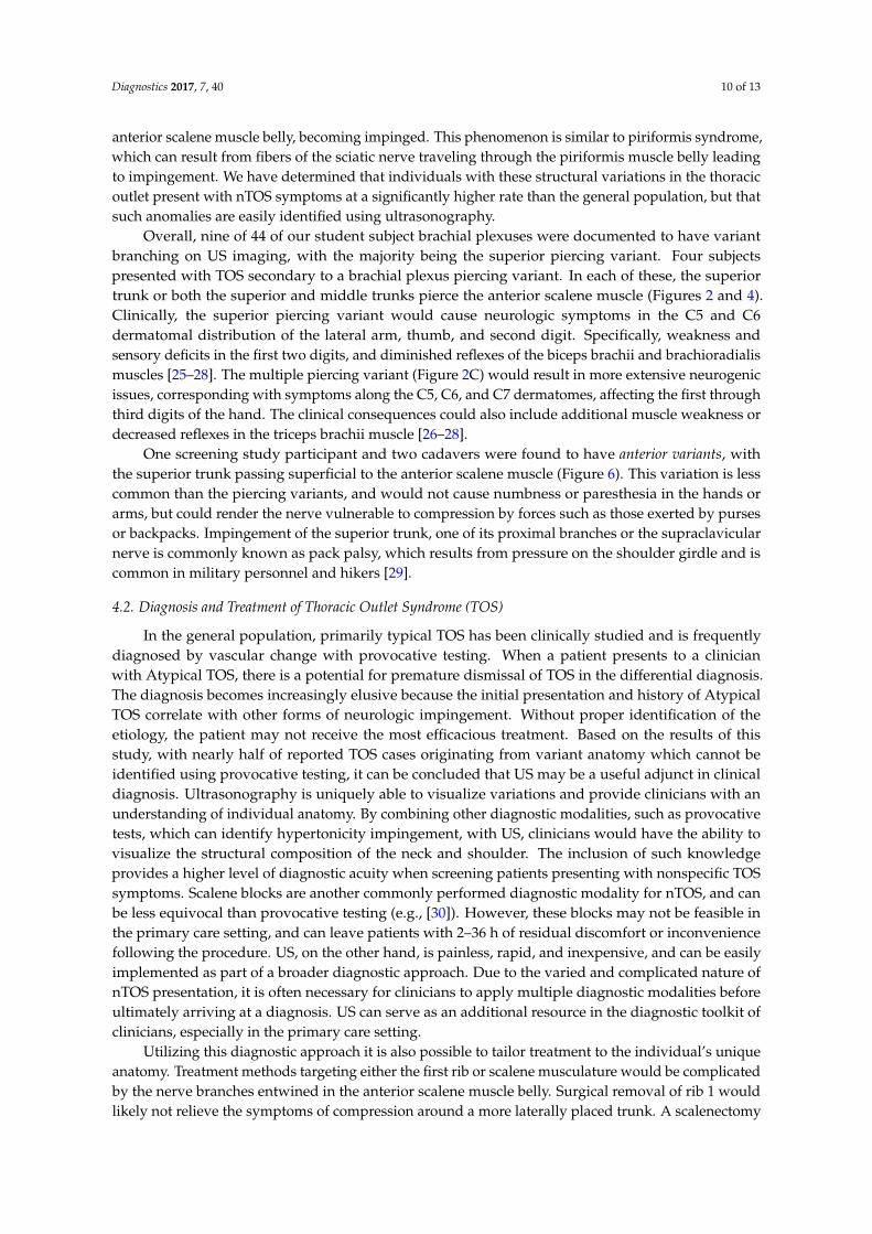
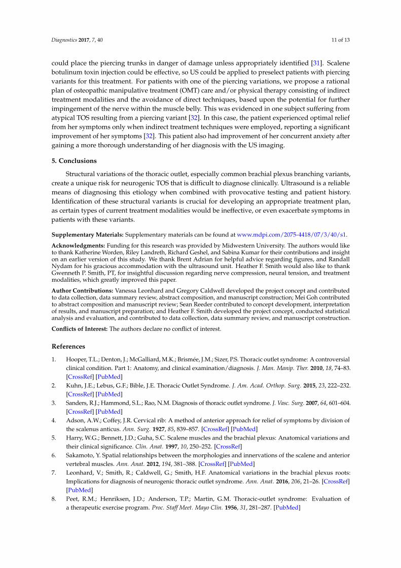
In the cadaveric sample (n = 95 plexi), brachial plexus branching variants were extremely common(Tables 2 and S1). Only 32 brachial plexi (33.7%) were found to possess the “classic” anatomical patternin which all three trunks of the brachial plexus course through the interscalene triangle (Figure 1).In the sample, 63 variations from the classic anatomical pattern were observed (Table 2, Figure 2B,C),such that 66.3% of the sample did not display the classic relationship between the scalene musculature
Diagnostics 2017, 7, 40 5 of 13
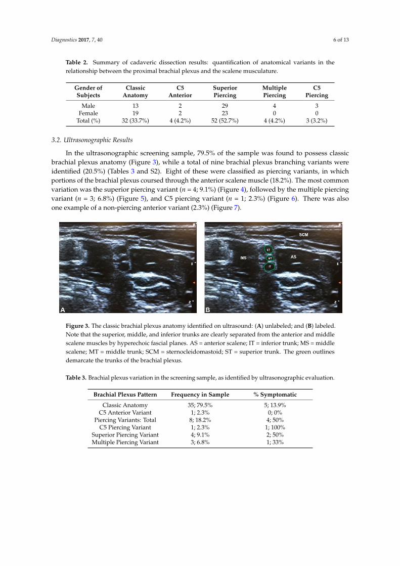
and proximal brachial plexus. These variations can be classified into four categories: superior piercing(54.7%), multiple piercing (4.2%), C5 piercing (3.2%), and C5 anterior variant (3.4%). In each variant,one or more components of the brachial plexus course(s) in a position of relative vulnerability whereit is more likely to become impinged. The most common clinically-relevant variants are depicted inFigure 2. The variant anatomy occurred more frequently in male cadavers than in females (74.5%vs. 56.8%); however, the t-test indicated that these differences between the sexes did not reach thestatistical threshold for significance (t = −1.83, p = 0.07).
Diagnostics 2017, 7, 40 5 of 12
categories: superior piercing (54.7%), multiple piercing (4.2%), C5 piercing (3.2%), and C5 anterior variant (3.4%). In each variant, one or more components of the brachial plexus course(s) in a position of relative vulnerability where it is more likely to become impinged. The most common clinically-relevant variants are depicted in Figure 2. The variant anatomy occurred more frequently in male cadavers than in females (74.5% vs. 56.8%); however, the t-test indicated that these differences between the sexes did not reach the statistical threshold for significance (t = −1.83, p = 0.07).
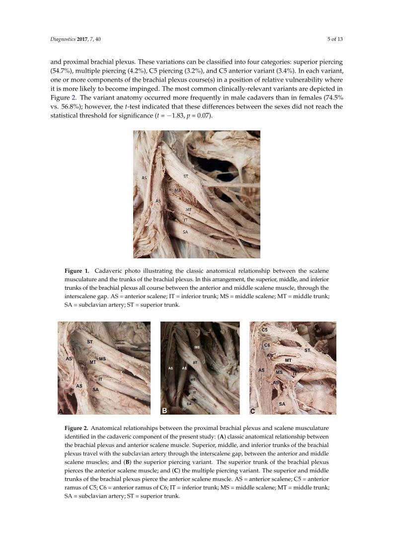
Figure 1. Cadaveric photo illustrating the classic anatomical relationship between the scalene musculature and the trunks of the brachial plexus. In this arrangement, the superior, middle, and inferior trunks of the brachial plexus all course between the anterior and middle scalene muscle, through the interscalene gap. AS = anterior scalene; IT = inferior trunk; MS = middle scalene; MT = middle trunk; SA = subclavian artery; ST = superior trunk.
Table 2. Summary of cadaveric dissection results: quantification of anatomical variants in the relationship between the proximal brachial plexus and the scalene musculature.
Gender of Subjects
Classic Anatomy
C5 Anterior
Superior Piercing
Multiple Piercing
C5 Piercing
Male 13 2 29 4 3 Female 19 2 23 0 0
Total (%) 32 (33.7%) 4 (4.2%) 52 (52.7%) 4 (4.2%) 3 (3.2%)
Figure 2. Anatomical relationships between the proximal brachial plexus and scalene musculature identified in the cadaveric component of the present study: (A) classic anatomical relationship between the brachial plexus and anterior scalene muscle. Superior, middle, and inferior trunks of the brachial plexus travel with the subclavian artery through the interscalene gap, between the anterior and middle scalene muscles; and (B) the superior piercing variant. The superior trunk of the brachial plexus pierces the anterior scalene muscle; and (C) the multiple piercing variant. The superior and
Figure 1. Cadaveric photo illustrating the classic anatomical relationship between the scalenemusculature and the trunks of the brachial plexus. In this arrangement, the superior, middle, and inferiortrunks of the brachial plexus all course between the anterior and middle scalene muscle, through theinterscalene gap. AS = anterior scalene; IT = inferior trunk; MS = middle scalene; MT = middle trunk;SA = subclavian artery; ST = superior trunk.
Diagnostics 2017, 7, 40 5 of 12
categories: superior piercing (54.7%), multiple piercing (4.2%), C5 piercing (3.2%), and C5 anterior variant (3.4%). In each variant, one or more components of the brachial plexus course(s) in a position of relative vulnerability where it is more likely to become impinged. The most common clinically-relevant variants are depicted in Figure 2. The variant anatomy occurred more frequently in male cadavers than in females (74.5% vs. 56.8%); however, the t-test indicated that these differences between the sexes did not reach the statistical threshold for significance (t = −1.83, p = 0.07).
Figure 1. Cadaveric photo illustrating the classic anatomical relationship between the scalene musculature and the trunks of the brachial plexus. In this arrangement, the superior, middle, and inferior trunks of the brachial plexus all course between the anterior and middle scalene muscle, through the interscalene gap. AS = anterior scalene; IT = inferior trunk; MS = middle scalene; MT = middle trunk; SA = subclavian artery; ST = superior trunk.
Table 2. Summary of cadaveric dissection results: quantification of anatomical variants in the relationship between the proximal brachial plexus and the scalene musculature.
Gender of Subjects
Classic Anatomy
C5 Anterior
Superior Piercing
Multiple Piercing
C5 Piercing
Male 13 2 29 4 3 Female 19 2 23 0 0
Total (%) 32 (33.7%) 4 (4.2%) 52 (52.7%) 4 (4.2%) 3 (3.2%)
Figure 2. Anatomical relationships between the proximal brachial plexus and scalene musculature identified in the cadaveric component of the present study: (A) classic anatomical relationship between the brachial plexus and anterior scalene muscle. Superior, middle, and inferior trunks of the brachial plexus travel with the subclavian artery through the interscalene gap, between the anterior and middle scalene muscles; and (B) the superior piercing variant. The superior trunk of the brachial plexus pierces the anterior scalene muscle; and (C) the multiple piercing variant. The superior and
Figure 2. Anatomical relationships between the proximal brachial plexus and scalene musculatureidentified in the cadaveric component of the present study: (A) classic anatomical relationship betweenthe brachial plexus and anterior scalene muscle. Superior, middle, and inferior trunks of the brachialplexus travel with the subclavian artery through the interscalene gap, between the anterior and middlescalene muscles; and (B) the superior piercing variant. The superior trunk of the brachial plexuspierces the anterior scalene muscle; and (C) the multiple piercing variant. The superior and middletrunks of the brachial plexus pierce the anterior scalene muscle. AS = anterior scalene; C5 = anteriorramus of C5; C6 = anterior ramus of C6; IT = inferior trunk; MS = middle scalene; MT = middle trunk;SA = subclavian artery; ST = superior trunk.
Diagnostics 2017, 7, 40 6 of 13
Table 2. Summary of cadaveric dissection results: quantification of anatomical variants in therelationship between the proximal brachial plexus and the scalene musculature.
Gender ofSubjects
ClassicAnatomy
C5Anterior
SuperiorPiercing
MultiplePiercing
C5Piercing
Male 13 2 29 4 3Female 19 2 23 0 0
Total (%) 32 (33.7%) 4 (4.2%) 52 (52.7%) 4 (4.2%) 3 (3.2%)
3.2. Ultrasonographic Results
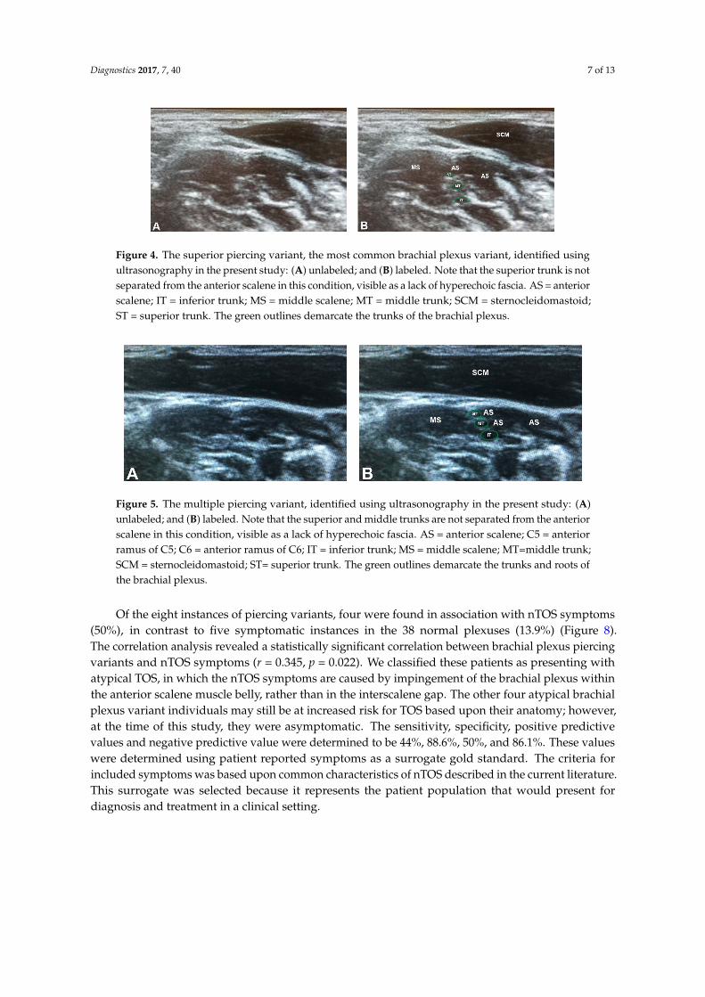
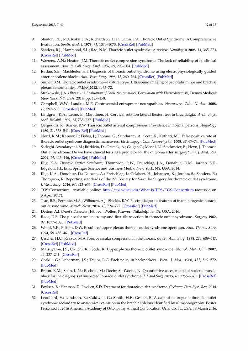
In the ultrasonographic screening sample, 79.5% of the sample was found to possess classicbrachial plexus anatomy (Figure 3), while a total of nine brachial plexus branching variants wereidentified (20.5%) (Tables 3 and S2). Eight of these were classified as piercing variants, in whichportions of the brachial plexus coursed through the anterior scalene muscle (18.2%). The most commonvariation was the superior piercing variant (n = 4; 9.1%) (Figure 4), followed by the multiple piercingvariant (n = 3; 6.8%) (Figure 5), and C5 piercing variant (n = 1; 2.3%) (Figure 6). There was alsoone example of a non-piercing anterior variant (2.3%) (Figure 7).
Diagnostics 2017, 7, 40 6 of 12
middle trunks of the brachial plexus pierce the anterior scalene muscle. AS = anterior scalene; C5 = anterior ramus of C5; C6 = anterior ramus of C6; IT = inferior trunk; MS = middle scalene; MT = middle trunk; SA = subclavian artery; ST = superior trunk.
3.2. Ultrasonographic Results
In the ultrasonographic screening sample, 79.5% of the sample was found to possess classic brachial plexus anatomy (Figure 3), while a total of nine brachial plexus branching variants were identified (20.5%) (Tables 3 and S2). Eight of these were classified as piercing variants, in which portions of the brachial plexus coursed through the anterior scalene muscle (18.2%). The most common variation was the superior piercing variant (n = 4; 9.1%) (Figure 4), followed by the multiple piercing variant (n = 3; 6.8%) (Figure 5), and C5 piercing variant (n = 1; 2.3%) (Figure 6). There was also one example of a non-piercing anterior variant (2.3%) (Figure 7).
Figure 3. The classic brachial plexus anatomy identified on ultrasound: (A) unlabeled; and (B) labeled. Note that the superior, middle, and inferior trunks are clearly separated from the anterior and middle scalene muscles by hyperechoic fascial planes. AS = anterior scalene; IT = inferior trunk; MS = middle scalene; MT = middle trunk; SCM = sternocleidomastoid; ST = superior trunk. The green outlines demarcate the trunks of the brachial plexus.
Table 3. Brachial plexus variation in the screening sample, as identified by ultrasonographic evaluation.
Brachial Plexus Pattern Frequency in Sample % Symptomatic Classic Anatomy 35; 79.5% 5; 13.9%
C5 Anterior Variant 1; 2.3% 0; 0% Piercing Variants: Total 8; 18.2% 4; 50%
C5 Piercing Variant 1; 2.3% 1; 100% Superior Piercing Variant 4; 9.1% 2; 50% Multiple Piercing Variant 3; 6.8% 1; 33%
Figure 3. The classic brachial plexus anatomy identified on ultrasound: (A) unlabeled; and (B) labeled.Note that the superior, middle, and inferior trunks are clearly separated from the anterior and middlescalene muscles by hyperechoic fascial planes. AS = anterior scalene; IT = inferior trunk; MS = middlescalene; MT = middle trunk; SCM = sternocleidomastoid; ST = superior trunk. The green outlinesdemarcate the trunks of the brachial plexus.
Table 3. Brachial plexus variation in the screening sample, as identified by ultrasonographic evaluation.
Brachial Plexus Pattern Frequency in Sample % Symptomatic
Classic Anatomy 35; 79.5% 5; 13.9%C5 Anterior Variant 1; 2.3% 0; 0%
Piercing Variants: Total 8; 18.2% 4; 50%C5 Piercing Variant 1; 2.3% 1; 100%
Superior Piercing Variant 4; 9.1% 2; 50%Multiple Piercing Variant 3; 6.8% 1; 33%
Diagnostics 2017, 7, 40 7 of 13
Diagnostics 2017, 7, 40 7 of 12
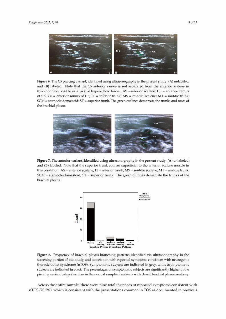
Figure 4. The superior piercing variant, the most common brachial plexus variant, identified using ultrasonography in the present study: (A) unlabeled; and (B) labeled. Note that the superior trunk is not separated from the anterior scalene in this condition, visible as a lack of hyperechoic fascia. AS = anterior scalene; IT = inferior trunk; MS = middle scalene; MT = middle trunk; SCM = sternocleidomastoid; ST = superior trunk. The green outlines demarcate the trunks of the brachial plexus.
Figure 5. The multiple piercing variant, identified using ultrasonography in the present study: (A) unlabeled; and (B) labeled. Note that the superior and middle trunks are not separated from the anterior scalene in this condition, visible as a lack of hyperechoic fascia. AS = anterior scalene; C5 = anterior ramus of C5; C6 = anterior ramus of C6; IT = inferior trunk; MS = middle scalene; MT=middle trunk; SCM = sternocleidomastoid; ST= superior trunk. The green outlines demarcate the trunks and roots of the brachial plexus.
Figure 6. The C5 piercing variant, identified using ultrasonography in the present study: (A) unlabeled; and (B) labeled. Note that the C5 anterior ramus is not separated from the anterior scalene in this condition, visible as a lack of hyperechoic fascia. AS =anterior scalene; C5 = anterior ramus of C5; C6 = anterior ramus of C6; IT = inferior trunk; MS = middle scalene; MT = middle trunk; SCM = sternocleidomastoid; ST = superior trunk. The green outlines demarcate the trunks and roots of the brachial plexus.
Figure 4. The superior piercing variant, the most common brachial plexus variant, identified usingultrasonography in the present study: (A) unlabeled; and (B) labeled. Note that the superior trunk is notseparated from the anterior scalene in this condition, visible as a lack of hyperechoic fascia. AS = anteriorscalene; IT = inferior trunk; MS = middle scalene; MT = middle trunk; SCM = sternocleidomastoid;ST = superior trunk. The green outlines demarcate the trunks of the brachial plexus.
Diagnostics 2017, 7, 40 7 of 12
Figure 4. The superior piercing variant, the most common brachial plexus variant, identified using ultrasonography in the present study: (A) unlabeled; and (B) labeled. Note that the superior trunk is not separated from the anterior scalene in this condition, visible as a lack of hyperechoic fascia. AS = anterior scalene; IT = inferior trunk; MS = middle scalene; MT = middle trunk; SCM = sternocleidomastoid; ST = superior trunk. The green outlines demarcate the trunks of the brachial plexus.
Figure 5. The multiple piercing variant, identified using ultrasonography in the present study: (A) unlabeled; and (B) labeled. Note that the superior and middle trunks are not separated from the anterior scalene in this condition, visible as a lack of hyperechoic fascia. AS = anterior scalene; C5 = anterior ramus of C5; C6 = anterior ramus of C6; IT = inferior trunk; MS = middle scalene; MT=middle trunk; SCM = sternocleidomastoid; ST= superior trunk. The green outlines demarcate the trunks and roots of the brachial plexus.
Figure 6. The C5 piercing variant, identified using ultrasonography in the present study: (A) unlabeled; and (B) labeled. Note that the C5 anterior ramus is not separated from the anterior scalene in this condition, visible as a lack of hyperechoic fascia. AS =anterior scalene; C5 = anterior ramus of C5; C6 = anterior ramus of C6; IT = inferior trunk; MS = middle scalene; MT = middle trunk; SCM = sternocleidomastoid; ST = superior trunk. The green outlines demarcate the trunks and roots of the brachial plexus.
Figure 5. The multiple piercing variant, identified using ultrasonography in the present study: (A)unlabeled; and (B) labeled. Note that the superior and middle trunks are not separated from the anteriorscalene in this condition, visible as a lack of hyperechoic fascia. AS = anterior scalene; C5 = anteriorramus of C5; C6 = anterior ramus of C6; IT = inferior trunk; MS = middle scalene; MT=middle trunk;SCM = sternocleidomastoid; ST= superior trunk. The green outlines demarcate the trunks and roots ofthe brachial plexus.
Of the eight instances of piercing variants, four were found in association with nTOS symptoms(50%), in contrast to five symptomatic instances in the 38 normal plexuses (13.9%) (Figure 8).The correlation analysis revealed a statistically significant correlation between brachial plexus piercingvariants and nTOS symptoms (r = 0.345, p = 0.022). We classified these patients as presenting withatypical TOS, in which the nTOS symptoms are caused by impingement of the brachial plexus withinthe anterior scalene muscle belly, rather than in the interscalene gap. The other four atypical brachialplexus variant individuals may still be at increased risk for TOS based upon their anatomy; however,at the time of this study, they were asymptomatic. The sensitivity, specificity, positive predictivevalues and negative predictive value were determined to be 44%, 88.6%, 50%, and 86.1%. These valueswere determined using patient reported symptoms as a surrogate gold standard. The criteria forincluded symptoms was based upon common characteristics of nTOS described in the current literature.This surrogate was selected because it represents the patient population that would present fordiagnosis and treatment in a clinical setting.
Diagnostics 2017, 7, 40 8 of 13
Diagnostics 2017, 7, 40 7 of 12
Figure 4. The superior piercing variant, the most common brachial plexus variant, identified using ultrasonography in the present study: (A) unlabeled; and (B) labeled. Note that the superior trunk is not separated from the anterior scalene in this condition, visible as a lack of hyperechoic fascia. AS = anterior scalene; IT = inferior trunk; MS = middle scalene; MT = middle trunk; SCM = sternocleidomastoid; ST = superior trunk. The green outlines demarcate the trunks of the brachial plexus.
Figure 5. The multiple piercing variant, identified using ultrasonography in the present study: (A) unlabeled; and (B) labeled. Note that the superior and middle trunks are not separated from the anterior scalene in this condition, visible as a lack of hyperechoic fascia. AS = anterior scalene; C5 = anterior ramus of C5; C6 = anterior ramus of C6; IT = inferior trunk; MS = middle scalene; MT=middle trunk; SCM = sternocleidomastoid; ST= superior trunk. The green outlines demarcate the trunks and roots of the brachial plexus.
Figure 6. The C5 piercing variant, identified using ultrasonography in the present study: (A) unlabeled; and (B) labeled. Note that the C5 anterior ramus is not separated from the anterior scalene in this condition, visible as a lack of hyperechoic fascia. AS =anterior scalene; C5 = anterior ramus of C5; C6 = anterior ramus of C6; IT = inferior trunk; MS = middle scalene; MT = middle trunk; SCM = sternocleidomastoid; ST = superior trunk. The green outlines demarcate the trunks and roots of the brachial plexus.
Figure 6. The C5 piercing variant, identified using ultrasonography in the present study: (A) unlabeled;and (B) labeled. Note that the C5 anterior ramus is not separated from the anterior scalene inthis condition, visible as a lack of hyperechoic fascia. AS =anterior scalene; C5 = anterior ramusof C5; C6 = anterior ramus of C6; IT = inferior trunk; MS = middle scalene; MT = middle trunk;SCM = sternocleidomastoid; ST = superior trunk. The green outlines demarcate the trunks and roots ofthe brachial plexus.Diagnostics 2017, 7, 40 8 of 12
Figure 7. The anterior variant, identified using ultrasonography in the present study: (A) unlabeled; and (B) labeled. Note that the superior trunk courses superficial to the anterior scalene muscle in this condition. AS = anterior scalene; IT = inferior trunk; MS = middle scalene; MT = middle trunk; SCM = sternocleidomastoid; ST = superior trunk. The green outlines demarcate the trunks of the brachial plexus.
Of the eight instances of piercing variants, four were found in association with nTOS symptoms (50%), in contrast to five symptomatic instances in the 38 normal plexuses (13.9%) (Figure 8). The correlation analysis revealed a statistically significant correlation between brachial plexus piercing variants and nTOS symptoms (r = 0.345, p = 0.022). We classified these patients as presenting with atypical TOS, in which the nTOS symptoms are caused by impingement of the brachial plexus within the anterior scalene muscle belly, rather than in the interscalene gap. The other four atypical brachial plexus variant individuals may still be at increased risk for TOS based upon their anatomy; however, at the time of this study, they were asymptomatic. The sensitivity, specificity, positive predictive values and negative predictive value were determined to be 44%, 88.6%, 50%, and 86.1%. These values were determined using patient reported symptoms as a surrogate gold standard. The criteria for included symptoms was based upon common characteristics of nTOS described in the current literature. This surrogate was selected because it represents the patient population that would present for diagnosis and treatment in a clinical setting.
Figure 8. Frequency of brachial plexus branching patterns identified via ultrasonography in the screening portion of this study, and association with reported symptoms consistent with neurogenic thoracic outlet syndrome (nTOS). Symptomatic subjects are indicated in grey, while asymptomatic subjects are indicated in black. The percentages of symptomatic subjects are significantly higher in the piercing variant categories than in the normal sample of subjects with classic brachial plexus anatomy.
Across the entire sample, there were nine total instances of reported symptoms consistent with nTOS (20.5%), which is consistent with the presentations common to TOS as documented in previous
Figure 7. The anterior variant, identified using ultrasonography in the present study: (A) unlabeled;and (B) labeled. Note that the superior trunk courses superficial to the anterior scalene muscle inthis condition. AS = anterior scalene; IT = inferior trunk; MS = middle scalene; MT = middle trunk;SCM = sternocleidomastoid; ST = superior trunk. The green outlines demarcate the trunks of thebrachial plexus.
Diagnostics 2017, 7, 40 8 of 12
Figure 7. The anterior variant, identified using ultrasonography in the present study: (A) unlabeled; and (B) labeled. Note that the superior trunk courses superficial to the anterior scalene muscle in this condition. AS = anterior scalene; IT = inferior trunk; MS = middle scalene; MT = middle trunk; SCM = sternocleidomastoid; ST = superior trunk. The green outlines demarcate the trunks of the brachial plexus.
Of the eight instances of piercing variants, four were found in association with nTOS symptoms (50%), in contrast to five symptomatic instances in the 38 normal plexuses (13.9%) (Figure 8). The correlation analysis revealed a statistically significant correlation between brachial plexus piercing variants and nTOS symptoms (r = 0.345, p = 0.022). We classified these patients as presenting with atypical TOS, in which the nTOS symptoms are caused by impingement of the brachial plexus within the anterior scalene muscle belly, rather than in the interscalene gap. The other four atypical brachial plexus variant individuals may still be at increased risk for TOS based upon their anatomy; however, at the time of this study, they were asymptomatic. The sensitivity, specificity, positive predictive values and negative predictive value were determined to be 44%, 88.6%, 50%, and 86.1%. These values were determined using patient reported symptoms as a surrogate gold standard. The criteria for included symptoms was based upon common characteristics of nTOS described in the current literature. This surrogate was selected because it represents the patient population that would present for diagnosis and treatment in a clinical setting.
Figure 8. Frequency of brachial plexus branching patterns identified via ultrasonography in the screening portion of this study, and association with reported symptoms consistent with neurogenic thoracic outlet syndrome (nTOS). Symptomatic subjects are indicated in grey, while asymptomatic subjects are indicated in black. The percentages of symptomatic subjects are significantly higher in the piercing variant categories than in the normal sample of subjects with classic brachial plexus anatomy.
Across the entire sample, there were nine total instances of reported symptoms consistent with nTOS (20.5%), which is consistent with the presentations common to TOS as documented in previous
Figure 8. Frequency of brachial plexus branching patterns identified via ultrasonography in thescreening portion of this study, and association with reported symptoms consistent with neurogenicthoracic outlet syndrome (nTOS). Symptomatic subjects are indicated in grey, while asymptomaticsubjects are indicated in black. The percentages of symptomatic subjects are significantly higher in thepiercing variant categories than in the normal sample of subjects with classic brachial plexus anatomy.
Across the entire sample, there were nine total instances of reported symptoms consistent withnTOS (20.5%), which is consistent with the presentations common to TOS as documented in previous

Diagnostics 2017, 7, 40 9 of 13
studies [9,10]. Given that the student population is predicted to be at higher risk for neurogenicsymptoms due to hypertonicity of the cervical musculature, a minor increase in cases was expected inthis study. Within the full clinically symptomatic group, three subjects (33.3%) had positive Adson’stests, while two had positive Wright tests (22.2%) (Table 4). These individuals represent the subset ofTypical TOS in which the compression occurs between hypertonic anterior and middle scalene muscles.
Table 4. Summary of findings of provocative testing and their association with self-reported neurogenicthoracic outlet syndrome (nTOS) symptoms across the entire screening sample.
Provacative Test Results and nTOS Symptoms Adson’s Test CostoclavicularTest
Hyperabduction/Wright Test
Positive test and reported nTOS symptomatic 3/16 (18.8%) 2/8 (25.0%) 3/13 (23.1%)Negative test and reported nTOS asymptomatic 22/28 (78.6%) 29/36 (80.6%) 25/31 (80.6%)
Within the group of participants who denied symptoms on questionnaire (n = 35), the provocativetests demonstrated substantial potential for false positives. There were seventeen instances in whicha positive result was found for at least one of the three provocative tests without a history of symptoms(48.6% false positives). Of these 17 overall positives, 13 were positive Adson’s tests (Figure 9).The correlation analyses revealed no statistically significant correlations between nTOS symptomsand any of the provocative tests (for all results, pulse, and symptoms). The partial correlationanalysis controlling for brachial plexus variation also revealed no significant correlation between nTOSsymptoms and the provocative tests.
Diagnostics 2017, 7, 40 9 of 12
studies [9,10]. Given that the student population is predicted to be at higher risk for neurogenic symptoms due to hypertonicity of the cervical musculature, a minor increase in cases was expected in this study. Within the full clinically symptomatic group, three subjects (33.3%) had positive Adson’s tests, while two had positive Wright tests (22.2%) (Table 4). These individuals represent the subset of Typical TOS in which the compression occurs between hypertonic anterior and middle scalene muscles.
Table 4. Summary of findings of provocative testing and their association with self-reported neurogenic thoracic outlet syndrome (nTOS) symptoms across the entire screening sample.
Provacative Test Results and nTOS Symptoms Adson’s Test Costoclavicular Test
Hyperabduction/Wright Test
Positive test and reported nTOS symptomatic 3/16 (18.8%) 2/8 (25.0%) 3/13 (23.1%) Negative test and reported nTOS asymptomatic 22/28 (78.6%) 29/36 (80.6%) 25/31 (80.6%)
Within the group of participants who denied symptoms on questionnaire (n = 35), the provocative tests demonstrated substantial potential for false positives. There were seventeen instances in which a positive result was found for at least one of the three provocative tests without a history of symptoms (48.6% false positives). Of these 17 overall positives, 13 were positive Adson’s tests (Figure 9). The correlation analyses revealed no statistically significant correlations between nTOS symptoms and any of the provocative tests (for all results, pulse, and symptoms). The partial correlation analysis controlling for brachial plexus variation also revealed no significant correlation between nTOS symptoms and the provocative tests.
Figure 9. Summary of brachial plexus pattern and Adson’s Test results as associated with nTOS symptoms across the full screening sample. Symptomatic subjects are indicated in light grey, while asymptomatic subjects are indicated in dark grey. The percentage of individuals with nTOS symptoms was significantly higher among the brachial plexus piercing variant subjects (50%) than in the subjects with classic brachial plexus anatomy (13.9%), but rates of correct diagnostic identification with Adson’s Test were slightly lower (50% in piercing variants vs. 61.1% in classic).
4. Discussion
4.1. Anatomical Variation Observed
The findings from this study support the hypothesis that some cases of disputed TOS may result from brachial plexus variations in which the roots or trunks of the plexus course through the anterior scalene muscle belly, becoming impinged. This phenomenon is similar to piriformis syndrome, which
Figure 9. Summary of brachial plexus pattern and Adson’s Test results as associated with nTOS symptomsacross the full screening sample. Symptomatic subjects are indicated in light grey, while asymptomaticsubjects are indicated in dark grey. The percentage of individuals with nTOS symptoms was significantlyhigher among the brachial plexus piercing variant subjects (50%) than in the subjects with classicbrachial plexus anatomy (13.9%), but rates of correct diagnostic identification with Adson’s Test wereslightly lower (50% in piercing variants vs. 61.1% in classic).
4. Discussion
4.1. Anatomical Variation Observed
The findings from this study support the hypothesis that some cases of disputed TOS mayresult from brachial plexus variations in which the roots or trunks of the plexus course through the
Diagnostics 2017, 7, 40 10 of 13
anterior scalene muscle belly, becoming impinged. This phenomenon is similar to piriformis syndrome,which can result from fibers of the sciatic nerve traveling through the piriformis muscle belly leadingto impingement. We have determined that individuals with these structural variations in the thoracicoutlet present with nTOS symptoms at a significantly higher rate than the general population, but thatsuch anomalies are easily identified using ultrasonography.
Overall, nine of 44 of our student subject brachial plexuses were documented to have variantbranching on US imaging, with the majority being the superior piercing variant. Four subjectspresented with TOS secondary to a brachial plexus piercing variant. In each of these, the superiortrunk or both the superior and middle trunks pierce the anterior scalene muscle (Figures 2 and 4).Clinically, the superior piercing variant would cause neurologic symptoms in the C5 and C6dermatomal distribution of the lateral arm, thumb, and second digit. Specifically, weakness andsensory deficits in the first two digits, and diminished reflexes of the biceps brachii and brachioradialismuscles [25–28]. The multiple piercing variant (Figure 2C) would result in more extensive neurogenicissues, corresponding with symptoms along the C5, C6, and C7 dermatomes, affecting the first throughthird digits of the hand. The clinical consequences could also include additional muscle weakness ordecreased reflexes in the triceps brachii muscle [26–28].
One screening study participant and two cadavers were found to have anterior variants, withthe superior trunk passing superficial to the anterior scalene muscle (Figure 6). This variation is lesscommon than the piercing variants, and would not cause numbness or paresthesia in the hands orarms, but could render the nerve vulnerable to compression by forces such as those exerted by pursesor backpacks. Impingement of the superior trunk, one of its proximal branches or the supraclavicularnerve is commonly known as pack palsy, which results from pressure on the shoulder girdle and iscommon in military personnel and hikers [29].
4.2. Diagnosis and Treatment of Thoracic Outlet Syndrome (TOS)
In the general population, primarily typical TOS has been clinically studied and is frequentlydiagnosed by vascular change with provocative testing. When a patient presents to a clinicianwith Atypical TOS, there is a potential for premature dismissal of TOS in the differential diagnosis.The diagnosis becomes increasingly elusive because the initial presentation and history of AtypicalTOS correlate with other forms of neurologic impingement. Without proper identification of theetiology, the patient may not receive the most efficacious treatment. Based on the results of thisstudy, with nearly half of reported TOS cases originating from variant anatomy which cannot beidentified using provocative testing, it can be concluded that US may be a useful adjunct in clinicaldiagnosis. Ultrasonography is uniquely able to visualize variations and provide clinicians with anunderstanding of individual anatomy. By combining other diagnostic modalities, such as provocativetests, which can identify hypertonicity impingement, with US, clinicians would have the ability tovisualize the structural composition of the neck and shoulder. The inclusion of such knowledgeprovides a higher level of diagnostic acuity when screening patients presenting with nonspecific TOSsymptoms. Scalene blocks are another commonly performed diagnostic modality for nTOS, and canbe less equivocal than provocative testing (e.g., [30]). However, these blocks may not be feasible inthe primary care setting, and can leave patients with 2–36 h of residual discomfort or inconveniencefollowing the procedure. US, on the other hand, is painless, rapid, and inexpensive, and can be easilyimplemented as part of a broader diagnostic approach. Due to the varied and complicated nature ofnTOS presentation, it is often necessary for clinicians to apply multiple diagnostic modalities beforeultimately arriving at a diagnosis. US can serve as an additional resource in the diagnostic toolkit ofclinicians, especially in the primary care setting.
Utilizing this diagnostic approach it is also possible to tailor treatment to the individual’s uniqueanatomy. Treatment methods targeting either the first rib or scalene musculature would be complicatedby the nerve branches entwined in the anterior scalene muscle belly. Surgical removal of rib 1 wouldlikely not relieve the symptoms of compression around a more laterally placed trunk. A scalenectomy
Diagnostics 2017, 7, 40 11 of 13
could place the piercing trunks in danger of damage unless appropriately identified [31]. Scalenebotulinum toxin injection could be effective, so US could be applied to preselect patients with piercingvariants for this treatment. For patients with one of the piercing variations, we propose a rationalplan of osteopathic manipulative treatment (OMT) care and/or physical therapy consisting of indirecttreatment modalities and the avoidance of direct techniques, based upon the potential for furtherimpingement of the nerve within the muscle belly. This was evidenced in one subject suffering fromatypical TOS resulting from a piercing variant [32]. In this case, the patient experienced optimal relieffrom her symptoms only when indirect treatment techniques were employed, reporting a significantimprovement of her symptoms [32]. This patient also had improvement of her concurrent anxiety aftergaining a more thorough understanding of her diagnosis with the US imaging.
5. Conclusions
Structural variations of the thoracic outlet, especially common brachial plexus branching variants,create a unique risk for neurogenic TOS that is difficult to diagnose clinically. Ultrasound is a reliablemeans of diagnosing this etiology when combined with provocative testing and patient history.Identification of these structural variants is crucial for developing an appropriate treatment plan,as certain types of current treatment modalities would be ineffective, or even exacerbate symptoms inpatients with these variants.
Supplementary Materials: Supplementary materials can be found at www.mdpi.com/2075-4418/07/3/40/s1.
Acknowledgments: Funding for this research was provided by Midwestern University. The authors would liketo thank Katherine Worden, Riley Landreth, Richard Geshel, and Sabina Kumar for their contributions and insighton an earlier version of this study. We thank Brent Adrian for helpful advice regarding figures, and RandallNydam for his gracious accommodation with the ultrasound unit. Heather F. Smith would also like to thankGwenneth P. Smith, PT, for insightful discussion regarding nerve compression, neural tension, and treatmentmodalities, which greatly improved this paper.
Author Contributions: Vanessa Leonhard and Gregory Caldwell developed the project concept and contributedto data collection, data summary review, abstract composition, and manuscript construction; Mei Goh contributedto abstract composition and manuscript review; Sean Reeder contributed to concept development, interpretationof results, and manuscript preparation; and Heather F. Smith developed the project concept, conducted statisticalanalysis and evaluation, and contributed to data collection, data summary review, and manuscript construction.
Conflicts of Interest: The authors declare no conflict of interest.
References
1. Hooper, T.L.; Denton, J.; McGalliard, M.K.; Brismée, J.M.; Sizer, P.S. Thoracic outlet syndrome: A controversialclinical condition. Part 1: Anatomy, and clinical examination/diagnosis. J. Man. Manip. Ther. 2010, 18, 74–83.[CrossRef] [PubMed]
2. Kuhn, J.E.; Lebus, G.F.; Bible, J.E. Thoracic Outlet Syndrome. J. Am. Acad. Orthop. Surg. 2015, 23, 222–232.[CrossRef] [PubMed]
3. Sanders, R.J.; Hammond, S.L.; Rao, N.M. Diagnosis of thoracic outlet syndrome. J. Vasc. Surg. 2007, 64, 601–604.[CrossRef] [PubMed]
4. Adson, A.W.; Coffey, J.R. Cervical rib: A method of anterior approach for relief of symptoms by division ofthe scalenus anticus. Ann. Surg. 1927, 85, 839–857. [CrossRef] [PubMed]
5. Harry, W.G.; Bennett, J.D.; Guha, S.C. Scalene muscles and the brachial plexus: Anatomical variations andtheir clinical significance. Clin. Anat. 1997, 10, 250–252. [CrossRef]
6. Sakamoto, Y. Spatial relationships between the morphologies and innervations of the scalene and anteriorvertebral muscles. Ann. Anat. 2012, 194, 381–388. [CrossRef] [PubMed]
7. Leonhard, V.; Smith, R.; Caldwell, G.; Smith, H.F. Anatomical variations in the brachial plexus roots:Implications for diagnosis of neurogenic thoracic outlet syndrome. Ann. Anat. 2016, 206, 21–26. [CrossRef][PubMed]
8. Peet, R.M.; Henriksen, J.D.; Anderson, T.P.; Martin, G.M. Thoracic-outlet syndrome: Evaluation ofa therapeutic exercise program. Proc. Staff Meet. Mayo Clin. 1956, 31, 281–287. [PubMed]
Diagnostics 2017, 7, 40 12 of 13
9. Stanton, P.E.; McClusky, D.A.; Richardson, H.D.; Lamis, P.A. Thoracic Outlet Syndrome: A ComprehensiveEvaluation. South. Med. J. 1978, 71, 1070–1073. [CrossRef] [PubMed]
10. Sanders, R.J.; Hammond, S.L.; Rao, N.M. Thoracic outlet syndrome: A review. Neurologist 2008, 14, 365–373.[CrossRef] [PubMed]
11. Warrens, A.N.; Heaton, J.M. Thoracic outlet compression syndrome: The lack of reliability of its clinicalassessment. Ann. R. Coll. Surg. Engl. 1987, 69, 203–204. [PubMed]
12. Jordan, S.E.; Machleder, H.I. Diagnosis of thoracic outlet syndrome using electrophysiologically guidedanterior scalene blocks. Ann. Vasc. Surg. 1998, 12, 260–264. [CrossRef] [PubMed]
13. Sucher, B.M. Thoracic outlet syndrome—Postural type: Ultrasound imaging of pectoralis minor and brachialplexus abnormalities. PM&R 2012, 4, 65–72.
14. Strakowski, J.A. Ultrasound Evaluation of Focal Neuropathies, Correlation with Electrodiagnosis; Demos Medical:New York, NY, USA, 2014; pp. 127–158.
15. Campbell, W.W.; Landau, M.E. Controversial entrapment neuropathies. Neurosurg. Clin. N. Am. 2008,19, 597–608. [CrossRef] [PubMed]
16. Lindgren, K.A.; Leino, E.; Manninen, H. Cervical rotation lateral flexion test in brachialgia. Arch. Phys.Med. Rehabil. 1992, 73, 735–737. [PubMed]
17. Gergoudis, R.; Barnes, R.W. Thoracic outlet arterial compression: Prevalence in normal persons. Angiology1980, 31, 538–541. [CrossRef] [PubMed]
18. Nord, K.M.; Kapoor, P.; Fisher, J.; Thomas, G.; Sundaram, A.; Scott, K.; Kothari, M.J. False positive rate ofthoracic outlet syndrome diagnostic maneuvers. Electromyogr. Clin. Neurophysiol. 2008, 48, 67–74. [PubMed]
19. Sadeghi-Azandaryani, M.; Bürklein, D.; Ozimek, A.; Geiger, C.; Mendl, N.; Steckmeier, B.; Heyn, J. ThoracicOutlet Syndrome: Do we have clinical tests as a predictor for the outcome after surgery? Eur. J. Med. Res.2009, 14, 443–446. [CrossRef] [PubMed]
20. Illig, K.A. Thoracic Outlet Syndrome; Thompson, R.W., Freischlag, J.A., Donahue, D.M., Jordan, S.E.,Edgelow, P.I., Eds.; Springer Science and Business Media: New York, NY, USA, 2014.
21. Illig, K.A.; Donohue, D.; Duncan, A.; Freischlag, J.; Gelabert, H.; Johansen, K.; Jordan, S.; Sanders, R.;Thompson, R. Reporting standards of the 271 Society for Vascular Surgery for thoracic outlet syndrome.J. Vasc. Surg. 2016, 64, e23–e35. [CrossRef] [PubMed]
22. TOS Consortium. Available online: http://tos.wustl.edu/What-is-TOS/TOS-Consortium (accessed on3 April 2017).
23. Tsao, B.E.; Ferrante, M.A.; Wilbourn, A.J.; Shields, R.W. Electrodiagnostic features of true neurogenic thoracicoutlet syndrome. Muscle Nerve 2014, 49, 724–727. [CrossRef] [PubMed]
24. Detton, A.J. Grant’s Dissector, 16th ed.; Wolters Kluwer: Philadelphia, PA, USA, 2016.25. Roos, D.B. The place for scalenectomy and first-rib resection in thoracic outlet syndrome. Surgery 1982,
92, 1077–1085. [PubMed]26. Wood, V.E.; Ellison, D.W. Results of upper plexus thoracic outlet syndrome operation. Ann. Thorac. Surg.
1994, 58, 458–461. [CrossRef]27. Urschel, H.C.; Razzuk, M.A. Neurovascular compression in the thoracic outlet. Ann. Surg. 1998, 228, 609–617.
[CrossRef] [PubMed]28. Matsuyama, J.S.; Okuchi, K.; Goda, K. Upper plexus thoracic outlet syndrome. Neurol. Med. Chir. 2002,
42, 237–241. [CrossRef]29. Corkill, G.; Lieberman, J.S.; Taylor, R.G. Pack palsy in backpackers. West. J. Med. 1980, 132, 569–572.
[PubMed]30. Braun, R.M.; Shah, K.N.; Rechnic, M.; Doehr, S.; Woods, N. Quantitative assessments of scalene muscle
block for the diagnosis of suspected thoracic outlet syndrome. J. Hand Surg. 2015, 40, 2255–2261. [CrossRef][PubMed]
31. Povlsen, B.; Hansson, T.; Povlsen, S.D. Treatment for thoracic outlet syndrome. Cochrane Data Syst. Rev. 2014.[CrossRef]
32. Leonhard, V.; Landreth, R.; Caldwell, G.; Smith, H.F.; Geshel, R. A case of neurogenic thoracic outletsyndrome secondary to anatomical variation in the brachial plexus identified by ultrasonography. PosterPresented at 2016 American Academy of Osteopathy Annual Convocation, Orlando, FL, USA, 18 March 2016.

Diagnostics 2017, 7, 40 13 of 13
© 2017 by the authors. Licensee MDPI, Basel, Switzerland. This article is an open accessarticle distributed under the terms and conditions of the Creative Commons Attribution(CC BY) license (http://creativecommons.org/licenses/by/4.0/).