thoracic outlet syndrome: orthopedic tests...thoracic outlet syndrome: orthopedic tests page 3 of 26...
TRANSCRIPT
Thoracic Outlet Syndrome: Orthopedic Tests Page 1 of 26
WSCC Clinics Protocol
Adopted: 7/08
Thoracic Outlet Syndrome: Orthopedic Tests
Adson’s Test Allen’s Test Costoclavicular Test (Eden’s Test) Cyriax Release Test
Halstead Maneuver Hyperabduction Test (Wright’s Test) Roos Test
OVERVIEW
Thoracic outlet syndromes (AKA, cervical rib, scalenus anticus, costoclavicular, hyperabduction and pectoralis minor syndrome) are a group of syndromes primarily associated with arm symptoms. Neurovascular entrapment is thought to be caused by compression of the brachial plexus, subclavian artery and/or vein at some combination of the following sites: within the interscalene triangle, between the first rib and clavicle, and between the corocoid process and the tendon of the pectoralis minor muscle. (Liebensen 1988) A variety of contributing factors have been suggested. Prolonged periods of using a computer keyboard and long periods of hyperabduction or elevation of the arm due to job, recreation or sleeping postures may lead to this condition. Fixed postures have also been implicated such as forward head carriage, shoulders rolled forward, “drooping” shoulder girdle, and large poorly supported breasts. Other factors occasionally include a cervical rib, a space occupying lesion (e.g., a Pancoast tumor), recent trauma or the delayed effects of trauma (including whiplash). A diagnosis of thoracic outlet syndrome (TOS) is often made based on the clinical symptoms (after excluding other diagnoses) and is not always confirmed by physical exam findings. The dominant symptoms include shoulder and arm pain, paresthesia of the fingers (often the 4th and 5th digit), a sense of heaviness or fatigue in the arm and sometimes pallor in the fingers. Sensory symptoms generally cover more than one
dermatome and precede motor symptoms. The hand may also demonstrate loss of grip strength, incoordination or clumsiness (Murphy 2000). Other symptoms may include neck pain or headache. Symptoms are usually unilateral. About 90-97% of patients have neurological symptoms. Far fewer have neurological signs or significant vascular involvement (Vanti 2007). Thoracic outlet syndromes can be grouped as following: 1) vascular TOS, which represents only 3-10% of cases; 2) true neurogenic TOS (N-TOS), which is an uncommon form of the condition and presents with neurological deficits demonstratable by physical exam or electrodiagnosis; and 3) nonspecific neurogenic TOS, which is the most common form and has neurological symptoms but no deficits or electrodiagnostic findings—this type of TOS is considered to be controversial since there is no gold standard to confirm the diagnosis (Vanti 2007). VASCULAR TOS
The vascular form of TOS is the most serious and requires urgent referral for further assessment and potential surgery. It is most common in young males, often who engage in strenuous activities (Huang 2004). This diagnosis is not based on loss of pulse during the classic TOS orthopedic tests, but rather on a constellation of more prominent vascular signs and symptoms. About 1-2% of TOS patients have significant venous compression (Vanti 2007). Signs and symptoms include swelling in the hand or arm, nonpitting edema, distended superficial veins in the upper extremity and chest, cyanosis,
Thoracic Outlet Syndrome: Orthopedic Tests Page 2 of 26
ecchymosis sometimes accompanied by a feeling of heaviness or fatigue in the arm. These symptoms may be aggravated by TOS tests, especially overhead tests. If these symptoms are constant and do not disappear with rest or arm dependency, thrombus formation may have occurred. Because of the potential of a pulmonary embolism, the patient should be referred urgently (Murphy 2000). About 1-5% have significant arterial compression (Vanti 2007). This form is characterized by unilateral cold sensation, pallor of the fingertips, splinter hemorrhages, Raynaud’s-like phenomenon, asymmetrical decreased radial pulse, an asymmetry of blood pressure equal to or more than 20 mmHg (the lower pressure in the symptomatic arm), subclavian bruits, mild signs of cramping or fatigue with repetitive use, and sometimes symptoms that also suggest neurogenic compression. TRUE NEUROGENIC TOS (N-TOS)
True neurogenic TOS is also thought to be rare with an estimated prevalence of 1/1,000,000 (Schenker 2001). Young thin females are the most common patients (Vanti 2007). There also appears to be a higher incidence of cervical ribs in this form of TOS. Neurologic signs dominate, often accompanied by little or no pain. Sensory loss is often the first presentation, with the loss classically restricted to the ulnar aspect of the hand and forearm. Symptoms are typically aggravated by overhead activities, carrying heavy objects and may be worse at the end of the day and when sleeping. Although they may come later, motor findings and muscle atrophy are often the most salient features. One classic finding is the “Gilliatt-Sumner hand” which displays a dramatic degree of atrophy of the abductor pollicis brevis, giving the thenar eminence a scooped out appearance where the muscle mass would usually be. The interossei and hypothenar eminence may also suffer a milder degree of atrophy.
NONSPECIFIC TOS
Nonspecific neurogenic TOS (AKA, the disputed form) makes up the bulk of diagnosed cases. There do not appear to be good prevalence estimates, but Vanti (2007) suggests it may account for up to 85% of TOS. Nonspecific TOS is most likely the form that chiropractic physicians see most commonly. It is more common among women and more prevalent in the 20- to 40-year-old age group. Pain and paresthesia dominate the clinical presentation. Symptoms often follow an ulnar distribution, as is the case with true N-TOS, but there are either no or only mild neurological findings. Symptoms may also present on the median side of the hand or affect the entire hand or forearm. Because of these variations, upper trunk and lower trunk forms of the syndrome have been suggested. Skeptics argue that the upper form or mixed form is more likely a separate condition and not caused by entrapment of the brachial plexus in the thoracic outlet. DIAGNOSIS
Diagnosis is based on a combination of clinical symptoms and exam findings, including positive thoracic outlet orthopedic tests that attempt to occlude some portion of the outlet and reproduce the patient’s symptoms. Ribbe and Lindgren (1989) proposed a cluster of findings that they claimed could be used to predict a diagnosis of TOS and identify a patient for conservative care targeting this condition. A follow-up study also used this TOS Index to make a TOS diagnosis and treat successfully with exercise (Lindgren 1997). The diagnosis was made by fulfilling at least three of the following criteria: 1) a history of symptom aggravation by having the arm in an elevated position; 2) a history of C8-T1 paresthesia; 3) supraclavicular tenderness over the brachial plexus; and 4) patients unable to continue the Roos test for 3 minutes. The true validity of the TOS Index, as well as the accuracy of all of the TOS orthopedic tests, is difficult to know because of the lack of a gold standard. The tests under study are often folded into the reference standard used
Thoracic Outlet Syndrome: Orthopedic Tests Page 3 of 26
for confirming the correct diagnosis, tainting the conclusion with incorporation bias. When TOS is suspected, a working diagnosis is usually arrived at based on the entire clinical presentation, which could include the TOS Index as well as the results of the other commonly performed orthopedic tests. The stronger the positive finding (e.g., repro-duction of pain symptoms vs. isolated loss of pulse) and the greater the number of positive tests, the more specific they are thought to become (Nannapaneni 2003, Rayan 1998, Vanti 2007). Because the diagnosis is made on such soft criteria, it is important to rule out other competing hypotheses. Additional procedures must be performed to further differentiate nonspecific TOS, true N-TOS, and vascular TOS. Evaluation of the joints and soft tissue may help focus a manual therapy and conservative care approach. Lastly, contributing factors may need to be identified and addressed.
A Work-Up Strategy
SUMMARY of Physical Exam Procedures
Postural analysis (standing and sitting)
Palpation of the scalenes, pectoralis and other cervical and shoulder girdle muscles
Neurological evaluation (e.g., DTRs, muscle tests, and sensory testing)
Vascular evaluation (check upper extremity pulses, nail blanching, temperature, swelling, auscultation for bruit, Allen’s test, bilateral blood pressure)
TOS tests (Adson’s, costoclavicular, hyperabduction, Roos , Tinel’s)
Focal stress tests over scalenes and upper portion of pectoralis minor muscles
Length testing of pectoralis and scalene muscles
Static and motion palpation of cervical and thoracic spine, ribs, AC and SC joints
Evaluate breathing pattern.
6 STEPS for Assessing Possible TOS
1. Correlate TOS test results. 2. Check for neurological deficits suggesting
true N-TOS. 3. Check for significant arterial or venous
compromise suggesting vascular TOS (and the need for urgent referral).
4. Assess joint and soft tissue structures that may be contributing to TOS.
5. Rule out other contributing or mimicking conditions.
6. Decide whether ancillary studies are necessary.
STEP 1. Correlate TOS test results. The following tests are routinely recommended (Brismee 2004, Karas 1990, Mackinnon 1996, Nannapaneni 2003, Nichols 1996, Oates 1996, Ouriel 1998, Rayan 1998, Schenker 2001, Schimp 1999, Talmage 1999). Recommended
A dson’s test Hyperabd uction test Co stoclavicular test Roos test Tinel’s test
Optional
Halstead (reverse Adson’s) test Cyriax release test Palpate or percuss supra- and infra-
clavicular space (Schenker 2001). It appears best to interpret the TOS tests in combination with each other (Nannapaneni 2003, Rayan 1998, Vanti 2007). For example, in Plewa’s study on healthy subjects (1997), the false positive rates were relatively low in the following circumstances: if pain was used to define a positive Adson’s test, costo-clavicular test or supraclavicular pressure; if a patient discontinued Roos test due to pain before 3 minutes; if at least 2 of the TOS tests reproduced pain in the upper extremity or at least 3 tests produced any symptoms in the same arm. Tinels’s test performed over the brachial plexus and/or direct compression of the associated nerves has also been
Thoracic Outlet Syndrome: Orthopedic Tests Page 4 of 26
recommended. (Nannapaneni 2003, Rayan 1998) A positive test is production of paresthesia in the arm (mostly commonly along the ulnar distribution) as opposed to local pain. Palpation in the supraclavicular fossa may produce radiating pain or paresthesia or reveal a fullness caused by a large cervical rib. STEP 2. Check for neurological deficits
suggesting true N-TOS.
Sensory testing (light touch, sharp and dull, vibration). Loss of sensation may be patchy over the medial forearm and ulnar side of the hand. Vibration may be the first sensory modality to be lost (Vanti 2007). Perform vibration testing over in the DIP of the little finger to assess the lower trunk of the brachial plexus (which is the most common portion compromised in true N-TOS).
Deep tendon reflexes (biceps, triceps, brachioradialis).
Motor status (especially C8-T1 weakness or atrophy of the intrinsic muscles of the hand). Check grip strength and pinch strength. (See CSPE protocol, Dynamometry (Grip) & Pinch Gauge.) Atrophy of the forearm or hand muscles may be due to disuse. However, more pronounced atrophy occurs with the true neurologic form, especially the hypothenar and thenar eminence, intrinsic muscles of the hand, and medial forearm flexors. (Atasoy 1996) Consider measuring forearm girth to establish a baseline for potential future atrophy.
Neurodynamic testing (optional). Consider performing the various upper limb tension tests.
STEP 3. Check for significant arterial or venous compromise suggesting vascular TOS (and the need for urgent referral).
Significant vascular compromise is rare and is based on more than simple loss of pulse during the TOS orthopedic tests.
Venous compression
Observe the hand and upper extremity for the following signs: swelling, cyanosis, distention of superficial veins in the upper extremity and chest (Vanti 2007) or a purplish-reddish color with the extremity in a dependent position. Arterial compression
The following findings are more significant when unilateral and consistent with the side of symptoms.
Observe fingers for pallor, a tendency to inappropriately blanch in a cold environment, capillary refill time when testing nail blanching, and nail beds for splinter hemorrhages.
Check for diminished or absent radial pulse (compare bilaterally).
Palpate for coldness. [Atasoy (1996) estimates that 50% of TOS will demonstrate this finding even when there is no serious vascular involvement.]
Perform Allen’s test. Auscultate for supraclavicular and infra-
clavicular bruit as well as carotid bruit. Measure blood pressure bilaterally (a
decrease of more than 20 mmHg in the symptomatic arm suggests an occlusion). (Huang 2004)
STEP 4. Assess joint and soft tissue
structures that may be contributing to TOS.
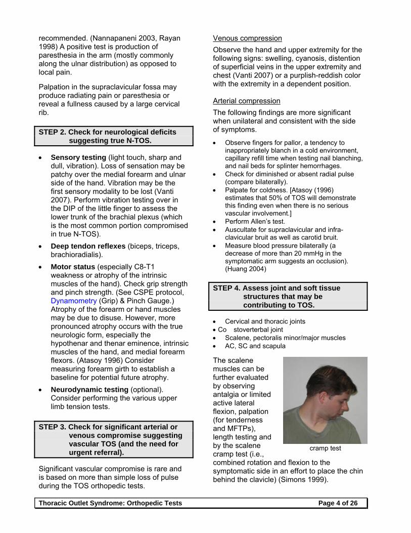
Cervical and thoracic joints Co stoverterbal joint Scalene, pectoralis minor/major muscles AC, SC and scapula The scalene muscles can be further evaluated by observing antalgia or limited active lateral flexion, palpation (for tenderness and MFTPs), length testing and by the scalene cramp test (i.e., combined rotation and flexion to the symptomatic side in an effort to place the chin behind the clavicle) (Simons 1999).
cramp test
Thoracic Outlet Syndrome: Orthopedic Tests Page 5 of 26
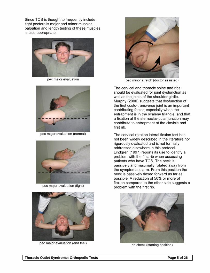
Since TOS is thought to frequently include tight pectoralis major and minor muscles, palpation and length testing of these muscles is also appropriate.
pec major evaluation
pec major evaluation (normal)
pec major evaluation (tight)
pec major evaluation (end feel)
pec minor stretch (doctor assisted)
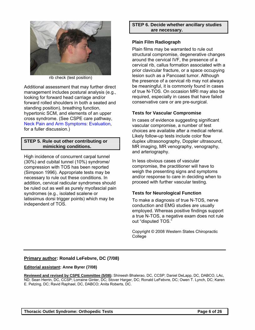
The cervical and thoracic spine and ribs should be evaluated for joint dysfunction as well as the joints of the shoulder girdle. Murphy (2000) suggests that dysfunction of the first costo-transverse joint is an important contributing factor, especially when the entrapment is in the scalene triangle, and that a fixation at the sternoclavicular junction may contribute to entrapment at the clavicle and first rib. The cervical rotation lateral flexion test has not been widely described in the literature nor rigorously evaluated and is not formally addressed elsewhere in this protocol. Lindgren (1997) reports its use to identify a problem with the first rib when assessing patients who have TOS. The neck is passively and maximally rotated away from the symptomatic arm. From this position the neck is passively flexed forward as far as possible. A reduction of 50% or more of flexion compared to the other side suggests a problem with the first rib.
rib check (starting position)
Thoracic Outlet Syndrome: Orthopedic Tests Page 6 of 26
rib check (test position)
Additional assessment that may further direct management includes postural analysis (e.g., looking for forward head carriage and/or forward rolled shoulders in both a seated and standing position), breathing function, hypertonic SCM, and elements of an upper cross syndrome. (See CSPE care pathway, Neck Pain and Arm Symptoms: Evaluation, for a fuller discussion.) STEP 5. Rule out other contributing or
mimicking conditions. High incidence of concurrent carpal tunnel (30%) and cubital tunnel (10%) syndrome/ compression with TOS has been reported (Simpson 1996). Appropriate tests may be necessary to rule out these conditions. In addition, cervical radicular syndromes should be ruled out as well as purely myofascial pain syndromes (e.g., isolated scalene or latissimus dorsi trigger points) which may be independent of TOS.
STEP 6. Decide whether ancillary studies are necessary.
Plain Film Radiograph
Plain films may be warranted to rule out structural compromise, degenerative changes around the cervical IVF, the presence of a cervical rib, callus formation associated with a prior clavicular fracture, or a space occupying lesion such as a Pancoast tumor. Although the presence of a cervical rib may not always be meaningful, it is commonly found in cases of true N-TOS. On occasion MRI may also be required, especially in cases that have failed conservative care or are pre-surgical. Tests for Vascular Compromise
In cases of evidence suggesting significant vascular compromise, a number of test choices are available after a medical referral. Likely follow-up tests include color flow duplex ultrasonography, Doppler ultrasound, MR imaging, MR venography, venography, and arteriography. In less obvious cases of vascular compromise, the practitioner will have to weigh the presenting signs and symptoms and/or response to care in deciding when to proceed with further vascular testing. Tests for Neurological Function
To make a diagnosis of true N-TOS, nerve conduction and EMG studies are usually employed. Whereas positive findings support a true N-TOS, a negative exam does not rule out “disputed TOS.” Copyright © 2008 Western States Chiropractic College
Primary author: Ronald LeFebvre, DC (7/08) Editorial assistant: Anne Byrer (7/08) Reviewed and revised by CSPE Committee (5/08): Shireesh Bhalerao, DC, CCSP; Daniel DeLapp, DC, DABCO, LAc, ND; Sean Herrin, DC, CCSP; Lorraine Ginter, DC; Stover Harger, DC; Ronald LeFebvre, DC; Owen T. Lynch, DC; Karen E. Petzing, DC; Ravid Raphael, DC, DABCO; Anita Roberts, DC.
Thoracic Outlet Syndrome: Orthopedic Tests Page 7 of 26
References Atasoy E. The thoracic outlet compression syndrome. Orthopedic Clincs of North Am 1996;27(2):265-303. Brismee J-M, Gilbert K, et al. Rate of false positive using the Cyriax release test for thoracic outlet syndrome in an
asymptomatic population. JMPT 2004;12(2):73-81. Huang JH, Zager EL.Thoracic outlet syndrome. Neurosurgery 2004;55:897-903. Karas SB. Thoracic outlet syndrome. Clinics Sports Medicine 1990 Apr;9(2):297-310. Liebensen CS. Thoracic outlet syndrome: Diagnosis and conservative management. JMPT 1988;(11):493-9. Lindgren K-A, Rytkonen H. Thoracic outlet syndrome: A functional dysfunction of the upper thoracic aperture? J Back
Musculoskel Rehab 1997;8:191-7. Lindgren K-A. Conservative treatment of thoracic outlet syndrome: A 2 year follow-up. Arch Phys Med Rehabil
1997;78:373-8. Mackinnon SE, Novak CB. Evaluation of the patient with thoracic outlet syndrome. Seminars in Thoracic and
Cardiovascular Surgery 1996 Apr;8(2):190-200. Murphy DR. Conservative management of cervical spine syndromes. New York: McGraw-Hill; 2000. Nannapaneni R, Marks SM. Neurogenic thoracic outlet syndrome. Br J Neurosurg 2003 Apr;17(2):144-8. Nichols AW. The thoracic outlet syndrome in athletes. J Am Board Fam Pract 1996;9:346-55. Oates SD, Daley RA. Thoracic outlet syndrome. Hand Clinics 1996 Nov;12(4):705-18. Ouriel K. Noninvasive diagnosis of upper extremity vascular disease. Seminars in Vascular Surgery 1998 Jun;11(2):54-9. Rayan GM. Thoracic outlet syndrome. J Shoulder Elbow Surg 1998;7(4):440-51. Ribbe E, Lindgren SH, Norgren L. Clinical diagnosis of thoracic outlet syndrome: Evaluation of patients with
cervicobrachial symptoms. Manual Med 1986;2:82-5. Schenker M, Kay SPJ. Mini-symposium: Nerve compression syndromes: (iv) Mechanical neuropathy at the thoracic
outlet and associated pain syndrome. Current Orthopedics 2001;15:264-74. Schimp DJ. The symptomatic upper extremity: An algorithmic approach to diagnosis. JACA 1999;Mar-Apr:32-37, 57. Simons DG, Travell JG. Myofascial Pain and Dysfunction: The Trigger Point Manual. Vol. 1: Upper half of the body.
Baltimore: Williams & Wilkins; 1999. Simpson RL, Fern SA. Multiple compression neuropathies and the double-crush syndrome. Orthopedic Clinics of North
Am 1996 Apr;27(2):381-8. Talmage DM, Lemke C. Thoracic outlet syndrome: How has it changed over the centuries? Top Clin Chiropr
1999;6(4):39-50. Vanti C, Natalini L, Romeo A, Tosarelli D, Pillastrini P. Conservative treatment of thoracic outlet syndrome: A review of
the literature. Eura Medicophys 2007;43:55-70.
Thoracic Outlet Syndrome: Orthopedic Tests Page 8 of 26
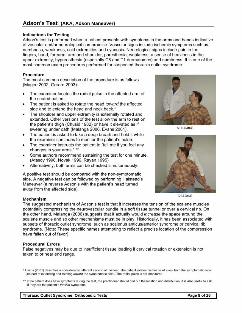
Adson’s Test (AKA, Adson Maneuver) Indications for Testing Adson’s test is performed when a patient presents with symptoms in the arms and hands indicative of vascular and/or neurological compromise. Vascular signs include ischemic symptoms such as numbness, weakness, cold extremities and cyanosis. Neurological signs include pain in the fingers, hand, forearm, arm and shoulder, paresthesia, weakness, a sense of heaviness in the upper extremity, hyperesthesia (especially C8 and T1 dermatomes) and numbness. It is one of the most common exam procedures performed for suspected thoracic outlet syndrome. Procedure The most common description of the procedure is as follows (Magee 2002, Gerard 2003): The examiner locates the radial pulse in the affected arm of
the seated patient. The patient is asked to rotate the head toward the affected
side and to extend the head and neck back.* The shoulder and upper extremity is externally rotated and
extended. Other versions of the test allow the arm to rest on the patient’s thigh (Chusid 1982) or have it elevated as if swearing under oath (Malanga 2006, Evans 2001).
The patient is asked to take a deep breath and hold it while the examiner continues to monitor the patient’s pulse.
The examiner instructs the patient to “tell me if you feel any changes in your arms.” **
Some authors recommend sustaining the test for one minute. (Atasoy 1996, Novak 1996, Rayan 1995)
Alternatively, both arms can be checked simultaneously. A positive test should be compared with the non-symptomatic side. A negative test can be followed by performing Halstead’s Maneuver (a reverse Adson’s with the patient’s head turned away from the affected side). Mechanism The suggested mechanism of Adson’s test is that it increases the tension of the scalene muscles potentially compressing the neurovascular bundle in a soft tissue tunnel or over a cervical rib. On the other hand, Malanga (2006) suggests that it actually would increase the space around the scalene muscle and so other mechanisms must be in play. Historically, it has been associated with subsets of thoracic outlet syndrome, such as scalenus anticus/anterior syndrome or cervical rib syndrome. (Note: These specific names attempting to reflect a precise location of the compression have fallen out of favor). Procedural Errors False negatives may be due to insufficient tissue loading if cervical rotation or extension is not taken to or near end range.
* Evans (2001) describes a considerably different version of this test. The patient rotates his/her head away from the symptomatic side
(instead of extending and rotating toward the symptomatic side). The radial pulse is still monitored. ** If the patient does have symptoms during the test, the practitioner should find out the location and distribution. It is also useful to ask
if they are the patient’s familiar symptoms.
unilateral
bilateral
Thoracic Outlet Syndrome: Orthopedic Tests Page 9 of 26
Interpretation A positive test suggests possible compromise of the neurovascular bundle somewhere along its course through the thoracic outlet. A positive test also suggests that the scalene muscles should be assessed for hypertonicity and trigger points. It would be reasonable to pay special attention to scalene muscle assessment. Other neurological causes of arm pain affected by neck movement such as a radicular syndrome (e.g., due to osteoarthritic encroachment) would also need to be ruled out. Positive test results can be seen as being on a continuum: loss of pulse is the least specific finding (and the most likely to be positive even in asymptomatic subjects), followed by production of paresthesia, followed by what may be the most specific finding which is pain production in the upper extremity. (Plewa 1998) In short, reproduction of pain or neurological symptoms is thought to be more clinically meaningful than isolated loss of pulse (Rayan 1995, Plewa 1998). Reproduction of the patient’s familiar symptoms may be considered even a stronger positive. Charting A positive test recorded in chart notes should indicate which limb is affected, what symptoms are reproduced, and the location. When part of a narrative report, the procedure must also be described. For example, “Cervical extension with rotation toward the affected side reproduced the right arm paresthesia as far as the forearm accompanied by loss of pulse (positive Adson’s test).” Test Validity Gillard (2001) reported that Adson’s test was one of the better performing tests of those commonly studied for TOS having a positive predictive value of 85% (79% sensitivity and 76% specificity). In this study, either loss of pulse or reproduction of symptoms was construed to be positive. A problem with the thoracic outlet tests on the whole is that many asymptomatic subjects will test positive, depending how a positive test is defined. In an asymptomatic population, Rayan (1998) found Adson’s to have a false positive rate of 13.5% for diminished/absent pulse but only 2% for neurological symptoms. Plewa (1998) found a comparable false positive rate of 11% for loss of pulse, a higher false positive rate for paresthesia (11%), but a very low rate of pain production (2%). Overall, Adson’s false positive rates were lower than those of either the hyperabduction and costoclavicular tests. Other studies have reported false positive rates (including isolated diminished pulse positives) to range as high as 53% (Rayan 1998) and even 92% (Malanga 2006). Although, overall, Adson’s test appears to be more useful than the costoclavicular or hyper-abduction test, using a diminished radial pulse to determine a positive Adson’s test should be done with caution. Even symptom reproduction during the procedure must be correlated with other findings. At least one retrospective post-surgical study fails to identify any “single preoperative diagnostic criterion” for thoracic outlet syndrome (Donaghy 1999). Rather, it is better to interpret the tests in combination (Nannapaneni 2003, Plewa 1998, Rayan 1998). Rayan (1998) and Nannapaneni et al. (2003) reported sensitivity of 94% using a combination of Adson’s, Eden’s, Wright’s and Roos tests with Tinel’s test or direct compression of the associated nerves. Likewise specificity appears to improve when multiple tests are combined. In Warrens’ study (1987), 58% of subjects given a battery of TOS tests (Adson’s, costoclavicular and hyperabduction) had at least one false positive, and only 2% had more than one test positive. Likewise, Plewa (1998) found that 2 or 3 positive tests dropped the overall false positive rate and improved the specificity. Unfortunately, most of the studies looking at specificity used asymptomatic patients rather than symptomatic patients with competing diagnoses, which tends to inflate test specificity values. Furthermore, because there is no gold standard to make a TOS diagnosis, most studies use the
Thoracic Outlet Syndrome: Orthopedic Tests Page 10 of 26
same orthopedic tests under investigation as part of the reference standard (incorporation bias), inflating the sensitivity values. Follow-up Testing See “A Work-Up Strategy” in the introduction to this document. Copyright © 2002, 2006, 2008 Western States Chiropractic College Primary author: Ronald LeFebvre, DC (7/02); Dave Peterson, DC (10/02) Revised: Charles Novak, DC (10/05); Ronald LeFebvre, DC (5/08) Reviewed and revised by CSPE Committee (5/08): Shireesh Bhalerao, DC, CCSP; Daniel DeLapp, DC, DABCO, LAc, ND; Sean Herrin, DC, CCSP; Lorraine Ginter, DC; Stover Harger, DC; Ronald LeFebvre, DC; Owen T. Lynch, DC; Karen E. Petzing, DC; Ravid Raphael, DC, DABCO; Anita Roberts, DC. References Atasoy E. The thoracic outlet compression syndrome. Orthopedic Clincs of North Am 1996;27(2):265-303. Brismee J-M, Gilbert K, et al. Rate of false positive using the Cyriax release test for thoracic outlet syndrome in an asymptomatic
population. JMPT 2004;12(2):73-81. Cipriano JJ. Photographic Manual of Orthopedic and Neurological Tests, 2nd edition. Baltimore: Williams and Wilkins;1991:22-3. Chusid JG. Correlative Neuroanatomy & Functional Neurology, 19th edition. Los Altos: Lange Medical Publications; 1982: 337. Donaghy M, Matkovic Z, Morris P. Surgery for suspected neurogenic thoracic outlet syndromes: A follow-up study. J Neurol,
Neurosurg & Psychiatry 1999 Nov;67(5):602-6. Evans RC. Illustrated Orthopedic Physical Assessment, 3rd edition. St. Louis, MO: Mosby; 2001: 192-5. Gerard JA, Kleinfeld SL. Orthopedic testing: A Rational Approach to Diagnosis. New York: Churchill Livingstone; 2003: 60-1. Gillard J, Perez-Cousin M, Hachulla E, et al. Diagnosing thoracic outlet syndrome: Contribution of provocation tests,
ultrasonography, electrophysiology, and helical computed tomography in 48 patients. Joint Bone Spine 2001;68:416-24. Grieve GP. Modern Manual Therapy of the Vertebral Column. Edinburgh: Churchill Livingstone; 1986: 359, 363, 507, 537. Ide J, Kataoka Y, et al. Compression and stretching of the brachial plexus in thoracic outlet syndrome: Correlation between
neuroradiographic findings and symptoms and signs produced by provocation maneuvers. J Hand Surg 2003;28B(3):218-23.
Karas SB. Thoracic outlet syndrome. Clinics Sports Medicine 1990 Apr;9(2):297-310. Mackinnon SE, Novak CB. Evaluation of the patient with thoracic outlet syndrome. Seminars in Thoracic and Cardiovascular
Surgery 1996 Apr;8(2):190-200. Magee DJ. Orthopedic Physical Assessment, 4th edition. Philadelphia: Saunders; 2002: 288. Malanga GA, Nadler SF. Musculoskeletal Physical Examination: An Evidence-Based Approach. Philadelphia: Mosby;2006:50-1. Mazion JM. Illustrated Manual of Neurological Reflexes/Signs/Tests and Orthopedic Signs/Tests/Maneuvers for Office
Procedure. Santa Cruz: JM Mazion; 1980: 211-12. Nannapaneni R, Marks SM. Neurogenic thoracic outlet syndrome. Br J Neurosurg 2003 Apr;17(2):144-8. Nichols AW. The thoracic outlet syndrome in athletes. J Am Board Fam Pract 1996;9:346-55. Novak CB, Mackinnon SE. Thoracic outlet syndrome. Occupational Disorder Management 1996;27(4):747-62. Oates SD, Daley RA. Thoracic outlet syndrome. Hand Clinics 1996 Nov;12(4):705-18. Ouriel K. Noninvasive diagnosis of upper extremity vascular disease. Seminars in Vascular Surgery 1998 Jun;11(2):54-9. Plewa MC, Delinger M. The false positive rate of thoracic outlet syndrome shoulder maneuvers in healthy individuals. Acad,
Emerg Med 1998:5:337-42. Rayan GM. Thoracic outlet syndrome. J Shoulder Elbow Surg 1998;7(4):440-51. Rayan GM, Jensen C. Thoracic outlet syndrome: Provocative examination maneuvers in a typical population. J Shoulder Elbow
Surg 1995;4:113-17. Schenker M, Kay SPJ. Mini-symposium: Nerve compression syndromes: (iv) Mechanical neuropathy at the thoracic outlet and
associated pain syndrome. Current Orthopedics 2001;15:264-74. Schimp DJ. The symptomatic upper extremity: An algorithmic approach to diagnosis. JACA 1999;Mar-Apr:32-37, 57. Simpson RL, Fern SA. Multiple compression neuropathies and the double-crush syndrome. Orthopedic Clinics of North Am
1996 Apr;27(2):381-8. Talmage DM, Lemke C. Thoracic outlet syndrome: How has it changed over the centuries? Top Clin Chiropr 1999;6(4):39-50. Warrens AN, Heaton JM. Thoracic outlet compression syndrome: The lack of reliability of its clinical assesment. Ann R Coll Surg
Engl 1987;69:203-4. White AA, Panjabi MM. Clinical Biomechanics of the Spine, 2nd edition. Philadelphia: J.B. Lippincott Co.; 1990: 412.
Thoracic Outlet Syndrome: Orthopedic Tests Page 11 of 26
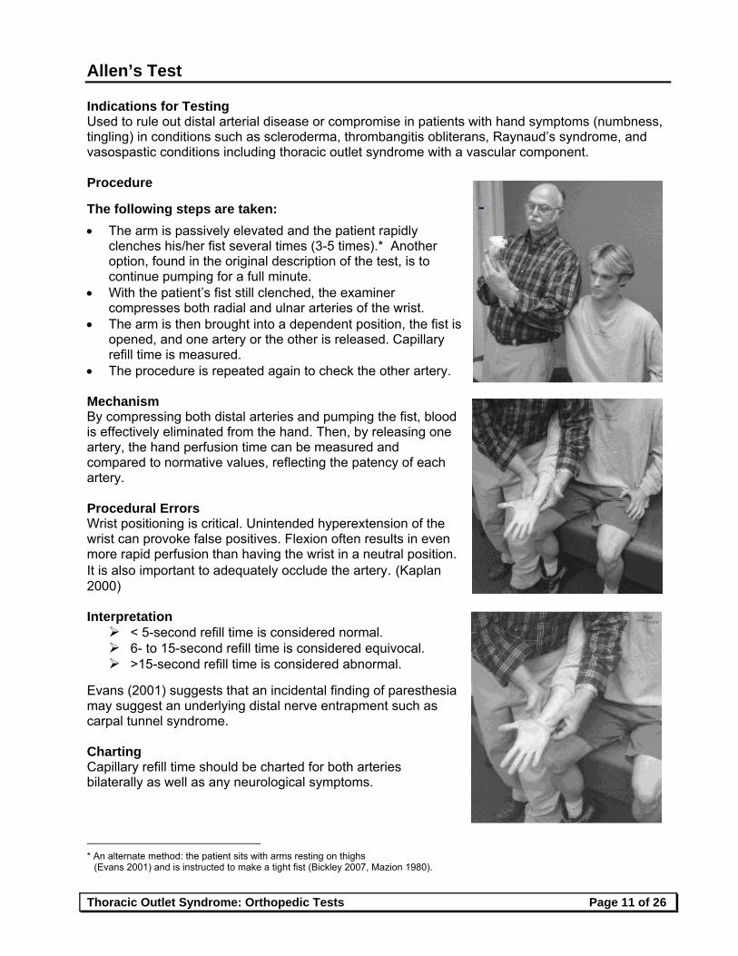
Allen’s Test Indications for Testing Used to rule out distal arterial disease or compromise in patients with hand symptoms (numbness, tingling) in conditions such as scleroderma, thrombangitis obliterans, Raynaud’s syndrome, and vasospastic conditions including thoracic outlet syndrome with a vascular component. Procedure
The following steps are taken:
The arm is passively elevated and the patient rapidly clenches his/her fist several times (3-5 times).* Another option, found in the original description of the test, is to continue pumping for a full minute.
With the patient’s fist still clenched, the examiner compresses both radial and ulnar arteries of the wrist.
The arm is then brought into a dependent position, the fist is opened, and one artery or the other is released. Capillary refill time is measured.
The procedure is repeated again to check the other artery. Mechanism By compressing both distal arteries and pumping the fist, blood is effectively eliminated from the hand. Then, by releasing one artery, the hand perfusion time can be measured and compared to normative values, reflecting the patency of each artery. Procedural Errors Wrist positioning is critical. Unintended hyperextension of the wrist can provoke false positives. Flexion often results in even more rapid perfusion than having the wrist in a neutral position. It is also important to adequately occlude the artery. (Kaplan 2000) Interpretation < 5-second refill time is considered normal. 6- to 15-second refill time is considered equivocal. >15-second refill time is considered abnormal.
Evans (2001) suggests that an incidental finding of paresthesia may suggest an underlying distal nerve entrapment such as carpal tunnel syndrome. Charting Capillary refill time should be charted for both arteries bilaterally as well as any neurological symptoms.
* An alternate method: the patient sits with arms resting on thighs
(Evans 2001) and is instructed to make a tight fist (Bickley 2007, Mazion 1980).

Thoracic Outlet Syndrome: Orthopedic Tests Page 12 of 26
Test Validity Sensitivity and specificity have both been reported to be 95% for distal artery compromise using angiography as the gold standard (Hirai 1980). However, Kaplan (2000) reports other studies resulting in poor positive predictive values. It likely has its greater value as a screening test when the results are normal. Confounding factors may compromise test validity, including patient cooperation, subjective evaluation, patient age, skin pigmentation, jaundice, prior hand injuries, bone deformities, edema, and wrist extension. According to Levinsohn and Sessler (1991):
Sens itivity Specificity PPV NPV Subjective evaluation 1.0 0.8 0.3 1.0 Pulse oximeter 0.3 1.0 1.0 0.9 Laser Doppler flowmetry 1.0 1.0 1.0 1.0
Follow-up Testing Phalen’s test, Tinel’s and wrist-drop should be performed to rule out neurological compromise. Consider medical referral for pulse oximeter and laser Doppler flowmetry, which are the gold standards for verification of arterial obstruction. Copyright © 2002, 2008 Western States Chiropractic College Primary authors: Charles Novak, DC; Dave Peterson, DC (11/02) Revised: Ronald LeFebvre, DC; Charles Novak, DC (5/08) Reviewed and revised by CSPE Committee (5/08): Shireesh Bhalerao, DC, CCSP; Daniel DeLapp, DC, DABCO, LAc, ND; Sean Herrin, DC, CCSP; Lorraine Ginter, DC; Stover Harger, DC; Ronald LeFebvre, DC; Owen T. Lynch, DC; Karen E. Petzing, DC; Ravid Raphael, DC, DABCO; Anita Roberts, DC. References Bickley LS, Szilagyi PG. Bates’ Guide to Physical Examination and History Taking, 9th edition. Philadelphia: Lippincott
Williams & Wilkins; 2007: 488. Evans RC. Illustrated Orthopedic Physical Assessment, 3rd edition. St. Louis, MO: Mosby; 2001: 192-5. Gerard JA, Kleinfeld SL. Orthopedic testing: A Rational Approach to Diagnosis., New York: Churchill Livingstone; 1993:
60-1. Hirai M, Kawai S. False positive and negative results in Allen test. J Cardiovasc Surg 1980;21:353-60. Kaplan DE, Levine SM. A critical reappraisal of the Allen test. Osler Medical J 2000;16. Levinsohn DG, Sessler DI. The Allen test: analysis of four methods. J Hand Surg 1991;16A:279-82. Magee DJ. Orthopedic Physical Assessment, 4th edition. Philadelphia: Saunders; 2002: 288. Malanga GA, Nadler SF. Musculoskeletal Physical Examination: An Evidence-Based Approach. Philadelphia: Mosby;
2006: 183. Mazion JM. Illustrated Manual of Neurological Reflexes/Signs/Tests and Orthopedic Signs/Tests/Maneuvers for Office
Procedure. Santa Cruz: JM Mazion; 1980: 213.
Thoracic Outlet Syndrome: Orthopedic Tests Page 13 of 26
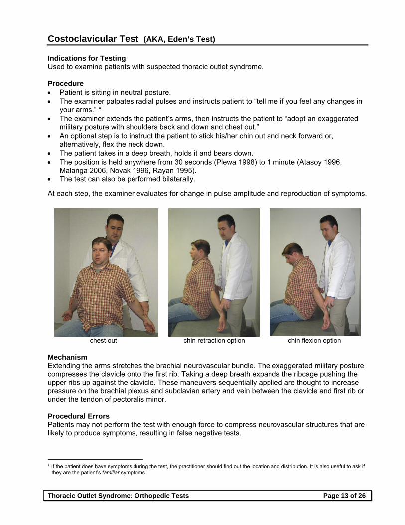
Costoclavicular Test (AKA, Eden’s Test) Indications for Testing Used to examine patients with suspected thoracic outlet syndrome. Procedure Patient is sitting in neutral posture. The examiner palpates radial pulses and instructs patient to “tell me if you feel any changes in
your arms.” * The examiner extends the patient’s arms, then instructs the patient to “adopt an exaggerated
military posture with shoulders back and down and chest out.” An optional step is to instruct the patient to stick his/her chin out and neck forward or,
alternatively, flex the neck down. The patient takes in a deep breath, holds it and bears down. The position is held anywhere from 30 seconds (Plewa 1998) to 1 minute (Atasoy 1996,
Malanga 2006, Novak 1996, Rayan 1995). The test can also be performed bilaterally. At each step, the examiner evaluates for change in pulse amplitude and reproduction of symptoms.
chest out chin retraction option chin flexion option
Mechanism Extending the arms stretches the brachial neurovascular bundle. The exaggerated military posture compresses the clavicle onto the first rib. Taking a deep breath expands the ribcage pushing the upper ribs up against the clavicle. These maneuvers sequentially applied are thought to increase pressure on the brachial plexus and subclavian artery and vein between the clavicle and first rib or under the tendon of pectoralis minor. Procedural Errors Patients may not perform the test with enough force to compress neurovascular structures that are likely to produce symptoms, resulting in false negative tests.
* If the patient does have symptoms during the test, the practitioner should find out the location and distribution. It is also useful to ask if
they are the patient’s familiar symptoms.
Thoracic Outlet Syndrome: Orthopedic Tests Page 14 of 26
Interpretation A positive test would be symptoms of upper limb neurovascular compression, such as cessation or dampening of radial pulse with reproduction of symptoms, ischemic color changes (e.g., pallor, blanching), paresthesia or extremity pain. A positive test suggests possible compromise of the neurovascular bundle somewhere along its course through the thoracic outlet. Positive test results can be seen as being on a continuum: loss of pulse is the least specific finding (and the most likely to be positive even in asymptomatic subjects), followed by production of paresthesia, followed by the most specific finding which is pain production in the upper extremity. (Plewa 1998)
In short, reproduction of pain or neurological symptoms is thought to be more clinically meaningful than isolated loss of pulse (Plewa 1998, Rayan 1995). Reproduction of the patient’s familiar symptoms may be considered even a stronger positive. Charting A positive test recorded in chart notes should indicate which limb is affected, what symptoms are reproduced, and the location. When part of a narrative report, the procedure must also be described. For example, “The radial pulse was lost with progressive pain and numbness down the right lateral arm and forearm when the patient adopted the exaggerated military posture with a chin jut and Valsalva maneuver suggesting vascular and/or neurologic thoracic outlet syndrome (positive costoclavicular test).” Test Validity The costoclavicular test has been studied a number of times along with Adson’s test. In general, it does not appear to perform as well. As in the case of Adson’s test, the vascular response rate of false positives appears to be much greater than for pain or neurological symptoms. As summarized by Plewa (1998), prior studies have suggested generally poor specificity for pulse change. This is reflected in false positive rates of 14% (Gergoudis 1980), 50% (Telford 1948), 27% (Warrens 1987), 47% (Nichols 1996, Rayan 1995) and 11% (Plewa 1998). In contrast, false positive findings have been reported to be 15% for paresthesia and 0% for pain (Plewa 1998). Follow-up Testing See “A Work-Up Strategy” in the introduction to this document. Copyright © 2006, 2008 Western States Chiropractic College Primary author: Charles Novak, DC (10/05)
Revised: Ronald LeFebvre, DC (5/08) Reviewed and revised by CSPE Committee (4/08): Shireesh Bhalerao, DC, CCSP; Daniel DeLapp, DC, DABCO, LAc, ND; Sean Herrin, DC, CCSP; Lorraine Ginter, DC; Stover Harger, DC; Ronald LeFebvre, DC; Owen T. Lynch, DC; Karen E. Petzing, DC; Ravid Raphael, DC, DABCO; Anita Roberts, DC.
References Atasoy E. The thoracic outlet compression syndrome. Orthopedic Clincs of North Am 1996;27(2):265-303. Brismee J-M, Gilbert, K, et al. Rate of false positive using the Cyriax release test for thoracic outlet syndrome in an
asymptomatic population. JMPT 2004;12(2):73-81. Gerard JA, Kleinfield SL. Orthopedic Testing: A Rational Approach to Diagnosis. New York: Churchill Livingstone; 1993:
64-5. Ide J, Kataoka Y, et al. Compression and stretching of the brachial plexus in thoracic outlet syndrome: Correlation
between neuroradiographic findings and symptoms and signs produced by provocation maneuvers. J Hand Surg 2003;28B(3):218-23.
Karas SB. Thoracic outlet syndrome. Clinics in Sports Medicine 1990 Apr;9(2):297-310.
Thoracic Outlet Syndrome: Orthopedic Tests Page 15 of 26
Mackinnon SE, Novak CB. Evaluation of the patient with thoracic outlet syndrome. Seminars in Thoracic and Cardiovascular Surgery 1996 Apr;8(2):190-200.
Magee DJ. Orthopedic Physical Assessment, 4th edition. Philadelphia: W.B. Saunders Company; 2002: 287. Malanga GA, Nadler SF. Musculoskeletal Physical Examination: An Evidence-Based Approach. Philadelphia: Mosby;
2006: 50-1. Nannapaneni R, Marks SM. Neurogenic thoracic outlet syndrome. Br J Neurosurg 2003 Apr;17(2):144-8. Nichols AW. The thoracic outlet syndrome in athletes. J Am Board Fam Pract 1996;9:346-55. Novak CB, Mackinnon SE. Thoracic outlet syndrome. Occupational Disorder Management 1996;27(4):747-62. Oates SD, Daley RA. Thoracic outlet syndrome. Hand Clinics 1996 Nov;12(4):705-18. Ouriel K. Noninvasive diagnosis of upper extremity vascular disease. Seminars in Vascular Surgery 1998 Jun;11(2):54-9. Plewa MC, Delinger M. The false positive rate of thoracic outlet syndrome shoulder maneuvers in healthy individuals.
Acad, Emerg Med 1998:5:337-42. Rayan GM. Thoracic outlet syndrome. J Shoulder Elbow Surg 1998;7(4):440-51. Rayan GM, Jensen C. Thoracic outlet syndrome: Provocative examination maneuvers in a typical population. J Shoulder
Elbow Surg 1995;4:113-7. Schenker M, Kay SPJ. Mini-symposium: Nerve compression syndromes: (iv) Mechanical neuropathy at the thoracic
outlet and associated pain syndrome. Current Orthopedics 2001;15:264-74. Schimp DJ. The symptomatic upper extremity: An algorithmic approach to diagnosis. JACA 1999;Mar-Apr:32-37, 57. Simpson RL, Fern SA. Multiple compression neuropathies and the double-crush syndrome. Orthopedic Clinics of North
Am 1996 Apr;27(2):381-8. Talmage DM, Lemke C. Thoracic outlet syndrome: How has it changed over the centuries? Top Clin Chiropr
1999;6(4):39-50. Warrens AN, Heaton JM. Thoracic outlet compression syndrome: The lack of reliability of its clinical assesment. Ann R
Coll Surg Engl 1987;69:203-4. White AA, Panjabi MM. Clinical Biomechanics of the Spine, 2nd edition. Philadelphia: J.B. Lippincott Co.; 1009: 412. Cited but not reviewed Gergoudis R, Barnes RW. Thoracic outlet compression: prevalence in normal persons. Angiology 1980;31:538-41. Telford ED, Mottershead S. Pressure at the cervico-brachial junction: an operative and anatomical study. J Bone Joint
Surg Br 1948;30:249-65.
Thoracic Outlet Syndrome: Orthopedic Tests Page 16 of 26
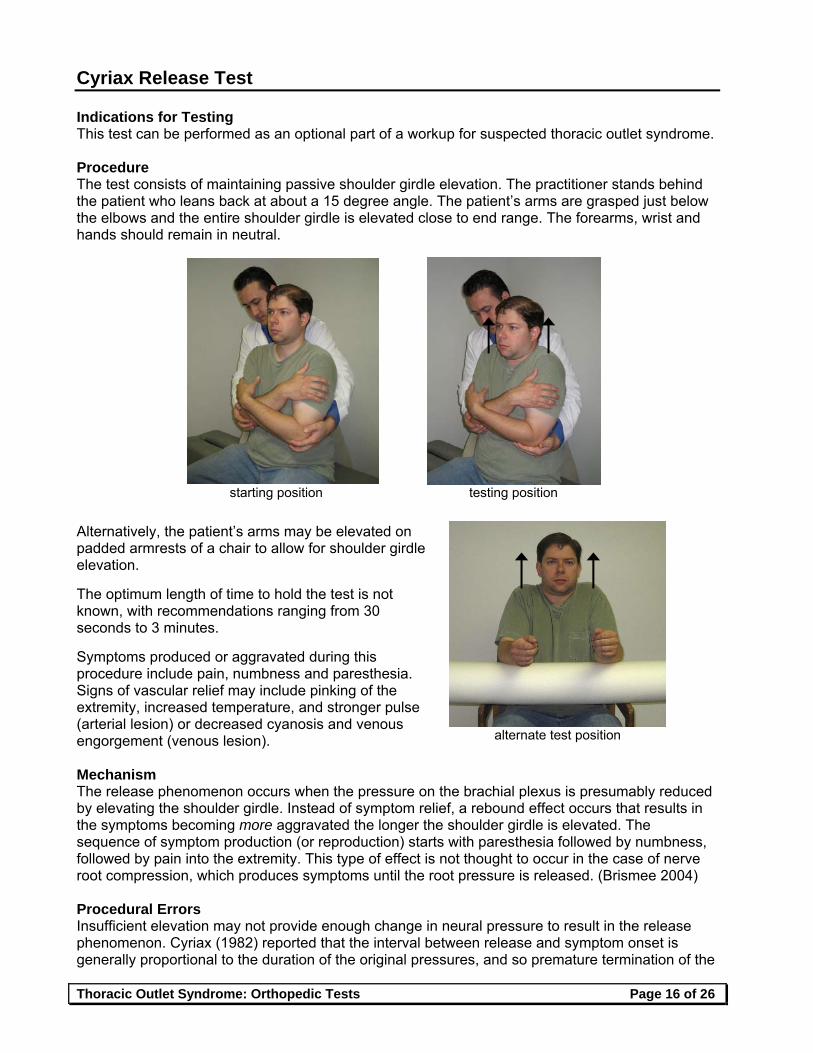
Cyriax Release Test
Indications for Testing This test can be performed as an optional part of a workup for suspected thoracic outlet syndrome. Procedure The test consists of maintaining passive shoulder girdle elevation. The practitioner stands behind the patient who leans back at about a 15 degree angle. The patient’s arms are grasped just below the elbows and the entire shoulder girdle is elevated close to end range. The forearms, wrist and hands should remain in neutral.
Alternatively, the patient’s arms may be elevated on padded armrests of a chair to allow for shoulder girdle elevation. The optimum length of time to hold the test is not known, with recommendations ranging from 30 seconds to 3 minutes. Symptoms produced or aggravated during this procedure include pain, numbness and paresthesia. Signs of vascular relief may include pinking of the extremity, increased temperature, and stronger pulse (arterial lesion) or decreased cyanosis and venous engorgement (venous lesion). Mechanism The release phenomenon occurs when the pressure on the brachial plexus is presumably reduced by elevating the shoulder girdle. Instead of symptom relief, a rebound effect occurs that results in the symptoms becoming more aggravated the longer the shoulder girdle is elevated. The sequence of symptom production (or reproduction) starts with paresthesia followed by numbness, followed by pain into the extremity. This type of effect is not thought to occur in the case of nerve root compression, which produces symptoms until the root pressure is released. (Brismee 2004) Procedural Errors Insufficient elevation may not provide enough change in neural pressure to result in the release phenomenon. Cyriax (1982) reported that the interval between release and symptom onset is generally proportional to the duration of the original pressures, and so premature termination of the
starting position testing position
alternate test position
Thoracic Outlet Syndrome: Orthopedic Tests Page 17 of 26
test may result in false negatives. For patients with chronic symptoms, the shoulder may need to be elevated for 3 minutes before the release phenomenon occurs. Interpretation Production of paresthesia, numbness and/or pain following release of pressure on the brachial plexus may be indicative of neurologic thoracic outlet syndrome. Reproduction of the patient’s familiar symptoms would be considered a stronger positive. Charting A positive test recorded in chart notes should indicate which limb is affected, what symptoms are reproduced, the location, and after what time duration. When part of a narrative report, the procedure must also be described. For example, “Passive elevation of the left shoulder reproduced the patient’s familiar paresthesia at 40 seconds and pain at 50 seconds.” Test Validity The Cyriax release test has not been well studied, and has been reported to have a low false positive rate in an asymptomatic population with a specificity of 97.4% when performing for 1 minute and 87.8% at three minutes. The specificity decreases dramatically after 3 minutes (Brismee 2004). Follow-up Testing See “A Work-Up Strategy” in the introduction to this document. Copyright © 2006, 2008 Western States Chiropractic College Primary author: Charles Novak, DC (10/05)
Revised: Ronald LeFebvre, DC (5/08) Reviewed and revised by CSPE Committee (5/08): Shireesh Bhalerao, DC, CCSP; Daniel DeLapp, DC, DABCO, LAc, ND; Sean Herrin, DC, CCSP; Lorraine Ginter, DC; Stover Harger, DC; Ronald LeFebvre, DC; Owen T. Lynch, DC; Karen E. Petzing, DC; Ravid Raphael, DC, DABCO; Anita Roberts, DC. References Brismee J-M, Gilbert K, et al. Rate of false positive using the Cyriax release test for thoracic outlet syndrome in an
asymptomatic population. JMPT 2004;12(2):73-81. Cyriax J. Textbook of Orthopedic Medicine: Volume 1, 8th edition. Philadelphia: Bailliere Tindall; 1982: 37-8. Karas SB. Thoracic outlet syndrome. Clinics in Sports Medicine 1990;9(2):297-310. Mackinnon SE, Novak CB. Evaluation of the patient with thoracic outlet syndrome. Seminars in Thoracic and
Cardiovascular Surgery 1996 Apr;8(2):190-200. Magee DJ. Orthopedic Physical Assessment, 4th edition. Philadelphia: Saunders; 2002. Nannapaneni R, Marks SM. Neurogenic thoracic outlet syndrome. Br J Neurosurg 2003 Apr;17(2):144-8. Nichols AW. The thoracic outlet syndrome in athletes. J Am Board Fam Pract 1996;9:346-55. Oates SD, Daley RA. Thoracic outlet syndrome. Hand Clinics 1996 Nov;12(4):705-18. Ouriel K. Noninvasive diagnosis of upper extremity vascular disease. Seminars in Vascular Surgery 1998 Jun;11(2):54-9. Rayan GM. Thoracic outlet syndrome. J Shoulder Elbow Surg 1998;7(4):440-51. Schenker M, Kay SPJ. Mini-symposium: Nerve compression syndromes: (iv) Mechanical neuropathy at the thoracic
outlet and associated pain syndrome. Current Orthopedics 2001;15:264-74. Schimp DJ. The symptomatic upper extremity: An algorithmic approach to diagnosis. JACA 1999;Mar-Apr: 32-37, 57. Talmage DM, Lemke C. Thoracic outlet syndrome: How has it changed over the centuries? Top Clin Chiropr
1999;6(4):39-50.
Thoracic Outlet Syndrome: Orthopedic Tests Page 18 of 26
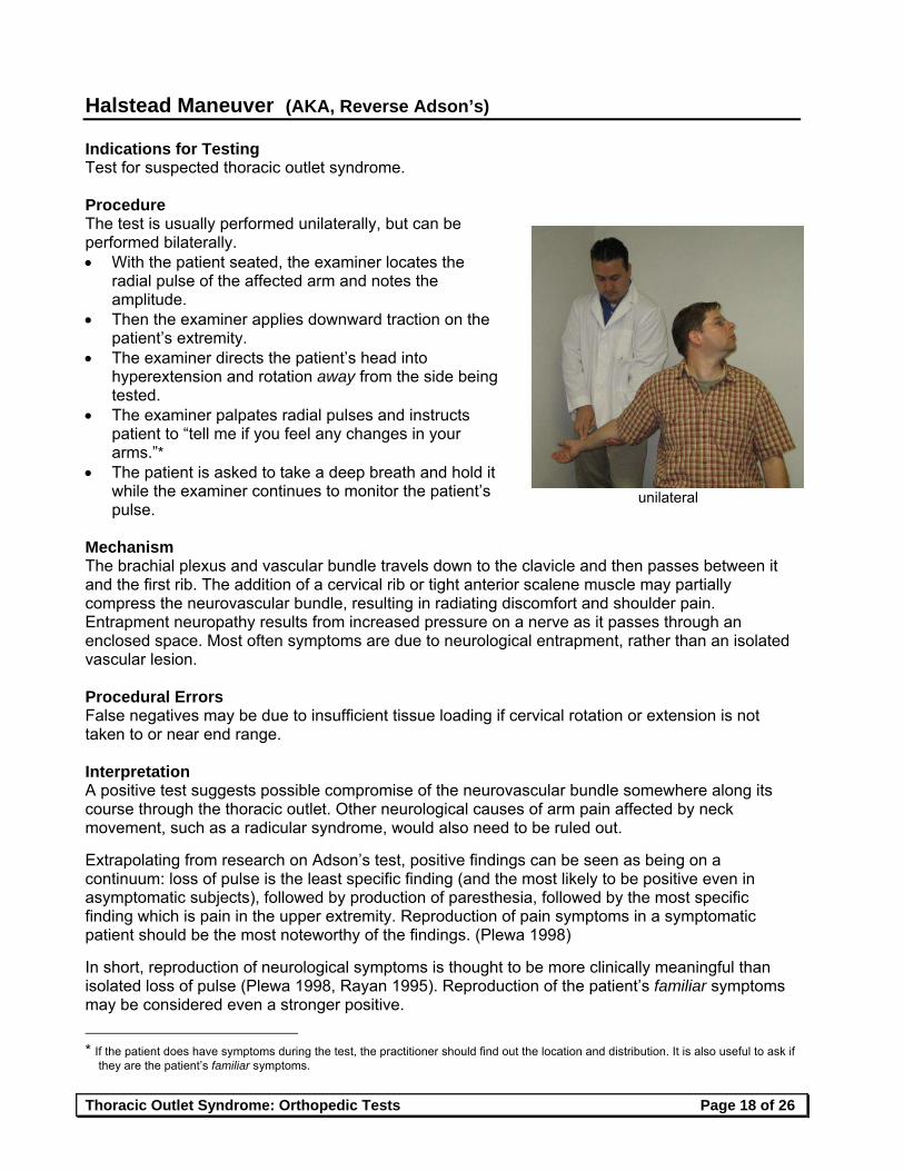
Halstead Maneuver (AKA, Reverse Adson’s) Indications for Testing Test for suspected thoracic outlet syndrome. Procedure The test is usually performed unilaterally, but can be performed bilaterally. With the patient seated, the examiner locates the
radial pulse of the affected arm and notes the amplitude.
Then the examiner applies downward traction on the patient’s extremity.
The examiner directs the patient’s head into hyperextension and rotation away from the side being tested.
The examiner palpates radial pulses and instructs patient to “tell me if you feel any changes in your arms.”*
The patient is asked to take a deep breath and hold it while the examiner continues to monitor the patient’s pulse.
Mechanism The brachial plexus and vascular bundle travels down to the clavicle and then passes between it and the first rib. The addition of a cervical rib or tight anterior scalene muscle may partially compress the neurovascular bundle, resulting in radiating discomfort and shoulder pain. Entrapment neuropathy results from increased pressure on a nerve as it passes through an enclosed space. Most often symptoms are due to neurological entrapment, rather than an isolated vascular lesion. Procedural Errors False negatives may be due to insufficient tissue loading if cervical rotation or extension is not taken to or near end range. Interpretation A positive test suggests possible compromise of the neurovascular bundle somewhere along its course through the thoracic outlet. Other neurological causes of arm pain affected by neck movement, such as a radicular syndrome, would also need to be ruled out. Extrapolating from research on Adson’s test, positive findings can be seen as being on a continuum: loss of pulse is the least specific finding (and the most likely to be positive even in asymptomatic subjects), followed by production of paresthesia, followed by the most specific finding which is pain in the upper extremity. Reproduction of pain symptoms in a symptomatic patient should be the most noteworthy of the findings. (Plewa 1998) In short, reproduction of neurological symptoms is thought to be more clinically meaningful than isolated loss of pulse (Plewa 1998, Rayan 1995). Reproduction of the patient’s familiar symptoms may be considered even a stronger positive.
* If the patient does have symptoms during the test, the practitioner should find out the location and distribution. It is also useful to ask if
they are the patient’s familiar symptoms.
unilateral
Thoracic Outlet Syndrome: Orthopedic Tests Page 19 of 26
Charting A positive test recorded in chart notes should indicate which limb is affected, what symptoms are reproduced, and the location. When part of a narrative report, the procedure must also be described. For example, “Examination revealed loss of the radial pulse on the right with arm extended and head turned toward the left suggesting scalene involvement in a suspected vascular thoracic outlet syndrome.” Note if the test reproduces familiar symptoms. Test Validity This test has not been studied as extensively as Adson’s, costoclavicular or hyperabduction test and so validity numbers are not available. Some authors recommend application of Halstead maneuver along with Adson’s, hyperabduction and Roos tests to increase sensitivity when screening for TOS. (Ouriel 1998, Schenker 2001) Follow-up Testing See “A Work-Up Strategy” in the introduction to this document. Copyright © 2006, 2008 Western States Chiropractic College Primary author: Charles Novak, DC (10/05)
Revised: Ronald LeFebvre, DC (5/08) Reviewed and revised by CSPE Committee (4/08): Shireesh Bhalerao, DC, CCSP; Daniel DeLapp, DC, DABCO, LAc, ND; Sean Herrin, DC, CCSP; Lorraine Ginter, DC; Stover Harger, DC; Ronald LeFebvre, DC; Owen T. Lynch, DC; Karen E. Petzing, DC; Ravid Raphael, DC, DABCO; Anita Roberts, DC. References Cipriano JJ. Photographic Manual of Regional Orthopedic and Neurological Tests, 4th edition. Philadelphia: Lippincott,
Williams & Wilkins; 2003; 160. Evans RC. Illustrated Orthopedic Physical Assessment, 3rd edition. St Louis: Mosby; 2001: 232-3. Gatterman M. Chiropractic Management of Spine Related Disorders. Baltimore: Williams and Wilkins; 1990; 221-2. Magee DJ. Orthopedic Physical Assessment, 3rd edition. Philadelphia: W.B. Saunders Co.; 1997: 220. Ouriel K. Noninvasive diagnosis of upper extremity vascular disease. Seminars in Vascular Surgery 1998
Jun;11(2):5459. Plewa MC, Delinger M. The false positive rate of thoracic outlet syndrome shoulder maneuvers in healthy individuals.
Acad, Emerg Med 1998:5:337-42. Rayan GM, Jensen C. Thoracic outlet syndrome: Provocative examination maneuvers in a typical population. J Shoulder
Elbow Surg 1995;4:113-7. Schenker M, Kay SPJ. Mini-symposium: Nerve compression syndromes: (iv) Mechanical neuropathy at the thoracic
outlet and associated pain syndrome. Current Orthopedics 2001;15:264-74.
Thoracic Outlet Syndrome: Orthopedic Tests Page 20 of 26
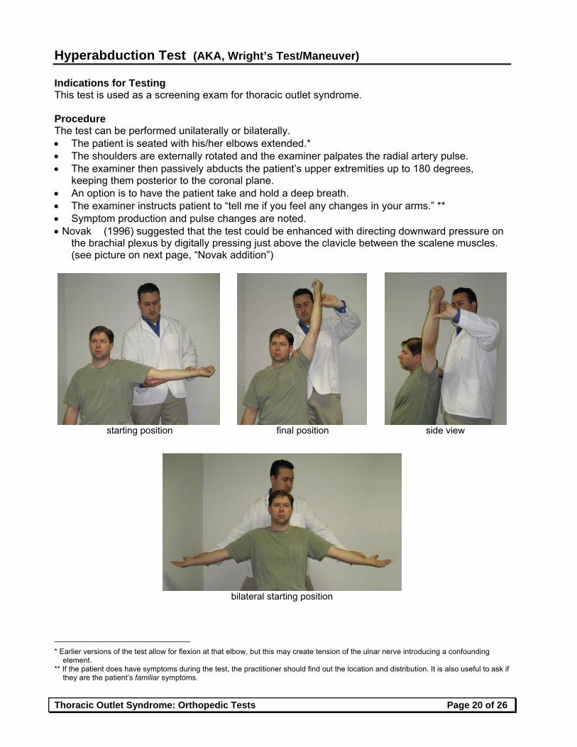
Hyperabduction Test (AKA, Wright’s Test/Maneuver) Indications for Testing This test is used as a screening exam for thoracic outlet syndrome. Procedure The test can be performed unilaterally or bilaterally. The patient is seated with his/her elbows extended.* The shoulders are externally rotated and the examiner palpates the radial artery pulse. The examiner then passively abducts the patient’s upper extremities up to 180 degrees,
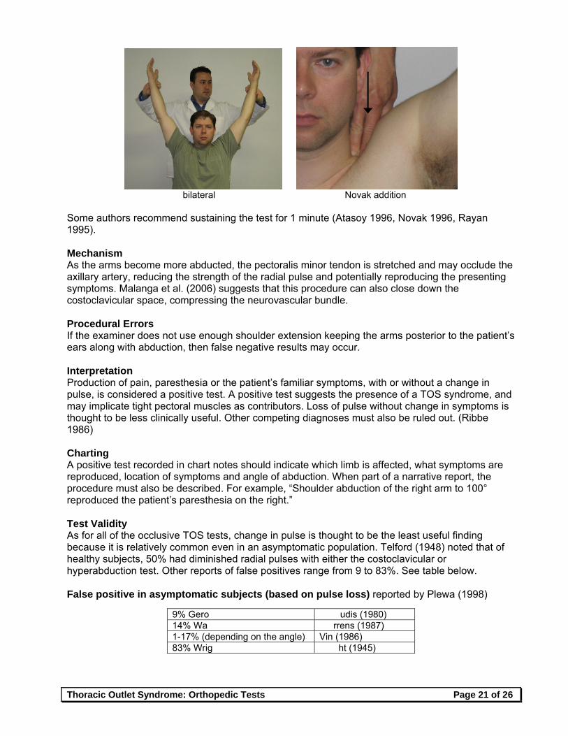
keeping them posterior to the coronal plane. An option is to have the patient take and hold a deep breath. The examiner instructs patient to “tell me if you feel any changes in your arms.” ** Symptom production and pulse changes are noted. Novak (1996) suggested that the test could be enhanced with directing downward pressure on
the brachial plexus by digitally pressing just above the clavicle between the scalene muscles. (see picture on next page, “Novak addition”)
starting position final position side view
bilateral starting position
* Earlier versions of the test allow for flexion at that elbow, but this may create tension of the ulnar nerve introducing a confounding
element. ** If the patient does have symptoms during the test, the practitioner should find out the location and distribution. It is also useful to ask if
they are the patient’s familiar symptoms.
Thoracic Outlet Syndrome: Orthopedic Tests Page 21 of 26
bilateral Novak addition
Some authors recommend sustaining the test for 1 minute (Atasoy 1996, Novak 1996, Rayan 1995). Mechanism As the arms become more abducted, the pectoralis minor tendon is stretched and may occlude the axillary artery, reducing the strength of the radial pulse and potentially reproducing the presenting symptoms. Malanga et al. (2006) suggests that this procedure can also close down the costoclavicular space, compressing the neurovascular bundle. Procedural Errors If the examiner does not use enough shoulder extension keeping the arms posterior to the patient’s ears along with abduction, then false negative results may occur. Interpretation Production of pain, paresthesia or the patient’s familiar symptoms, with or without a change in pulse, is considered a positive test. A positive test suggests the presence of a TOS syndrome, and may implicate tight pectoral muscles as contributors. Loss of pulse without change in symptoms is thought to be less clinically useful. Other competing diagnoses must also be ruled out. (Ribbe 1986) Charting A positive test recorded in chart notes should indicate which limb is affected, what symptoms are reproduced, location of symptoms and angle of abduction. When part of a narrative report, the procedure must also be described. For example, “Shoulder abduction of the right arm to 100° reproduced the patient’s paresthesia on the right.” Test Validity As for all of the occlusive TOS tests, change in pulse is thought to be the least useful finding because it is relatively common even in an asymptomatic population. Telford (1948) noted that of healthy subjects, 50% had diminished radial pulses with either the costoclavicular or hyperabduction test. Other reports of false positives range from 9 to 83%. See table below. False positive in asymptomatic subjects (based on pulse loss) reported by Plewa (1998)
9% Gero udis (1980) 14% Wa rrens (1987) 1-17% (depending on the angle) Vin (1986) 83% Wrig ht (1945)
Thoracic Outlet Syndrome: Orthopedic Tests Page 22 of 26
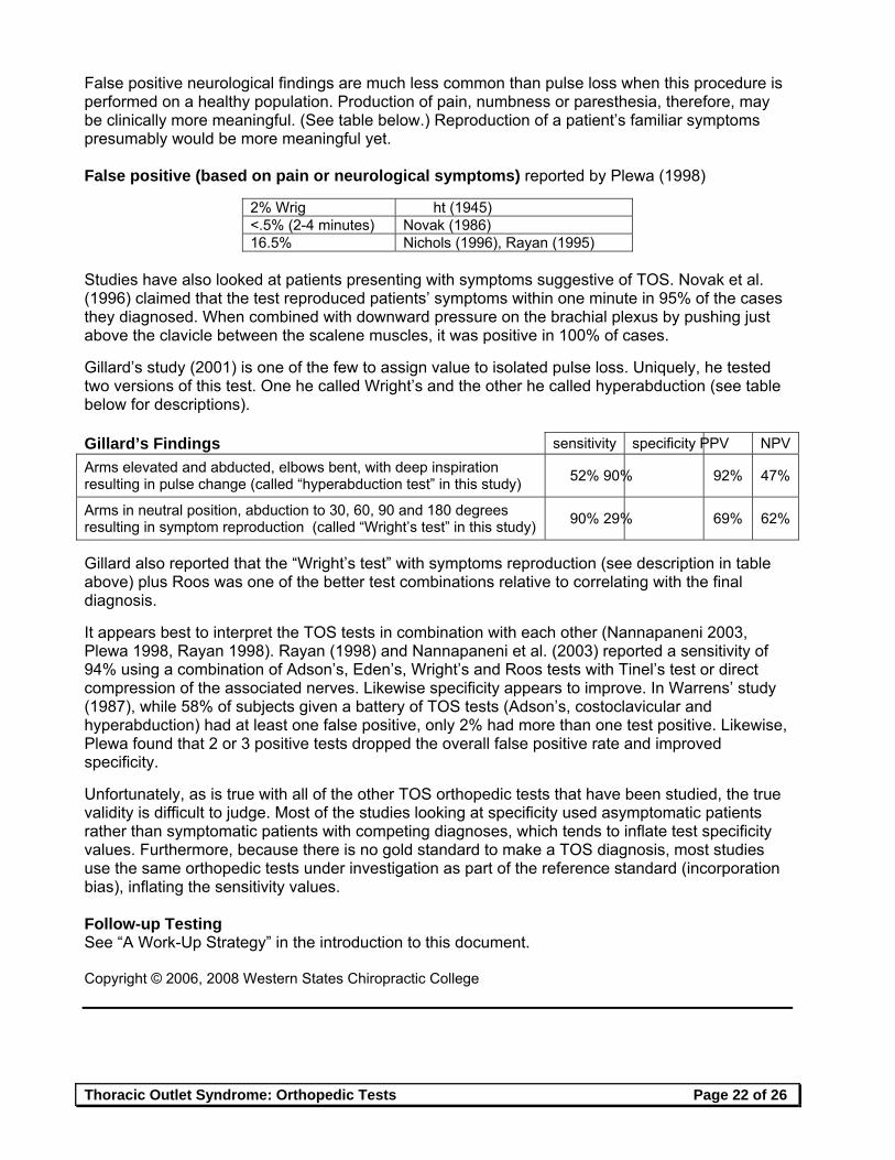
False positive neurological findings are much less common than pulse loss when this procedure is performed on a healthy population. Production of pain, numbness or paresthesia, therefore, may be clinically more meaningful. (See table below.) Reproduction of a patient’s familiar symptoms presumably would be more meaningful yet. False positive (based on pain or neurological symptoms) reported by Plewa (1998)
2% Wrig ht (1945) <.5% (2-4 minutes) Novak (1986) 16.5% Nichols (1996), Rayan (1995)
Studies have also looked at patients presenting with symptoms suggestive of TOS. Novak et al. (1996) claimed that the test reproduced patients’ symptoms within one minute in 95% of the cases they diagnosed. When combined with downward pressure on the brachial plexus by pushing just above the clavicle between the scalene muscles, it was positive in 100% of cases. Gillard’s study (2001) is one of the few to assign value to isolated pulse loss. Uniquely, he tested two versions of this test. One he called Wright’s and the other he called hyperabduction (see table below for descriptions). Gillard’s Findings sensitivity specificity PPV NPVArms elevated and abducted, elbows bent, with deep inspiration resulting in pulse change (called “hyperabduction test” in this study) 52% 90% 92% 47%
Arms in neutral position, abduction to 30, 60, 90 and 180 degrees resulting in symptom reproduction (called “Wright’s test” in this study) 90% 29% 69% 62%
Gillard also reported that the “Wright’s test” with symptoms reproduction (see description in table above) plus Roos was one of the better test combinations relative to correlating with the final diagnosis.
It appears best to interpret the TOS tests in combination with each other (Nannapaneni 2003, Plewa 1998, Rayan 1998). Rayan (1998) and Nannapaneni et al. (2003) reported a sensitivity of 94% using a combination of Adson’s, Eden’s, Wright’s and Roos tests with Tinel’s test or direct compression of the associated nerves. Likewise specificity appears to improve. In Warrens’ study (1987), while 58% of subjects given a battery of TOS tests (Adson’s, costoclavicular and hyperabduction) had at least one false positive, only 2% had more than one test positive. Likewise, Plewa found that 2 or 3 positive tests dropped the overall false positive rate and improved specificity. Unfortunately, as is true with all of the other TOS orthopedic tests that have been studied, the true validity is difficult to judge. Most of the studies looking at specificity used asymptomatic patients rather than symptomatic patients with competing diagnoses, which tends to inflate test specificity values. Furthermore, because there is no gold standard to make a TOS diagnosis, most studies use the same orthopedic tests under investigation as part of the reference standard (incorporation bias), inflating the sensitivity values. Follow-up Testing See “A Work-Up Strategy” in the introduction to this document. Copyright © 2006, 2008 Western States Chiropractic College
Thoracic Outlet Syndrome: Orthopedic Tests Page 23 of 26
Primary author: Charles Novak, DC (10/05)
Revised: Ronald LeFebvre, DC (5/08) Reviewed and revised by CSPE Committee (4/08): Shireesh Bhalerao, DC, CCSP; Daniel DeLapp, DC, DABCO, LAc, ND; Sean Herrin, DC, CCSP; Lorraine Ginter, DC; Stover Harger, DC; Ronald LeFebvre, DC; Owen T. Lynch, DC; Karen E. Petzing, DC; Ravid Raphael, DC, DABCO; Anita Roberts, DC. References Atasoy E. The thoracic outlet compression syndrome. Orthopedic Clincs of North Am 1996;27(2):265-303. Brismee J-M, Gilbert K, et al. Rate of false positive using the Cyriax release test for thoracic outlet syndrome in an
asymptomatic population. JMPT 2004;12(2):73-81. Gerard JA, Kleinfeld SL. Orthopedic testing: A Rational Approach to Diagnosis. New York: Churchill Livingstone; 1993:
60-1. Gillard J, Perez-Cousin M, Hachulla E, et al. Diagnosing thoracic outlet syndrome: Contribution of provocation tests,
ultrasonography, electrophysiology, and helical computed tomography in 48 patients. Joint Bone Spine 2001;68:416-24.
Ide J, Kataoka Y, et al. Compression and stretching of the brachial plexus in thoracic outlet syndrome: Correlation between neuroradiographic findings and symptoms and signs produced by provocation maneuvers. J Hand Surg 2003;28B(3):218-23.
Karas SB. Thoracic outlet syndrome. Clinics in Sports Medicine 1990 Apr;9(2):297-310. Mackinnon SE, Novak CB. Evaluation of the patient with thoracic outlet syndrome. Seminars in Thoracic and
Cardiovascular Surgery 1996 Apr;8(2):190-200. Magee DJ. Orthopedic Physical Assessment, 3rd edition. Philadelphia: W.B. Saunders; 1997: 219. Malanga GA, Nadler SF. Musculoskeletal Physical Examination: An Evidence-Based Approach. Philadelphia: Mosby;
2006: 50-1. Nannapaneni R, Marks SM. Neurogenic thoracic outlet syndrome. Br J Neurosurg 2003 Apr;17(2):144-8. Nichols AW. The thoracic outlet syndrome in athletes. J Am Board Fam Pract 1996;9:346-55. Novak CB, Mackinnon SE. Thoracic outlet syndrome. Occupational Disorder Management 1996;27(4):747-62. Oates SD, Daley RA. Thoracic outlet syndrome. Hand Clinics 1996 Nov;12(4):705-18. Ouriel K. Noninvasive diagnosis of upper extremity vascular disease. Seminars in Vascular Surgery 1998 Jun;11(2):54-9. Plewa MC, Delinger M. The false positive rate of thoracic outlet syndrome shoulder maneuvers in healthy individuals.
Acad, Emerg Med 1998:5:337-42. Rayan GM, Jensen C. Thoracic outlet syndrome: Provocative examination maneuvers in a typical population. J Shoulder
Elbow Surg 1995;4:113-7. Rayan GM. Thoracic outlet syndrome. J Shoulder Elbow Surg 1998;7(4):440-51. Ribbe EB, Lindgren SH, et al. Clinical diagnosis of thoracic outlet syndrome – evaluation of patients with cervicobrachial
symptoms. Manual Medicine 1986;2:82-5. Sapira JD. The Art & Science of Bedside Diagnosis. Baltimore-Munich: Urban & Schwarzenberg; 1990: 344. Schenker M, Kay SPJ. Mini-symposium: Nerve compression syndromes: (iv) Mechanical neuropathy at the thoracic
outlet and associated pain syndrome. Current Orthopedics 2001;15:264-74. Schimp DJ. The symptomatic upper extremity: An algorithmic approach to diagnosis. JACA 1999;Mar-Apr:32-37, 57. Simpson RL, Fern SA. Multiple compression neuropathies and the double-crush syndrome. Orthopedic Clinics of North
Am 1996 Apr;27(2):381-8. Souza TA. Differential Diagnosis and Management for the Chiropractor, 2nd edition. Sudbury, MA: Jones and Bartlett
Publishers; 2001: 52-3. Talmage DM, Lemke C. Thoracic outlet syndrome: How has it changed over the centuries? Top Clin Chiropr
1999;6(4):39-50. Warrens AN, Heaton JM. Thoracic outlet compression syndrome: The lack of reliability of its clinical assesment. Ann R
Coll Surg Engl 1987;69:203-4. White AA, Panjabi MM. Clinical Biomechanics of the Spine, 2nd edition. Philadelphia: J.B. Lippincott Co.; 1990: 412. Cited but not reviewed Telford ED, Mottershead S. Pressure at the cervico-brachial junction: an operative and anatomical study. J Bone Joint
Surg Br 1948;30:249-65.
Thoracic Outlet Syndrome: Orthopedic Tests Page 24 of 26
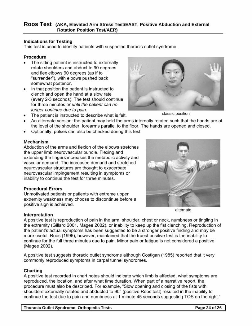
Roos Test (AKA, Elevated Arm Stress Test/EAST, Positive Abduction and External Rotation Position Test/AER)
Indications for Testing This test is used to identify patients with suspected thoracic outlet syndrome. Procedure The sitting patient is instructed to externally
rotate shoulders and abduct to 90 degrees and flex elbows 90 degrees (as if to “surrender”), with elbows pushed back somewhat posterior.
In that position the patient is instructed to clench and open the hand at a slow rate (every 2-3 seconds). The test should continue for three minutes or until the patient can no longer continue due to pain.
The patient is instructed to describe what is felt. An alternate version: the patient may hold the arms internally rotated such that the hands are at
the level of the shoulder, forearms parallel to the floor. The hands are opened and closed. Optionally, pulses can also be checked during this test. Mechanism Abduction of the arms and flexion of the elbows stretches the upper limb neurovascular bundle. Flexing and extending the fingers increases the metabolic activity and vascular demand. The increased demand and stretched neurovascular structures are thought to exacerbate neurovascular impingement resulting in symptoms or inability to continue the test for three minutes. Procedural Errors Unmotivated patients or patients with extreme upper extremity weakness may choose to discontinue before a positive sign is achieved. Interpretation A positive test is reproduction of pain in the arm, shoulder, chest or neck, numbness or tingling in the extremity (Gillard 2001, Magee 2002), or inability to keep up the fist clenching. Reproduction of the patient’s actual symptoms has been suggested to be a stronger positive finding and may be more useful. Roos (1996), however, maintained that the truest positive test is the inability to continue for the full three minutes due to pain. Minor pain or fatigue is not considered a positive (Magee 2002). A positive test suggests thoracic outlet syndrome although Costigan (1985) reported that it very commonly reproduced symptoms in carpal tunnel syndromes. Charting A positive test recorded in chart notes should indicate which limb is affected, what symptoms are reproduced, the location, and after what time duration. When part of a narrative report, the procedure must also be described. For example, “Slow opening and closing of the fists with shoulders externally rotated and abducted to 90° (positive Roos test) resulted in the inability to continue the test due to pain and numbness at 1 minute 45 seconds suggesting TOS on the right.”
classic position
alternate
Thoracic Outlet Syndrome: Orthopedic Tests Page 25 of 26
Test Validity Some authors have reported this to be most sensitive of the TOS tests and more specific than the other TOS tests. (Evans 2001, Novak 1996, Roos 1996) These claims are based mostly on expert opinion. Gillard et al. (2001) reported an 84% sensitivity and 30% specificity in a patient population suspected of having TOS (translating into a positive predictive value of 68% and a negative predictive value of 50%). In this study a positive test was defined as reproducing the patient’s symptoms. Cositgan (1985) reported a false positive rate of 74% in asymptomatic subjects complaining of pain or discomfort, but none discontinued the test due to pain. Likewise, Plewa (1998) reported a 36% false positive rate for paresthesia, but only 3 % of subjects terminated the test due to pain. In Plewa’s study, the arms were held in more abduction, greater than 90 degrees. Roos himself suggested that a true positive for his test was the patient’s inability to continue due to pain. Plewa (1998) found that many of the false positives in his cohort occurred after 90 seconds. Shortening the test might add to test specificity, although may decrease its sensitivity. Relative to pulse change, in one study only 7.5% of normal individuals developed a decrease in pulse with this maneuver. Plewa reported a much higher rate of pulse loss (62%). As with other TOS tests, Roos test appears to perform better when combined with other positive TOS tests. Gillard et al. (2001) found that the combination of a positive Roos and symptom reproduction with hyperabduction/Wright’s test was the best combination (comparing Roos, Wright’s and Adson’s tests). Ribbe (1986) reported that 94% of patients diagnosed with TOS had at least 3 out of 4 of the following items positive in a TOS Index: 1) history of aggravation of symptoms with the arm in an elevated position; 2) a history of paresthesia in the segments along the medial aspect of the hand, forearm and upper arm (C8-T1); 3) tenderness over the brachial plexus supraclavicularly; and 4) positive Roos test (not described). Lindgren et al. (1997) used the same index to identify surgical candidates, 12 of 15 whom had successful outcomes.
As is true with all of the TOS physical exam procedures, most of the studies looking at specificity used asymptomatic patients rather than symptomatic patients with competing diagnoses, which tends to inflate test specificity values. Furthermore, because there is no gold standard to make a TOS diagnosis, most studies, including Ribbe’s trial, used the same orthopedic tests under investigation as part of the reference standard (incorporation bias), inflating the sensitivity values. Follow-up Testing See “A Work-Up Strategy” in the introduction to this document. Copyright © 2006, 2008 Western States Chiropractic College Primary author: Charles Novak, DC (10/05)
Revised: Ronald LeFebvre, DC (5/08) Reviewed and revised by CSPE Committee (4/08): Shireesh Bhalerao, DC, CCSP; Daniel DeLapp, DC, DABCO, LAc, ND; Sean Herrin, DC, CCSP; Lorraine Ginter, DC; Stover Harger, DC; Ronald LeFebvre, DC; Owen T. Lynch, DC; Karen E. Petzing, DC; Ravid Raphael, DC, DABCO; Anita Roberts, DC.
Thoracic Outlet Syndrome: Orthopedic Tests Page 26 of 26
References Brismee J-M, Gilbert K, et al. Rate of false positive using the Cyriax release test for thoracic outlet syndrome in an
asymptomatic population. JMPT 2004;12(2):73-81. Cositgan DA, Wilbourn AJ. The elevated arm stress test: specificity in the diagnosis of the thoracic outlet syndrome.
[Abstract] Neurology 1985;35(suppl 1):74-5. Evans RC. Illustrated Orthopedic Physical Assessment, 3rd edition. St. Louis, MO: Mosby; 2001: 192-5. Gillard J, Perez-Cousin M, Hachulla E, et al. Diagnosing thoracic outlet syndrome: Contribution of provocation tests,
ultrasonography, electrophysiology, and helical computed tomography in 48 patients. Joint Bone Spine 2001;68:416-24.
Ide J, Kataoka Y, et al. Compression and stretching of the brachial plexus in thoracic outlet syndrome: Correlation between neuroradiographic findings and symptoms and signs produced by provocation maneuvers. J Hand Surg 2003;28B(3):218-23.
Karas SB. Thoracic outlet syndrome. Clinics in Sports Medicine 1990 Apr;9(2):297-310. Liebensen CS. Thoracic outlet syndrome: Diagnosis and conservative management. JMPT 1988;(11):493-9. Lindgren K-A, Rytkonen H. Thoracic outlet syndrome: A functional dysfunction of the upper thoracic aperture? J Back
Musculoskel Rehab 1997;8:191-7. Magee DJ. Orthopedic Physical Assessment, 4th edition. Philadelphia: Saunders; 2002. Mackinnon SE, Novak CB. Evaluation of the patient with thoracic outlet syndrome. Seminars in Thoracic and
Cardiovascular Surgery 1996 Apr;8(2):190-200. Nannapaneni R, Marks SM. Neurogenic thoracic outlet syndrome. Br J Neurosurg 2003 Apr;17(2):144-8. Nichols AW. The thoracic outlet syndrome in athletes. J Am Board Fam Pract 1996;9:346-55. Novak CB, Mackinnon SE. Thoracic outlet syndrome. Occupational Disorder Management 1996;27(4):747-62. Oates SD, Daley RA. Thoracic outlet syndrome. Hand Clinics 1996 Nov;12(4):705-18. Ouriel K. Noninvasive diagnosis of upper extremity vascular disease. Seminars in Vascular Surgery 1998 Jun;11(2):54-9. Plewa MC, Delinger M. The false positive rate of thoracic outlet syndrome shoulder maneuvers in healthy individuals.
Acad, Emerg Med 1998:5:337-42. Ribbe EB, Lindgren SH, et al. Clinical diagnosis of thoracic outlet syndrome – evaluation of patients with cervicobrachial
symptoms. Manual Medicine 1986;2:82-5. Roos DB. The place for scalenectomy and first-rib resection in thoracic outlet syndrome. Surgery 1982;(92):1077-85. Roos D. Historical perspectives and anatomic considerations. Seminars in Thoracic and Cardiovascular Surgery 1996
Apr;8(2):183-9. Rayan GM. Thoracic outlet syndrome. J Shoulder Elbow Surg 1998;7(4):440-51. Sapira JD. The Art & Science of Bedside Diagnosis. Baltimore-Munich: Urban & Schwarzenberg; 1990: 344. Schenker M, Kay SPJ. Mini-symposium: Nerve compression syndromes: (iv) Mechanical neuropathy at the thoracic
outlet and associated pain syndrome. Current Orthopedics 2001;15:264-74. Schimp DJ. The symptomatic upper extremity: An algorithmic approach to diagnosis. JACA 1999; Mar-Apr:32-37, 57. Simpson RL, Fern SA. Multiple compression neuropathies and the double-crush syndrome. Orthopedic Clinics of North
Am 1996 Apr;27(2):381-8. Talmage DM, Lemke C. Thoracic outlet syndrome: How has it changed over the centuries? Top Clin Chiropr
1999;6(4):39-50.