thoracic outlet syndrome tos. thoracic outlet syndrome thoracic outlet syndrome results from...
TRANSCRIPT
Thoracic Outlet SyndromeTOS
Thoracic Outlet Syndrome
Thoracic outlet syndrome results from compression of the subclavian vessels and brachial plexus.
Patients may complain of neck and shoulder pain with numbness and tingling in the upper extremity.
The ulnar side is typically involved.
Using the extremity in an overhead or elevated position is difficult.

Clinical Signs and Symptoms
Upper extremity pain.
Upper extremity paresthesias.
Grip weakness.
Clinical Signs and Symptoms
Upper extremity edema.
Upper extremity coldness.
Excessive dryness of the arm or hand.
Excessive sweating of the arm or hand.
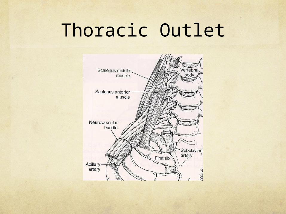
Thoracic Outlet
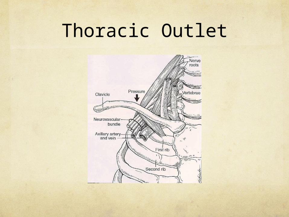
Thoracic Outlet
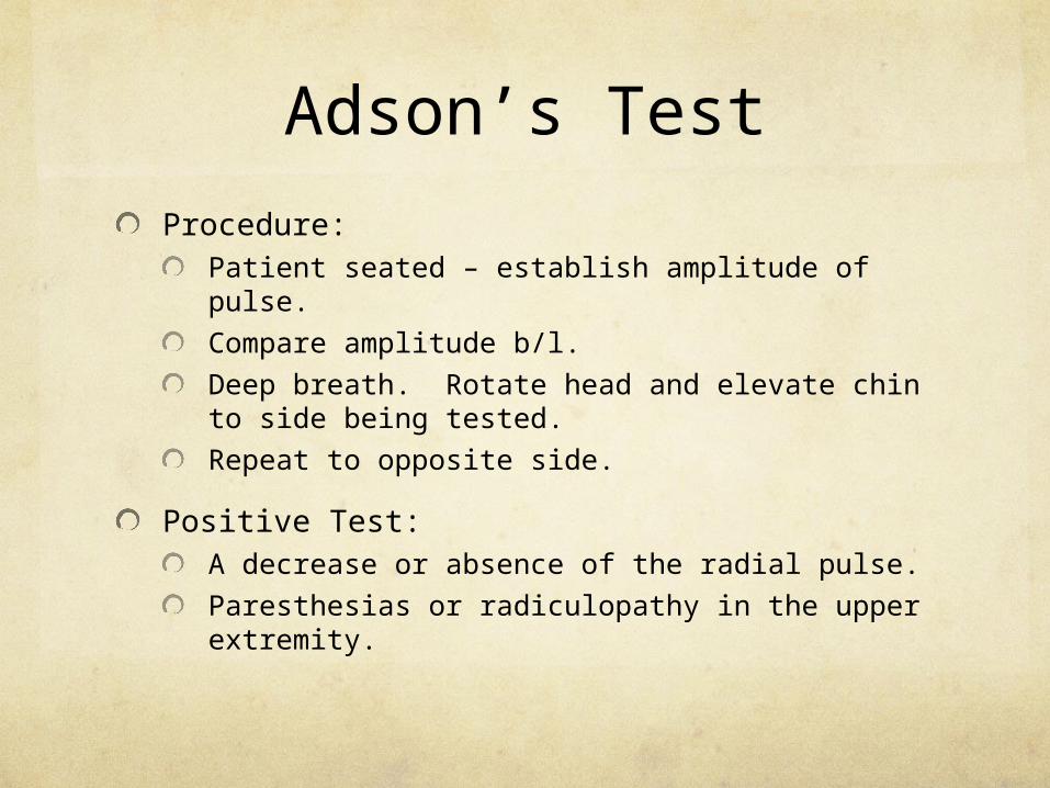
Adson’s Test
Procedure:Patient seated – establish amplitude of pulse.
Compare amplitude b/l.
Deep breath. Rotate head and elevate chin to side being tested.
Repeat to opposite side.
Positive Test:A decrease or absence of the radial pulse.
Paresthesias or radiculopathy in the upper extremity.
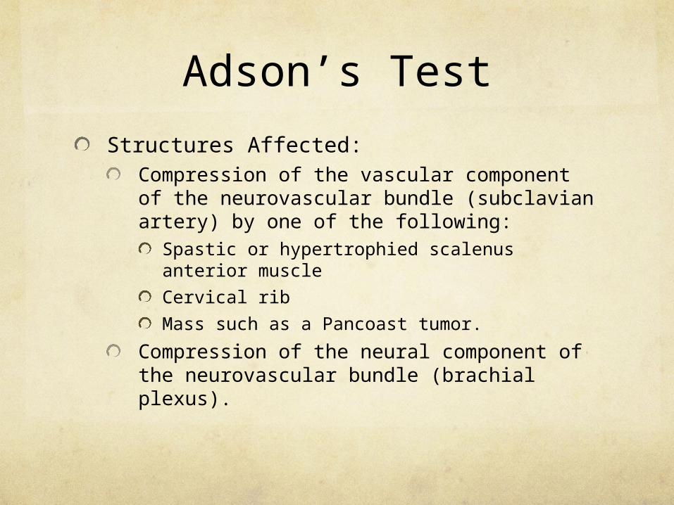
Adson’s Test
Structures Affected:Compression of the vascular component of the neurovascular bundle (subclavian artery) by one of the following:
Spastic or hypertrophied scalenus anterior muscle
Cervical rib
Mass such as a Pancoast tumor.
Compression of the neural component of the neurovascular bundle (brachial plexus).

Adson’s Test
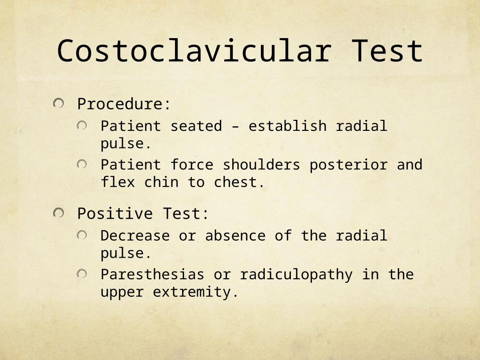
Costoclavicular Test
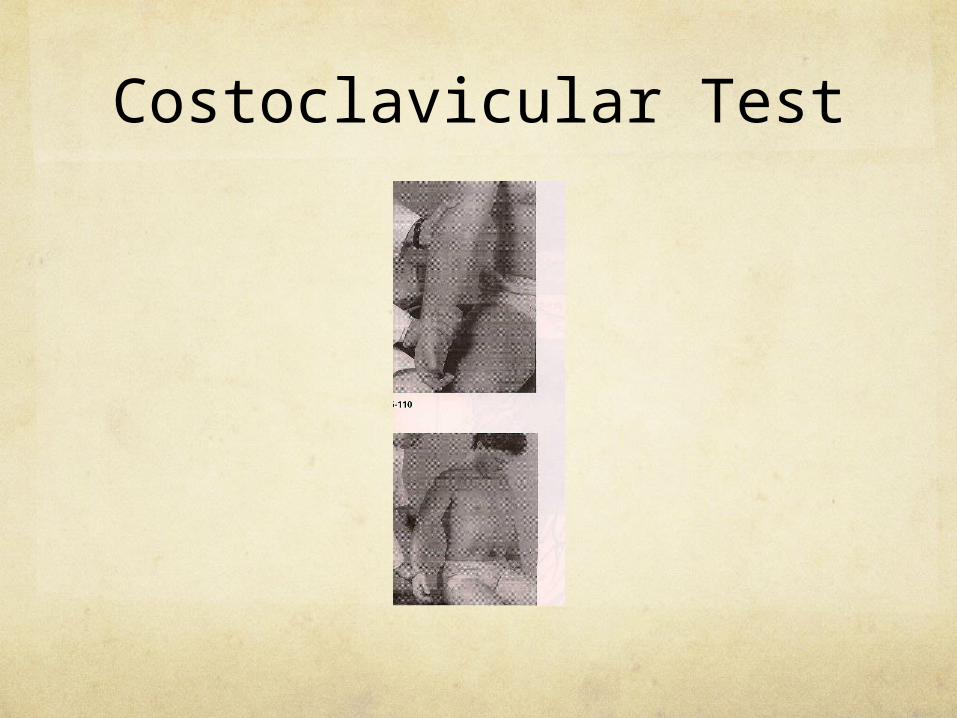
Procedure:Patient seated – establish radial pulse.
Patient force shoulders posterior and flex chin to chest.
Positive Test:Decrease or absence of the radial pulse.
Paresthesias or radiculopathy in the upper extremity.
Costoclavicular Test
Structures Affected:Compression is caused by a decrease in the space between the clavicle and the first rib.
A recent or healed fracture of the clavicle or first rib with or without callus formation, dislocation of the medial aspect of the clavicle, or spastic or hypertrophied subclavius muscle could cause the compression.
Costoclavicular Test
Wright’s Test
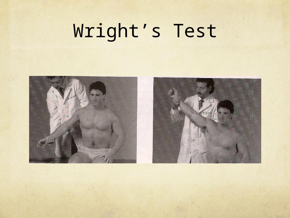
Procedure:Patient seated – establish radial pulse.
Hyperabduct the arm and take the pulse again.
Positive Test:Decrease or absence of the radial pulse.
Structures Affected:Compression of the axillary artery by a spastic or hypertophied pectoralis muscle or a deformed or hypertrophied coracoid process.
Wright’s Test
Traction Test
Procedure:Patient seated – establish radial pulse.
Maintain pulse, extend and traction arm.
Positive Test:A decreased or obliterated pulse alone is not diagnostic; however, when if the test is repeated on the other side and reveals no change it is positive.
Structures Affected:A subluxated or malpositioned first rib or a cervical rib.

Traction Test
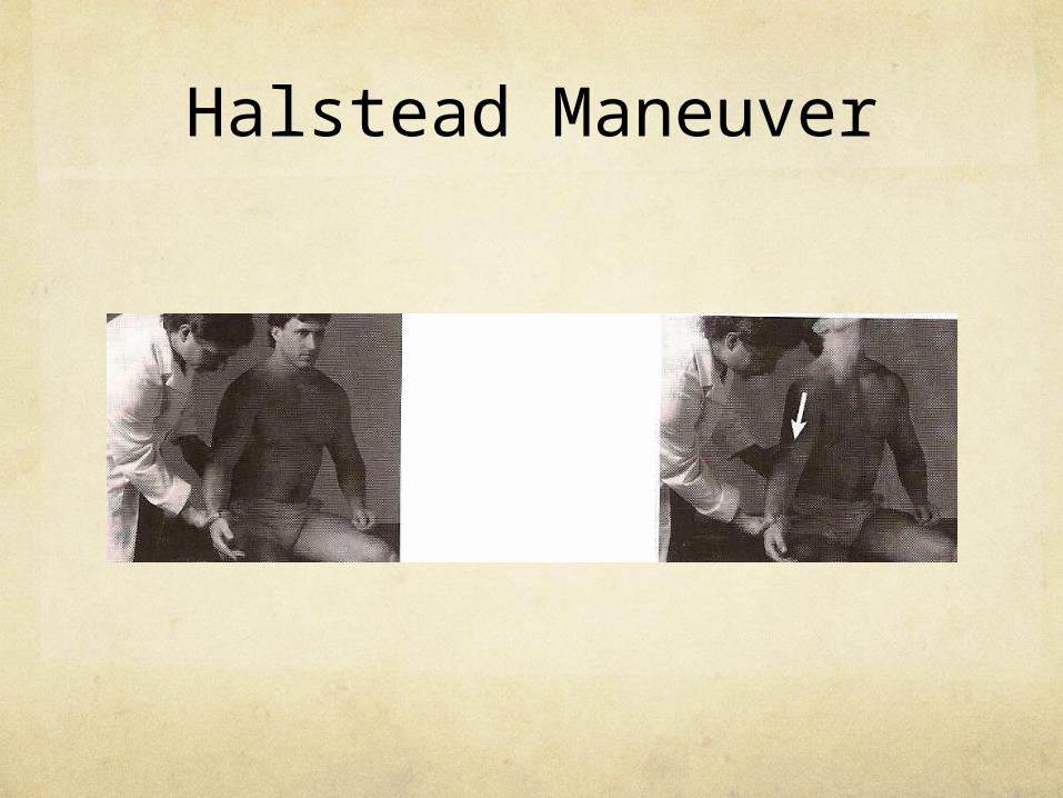
Halstead Maneuver
Procedure:Patient seated – establish radial pulse and note amplitude.
With opposite hand, pull on the patient’s arm and have him hyperextend the neck.
Repeat on opposite side.
Positive Test:Decrease or absence of the radial pulse.
Radiculopathy in the upper extremity.

Halstead Maneuver
Structures Affected:Decrease or absence of the pulse indicates a cervical rib, subluxation, or malposition of the first rib.
A radicular component indicates compression of the brachial plexus by the scalenus anterior muscle.
Halstead Maneuver
Bracial Plexus Irritation
Irritation of the brachial plexus may be due to various factors such as the following:
Cervical rib
Severe upper traction of the arm
Fractured clavicle– Pulmonary apical mass
Clinical Signs and Symptoms
• Upper extremity radicular pain
• Upper extremity paresthesias
• Grip weakness

Brachial Plexus Stretch Test
• Procedure:– Patient seated.– Laterally flex the head opposite to the side
affected.– Extend the shoulder and elbow on the
affected side.
Brachial Plexus Stretch Test
• Positive Test:– Pain and/or paresthesia along the
distribution of the brachial plexus.– Pain on the side of lateral bending may
indicate a nerve root problem.– Local cervical pain on the side of bending
could be a facet joint problem on that side.
Brachial Plexus Stretch Test