thoracic outlet syndrome - pdfs.semanticscholar.org · the effectiveness of tests and provocations...
TRANSCRIPT
The Effectiveness Of Tests And Provocations Used In Diagnosis Of Thoracic Outlet Syndrome Willem Stegeman
Thoracic Outlet Syndrome
The Effectiveness Of Tests And Provocations Used In Diagnosis Of Thoracic Outlet
Syndrome: A Literature Review
By Willem R. Stegeman, PT, DPT, MTC, CEAS
The Effectiveness Of Tests And Provocations Used In Diagnosis Of Thoracic Outlet Syndrome Willem Stegeman
Abstract:
This literature review investigates several tests for clinical evaluation of Thoracic Outlet
Syndrome. Several authors have evaluated provocative tests. Effectiveness and usefulness of
the different research studies is reported. A comparison and contrast of the tests is made
and discussed. Tests for evaluation of vascular response show a high number of false
positive results in the healthy population. Tests for neurologic response have questionable
specificity and sensitivity. Tests for evaluation of neural integrity through nervous
stretching have high specificity and reliability but a lack of controlled studies. The impact of
the available data on clinical decision-making is discussed, after which suggestions for
clinical examination of patients with TOS symptoms are made. Combinations of tests for of
TOS result in increased specificity and sensitivity, and are recommended for clinical
evaluation of this syndrome.
The Effectiveness Of Tests And Provocations Used In Diagnosis Of Thoracic Outlet Syndrome Willem Stegeman
Introduction:
The diagnosis of Thoracic Outlet Syndrome (TOS) remains a highly controversial topic
in healthcare. Much of the controversy relates to the patient with complaints of paresthesias,
numbness and pain but without any definitive test to identify the cause. Clinical diagnosis of TOS
is currently mostly based on provocative positional and compression tests, and on the
reproduction of symptoms. Many of the traditional TOS tests are reported to have low specificity
and reliability, as well as a high incidence of false-positive and false-negative findings.
Electrodiagnostic tests are useful in ruling out other, more distal nerve entrapments. Radiographic
test results are frequently normal in the patient population with TOS symptoms. By contrast,
clinical testing with vascular TOS tests is frequently abnormal in the asymptomatic population. A
number of clinical, radiographic and electrodiagnostic tests have been described in the literature.
However, many tests are considered unreliable, and not one test is regarded as the “gold
standard”. The symptomatology is complex by nature of the structures involved (1,2,3). TOS
consists of a group of impingement syndromes affecting nerves in the brachial plexus and, less
commonly, the arteries, veins and lymph vessels in their passage from cervical spine into the
axillary region. Historically the vascular component of TOS appears to have been largely
overrated. Only in the last decades has the neurogenic etiology been accepted as the dominant
factor in this syndrome.
The purpose of this paper is to provide a better understanding of the TOS syndrome. This
literature review will describe the anatomy of the thoracic outlet as well as the different
dysfunctions of TOS. Provocative tests of the neural integrity, the vascular integrity and
neurovascular integrity are categorized and discussed. Research studies investigating the different
tests are described and compared. Diagnostic difficulties for the physical therapist in the clinical
The Effectiveness Of Tests And Provocations Used In Diagnosis Of Thoracic Outlet Syndrome Willem Stegeman
diagnosis of TOS are discussed, after which suggestions for clinical examination and treatment of
patients with TOS symptoms are made.
Anatomy of the Thoracic Outlet:
The thoracic outlet is the upper lid (operculum) of the chest cage, so named because it is
the site from which the arterial flow of the thorax exits out. The thoracic outlet is approximately a
four-centimeter area with boundaries anteriorly by the manubrium of the sternum, laterally by the
first rib and its costal cartilage, and posteriorly by the body of T1 (4,5,6).
This tightly confined area is covered by Sibson’s fascia and surrounded by the scalene
muscles, which border the nerve roots and trunks of the brachial plexus on their route to the upper
limb. It is further surrounded by the sternocleidomastoideus muscle and the trapezius muscle
which serve as the exit site of the subclavian artery, the subclavian vein and the brachial plexus
(i.e. the neurovascular bundle). The rigid osseous boundaries, and strong muscular and
ligamentous structures, are the underlying anatomical sources of impingement or compression in
the thoracic outlet (4,5,6).
The C4, C5, C6, C7, C8 and T1 spinal nerves exit the cervical foramina and form the
three trunks of the brachial plexus. These pass in between the anterior scalenus muscle and the
medial scalenus muscle. The subclavian artery accompanies the brachial plexus while the
subclavian vein is located in front of the anterior scalenus muscle. The trunks of the brachial
plexus divide into anterior and posterior divisions, which travel under the clavicle, in the space
formed by the first rib and the clavicle (5,6). The cords then form the peripheral nerves into the
upper extremity, namely the musculocutaneus nerve, the axillary nerve, the radial nerve, the
median nerve and the ulnar nerve (7).
The Effectiveness Of Tests And Provocations Used In Diagnosis Of Thoracic Outlet Syndrome Willem Stegeman
Common underlying musculoskeletal and neurological impairments of Thoracic Outlet
Syndrome:
Several anatomical soft tissue structures can be involved in the thoracic outlet syndrome.
Arterial, nerve, vein, or lymphatic system could be compressed, resulting in a variety of signs and
symptoms. Signs and symptoms of compression of the anatomical structures mentioned above
include pain in the upper extremities, at times with radiating character towards the hands and
often only into the fifth digit. At times pain is radiating posteriorly to the head. Paresthesias are
felt in dermatomes C2 through C8 and T1. A sensation of weakness during and after activity can
be experienced. Because of the compression of structures being mostly intermittent, paralysis,
thrombosis and emboli are rarely seen (4).
The subclavian artery is compressed in one to two percent of the cases. The passing of
the artery through the interscalene triangle places it at risk for compression by the muscle bodies
of the scalene whenever there can be muscular hypertrophy. Patients with asthma bronchiale,
chronic obstructive pulmonary disease, hyperventilation, and in practitioners of certain sports as
weight lifting and bodybuilding are at risk. Abnormally wide insertions or fusions of insertions of
both muscles have also been found to cause compression of the subclavian artery. Compression
of the subclavian artery gives rise to ischemia, decreased peripheral pulses, coolness, pallor, and
possible cyanosis of the upper extremity. Overhead activities will result in increasing complaints,
including a temporary “dropping hand”. Signs and symptoms disappear after rest or change of
posture, as with elevation of the shoulders. Possible serious complications of compression of the
subclavian artery are micro-emboli, ulcerations and gangrene, which are rarely seen (4,5,6).
The Effectiveness Of Tests And Provocations Used In Diagnosis Of Thoracic Outlet Syndrome Willem Stegeman
Running directly beside the subclavian artery, with the exception of passage anterior of
the anterior scalenus insertion, and posteriorly to the sternocleidomastoid muscle insertion, is the
subclavian vein. Due to the vein’s escape from very tight inter-muscular passage, venous
compression syndromes are rare (three to four percent), and involve swelling in the hands
whenever the vein is involved. Cyanosis can be present. Patients complain of a “heavy” sensation
in the upper extremity, combined with pain. Serious complications of compression of the
subclavian vein are, as with arterial compression, micro-emboli, ulcerations of the upper
extremity, and gangrene, which is rarely seen (4,5,6).
The third component of the neurovascular bundle, the brachial plexus, follows the same
course as the subclavian artery through the interscalene triangle, only slightly more posterior and
lateral. It is the most frequently involved structure in thoracic outlet compression (90-95 % of all
cases) and produces paresthesias, anesthesias, pain, weakness, and atrophy in the upper extremity
as a result of its compression. Motor deficit is seldom significant but compression of the
neurovascular bundle may present as isolated intrinsic muscle atrophy of the hand without any
pain, at times referred to as a Gilliat-Sumner hand (5,6). The release-phenomenon, as described
by James Cyriax in 1978, can be present. Cyriax was the first to recognize that removal of
causative pressure resulted in increase of neurovascular symptoms, thus creating the release-
phenomenon (8).

The Effectiveness Of Tests And Provocations Used In Diagnosis Of Thoracic Outlet Syndrome Willem Stegeman
Classification of syndromes:
Thoracic outlet compression syndrome is classified according to the three anatomical
compressive regions, which are involved. These regions consist of the interscalene triangle, the
costoclavicular space and the subcoracoid region, and give the three different syndromes their
names.
The scalenus anticus syndrome or Naffziger’s syndrome involves compression of the
brachial plexus and/or the subclavian artery as these structures pass through the interscalene
triangle. Both muscles attach to the first rib and serve as a support structure for the brachial
plexus. Muscle hypertrophy, accessory muscles and C7 transversomegaly (abnormally enlarged
transverse process) usually impose pressure (8,9).
A variation of the syndrome described above is called the cervical rib syndrome.
Accessory ribs of varying size, mono- or bilateral, are found in approximately 0.5 to 1.2 percent
of the general population. Also 14 types of cervical bands have been described, increasing the
risk of compression. The most frequent is the type III band extending from the neck to the scalene
tubercle of the first rib, crossing the concavity of the rib (5). The posterior portion of the brachial
plexus formed by mainly C8 and T1 is mostly involved in this syndrome. Symptoms of
neuropathy most often involve paresthesia in the fourth and fifth fingers, the lateral side of the
hand and possibly the forearm, and decreased sensory appreciation of light touch or pinprick in
The Effectiveness Of Tests And Provocations Used In Diagnosis Of Thoracic Outlet Syndrome Willem Stegeman
the 5th digit. Atrophy, hypertonicity and cramping of the finger flexors, as well as weakness can
also be found (4).
The second major site of compression is found between the mobile clavicle and the
relatively fixed first rib, and is called the costoclavicular syndrome. It may be found in the
normal athletic male with massive shoulders, or may be seen in any individual who typically has
a backwards, downwards thrust of the shoulders as in a military posture. Fractures of the
clavicula producing a large callus, congenitally bifid clavicles, posture, or a thickened and tight
clavipectoral fascia may compromise the space between first rib and clavicula thus decreasing the
available space for passage of the neurovascular bundle (4,5,6,10). Narrowing of the
costoclavicular space places pressure on the subclavian artery and vein, and at times on the
brachial plexus. The costoclavicular signs and symptoms include coolness, pallor, weakness,
paresthesias, diminished distal pulses and edema in the fingers and dorsum of the hand (4,6).
The third syndrome is called the hyperabduction syndrome. The potential area of
compression is in the subcoracoid region adjacent to the pectoralis minor muscle, or the space
where the neurovascular bundle passes through the retroclavicular space, between the clavicle
and the first rib. With full circumduction of the arm, the coracoid process almost forms a fulcrum
under which the subclavian vessels and the neurovascular bundle must pass (5,6,10).
Compression results in pain, numbness, paresthesias and weakness of the upper extremity (4).
Thoracic outlet compression needs to be differentiated from ulnar and median nerve
compression syndromes and cervical radicular signs. Other diagnoses mimicking thoracic outlet
syndrome are a Pancoast tumor, complex regional pain syndrome (type I), cervical disc
protrusions, cervical degenerative processes, poliomyelitis, tuberculosis, syringomyelitis,
inflammatory processes, trauma to the brachial plexus, entrapment neuropathies, M. Raynaud,
migraine occipitalis, hyperventilation and M. Paget-Schroetter (4,5,8,9).
The Effectiveness Of Tests And Provocations Used In Diagnosis Of Thoracic Outlet Syndrome Willem Stegeman
Tests and provocations used to evaluate Thoracic Outlet Syndrome:
Multiple tests for clinical evaluation of TOS have been developed since Adson and
Caffey reported on cervical rib and scalenus anticus syndrome in 1927 (11). The earlier tests used
for provocative testing, described in the literature for the diagnosis of TOS, evaluated vascular
integrity. These tests initially were considered positive when detecting a loss or decrease of the
radial pulse, and for some tests, with reproduction of symptoms. Several specific maneuvers have
been described:
Adson Test or Scalene Maneuver:
The examiner locates the radial pulse. The patient rotates the head toward the tested arm
(the scalene muscles will contract) or rotates away from the tested arm (the scalene muscles will
stretch and tighten thus compressing in a “scissor-like” manner). A positive test is indicated by a
disappearance of the radial pulse.
Allen Maneuver:
The Effectiveness Of Tests And Provocations Used In Diagnosis Of Thoracic Outlet Syndrome Willem Stegeman
The examiner flexes the patient’s elbows to 90 degrees while the shoulder is extended
horizontally and rotated externally. The patient then rotates the head away from the side tested. A
positive test is indicated by a disappearance of the radial pulse.
Halstead Maneuver:
The examiner finds the radial pulse and applies downward traction on the tested
extremity while the patient’s neck is hyper extended and the head rotated to the opposite side.
Absence or disappearance of the radial pulse indicates a positive test.
Eden test or costoclavicular test:
The examiner locates the radial pulse. The patient’s shoulder girdle is depressed and
retracted with the patient leaning away from the arm being tested. The patient then breathes in
deeply. A positive test is indicated by pain, tingling, and by a disappearance of the radial pulse.
Wright’s test (test of the costo-thoraco-pectoral opening):
The examiner elevates and abducts the patient’s arm while palpating the radial pulse. A
determination is made of the speed of, and the angle in the trajectory when symptoms are
replicated.
Hyperabduction maneuver (test of the costo-thoraco-pectoral opening):
The arms are elevated and abducted with the elbows bent. The patient is then asked to
inhale deeply. The test is positive when the radial pulse is abolished or symptoms are replicated.
Novak and McKinnon have adapted this test by keeping the elbow extended and the wrist in
neutral as not to provoke cubital or carpal tunnel syndrome (3).
The Effectiveness Of Tests And Provocations Used In Diagnosis Of Thoracic Outlet Syndrome Willem Stegeman
The EAST test (Elevated Arm Stress Test), Abduction External Rotation test (AER test), Stick up
test, Hands up test or Roos test:
This test may be a good screening test for all types of thoracic outlet compression.
The patient brings the arms up into abduction and external rotation with elbows bend and slightly
behind the head. The patient then opens and closes the hands for three minutes. A positive test is
indicated if pain, heaviness or profound arm weakness or numbness and tingling of the hand are
present.
Not one of the tests described thus far has been accepted as the “gold standard” for TOS,
and many of the tests for vascular insufficiency are reported to show high numbers of false-
positive and false-negative findings. These tests might be inaccurate and insufficient in many
patients with TOS because the vast majority of patients (95%) appear to have complaints relating
to brachial plexus nerve compression, with or without compression of the subclavian vessels
(3,12,13). Use of the previous tests for symptom reproduction shows better specificity than with
testing for vascular integrity. Other tests, which include positional and stretching maneuvers, may
be used to elicit and reproduce symptoms in patients with TOS. The following tests are used:
Tinel’s sign:
Tapping with the examiner’s finger over the nerves may produce a tingling sensation
within the distribution of the nerve. The test is performed over the common entrapment sites in
the upper extremity as the carpal tunnel, the median nerve in the forearm, the cubital tunnel, and
The Effectiveness Of Tests And Provocations Used In Diagnosis Of Thoracic Outlet Syndrome Willem Stegeman
the brachial plexus in the infra- and supraclavicular fossa. This test tends to be positive in later
stages of chronic nerve compression.
Pressure Provocative testing (mechanical allodynia):
The examiner applies direct pressure with thumb or finger tips over nervous tissue as the
brachial plexus, ulnar nerve and median nerve. The test is considered positive when producing
symptoms of paresthesia or numbness in the distribution of the provoked nerve.
Erb’s test or Lasegue of the arm (test for differentiation between radicular and pseudo- radicular
symptoms):
The examiner brings the patient’s head into hetero lateral side bending, and the arm into
extension and abduction, elbow into extension and pronation, wrist and fingers into flexion. A
shooting pain with other symptoms of a herniated nucleus pulposi would indicate radicular
symptoms. Burning pain with slowly increasing tingling sensations and other complaints obtained
in the history would indicate TOS.
Upper Limb Nerve Tension Testing:
Butler (1) describes tension testing utilizing upper limb nerve tension tests, with
emphasis on the upper limb nerve tension test 3 (ULTT 3), which is more sensitive for ulnar
nerve irritation. These tension tests involve the increase of tension of nerves, through stretching
by means of positioning joints in predetermined positions, to tension nerves from cervical nerve
roots to the peripheral nerves in the fingers. The tests are considered positive when reproduction
of symptoms is obtained or range of motion is limited. Butler also recommends performance of
the slump test in longsitting and sitting to investigate the spinal canal components for adverse
neural tension. He also suggests that the possibility of double or multiple crush syndromes should
The Effectiveness Of Tests And Provocations Used In Diagnosis Of Thoracic Outlet Syndrome Willem Stegeman
be investigated, as well as the contribution of the sympathetic nervous system to the symptoms
presented (1,14).
Hypomobility of the first rib has also been hypothesized as a possible cause of TOS.
Lindgren et al (15) investigated patients with hypomobile first ribs, and indicated that
compression of the C8-T1 nerve roots and the stellate ganglion could result in irritation, thus
generating TOS symptoms. Lindgren et al described the following test to evaluate this:
Cervical Rotation Lateral Flexion Test:
This test is used to detect possible restriction of the first rib, which may lead to TOS
symptoms and restricted cervical range of motion. The test is performed by passively and
maximally rotating the neutrally positioned cervical spine away from the painful side, after which
a passive lateral flexion is performed. The test is considered positive when a bony restriction
totally blocks the movement, indicating a hypomobile first rib (15,16,17).
Contrasting and comparing the provocative tests:
Controversy has existed regarding the accuracy of maneuvers for the diagnosis of TOS.
This is partially because of the varying definitions for a positive test for TOS. Furthermore, the
tests that are deemed positive with detection of a decreased radial pulse are considered unreliable
because of the high percentage of positive pulse changes in healthy subjects (18).
Many authors have investigated the reliability, validity, specificity and sensitivity of tests
for clinical evaluation of TOS. Reliability is the extent to which repeated measurements of a
relatively stable phenomenon are close to each other. Validity is the degree to which the results of
a measurement correspond to the true state of the phenomenon being investigated. The sensitivity
of a test is the probability of a positive test among patients with the dysfunction or disease. The
The Effectiveness Of Tests And Provocations Used In Diagnosis Of Thoracic Outlet Syndrome Willem Stegeman
specificity is probability of a negative test among patients without the dysfunction or disease. For
any tests to be a useful tool in clinical evaluation of TOS, the tests should both reliable and valid
(19).
Testing for vascular integrity:
Many authors have investigated testing for vascular integrity in healthy subjects. The
original intent of tests used for provocative testing, described in the literature for the diagnosis of
TOS, evaluated vascular integrity. These tests initially were considered positive when detecting a
loss or decrease of the radial pulse, and for some tests, with reproduction of symptoms. Others
have investigated tests, combining evaluation of vascular and neural integrity.
Rayan and Jensen (11) assessed the prevalence of positive responses to provocative tests
in a healthy population. They investigated the Adson test, the costoclavicular and the hyper
abduction maneuvers for vascular and neurologic responses, as well as Tinel’s sign for a
neurologic response. Tinel’s sign was positive in 7.5% of the extremities, as well as 2% for the
Adson maneuver, 10% for the costoclavicular maneuver and 16.5% for the hyper abduction
maneuver for a neurologic response. A vascular response was positively identified in 13.5 % of
the extremities for the Adson maneuver, 47% for the costoclavicular maneuver and 57% for the
hyperabduction maneuver. The authors concluded that the vascular response was greater than the
neurologic response in all three tests in a healthy population.
Gergoudis and Barnes (20) found high prevalence of thoracic outlet compression of the
subclavian artery in normal individuals. They found significant arterial obstruction in 60% of the
subjects, utilizing the Adson, costoclavicular and hyperabduction maneuvers. The Adson
maneuver resulted in arterial occlusion (complete obstruction of digital pulse amplitude) in 51%
of the subjects and arterial stenosis (more than 75% reduction in digital pulse amplitude) in 2%.
The costoclavicular maneuver resulted in arterial occlusion in 14% without any instance of
The Effectiveness Of Tests And Provocations Used In Diagnosis Of Thoracic Outlet Syndrome Willem Stegeman
arterial stenosis. The hyperabduction maneuver resulted in arterial occlusion in 9% of the
subjects, and arterial stenosis in 10%. They concluded that provocative tests, as the Adson test, as
well as the costoclavicular and hyperabduction maneuvers, used to demonstrate vascular
compression, have little importance in evaluating patients with symptoms suggestive of thoracic
outlet compression. The authors stated this because of brachial plexus compression and irritation
being related to TOS in the majority (98%) of patients. However, they also suggested that the
maneuvers investigated might be helpful in assessing patients with vascular complications of
TOS.
Plewa and Delinger (21) estimated the incidence of false-positive findings of thoracic
outlet syndrome shoulder maneuvers. They tested the Adson test (AT), costoclavicular maneuver
(CCM), elevated arm stress test (EAST), and supraclavicular pressure (SCP) in healthy subjects.
The AT, CCM, EAST, and SCP resulted in an altered pulse in 11%, 11%, 62%, and 21% of
cases, respectively; pain in 0%, 0%, 21%, and 2% of cases, respectively; and paresthesias in 11%,
15%, 36%, and 15% of cases, respectively. These authors concluded that in this study of TOS
shoulder maneuvers in healthy subjects, the outcomes of pulse alteration or paresthesias were
unreliable in general. However, they also found TOS shoulder maneuvers to have reasonably low
false-positive rates with a positive outcome defined as: pain in shoulder, arm or hand during AT,
CCM, or SCP; discontinuation of the EAST secondary to pain in the arm; pain in the same arm
with 2 or more of the tested maneuvers; or any symptom (pain, paresthesias or altered pulse) in
the same arm with 3 or more maneuvers.
Warrens and Heaton (22) described false-positive testing in 64 randomly chosen
volunteers. The tests these authors investigated were the costoclavicular test with 27% positive
findings, the Adson test with 15% and the hyperabduction test with 14%. Although 17% of the
subjects had any symptoms of TOS, 58% of the subjects had a positive result with at least one

The Effectiveness Of Tests And Provocations Used In Diagnosis Of Thoracic Outlet Syndrome Willem Stegeman
maneuver. Only 2% were positive for more than two maneuvers. They suggested that the low
specificity of the tests investigated, devalued these tests in clinical practice.
Marx et al (19) reviewed literature regarding reliability and validity of commonly used
clinical tests for disorders of the upper extremity. They found for the Adson maneuver a
specificity of 32% to 87%, for the costoclavicular maneuver a sensitivity of 94% and specificity
of 53% to 85%, and for the Allen test specificity of 18% to 43%. Ranges were given to reflect the
conflicting findings of the different authors, which is likely due to the fact that the authors chose
varying methods to define the presence of TOS. Since true sensitivity and specificity could not
accurately be determined based on the available literature, the tests were considered to have
insufficient evidence for use in clinical practice.
Testing for neural integrity through symptom reproduction:
Positive findings in many other tests include and emphasize symptom reproduction and
exacerbation of symptoms of TOS. A few studies have described prospectively the false positive
rate of provocative maneuvers in healthy subjects. The EAST test (Elevated Arm Stress Test,
Abduction External Rotation test (AER test), Stick up test, Hands up test or Roos test) has been
the most frequently investigated test. Other tests investigated are the pressure provocation test,
Tinel’s sign and the ULNTT.
Costigan and Wilbourn (23) investigated the sensitivity and specificity of the Elevated
Arm Stress Test (EAST test) for evaluation of patients with neurogenic TOS, a type of TOS
allegedly presenting with sensory symptoms alone, without muscle wasting or bony
abnormalities. They performed testing on 65 patients with clinical and EMG evidence of carpal
tunnel syndrome and 24 a-symptomatic controls. The authors found a positive EAST test in 92%
of carpal tunnel syndrome patients and in 74% of a-symptomatic controls. They concluded the
EAST test not to be specific for TOS. The authors suggested, in patients suspected of neurogenic
The Effectiveness Of Tests And Provocations Used In Diagnosis Of Thoracic Outlet Syndrome Willem Stegeman
TOS, an EMG to be essential to exclude carpal tunnel syndrome before surgical intervention for
TOS is considered. They also found carpal tunnel syndrome to be an unlikely differential
diagnoses in absence of a positive EAST test.
Barsotti and Chiaroni evaluated the EAST test in 150 normal subjects (24). They found
the test to be positive in “nearly all” of the 150 subjects at 2 minutes into the test, because of the
onset of interscapular and forearm pain. This had, according to the authors, nothing to do with the
usual positional syndrome, but was intolerable. In persons with TOS, the test was positive earlier
than in persons without TOS symptoms, namely after 1-2 minutes.
Toomingas et al (25) investigated the results of the Abduction External Rotation (AER)
Test among manual and office workers. They found the sensitivity of the AER test to be moderate
(33% and 80% respectively) while the specificity was considered good (83% and 90%
respectively). A positive association between the AER test and present problems in neck,
shoulder, scapula, upper arm including carpal tunnel syndrome at the time of testing was noted.
Toomingas et al (26) in a different study investigated the predictive aspects of the AER
test. The AER test was conducted among 137 male industrial and office workers at baseline and
after 5 years follow-up, and nerve conduction measurements in the wrist regions were made.
Testing after five years of the subjects with “AER signs” resulted in detection of slowed nerve
conduction in the wrist area in these subjects. They concluded that the AER test predicted future
neck and upper extremity symptoms and signs of nerve compression. According to the
investigators, the results of this study gave support to the "double or multiple crush" theory of
nerve compression. They found the AER test to be a supplementary tool, valuable in
epidemiological and occupational health settings.
Supraclavicular pressure (a pressure provocation test of nerves for mechanical allodynia)
was described by Roos as one of the most helpful tests in the diagnosis of TOS (13). It has been
the subject of few studies. In a study by Costigan and Wilbourn (23) supraclavicular tenderness
The Effectiveness Of Tests And Provocations Used In Diagnosis Of Thoracic Outlet Syndrome Willem Stegeman
was noted in only 4% of the subjects. Plewa and Delinger (21) found the supraclavicular pressure
test rarely painful (2%), with paresthesias noted in 15% of healthy subjects tested.
Rayan and Jensen (11) described Tinel’s sign for clinical evaluation of TOS. These
authors assessed the prevalence of positive responses to provocative tests in a healthy population.
Tinel’s sign was positive in 7.5% of the extremities of healthy subjects. Gillard et al (28)
investigated the usefulness of provocative tests in 48 patients with clinical suspicion of TOS.
Tinel’s sign showed poor results with sensitivity and specificity of respectively 46% and 56%.
Furthermore a positive predictive value of 63% and negative predictive value of 39% was
reported.
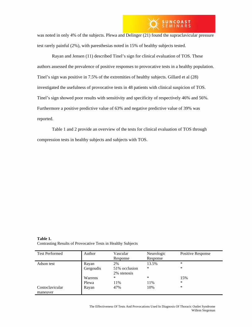
Table 1 and 2 provide an overview of the tests for clinical evaluation of TOS through
compression tests in healthy subjects and subjects with TOS.
Table 1. Contrasting Results of Provocative Tests in Healthy Subjects Test Performed Author Vascular
Response Neurologic Response
Positive Response
Adson test Rayan 2% 13.5% * Gergoudis 51% occlusion
2% stenosis * *
Warrens * * 15% Plewa 11% 11% * Costoclavicular maneuver
Rayan 47% 10% *
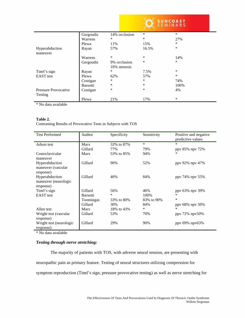
The Effectiveness Of Tests And Provocations Used In Diagnosis Of Thoracic Outlet Syndrome Willem Stegeman
Gergoudis 14% occlusion * * Warrens * * 27% Plewa 11% 15% * Hyperabduction maneuver
Rayan 57% 16.5% *
Warrens * * 14% Gergoudis 9% occlusion
10% stenosis * *
Tinel’s sign Rayan * 7.5% * EAST test Plewa 62% 57% * Costigan * * 74% Barsotti * * 100% Pressure Provocative Testing
Costigan * * 4%
Plewa 21% 17% * * No data available
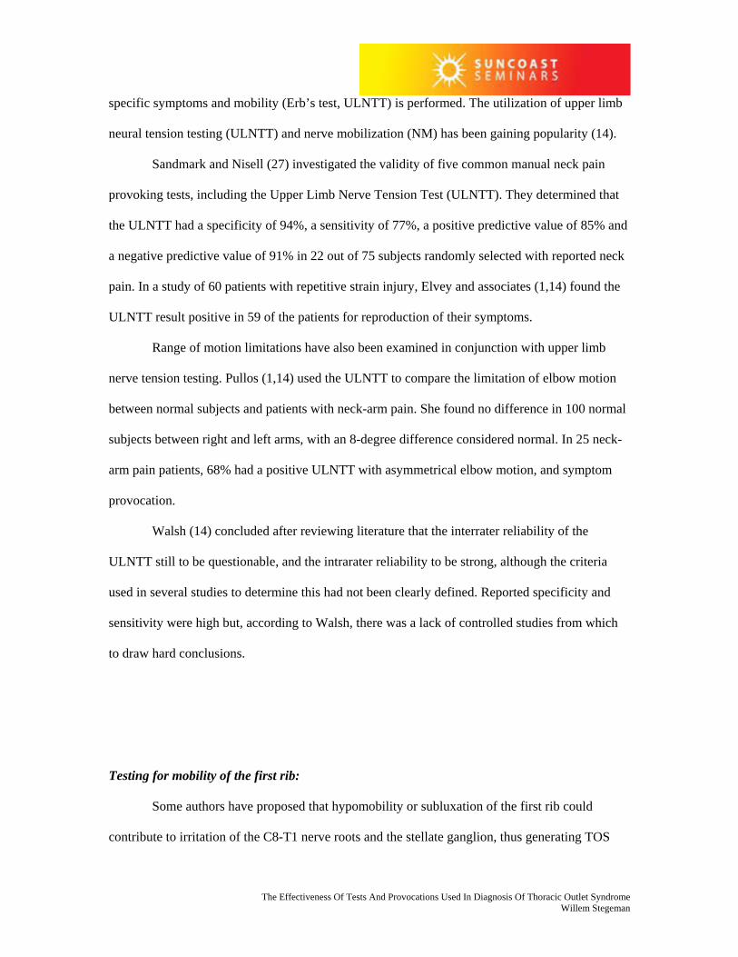
Table 2. Contrasting Results of Provocative Tests in Subjects with TOS Test Performed Author Specificity Sensitivity Positive and negative
predictive values Adson test Marx 32% to 87% * * Gillard 77% 79% ppv 85% npv 72% Costoclavicular maneuver
Marx 53% to 85% 94% *
Hyperabduction maneuver (vascular response)
Gillard 90% 52% ppv 92% npv 47%
Hyperabduction maneuver (neurologic response)
Gillard 40% 84% ppv 74% npv 55%
Tinel’s sign Gillard 56% 46% ppv 63% npv 39% EAST test Barsotti * 100% * Toomingas 33% to 80% 83% to 90% * Gillard 30% 84% ppv 68% npv 50% Allen test Marx 18% to 43% * * Wright test (vascular response)
Gillard 53% 70% ppv 72% npv50%
Wright test (neurologic response)
Gillard 29% 90% ppv 69% npv63%
* No data available
Testing through nerve stretching:
The majority of patients with TOS, with adverse neural tension, are presenting with
neuropathic pain as primary feature. Testing of neural structures utilizing compression for
symptom reproduction (Tinel’s sign, pressure provocative testing) as well as nerve stretching for
The Effectiveness Of Tests And Provocations Used In Diagnosis Of Thoracic Outlet Syndrome Willem Stegeman
specific symptoms and mobility (Erb’s test, ULNTT) is performed. The utilization of upper limb
neural tension testing (ULNTT) and nerve mobilization (NM) has been gaining popularity (14).
Sandmark and Nisell (27) investigated the validity of five common manual neck pain
provoking tests, including the Upper Limb Nerve Tension Test (ULNTT). They determined that
the ULNTT had a specificity of 94%, a sensitivity of 77%, a positive predictive value of 85% and
a negative predictive value of 91% in 22 out of 75 subjects randomly selected with reported neck
pain. In a study of 60 patients with repetitive strain injury, Elvey and associates (1,14) found the
ULNTT result positive in 59 of the patients for reproduction of their symptoms.
Range of motion limitations have also been examined in conjunction with upper limb
nerve tension testing. Pullos (1,14) used the ULNTT to compare the limitation of elbow motion
between normal subjects and patients with neck-arm pain. She found no difference in 100 normal
subjects between right and left arms, with an 8-degree difference considered normal. In 25 neck-
arm pain patients, 68% had a positive ULNTT with asymmetrical elbow motion, and symptom
provocation.
Walsh (14) concluded after reviewing literature that the interrater reliability of the
ULNTT still to be questionable, and the intrarater reliability to be strong, although the criteria
used in several studies to determine this had not been clearly defined. Reported specificity and
sensitivity were high but, according to Walsh, there was a lack of controlled studies from which
to draw hard conclusions.
Testing for mobility of the first rib:
Some authors have proposed that hypomobility or subluxation of the first rib could
contribute to irritation of the C8-T1 nerve roots and the stellate ganglion, thus generating TOS
The Effectiveness Of Tests And Provocations Used In Diagnosis Of Thoracic Outlet Syndrome Willem Stegeman
symptoms. Lindgren and Leino (17) reported on patients with subluxation of the first rib as a
possible mechanism for creating symptoms of TOS. They described 22 cases of both TOS and
complex regional pain syndrome, type I. Each case had a hypomobile first rib on the affected
side. After isometric exercises of the scalene muscles, the mobility of the first rib was restored
and symptoms completely relieved in 13 subjects. They concluded that subluxation of the first rib
may irritate the neural network and the stellate ganglion close to the first costotransverse joint.
Lindgren et al (15,16) also investigated the use of testing the combined motion of
cervical spine rotation and lateral flexion (CRLF test) for detection of subluxation and restricted
movement of the first rib. They found excellent interexaminer reliability. The number of false
positive tested ribs was 3 out of 23 subjects; the false negative rate was 2 out of 23 subjects. They
suggested inclusion of the test in examination for cervical mobility, as well as for patients
presenting with symptoms of brachialgia and TOS.
Testing through combinations of multiple tests:
Several authors have reported on improved specificity and reliability of tests for
diagnosis of TOS by combining multiple tests with other areas of the clinical evaluation, as the
history, range of motion and muscle tightness. Combinations of tests and specific areas of clinical
evaluation have shown significantly improved sensitivity and specificity.
Ribbe and Lindgren (18) studied 315 patients with cervicobrachial symptoms. They
found the most reliable symptoms to detect TOS to be: (1) a history of aggravation of symptoms
with the arm elevated, (2) a history of paresthesias in C8-Th1, (3) tenderness over the brachial
plexus supraclavicularly, (4) a positive abduction and external rotation test (AER test). They
found three out of four tests present in 94% of the TOS cases investigated. Thirty-three percent of
the time they had false-positive results. The investigators referred to these four tests as the
“thoracic outlet syndrome index”. Findings were confirmed by Plewa and Delinger (21). They
The Effectiveness Of Tests And Provocations Used In Diagnosis Of Thoracic Outlet Syndrome Willem Stegeman
reported reasonable specificity when tests resulted in pain in the same arm with 2 or more of the
tested maneuvers, or any symptom in the same arm with 3 or more maneuvers.
Gillard et al (28) investigated the usefulness of provocative tests in 48 patients with
clinical suspicion of TOS. They investigated the Adson test, the hyperabduction maneuver, the
Wright test, the Roos test and Tinel’s sign. They found limited sensitivity and specificity of the
tests. Considering all five provocative tests, the tests showed a mean sensitivity and specificity of
respectively 72% (deemed acceptable) and 53% (deemed poor). The Adson test had the best
outcome for sensitivity and specificity with respectively 79% and 76%. Tinel’s sign showed the
worst outcome for sensitivity and specificity with respectively 46% and 56%. Pairs of tests
improved the specificity compared with tests alone; the gain in sensitivity, however, was smaller
then the gain in specificity. Combinations of three, four and five maneuvers involving the Adson
test, the Wright test and hyperabduction maneuver with abolition of radial pulse, the Wright test
with reproduction of symptoms, the Roos test and Tinel’s sign increased specificity but not
sensitivity. The sensitivity and specificity mostly improved when several provocative tests were
used in combination with the best test being the Adson test, particularly when used in
combination with the Roos test (sensitivity 72%, specificity 82%), the hyperabduction maneuver
(sensitivity 72%, specificity 88%), or the Wright test (sensitivity 79%, specificity 76%).
Pascarelli and Hsu (29) investigated work related upper extremity disorders in 485
computer users, musicians and others engaged in repetitive work. They found that 70% of the
persons had clinical evidence of TOS after testing for a positive reaction in two out of five tests.
These tests consisted of the EAST test, Wright’s maneuver, positive supra- and infraclavicular
mechanical allodynia, and scalene tightness with decreased range of motion in the neck.
Roos (13) uses five tests, which he believes are the most reliable for diagnosing
neurogenic TOS: Tinel’s sign (performed over scalene, and supra- and infraclavicular), pressure
The Effectiveness Of Tests And Provocations Used In Diagnosis Of Thoracic Outlet Syndrome Willem Stegeman
provocative testing (supraclavicular), strength testing (triceps, interosseous hand muscles and
handgrip testing), hypesthesia (C8, T1 dermatomes) and the EAST test.
Summary of the tests:
Results of the tests as described in table 1 and 2 vary strongly. Several reasons for these
variations can be identified in the articles investigated. Many of these reasons are identified and
described.
Reasons for variation of tests:
The definitions for a positive test might have been different in the investigation of tests
making a comparison less effective. When based on findings of a diminished radial pulse, many
maneuvers are thought to be unreliable, since pulse alterations may be found in high percentages
of healthy individuals. Execution of the tests may have been different, as well as the duration of
performed tests, which is not always reported. Furthermore, provocative testing can exacerbate
upper limb symptoms, even when related to other causes than TOS (28). Positive tests may have
identified patients with asymptomatic carpal tunnel syndrome, cervical disc disease or individuals
with a predisposition for TOS (21) and have influenced outcomes. Sample size varied strongly,
from 22 subjects in the investigation of Lindgren (15) to 485 subjects in the review by Pascarelli
and Hsu (29). Also the composition of the samples has influenced the results, since females show
a greater response than males. This difference correlates with the clinically observed prevalence
of TOS in the general population, probably due to anatomical differences of the chest wall
between the two sexes (18). Age composition of the samples may have played a role. The
younger age group in the article by Rayan and Jensen (11) had a tendency for a greater vascular
response than the older age group. Occupation of the patients has a correlation with positive
testing for TOS. Toomingas et al (26) reported that vibration exposure and the vibration
The Effectiveness Of Tests And Provocations Used In Diagnosis Of Thoracic Outlet Syndrome Willem Stegeman
acceleration level experienced during work were associated with a positive AER test.
Furthermore, they found a positive correlation between the AER test and neck and arm pain and,
to some extent, carpal tunnel syndrome. This indicates that the presence of minor serial
impingements along a peripheral nerve may result in a positive test for TOS and has to be taken
into account. Therefore these authors recommended inclusion of testing of more distal sites of
possible nerve compression.
Comparing the tests:
Some authors report significantly higher or lower outcomes than others. The investigation
of the Adson test by Gergoudis (20) reveals a very high positive response in comparison with
other authors. No duration of the test performed was given in the article, which might have
influenced the outcome. Marx et al (19) found, in his literature review, a range of specificity of
32% to 87% for the Adson test in patients with TOS, as well as ranges of 53% to 85% and 18% to
43% for respectively the costoclavicular and Allen test. They indicated that the conflicting results
were probably due to the fact that the investigators choose varying methods to define the presence
of TOS. Rayan (18) reports a high vascular response for the costoclavicular test as well as the
hyperabduction maneuver. The composition of his samples for age and gender might have
influenced the outcomes in their study. The EAST test shows high false-positive results in healthy
subjects, in the literature reviewed, ranging from 57% neurologic response to 100% positive
response (19, 23). The duration of the test plays an important part in this test. Roos (30) indicates
that the test should be performed for three minutes, which according to several authors results in
poor specificity (20, 23, 24). Plewa (21) indicated that shortening the test to 90 seconds might
increase the specificity, since many of their healthy subjects did not develop symptoms until after
90 seconds. Many other tests as the Allen maneuver, the Halstead maneuver, the CRLF test and
the Wright test have not been extensively reported on.
The Effectiveness Of Tests And Provocations Used In Diagnosis Of Thoracic Outlet Syndrome Willem Stegeman
Results of the tests:
Sensitivity and specificity of the tests for vascular integrity, when used in isolation, are
mostly low. False-positive results are high. Tests used in isolation often result in unreliable tests.
However, combinations of tests appear to increase specificity and sensitivity, as well as decrease
the rate of false-positive results (21). The Adson test, particularly when used in combination with
the hyperabduction maneuver, the Roos test and the Wright test show satisfactory specificity and
sensitivity (28). Of the tests for neural integrity, the ULNTT appear to have satisfactory outcomes
although a lack of controlled studies from which to draw hard conclusions is reported (14).
Testing for mobility of the first rib has been the subject of few studies (15,16,17). The CRLF test
appears to be a good addition to the “standard “ TOS tests, although further investigation with
larger samples is warranted before hard conclusions regarding reliability can be drawn.
Suggestions:
Future investigation of tests used for clinical evaluation should include a sufficient
sample size. Depending on the type of investigation, it should represent the proper population
investigated, in gender, age and occupation, since this may have significant influence on the
outcomes. Degree of pressure or traction given during testing should also be measured, as well as
duration of the tests performed, to improve objectivity of the tests.
The Effectiveness Of Tests And Provocations Used In Diagnosis Of Thoracic Outlet Syndrome Willem Stegeman
Diagnostic difficulties:
Several difficulties regarding the anatomy and pathofysiology of the thoracic outlet exist,
making the diagnosis of TOS through clinical testing complicated. Experts regarding the topic of
TOS have diverging views on the pathogeneses. There is no agreement on whether the basic
injury is one of compression or traction.
Several theories regarding the pathogenesis of TOS have been proposed (31). The first
theory suggests that, since anatomical anomalies are prevalent in the normal population, they
might predispose healthy subjects for symptoms of TOS, as well as to false-positive provocative
testing (13,32).
As second theory occupational and postural factors as descending of the shoulder girdle in
middle-aged adults has been implicated in the pathofysiology of TOS (33). Normal rib and
muscle structures could cause brachial plexus compression and stretching because of muscle
disuse atrophy, muscle imbalance, muscle shortening, compensatory muscle overuse, protraction
of the shoulders and forward head posture (2, 18).
A third theory proposes that trauma could result in scarring of the brachial plexus. Nerve
tissue could adhere to other structures causing decreased range of motion. Bleeding and
subsequent fibrosis, contractures and shortening of connective tissue could be the result.
Eventually a change in muscle type of the scalene muscles might be seen (31).
Diagnosis of TOS:
Accurate diagnosis of thoracic outlet syndrome is difficult for many reasons. Clinical
diagnosis of TOS is presently based predominantly on subjective complaints of the upper
The Effectiveness Of Tests And Provocations Used In Diagnosis Of Thoracic Outlet Syndrome Willem Stegeman
extremity as paresthesia, anesthesia and pain. Many provocative tests, resulting in subjective
complaints, reveal low specificity and sensitivity. Objective tests, mostly used by other
professionals than Physical Therapists, as X-ray films, computed tomography scans, nerve
conduction studies and somatosensory evoked potentials appear most useful for diagnosing
moderate to severe cases of vascular or neural compromise (31) as well as for ruling out of other
diagnoses (2,3).
Several provocative tests and maneuvers have been described in the literature. The original
provocative tests described for the diagnosis of TOS evaluated vascular integrity, noting a loss or
decrease of radial pulses with or without the reproduction of symptoms. These tests, which
evaluated the effects of positional changes on the radial pulse for diagnosis of TOS, and other
tests have proven unreliable because of the high incidence of false- positive findings in healthy
subjects. (18,19,20,21,22,23,24). Testing for neurologic response has shown poor to fair
specificity and fair to good sensitivity. Only nerve tension tests and testing for mobility of the
first rib have shown good specificity and sensitivity. Unfortunately, there is a lack of controlled
studies from which to draw hard conclusions (14).
Influence of anatomical abnormalities and posture on testing:
Juvonen et al (32) investigated the rate of anomalies at the thoracic outlet in the general
population. They found normal bilateral thoracic anatomy in 5 out of 50 cadavers (10%) during
98 dissections. They concluded that congenital anatomic abnormalities are frequent in the normal
population. They also suggested that the abnormalities would predispose subjects to TOS after
stress, related to occupation and cervical injuries Roos initially described 7 different types of
congenital bands and ligaments, and finding these in 33% of the general population. Roos later
increased this number after finding 7 more anomalies to 14 (13,31,32).
The Effectiveness Of Tests And Provocations Used In Diagnosis Of Thoracic Outlet Syndrome Willem Stegeman
Occupational factors have been hypothesized to be a factor in development of TOS.
Hairdressers, painters, construction and industrial workers, nursing staff, switchboard operators,
and other workers performing repetitive manual work (rather than heavy work) have an increased
prevalence of TOS. Juvonen et al reported 60-80% of the normal population to have positive
findings in clinical tests aimed at irritating the neurovascular bundle in the thoracic outlet. They
suggested that the presence of abnormal fibromuscular bands often resulted in positive findings in
during provocative tests for TOS. This would predispose subjects to TOS after certain stress,
possibly related to occupation or cervico-brachial injury (32).
Machleder (34) indicated that anatomical abnormalities represent stages in a spectrum of
developmental variation of the neurovascular and musculoskeletal structures at the base of the
neck. He concluded that the abnormalities could become significant in settings of unusual
physical requirements, repetitive stress, or injury. He indicated that repetitive mechanical stress
on the job could eventually expose anatomical weaknesses in the thoracic outlet.
Several authors describe congenital abnormalities, which could contribute to the
pathogenesis of TOS. Examples are cervical ribs, abnormal thoracic ribs, and scalene muscle- and
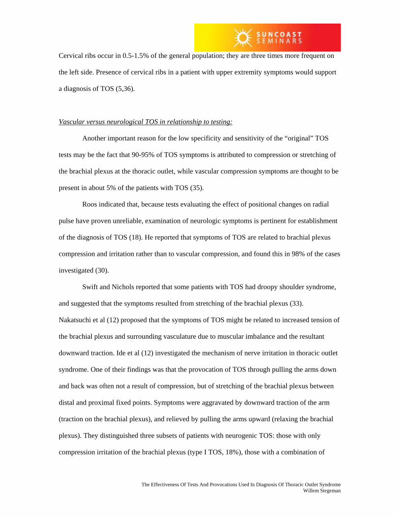
insertion abnormalities. Table 3 shows a classification Gruber made of the cervical rib anomalies
in 1842.
Table 3. Gruber Classification of Cervical Rib Anomalies: Anomaly Type Description
Type I large, hypertrophic transverse process of C7
Type II rudimentary rib, but with free extremity and no connection to the first rib
Type III incomplete rib connected to the first thoracic rib by a fibrous band
Type IV complete cervical rib fusing with the first rib or connecting with it by a cartilaginous pseudo-articulation
The Effectiveness Of Tests And Provocations Used In Diagnosis Of Thoracic Outlet Syndrome Willem Stegeman
Cervical ribs occur in 0.5-1.5% of the general population; they are three times more frequent on
the left side. Presence of cervical ribs in a patient with upper extremity symptoms would support
a diagnosis of TOS (5,36).
Vascular versus neurological TOS in relationship to testing:
Another important reason for the low specificity and sensitivity of the “original” TOS
tests may be the fact that 90-95% of TOS symptoms is attributed to compression or stretching of
the brachial plexus at the thoracic outlet, while vascular compression symptoms are thought to be
present in about 5% of the patients with TOS (35).
Roos indicated that, because tests evaluating the effect of positional changes on radial
pulse have proven unreliable, examination of neurologic symptoms is pertinent for establishment
of the diagnosis of TOS (18). He reported that symptoms of TOS are related to brachial plexus
compression and irritation rather than to vascular compression, and found this in 98% of the cases
investigated (30).
Swift and Nichols reported that some patients with TOS had droopy shoulder syndrome,
and suggested that the symptoms resulted from stretching of the brachial plexus (33).
Nakatsuchi et al (12) proposed that the symptoms of TOS might be related to increased tension of
the brachial plexus and surrounding vasculature due to muscular imbalance and the resultant
downward traction. Ide et al (12) investigated the mechanism of nerve irritation in thoracic outlet
syndrome. One of their findings was that the provocation of TOS through pulling the arms down
and back was often not a result of compression, but of stretching of the brachial plexus between
distal and proximal fixed points. Symptoms were aggravated by downward traction of the arm
(traction on the brachial plexus), and relieved by pulling the arms upward (relaxing the brachial
plexus). They distinguished three subsets of patients with neurogenic TOS: those with only
compression irritation of the brachial plexus (type I TOS, 18%), those with a combination of
The Effectiveness Of Tests And Provocations Used In Diagnosis Of Thoracic Outlet Syndrome Willem Stegeman
stretching and compression irritation to the brachial plexus (type II TOS, 74%), and those with
only stretching irritation of the brachial plexus (type III TOS, 8%). The three subsets were found
to have characteristic responses to provocative maneuvers, with traction rather than compression,
producing signs and symptoms in a majority of the patients with type II and type III TOS.
Disputed neurologic TOS and testing:
Wilbourn (31) divided TOS into four distinct subgroups: arterial vascular, venous
vascular, true neurologic and disputed neurologic TOS (disputed N-TOS). Several other names
for disputed N-TOS have been proposed including “assumed N-TOS”, “aspecific N-TOS”,
“nonspecific N-TOS”, “symptomatic N-TOS”, “TOS without objective findings”, and “cervical-
brachial neuralgia”. The first three subgroups are uncontroversial. They share several features
including presentation with very low incidence, a characteristic presentation, and one or more
reliable, confirming laboratory procedures. The fourth subgroup of disputed N-TOS is unclear in
etiology. Several theories regarding the pathogenesis of disputed N-TOS have been proposed.
They can be divided into three categories: (1) congenital anomalies, (2) postural factors and (3)
traumatic injury.
Pascarelli and Hsu stated that “neurogenic TOS is a clinical diagnosis and as such, is a
victim of the tendency to discount physical findings in favor of laboratory tests, which in case of
disputed N-TOS are mostly negative” (29).
Chronic nerve compression and its influence on testing:
A further explanation for the difficulty of diagnosing TOS might be the intermittent
nature of the dysfunction. Novak et al (2) describe the presence of chronic nerve compression
with possible histological changes. These can progress from changes in the blood nerve barrier
function, to connective tissue thickening, to local followed by diffuse changes of segmental
demyelination, and finally to Wallerian degeneration with nerve fiber loss. Patient presentation
The Effectiveness Of Tests And Provocations Used In Diagnosis Of Thoracic Outlet Syndrome Willem Stegeman
reflects these patho-histological findings. In the early stages, the patient may present symptom
free and will describe symptoms only when provoked with the extremity in certain positions.
Physical findings will be limited to positive provocative tests (pressure or positional). In moderate
degrees of nerve compression, changes in vibration and pressure thresholds and muscle weakness
may be apparent. In severe nerve compression, loss of nerve fibers will be present, combined with
atrophy and decreased two-point discrimination. Sensory complaints parallel the changes,
beginning with intermittent paresthesia in certain positions. Progression into persistent
paresthesia and finally constant numbness may occur. When pain is prevalent, patients may avoid
aggravating positions and modify activities to minimize discomfort, and thus avoid progressing
into the moderate and severe stages. These patients present with positive positional and pressure
provocative tests, but rarely with positive objective test abnormalities (2,31).
Double crush phenomenon and testing for TOS:
Another confusing entity could be the presence of the double crush phenomenon. Upton
and McComas postulated their double crush phenomenon theory (1,2,18) in 1973, suggesting that
minor serial impingements along a peripheral nerve could have an additive effect and result in
neuropathy. The basis of the distal neuropathy was considered to be altered axoplasmatic flow.
The phenomenon is applicable to TOS in the thoracic inlet and outlet, as well as to the more distal
sites of nerve compression, as compression of a number of sites may contribute to the patient’s
symptoms, distally and proximally through retrograde pain radiation. In a study by Costigan and
Wilbourn (23) supraclavicular tenderness was noted in only 4% of the subjects. However, they
also found a positive supraclavicular pressure test in 23% of with confirmed carpal tunnel
syndrome. Therefore the authors recommended including of more distal sites of possible nerve
compression to be tested, as the cubital and carpal tunnel, in examination of the brachial plexus in
patients with symptoms of TOS (2,38). Toomingas et al (26) recommend that prevention,
The Effectiveness Of Tests And Provocations Used In Diagnosis Of Thoracic Outlet Syndrome Willem Stegeman
evaluation, and management of neck and upper extremity nerve compression diseases should
attend to all probable locations of such compression.
The role of posture as a cause of Thoracic Outlet Syndrome:
This chapter describes the possible influence of posture on the pathogeneses of TOS.
Several problems are described including the role of normal and abnormal development of
posture, muscle imbalance and its effect on posture, neural involvement and its effect on posture,
and predisposing functional activities and work duties, all relating to TOS.
Development of posture relating to TOS:
During normal development of the human shoulder girdle, the scapula descends from a
relatively high position at birth to a lowered position in adulthood. These relationships are
influenced by hypertrophy and atrophy of musculature as well as chronic postural positions. The
scapulae are entirely suspended by musculature. Especially the rhomboid and the levator scapulae
muscles are therefore important in considering pathology and possible treatment (8).
Many problems in the thoracic outlet are congenital rather than acquired. Because of the
(changing) configuration of the shoulder girdle throughout life, true thoracic outlet compression
is rarely found before puberty. Several factors influencing posture and leading to increasing
angulation of the neurovascular structures may result in symptoms, as previously described (8).
The scapula tends to descend more in females then in males, partly explaining a greater incidence
of thoracic outlet syndrome in women then in men. Increasing age and decreasing mobility as
well as changes in the shoulder girdle, in combination with excessive body weight and breast
hypertrophy, appear predisposing factors for symptoms to become manifest (4,8).
The Effectiveness Of Tests And Provocations Used In Diagnosis Of Thoracic Outlet Syndrome Willem Stegeman
Muscle imbalance and posture relating to TOS:
Muscle imbalance or muscle strain can be a major source of symptoms in patients with
thoracic outlet compression. With persistent positional changes, musculature is able to change its
resting length. This may have a significant functional impact because of weakening of
musculature. The typical “slumped” rolled-forward posture of the shoulders and neck are
comfortable, but is a potentially damaging posture for the scapular and neck musculature (2).
Janda describes the effects of myofascial imbalances on postural equilibrium extensively.
His principles include the relationship of postural versus phasic musculature and their correlation
with agonist/antagonist muscle groups; postural musculature having a predisposition to react to
dysfunction with shortening and tightening, and phasic musculature reacting with weakening. His
description of the “Oberkreuz- and Unterkreuz-syndrome” describes the vital role of
agonist/antagonist relationships in postural problems involving the spine and extremities (2,13).
Sucher and Heath (1993) describe several structural and postural considerations for TOS. They
indicate that thoracic outlet compression rarely is confined to one “primary” dysfunction. The
localized dysfunction at the thoracic outlet may be one aspect or a regional component of a global
musculoskeletal or systemic viscero-somatic dysfunction. (14).
In osteopathic literature mechanical linkage in TOS is described through decompensation
in the frontal plane, as with leg length discrepancies. This would result in development of trigger
points or somatic dysfunction. “Thoracic distress” and shoulder pain have been noted with this
decompensation, as well as the perpetuation of shoulder girdle dysfunction leading to the
symptoms of thoracic outlet compression. Sagittal plane decompensation through excessive
The Effectiveness Of Tests And Provocations Used In Diagnosis Of Thoracic Outlet Syndrome Willem Stegeman
hyper- or hypolordosis can result in compensatory protraction of the shoulder girdle thus
encouraging the “thoracic outlet posture” (14).
Robin Mckenzie has developed a concept of treatment, describing the presence of pain and
decreased mobility, in conjunction with forward head posture, protracted shoulders, decreased
lumbar lordosis, and a bent and stooped posture. He relates three predisposing factors related to
TOS: poor sitting posture, increased frequency of flexion in the cervico-thoracic spine and loss of
cervical lordosis and mobility (15).
Neural involvement and posture relating to TOS:
Neural involvement has been proposed to play a significant role in TOS. Hyper-excited
or “facilitated” segments in the spine may result in an exaggerated response to even mild stimuli
from remote sites. Trigger points can play a role through perpetuating and maintaining TOS
symptoms. Sympathetic nerve involvement might further influence symptoms, as well as
peripheral nociceptor branches from the upper thoracic region, which are believed to project to
the brachial plexus (14).
Double or multiple crush syndromes have been suggested to play a role in TOS. The alteration of
axoplasmatic transport at proximal level would account for the increased susceptibility at other
sites, distally, along the periferal nerves. In the case of brachial nerve compression multiple sites
of compression contribute to patient symptoms. High incidences of cubital and carpal tunnel
syndrome are associated with nerve root compression and alterations of axoplasmatic flow
resulting in subsequent pathologic changes. Identification and treatment of all sites of
compression is needed for successful treatment of TOS (1,2).
Functional activities and posture relating to TOS:
The Effectiveness Of Tests And Provocations Used In Diagnosis Of Thoracic Outlet Syndrome Willem Stegeman
Muscle swelling from trauma, exercise or hypertrophy may initiate the syndrome.
Wearing of heavy backpacks, which these days appear to be normal in schools, will pull the
shoulders back and down. Also individuals who have acquired a backwards and downwards
posture of the scapulae as in military posture may develop symptoms (3,8).
Athletes (swimmers, volleyball players, tennis players, baseball pitchers) and individuals with
high development of the trapezius muscle and other neck musculature are reported to run the
highest probability of developing symptoms of thoracic outlet compression (8).
Individuals who perform work duties requiring prolonged overhead activities (painters,
electricians, plasterers, mechanics) are at risk. Workers using static work postures (assembly line
workers, cash register operators, students, needle workers) may accentuate forward head posture,
depressed scapulae and development of symptoms (4,5). Abnormal static and repetitive work
postures can have three effects. First, work postures and positions can result in increased nerve
tension. Examples would be increase of brachial plexus tension during overhead reaching, and
elbow flexion, increasing tension in the ulnar nerve. Second, certain work postures can maintain
muscles in abnormally shortened positions, resetting them to new length. This may result in pain
upon stretching of muscles, as well as compression of neurovascular structures if being crossed
by shortened and tight musculature. An example would be head-forward scapular abducted
positions resulting in tightness of the scalene and/or pectoralis minor muscle, compressing the
brachial plexus. Third, abnormal work postures will result in some muscles being placed in
shortened positions and others in lengthened positions, placing both at mechanical disadvantage.
With weakness of one set of muscles and overuse of another, the result will be muscle imbalance.
This can evolve in forward head position, increase of thoracic kyphosis and scapular abduction.
For these reasons addition of ergonomic assessment of the workplace to a treatment plan might be
necessary to remove etiology (4,5,13).
The Effectiveness Of Tests And Provocations Used In Diagnosis Of Thoracic Outlet Syndrome Willem Stegeman
Examination of the patient with suspected TOS should include several areas of evaluation. It
should include history including questionnaires, clinical investigation of TOS, and investigation
of other problems. This may include cervical ribs, peripheral neuropathy and other pathology,
which has to be excluded.
History:
The patient’s history of the complaints may assist in diagnosing a dysfunction.
Neurogenic problems can result in complaints of paresthesias and numbness, especially in the
ulnar distribution of the forearm, and could be related to nerve compression in the region of the
brachial plexus. Pain questionnaires may assist in determining the impact of the patient’s
dysfunction on his life. Overhead activities and prolonged abduction as well as traction (lifting
groceries) will exacerbate the upper extremity complaints. At times, patients may report an injury
or accident with a slowly progressive onset of upper extremity pain. Arterial compression will
often result in coldness of the hands, and Raynaud’s phenomenon. Venous abnormalities will
present with edema and venous congestion. However, the majority of patients with TOS will
present with neurogenic symptoms.
Clinical testing:
The Effectiveness Of Tests And Provocations Used In Diagnosis Of Thoracic Outlet Syndrome Willem Stegeman
Sensory testing for vibration, stationary and moving touch, as well as two point
discrimination may be helpful to determine the degree of nerve compression. Posture could be an
important component of TOS as well as muscle imbalance in the neck, shoulder and upper back.
Mobility testing of several joints may be important (3). Testing of the mobility of the first rib
(CRLF test), cervicothoracic junction, acromioclavicular joints, the sternoclavicular joints and the
scapulothoracic joints should be performed. Depending on the patient’s history, vascular testing
and/or testing for neural integrity should be performed. The Adson test, particularly when used in
combination with the hyperabduction maneuver and the Wright test show satisfactory specificity
and sensitivity and are probably the best tests for vascular integrity. Use of more tests may
increase the specificity for TOS. Neural integrity can be investigated with the ULNTT for
investigation of the brachial plexus and the peripheral nerves.
Other investigation:
Cervical ribs occur in 0.5-1.5% of the general population, and are three times more
frequent on the left side. These may be palpated or may be seen in radiographic investigation.
Presence of cervical ribs in a patient with upper extremity symptoms would support a diagnosis
of TOS (5,36). A request for radiographic investigation might be warranted. Other relevant
pathology should be excluded. Evaluation of more distal sites of possible nerve compression
should be tested. Compression of nervous tissue, especially in the cubital and carpal tunnel, may
result in complaints mimicking the symptoms of TOS in the upper extremity (3). Objective
testing may exclude pathology or could diagnose moderate to severe vascular or neurogenic TOS.
Evaluation of coexistent distal nerve compression should be performed (18,37).
The Effectiveness Of Tests And Provocations Used In Diagnosis Of Thoracic Outlet Syndrome Willem Stegeman
Relevance of physical therapy in treatment of TOS / treatment options:
The following chapter will discuss effectiveness of physical therapy intervention in
treatment of TOS, as well as provide a summary of several treatment options, available to the
physical therapist.
Physical therapy plays an important role in conservative treatment of TOS. Lindgren
(1997) reports in his study that 88% of the patients were satisfied with the outcome of their
treatment, and that the ranges of motion of the cervical spine and upper thoracic aperture had
normalized in 8 of 10 patients (16). Novak (1995, 2002) reports success with conservative
treatment of TOS ranging from 50% - 90%. The achievement of poor results was related to
obesity, workers' compensation and associated carpal or cubital tunnel syndrome (2,16,17).
Physical therapy treatment of TOS requires treatment of the impairments causing the
brachial plexus compression and other levels of nerve compression, and treatment of muscle
imbalance in the cervico-scapular region. It also requires treatment of other conditions present,
but not clearly directly related to the TOS, such as foraminal compression, cervical disc disease,
The Effectiveness Of Tests And Provocations Used In Diagnosis Of Thoracic Outlet Syndrome Willem Stegeman
rotator cuff tendonitis and epicondylitis (2). Attention to only one component of the TOS problem
will not result in total relief of symptoms.
The following treatment options can assist in treatment of TOS:
Patient Education:
Patient education is a vital component of a comprehensive treatment plan for TOS, and
patients with highly irritable conditions must begin modifying their activities to spend less time in
irritating positions. Sleeping postures should be addressed since patients frequently wake up with
exacerbated symptoms. Patients awakening with pain, paresthesias, numbness, headaches and
neck stiffness are possibly aggravating their symptoms in the cervico-scapular region. Change of
neck posture through use of different pillows or cervical supports may be prescribed. Advise or
referral for weight reduction and breast support may be needed if necessary (2).
Treatment of head posture and cervical mobility:
Head posture and cervical mobility should be examined to determine the presence of
forward head posture, for which McKenzie propagated a treatment approach. He indicates that
forward head posture may result in three predisposing factors related to TOS: poor sitting posture,
increased frequency of flexion in the cervico-thoracic spine and loss of cervical lordosis and
mobility. Evaluation often reveals decreased cervical retraction. Stretching exercises are started
for improvement of upper cervical spine mobility, and are advanced towards cervical spine
extension, side bending, rotation and flexion (15). Manipulation techniques may also be used to
improve cervical mobility and to decrease dysfunction or aberrant motion. Decrease of forward
head posture may further influence shoulder mobility by relieving restrictions into abduction and
external rotation, thus decreasing TOS symptoms (18).
The Effectiveness Of Tests And Provocations Used In Diagnosis Of Thoracic Outlet Syndrome Willem Stegeman
Muscle re-education and stretching:
Janda describes the muscle imbalance in the cervico-thoracic region, with other authors
adding to his work. The group of muscles considered postural agonists, prone to tightening and
shortening, consists of the upper trapezius, sternocleidomastoideus, levator scapulae, pectoralis
major and minor, and cervical erector spinae musculature (suboccipital musculature). The group
of muscles considered the phasic antagonist group, prone to weakening, consists of the latissimus
dorsi, mid and lower trapezius, rhomboids and anterior cervical musculature (2). Because patients
with TOS usually present with tightness in the postural musculature, stretching of the tight
musculature will be effective. Stretches are most effective with low force and long duration. The
stretching should be performed multiple times a day, progressively stretching a little further each
time, to create and maintain true elongation of the muscle. Discomfort after stretching should
subside within seconds to minutes although persistent pain for 24 hours has been reported,
probably due to breaking up of adhesions or irritation of neurovascular structures (3,19).
Soft Tissue Manipulation:
Myofascial release and soft tissue manipulation may assist with decrease of pain and
improvement of mobility. This is accomplished by treatment of trigger points, freeing up of
restrictions in musculature, fascia and joints, and through increase of circulation. Three
approaches can be distinguished, namely the autonomic approach (Connective Tissue Massage,
Hoffa massage), the mechanical approach (Rolfing®, Tragering®), as well as use of movement
approaches (Alexander®, Feldenkrais®) for postural education (13).
Neural mobilization:
Neural tissue stretching should be included in treatment since neural connective tissue
can become short and tight with abnormal postures. Butler describes several techniques for
The Effectiveness Of Tests And Provocations Used In Diagnosis Of Thoracic Outlet Syndrome Willem Stegeman
evaluation and treatment of the peripheral nerves, spinal canal components and sympathetic fibers
involved in TOS (1).
Strengthening exercises:
Strengthening exercises are not started until patients have achieved adequate pain-free
range of motion. These should be aimed towards strengthening of the phasic muscles, consisting
of the latissimus dorsi, mid and lower trapezius, rhomboids and anterior cervical musculature, as
well as the serratus anterior muscle. Improvement of strength will result in decreased
hyperactivity of the postural musculature thus breaking the cycle of muscular imbalance in
cervico-thoracic region. Strengthening should be directed at both power and endurance,
necessitating the aerobic conditioning of the patient. Often poor aerobic condition is accompanied
by poor posture. This results in overuse of accessory respiratory musculature, in particular the
scalene, the sternocleidomastoideus and trapezius muscles. Diaphragmatic breathing exercises
will result in a more efficient breathing pattern and thereby decrease use of accessory respiratory
musculature. Postural correction in a more upright posture will decrease flexion and improve
chest expansion with inspiration (2).
Modalities:
Modalities as heat, laser, transcutaneus electrical stimulation, ultrasound, diathermia,
cold, or massage are not necessary for successful treatment of the patient with TOS. They can
however facilitate treatment by decreasing pain and/or preparing connective tissue for further
manipulation by increasing or decreasing extensibility (2,3,14). Influence of analgesics/anti-
inflammatories and muscle relaxants during treatment should be considered because these
medications have often been prescribed to the patient, to improve ability to sleep and function
(19).
The Effectiveness Of Tests And Provocations Used In Diagnosis Of Thoracic Outlet Syndrome Willem Stegeman
Postural education:
Postural instruction starts with patient education about symptom-exacerbating postures
and symptom-relieving postures. Positions during work, at home and during sleeping should be
modified for long-term relief (2). Movement approaches as the Alexander® and Feldenkrais®
techniques for postural education and improved postural awareness may also prove beneficial
(13).
Conclusion: Many provocative tests have been described for the diagnosis of TOS. This paper
categorizes the most commonly used tests. It also compares and contrasts information regarding
reliability, validity, sensitivity and specificity in available literature of tests and provocations used
to evaluate Thoracic Outlet Syndrome. Most tests either investigate vascular integrity, through
decrease or obliteration of the radial pulse, or neural integrity through exacerbation and
reproduction of symptoms, through compression and stretching of nervous tissue. Individually,
the tests that use compression show, as investigated by several authors, results ranging from poor
to fair. When these tests are used in combination the Adson test, particularly when used in
combination with the hyperabduction maneuver, the Roos test and the Wright test show improved
specificity and sensitivity. Increase of the number of tests may increase the reliability. The
ULNTT as test for neural integrity through stretching appears to have satisfactory outcomes
although a lack of controlled studies from which to draw hard conclusions is reported. Testing of
The Effectiveness Of Tests And Provocations Used In Diagnosis Of Thoracic Outlet Syndrome Willem Stegeman
mobility of the first rib joint is highlighted in this paper through the CRLF test and shows, as far
as investigated, excellent reliability.
The diagnosis of thoracic outlet syndrome remains mostly a clinical decision. The
diagnosis appears best made by obtaining the patients history. Then a combination of appropriate
tests for TOS, resulting in an increase of specificity and sensitivity of these tests over individually
performed tests, should be performed.
Examination and management of TOS should be directed towards altering all the
aggravating factors and sites of compression that result in increased symptomatology, since
treatment of one site or factor will likely result in partial relief (2,3, 37,38).
References:
1. Butler DS. Mobilisation of the Nervous System. Melbourne, Australia: Churchill Livingstone;
1991: 139-159, 222.
2. Novak CB, Mackinnon SE. Thoracic outlet syndrome. Curr Probl Surg. 2002; 39: 1070-1145.
3. MacKinnon SE, Novak CB. Evaluation of the patient with thoracic outlet syndrome. Seminars
in Thoracic and Cardiovascular Surgery. 1996 Apr; 8(2): 190-200.
4. Winkel D, Fisher S, Vroege C. Soft Tissue Injuries: part II, diagnostics. Utrecht, The Netherlands:
Bohn, Scheltema & Holkema; 1984: 93-104.
5. Dubuisson AS. The Thoracic Outlet Syndrome. Website. Available at:
http://www.medschool.lsuhsc.edu/neurosurgery/nervecenter/TOS.html Accessed on May 26th,
2004.
6. Wallace E. Osteopathic Principles Key to Treating Patients with Thoracic Outlet Syndromes.
Website. Available at: http://www.acofp.org/member_publications/thoracic.htm Accessed on May
26th, 2004.

The Effectiveness Of Tests And Provocations Used In Diagnosis Of Thoracic Outlet Syndrome Willem Stegeman
7. Kendall HO, Kendall FP, Wadsworth GE. Muscles, Testing and Function. Baltimore, The
Williams and Wilkins Company, 1971.
8. Cyriax J. Textbook of Orthopaedic Medicine: Volume I, Diagnoses of Soft Tissue Lesions. London,
England: Balliere Tindall; 1975: 169-175.
9. Grieve G. Common Vertebral Joint Problems. London, Churchill Livingstone; 1981.
10. Zachazewski JE, Magee DJ, Quillen WS. Athletic Injuries and Rehabilitation. WB Saunders
Company; 1996; 536-538.
11. Rayan GM, Jensen C. Thoracic outlet syndrome: Provocative examination maneuvers in a typical
population. J Shoulder Elbow Surg 1995 Mar-Apr; 4(2): 113-117.
12. Ide J, KataokaY, Yamaga M, Kitamura T, Takagi K. Compression and stretching of the brachial
plexus in thoracic outlet syndrome: correlation between neuroradiographic findings and symptoms
and signs produced by provocation maneuvers. J Hand Surg (British and European Volume).
2003; 28B: 3: 218-223.
13. Roos DB. Congenital anomalies associated with thoracic outlet syndrome. Anatomy, symptoms,
diagnosis, and treatment. Am J Surg. 1976 Dec; 132(6): 771-778.
14. Walsh MT. Upper limb neural tension testing and mobilization. Fact, fiction, and a practical
approach J Hand Ther. 2005 Apr-Jun; 18(2): 241-258.
15. Lindgren KA, Leino E, Manninen H. Cervical rotation lateral flexion test in brachialgia. Arch
Phys Med Rehabil. 1992 Aug; 73: 735-737.
16. Lindgren KA, Leino E, Hakola M, Hamberg J. Cervical spine rotation and lateral flexion
combined motion in the examination of the thoracic outlet. Arch Phys Med Rehabil. 1989; 71:
343-344.
17. Lindgren KA, Leino E. Subluxation of the first rib: A possible thoracic outlet syndrome
mechanism. Arch Phys Med Rehabil. 1988; 69: 692-695.
18. Liebenson CS. Thoracic outlet syndrome: Diagnosis and conservative management.
J Manipulative Physiol Ther. 1988 Dec; 11(6): 493-499.
19. Marx RG, Bombardier C, Wright JG. What do we know about the reliability and validity of
physical examination tests used to examine the upper extremity? J Hand Surg. 1999 Jan; 24(1):
The Effectiveness Of Tests And Provocations Used In Diagnosis Of Thoracic Outlet Syndrome Willem Stegeman
185-193.
20. Gergoudis R, Barnes RW. Thoracic outlet arterial compression: prevalence in normal persons.
Angiology 1980 Aug; 31(8): 538-541.
21. Plewa MC, Delinger M. The false-positive rate of thoracic outlet syndrome shoulder maneuvers in
healthy subjects. Acad Emerg Med 1998 Apr; 5(4): 337-342.
22. Warrens AN, Heaton JM. Thoracic outlet compression syndrome: the lack of reliability of its
clinical assessment. Ann R Coll Surg Engl. 1987 Sep; 69(5): 203-204.
23. Costigan DA, Wilbourn AJ. The elevated arm stress test: Specificity in the diagnosis of thoracic
outlet syndrome. Neurology. 1985; 35(suppl 1): 74-75.
24. Barsotti J, Chiaroni DP, Chiaroni P. Syndrome de traversée thoraco-brachiale. Diagnostic par le
test de Roos. Presse Med. 1984 May; 13(21): 1335.
25. Toomingas A, Hagberg M, Jorulf L, Nilsson T, Burstrom L, Kihlberg S. Outcome of the
abduction external rotation test among manual and office workers. Am J Ind Med. 1991; 19(2):
215-227.
26. Toomingas A, Nilsson T, Hagberg M, Lundstrom R. Predictive aspects of the abduction external
rotation test among male industrial and office workers. Am J Ind Med. 1999 Jan; 35(1): 32-42.
27. Sandmark H, Nisell R. Validity of five common manual neck pain provoking tests. Scand J
Rehabil Med. 1995 Sep; 27(3): 131-136.
28. Gillard J, Pérez-Cousin M, Hachulla E, Remy J, Hurtevent J, Vinckier L, Thevenon A, Duquesnoy
B. Diagnosing thoracic outlet syndrome: contribution of provocative tests, ultrasonography,
electrophysiology, and helical computed tomography in 48 patients. Joint Bone Spine. 2001; 68:
416-424.
29. Pascarelli EF, Hsu YP. Understanding work-related upper extremity disorders: Clinical
findings in 485 computer users, musicians, and others. J Occup Rehabil. 2001 Mar; 11(1): 1-21.
30. Roos DB. Thoracic outlet syndrome is underdiagnosed. Muscle Nerve. 1999 Jan; 22(1): 126-
129; discussion 137-138.
The Effectiveness Of Tests And Provocations Used In Diagnosis Of Thoracic Outlet Syndrome Willem Stegeman
31. Wilbourn AJ. Thoracic outlet syndrome is overdiagnosed. Muscle Nerve. 1999 Jan; 22(1): 130-
136; discussion 136-137.
32. Juvonen T, Satta J, Laitala P, Luukkonen K, Nissinen J. Anomalies at the thoracic outlet are
frequent in the general population. Am J Surg. 1995 Jul; 170(1): 33-37.
33. Swift TR, Nichols FT. The droopy shoulder syndrome. Neurology. 1984 Feb; 34(2): 212-215.
34. Machleder HI. Editorial comment in: Juvonen T, Satta J, Laitala P, Luukkonen K, Nissinen J.
Anomalies at the thoracic outlet are frequent in the general population. Am J Surg. 1995 Jul;
170(1): 37.
35. Schwartzman RJ. Brachial plexus traction injuries. Hand Clin. 1991 Aug; 7(3): 547-556.
36. Nannapaneni R, Marks SM. Neurogenic thoracic outlet syndrome. Br J Neurosurg. 2003 Apr;
17(2): 144-148.
37. Novak CB, Collins ED, Mackinnon SE. Outcome following conservative management of
thoracic outlet syndrome. J Hand Surg. 1995 Jul; 20(4): 542-548.
38. Cooke RA. Thoracic outlet syndrome--aspects of diagnosis in the differential diagnosis of hand-
arm vibration syndrome. Occup Med. 2003 Aug; 53(5): 331-336.