ultrasonographic diagnosis of round ligament varicosities...
TRANSCRIPT
216 Ultrasonography 33(3), July 2014 e-ultrasonography.org
Ultrasonographic diagnosis of round ligament varicosities mimicking inguinal hernia: report of two cases with literature review
Kyeong Hwa Ryu, Jung-Hee Yoon
Department of Radiology, Inje University Haeundae Paik Hospital, Inje University College of
Medicine, Busan, Korea
http://dx.doi.org/10.14366/usg.14006pISSN: 2288-5919 • eISSN: 2288-5943
Ultrasonography 2014;33:216-221
Round ligament varicosities are rare, and the mass mimics an inguinal hernia. Round ligament varicosities should be considered in the differential diagnosis of a groin swelling in a female, especially during pregnancy. The diagnosis of round ligament varicosities can be established on grayscale and color Doppler ultrasonography. We report two cases of round ligament varicosities in a 33-year-old non pregnant woman and a 28-year-old pregnant woman, and these patients were diagnosed using ultrasonography. We also reviewed the literature on round ligament varicosities including the present cases. Ultrasonography is diagnostic and can prevent unnecessary surgical intervention and associated morbidity.
Keywords: Round ligament; Varice veins; Ultrasonography; Pregnancy; Inguinal hernia
Received: January 20, 2014Revised: February 16, 2014Accepted: March 18, 2014
Correspondence to:Jung-Hee Yoon, MD, Department of Radiology, Inje University Haeundae Paik Hospital, 875 Haeun-daero, Haeundae-gu, Busan 612-862, Korea
Tel. +82-51-797-0355Fax. +82-51-797-0379E-mail: [email protected]
CASE REPORT
This is an Open Access article distributed under the terms of the Creative Commons Attribution Non-Commercial License (http://creativecommons.org/licenses/by-nc/3.0/) which permits unrestricted non-commercial use, distribution, and reproduction in any medium, provided the original work is properly cited.
Copyright © 2014 Korean Society of Ultrasound in Medicine (KSUM)
How to cite this article: Ryu KH, Yoon JH. Ultrasonographic diagnosis of round ligament varicosities mimicking an inguinal hernia: a report of two cases with a literature review. Ultrasonography. 2014 Jul;33(3):216-221.
Introduction
Round ligament varicosities (RLVs) have not been widely reported, and all cases have been described during pregnancy to the best of our knowledge [1-4]. The swelling mimics an inguinal hernia and should be considered in the differential diagnosis of a groin swelling, especially during pregnancy. A clinical distinction between the two disease entities is difficult. Unnecessary surgical intervention and associated morbidity is a critical problem.
Ultrasonography can accurately diagnose RLV and prevent unnecessary treatment [1,5-7]. We describe two cases of RLVs in a non-pregnant woman and a pregnant woman, both of which were diagnosed using ultrasonography, and review the literature.
Case Reports
Case 1A 33-year-old non-pregnant woman, parity 2, visited our hospital with a palpable mass and pain in the left groin. Her previous pregnancies had been uneventful. Ultrasonography was performed with the 12-MHz linear array transducer of an iU22 scanner (Philips Healthcare, Bothell, WA,
Round ligament varicosities
e-ultrasonography.org Ultrasonography 33(3), July 2014 217
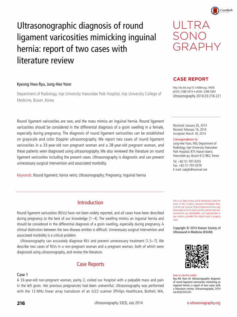
USA). Grayscale ultrasonography showed a mass in the left groin composed of dilated, echo-free, tubular channels. The color Doppler ultrasonography identified a mass composed of multiple echo-free tubular channels with hypervascularity that became more prominent during a Valsalva maneuver. There was no ultrasonographic evidence of a herniated bowel or lymphadenopathy (Fig. 1A, B). The patient was treated conservatively.
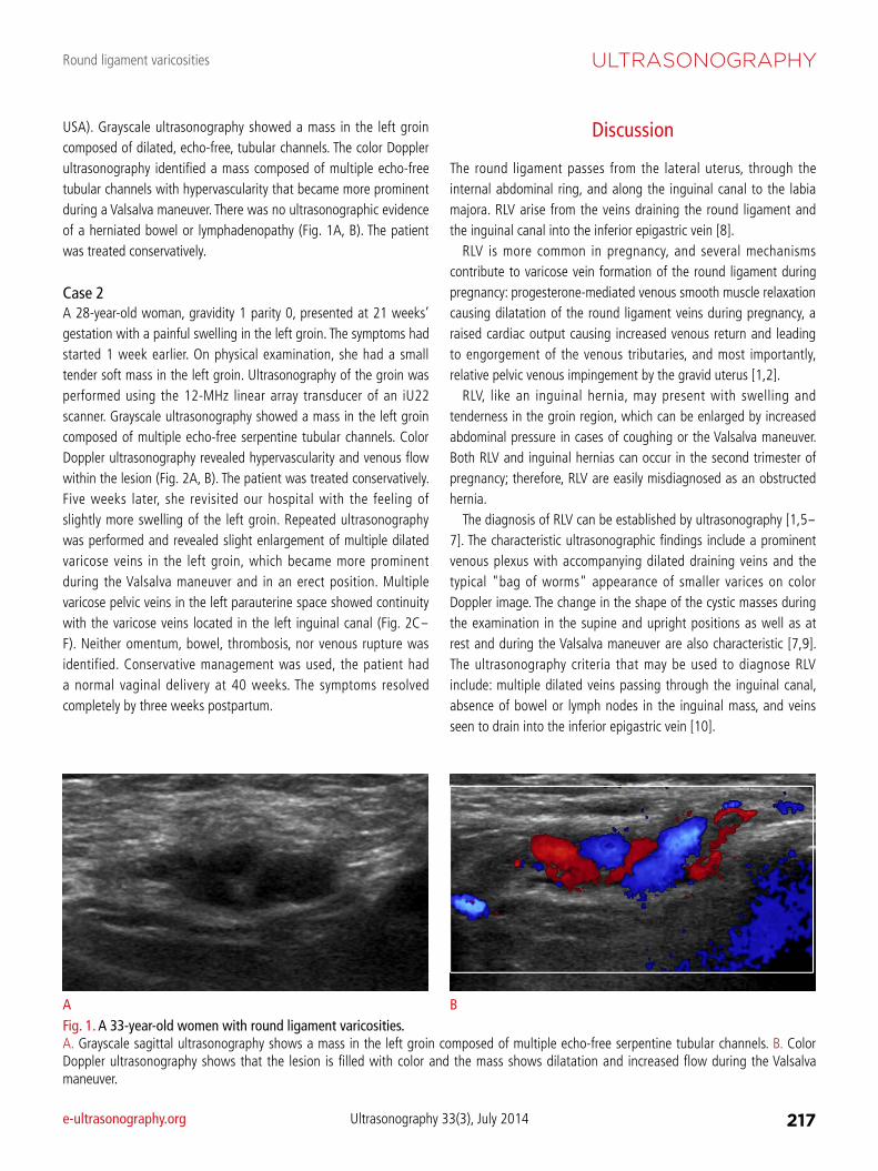
Case 2A 28-year-old woman, gravidity 1 parity 0, presented at 21 weeks’ gestation with a painful swelling in the left groin. The symptoms had started 1 week earlier. On physical examination, she had a small tender soft mass in the left groin. Ultrasonography of the groin was performed using the 12-MHz linear array transducer of an iU22 scanner. Grayscale ultrasonography showed a mass in the left groin composed of multiple echo-free serpentine tubular channels. Color Doppler ultrasonography revealed hypervascularity and venous flow within the lesion (Fig. 2A, B). The patient was treated conservatively. Five weeks later, she revisited our hospital with the feeling of slightly more swelling of the left groin. Repeated ultrasonography was performed and revealed slight enlargement of multiple dilated varicose veins in the left groin, which became more prominent during the Valsalva maneuver and in an erect position. Multiple varicose pelvic veins in the left parauterine space showed continuity with the varicose veins located in the left inguinal canal (Fig. 2C-F). Neither omentum, bowel, thrombosis, nor venous rupture was identified. Conservative management was used, the patient had a normal vaginal delivery at 40 weeks. The symptoms resolved completely by three weeks postpartum.
Discussion
The round ligament passes from the lateral uterus, through the internal abdominal ring, and along the inguinal canal to the labia majora. RLV arise from the veins draining the round ligament and the inguinal canal into the inferior epigastric vein [8].
RLV is more common in pregnancy, and several mechanisms contribute to varicose vein formation of the round ligament during pregnancy: progesterone-mediated venous smooth muscle relaxation causing dilatation of the round ligament veins during pregnancy, a raised cardiac output causing increased venous return and leading to engorgement of the venous tributaries, and most importantly, relative pelvic venous impingement by the gravid uterus [1,2].
RLV, like an inguinal hernia, may present with swelling and tenderness in the groin region, which can be enlarged by increased abdominal pressure in cases of coughing or the Valsalva maneuver. Both RLV and inguinal hernias can occur in the second trimester of pregnancy; therefore, RLV are easily misdiagnosed as an obstructed hernia.
The diagnosis of RLV can be established by ultrasonography [1,5-7]. The characteristic ultrasonographic findings include a prominent venous plexus with accompanying dilated draining veins and the typical "bag of worms" appearance of smaller varices on color Doppler image. The change in the shape of the cystic masses during the examination in the supine and upright positions as well as at rest and during the Valsalva maneuver are also characteristic [7,9]. The ultrasonography criteria that may be used to diagnose RLV include: multiple dilated veins passing through the inguinal canal, absence of bowel or lymph nodes in the inguinal mass, and veins seen to drain into the inferior epigastric vein [10].
Fig. 1. A 33-year-old women with round ligament varicosities. A. Grayscale sagittal ultrasonography shows a mass in the left groin composed of multiple echo-free serpentine tubular channels. B. Color Doppler ultrasonography shows that the lesion is filled with color and the mass shows dilatation and increased flow during the Valsalva maneuver.
A B
Kyeong Hwa Ryu, et al.
218 Ultrasonography 33(3), July 2014 e-ultrasonography.org
The Table 1 summarizes the clinico-radiological features of the previously reported cases in the English-language literature including the present cases [1,4,9-19]. The mean age of previously
reported cases of RLV was 29.7 years (range, 18 to 40 years) and site of development site was the right, followed by the left and then by both sides in the ratio of 11:9:6. All of the cases except one of
A B
C D
Fig. 2. A 28-year-old women at the 21st week of pregnancy with round ligament varicosities. A. Grayscale transverse ultrasonography shows an ovoid multiseptated cystic mass in the left groin. B. Color Doppler ultrasonography shows that the lesion is hypervascular. After 5 weeks, follow-up ultrasonography was performed (C-F). The mass in the left groin shows slightly enlarged and composed of multiple anechoic serpentine tubular channels on grayscale ultrasonography (not shown). C. Color Doppler sagittal ultrasonography during Valsalva maneuver. The mass expands and shows marked flow augmentation. D, E. Sagittal ultrasonography of the left groin through the inguinal canal in an erect position. The lesion is enlarged and the vascularities of the mass are markedly engorged. F. These varicose veins between the markers continue to the left parauterine space (arrows) through the inguinal canal (arrowheads).
E F
Round ligament varicosities
e-ultrasonography.org Ultrasonography 33(3), July 2014 219
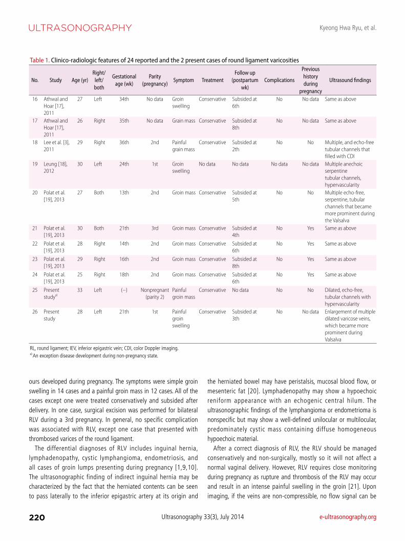
Table 1. Clinico-radiologic features of 24 reported and the 2 present cases of round ligament varicosities
No. Study Age (yr)Right/left/both
Gestational age (wk)
Parity (pregnancy)
Symptom TreatmentFollow up
(postpartum wk)
Complications
Previous history during
pregnancy
Ultrasound findings
1 Cheng et al. [1], 1997
22 Left 28th No data Painful groin mass
Conservative Subsided after normal delivery
No No data Thickened RL with varicosities, both within and around it, draining into the IEV with good flow signal
2 Chi et al. [11], 2005
30 Left 33th 2nd Groin swelling
Conservative Subsided at 6th
No Yes Multiple dilated varicosities within and around the RL extending from the left inguinal ring
3 Murphy et al. [12], 2007
37 Left 28th 2nd Groin swelling
No data No data No data No Multiple serpentine tubular channels that filled with color
4 Nguyen and Gruenewald [13], 2008
18 Right 33th 1st Painful groin swelling
Conservative Subsided at 4th
No No data Circumscribed hypoechoic soft tissue mass, minor venous flow at rest with marked flow during Valsalva
5 McKenna et al. [10], 2008
35 Left 23th 3rd Inguinal swelling
Conservative Subsided at 8th
No No data No description
6 McKenna et al. [10], 2008
35 Right 30th 4th Inguinal swelling
Conservative Subsided at 8th
No No data Multiple prominent vessels with venous flow
7 McKenna et al. [10], 2008
37 Both 31th 6th Painful groin swelling
Conservative Subsided at 8th
No Yes Prominent engorged pelvic vessels, extending from the pelvic side wall to the labia majora
8 McKenna et al. [10], 2008
38 Both 19th 3rd Painful groin swelling
Conservative Subsided at 12th
No No data Abnormal venous structures within the inguinal canal
9 McKenna et al. [10], 2008
40 Right 2nd trimester 2nd Groin mass Conservative Subsided at 4th
No No data Engorged blood vessels along the inguinal canal
10 Tokue et al. [14], 2008
37 Right 35th 2nd Painful groin mass
Conservative Improved slowly
Thrombosed varices of the RL
No data Hypoechoic moniliform mass lesion
11 Ijpma et al. [4], 2009
29 Both 14th of 2nd pregnancy, 8th, 26th
of 3rd pregnancy
3rd Painful groin swelling
Conservative at 2nd, surgical excision at 3rd
No data No No data Multiple dilated veins in the inguinal canal
12 Uzun et al. [9], 2010
24 Right 26th No data Painful groin swelling
Conservative Subsided at 2th
No No data Multiple echo-free serpentine tubular channels with hypervascularity
13 Dent et al. [15], 2010
28 Left 20th 1st Painful groin mass
Conservative Subsided after delivery
No No data A leash of veins extending into the inguinal canal
14 Kahriman et al. [16], 2010
22 Both 28th No data Painful groin swelling
Conservative Subsided at 4th
No No data Cystic mass, demonstrated multiple dilated varicose veins within the RL
15 Athwal and Hoar [17], 2011
28 Right 24th No data Groin mass Conservative Subsided at 4th
No No data Multiple prominent varicosities
continued
Kyeong Hwa Ryu, et al.
220 Ultrasonography 33(3), July 2014 e-ultrasonography.org
ours developed during pregnancy. The symptoms were simple groin swelling in 14 cases and a painful groin mass in 12 cases. All of the cases except one were treated conservatively and subsided after delivery. In one case, surgical excision was performed for bilateral RLV during a 3rd pregnancy. In general, no specific complication was associated with RLV, except one case that presented with thrombosed varices of the round ligament.
The differential diagnoses of RLV includes inguinal hernia, lymphadenopathy, cystic lymphangioma, endometriosis, and all cases of groin lumps presenting during pregnancy [1,9,10]. The ultrasonographic finding of indirect inguinal hernia may be characterized by the fact that the herniated contents can be seen to pass laterally to the inferior epigastric artery at its origin and
the herniated bowel may have peristalsis, mucosal blood flow, or mesenteric fat [20]. Lymphadenopathy may show a hypoechoic reniform appearance with an echogenic central hilum. The ultrasonographic findings of the lymphangioma or endometrioma is nonspecific but may show a well-defined unilocular or multilocular, predominately cystic mass containing diffuse homogeneous hypoechoic material.
After a correct diagnosis of RLV, the RLV should be managed conservatively and non-surgically, mostly so it will not affect a normal vaginal delivery. However, RLV requires close monitoring during pregnancy as rupture and thrombosis of the RLV may occur and result in an intense painful swelling in the groin [21]. Upon imaging, if the veins are non-compressible, no flow signal can be
Table 1. Clinico-radiologic features of 24 reported and the 2 present cases of round ligament varicosities
No. Study Age (yr)Right/left/both
Gestational age (wk)
Parity (pregnancy)
Symptom TreatmentFollow up
(postpartum wk)
Complications
Previous history during
pregnancy
Ultrasound findings
16 Athwal and Hoar [17], 2011
27 Left 34th No data Groin swelling
Conservative Subsided at 6th
No No data Same as above
17 Athwal and Hoar [17], 2011
26 Right 35th No data Grain mass Conservative Subsided at 8th
No No data Same as above
18 Lee et al. [3], 2011
29 Right 36th 2nd Painful grain mass
Conservative Subsided at 2th
No No Multiple, and echo-free tubular channels that filled with CDI
19 Leung [18], 2012
30 Left 24th 1st Groin swelling
No data No data No data No data Multiple anechoic serpentine tubular channels, hypervascularity
20 Polat et al. [19], 2013
27 Both 13th 2nd Groin mass Conservative Subsided at 5th
No No Multiple echo-free, serpentine, tubular channels that became more prominent during the Valsalva
21 Polat et al. [19], 2013
30 Both 21th 3rd Groin mass Conservative Subsided at 4th
No Yes Same as above
22 Polat et al. [19], 2013
28 Right 14th 2nd Groin mass Conservative Subsided at 6th
No Yes Same as above
23 Polat et al. [19], 2013
29 Right 16th 2nd Groin mass Conservative Subsided at 8th
No Yes Same as above
24 Polat et al. [19], 2013
25 Right 18th 2nd Groin mass Conservative Subsided at 6th
No Yes Same as above
25 Present studya)
33 Left (-) Nonpregnant (parity 2)
Painful groin mass
Conservative No data No No Dilated, echo-free, tubular channels with hypervascularity
26 Present study
28 Left 21th 1st Painful groin swelling
Conservative Subsided at 3th
No No data Enlargement of multiple dilated varicose veins, which became more prominent during Valsalva
RL, round ligament; IEV, inferior epigastric vein; CDI, color Doppler imaging.a) An exception disease development during non-pregnancy state.
Round ligament varicosities
e-ultrasonography.org Ultrasonography 33(3), July 2014 221
pregnancy. J Med Soc N J 1962;59:24-26. 9. Uzun M, Akkan K, Coskun B. Round ligament varicosities mimicking
inguinal hernias in pregnancy: importance of color Doppler sonography. Diagn Interv Radiol 2010;16:150-152.
10. McKenna DA, Carter JT, Poder L, Gosnell JE, Maa J, Pearl JM, et al. Round ligament varices: sonographic appearance in pregnancy. Ultrasound Obstet Gynecol 2008;31:355-357.
11. Chi C, Taylor A, Munjuluri N, Abdul-Kadir R. A diagnostic dilemma: round ligament varicosities in pregnancy. Acta Obstet Gynecol Scand 2005;84:1126-1127.
12. Murphy IG, Heffernan EJ, Gibney RG. Groin mass in pregnancy. Br J Radiol 2007;80:588-589.
13. Nguyen QH, Gruenewald SM. Doppler sonography in the diagnosis of round ligament varicosities during pregnancy. J Clin Ultrasound 2008;36:177-179.
14. Tokue H, Aoki J, Tsushima Y, Endo K. Characteristic of computed tomography and magnetic resonance imaging finding of thrombosed varices of the round ligament of the uterus: a case report. J Comput Assist Tomogr 2008;32:559-561.
15. Dent BM, Al Samaraee A, Coyne PE, Nice C, Katory M. Varices of the round ligament mimicking an inguinal hernia: an important differential diagnosis during pregnancy. Ann R Coll Surg Engl 2010;92:W10-W11.
16. Kahriman G, Donmez H, Mavili E, Ozcan N. Bilateral round ligament varicosities mimicking an inguinal hernia in pregnancy: case report. J Clin Ultrasound 2010;38:512-514.
17. Athwal R, Hoar F. Round ligament varicosity: a rare cause of inguinal swelling. Phlebology 2011;26:213-214.
18. Leung JL. Round ligament varicosities: a rare cause of groin swelling in pregnancy. Hong Kong Med J 2012;18:256-257.
19. Polat AV, Aydin R, Polat AK, Kececi IS, Karahan G, Taskin GO. Round ligament varicosities: a rare cause of groin swelling in pregnancy. Abdom Imaging 2013;38:1178-1181.
20. Jamadar DA, Jacobson JA, Morag Y, Girish G, Ebrahim F, Gest T, et al. Sonography of inguinal region hernias. AJR Am J Roentgenol 2006;187:185-190.
21. Al-Qudah MS. Postpartum pain due to thrombosed varicose veins of the round ligament of the uterus. Postgrad Med J 1993;69:820-821.
obtained, and/or there is a visible clot within the lumen, complicated RLV should be suspected and emergency surgical exploration is recommended [10,21].
RLV should be considered a part of the differential diagnosis of an inguinal mass of women, especially during pregnancy, and ultrasonography can accurately diagnose RLV. By becoming familiar with the ultrasonographic findings of RLV, RLV can be treated optimally, and unnecessary surgical intervention and associated morbidity can be prevented.
ORCID: Kyeong Hwa Ryu: http://orcid.org/0000-0003-1599-8881; Jung-Hee Yoon:
http://orcid.org/0000-0001-5152-6668
Conflict of InterestNo potential conflict of interest relevant to this article was reported.
References
1. Cheng D, Lam H, Lam C. Round ligament varices in pregnancy mimicking inguinal hernia: an ultrasound diagnosis. Ultrasound Obstet Gynecol 1997;9:198-199.
2. Smith P, Heimer G, Norgren A, Ulmsten U. The round ligament: a target organ for steroid hormones. Gynecol Endocrinol 1993;7:97-100.
3. Lee DK, Bae SW, Moon H, Kim YK. Round ligament varicosities mimicking inguinal hernia in pregnancy. J Korean Surg Soc 2011;80:437-439.
4. Ijpma FF, Boddeus KM, de Haan HH, van Geldere D. Bilateral round ligament varicosities mimicking inguinal hernia during pregnancy. Hernia 2009;13:85-88.
5. Pilkington SA, Rees M, Jones O, Green I. Ultrasound diagnosis of round ligament varicosities mimicking inguinal hernias in pregnancy. Ann R Coll Surg Engl 2004;86:400-401.
6. Frede TE. Ultrasonic visualization of varicosities in the female genital tract. J Ultrasound Med 1984;3:365-369.
7. Oh SN, Jung SE, Rha SE, Lim GY, Ku YM, Byun JY, et al. Sonography of various cystic masses of the female groin. J Ultrasound Med 2007;26:1735-1742.
8. Reisfield DR. Varicosities in veins of the inguinal canal during