ultrasound guided low approach interscalene brachial ... · ultrasound guided low approach...
TRANSCRIPT

Korean J Pain 2016 January; Vol. 29, No. 1: 18-22pISSN 2005-9159 eISSN 2093-0569http://dx.doi.org/10.3344/kjp.2016.29.1.18
| Original Article |
Ultrasound Guided Low Approach Interscalene Brachial Plexus Block for Upper Limb Surgery
Department of Anesthesiology and Pain Medicine, Jeju National University Hospital, Jeju, *Ulsan University Hospital, University of Ulsan College of Medicine, Ulsan, Korea
Sun Kyung Park, Min Ha Sung*, Hae Jin Suh, and Yun Suk Choi
Background: The interscalene brachial plexus block is widely used for pain control and anesthetic purposes during shoulder arthroscopic surgeries and surgeries of the upper extremities. However, it is known that interscalene brachial plexus block is not appropriate for upper limb surgeries because it does not affect the lower trunk (C8−T1, ulnar nerve) of the brachial plexus.
Methods: A low approach, ultrasound-guided interscalene brachial plexus block (LISB) was performed on twenty-eight patients undergoing surgery of the upper extremities. The patients were assessed five minutes and fifteen minutes after the block for the degree of block in each nerve and muscle as well as for any complications.
Results: At five minutes and fifteen minutes after the performance of the block, the degree of the block in the ulnar nerve was found to be 2.8 ± 2.6 and 1.1 ± 1.8, respectively, based on a ten-point scale. Motor block occurred in the median nerve after fifteen minutes in 26 of the 28 patients (92.8%), and in all of the other three nerves in all 28 patients. None of the patients received additional analgesics, and none experienced complications.
Conclusions: The present study confirmed the achievement of an appropriate sensory and motor block in the upper extremities, including the ulnar nerve, fifteen minutes after LISB, with no complications. (Korean J Pain 2016; 29: 18-22)
Key Words: Complication; Interscalene brachial plexus block; Nerve stimulator; Sensory block; Ultrasound- guided; Upper limb surgery.
Received September 3, 2015. Revised October 8, 2015. Accepted October 16, 2015.Correspondence to: Yun Suk ChoiDepartment of Anesthesiology and Pain Medicine, Jeju National University Hospital, 15 Aran 13-gil, Jeju 63241, KoreaTel: +82-64-717-2025, Fax: +82-64-717-2042, E-mail: [email protected]
This is an open-access article distributed under the terms of the Creative Commons Attribution Non-Commercial License (http:// creativecommons.org/licenses/by-nc/4.0/), which permits unrestricted non-commercial use, distribution, and reproduction in any medium, provided the original work is properly cited.Copyright ⓒ The Korean Pain Society, 2016
INTRODUCTION
The brachial plexus runs from the C5-T1 ventral rami,
forms the superior, middle, and inferior trunks, divides un-
der the clavicle, leads to the lateral, posterior, and medial
cords, and finally forms the peripheral nerves running to
the arms. The brachial plexus block is popular for anes-
thetic and pain control purposes in the upper limbs. There
are a few approaches to the block, including the inter-
scalene approach, supraclavicular approach, infracla-
Park, et al / Ultrasound Guided Low Interscalene Block 19
www.epain.org
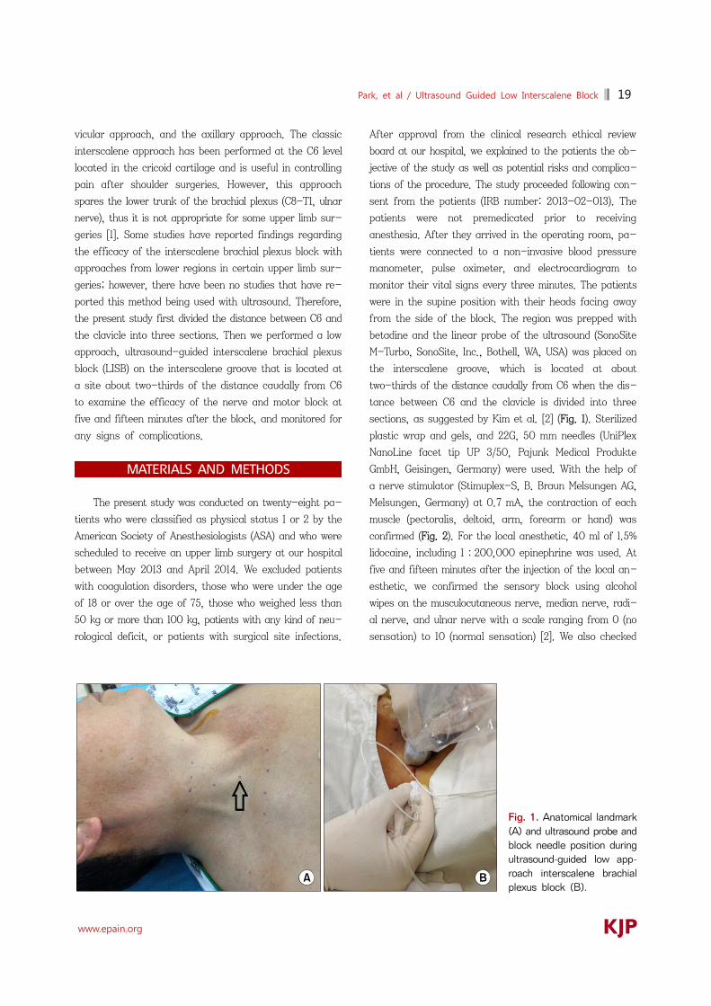
Fig. 1. Anatomical landmark (A) and ultrasound probe and block needle position during ultrasound-guided low app-roach interscalene brachial plexus block (B).
vicular approach, and the axillary approach. The classic
interscalene approach has been performed at the C6 level
located in the cricoid cartilage and is useful in controlling
pain after shoulder surgeries. However, this approach
spares the lower trunk of the brachial plexus (C8-T1, ulnar
nerve), thus it is not appropriate for some upper limb sur-
geries [1]. Some studies have reported findings regarding
the efficacy of the interscalene brachial plexus block with
approaches from lower regions in certain upper limb sur-
geries; however, there have been no studies that have re-
ported this method being used with ultrasound. Therefore,
the present study first divided the distance between C6 and
the clavicle into three sections. Then we performed a low
approach, ultrasound-guided interscalene brachial plexus
block (LISB) on the interscalene groove that is located at
a site about two-thirds of the distance caudally from C6
to examine the efficacy of the nerve and motor block at
five and fifteen minutes after the block, and monitored for
any signs of complications.
MATERIALS AND METHODS
The present study was conducted on twenty-eight pa-
tients who were classified as physical status 1 or 2 by the
American Society of Anesthesiologists (ASA) and who were
scheduled to receive an upper limb surgery at our hospital
between May 2013 and April 2014. We excluded patients
with coagulation disorders, those who were under the age
of 18 or over the age of 75, those who weighed less than
50 kg or more than 100 kg, patients with any kind of neu-
rological deficit, or patients with surgical site infections.
After approval from the clinical research ethical review
board at our hospital, we explained to the patients the ob-
jective of the study as well as potential risks and complica-
tions of the procedure. The study proceeded following con-
sent from the patients (IRB number: 2013-02-013). The
patients were not premedicated prior to receiving
anesthesia. After they arrived in the operating room, pa-
tients were connected to a non-invasive blood pressure
manometer, pulse oximeter, and electrocardiogram to
monitor their vital signs every three minutes. The patients
were in the supine position with their heads facing away
from the side of the block. The region was prepped with
betadine and the linear probe of the ultrasound (SonoSite
M-Turbo, SonoSite, Inc., Bothell, WA, USA) was placed on
the interscalene groove, which is located at about
two-thirds of the distance caudally from C6 when the dis-
tance between C6 and the clavicle is divided into three
sections, as suggested by Kim et al. [2] (Fig. 1). Sterilized
plastic wrap and gels, and 22G, 50 mm needles (UniPlex
NanoLine facet tip UP 3/50, Pajunk Medical Produkte
GmbH, Geisingen, Germany) were used. With the help of
a nerve stimulator (Stimuplex-S, B. Braun Melsungen AG,
Melsungen, Germany) at 0.7 mA, the contraction of each
muscle (pectoralis, deltoid, arm, forearm or hand) was
confirmed (Fig. 2). For the local anesthetic, 40 ml of 1.5%
lidocaine, including 1:200,000 epinephrine was used. At
five and fifteen minutes after the injection of the local an-
esthetic, we confirmed the sensory block using alcohol
wipes on the musculocutaneous nerve, median nerve, radi-
al nerve, and ulnar nerve with a scale ranging from 0 (no
sensation) to 10 (normal sensation) [2]. We also checked

20 Korean J Pain Vol. 29, No. 1, 2016
www.epain.org
Table 1. Patient Dermographic Data
n = 28
Age (yr) Height (cm) Weight (kg) Sex (M/F)Operation time (min) Block performance time (sec)
47.8 ± 14.6160.0 ± 8.6
59.7 ± 10.610/18
59.6 ± 33.0341.7 ± 59.2
Values are mean ± standard deviation or number of patients.
Fig. 2. Ultrasound image showing low approach inter-scalene brachial plexus block. Needle pathway (arrows), SCM: sternocleidomastoid muscle, IJV: internal jugular vein,ST: superior trunk of brachial plexus, MSM: middle scalene muscle.
Table 3. Characteristics of the Sensory Block Using a Low Approach Interscalene Brachial Plexus Block
5 min after injection
15 min after injection
Sensory block (0–10) Musculocutaneous nerve Median nerve Radial nerve Ulnar nerve
0.7 ± 1.31.6 ± 2.30.5 ± 1.12.8 ± 2.6
0.2 ± 0.60.9 ± 1.90.1 ± 0.31.1 ± 1.8
Values are mean ± standard deviation. Sensory block (0−10); 0: loss of sensation, 10: normal sensation.
Table 4. Characteristics of the Motor Block Using a Low ApproachInterscalene Brachial Plexus Block
5 min after injection No. (%)
15 min after injection No. (%)
Musculocutaneous nerve Median nerve Radial nerve Ulnar nerve
26 (92.8%)20 (71.4%) 25 (89.2%)23 (82.1%)
28 (100%)26 (92.8%)28 (100%)28 (100%)
Values are number of patients (percentage).
Table 2. Type of Surgery
Type of surgery N=28
Elbow curettage & drillingDistal radius ORIFFinger ORIFUlnar metal removalGanglion excisionTendon repositionArthroscopic debridement, wristTenosynovectomy
86333311
Values are number of patients. ORIF: open reduction & internal fixation.
for muscular contractions by assessing flexion of the elbow
(musculocutaneous nerve), extension of the elbow and
wrist (radial nerve), pronation of the arm and flexion of
the wrist (median nerve), and flexion and opposition of the
fourth and fifth fingers toward the thumb (ulnar nerve),
and considered signs of paralysis (loss of contraction) to
indicate a successful motor block [3]. One anesthesiologist
performed the LISB procedure and one orthopedist per-
formed the surgery. We confirmed cases of hemi-
diaphragmatic paralysis after the surgery by performing
a chest X-ray and consulting a radiologist regarding the
results.
RESULTS
Among the 28 subjects of this study, 10 were male and
18 were female. The patients’ demographic and clinical da-
ta including age, body weight, height, gender, surgery
length, and type of surgery are illustrated in Tables 1 and 2.
At five and fifteen minutes after the block procedure, the
degree of sensory block in the ulnar nerve was found to
be 2.8 ± 2.6 and 1.1 ± 1.8, respectively, on a scale of
ten. Muscular block occurred in the median nerve after fif-
teen minutes in 26 of the 28 patients (92.8%), and in all of
the other three nerves in all 28 patients (Tables 3 and 4).
None of the patients received additional analgesics after
the surgery, and there were no abnormalities during the
surgeries. In addition, there were no signs of complica-

Park, et al / Ultrasound Guided Low Interscalene Block 21
www.epain.org
Table 5. Analgesic Requirement and Complications after a Low Approach Interscalene Brachial Plexus Block
N = 28
Patients requiring analgesics during operation Patients with complications Nausea Horner syndrome Dyspnea
0
000
Values are number of patients.
tions, such as dyspnea or Horner syndrome, during the
surgery, in the recovery room, or in the wards (Table 5).
DISCUSSION
This study confirmed that an appropriate sensory and
motor block was achieved in the upper extremities, includ-
ing the ulnar nerve, fifteen minutes after LISB, and that
there were no complications associated with the block.
Two methods of LISB have been introduced, namely the
anatomical landmark approach and the ultrasound-guided
approach. There are three approaches to the anatomical
landmark method: first, it can be performed in between
the cricoid cartilage and the clavicle. Second, it can be
performed 2 cm above the clavicle, and third, it can be
performed on the interscalene groove, which is located at
about two-thirds of the distance caudally from C6 after
dividing the distance between C6 and the clavicle into three
sections. There are two methods for the ultrasound-guided
approach: first, it can be performed on the superior trunk
where C5 and C6 are combined. Second, the injection can
be performed on the caudal side of the C6 nerve root
[2,4,5]. Although different studies define and name the
procedures slightly differently (low approach, lower inter-
scalene approach, or superior trunk approach), these pro-
cedures are identical in terms of using an approach
through the lower regions of the C6 level compared to the
existing ISB.
Owing to the advances in procedural techniques and
the application of ultrasound technology, several studies
have reported that the use of local anesthetics can be re-
duced when performing ISB. However, most of these stud-
ies limit their scope to shoulder surgeries, which are irrele-
vant to the block of the inferior trunk (C8-T1, ulnar nerve)
[6-8]. It has been known that the ulnar nerve is not af-
fected in about 30-50% of ISBs performed with the classic
approach [1]. Furthermore, it has been reported that the
ulnar nerve was not blocked in about 7-33.3% of the cases
that took the classic approach ISB and used 30 ml of local
anesthetics on the caudal side of the C6 nerve root [9,10].
Hence, in the present study, we used 40 ml of local anes-
thetics to ensure a quick onset and complete block of the
ulnar nerve.
LISB is known to involve a short effect distance (from
the C5 nerve root to the C8 nerve root) and to diffuse local
anesthetics via the deep cervical fascia. In addition, LISB
has been reported to bring about appropriate sensory and
motor blocks required for upper limb surgeries even with
a single injection [10]. Against this backdrop, the present
study was planned and conducted.
Moreover, according to Plante et al., [9] who compared
two groups of patients who were injected with local anes-
thetics either in the upper region of the C5 nerve root or
the lower region of the C6 nerve root during an ultra-
sound-guided interscalene brachial plexus block for an-
algesia in arthroscopic shoulder surgeries, the group of
patients who received local anesthetics in the lower region
of the C6 nerve root had appropriate sensory blocks in all
of the nerves; they reported that the sensory and motor
blocks were especially noticeable in the ulnar nerve and
that there was a rapid onset in the ulnar nerve. The results
of this study showed that the sensory block in the ulnar
nerve was 2.8 ± 2.6 on a 10-point scale at five minutes
after LISB and 1.1 ± 1.8 at fifteen minutes after LISB.
About 82.1% of motor neurons were blocked at five mi-
nutes, but 100% were blocked after fifteen minutes. In ad-
dition, there were no additional injections of analgesics
during the surgery. Therefore, it can be said that the ulnar
nerve was appropriately blocked via LISB.
Meanwhile, ISB is known to induce a temporary para-
lysis in the ipsilateral hemidiaphragm due to phrenic nerve
palsy. The phrenic nerve is located within 2 mm of the
brachial plexus of the cricoid cartilage and divides 3 mm
per 1 cm as it descends caudally. Thus, it can be predicted
that the incidence of phrenic nerve palsy-induced hemi-
diaphragmatic paralysis can be reduced if ISB is performed
more caudal to the C6 level or on the superior trunk [11,
12]. In the present study, there were no signs of dyspnea
or hemidiaphragmatic paralysis. In addition, LISB is known
to reduce the damage to the dorsal scapular and long
thoracic nerves, both of which split from the C5 nerve root

22 Korean J Pain Vol. 29, No. 1, 2016
www.epain.org
[4]. Although we did not assess whether any such damages
occurred in the present study, none of the patients experi-
enced any such problems.
As mentioned above, there were no complications in
the current study. We presume that we were able to reduce
the risk of complications, such as vascular injection or
nerve injury, by using an ultrasound nerve stimulator in
addition to the inherent merits of the LISB method.
In the present study, the motor block in the median
nerve was shown to be about 71.4% at five minutes after
the procedure was performed. The block increased to
92.8% at fifteen minutes, and there were no additional an-
algesics injected and no additional block was performed.
Other studies have also reported similarly slow blocks in
the median nerve within fifteen minutes [9], which is
thought to be due to the fact that the median nerve in-
nervates all of C5, C6, C7, and T1.
There are a few limitations to this study. First, we did
not have a large pool of subjects. Second, we could not
observe the diffusion of local anesthetics through injecting
contrast medium. Third, we did not compare the procedure
of interest with other approaches. Thus far, studies on
LISB are only in the form of case reports or brief reports;
hence, in the future, LISB should be compared with other
approaches, and cases of LISB using different doses of lo-
cal anesthetics should be compared as well.
In conclusion, the present study confirmed that the
nerves in the upper extremities, including the ulnar nerve,
were appropriately blocked fifteen minutes after the per-
formance of LISB, and that there were no complications
induced by the block.
ACKNOWLEDGEMENTS
This work was supported by the research grant of Jeju
National University in 2013.
REFERENCES
1. Neal JM, Gerancher JC, Hebl JR, Ilfeld BM, McCartney CJ, Franco CD, et al. Upper extremity regional anesthesia:
essentials of our current understanding, 2008. Reg Anesth Pain Med 2009; 34: 134-70.
2. Kim JH, Chen J, Bennett H, Lesser JB, Resta-Flarer F, Barczewska-Hillel A, et al. A low approach to interscalene brachial plexus block results in more distal spread of sensory-motor coverage compared to the conventional approach. Anesth Analg 2011; 112: 987-9.
3. Liu FC, Liou JT, Tsai YF, Li AH, Day YY, Hui YL, et al. Efficacy of ultrasound-guided axillary brachial plexus block: a comparative study with nerve stimulator-guided method. Chang Gung Med J 2005; 28: 396-402.
4. Burckett-St Laurent D, Chan V, Chin KJ. Refining the ultrasound-guided interscalene brachial plexus block: the superior trunk approach. Can J Anaesth 2014; 61: 1098- 102.
5. Gadsden JC, Tsai T, Iwata T, Somasundarum L, Robards C, Hadzic A. Low interscalene block provides reliable anesthesia for surgery at or about the elbow. J Clin Anesth 2009; 21: 98-102.
6. Renes SH, Rettig HC, Gielen MJ, Wilder-Smith OH, van Geffen GJ. Ultrasound-guided low-dose interscalene brachial plexus block reduces the incidence of hemidiaphragmatic paresis. Reg Anesth Pain Med 2009; 34: 498-502.
7. Gautier P, Vandepitte C, Ramquet C, DeCoopman M, Xu D, Hadzic A. The minimum effective anesthetic volume of 0.75% ropivacaine in ultrasound-guided interscalene brachial plexus block. Anesth Analg 2011; 113: 951-5.
8. Fredrickson MJ, Smith KR, Wong AC. Importance of volume and concentration for ropivacaine interscalene block in preventing recovery room pain and minimizing motor block after shoulder surgery. Anesthesiology 2010; 112: 1374-81.
9. Plante T, Rontes O, Bloc S, Delbos A. Spread of local anesthetic during an ultrasound-guided interscalene block: does the injection site influence diffusion? Acta Anaesthesiol Scand 2011; 55: 664-9.
10. Bharti N, Bhardawaj N, Wig J. Comparison of ultrasound- guided supraclavicular, infraclavicular and below-C6 inter-scalene brachial plexus block for upper limb surgery: a randomised, observer-blinded study. Anaesth Intensive Care 2015; 43: 468-72.
11. Nadeau MJ, Lévesque S, Dion N. Ultrasound-guided regional anesthesia for upper limb surgery. Can J Anaesth 2013; 60: 304-20.
12. Kessler J, Schafhalter-Zoppoth I, Gray AT. An ultrasound study of the phrenic nerve in the posterior cervical triangle: implications for the interscalene brachial plexus block. Reg Anesth Pain Med 2008; 33: 545-50.