ultrastructure of mitosis and gametogenesis in cladophora
TRANSCRIPT

W&M ScholarWorks W&M ScholarWorks
Dissertations, Theses, and Masters Projects Theses, Dissertations, & Master Projects
1973
Ultrastructure of Mitosis and Gametogenesis in Cladophora Ultrastructure of Mitosis and Gametogenesis in Cladophora
flexuosa (Dillwyn) Harvey flexuosa (Dillwyn) Harvey
Kenneth Wilson Bullock College of William & Mary - Arts & Sciences
Follow this and additional works at: https://scholarworks.wm.edu/etd
Part of the Developmental Biology Commons
Recommended Citation Recommended Citation Bullock, Kenneth Wilson, "Ultrastructure of Mitosis and Gametogenesis in Cladophora flexuosa (Dillwyn) Harvey" (1973). Dissertations, Theses, and Masters Projects. Paper 1539624845. https://dx.doi.org/doi:10.21220/s2-td1w-ff67
This Thesis is brought to you for free and open access by the Theses, Dissertations, & Master Projects at W&M ScholarWorks. It has been accepted for inclusion in Dissertations, Theses, and Masters Projects by an authorized administrator of W&M ScholarWorks. For more information, please contact [email protected].
ULTRASTRUCTURE OF MITOSIS AND G AMETOGENESISi \IN CLADOPHORA FLEXU03A (DILLWYN) HARVEY
A Thesis Presented to
The Faculty of the Department of Biology The College of William and Mary in Virginia
In Partial Fulfillment of the Requirements for the Degree of
Master of Arts
ByKenneth Wilson Bullock
197^

APPROVAL SHEET
This thesis is submitted in partial fulfillment of .the requirements for the degree of
Master of Arts
Approved, September 197^
U7Martin C. Mathes, Ph.D.
Lawrence L. Wiseman, Ph.D.

TABLE OF CONTENTSPage
ACKNOWLEDGMENTS ................................... ivLIST OF FIGURES...................................... vABSTRACT........................ viiINTRODUCTION ...................................... 2MATERIALS AND METHODS ............................ 15RESULTS.............................................. 1?D I S C U S S I O N .........................................25KEY TO ABBREVIATIONS............................... 33FIGURES...............................................3&BIBLIOGRAPHY ...................................... 52

ACKNOWLEDGMENTS
The author wishes to express his appreciation to Dr. Joseph L. Scott under whose guidance and criticism this investigation was completed. The author is also indebted to Dr, Martin C. Mathes and Dr. Lawrence L. Wiseman for their critical reading of the manuscript. Thanks are also due to Mrs. Jewel P. Thomas for technical assistance rendered during the course of this study.

LIST OF FIGURES Figure Page
1. Light micrograph of apical regionof filament............... 3^
2, Longitudinal section of apical cellt i p ................................... 3^
3* Cross section of a vegetative cell . . . 364. Longitudinal section of basal region
of cell 365. Interphase condition of nucleus
and c y t o p l a s m ..........................366. Nuclear envelope ............... 387. Interphase n u c l e u s ..................... 388. Nuclear pores and centioles ............ ^09. Prophase nucleus . . . . . . . . 4-0
10. Nuclear pore-centriole arrangementin prophase . ^0
11. Metaphase plate . . . . . . . . f̂012. Longitudinal section of early
anaphase n u c l e u s ................... k 213. Area of constriction in late
anaphase nucleus ................... ^2
lA. Endoplasmic reticulum in lateanaphase condition ................ kZ
15. Polar region of late anaphase nucleus . .16, Longitudinal section of late
anaphase nucleus . . . . . . ^
v

Figure Page1?, Interphase nucleus following mitosis , . 4618. High magnification of mitochondria . . . 4-619« Cytoplasm at 'the beginning of
gametogenesis . . . . . . . 4620. Formation of the gametes by
vacuolation ................... 4621. Newly formed gametes . . . . . . . 4822. Light micrograph of gamete . . . . . 4823. Longitudinal section of newly formed
g a m e t e ............................ 4824. High magnification of eyespot from
g a m e t e ............................ 5025. Typical mitochondrion from gamete . . . 5026. Gamete showing the biflagellated
condition ................ 5027. High magnification of flagellar
apparatus . 5228. Flagellar apparatus of gamete . . . . 5229. Cross section of flagellum . . . . . . 52
vi
ABSTRACT
Plants of Cladophora flexuosa, a marine green alga, were collected from nature and examined with the electron microscope. The vegetative morphology shows a coenocytic condition with numerous nuclei per cell. Channels were noted passing through the peripheral cytoplasmic ring which could allow for communication between the central vacuole and external environment. In prophase the chromosomes condense and the nucleolus disperses, but the nuclear envelope remains intact with no fenestrae. At metaphase the chromosomes align in a metaphase plate. In anaphase the nucleus elongates and assumes a dumbbell shape. This elongation continues to give two daughter nuclei. The beginning of gametogenesis is signaled by a vacuolation of the cytoplasm and this continues until numerous gametes are formed per cell. These gametes are typical of green algal swarmers and are biflagellated. During mitosis and gametogenesis mitochondria were observed containing numerous double membrane bound areas of DNA. A possible relationship between this unusual morphology and division of the mitochondria preceding reproductive differentiation is suggested.

ULTRASTRUCTURE OF MITOSIS AND GAMETOGENESIS IN CLADOPHORA FLEXUOSA (DILLV/YN) HARVEY

INTRODUCTION
The possible mechanisms by which genome separation is achieved in the green algae will be examined from an ultra- structural level. Mitosis essentially involves a longitudinal replication of the chromosomes and a segregation of sister chromatids into daughter nuclei. This segregation is accomplished by means of an elaborate mitotic apparatus.
In classical vascular plant mitosis there is a condensation of the interphase chromosomes, each of which has already replicated into two chromatids. The nucleolus and nuclear envelope break down next and a bipolar spindle is formed during late prophase. The spindle is composed of numerous parallel microtubules running from pole to pole and many of these microtubules become clustered in groups to form spindle fibers. Centrioles are not found in the polar regions of vascular plants during mitosis. Chromosomal microtubules which run from the kinetochores on sister chromatids to opposite poles are also formed during late prophase. This prophase stage is followed by metaphase as the chromosomes align at the spindle equator to form an equatorial plate. Anaphase begins when the chromatids are moved by the spindle in opposite directions toward the poles. At telophase the nuclear envelope is reformed around the two daughter nuclei, one or more nucleoli are formed in each nucleus,
2

3
and the chromosomes uncoil. Many of the green algae previously studied possess mitotic mechanisms similar to this classical model with only minor differences, but a number display major differences.
Among the examples of mitosis studied in green algae which conform to the classical pattern is Chara sp. (Pickett-Heaps,1967). During prophase the chromosomes condense, the nucleolus disperses, the nuclear envelope disappears, and the microtubules become evident in the nucleus. The chromosomes are aligned at the equator during metaphase and interzonal microtubules appear and proliferate as the chromosomes move to the poles during anaphase. The nuclear envelope is reformed in late anaphase and the chromosomes disperse during telophase-as the nucleolus is reformed in each of the two daughter nuclei. Centrioles are present and associate with the mitotic spindle.
Ulva mutabilis, a multicellular green alga, exhibits a mitotic process very similar to the classical model. The normal sequence of events occurs during prophase with centrioles being present. These centrioles are rather poorly defined and are present at the ends of the nucleus, somewhat displaced from a true polar position. Unlike the classical pattern of mitosis the nuclear envelope persists in Ulva during division except for polar fenestrae through which the spindle microtubules pass into the cytoplasm. Separation of the chromatids occurs as usual and formation of the two daughter nuclei procedes by a lateral elongation and constriction of the nucleus (Lovlie and Braten,1970)i

4
The ultrastructure of mitosis in Cyanophora paradoxa reveals much that follows the classical pattern (Pickett- Heaps, 1971). Normal prophase events occur so that by metaphase the nuclear envelope has broken down and the nucleus has been invaded by microtubules. At anaphase the spindle contains both continuous and chromosomal microtubules.Nuclear separation is achieved by an asymmetric, longitudinal constriction of the cell membrane which passes between the two replicated flagellar apparatuses and does not involve microfibrillar elements. The telophase daughter nuclei reform in the normal manner.
The ultrastructure of mitosis in Klebsormidium flaccidum has been described (Floyd, Stewart, and Mattox,1972) and bears many similarities to the classical model. The nuclear envelope disperses, centrioles are present at the poles, and the metaphase spindle is open. The chromosomes separate during anaphase as two flat plates. Y/hen chromosomal separation is about one half completed, two vacuoles appear between the chromosomal plates; these vacuoles fuse and the resulting single vacuole enlarges. During anaphase the nuclear envelope becomes partially reorganized at the poles and the two sets of chromosomes continue to separate until the nuclear envelope reforms at telophase around these two sets of chromosomes to give two daughter nuclei.
Mitosis in Coleochaete scutata has also been described and follows the classical model (Marchant and Pickett-Heaps, 1973). In addition to the usual nuclear events in prophase
5
the nucleus becomes ensheathed in microtubules. During pro- metaphase the nuclear envelope fragments and an area near the nucleus accumulates dictyosomes and mitochondria. No distinct kinetochores have been observed. Fragments of the nuclear envelope become appressed to the chromosomes and microtubules persist between the daughter nuclei as they are formed during anaphase and telophase which proceed in the normal manner.
Ultrastructural studies of mitosis in the green algae reveal some cases where there are substantial differences from the classical pattern. Mitosis in an unidentified species of Spirogyra (Fowke and Pickett-Heaps,1969) involves a persistence of nucleolar material as a coating on the chromosomes, a lack of centrioles, and no distinct kinetochores. During prophase microtubules appear in the cytoplasm adjacent to the poles of the nucleus, fenestrae are present in the nuclear envelope which is otherwise intact, and the microtubules penetrate into the nucleus. At metaphase many microtubules extend into the nucleus through the fenestrae in the poles. Tufts of microtubules extend from the chromosomes to the poles of the nucleus, but kinetochores are not present.The nuclear envelope finally disperses as the spindle expands laterally during anaphase and this separation continues until two daughter nuclei are formed. At telophase the two daughter nuclei once again become enclosed by a nuclear envelope.
The ultrastructure of mitosis in Kirchneriella lunaris reveals certain deviations from the clasical pattern. Rudimentary centrioles appear prior to mitosis and become asso

6
ciated with a sheath of extranuclear microtubules during prophase (Pickett-Heaps,1970). The nuclear envelope remains intact except for fenestrae at the poles through which both microtubules and the spindle migrate preceding formation of the intranuclear spindle. The nucleus is enveloped by a layer of endoplasmic reticulum during division and chromosome separation is achieved by a considerable elongation of the spindle apparatus. Finally, at telophase the reforming nuclear envelope excludes both the centrioles and the interzonal spindle apparatus from the two daughter nuclei.
An unidentified species of Qedogonium, a filamentous green alga, possesses some mitotic events which differ from those encountered in the standard example of mitosis. At prophase the chromosomes condense, the nucleolus disperses, and microtubules appear within the nucleus (Pickett-Heaps and Fowke,1969). The nuclear membrane persists although fenestrae are present at the poles and paired kinetochores are found on the chromosomes with microtubules attached to them. The kinetochores separate at anaphase and the chromatids begin to pull apart. The spindle continues to elongate until the nuclear envelope begins to close around the two separated sets of chromosomes to form two daughter nuclei. Microtubules pass through the fenestrae at the poles and no centrioles are evident during division.
Additional information has been presented on nuclear division in Qedogonium cardiacum (Pickett-Heaps and Fowke,1970) which shows differences from the classical pattern of

events. The nucleus enlarges prior to division and at prophase kinetochores appear on the chromosomes and the nucleolus disperses as usual but remains as a loose arrangement of granular material on the chromosomes. During prophase and metaphase the nuclear envelope at the poles forms channels that extend for some distance into the cytoplasm. The kinetochore pairs split and then move polewards at anaphase with the rest of the chromatid trailing behind. Large numbers of microtubules run from the kinetochore into evaginations of the nuclear envelope. The spindle apparatus grows in length during anaphase coinciding with a proliferation of interzonal microtubules. The nuclear envelope becomes severely stretched and contracts closely around each of the daughter nuclei, isolating them from the rest of the spindle. The spindle then collapses, the nuclei come together and flatten against one another.
Mitosis has also been described at the electron microscopic level in male colonies of Vo1vox aureus (Deason and Deason, 19?1). The interphase nucleus is of the usual type with pores and evenly dispersed chromatin. During prophase the nucleolus breaks down and the chromatin becomes more condensed. Basal body replication takes place during early prophase and they are present near the poles of the spindle during metaphase and anaphase. There is no evidence of any role by the basal bodies in formation of the spindle or in polarization of the cell during division. During metaphase and anaphase the nucleus elongates and the chromosomes and

8spindle are better defined. The nuclear envelope remains intact except for polar fenestrae. Interzonal microtubules are evident between the separating chromosomes while spindle fibers pass through the polar fenestrae. No kinetochores are evident during mitoses. In telophase the region of the nucleus between the poles constricts until the nuclear envelope joins and two daughter nuclei are formed which are connected by rough endoplasmic reticulum until cell division.
Mitosis in an unidentified species of Tetraedon has been described on the ultrastructural level (Pickett-Heaps,1972). Persistent centrioles replicate before prophase and the pairs of centrioles separate to define the future poles of the spindle while increasing numbers of microtubules become associated with these centrioles. At prophase the centrioles become enclosed within an envelope of endoplasmic reticulum. The nuclear envelope becomes fenestrated at the poles during metaphase and the extranuclear microtubules migrate through the fenestrae into the forming spindle.The normal elongation of the nucleus proceeds during anaphase and telophase to give two daughter nuclei.
An ultrastructural study of mitosis in Chlamydomonas reinhardi reveals several variations from the classical model of mitosis (Johnson and Porter, 1968). The nucleolus disperses, the chromosomes condense, the nucleus elongates as usual, and spindle microtubules are formed in the normal manner. However centrioles are not observed and the nuclear membrane remains intact except for fenestrae at the poles.

9
The spindle microtubules pass through these fenestrae into a polar region free of both ribosomes and any strucutre associated with spindle organization. The nucleus continues to expand laterally and the chromosomes separate until late anaphase when they are pressed against the nuclear envelope at the poles. During telophase a complex of rough endoplasmic reticulum connects the nuclear envelopes of the two daughter nuclei. This complex of endoplasmic reticulum appears to be a result of the formation of the nuclear envelopes of the two daughter nuclei and is present in the cytoplasm as they move apart.
Four sets of microtubules participate in mitosis and cell division in Chlamydomonas reinhardi (Johnson and Porter,1968). The spindle microtubules are involved in mtiosis while a band of microtubules arcs across the mitotic nucleus indicating the future plane of cleavage. A third set of microtubules appears between the daughter nuclei at telophase and lies at right angles to the spindle. The fourth set of microtubules comprises the cleavage apparatus and radiates from the basal bodies to extend along both sides of the cleavage furrow during cytokinesis. It is during prophase that the flagellar basal bodies replicate and lose their association with the flagella. The four basal bodies, resembling centrioles, do not have centriolar activity but are involved in organizing the plane of cytokinesis.
Mitosis has also been studied on the ultrastructural level in Ghlamyd om ona s moewusii (Triemer and brown,197^) with a number of differences from Ch1amydomonas reinhardi.

10
During prophase the hasal bodies move to the nuclear envelope and microtubules appear between the basal bodies and the nucleus. The nucleolus fragments, prior to replication of the basal bodies and the spindle fibers become prominent in the nucleus. The nuclear envelope remains intact in the absense of polar fenestrae. During metaphase the nucleus assumes a crescent shape and microtubules are seen converging on the chromosomes. At anaphase kinetochores appear and the interzonal spindle elongates as the chromosomes move to the poles of the nucleus. The chromosomes become associated with the nuclear envelope at the poles which are free of ribosomes. During telophase the daughter are separated by an ingrowth of the nuclear envelope and the interzonal spindle degenerates. Ribosomes appear on the nuclear envelopes of the daughter nuclei and the nucleoli reform.
In many green algae mitosis is followed by the formation of "swarraers" which may be either gametes or zoospores (Fritsch, 1961), Since the morphology of both gametes and zoospores is very similar and the majority of the literature and research has involved zoospores, the ultrastructural morphology of both will be considered. Zoospores of Microthamnion are fairly typical of the pattern found in most chlorophycean zoospores although the number and arrangement of the organelles may vary somewhat, hiicrotharnnion zoospores have one chloro- plast which contains many starch granules but no pyrenoid (Watson and Arnott,1973). Thylakoids run from one end of the chloroplast to the other and anastomose into 2-8 mem- bered stacks. One large mtiochondrion is found in the

11
anterior neck of the zoospore with cristae which are evenly spaced and parallel. A fibrous rhizoplast, not a usual feature in zoospores, is situated beside the mitochondrion and appears to connect the flagellar apparatus with the outer membrane of the nucleus. Numerous oil vacuoles and lipid bodies are restricted to the posterior end of the zoospore.A single nucleus of typical structure is found in the anterior end. A Golgi apparatus consisting of two dictyosomes plus a number of vesicles is present in the cell. Two flagella of typical structure are evident in the anterior end of the cell with two contractile vacuoles located at their bases.
The morphology of zoospores of StigeocIonium (Hanton,1964) is of the usual structure for chlorophycean zoospores with four flagella of 9+2 axonemal structure plus two contractile vacuoles situated between the flagella and the nucleus. The zoospore is dominated by one large chloroplast which contains an eyespot of closely packed pigment chambers near the flagellar root. Numerous vesicles, ribosomes, and profiles of endoplasmic reticulum are situated near the nucleus which is of the usual structure. Mitochondria are also widespread and a single Golgi body is found near the nucleus.
Enteromorpha intestinalis zoospores contain four flagella, one basin shaped chloroplast with an eyespot and a pyrenoid surrounded by a starch shell, and numerous vesicles, Golgi bodies, and mitochondria (Nvans and Christie,19?0). Hoospores of Ophiocytium majus (Hibberd and Leedale,1971)» Peaiastrum spp. (Hawkins and Leedale,1971)# and Hydrodictyon reticulatam (Hawkins and Leedale,1971) exhibit features which are not

12
significantly different from the typical chlorophycean strucutre. Zoospore morphology in PseudondocIonium basiliense and Trichosarcina polymorpha (Mattox and Stewart,1973) appear much the same as other green algal zoospores except for the pyrenoid matrix which is bisected by a single thylakoid.
The actual events of zoosporogenesis and gametogenesis have been described in a few of the green algae. Gametogenesis in Bryopsis hypnoides (Burr and West,1970) is preceded by the formation of a plug in the basal constriction of a side branch. Cytoplasmic streaming ceases, the chloro- plasts increase in number, and a large amount of stromal starch appears. The nucleolus and nuclear envelope persist with fenestrae and centrioles at the poles. The nucleus has a peak where the flagella are ultimately formed and cleavage is indicated by the accumulation of large flat vesicles around the nucleus. The contents of the cell become progressively fenestrated until the gametes are formed and discharged. Gametes are anisogamous with the male gametes resembling a typical chlorophycean zoospore except for the absence of a pyrenoid and eyespot and the presence of one large mitochondrion. The female gametes are larger and also of typical zoospore morphology with several mitochondria and one uniseriate eyespot of osmiophilic granules.
The first indication of zoosporogenesis in Hormidium flaccldum is the appearance of small papillae on the lateral walls of the cells of the filament (Mattox,1971). The entire contents of the cell squeezes through the pore at the papilla

13
to give one zoospore per cell. There is no apparent change in the vegetative cell on the light microscopic level to indicate the onset of zoosporogenesis. Ultrastructural studies on this particular species are lacking at this time. The released zoospore is of typical chlorophycean morphology.
The first indication of zoosporogenesis in Klebsormidium flaccidum is the appearance of a vacuolate papilla on the lateral wall of the cell. Golgi bodies, a multilayered strcuture, and the centrioles move to the periphery of the cell near the papilla and extruding flagella (Cain, Mattox, and Stewart,1973). Two large terminal vesicles disappear as contractile vacuoles atise near the basal bodies while there is progressive loss of vacuolar volume until the papilla ruptures to release one zoospore (Marchant, Pickett- Heaps, and Jacobs,1973) * Zoospore morphology is typical for green algal "swarmers" except for the presence of a multilayered structure which consists of an amorphous layer over a lamellar layer and another layer which is not membrane bound.
The purpose of this investigation is an ultrastructural study of mitosis and gamete formation in Cladophora flexuosa. No previous electron micoscopic studies have been done on any aspect of either the vegetative structure or mitosis in Cladophora flexuosa. Ultrastructural studies on reproductive differentiation in Cladophora rupestris and Chaeto- morpha melagonium (Robinson, White and Preston,1972) indicates that the morphology of these two specie.' of swarmex^s follows the typical pattern found in most green algae except for

Ik
the marked accumulation of vacuoles in the outer region of the cytoplasm and a well-defined double layer at the periphery of the cell, The innermost of these two layers is the plasma- lemma while the outermost is a fibrous layer with granular contents between them. There are two chloroplasts present in Cladophora rupestris with a pyrenoid and an accumulation of starch granules in some cases. Otherwise the morphology of both Cladophora rupestris and Chaetomorpha melagonium resembles that of other green algal swarmers. This work was done on swarmers which had not settled and begun to germinate. No ultrastructural study has as yet been done on the actual events of gamete formation in Cladophora flexuosa.

MATERIALS AND METHODS
Specimens of Cladophora flexuosa (Cladophoraceae, Chlorophyta) were collected from Fort Monroe, Virginia during the fall of 1973 and from the York River near Yorktown during May, 197^. The material was gathered during high and low tides and was found either attached to rocks at shallow depths or floating freely in the water.
Apical portions of the plants collected were fixed following collection in a mixture of y/o glutaraldehyde- 3% paraformaldehyde in a 0.1 M phosphate buffer (pH 6.6) with 0.2 M sucrose, while others were stored in culture with von Stosch's medium and allowed to produce swarmers for later observation with the light microscope. Post fixation was performed using 1% OsQ/f in the same buffer as mentioned earlier. Both the fixations were carried out for two hours each. Early in the dehydration schedule, which was accomplished using increasing concentrations of acetone, the material was stained overnight (12-16 hours) in a 2% uranyl acetate - 70f° acetone solution at C. Epon 812 was used for infiltration and embedding of the material.
Thin sectioning was done with a diamond knife on aPorter-Blum MT-2B ultramicrotome. The sections were collectedon one hole copper grids coated with formvar and stainedfor one minute in lead citrate (Venable and Coggleshall,1965) before examination in a 2eiss EM 9S-2 electron
15

16
microscope.Epon-embedded material prepared for electron microscopy
was cut with a glass knife at 0.5-1.0 um and stained with toluidine blue. All material examined by light microscope, both living (apical portions and swarmers) and resin embedded, was examined and photographed with a Zeiss Photomicroscope II on 35 nun Panatomic X film
RESULTS
The marine green algae Cladophora flexuosa exhibits a filamentous structure in which the branching of filaments is primarily bilateral and is accomplished through lateral outgrowths of cells with the branch outgrowth undergoing transverse septation similar to the main filament (Smith,1951) (Fig.l), The areas of the plant examined in this study were the apical portions of both the main filaments and outgrowths with only the terminal 2-3 cells being prepared and sectioned.
Cells in the apical portions are characterized by a thick multi-layered cell wall and a large central vacuole (Figs.2,3# and4). The cytoplasm exists as a peripheral layer surrounding the central vacuole (Fig.2). The cytoplasm of the cell contains numerous indentations or perforations (Figs. 2,3»and^) with actual discontinuities present in the cytoplasm which would allow for communication between the central vacuole and the area between the cytoplasm and cell wall which appears to be a continuation of the central vacuole (Fig.2). In the basal regions of some cells a large amount of cytoplasm is absent resulting in a large discontinuity in the cytoplasm of the cell-as it transverses the cross wall region (Fig.AL).
A considerable number of nuclei are present in the cell with no regular spatial arrangement evident (Figs.2,3»andd).At the ultrastructural level the conspicuous and numerous
17
18
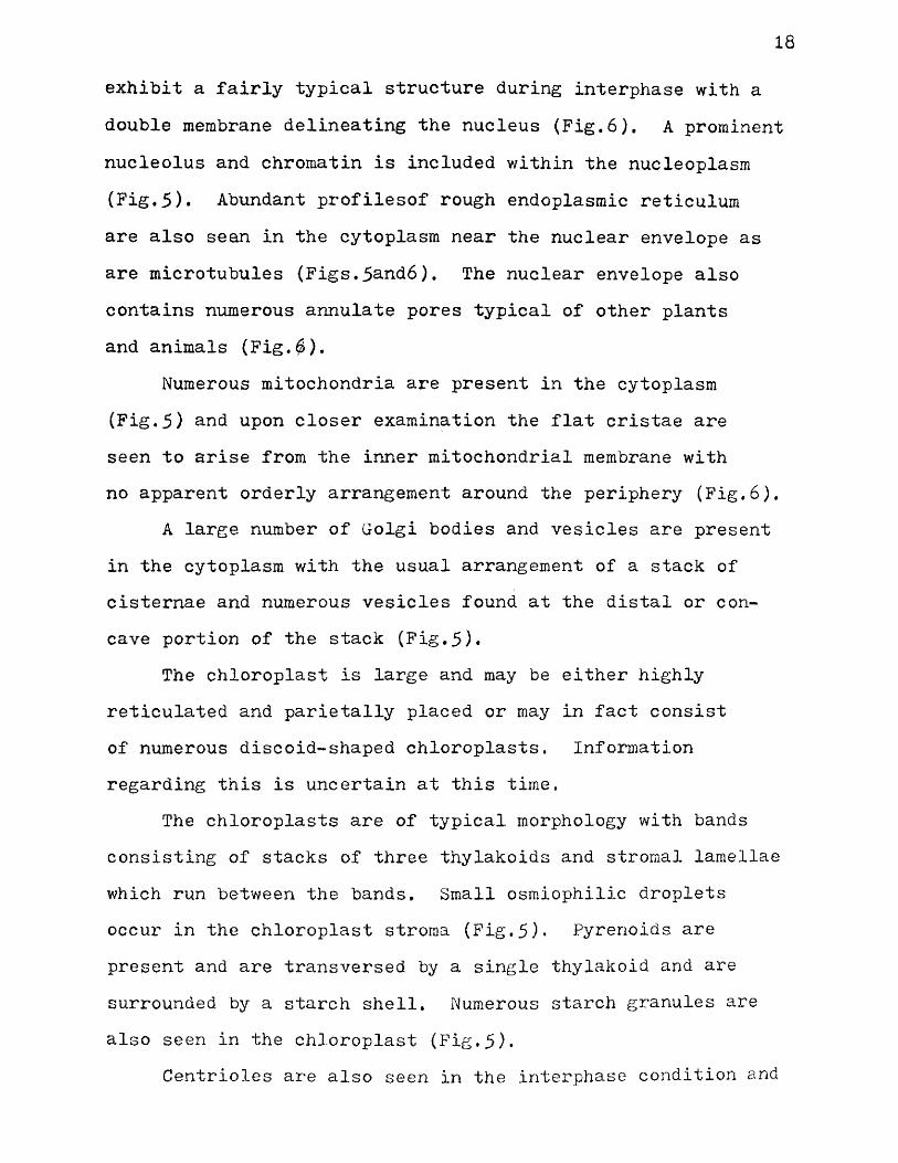
exhibit a fairly typical structure during interphase with a double membrane delineating the nucleus (Fig.6). A prominent nucleolus and chromatin is included within the nucleoplasm (Fig.5). Abundant profilesof rough endoplasmic reticulum are also sean in the cytoplasm near the nuclear envelope as are microtubules (Figs.5and6). The nuclear envelope also contains numerous annulate pores typical of other plants and animals (Fig.#).
Numerous mitochondria are present in the cytoplasm (Fig.5) and upon closer examination the flat cristae are seen to arise from the inner mitochondrial membrane with no apparent orderly arrangement around the periphery (Fig.6).
A large number of Golgi bodies and vesicles are present in the cytoplasm with the usual arrangement of a stack of cisternae and numerous vesicles found at the distal or concave portion of the stack (Fig.5).
The chloroplast is large and may be either highly reticulated and parietally placed or may in fact consist of numerous discoid-shaped chloroplasts. Information regarding this is uncertain at this time.
The chloroplasts are of typical morphology with bands consisting of stacks of three thylakoids and stromal lamellae which run between the bands. Small osmiophilic droplets occur in the chloroplast stroma (Fig.5)* Pyrenoids are present and are transversed by a single thylakoid and are surrounded by a starch shell. Numerous starch granules are also seen in the chloroplast (Fig.5).
Centrioles are also seen in the interphase condition and
19
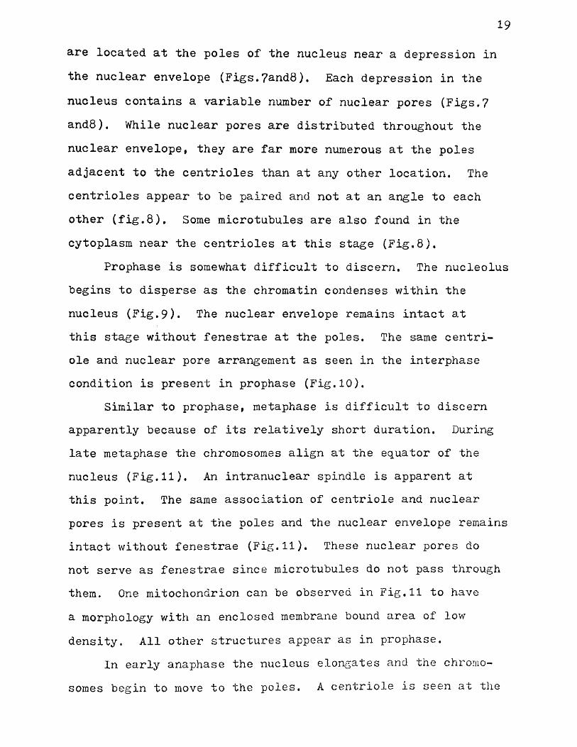
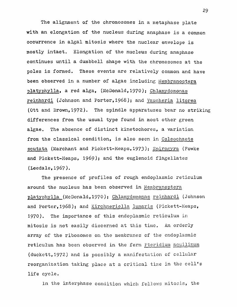
are located at the poles of the nucleus near a depression in the nuclear envelope (Figs.?and8). Each depression in the nucleus contains a variable number of nuclear pores (Figs.7 and8). While nuclear pores are distributed throughout the nuclear envelope, they are far more numerous at the poles adjacent to the centrioles than at any other location. The centrioles appear to be paired and not at an angle to each other (fig.8). Some microtubules are also found in the cytoplasm near the centrioles at this stage (Fig.8),
Prophase is somewhat difficult to discern. The nucleolus begins to disperse as the chromatin condenses within the nucleus (Fig.9). The nuclear envelope remains intact at this stage without fenestrae at the poles. The same centri- ole and nuclear pore arrangement as seen in the interphase condition is present in prophase (Fig.10).
Similar to prophase, metaphase is difficult to discern apparently because of its relatively short duration. During late metaphase the chromosomes align at the equator of the nucleus (Fig.11). An intranuclear spindle is apparent at this point. The same association of centriole and nuclear pores is present at the poles and the nuclear envelope remains intact without fenestrae (Fig.11). These nuclear pores do not serve as fenestrae since microtubules do not pass through them. One mitochondrion can be observed in Fig,11 to have a morphology with an enclosed membrane bound area of low density. All other structures appear as in prophase.
In early anaphase the nucleus elongates and the chromosomes begin to move to the poles. A centriole is seen at the

20
pole with a number of microtubules present in the adjacent cytoplasm. The nuclear envelope is still intact with no fenestrae seen at the polar regions (Fig.12).
During late anaphase the nucleus has assumed a dumbbell shape (Fig.16). The intranuclear spindle is clearly seen at this stage (Fig,l6). The nuclear envelope is intact with no fenestrae although it is somewhat modified in certain areas (Figs,15andl6). Nuclear pores are easily seen in the membrane adjacent to the centrioles (Fig.16). Kinetochores are not observed on the chromosomes in the anaphase nucleus (Figs.15 andl6).
Profiles of rough endoplasmic reticulum are seen in the cytoplasm adjacent to the nucleus as are numerous ribosomes (Figs,13andl6). Some of these ribosomes have a close attachment to the nuclear envelope, especially in the area of constriction (Fig.13). Upon closer examination these ribosomes appear to have an orderly arrangement on the membranes of the endoplasmic reticulum (Fig.14). A Golgi body is seen near each end of the anaphase nucleus with vesicles being pinched off in the direction of the nuclear envelope (Fig.16).
During anaphase microtubules are also present in the cytoplasm near the constricted area of the nucleus (Fig.13). The spindle apparatus is seen with some of the microtubules in close proximity to the nuclear envelope in the area of constriction (Figs. 13stndl5) •
Telophase nuclei were difficult to discern and were therefore not detected. It is assumed that the nucleus will continue its constriction to give two daughter nuclei,

21
In the interphase condition which follows mitosis the nucleus contains a nucleolus and has centrioles at the poles with the adjacent pores in the nuclear envelope (Fig.17). Chloroplast structure remains the same with the pyrenoid having a single transverse thylakoid and a shell of starch. Numerous starch granules and osmiophilic droplets are seen in the stroma (Fig.18). Profiles of rough endoplasmic reticulum are found throughout the cytoplasm (Fig.17) with the same orderly array of ribosomes on the membrane (F ig.18).
A major difference from the parent cell is the presence of large osmiophilic droplets in the cytoplasm (Fig.17). Numerous mitochondria are also present with some of them exhibiting an unusual morphology (Fig.17). Regions of lower density can be seen within the mitochondria and are bound by a double membrane. One area includes an invagination of the cytoplasm in which the ribosomes are present (Figs.17andl8). The material within these areas of lower density has a fibrillar appearance. Some flat cristae arise from the inner mitochondrial membrane and are found around the periphery of the mitochondria (Fig.18).
Following mitosis the beginning of reproductive differentiation is first observed as a cleavage of the cytoplasm. This cleavage may be due to either a vacuolation of the cytoplasm or more likely it is a result of both vacuolation and an evagination of the remainder of the central vacuole into the cytoplasm (Fig.19). The mitochondria previously described in Fig.18 are still present but no mitotic nuclei are evident.

22
Numerous osmiophilic droplets are seen in the chloroplasts and cytoplasm with all other features remaining the same as in interphase prior to mitosis (Fig.19).
The vacuolation continues with the general boundaries of the swarmers becoming evident (Fig.20). This vacuolation becomes quite extensive with the cytoplasm in the area of the vacuoles degenerating. Each future swarmer now contains a nucleus and single chloroplast (Fig.20).
In the final stages of swarmer production prior to release, most of the cytoplasm has degenerated except in the swarmers (Fig.21). Several of the swarmers contain osmio- philic droplets. Numerous flage11a are present but none are seen attached to the swarmers due to the plane of sectioning (Fig.21).
The swarmers are released from the cell through a rupture in the wall and upon closer examination are seen to be biflagellated, indicating they are gametes and that these are gametangial plants (Fritsch,1961).(Figs.22and26).
Prior to release from the cell the gamete has a morphology which is fairly typical of other chlorophycean algal swarmers. The nucleus is somewhat irregular in shape, contains a prominent nucleolus, and is defined by a double membrane. An osmiophilic droplet is situated adjacent to the nucleus and on occasion, can actually be seen protruding into it (Fig.23).
Chlorplasts are perforated with many starch granules.Once again the chloroplast may be highly reticulated and parietally placed or may consist of numerous discoid shaped

23
chloroplasts. Further work is necessary to clarify this point. One pyrenoid is present with a single thylakoid transversing it and a starch shell (Fig23). An eyespot is seen in the chloroplast in the anterior half of the cell (Fig.23). This eyespot is closely appressed to the outer cell membrane and is composed of 15-16 oval osmiophilic granules. This eyespot is 3-4 times as long as wide (Fig.24).
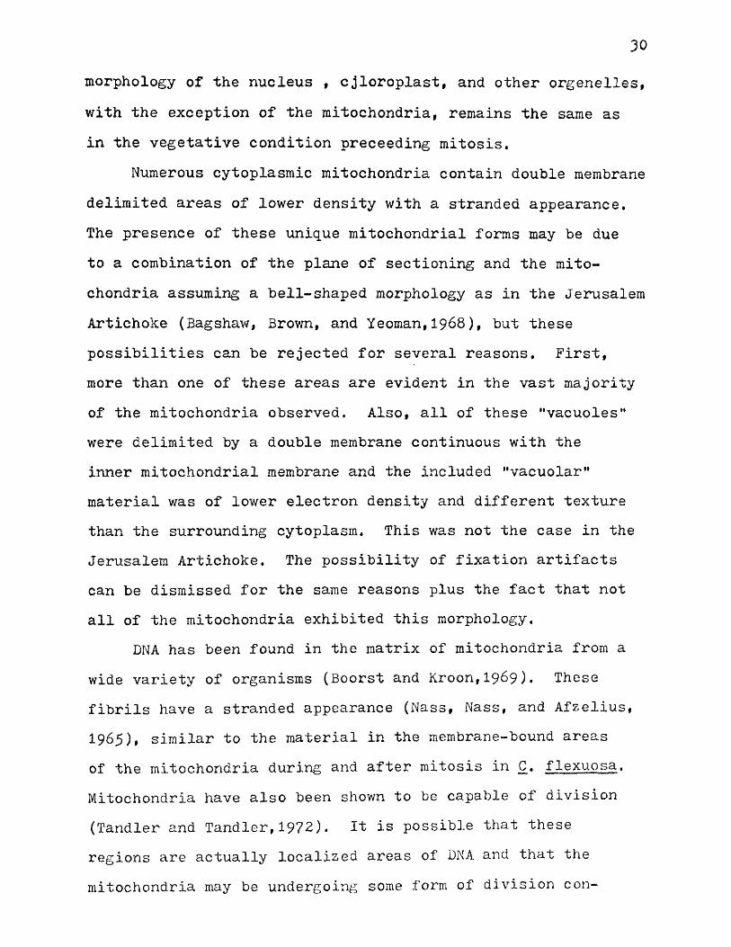
A flagellar apparatus is present in the anterior end of the cell and consists of two basal bodies with a connecting fiber (Figs.23and28). The basal bodies are of typical 9+0 structure when seen in cross-section (Fig.28), Numerous osmiophilic droplets and microtubules are seen in the area of the flagella (Figs,23and28). A contractile vacuole is also found in this area (Figs.23and26).
The falgella emerge from the cell through a specialized region in the cell wall (Fig.2?). The basal bodies lie in a concave shaped configuration and are joined by a connecting fiber (Fig.27). A band of supporting microtubules emerges from the area between the basal bodies and is attached to the cytoplasm (Fig.27). A cross-section of the flagellum shows the typical 9+2 axonemal structure (Fig,29).
Numerous mitochondria are found throughout the cytoplasm (Fig.23) and show an arrangement of the flat cristae where they are stacked upon each other in the middle of the cell as opposed to the earlier peripheral arrangement (Fig,25). Only a few profiles of endoplasmic reticulum are seen in the gamete (Fig.23).

Zk
A single Golgi body may be found associated with a number of vesicles at the posterior end of the nucleus. A large osmiophilic droplet is found between the Golgi body and the nucleus (Fig.23). A number of large vacuoles are seen around the periphery of the cell and appear to be a result of the vacuolation leading to gamete formation (Fig.23).

DISCUSSION
Observations on the vegetative structure of Cladophora flexuosa show a relationship to certain other coenocytic or siphonous green algae even though Cladophora has a septum dividing the filament into cells. Ultrastructural morphology
Bryopsis hypnoides (Burr and West,1970); Caulerpa prolifera (Sabnis,1969)! and Dichotomosiphon tuberosus (Moestrup and Hoffman,1973) reveals a large central vacuole with all other organelles limited to a thin peripheral layer of cytoplasm.In the case of Bryopsis hypnoides (Burr and West,1970) and Caulerpa prolif era (Sabnis, 1969) cytoplasm was penetrated by evaginations of the central vacuole, but no channels were noted passing through the cytoplasm. The presence of narrow channels through the cytoplasm of Cladophora flexuosa which would allow for communication between the central vacuole and the cell wall appears to be a unique situation. These channels might be a fixation artifact, but their persistent occurrence plus the presence of numerous evaginations of the central vacuole would indicate otherwise. Very possibly these evaginations and channels serve to facilitate cytoplasmic-vacuolar exchanges as suggested in Bryopsis hypnoides (Burr and West, 1970).
No regular spatial arrangement of the cytoplasm has beenobserved in both Caulerpa prolifera (Sabnis,19^9 ) andDichotomosiphon tuberosus (gay.^trup and Hoffman, 1973) •
2.5
26
The cytoplasm of Bryopsis hypnoides (Burr and V/estfl9?0) is limited to two different organized layers which differs from Cladophora flexuosa where organization is absent.
In most algae the interphase nucleus is typically eukaryotic with chromosomes# a nucleolus, and nucleoplasm delimited from the cytoplasm by a nuclear envelope. In all eukaryotic algae the nuclear envelope consists of two unit membranes separated by a perinuclear space. The outer nuclear membrane is often continuous with membranes of the endoplasmic reticulum. Nuclear pores transverse the nuclear envelope in all groups of green algae (Leedale,1970). Cladophora flexuosa is typically eukaryotic in all these respects of interphase morphology.
The mitochondria and Golgi bodies exhibit no unusual morphological features. The presence of bands of thylakoids in the chloroplast is typical of the Chlorophyceae as is the occurrence of 2-6 thylakoids per band with no girdle bands present (Gibbs,1970). The presence of osmiophilic droplets in the chloroplast matrix is also a general feature of algal chloroplasts (Gibbs,1970). Cladophora flexuosa remains typical in these respects. All groups of algae except for the Cyanophyta contain species which possess pyrenoids.The morphology of embedded pyrenoids consists of a granular matrix often transversed by thylakoids and surrounded by a starch shell of two hemispheres, A limiting membrane is not present (Griffiths,1970). In some algae, including Cladophora f racta (Strugger and Peveling, 1.961), the pyrenoid is divided into two hemispheres by a single membrane-limited disc.
27
Pyrenoid structure in Cladophora flexuosa remains consistent with other species containing embedded pyrenoids.
While centrioles are sometimes seen in the interphase condition at the poles, the only previously reported case of nuclear pores adjacent to the centrioles was in the golden- brown alga, Vaucheria litorea (Ott and Brown,1972). In Vaucheria litorea a pair of persistent centrioles lie at right angles to each other near the anterior end of the nucleus. A nuclear pore is present in the nuclear envelope opposite the centrioles. The existence of a depression in the nuclear envelope containing a number of pores, as in Cladophora flexuosa, has not been reported in other green algae. These pores could serve as a means of communication between the nucleus and the centrioles much as the fenestrae in other species of green algae, but microtubules have not been observed to pass through these pores.
Mitosis in Cladophora flexuosa is unique because it is independant of cytokinesis and often precedes gametogenesis whereas in some other green algae, (Spirogyra. Fowke and Pickett-Heaps,1969; Qedogonium, Fickett-Heaps and Fowke,1969; . and Chlamydomonas reinhardi, Johnson and Porter,1968), mitosis precedes cytokinesis. Evidence for this comes from the fact that swarmer formation in Cladophora takes place in a definite basipetal succession (Fritsch,1961) with an apical cell often containing gametes v/hile the cell immediately below it showed a number of mitotic figures. Also the production of iso- gamous, biflagellated gametes (Fritsch,1961) signifies that they are the end result of mitosis in the garaetophyte generation.

28
Dispersion of the nucleolus and condensation of the chromosomes during prophase are events which occur in all green algae in the same manner,
The,persistence of the nuclear envelope throughout mitosis with no polar fenestrae appears to be an unusual feature in the green algae, being observed only in Cladophora flexuosa and Chlamydomonas moewusii (Triemer and Brown,197^).The persistence of the nuclear envelope in Cladophora flexuosa is not absolute since the membrane appears to be modified to some extent in several cases. The absence of fenestrae at the poles may be attributable to a dynamic nature so that fenestrae may actually be present for a short period but were absent due to the time of fixation (Triemer and Brown,197^). The nuclear envelope is also seen to persist in Vaucheria litorea (Ott~and Brown,1972) and the euglenoid flagellates (Leedale, 1967,1968) with no gaps or fenestrae at the poles. The nuclear envelope also persists during mitosis in two species of marine dinoflagellates, Woloszynkia micra (Leadbeater and Dodge,1967) and Gyrodinium cohnii (Kubai and Ris,1969) and in some fungi including Fusarum oxysporum (Aist and Williams,1972) and Mucor hiemalis (McCully and Robinow,1973). The persistence of the nuclear envelope appears to be a fairly widespread event in homokaryotic and dikaryotic micelia of numerous Basidiomycetes (Thielke,1973)• It is apparent that the persistence of the nuclear envelope with no fenestrae at the poles is a fairly rare occurrence in algal mitosis and is much more common in fungal mitosis, especially Basidiomycetes,
29The alignment of the chromosomes in a metaphase plate
with an elongation of the nucleus during anaphase is a common occurrence in algal mitosis where the nuclear envelope is mostly intact. Elongation of the nucleus during anaphase continues until a dumbbell shape with the chromosomes at the poles is formed. These events are relatively common and have been observed in a number of algae including Membranoptera platyphylla, a red alga, (McDonald,1970)j Chlamydomonas reinhardi (Johnson and Porter,1968); and Vaucheria litorea (Ott and Brown,1972). The spindle apparatuses bear no striking differences from the usual type found in most other green algae. The absence of distinct kinetochores, a variation from the classical condition, is also seen in Coleochaete scutata (Marchant and Pickett-Heaps,1973); Spirogyra (Fowke and Pickett-Heaps, 1969); an(̂ euglenoid flagellates (Leedale,1967)•
The presence of profiles of rough endoplasmic reticulum around the nucleus has been observed in Membranoptera platyphylla (McDonald,1970); Chlamydomonas reinhardi (Johnson and Porter,1968); and Kirchneriella lunaris (Pickett-Heaps, 1970). The importance of this endoplasmic reticulum in mitosis is not easily discerned at this time. An orderly array of the ribosomes on the membranes of the endoplasmic reticulum has been observed in the fern Pterldium aquilinum (duckett,1972) and is possibly a manifestation of cellular reorganization taking place at a critical time in the ceil*s life cycle.
In the interphase condition which follows mitosis, the
30
morphology of the nucleus , cjloroplast, and other orgenelles, with the exception of the mitochondria, remains the same as in the vegetative condition preceeding mitosis.
Numerous cytoplasmic mitochondria contain double membrane delimited areas of lower density with a stranded appearance. The presence of these unique mitochondrial forms may be due to a combination of the plane of sectioning and the mitochondria assuming a bell-shaped morphology as in the Jerusalem Artichoke (Bagshaw, Brown, and Yeoman,1968), but these possibilities can be rejected for several reasons. First, more than one of these areas are evident in the vast majority of the mitochondria observed. Also, all of these "vacuolesM were delimited by a double membrane continuous with the inner mitochondrial membrane and the included "vacuolar" material was of lower electron density and different texture than the surrounding cytoplasm. This was not the case in the Jerusalem Artichoke. The possibility of fixation artifacts can be dismissed for the same reasons plus the fact that not all of the mitochondria exhibited this morphology.
DNA has been found in the matrix of mitochondria from a wide variety of organisms (Boorst and Kroon,1969). These fibrils have a stranded appearance (Nass, Nass, and Afzelius, 1965), similar to the material in the membrane-bound areas of the mitochondria during and after mitosis in C. flexuosa. Mitochondria have also been shown to be capable of division (Tandler and Tandler,1972). It is possible that these regions are actually localised areas of DNA and that the mitochondria may be undergoing some form of division con-

31
current with a proliferation of the cytoplasm prior to gametogenesis. Definitive evidence to support this hypothesis is absent at this time.
The progressive vacuolation of the cytoplasm to form gametes in Cladophora flexuosa has been described in another coenocytic green alga, Bryopsis hypnoides (Burr and West,1970). ln Bryopsis hypnoides as in C. flexuosa repeated intranuclear mitoses precede the cleavage of the cytoplasm into gametes by cleavage vesicles. These vesicles in Bryopsis hypnoides increase in number and fuse to partition portions of the cytoplasm into gametes, as in C. flexuosa. This similarity in the mechanism of gametogenesis is of special notice because both algae are coenocytic forms although Cladophora flexuosa is divided into cells. Mechanisms of zoosporogenesis in Hormidium flaccidum (Mattox,1971) and Klebsormidium flaccidum (Cain, Mattox, and Stewart,1973) are of little importance except for the zoospore morphology since only one zoospore is produced per cell.
Gamete morphology in Cladophora flexuosa closely resembles that of other chlorophycean swarmers. The nucleus, Golgi bodies, and mitochondria show similarities to zoospores of Microthamnion (Watson and Arnott,1973); Stigeoclonium (Manton, 196^); and Cladophora rupestris (Robinson, White, and Preston, 1972) among others. The presence in the chloroplast of a pyrenoid bisected by a single thylakoid is similar to the situation found in zoospores of Enteromorpha intestinalis (Evans and Christie,1970)j PseudondocIonium basildense and Trichosarcina polymorpha (Mattox and Stewart,1973)• Eyespot

32
morphology resembles that described for Tetracystis excentrica (Amott and Brown,1967),
The flagellar apparatus, similar to Chlamydomonas reinhardi (Ringo,1967), consists of two basal bodies at right angles to each other joined by a connecting fiber at the top with bands of supporting microtubules running into the cytoplasm. The 9+2 axonemal structure of the flagella is found in all fla- gella including those of Stigeoclonium (Manton,196^) and Chlamydomonas reinhardi (Ringo,19^7).
The present study has shown that the vegetative structure of Cladophora flexuosa is similar to other coenocytic green algae with the exception of the cytoplasm being divided into multinucleate cells by septae. Mitosis in C. flexuosa precedes ganetogenesis and bears many similaritities to the classical model of mitosis except that the nuclear envelope remains intact with no fenestrae. Gametogenesis by vacuolation has been described in Bryopsis hypnoides (Burr and West,1970), a coenocytic green alga* and is therefore not unique. The unusual morphology of the mitochondria following mitosis has not been previously described while gamete morphology is fairly typical of other green algal swarmers. Cladophora flexuosa possesses some unusual features in mitosis and gametogenesis but overall is not entirely unique.

Key To Abbreviations
bb Basal Bodyce Centriolecf Connecting Fiberch Chloroplastcn Contractile Vacuolecv Central Vacuolecw Cell Waller Endoplasmic Reticulumey EyespotF Flagellar ApparatusG Golgi BodyLi Lipid Dropletm Mitochondrionmt MicrotubuleN Nucleusnc Nucleolusnp Nuclear PoreP Pyrenoids Starch GranuleV Vacuole
33

34
Figure 1,
Light micrograph of the apical region of plant* 386X.
Figure 2.
Longitudinal section of tip of apical cell. Note the invaginations of the cytoplasm (arrows). 3#51 OX.


36
Figure 3.
Cross section of a vegetative cell. Note the channels in the cytoplasm (arrows). 3,2ifOX.
Figure
Longitudinal section of the "basal region of a vegetative cell showing the discontinuities in the cytoplasm. 3»25OX.

w
%
«L If g(g//' -'Jr-7 -tii* < x
l iferr. * - * / ' j f ^ 4S "&
t & TKV?/ . **>/ J/-/* T]

38
Figure 5«
View of typical interphase condition of nucleus and cytoplasm. Note the single thylakoid transversing the pyrenoid (arrow). l^f280X.
Figure 6.
High magnification of nuclear envelope. 32,000X.
Figure ?.
Typical interphase nucleus with centrioles at each pole (arrows). 15»230X.

Aj

40
Figure 8.
High magnification view of nuclear pore region with centrioles. 29,000X.
Figure 9.
Prophase nucleus (N). 11,900X.
Figure 10.
Nuclear pore-centriole arrangement in prophase. 24,000X.
Figure 11.
Metaphase plate. 11,900X.
Sffi

kz
Figure 12.
Longitudinal section of early anaphase nucleus. 1^,280X.
Figure 13.
Area of constriction in late anaphase nucleus, 30*000X.
Figure 14.
Endoplasmic reticulum from late anaphase condition showing the arrangement of ribosomes on the membrane of the endoplasmic reticulum. 30|000X.
Figure 15.
Polar region of late anaphase nucleus. Note the lack of evident kinetochores. 28,000X.

1§Iiff*.m

44
Figure 16.
Longitudinal section of late anaphase nucleus. Note the depression in the nuclear envelope at the poles. 22,850X.

(Sisw
RpRs

46
Figure 17.
Interphase nucleus following mitosis. Note the mitochondria and depression in the nuclear envelope.12,380X.
Figure 18.
High magnification of the unusual mitochondria found during and after mitosis. Note the invagination of the cytoplasm and ribosomes in the invagination. 32,000X.
Figure 19.
Cytoplasm at the beginning of gametogenesis with the vacuoles. 3*990X.
Figure 20.
Formation of the gametes by a vacuolation of the cytoplasm. 3*220X.

Mflr-

Figure 21.
Newly formed gametes in the cell prior to release. 2
Figure 22.
Light micrograph of gamete showing the biflagellated condition. 3i000X.
Figure 23*
48
50 ox.
Longitudinal section of newly formed gamete prior to release from the cell. 21f?00X.


Figure Z h .
High magnification of eyespot of gamete. 3^f000X.
Figure 25.
Typical mitochondrion from gamete. 31f000X.
Figure 26.
Gamete showing the.biflagellated condition. 22,850X.


Figure 27.
High magnification of flagellar apparatus. Note the band of microtubules (arrow), 2^,280X..
Figure 28,
Flagellar apparatus of gamete. 27f000X.
Figure 29.
Cross section of flagellum, 126,900X,

BIBLIOGRAPHY
Aist, J.R. and P.H. Williams. 1972. Ultrastruclure andtime course of mitosis in the fungus Fusarum oxysporum.J. Cell Biol. 55; 369-391.
Arnott, H.J. and R.M. Brown. 1967. Ultrastructure of theeyespot and its possible significance in phototaxis of Tetracystis excentrica. J. Protozool. 14(4): 529-539.
Bagshaw, V., R. Brown, and M.M. Yeoman. 1969. Changes in the mitochondrial complex accompanying callus growth.Ann. Bot. 33: 35-44,
Borst, P. and A.M. Kroon. 1969. Mitochondrial DNA; Physicochemical properties, replication, and genetic function.Int. Rev. Cytol. 26: 107-190.
Burr, F.A. and J.A, West. 1970. Light and electron microscopicobservations on the vegetative and reproductive strucutres °T Bryopsis hypnoides. Phycologia. 9(1): 17-37.
Cain, J.R., K.R. Mattox, and K.D. Stewart. 1973. The cytology of zoosporogenesis in the filamentous green algal genus Klebsormidium. Trans. Amer. Micros. Soc, 92(3): 398-404.
Deason, T.R. and W.H. Darden. 1971. The male initial and mitosis in Volvox aureus. In Parker, B.C. and R.M.Brown (eds.), Contributions in Phycology, Allen Press,Inc., Lawrence, Kansas'! 87-79.
Duckett, J.S. 1972. Pentagonal arrays of ribosomes infertilized eggs of Pteridium aquilinum. J. Ultrastructure Res. 38: 390-397.
Evans, L.V. and A.0, Christie. 1972. Studies on the ship fouling alga Enteromorpha. I. Aspects of the fine structure and biochemistry of swimming and newly settled zoospores. Ann. Bot. 34: 451-466.
Floyd, G.L., K.D. Stewart, and K.R. Mattox. 1971. Cytokinesis and plasmodesmata in Ulothrix. J. Phycol. 7: 306-309.
Fowke, L.C. and J.D. Pickett-Heaps. 1969. Cell division in Spirogyra. I. Mitosis. J. Phycol. 5: 240-259.
Fritsch, F.E. I96I. The Structure and Reproduction of the Algae. Cambridge University Press, London, 229-249.
52

53
Gibbs, S.P. 1971. The comparative ultrastructure of thealgal chloroplast. Ann. N.Y. Academ. Sci. 175:,^5^-^73.
Griffiths, D.J. 1970. The pyrenoid. Bot, Review, 36: 29-5^.Hawkins, A.F. and G.F. Leedale, 1971* Zoospore structure and
colony formation in Pediastrum sp. and Hydrodictyon reticulatum. Ann, Bot. 35: 201-211.
Hibberd, D.J. and G.F, Leedale. 1971, Cytology and ultrastructure of the Xanthophyceae. IX. The zoospore and vegetative cell of coccoid forms, with special reference ’to Ophiocytium ma.jus, Br. Phycol. J. 6(1): 1-23.
Hoffman, L. and I. Manton. 1962. Observations on the fine structure of the.zoospore of Oedogonium cardiacum with special reference to the flage11a apparatus. J. Exptl.Bot. 13: 443-449.
Johnson, U.G, and K.R. Porter. 1968. Fine structure of cell division in Chlamydomonas reinhardi. J. Cell Biol.38: 403-425.
Kubai, D.F. and H. Ris. 1969. Division in the dinoflagellate Gyrodinium cohnii. J. Cell Biol. 40: 508-528.
Leadbeater, B. and J.D. Dodge. 1967. An electron microscope study of nuclear and cell division in a dinoflagellate, Archiv. Mikrobiol. 57: 239-25^.
Leedale, G.F. 1967, Suglenoid Flagellates. Prentice-Hall, Englewood Cliffs, N.J. 216-235.
Leedale, G.F. 1968. The nucleus in Euglena. In the Biologyof Euglena, D.E. Buetow, ed., Academic Press, N.Y. 185-242.
Leedale, G.F. 1970. Phylogenetic aspects of nuclear cytology in the algae. Ann. N.Y. Acad. Sci. 175: ^29-453.
Lovlie, A. and T. Braten. 1970. On mitosis in the multicellular alga, Ulva mutabilis. J. Cell Sci. 6: 109-129.
Manton, I. 1964. Observations on the fine structure of thezoospore and young germling of Stigeoclonium. J. Exptl, Bot. 15: 399-^11.
Marchant, H.J. and J.D. Pickett-Heaps, 1973. Mitosis andcytokinesis in Coleochaete scutata. J. Phycol. 9: 461-471.
Mattox, K.R. 1971. Zoosporogenesis and resistant-cell formation in Hormidium flaccidum. Contrib. in Phycol.Sept. 1971, 137-1^.

54
Mattox, K.R. and K.D, Stewart. 1973* Observations on thezoospores of PseudondocIonium basiliense and Trichosarcina polymorpha. Gan. J. of Bot. 51(7): 1425-1430.
McCully, E.K. and G.F. Robinow. 1973* Mitosis in Mucor hiemalis. Archiv, Mikrobiol. 94: 133-148,
Moestrup, 0. and L.R. Hoffman. 1973. Ultrastructure of the green alga Dichotomosiphon tuberosus with special reference to the occurrence of striated tubules in the chloroplast. J. Phycol. 9: 430-437.
Nass, M.M.K., S. Nass, and B.A. Afzelius. 1965. The general occurrence of mitochondrial DNA, Exptl. Cell Res.37: 516-539.
Ott, D. and R.M. Brown. 1972. Light and electron microscopical observations on mitosis in Vaucheria litorea. Br. phycol.J. 7: 361-374.
Pickett-Heaps, J.D, 1967. Ultrastructure and differentiationin Chara sp. II. Mitosis. Aust. J. Biol. Sci. 20.* 883-894.
Pickett-Heaps, J.D. and L.G, Fowke. 1969. Cell division inOedogonium. I. Mitosis, cytokinesis, and cell elongation. Aust. J. Biol. Sci. 22s 857-894.
Pickett-Heaps, J.D. and L.C. Fowke. 1970. Cell division inOedogonium. II. Nuclear division in 0. cardiacum.Aust. J. Biol. Sci. 23: 71-92.
Pickett-Heaps, J.D. 1970. Mitosis and autospore formation in the green alga Kirchneriella lunaris. Protoplasma.70: 325-34?.
Pickett-Heaps, J.D. 1972. Cell division in Cyanophoraparadoxa. New Phytol. 71: 561-567.
Pickett-Heaps, J.D. 1972. Cell division in Tetraedon. Ann.Bot, 36: 693-701.
Ringo, D.L. 1967. Flagellar motion and fine structure of the flagellar apparatus in Chlamydomonas. J. Cell Biol. 33: 543-571.
Robinson, D.G., R.K. White, and R.D. Preston. 1972. Fine structure of Cladophora and Chaetomorpha. III. Wall synthesis and development. Planta. 107: 131-l44,
Sabnis, D.D. 1969. Observations on the ultrastructure ofthe coenocytic alga Caulerpa prolifera, with special reference to some unusual cytoplasmic components.Phycologia. 7(1): 24-42.

55
Smith, G.M. 1951* Manual of Phycology. The Ronald Press Company, New York. 27-28.
Strugger, J.R. and S. Peueling. 1961. Uber den feinbau der chromatophoren von Cladophora fracta, Kutz. Protoplasma. 54: 254-262.
Tandler, B. and C.L. Hoppel. 1972. Possible division ofcardiac mitochondria. Anatomical Rec. 173(3)* 309-324.
Thielke, Ch. 1973* Intranuclear mitosen in hemekaryotischen und dikaryotischen mycelien der Basidiomyceten. Archiv. Mikrobiol. 94: 341-350.
Triemer, R.E. and R.M, Brown. 1974. Cell division in Chlamydomonas moewusii. unpublished manuscript
Venable, J.H. and R. Coggleshall. 1965* A simplified lead citrate stain for electron microscopy. J. Cell Biol.25: 407-408.
Watson, M.W. and H.J. Arnott. 1973< Ultrastructural morphology of Pflicrothamnion zoospores. J. Phycol. 9(1)* 15-29*

VITA
Kenneth Wilson Bullock
Born in Fredericksburg, Virginia, April 22, 1950* Graduated from Stafford High School in Stafford, Virginia June, 1968. Received B.S. degree in biology from the College of William and Mary, June, 1972. Entered gradual studies at the College of William and Mary in September, 1972. Candidate for Master of Arts degree in biology, September, 1974.