unique pathogenic escherichia strain - proceedings of the national
TRANSCRIPT

Proc. Natl. Acad. Sci. USAVol. 93, pp. 11149-11154, October 1996Microbiology
Unique chromosomal regions associated with virulence of an avianpathogenic Escherichia coli strainPETER K. BROWN AND RoY CURTISS III*Department of Biology, Washington University, Campus Box 1137, One Brookings Drive, St. Louis, MO 63130
Communicated by Allan M. Campell, Stanford University, Stanford, CA, July 15, 1996 (received for review May 28, 1996)
ABSTRACT The avian pathogenic Escherichia coli strainX7122 (serotype 078:K80:H9) causes airsacculitis and coli-septicemia in chickens. To identify genes associated with aviandisease, a genomic subtraction technique was performedbetween strain X7122 and the E. coli K-12 strain X289. TheDNA isolated using this method was found only in strainX7122 and was used to identify cosmid clones carrying uniqueDNA from a library of X7122 that were then used to map theposition of unique DNA on the E. coli chromosome. A total of12 unique regions were found, 5 of which correspond topreviously identified positions for unique DNA sequence in E.coli strains. To assess the role each unique region plays invirulence, mutants of X7122 were constructed in which asegment of unique DNA was replaced with E. coli K-12 DNAby cotransduction of linked transposon insertions in DNAflanking the unique sequence. The resulting replacementmutants were assessed for inability to colonize the air sac andcause septicemia in 2-week-old white Leghorn chickens. Twomutants were found to be avirulent when injected into theright caudal air sac of 2-week-old chickens. One avirulentmutant, designated X7145, carries a replacement of the rjblocus at 44 min, generating a rough phenotype. The secondmutant is designated X7146, and carries a replacement atposition 0.0 min on the genetic map. Both mutants could becomplemented to partial virulence by cosmids carrying se-quences unique to X7122.
Escherichia coli is a diverse species, the majority of strains ofwhich are normal inhabitants of the intestinal tract. However,certain strains are capable of causing intestinal or extraintes-tinal infections in specific hosts.An example of strain dependent host specificity is avian
pathogenic E. coli (APEC), which is the causative agent ofairsacculitis, pericarditis, perihepatitis, and colisepticemia inpoultry (1). The disease is caused by a limited number ofserotypes, the most predominant being O1:K1, 02:K1, and078 (2-5). The bacteria are thought to enter the respiratorytract of chickens following inhalation of feces contaminateddust, where they colonize the air sac (6). Under certaincircumstances, the bacteria are capable of establishing asystemic infection resulting in a fatal septicemia. Prior infec-tion with other pathogens including infectious bronchitis dis-ease virus, Newcastle disease virus, and Mycoplasma spp., orstresses such as starvation, overheating, and overcrowdingpredispose birds to colisepticemia (1, 7, 8). With the increaseduse of intensive confinement housing, colisepticemia has be-come the predominant bacterial disease affecting the poultryindustry (1). However, little is known about the factors whichfacilitate colonization of the air sac, entry into the bloodstream, or any subsequent steps involved in the virulence ofavian pathogenic E. coli.
In a number of pathogenic isolates of E. coli, the associationof specialized unique DNA regions with virulence is well
established. The genes for class II capsule synthesis (kps) andfor 0 antigen synthesis (rib gene cluster) are either absent ornonfunctional in E. coli K-12 (9, 10). Recently, the pathoge-nicity islands PAI I and PAI II of uropathogenic strains (11,12), and the locus of enterocyte effacement (LEE) of entero-pathogenic and enterohemorrhagic isolates represent exam-ples of large unique regions of chromosome encoding knownand/or putative virulence attributes that are not found in E.coli K-12 or natural E. coli isolates from other sources (13).
It is likely that APEC strains will possess special attributesnecessary for colonization and infection of the respiratorytract, especially in avian species. This report describes the useof a genomic subtractive hybridization technique to identifyregions of the chromosome likely to be associated with viru-lence of APEC strains.
MATERIALS AND METHODSBacterial Strains, Plasmids, and Culture Conditions. The
naladixic acid resistant virulent APEC strain X7122 (14) wasused in subtractive hybridization, cloning and mutant con-struction. E. coli strains used in this study are listed in Table1. Cells were grown at 37°C in Lennox broth (L broth) (18)unless otherwise stated. Minimal M9 media (18) was used tocompare growth rates of various strains. Solid growth mediawere made by addition of 1.5% agar. Tetracycline (8 ,tg/ml),kanamycin (25 ,tg/ml), chloramphenicol (25 ,ug/ml), naladixicacid (12.5 p,g/ml), and ampicillin (100 ,tg/ml) were added asrequired. Plasmid pYA3107 carries the tsh gene from strainx7122 (19).
General Molecular Techniques. Chromosomal DNA wasisolated as described by Hull et al. (20). ColV plasmid andcosmid DNA isolation and Southern and colony hybridizationswere performed by procedures described by Sambrook et. al.(21). Hybridizations using the Kohara miniset (22) wereperformed according to the manufacturers protocol (TakaraBiomedicals, Madison, WI). Radioactive probes were gener-ated by random priming of template DNA or, where stated, byextension of a complementary primer using Klenow polymer-ase (21). Polymerase chain reaction (PCR) for the subtractivehybridization procedure was performed as described by Strausand Ausubel (23) using Amplitaq DNA polymerase (Perkin-Elmer/Cetus) and a 480 DNA Thermal Cycler (Perkin-Elmer/Cetus).Cosmid Library Construction. A genomic library of X7122
was constructed in the low copy number vector pYA3174.Plasmid pYA3174 carries an ampicillin resistance gene, apSC101 replicon, dual cos sites from pCos2embl, a BamHIcloning site flanked by symmetrical EcoRI and NotI sites, andthe mob site from pGP704, permitting mobilization of cosmidsby conjugal transfer from strain SMlOApir (16). ChromosomalDNA from X7122 was partially digested with Sau3A and size
Abbreviations: APEC, avian pathogenic E. coli; PAI, pathogenicityisland; LEE, locus of enterocyte effacement; L broth, Lennox broth;cfu, colony-forming units.*To whom reprint requests should be addressed.
11149
The publication costs of this article were defrayed in part by page chargepayment. This article must therefore be hereby marked "advertisement" inaccordance with 18 U.S.C. §1734 solely to indicate this fact.
11150 Microbiology: Brown and Curtiss
Table 1. E. coli strains
Strain Genotype or characteristics Source, construction, or ref.
X7122 078:K80:H9 14X289 W1485 A- ginV44 F- This laboratoryMG1655 Wild type 15SMlOApir thi thr leu tonA lacY supE 16
A(pirR6K) recA::RP4-2-Tc::MuKm
X2819 F- lacYI glnV44 galK2 galT22 17A(cI857 b2 red,(3 S7) recA56AthyA57 metBI hsdR2
CAG12093 MG1655 car-96::TnlO 15CAG18425 MG1655 thr-3091::TnlOkan 15CAG18447 MG1655 proAB81::TnlO 15CAG18633 MG1655 zag-3198::TnlOkan 15NK5526 hisG213::TnlO, A-, IN(rrnD-rrnEl) N. KlecknerCAG12176 MG1655 zee-3189::TnlOkan 15CAG18604 MG1655 zgd-3156::TnlOkan 15CAG18472 MG1655 nupG3157::TnlO 15x7176 x7122 car-96::TnlO Pl(CAG12093) - X7122X7177 X7176 thr-3091::TnlOkan P1(CAG18425) X7176X7146 X7122 (X289:thr-car) Pl(X289) --> X7177X7180 X7122 zag-3198::TnlOkan P1(CAG18633) - X7122X7181 X7180proAB81::Tn1O P1(CAG18447) > X7180X7148 X7122 (x289:proAB-zag) Pl(X289) --X7181x7178 X7122 zee-3189::TnlOkan Pl(CAG12176) -> X7122x7179 X7178 hisG::TnJO Pl(NK5526) -- X7178X7145 X7122 (X289:hisG-zee) Pl(X289) ->x7179X7175 MG1655 zgd-3156::TnlOkan Pl(CAG18472) >
nupG3157::TnlO CAG18604X7147 X7122 (MG1655:zgd-nupG) Pl(X7175) -- X7122
fractionated through a 10-25% NaCI gradient (20). DNAfragments of 35-50 kb were ligated to pYA3174 digested withBamHI and PvuII. The ligation mixtures were packaged in vitroand transduced into E. coli K-12 strain X2819, followed by invivo amplification and repackaging (17). Clones capable ofcomplementing E. coli K-12 strains carrying auxotrophic mu-tations in thr (at 0.0 min), purE (at 12.0 min), trp (at 28.0), his(at 45.0 min), thyA (at 63.8 min), and purA (at 95.0 min) wereobtained at frequencies between 10-3 and 10-6 per cosmid-bearing A particle, indicating that DNA inserts from aroundthe chromosome were present and that the library is thereforerepresentative of the genome of X7122.
Conjugal Transfer ofCosmid DNA. In vivo packaged cosmidclones were transferred to E. coli strain SMlOApir, and then tomutant derivatives of X7122 via conjugation (16).
Subtractive Hybridization. Chromosomal DNA from theprototrophic E. coli K-12 strain X289 was mixed with A DNAin a ratio of 100:1, sheared to a length of 1-3 kb, andbiotinylated using photoactivatable biotin (Clontech). Theresulting biotinylated DNA mixture (10 jig) was hybridizedwith Sau3A-digested X7122 DNA (0.5 jig) in a subtractivehybridization procedure as described by Straus and Ausubel(23), except that hybrids were removed using streptavidin-coated magnetic beads (Promega). After five rounds of sub-tractive hybridization, adaptors were ligated to the Sau3A endsof the unbound fraction, and the resulting ligation was PCRamplified (23). The PCR products were then radioactivelylabeled with [a-32P]dCTP by extension with one of the strandsof the adaptor using DNA polymerase (Klenow fragment)(21).
Bacteriophage Transduction and Replacement of UniqueRegions. Bacteriophage P1 clrlOO Cm (24) was used fortransduction of E. coli (25). Optimum transduction of X7122was obtained following growth at 30°C to an OD6wo of 0.3.To replace the unique DNA of X7122 with E. coli K-12 DNA
using P1 clr 100 Cm-mediated transduction, two approaches
were used. In one approach, E. coli K-12 strains were con-structed with TnlO and TnlOkan insertions flanking the po-sition of the unique DNA sequence as determined by hybrid-ization to the Kohara miniset (see below). The transposonswere cotransduced into strain X7122, followed by selection fortetracycline and kanamycin resistance.An alternate approach was used when hybridization to
multiple Kohara clones made positioning of the unique regionless precise. An individual TnlOkan insertion was transducedinto X7122, and the resulting strain was used as the recipientfor transduction of a linked TnlO insertion. Transductantswere screened for loss of kanamycin resistance to ensure thatDNA extending to the position of the TnlOkan insertion wascotransduced.
In either approach, to ensure that insertions in auxotrophicmarkers did not affect virulence, mutant strains were trans-duced with lysates made on X289 and prototrophic derivativeswere selected. The resulting strains thus have E. coli K-12 DNAin place of the unique DNA segment of X7122 and are deletedfor the specific unique DNA region.
Virulence Studies. For LD50 determinations, bacteria weregrown to OD600 of 0.4 in L broth, pelleted by centrifugation,and resuspended in phosphate-buffered saline (PBS) to adensity of 109 colony-forming units (cfu)/ml. Suspensions (100gl) of 10-fold dilutions were injected into the right caudal airsac of 2-week-old specific-pathogen-free white Leghorn chick-ens. In vivo selection of cosmid clones that restore virulence toavirulent mutants was performed by injecting _108 cfu ofpools of 15 cosmid clones in the avirulent mutant into the rightcaudal air sac of 2-week-old birds. Birds were euthanatized 3days after inoculation, and heart blood was drawn and platedon L agar containing naladixic acid and ampicillin.
RESULTSEnrichment of Sequences Unique to X7122. The prototro-
phic E. coli K-12 strain X289 is avirulent (LD5o > 108) in
Proc. Natl. Acad. Sci. USA 93 (1996)
Proc. Natl. Acad. Sci. USA 93 (1996) 11151
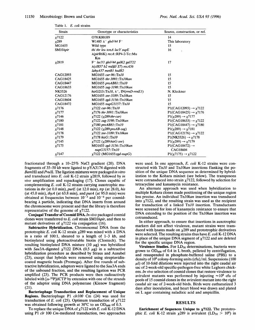
2-week-old chickens and is thus lacking some of the attributespresent in the virulent APEC strain X7122 (LD5o = 9.4 x 10⁵cfu). To isolate DNA that is present in X7122 but not in E. coliK-12, a subtractive hybridization was performed in which DNAfragments of X7122 that hybridize with DNA from strain X289or A are selectively removed. Southern hybridization experi-ments using radioactively labeled A DNA to probe digests ofthe genome of X7122 showed that this strain carries lambdoidprophage sequences, and since strain X289 does not, theselambdoid sequences would be represented among uniqueDNA following subtractive hybridization. Exogenous A DNAwas therefore included in the subtractive hybridization proce-dure to remove lambdoid sequences of X7122 that wouldotherwise result in high background signals in subsequenthybridization experiments using cosmid vectors and the lyso-genicE. coli strain X2819. Following five rounds of subtraction,PCR amplification of the X7122 unique DNA gave a productranging in size from <0.3 kb to 3.0 kb (data not shown).To verify that sequences common to X7122 and X289 were
removed by the subtraction procedure, a Southern blot ofHincIl-digested genomic DNA from X289 and X7122 wasprobed with the radioactively labeled PCR product. The probehybridized to DNA fragments of X7122 varying in size from 0.5kb to 15 kb (Fig. 1). No hybridization was detected with DNAof X289.
Identification of Cosmid Clones Carrying Unique DNA. Acosmid library representing the genome of X7122 in pYA3174was probed with the radioactively labeled X7122 unique PCRproduct. From a total of 1000 cosmid clones, 145 hybridized tothe unique DNA probe (data not shown). To verify that knownunique sequences were represented among the positive clones,colony hybridizations were performed using the ColV plasmidfrom X7122, and a Scal fragment from pYA3107 that containsthe tsh gene as probes (19, 26). Since the tsh gene and the ColVplasmid are present in X7122 but not in X289, these sequencesshould be represented among clones identified as carryingunique DNA. The tsh gene and ColV plasmid hybridized to 19
150Okb-: -
6.8kb-.
4.7kb
3.1kb2.5kb
2At 2.
1 8kb1 4kb
1. kb
0.8kb
FIG. 1. Results of probing Southern -blots of. Hin.cll-digestedgenomic DNA of strains X7122 and X289 with the radioactively labeledPCR product from the subtractive hybridization technique. Equalamounts (5 gig) of DNA were loaded in each lane. Strains are labeledas follows: lane A, X7122; lane B, X289. Molecular sizes are given inkilobases on the left.
(13.1%) and 62 (42.7%) of the 145 clones, respectively (datanot shown), indicating that the unique probe was able toidentify cosmid clones carrying known unique sequences.
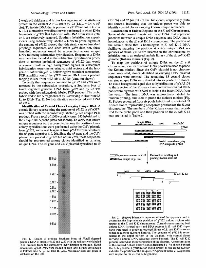
Localization of Unique Regions on the E. coli Chromosome.Some of the cosmid inserts will carry DNA that representjunctions between a unique DNA sequence and DNA that ishomologous to the E. coli K-12 chromosome. The portion ofthe cosmid clone that is homologous to E. coli K-12 DNAfacilitates mapping the position at which unique DNA se-quences of strain X7122 are inserted in the chromosome byhybridization to an ordered lambda library of the E. coli K-12genome (Kohara miniset) (Fig. 2).To map the positions of unique DNA on the E. coli
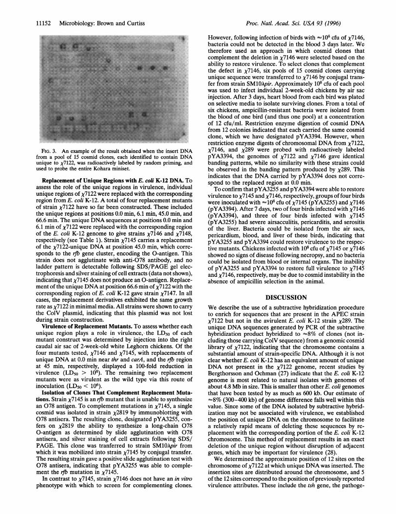
chromosome, a series of cosmid DNA pools were used to probethe Kohara miniset. Since the ColV plasmid is not chromo-some associated, clones identified as carrying ColV plasmidsequences were omitted. The remaining 83 cosmid clonescarrying unique DNA were divided into six pools of 15 clones.To avoid background signal due to hybridization of pYA3174to the A vector of the Kohara clones, individual cosmid DNApools were digested with NotI to isolate the insert DNA fromthe vector. The insert DNA was radioactively labeled byrandom priming, and used to probe the Kohara miniset (Fig.3). Probes generated from six pools hybridized to a total of 33Kohara clones, representing 12 separate positions on the E. colichromosome. The numbers of the Kohara clones that hybrid-ized to the probe pools and their position on the E. coli K-12map are listed in Table 2.
unique DNA carAthr araDABC
Pooled cosmids carryingDNA unique to X7122
JSequence common to K-12 Radioactive labeling andDNA uique to X7122 probing Kohara mini-set
serB thr carA araDABC1
E. coli K-122 3
4 - _6 7
FIG. 2. (Upper) Schematic representation of the approach used todetermine the approximate position of X7122 unique regions withrespect to the E. coli K-12 chromosome. Cosmid clones carrying bothunique DNA (striped bars) and DNA present in E. coli K-12 (openbars) were used to probe an ordered library of E. coli K-12 chromo-somal sequences (Kohara library). The genome of X7122 is repre-sented in the upper portion of the diagram, with cosmid clonescarrying a unique DNA sequence shown beneath. The E. coli K-12genome is shown in the lower portion of the diagram. A representationof the ordered Kohara library clones designated 1-7 is shown beneaththe K-12 genome. Hybridization (solid circles) to the clones (Lower)indicates the position of the unique DNA present in the X7122 genomewith respect to the E. coli K-12 genome.
345°88g88 888 s88 588
000 000 000 000 000000 00 0 000 000
0888 888800880888 888000 000 000 000 000000 000 000 000 000008000oooo888 8880888
Microbiology: Brown and Curtiss
11152 Microbiology: Brown and Curtiss
FIG. 3. An example of the result obtained when the insert DNAfrom a pool of 15 cosmid clones, each identified to contain DNAunique to X7122, was radioactively labeled by random priming, andused to probe the entire Kohara miniset.
Replacement of Unique Regions with E. coli K-12 DNA. Toassess the role of the unique regions in virulence, individualunique regions of X7122 were replaced with the correspondingregion from E. coli K-12. A total of four replacement mutantsof strain X7122 have so far been constructed. These includedthe unique regions at positions 0.0 min, 6.1 min, 45.0 min, and66.6 min. The unique DNA sequences at positions 0.0 min and6.1 min of X7122 were replaced with the corresponding regionof the E. coli K-12 genome to give strains X7146 and X7148,respectively (see Table 1). Strain X7145 carries a replacementof the x7122-unique DNA at position 45.0 min, which corre-sponds to the rjb gene cluster, encoding the 0-antigen. Thisstrain does not agglutinate with anti-078 antibody, and noladder pattern is detectable following SDS/PAGE gel elec-trophoresis and silver staining of cell extracts (data not shown),indicating that X7145 does not produce an 0-antigen. Replace-ment of the unique DNA at position 66.6 min of X7122 with thecorresponding region of E. coli K-12 gave strain x7147. In allcases, the replacement derivatives exhibited the same growthrate as X7122 in minimal media. All strains were shown to carrythe ColV plasmid, indicating that this plasmid was not lostduring strain construction.
Virulence of Replacement Mutants. To assess whether eachunique region plays a role in virulence,, the LD50 of eachmutant construct was determined by injection into the rightcaudal air sac of 2-week-old white Leghorn chickens. Of thefour mutants tested, X7146 and x7145, with replacements ofunique DNA at 0.0 min near thr and carA, and the rfb regionat 45 min, respectively, displayed a 100-fold reduction invirulence (LD5o > 108). The remaining two replacementmutants were as virulent as the wild type via this route ofinoculation (LD5o < 106).
Isolation of Clones That Complement Replacement Muta-tions. Strain X7145 is an rIb mutant that is unable to synthesizean 078 antigen. To complement mutations in x7145, a singlecosmid was isolated in strain X2819 by immunoblotting with078 antisera. The resulting clone, designated pYA3255, con-fers on X2819 the ability to synthesize a long-chain 0780-antigen as determined by slide agglutination with 078antisera, and silver staining of cell extracts following SDS/PAGE. This clone was tranferred to strain SMlOApir fromwhich it was mobilized into strain X7145 by conjugal transfer.The resulting strain gave a positive slide agglutination test with078 antisera, indicating that pYA3255 was able to comple-ment the rfl mutation in x7145.
In contrast to x7145, strain X7146 does not have an in vitrophenotype with which to screen for complementing clones.
However, following infection of birds with _ 108 cfu of X7146,bacteria could not be detected in the blood 3 days later. Wetherefore used an approach in which cosmid clones thatcomplement the deletion in X7146 were selected based on theability to restore virulence. To select clones that complementthe defect in X7146, six pools of 15 cosmid clones carryingunique sequence were transferred to X7146 by conjugal trans-fer from strain SM10pir. Approximately 108 cfu of each poolwas used to infect individual 2-week-old chickens by air sacinjection. After 3 days, heart blood from each bird was platedon selective media to isolate surviving clones. From a total ofsix chickens, ampicillin-resistant bacteria were isolated fromthe blood of one bird (and thus one pool) at a concentrationof 12 cfu/ml. Restriction enzyme digestion of cosmid DNAfrom 12 colonies indicated that each carried the same cosmidclone, which we have designated pYA3394. However, whenrestriction enzyme digests of chromosomal DNA from X7122,X7146, and X289 were probed with radioactively labeledpYA3394, the genomes of X7122 and X7146 gave identicalbanding patterns, while no similarity with these strains couldbe observed in the banding pattern produced by X289. Thisindicates that the DNA carried by pYA3394 does not corre-spond to the replaced region at 0.0 min.To confirm that pYA3255 and pYA3394 were able to restore
virulence to X7145 and X7146, respectively, groups of four birdswere inoculated with _108 cfu of X7145 (pYA3255) and X7146(pYA3394). After 7 days, two of four birds infected with X7146(pYA3394), and three of four birds infected with X7145(pYA3255) had severe airsacculitis, pericarditis, and serositisof the liver. Bacteria could be isolated from the air sacs,pericardium, blood, and liver of these birds, indicating thatpYA3255 and pYA3394 could restore virulence to the respec-tive mutants. Chickens infected with 108 cfu of X7145 or X7146showed no signs of disease following necropsy, and no bacteriacould be isolated from blood or internal organs. The inabilityof pYA3255 and pYA3394 to restore full virulence to X7145and X7146, respectively, may be due to cosmid instability in theabsence of ampicillin selection in the animal.
DISCUSSIONWe describe the use of a subtractive hybridization procedureto enrich for sequences that are present in the APEC strainX7122 but not in the avirulent E. coli K-12 strain X289. Theunique DNA sequences generated by PCR of the subtractivehybridization product hybridized to -8% of clones (not in-cluding those carrying ColV sequence) from a genomic cosmidlibrary of X7122, indicating that the chromosome contains asubstantial amount of strain-specific DNA. Although it is notclear whether E. coli K-12 has an equivalent amount of uniqueDNA not present in the X7122 genome, recent studies byBergthorsson and Ochman (27) indicate that the E. coli K-12genome is most related to natural isolates with genomes ofabout 4.8 Mb in size. This is smaller than other E. coli genomesthat have been tested by as much as 600 kb. Our estimate of-8% (300-400 kb) of genome difference falls well within thisvalue. Since some of the DNA isolated by subtractive hybrid-ization may not be associated with virulence, we establishedthe position of unique DNA on the chromosome to facilitatea relatively rapid means of deleting these sequences by re-placement with the corresponding portion of the E. coli K-12chromosome. This method of replacement results in an exactdeletion of the unique region without disruption of adjacentgenes, which may be important for virulence (28).We determined the approximate position of 12 sites on the
chromosome of X7122 at which unique DNA was inserted. Theinsertion sites are distributed around the chromosome, and 5of the 12 sites correspond to the position of previously reportedvirulence attributes. These include the tsh gene, the pathoge-
Proc. Natl. Acad. Sci. USA 93 (1996)

Proc. Natl. Acad. Sci. USA 93 (1996) 11153
Table 2. Positions of unique DNA sequence of X7122 relative to the E. coli K-12 genome
Position (min)* ofunique sequence Kohara clone numbers Flanking genes*
20 kb (0.0) 9E4, 6H3, 22B12, 2F7 thr, carA285 kb (6.1) 5A5, 3C7, 21C1, 5E5 proAB, bet1065 kb (22.8) 2F1, 5A12 appA, agpA1480 kb (31.7) 1A7, 7F12 trkG, cybB2115 kb (45.0) 6D9, 21H10 his, alkA2420 kb (51.8) 9C2 menD, ackAc3110 kb (66.6) 23G4, 12C6 metK, nupG3460 kb (74.0) 18C4, 5F12 aroE, rplN3610 kb (77.2) 1OF5 asd, ugp3750 kb (80.8) 6F2, 10C8, 7C3, 9B3 glyS, selB4430 kb (94.8) 3H6, 3A1, 6G4, lGlO ampC, purA4530 kb (96.8) E1F5, 5C4, E4D8, 16H7S, 7G7 pyrB, fecAThe position of unique DNA sequences were determined by hybridization of cosmid pools, each
identified to contain DNA uniques to X7122, to the Kohara miniset.*Based on the E. coli K-12 map (18).
nicity islands PAI I (and LEE) and PAI II, the group II capsulegenes, and the rfb gene cluster.We previously reported the sequence of the tsh gene from
strain X7122 (19). Sequence immediately downstream of thetsh gene exhibits 100% identity with that of the fins gene of E.coli K-12, indicating that the tsh gene lies at the edge of theunique region at 74.0 min. The pathogenicity island PAI I ofurinary tract pathogenic strain 536, and the LEE of entero-pathogenic strains lies at 82.4 min on the E. coli chromosome(12, 13), and strain X7122 carries a unique region at 80.8 min,which is within 80 kb of the insertion position ofPAI I and LEE(the selC gene). PAI I is 70 kb long and includes the geneencoding hemolysin I (11, 12), while the LEE locus is 35 kblong and carries the genes for attachment and effacement ofepithelial cells (13). In addition, strain X7122 carries a uniqueregion at 96.8 min, which is the insertion position of PAI II(near leuX) of UPEC strains (11). The PAI II region is 190 kblong in UPEC strains, and carries the genes for Pap-relatedfimbriae and hemolysin II (12). However, strain X7122 does notexhibit hemolysis of erythrocytes or attachment and efface-ment of epithelial cells (unpublished data), and it is thereforelikely that the unique regions at 80.8 min and 96.8 min in thisstrain encode proteins different to those encoded by PAT I,LEE, and PAI IT. We are at present constructing replacementsin these regions to test whether they also play a role in thevirulence of APEC strains.The region between nupG and metK at 66.6 min has been
shown to carry the genes for synthesis and export of group TIcapsules in E. coli isolates (29). However, strain X7122 pro-duces a K80 capsule that is not classified as group II, butbelieved to be an 0 antigen capsule (30). The K80 capsule isthought to be involved in prevention of phagocytosis bypolymorphonuclear leukocytes (31), and while it is not knownwhether the unique region at 66.6 min contributes to K80capsule synthesis, replacement of this region with DNA fromE. coli K-12 did not reduce virulence. This indicates that theunique DNA at 66.6 min is not necessary for virulence of thisstrain following air-sac inoculation, or that it encodes avirulence attribute which is duplicated in function by someother sequences.
In contrast, the genes necessary for 0-antigen synthesis areessential for virulence of X7122. The 0-antigen is encoded bythe rfb gene cluster at 45.0 min, and this region was identifiedas carrying unique sequences in X7122. The rjb region ofE. coliK-12 carries an insertion element, resulting in a rough phe-notype (10). Replacement of the 078 rfb region of strain X7122with that from E. coli K-12 results in a rough strain, the LD50ofwhich is at least 100-fold higher than the wild type. A cosmidexpressing the 078 0-antigen was able to restore partialvirulence to x7145, indicating that an 0-antigen is required for
virulence. Strains with an 078 0-antigen are one of the mostpredominant serotypes associated with colisepticemia of poul-try (1), and virulent 078 isolates belong to a limited numberof clonal groups (30, 32). It is not clear, however, whether the078 serotype predominates due to the clonal nature of avianE. coli, or because this 0-antigen possesses inherent structuralproperties that facilitate success of strains expressing thisserotype in colisepticemia.A second unique region that is involved in virulence lies at
0.0 min near the thr and carA loci. Replacement of this regionof the chromosome of X7122 with the corresponding region ofE. coli K-12 increases the LD50 by at least 100-fold. No knownvirulence factor gene has been described in this position,emphasizing the ability of the method described above todetect novel regions on the chromosome associated withvirulence. Using an in vivo selection procedure, we isolated acosmid clone which increases the ability of X7146 to persist inthe bird, but does not correspond to the replaced region ofX7146. We are at present working to determine how extracopies of this unique DNA can compensate for the replace-ment at 0.0 min. We are also continuing attempts to isolateclones that correspond to the replaced region, and that com-plement the virulence defect of strain X7146.The remaining seven regions that carry unique DNA do not
correspond to positions of genes for known virulence factors.Replacement of the unique DNA of X7122 betweenproAB andbetA at 6.1 min does not reduce virulence following air-sacinoculation. However, inoculation by air-sac injection will notidentify mutants attenuated in the early stages of respiratorytract colonization, and replacement mutants showing wild-type levels of virulence by this route of infection may exhibitattenuation when administered by a different route. In addi-tion, replacement of a single unique DNA region may not besufficient to reduce virulence owing to the existence of mul-tiple mechanisms by which avian E. coli can cause disease. Wehave shown previously that a knockout mutation of the tshgene does not eliminate hemagglutination in X7122 (19),indicating that other adhesins are expressed in this strain,illustrating the existence of back-up mechanisms in avian E.coli. In addition, virulence attributes that are also present in E.coli K-12 will not be deleted using a replacement approach, asis the case with type 1 fimbriae, which are found in many E. coliisolates including avian pathogens and E. coli K-12 (33). Inavian strains, type 1 fimbriae can mediate adhesion to avianrespiratory tissue (34), and expression of these structures canbe detected in the air sacs and trachea of birds experimentallyinfected with avian E. coli, suggesting that type 1 fimbriae playa role in colonization of the avian respiratory tract (35).Therefore, replacement of unique regions that encode ad-hesins involved in colonization of the respiratory tract may not
Microbiology: Brown and Curtiss

11154 Microbiology: Brown and Curtiss
exhibit a measurable phenotype in the presence of type 1fimbriae. We are currently constructing single and multiplereplacements of the remaining sites in X7122, and combiningthese with mutations in the genes for type 1 fimbriae and curlito determine which unique DNA regions contribute to thevirulence of APEC strains. We are also, in collaboration withothers, evaluating the virulence of all the replacement mutantswhen administered to chickens by an aerosol route.The ability to identify and replace unique DNA on the
genome of E. coli is an effective means of rapidly determiningregions of virulence on the chromosome. Knowledge of thelocation and function of unique DNA in different E. coliisolates should lead to an understanding of the interactionbetween specific bacteria and their hosts, and to how an E. colipathogen evolves by acquiring DNA that facilitates adaption toa particular niche.
This work was supported by U.S. Department of AgricultureNational Research Initiative Competitive Grants Program Grant94021002, and an unrestricted grant award from Bristol-Myers Squibb.We thank Barbara Bachmann, Carol Gross and Willy Walter for
supplying E. coli strains, Teresa A. Doggett, Allen L. Honeyman andMarinella Messina for critical review of the manuscript, and Lisa L.Burns-Keliher for preparation of Fig.s.
1. Gross, W. B. (1984) in Diseases of Poultry, eds. Hofstad, M. S.,Barnes, H. J., Calnek, B. W., Reid, W. M. & Yoder, H. W. (IowaState Univ. Press, Ames), pp. 270-277.
2. Cloud, S. S., Rosenberger, J. K, Fries, P. A., Wilson, R. A. &Odor, E. M. (1985) Avian Dis. 29, 1084-1093.
3. Glantz, P. J., Narotsky, S. & Bubash, G. (1962) Avian Dis. 6,322-328.
4. Harry, E. G. (1964) Vet. Rec. 76, 466-470.5. Heller, E. D. & Perek, M. (1968) Br. Vet. J. 124, 509-513.6. Carlson, H. C. & Whenham, G. R. (1968)Avian Dis. 12,297-302.7. Nakamura, K., Yuasa, Y., Abe, F. & Narita, M. (1990) Avian
Pathol. 19, 713-721.8. Leitner, G. & Heller, E. D. (1992) Avian Dis. 36, 211-220.9. Boulnois, G. J. & Roberts, I. S. (1990) Curr. Top. Microbiol.
Immunol. 150, 1-18.10. Liu, D. & Reeves, P. R. (1994) Microbiology 140, 49-57.11. Hacker, J., Bender, L., Wingender, J., Lund, B., Marre, R. &
Goebel, W. (1990) Microb. Pathog. 8, 213-225.12. Blum, G., Ott, M., Lischewski, A., Ritter, A., Imrich, H., Tschape,
H. & Hacker, J. (1994) Infect. Immun. 62, 606-614.
13. McDaniel, T. K., Jarvis, K. G., Donnenberg, M. S. & Kaper, J. B.(1995) Proc. Natl. Acad. Sci. USA 92, 1664-1668.
14. Provence, D. L. & Curtiss, R., III (1992) Infect. Immun. 60,4460-4467.
15. Singer, M., Baker, T. A., Schnitzler, G., Deischel, S. M., Goel, M.,Dove, W., Jaacks, K. J., Grossman, A. D., Erickson, J. W. &Gross, C. A. (1989) Microbiol. Rev. 53, 1-24.
16. Miller, V. L. & Mekalanos, J. J. (1988) J. Bacteriol. 170, 2575-2583.
17. Jacobs, W. R., Barrett, J. F., Clark-Curtiss, J. E. & Curtiss, R., III(1986) Infect. Immun. 52, 101-109.
18. Miller, J. H. (1992) A Short Course in Bacterial Genetics: ALaboratoryManualandHandbookforEscherichia coli andRelatedBacteria (Cold Spring Harbor Lab. Press, Plainview, NY).
19. Provence, D. L. & Curtiss, R., III, (1994) Infect. Immun. 62,1369-1380.
20. Hull, R. A., Gill, R. E., Hsu, P., Minshew, B. H. & Falkow, S.(1981) Infect. Immun. 33, 933-938.
21. Sambrook, J., Fritsch, E. F. & Maniatis, T. (1989) MolecularCloning: A Laboratory Manual (Cold Spring Harbor Lab. Press,Plainview, NY), 2nd Ed.
22. Kohara, Y. (1990) in The Bacterial Chromosome, eds. Drlica, K.& Riley, M. (Am. Soc. for Microbiol., Washington, DC), pp.29-42.
23. Straus, D. & Ausubel, F. (1990) Proc. Natl. Acad. Sci. USA 87,1889-1893.
24. Goldberg, R. B., Bender, R. A. & Streicher, S. L. (1974) J.Bacteriol. 118, 810-814.
25. Stemnberg, N. L. & Maurer, R. (1991) Methods Enzymol. 204,18-43.
26. Waters, V. L. & Crosa, J. H. (1991) Microbiol. Rev. 55, 437-450.27. Bergthorsson, U. & Ochman, H. (1995) J. Bacteriol. 177, 5784-
5789.28. Ritter, A., Blum, G., Emody, L., Kerenyi, M., Bock, A., Neuhierl,
B., Rabsch, W., Scheutz, F. & Hacker, J. (1995) Mol. Microbiol.17, 109-121.
29. Vimr, E. R. (1991) J. Bacteriol. 173, 1335-1338.30. Cherifi, A., Contrepois, M., Picard, B., Goullet, P., Orskov, I. &
Orskov, F. (1994) J. Clin. Microbiol. 32, 1197-1202.31. Rozenberg-Arska, M., Salters, M. E. C., Van Strijp, J. A. G.,
Geuze, J. J. & Verhoef, J. (1985) Infect. Immun. 50, 852-859.32. White, D. G., Dho-Moulin, M., Wilson, R. A. & Whittam, T. S.
(1993) Microb. Pathog. 14, 399-409.33. Dozois, C. M., Fairbrother, J. M., Harel, J. & Bosse, M. (1992)
Infect. Immun. 60, 2648-2656.34. Gyimah, J. E. & Panigrahy, B. (1988) Avian Dis. 32, 74-78.35. Dozois, C. M., Chanteloup, N., Dho-Moulin, M., Bree, A., De-
sautels, C. & Fairbrother, J. M. (1994) Avian Dis. 38, 231-239.
Proc. Natl. Acad. Sci. USA 93 (1996)