universita’ degli s napoli “federico ii” - fedoa - fedoa · dimostrato che tale mediatore è...
TRANSCRIPT

UNIVERSITA’ DEGLI STUDI DI NAPOLI “FEDERICO II”
Dottorato di ricerca in “Scienza del farmaco”
XX ciclo
Indirizzo Farmacologia, farmacognosia e tossicologia
“Hydrogen Sulphide, a novel gasotransmitter regulating pathophysiological processes and
cross-talk with Nitric Oxide”
2004-2007
Tutor: Prof. Giuseppe Cirino
Dottorando: Dott. Vincenzo Brancaleone
Coordinatore: Prof.ssa Maria Valeria D’Auria

- 2 -
Acknowledgements
During my PhD studentship, I have been so lucky to meet a lot of people that
now deserve my thanks.
First of all, I wish to thank my tutor, Prof. Giuseppe Cirino, who has given to
me the opportunity to work in his lab and has been a precious master and teacher,
always lavishing suggestions and trusting and supporting me all the time, since I was an
undergraduate student.
I thank Dr. Fiorentina Roviezzo and Dr. Mariarosaria Bucci for their massive
contribution to my professional growth and not only professional! They first have
stirred up in me the passion for pharmacology, encouraging and stimulating me to
make me doing things in the best way possible.
I like to thank Dr. Roberta d’Emmanuele di Villa Bianca, Dr. Annarita Di
Lorenzo, Dr. Valentina Vellecco, Dr. Rosalinda Sorrentino, Ms. Luana De Gruttola
and Ms. Emma Mitidieri, my friends and co-workers, for their invaluable friendship
and help, as well as for useful discussions and criticisms.
Thanks also to all past and current lab-mates, with whom I have been sharing
lab-life, as well as to all my friends and colleagues of the Department of Experimental
Pharmacology in Naples.
A special thank for his excellent hospitality and continuous support, to Prof.
Mauro Perretti of the Centre for Biochemical Pharmacology at William Harvey
Research Institute, “Queen Mary University of London”. He gave me the great chance
to join his lab, where I met new friends and colleagues, learning a lot, not only about
science, but also about life.
I wish to thank Prof. Enrico Abignente and Prof. Maria Valeria D’Auria, old
and new chief organizer of this PhD respectively, for their helpful advices.

- 3 -
At the end, but not last, I have to thank God for everything He gave me in my life
and also thank my family, overall my brother Angelo, since they have had a critical role
in my life, supporting me in both everything I have done and I have not in my life.

- 4 -
INDEX Abstract pag 7
Summary pag 10
1. INTRODUCTION pag 13
1.1 The endothelium as a detector inducing vascular change pag 16
1.1.1 Vascular andothelium and its multi-functional rol pag 16
1.1.2 Endothelial cells in the regulation if vascular tone pag 19
1.1.3 Inflammation and endothelial cells: interplay and communication
pag 23
1.2 Hydrogen sulphide (H2S): gaseous chemical with biological activity pag 27
1.2.1 H2S: an overview on chemical and biochemical properties pag 27
1.2.2 H2S producing enzymes cystathionine-β-synthase (CBS)
and cystathionine-γ-lyase (CSE) pag 32
1.2.3 Endogenous H2S role in cardiovascular system pag 35
1.2.4 Involvement of H2S in inflammatory processes pag 37
1.2.5 How does H2S behave in other tissues ? pag 38
1.3 Scientific basis and aim of the study pag 40
2. MATERIALS AND METHODS pag 42
2.1 H2S in vascular tone: functional and molecular studies pag 42
2.1.1 Reagents pag 42
2.1.2 Non Obese Diabetic (NOD) and Non Obese Resistant (NOR) mice
pag 42
2.1.3 Determination of glycosuria pag 43
2.1.4 Tissue preparation and experimental protocol pag 44

- 5 -
2.1.5 Cell experiment with normal and high glucose environment pag 45
2.1.6 Measurements of H2S: plasma and tissues pag 45
2.1.7 CSE and CBS expression: qRT-PCR pag 46
2.1.8 Western Blot analysis pag 48
2.1.9 Statistical analysis pag 48
2.2 H2S and inflammation: functional study pag 49
2.2.1 Reagents pag 49
2.2.2 Animals pag 49
2.2.3 Intravital microscopy study pag 49
2.2.4 Paw oedema pag 51
2.2.5 Statistical analysis pag 51
2.3 H2S and inflammation: molecular study pag 52
2.3.1 Human neutrophils isolation and AnxA1 mobilization analysis by
western blot pag 52
2.3.2 Animals and Bone Marrow Derived Macrophages (BMDM) pag 53
2.3.3 qRT-PCR on AnxA1-/- mice pag 53
2.3.4 Statistical analysis pag 54
3. RESULTS pag 56
3.1 H2S and vascular system: signalling in regulating vascular tone pag 56
3.1.1 Effect of NaHS and L-cysteine on aortic rings harvested from NOR or
NOD mice pag 56

- 6 -
3.1.2 Contribution of cAMP and cGMP in H2S-induced vasorelaxation
pag 59
3.2 H2S and vascular system: release and molecular changes in production
pag 60
3.2.1 Impairment of CBS/CSE activity in NOD mice pag 60
3.2.2 High glucose environment affects stimulated H2S production in BAEC
pag 62
3.3 H2S and inflammation pag 63
3.3.1 H2S donors decrease ASA-induced leukocyte adhesion through the
activation of KATP channels pag 63
3.3.2 H2S donors inhibit fMLP-induced leukocyte adherence pag 64
3.3.3 H2S modulates oedema formation via effects on KATP channels
pag 65
3.3.4 H2S induced externalization of AnxA1 onto membrane surface
pag 66
3.3.5 H2S reverted CSE, COX-2 and i-NOS over-expression in LPS treated
BMDM through AnxA1-dependent mechanism pag 67
3.3.6 H2S production in AnxA1-/- mice pag 67
4. DISCUSSION pag 74
5. REFERENCES pag 83

- 7 -
Abstract
L’acido sulfidrico (H2S) è stato considerato per lungo tempo come un gas
tossico, ma recentemente i suoi effetti farmacologici sono stati rivalutati. E’ stato
dimostrato che tale mediatore è prodotto in vari tessuti grazie all’azione di due enzimi
quali cistationina-γ-liasi (CSE) e cistationina-β-sintasi (CBS), ed è coinvolto in vari
processi fisiopatlogici. In particolare da questi studi emerge per l’acido sulfidrico un
ruolo di primo piano nella regolazione dell’omeostasi vascolare e nei processi
infiammatori. E’ noto, infatti, che H2S induce vasodilatazione mediante interazione con
i canali del potassio ATP-dipendenti (K+ATP) e che una riduzione dei livelli plasmatici
di H2S rappresenta un fattore che contribuisce allo sviluppo dell’ipertensione
spontanea. Viceversa, in stati infiammatori ed in particolare in seguito a
somministrazione di LPS nei topi, si riscontra un aumento dei livelli plasmatici di tale
mediatore gassoso.
Queste evidenze hanno portato alla volontà di studiare il ruolo di H2S nel
compartimento vascolare ed in particolare in che modo esso possa interagire con
l’endotelio e con i mediatori da esso prodotti, quali ad esempio il monossido d’azoto
(NO) nella regolazione dell’omeostasi vascolare e quindi anche dei processi
infiammatori. Pertanto, è stato utilizzato un primo approccio di tipo funzionale allo
scopo di indagare sul coinvolgimento di tale molecola in un modello di disfunzione
vascolare associato al diabete mellito insulino-dipendente (IDDM), utilizzando aorte
espiantate da animali spontaneamente diabetici (NOD/Ltj).
E’ da sottolineare il fatto che in questi animali la disfunzione endoteliale è
principalmente legata ad una drastica riduzione dei livelli di NO. Il passo successivo è
stato poi caratterizzato da uno studio molecolare mirato alla valutazione
dell’espressione degli enzimi responsabili della sintesi di H2S e alla quantificazione

- 8 -
della produzione di tale mediatore. In ultima analisi, è stato poi valutato il ruolo di H2S
nella regolazione della risposta infiammatoria, utilizzando diversi modelli animali e
cellulari.
I primi risultati ottenuti hanno mostrato un chiaro coinvolgimento di NO nella
vasodilatazione indotta da H2S, in particolare NO sembra regolare positivamente la
produzione enzimatica di H2S a partire da L-cisteina, suo precursore endogeno,
mediata da CBS e CSE. Inoltre, lo studio condotto sugli animali diabetici evidenzia una
riduzione della capacità vasodilatante della L-cisteina, sintomo di un’alterata attività
enzimatica probabilmente correlata ai ridotti livelli di NO.
Lo studio molecolare effettuato attraverso analisi di western blot e qRT-PCR ha
mostrato un aumento nella espressione di CBS e CSE, sebbene i livelli di H2S
plasmatici e tissutali siano significativamente ridotti negli animali diabetici. Ciò
potrebbe essere ascrivibile ad un fenomeno di tipo compensatorio in risposta ad una
riduzione dei livelli di H2S circolanti.
Infine, i risultati ottenuti attraverso l’utilizzo di diversi modelli animali di
infiammazione hanno mostrato che H2S è in grado di inibire la migrazione dei
polimorfonucleati (PMN) nei tessuti in seguito a stimolazione con agenti flogogeni; in
particolare, questa azione è mediata dai canali del potassio ATP-dipendenti. Inoltre,
l’edema indotto da carragenina veniva significativamente ridotto dal trattamento con
diversi donatori di H2S.
In conclusione i risultati sperimentali ottenuti indicano in primo luogo che
l’ossido nitrico è coinvolto nei meccanismi di vasodilatazione indotti da H2S,
suggerendo l’esistenza di un legame tra le vie metaboliche L-arginina/NO e L-
cisteina/H2S nella regolazione del tono vascolare. Inoltre, H2S mostra un effetto anti-
infiammatorio, che si esplica principalmente attraverso il blocco della migrazione

- 9 -
leucocitaria. Queste evidenze, nel loro insieme, suggeriscono per la prima volta che
H2S agisce in maniera diretta sulle cellule endoteliali vascolari influenzando molteplici
fenomeni che avvengono all’interfaccia tra endotelio e sangue, in particolare
intervenendo sul mantenimento dell’omeostasi vascolare e sulla regolazione della
migrazione leucocitaria.

- 10 -
Summary
Sulphidric acid has been considered for a long time just a polluting gas and only
recently its pharmacological effects have been investigated. H2S is produced in many
mammalian tissues by two enzymes, cystathionine-γ-lyase (CSE) and cystathionine-β-
synthase (CBS) and it is involved in many physiological and pathological events. In
particular this gas seems to have a critical role in vascular homeostasis and in
inflammatory processes. It has been demonstrated that H2S induces K+ATP channels-
dependent vasorelaxation and that spontaneous hypertension is associated to its
lowered plasmatic levels. On the other hand, during inflammatory process, such as LPS
administration in mice, plasmatic H2S levels result increased.
These evidences put us on way to investigate the role of H2S in vascular tone
regulation and the possible relationship with nitric oxide (NO). Furthermore the aim of
this work was also to assess involvement of this gaseous molecule in inflammatory
events. For these reasons, we first used a functional approach in order to study H2S role
in a model of endothelial dysfunction associated to diabetes, by using aortic tissue
harvested from Non ObeseDiabetic mice (NOD/Ltj). These animals show endothelial
disorder mainly associated to a reduction in NO levels.
The following step was characterized by a molecular approach in order to
verify the expression of CBS and CSE and their enzymatic activity leading to H2S
production. The last part of this work dealt with evaluation of hydrogen sulphide in
inflammatory responses. First data obtained showed a marked involvement of NO in
H2S-induced vasodilatation and in particular NO seemed to regulate production of H2S
from L-cysteine, its biological precursor. Furthermore, results of experiments carried
out on diabetic mice highlighted a reduce vasodilatory effect by L-cysteine, likely linked
to reduced NO availability. Molecular data showed that both CBS and CSE expression

- 11 -
are significantly increased, although plasmatic levels of H2S were reduce in diabetic
mice. This evidence could reflect a compensatory response in order to restore H2S
levels.
Finally, data obtained from experiments performed on different animal model of
inflammation clearly demonstrated that H2S inhibited polymorphonuclear (PMN)
transmigration, following an inflammatory stimulus. In addition, this effect is mediated
by K+ATP channels.
In conclusion, our experimental data indicate for the first time that nitric oxide
is involved in vasodilatory effect induced by H2S, suggesting a link between L-
arginine/NO and L-cysteine/H2S pathway in vascular tone regulation.
Furthermore, hydrogen sulphide showed an anti-inflammatory effect, mainly
exerted through blockade of PMN transmigration. Altogether, these evidences suggest
that H2S is able to act directly on endothelial cells, where it is also produced, affecting
all phenomenon occurring at blood-endothelium interface, particularly intervening on
maintenance of vascular homeostasis and regulation of leukocytes migration.

- 12 -
CHAPTER 1

- 13 -
1. INTRODUCTION
In the second century A.D. Galen said: “Throughout the body the animal
arteries are mingled with veins and veins with arteries, and both veins and arteries are
mingled with nerves and the nerves with these…And of course the usefulness of such a
complete interweaving is very evident, if, that is to say, it is a useful thing for all parts
of the animal to be nourished”. Then, in despite of a very sophisticated and fine
regulated human body, the most important and easy way for maintaining it alive and
well working in all its functions is just represented by blood. Its flowing through a
massive network of “tubes” makes all cells being fed with all nutrients needed and keep
in constant communication all organs and tissues, even if they are far away from each
other. In this kind of processes , these “tubes”, called vessels, are simply fundamental
(fig. 1.1)
Vessels are divided into two main categories, such as arteries and veins. Both of
them have a particular structure formed by three layer of different cells types, even if
there are some quantitative differences. The outer one is represented by adventitia,
mainly formed by collagen and fibroblasts; in the middle, there is the media, constituted
by smooth muscle cells; finally, the inner layer is the endothelium, formed by a
monolayer of endothelial cells that have a proper direct contact with all molecules going
around the body through the blood flow (fig. 1.2). The possibility of this interface
contact between endothelium and blood makes the endothelium one of the most
important actor in regulating physiology of vascular tone; and this also because it
actually represents a “barrier” allowing or not molecules passing through it and going
into tissues, and then exerting their own effects.

- 14 -
The activity of this “barrier” is regulated at various levels by a wide range of
molecules but for sure the one playing a pivotal role is nitric oxide (NO). Through
years, NO and its effects have been described in details and its scenario has been well
drawn up (Palmer RM et al., 1988; Moncada S et al., 1991). Particularly, it has been
appearing clear how just one simple molecule like NO could be responsible for vascular
relaxation, pro-inflammatory actions and proliferation affecting stimuli (Cirino G et al.,
2003; Garcia X et al., 2006; Moncada S et al., 1991). Furthermore, alteration of NO
levels is consistent with many pathological conditions, overall endothelial dysfunction
(Nagareddy PR et al., 2005; Bucci M et al., 2004; Pechanova O et al., 2007).
In this contest, another important gaseous mediator has been recently
considered: the gaseous molecule in question is hydrogen sulphide (H2S). H2S has been
known since early times and its chemistry has been studied since the 1600s; it is a
colourless gas with a typical rotten eggs smell that represents the main reason it is well
known for. This molecule was thought just like a toxic and polluting gas for a long time,
even if pollution was later discovered to occur at concentration 400-fold higher than
physiological one. Only in last decade its involvement in many physiological and
pathological processes has been found out, and the aim of this work is just to assess its
functions, contributing to the latest findings in the “pharmacology of hydrogen
sulphide”.
.

- 15 -
Fig. 1.2 Structure of blood vessels. Arteries and veins are a complex of
multiple layers where endothelium represents the inner one and smooth
muscle cells and connective tissue are stratified over it
Fig. 1.1 Medieval images of circulatory tree

- 16 -
1.1 THE ENDOTHELIUM AS A DETECTOR INDUCING VASCULAR CHANGES
1.1.1 Vascular endothelium and its multi-functional role
The endothelium is a specialized type of epithelial tissue consisting in a thin
layer of cells that line the interior surface of blood vessels and forming an interface
between circulating blood in the lumen and the rest of the vessel wall. In all types of
blood vessels the endothelium of the tunica intima is highly specialised with endocrine,
exocrine, cell adhesion, clotting and transport functions. Endothelial cells line the entire
circulatory system, from the heart to the smallest capillaries and they reduce friction of
blood flow allowing the fluid to be pumped further.
Endothelium is composed of flattened cells and in routine histological sections
the cytosol of most cells is barely visible and only the small flattened nuclei are seen.
Ultrastructurally, each cell can be seen to be anchored to an underlying basal lamina;
individual cells are anchored together by adherent junctions, including prominent tight
junctions which prevent diffusion between cells.
A major feature of endothelial cells is represented by pinocytotic vesicles which
are involved in the process of transport of substances from one side of the cell to the
other. In small blood vessels of the nervous system, endothelial cells express transport
proteins which are responsible for active transport of all substances, for example
glucose into the brain. Furthermore, they are able to sense changes in blood pressure,
oxygen tension and blood flow by as yet unknown mechanisms. In response to changes
in local conditions they respond by secreting substances which have powerful effects on
the tone of vascular smooth muscle. Endothelium is also important in the control of

- 17 -
blood coagulation, since under normal circumstances its surface prevents blood clotting
and allows a smooth blood flux (Santiago-Delpin EA, 2004; Ppova EN et al., 2004).
Another important feature of endothelium is characterized by its ability in
adapting rapidly to changes in its environment. In fact, under certain circumstances,
especially in response to adverse stimuli such as wounds, infections or irritation (e.g.
insect sting), it becomes activated and changes its function (Pober JS et al., 2002). This
activation is mainly operated by cytokines and develops specialisation for lymphoid
cells migration. The endothelial cells become cuboidal in shape and express surface
adhesion molecules which facilitate lymphocytes adhesion and migration. This type of
endothelium is particularly present in the specialised venules in the lymph node
paracortex (high endothelial venules). These changes normally occur after tissue
damages and allows neutrophils to migrate into local tissues during acute inflammation.
One of the first step in this pathway is represented by exposure of P-selectin, a cell
adhesion molecule stored in special vesicles (Weibel-Palade bodies) inside the
endothelium. Upon an appropriate stimulation, these vesicles dock with the endothelial
cell membrane and P-selectin is then available on the cell surface for neutrophils
adhesion (Arribas M et al., 2000; Burns AR et al., 1999) (fig. 1.3).
In physiological conditions, endothelium is locally impermeable to substances
circulating in the blood but in presence of certain molecules, such as histamine, they
lose attachment to each other and retract. This change allows fluid and proteins to
diffuse out into local tissues causing its swelling, termed oedema. The reorganisation of
cell-cell junctions is rapid and reversible and takes place in the space of a few minutes
(Tab I).

- 18 -
Tab 1 - Properties of endothelial cells of blood vessels
a) Cells are bound together by junctional complexes and have many pinocytic vesicles b) Cells, although simple in appearance, have many complex roles c) Under normal circumstances secretes substances which prevent blood clotting
d) Also under normal circumstances secretes substances which maintain the tone of vascular smooth muscle
e) Can be activated by cytokines to express cell adhesion molecules which allow white blood cells to stick
Fig 1.3 P-selectin and E-selectin are involved in rolling of circulating
leukocytes and in transmigration process in inflamed tisues

- 19 -
1.1.2 Endothelial cells in the regulation of vascular tone
Arterial blood pressure is mainly regulated by a tight balance between different
molecules having vasoconstrictor and vasorelaxant activities Pechanova O et al., 2007).
Since endothelium represents a proper barrier between blood and tissues, it actually is
the first element which comes in contact with all circulating molecules and it is also
able to counteract any kind of misbalancing events. Endothelial cells produce and/or
release a wide range of molecules responsible for regulation of vascular tone (Tab II).
An alteration in production or release of these mediators could lead to an endothelial
dysfunction, a hallmark for vascular diseases, typically associated to diabetes and
hypertension (Bucci M et al., 2004; Li J et al., 2007; Punthakee Z et al., 2007).
One of the most important factor produced by endothelium is NO, released by
enzymatic degradation of L-arginine operated by the endothelial isoform of nitric oxide
synthase (eNOS). This radicalic molecule activates the soluble guanylate cyclase (sGC),
increasing the amount of cGMP and leading to the final effect consisting in relaxation
of smooth muscle cells. Furthermore, NO-dependent pathway activates cGMP-
dependent protein kinase (PKG) that acts through opening potassium (K+) channels and
externalization of K+ ions outside the cell. NO is extremely important in this
environment since, its impaired release is associated to endothelial dysfunction (fig
1.4a).
Another important mediator involved in homeostasis of vascular tone is
prostacyclin (PGI2). As NO, also PGI2 exerts vasorelaxant activity but through a
different mechanism. Indeed, it activates adenylate cyclase and induces an increase in
cAMP levels. Its increase activates PKA, a cAMP-dependent protein kinase, that in
turn induces opening of K+ channels PKG (fig 1.4b).

- 20 -
Belong vasoconstrictor mediators released by endothelium, there are endothelins
(ETs). Endothelin-1 (ET-1), a peptide of 21 amino acid residues, is the most potent
vasoconstrictor substance known. Originally isolated from porcine aortic endothelial
cells,
Fig 1.4 a) Nitric oxide signalling in vascular tissue. NO diffuses from
endothelium to smooth muscle cells and there activate soluble guanylate
cyclase, increasing production of cGMP that leads to vasorealxation. b) PGI2
signalling in vascular tissues. PGI2 activates adenylate cyclase increasing
release of cAMP, responsible for relaxation through PKA activation.
a)
b)

- 21 -
ET-1 is now known to belong to a family of three mammalian vasoactive peptides that
also includes endothelin-2 (ET-2) and endothelin-3 (ET-3). ETs have a double effect
depending upon activation of two different receptors, ETA and ETB. Both receptors are
expressed on smooth muscle cells and binding of ETs on these receptors is able to
induce vasoconstriction. Conversely, only ETB is expressed on endothelial cells and its
activation leads to vasodilation via NO production (Rubanyi GM et al., 1994; Luscher T
et al., 2000; Suzaki Y et al., 2003) (fig 1.5a).
All mediators, so far mentioned, are just a part of the very complex network of
molecules underlying the regulation of vascular functions and it has to be said about the
existence of a neural system capable to interplay with NO, PGI2 and ETs. In this
network a prominent role is occupied by sympathetic system supplying hormones like
noradrenalin and adrenalin, the major neuropeptides released upon nervous fibers
activation.
In addition, it is noteworthy to underline the activity of the renin-angiotensin
system and of vasopressin responsible, in association with ETs, of the constrictor effects
in complex equilibrium that leads to the vascular homeostasis (fig 1.5b).
Tab II - Molecules produced by endothelial cells
Factor secreted by the endothelium Activities Prostacylin (PGI2) vasodilation, inhibits platelet aggregation Nitric Oxide (NO) vasodilation, inhibits platelet adhesion and aggregation Tissue Plasminogen Activator (tPA) regulates fibrinolysis thrombomodulin anticoagulant activity thromboplastin promotes blood coagulation Platelet Activating Factor (PAF) activation of platelets and neutrophils von Willebrand Factor (vWF) promotes platelet adhesion and activation of blood coagulation

- 22 -
Fig 1.5 a) Endothelins signalling pathway. ETs have both constrictor and
relaxant effects, depending upon activating ETA or ETB receptor. b) Balancing
and interplay of vasoactive molecules in vascular tissue.
a)
b)

- 23 -
1.1.3 Inflammation and endothelial cells: interplay and communication
An inflammatory condition is characterized by a wide range of events one of
which is the leukocyte transmigration. This event starts following an injury generating
activation and release of cytokines by macrophages (IL-1, TNF-α), that in turn induce
the expression of selectins (P- and E-selectin) on endothelial cell surface. Together P-
and E-selectin slow the motion of leukocytes through the bloodstream by causing them
to roll along the endothelial surface, allowing other molecules to interact with the
slowed leukocytes in order to stop them and to promote their movement into the tissues
(Leeuwenberg et. al. 1992). Leukocyte rolling occurs due to the brief, reversible binding
of E and P-selectin with their complementary molecules, which are expressed on the
surface of leukocytes.
At the same time, chemokines released by macrophages activate the rolling
leukocytes and cause activation of integrins, another type of adhesion molecules. This
activation consists in switching leukocytes from the default low-affinity state to a high-
affinity state. This is assisted by juxtacrine activation of integrins by chemokines and
soluble factors released by endothelial cells. In the activated state, integrins bind tightly
to complementary receptors expressed on endothelial cells, with high affinity. The
overall effect is the immobilization of the leukocytes, despite the shear forces of the
ongoing blood flow (Aplin AE et al., 1998).
In order to migrate the cytoskeleton of the leukocytes is re-organized in such a
way that the leukocytes are spread out over the endothelial cells. In this form,
leukocytes extend pseudopodia and pass through gaps between endothelial cells.
Transmigration of the leukocytes occurs as PECAM proteins, found on the leukocyte
and endothelial cell surfaces, interact and effectively pull the cell through the

- 24 -
endothelium. The leukocytes secrete proteases that degrade the basement membrane,
allowing them to escape the blood vessel – a process known as diapedesis. Once in the
interstitial fluid, leukocytes migrate along a chemotactic gradient towards the site of
injury or infection and become ready to counteract the inflammatory events (fig. 1.6).
Acute inflammation normally resolves by mechanisms that have remained
somewhat elusive. Emerging evidence now suggests that an active, coordinated
program of resolution initiates in the first few hours after an inflammatory response
begins. After entering tissues, leukocytes promote the switch of arachidonic acid–
derived prostaglandins and leukotriens of the starting phase of inflammation to lipoxins,
which initiate the termination sequence. Belonging to this class of protein, we find
Annexin 1 (AnxA1), well known as Lipocortin-1 too; this protein is playing a critical
role in endothelial transmigration of neutrophils, being activated and exposed on cell
surface once inflammation starts. In particular, it blocks adherence of leukocytes and
diapedesis, promoting an anti-inflammatory effect (Kamal H. et al., 2005) (fig. 1.7).
Thus, when neutrophil recruitment ceases, programmed death by apoptosis is
engaged and this event coincides with the biosynthesis of resolvins and protectins
(Serhan CN et al., 2005). Then, apoptotic neutrophils undergo phagocytosis by
macrophages, leading to neutrophil clearance and release of anti-inflammatory and
reparative cytokines such as transforming growth factor-β1 (TGF-β1). The anti-
inflammatory program ends with the departure of macrophages through the lymphatics.

- 25 -
Fig 1.6 Rolling of leukocytes onto endothelial cells surface represents the first
step of diapedesis. Then, leukocytes tightly adhere to adhesion molecules and
changes in shape occur, allowing them to go through endothelium towards tissue.

- 26 -
Fig 1.7 The concept of resolution and inflammatory pathology. This scheme
summarises the concept behind the study of antiinflammation. It is proposed that the
physiological scenario (1) is observed when a pro-inflammatory phase of
inflammation is followed by an active phenomenon of resolution. Inflammatory
pathology could be due to an overshooting of the pro-inflammatory phase (2) or by a
lack of active resolution, with consequent prolongation of the inflammatory aetiology
and symptoms (3). It is worth noting that scenario no. 2 could also due, at least in
part, by lack of activity of anti-inflammatory mediators, tonically active to down-
regulate the pro-inflammatory phase (classical example being the one of
glucocorticoids, in view of the exacerbation of the response observed after
adrenalectomy) (Flower RJ et al. 1986, Perretti M et al. 1989).

- 27 -
1.2 HYDROGEN SULPHIDE (H2S): GASEOUS CHEMICAL WITH BIOLOGICAL
ACTIVITIES
1.2.1 H2S: an overview on chemical and biochemical properties
H2S structure is very similar to that of water and this is where the similarity ends
(fig 1.8). The sulphur atom is not nearly as electronegative as oxygen so that hydrogen
sulphide is not nearly as polar as water. Because of this, comparatively weak
intermolecular forces exist for H2S and the melting and boiling points are much lower
than they are in water. In fact, boiling temperature of hydrogen sulphide and water are -
60.7 °C and +100.0 °C , respectively.
Hydrogen sulphide is weakly acidic, dissociating in aqueous solution into
hydrogen cations H+ and the hydrosulphide anion HS-, which subsequently may
decompose to H+ and sulphide ion S2- (Ka1 = 1.3x10⁻⁷ M, Ka2 =1x10-19 M; Giggenbach
W, 1971; Myers RJ, 1986).
H2S HS-H+ H+ S2-+ +Ka1 Ka2H2S HS-H+ H+ S2-+ +Ka1 Ka2
Fig 1.8 Similarity in molecular structure between water (H2O) and hydrogen sulphide (H2S)

- 28 -
Under physiological conditions, i.e. at pH 7.4, one third of hydrogen sulphide is
undissociated and this means that more than 30% of this gas may be revealed in
biological fluids as H2S. Conversely, the chemical form S2- is not present in appreciable
amounts, since the dissociation of HS- occurs only at high pH values.
In a similar way to other biological relevant gases like carbon monoxide (CO)
and nitric oxide (NO), H2S has lipophilic features that confer on it the capability to
permeate plasmamembrane. Nevertheless, due to partial dissociation, membrane are
relatively less permeable to H2S than to both other gases.
In mammalian, the “smelling gas” is endogenously produced by enzymatic
reactions, even if some non-enzymatic pathways are involved in biochemistry of
hydrogen sulphide. Enzymes regulating its release are cystathionine-β-synthase (CBS)
and cystathionine-γ-lyase (CSE) that work differently but both have a key role in H2S
metabolic pathway (Chen X et al., 2004). However, the only substrate for the generation
of endogenous H2S from these enzymes is L-cysteine, a sulphur-containing amino acid
derived from alimentary sources, synthesized from L-methionine through a so called
“trans-sulfuration pathway” with homocysteine (Hcy) as an intermediate, or liberated
from endogenous protein (Meier M et al., 2001; Yap S et al., 2000) (fig 1.9).

- 29 -
Fig 1.9 Endogenous hydrogen sulphide synthesizing pathways. Homocysteine is the
physiological substrate for CBS, that in presence of cysteine releases both H2S and
cystathionine. Cystine and cystathionine are both substrate for CSE, that is able to
produce both cysteine and thiocysteine; the latter releases H2S in a non-enzymatic way

- 30 -
Another pathway that leads to the release of H2S is represented by a
“desulfhydration” reaction, consisting in the removal of cysteine sulphur atom without
its oxidation and this process may be catalyzed by both trans-sulfuration pathway
enzymes, CBS and CSE. On the other hand, H2S catabolic pathway is not well defined
yet, and most of the data we have been obtained by using exogenous H2S; this implies
that most of the studies carried out till now may not have biological and physiological
relevance. Hydrogen sulphide is rapidly oxidized, mainly in mitochondria, initially to
thiosulfate which is further converted to sulphite and sulphate. This oxidation is not
enzymatically driven, whilst thiosulfate conversion in sulphate and/or sulphite is
catalyzed by thiosulfate-cyanide sulfurtransferase (TST).
Also, sulfite originated by this reaction is quickly oxidized to sulphate, as
sulphate is the major end-product of H2S metabolism under physiological conditions;
however urinary thiosulfate is considered to be an aspecific marker of whole-body H2S
production (Belardinelli MC et al., 2001).
Another catabolic pathway is represented by methylation by thiol-S-
methyltransferase (TSMT) to methanethiol and dimethylsulfide. This reaction occurs
mainly in cytosol and some studies question the significance of this pathway in the
gastrointestinal tract (Levitt MD et al., 1999; Picton R et al., 2002). Finally, hydrogen
sulphide is able to bind to methaemoglobin to form sulfhaemoglobin and since
haemoglobin may also bind CO and NO, it is considered a “common sink” for all three
endogenous gases (Kurzban GP et al., 1999) (fig 1.10)

- 31 -
.
a)
b)
c)
Fig 1.10 Catabolism of H2S. a) mytocondrial oxidation; b) cytosolic methylation; c)
bindind to haemoglobin. SO: sulfite oxidase; TMST: thiol-S-methyl transferase; TST:
thisulfate-cyanide sulfurtransferase (rhodanese)

- 32 -
1.2.2 H2S producing enzymes cystathionine-β-synthase (CBS) and cystathionine-γ-lyase
(CSE)
H2S formation is related to the activity of both cystathionine-β-synthase (CBS),
and cystathionine-γ-lyase (CSE), two pyridoxal-5’-phosphate(PLP)-dependent enzymes
involved in trans-sulfuration pathway that differ in their specific mechanism (fig 1.9).
CSE, also known as cystathionase (CTH), is mainly able to convert
cystathionine in cysteine; basically, it also catalyses elimination reactions of L-
homoserine to form H2O, NH3 and 2-oxobutanoate, of L-cystine, producing thiocysteine,
pyruvate and NH3, and of L-cysteine producing pyruvate, NH3 and H2S (Braunstein AE
et al., 1950; Braunstein AE et al., 1952; Flavin M et al., 1964). CSE is a protein of
405aa and it structurally is a tetramer formed by two homodimers both contributing to
the active site pocket (fig 1.11a).
On the other hand, CBS is a cystathionine forming enzyme even if, in general, it
catalyses β-replacement reactions between L-serine, L-cysteine, cysteine thioethers, or
some other β-substituted α-L-amino acids, and a variety of mercaptans (Braunstein AE
et al., 1971; Nakagawa H et al., 1968; Schlossmann K et al., 1962). In case that
condensation reaction involves cysteine and homocysteine, the final product will be H2S
as well as cystathionine. The human CBS is a homotetramer consisting of 63 kDa
subunits, which binds two cofactors, pyridoxal-5’-phosphate (PLP) and heme (Skovby
et al., 1984; Kery et al., 1994). Each CBS subunit of 551 amino acid residues binds two
substrates (homocysteine and serine) and is further regulated by S-adenosyl-L-
methionine (AdoMet) (Kery et al., 1994). While the role of heme in CBS is unknown,
catalysis by CBS can be explained solely by participation of PLP in the reaction
mechanism (Kery et al., 1999) (fig 1.11b).

- 33 -
From a biological point of view, one thing to be considered is that CSE seems is
described as the major H2S-generating enzyme in cardiovascular system, while CBS is
mainly involved in central nervous system (CNS) (Wang R, 2002). However, the
presence of both enzymes could be required for producing H2S in some tissues. Thus, it
comes as no surprise that the expression of CBS and CSE is tissue specific. The
expression of CBS (Hosoki R et al., 1997; Meier M et al., 2001) and CSE (Levonen AL
et al., 2000; Lu Y et al., 1992; van der Molen EF et al., 1997) has been identified in
many human and other mammalian cells; indeed, both were found in liver, kidney,
brain, fibroblast and blood cells, and their activity is regulated by H2S-dependent
negative feedback mechanism since H2S, the end product of cysteine metabolism,
blocks CBS and CSE.
Furthermore, other regulatory mechanisms intervening in this pathway should be
still clarified, although nitric oxide has already been shown upregulating CSE
expression in smooth muscle cells in certain conditions (Sheng L et al., 2006; Zhang Y
et al., 2005).

- 34 -
a)
b)
Fig 1.11 a) Cystathionine-γ-lyase (CSE) molecular structure. Red circles evidence
pyridoxal-5’-phosphate (PLP) residues in quaternary folding of enzyme. b) Cystathionine-β-
synthase (CBS) molecular structure. Red circles show pyridoxal-5’-phosphate (PLP)
residues and yellow circles highlight the position of heme-groups.
Pyridoxal-5’-phosphate (PLP)
Pyridoxal-5’-phosphate (PLP)
Heme-group

- 35 -
1.2.3 Endogenous H2S role in cardiovascular system
Hydrogen sulphide is a new gaseous mediator involved in both physiological
and pathological processes in many mammalian tissues and the latest studies have
shown a wide range of processes where it plays a regulatory role.
One of the first finding published in literature is the relaxant effect on
vasculature exerted by H2S, consisting in a decrease in tension in vitro and a transient
fall in systolic blood pressure in vivo (Zhao W et al., 2001). This relaxant effect was
reproducible in some vascular districts such as mesenteric artery, aorta and portal vein
and importantly, the concentration of H2S needed for obtaining the relaxing effect was
quite lower in mesenteric artery rather than the latter two (EC50: 25.2±3.6 µM for rat
mesenteric artery; EC50: 125.0±14 µM for rat aorta) (Cheng Y et al., 2004). Some
earlier studies showed that neither endothelial denudation nor vascular denervation
affected hydrogen sulphide-mediated vasorelaxation, suggesting a prominent role of
smooth muscle cells in this process. However, subsequent and more detailed studies
revealed that both NOS inhibitors and endothelial removal shifted the dose-response
curve to the right side, i.e. higher EC50, without affect the maximum response (Zhao W
et al., 2002). In 1997 Hosoki R et al. showed a possible role of nitric oxide in H2S
induced vasodilation; in particular, they showed that relaxant effect of NO was
increased in presence of hydrogen sulphide, although there was no apparent explication
about mechanism through which H2S exerted its effect. But in 2001, Zhao’s research
group described ATP-dependent potassium channel (K+ATP) as the target for molecular
action of hydrogen sulphide. In fact, using glibenclamide, a specific K+ATP channels
blocker, the vasorelaxant effect of H2S was reverted, even if not completely abrogated.
However, if this is the only molecular target for its action is to be further investigated.

- 36 -
As previously said, H2S effects in vascular system are not only dealing with
physiological environment, but there are some evidences suggesting a role for this
endogenous mediator in many pathologies, other than in cardiovascular.
One of the pathological conditions involving H2S levels impairment is
represented by hypertension. This condition has a multifactorial aetiology since that the
actors playing on the stage of vascular homeostasis regulation are several and one of the
most important is for sure represented by NO. In fact, its reduced availability is mainly
responsible of the increase in systolic blood pressure, even if sometimes this is only an
effect and not properly the cause generating hypertensive conditions (Busse R et al.,
1999).
In the same way, H2S levels are reduced in plasma of two animal models of
hypertension, such as treatment with L-ωNitro-Arginine Methylester (L-NAME), a well
known NOS inhibitor, and Spontaneous Hypertensive Rats (SHR). In particular,
reduction in H2S amount has been related to the downregulation of CSE mRNA
expression and of its enzymatic activity (Zhong G et al., 2003; Yan H et al., 2004).
Moreover, daily administration of CSE inhibitor D,L-propargylglycine (PAG) for a
sustained time, i.e. 2-3 weeks, induces an elevation in systolic blood pressure in rats
(Zhao W et al., 2003).
All these are good evidences supporting a clear involvement of hydrogen
sulphide in regulation of vascular tone, even if the poor amount of clinical studies on
the H2S role in cardiovascular pathologies do not allow convincing conclusions.

- 37 -
1.2.4 Involvement of H2S in inflammatory processes
Inflammation is a simple word hiding a very complex meaning. It is
characterized by a massive amount of reactions and processes having the role of
preserving the whole body functions. In general, inflammation occurs when a tissue
damage is originated by several types of injuries, such as pathogen, irritant or
mechanical stimuli. Moreover, it represents the starting point of healing process.
The first system triggering inflammatory reaction is the endothelium, which
changes its features and starts to counteract the injury. The primary step is represented
by release of preformed mediators, such as bradikinin (BK), histamine (H) and
serotonin (5-HT) and leads to swelling, redness, pain, heat, and loss of function, all
typical for inflammation. Blood vessels also permit the extravasation of leukocytes
through the endothelium and basement membrane constituting the blood vessel. Once in
the tissue, the cells migrate along a chemotactic gradient to reach the site of injury,
where they can attempt to remove the stimulus and repair the tissue. The first cells to
reach inflammation site are neutrophils, followed by macrophages. In addition, i-NOS-
derived NO and COX-2-derived prostaglandins play a major role in maintaining of
inflammatory state.
Recently, many studies were published on the involvement of hydrogen sulphide
in inflammatory events, although there is not a clear evidence for pro- or anti-
inflammatory role of H2S. Hui et al., have demonstrated that H2S levels were increased
during septic shock in rats (Hui Y et al., 2003) and the same evidence has been shown
in haemorrhagic shock conditions (Mok YY et al., 2004). Furthermore, H2S inhibited
chemotaxis and degranulation of polymorphonuclear leukocytes (PMN) and their
adhesion to endothelium (Mariggiò MA et al., 1997; Zanardo RC et al., 2006). Several

- 38 -
subsequent studies confirmed that plasma H2S concentration as well as CSE expression
and activity were increased in LPS-treated mice (Li L et al., 2005). On the other hand, at
least one study showed that inhibitor of H2S synthesis propargylglycine (PAG) was able
to reduce carragenaan-induced edema in mice paws (Bhatia M et al., 2005). This
emerging discrepancy has not been clarified as yet and may be it is due to a different
behaviour of H2S in various tissues and at different concentrations.
1.2.5 How does H2S behave in other tissues ?
As previously said, hydrogen sulphide is not only involved in vascular system
and inflammation, but there are some scientific evidences showing important role for it
in many other tissues.
The presence of H2S and enzymes responsible for its synthesis have been largely
detected in the nervous system, where the contribution of CBS seems to be predominant
(Eto K et al., 2002). In particular, H2S enhances NMDA-induced currents and increases
the sensitivity of NMDA receptors to glutamate in cAMP-dependent manner (Kimura
H, 2002). In addition, H2S donors are able to decrease potassium-stimulated release of
corticotropin release hormone (CRH) in rat hypothalamus (Dello Russo C et al., 2000).
Moreover, another source of endogenous H2S is represented by the
gastrointestinal system, where both CBS and CSE are markedly expressed. In this
particular environment, H2S seems to be a protective factor against gastric mucosa
injury. In particular it has been shown that NaHS, a H2S donor, prevents the reduction
of mucosal blood low originated by NSAIDs (Fiorucci S et al., 2005 a). But H2S effect

- 39 -
is also visible in isolated tissues, such as ileum, where it reduces both spontaneous and
Ach-induced contractility (Teague B et al., 2002; Hosoki R et al., 1997).
Other effects for “the new gasotransmitter” have been identified in liver, where
it contributes to relaxation of hepatic microvessels. It has been shown that in cirrhotic
animals there is an increase in vascular resistance, likely related to a reduced expression
in CSE and endogenous H2S production (Fiorucci S et al., 2005 b).
These scientific evidences show that H2S signalling is widely involved in many
cellular pathways and more likely, many other implications are so far to be discovered.

- 40 -
1.3 SCIENTIFIC BASIS AND AIM OF THE STUDY
The role of hydrogen sulphide is still poorly understood although a large amount
of studies has been already published. Furthermore the majority of experiments
performed showed that endothelium seems do not have a key role in H2S induced effect,
since experiments performed in endothelium-denuded tissues do not highlight any
significant difference in H2S-induced vasorelaxation. It has to be underlined that
mechanical endothelium removal is not necessarily reflecting the pathological condition
known as endothelial dysfunction. Indeed, the alteration of endothelium functionality is
a gradual and progressive process and cannot be directly related only to a endothelium
disruption. For this reason, in the first part of this study we focussed on the role of
endothelium in vasorelaxant effect generated by H2S, by using Non Obese Diabetic
(NOD) mice. This strain spontaneously and gradually develops type I diabetes
associated to a progressive and gradual endothelial dysfunction (Bucci M et al, 2004).
Since the role of endothelium was demonstrated to be critical by our findings, in
the second part of this study we focussed our attention on the ability of H2S in
modulating inflammatory events. In order to assess how this gasotransmitter could
interfere with pathophysiological response to injuries, we used different approaches;
first, we performed experiment of cell migration in animal model such as carragenaan-
induced paw oedema and air-pouch model, in parallel with intravital microscopy (IVM)
studies. Then, we used a cellular and molecular approach in order to identify a mediator
for H2S activity.

- 41 -
CHAPTER 2

- 42 -
2. MATERIALS AND METHODS
2.1 H2S IN VASCULAR TONE: FUNCTIONAL AND MOLECULAR STUDIES
2.1.1 Reagents
Acetylcholine (Ach), serotonin (5-HT), Nω-nitro-arginine methyl ester (L-
NAME), SQ-22,536 (SQ) and ODQ were purchased from Sigma Chemical Co. (Milano,
Italy). Sodium hydrosulphide hydrate (NaHS) and L-Cysteine hydrochloride (L-Cys)
were purchased from Aldrich (Milano, Italy). Kit for enzymatic measurement of glucose
levels (Trinder reaction kit) was purchased from Bio-Gamma (Roma, Italy).
2.1.2 Non Obese Duiabetic(NOD) and Non Obese Resistant (NOR) mice
Non obese diabetic (NOD) mice is a strain with an elevated susceptibility in
developing type 1 diabetes (IDDM) (Makino S et al., 1980). These mice show changes
within evolution of pathology, in particular there is an early phase characterized by a
localization of inflammatory cells, such as T-cells and activated macrophages, around
pancreatic islet, inducing peri-insulite (4-10 weeks of age); consequently, these cells
infiltrate islets and initiate a progressive destruction of pancreatic β-cells, resulting in a
drastic reduction in insulin plasma level (12-30 weeks of age). The progression of
diabetic pathology and its clinical evidences in these animals are similar to human ones
and are associated to vascular disorders.
NOD mice were divided into three groups, as follows, by measurement of
glycosuria and each group can be considered representative of different diabetic states.

- 43 -
Indeed, we can consider NOD-I group where diabetic state is not yet assessed; NOD-II
group where we have the established pathology; NOD-III group where we find severe
pathological conditions with all clinical manifestations of diabetes, followed by death
for circulatory and renal complicances (Doi T et al., 1990). In aortic tissues of these
groups, a gradual reduction of acetylcholine-induced relaxation was found and this
correlated with a reduced availability of circulating nitric oxide (Bucci M et al., 2004).
NOR mice were used as control mice for NOD and both strains were purchased
from Charles River Laboratories (Lecco, Italy) and then housed in an animal care
facility.
2.1.3 Determination of glycosuria
Diabetic state was assessed by measurement of glycosuria. Animals were
deprived of food for 2 hours to normalize glucose plasma levels and then put in
metabolic cages for 4 hours, where urine samples were collected. Glycosuria levels
were measured by glucose-6-oxidase colorimetric assay (Trinder reaction kit).
According to glucose determination, mice were divided into three group, NOD-I (0
mg/dl<[glucose]<20 mg/dl), NOD-II (20 mg/dl<[glucose]<500 mg/dl), NOD-III
([glucose]>500 mg/dl), and sacrificed at different state of pathology, as described in
following paragraph (fig. 2.1).
0
5
10
NOR
NOD I
NOD II
NOD III
400
800
1200
1600
4 8 18 26age (weeks)
glyc
osur
ia (m
g/dl
)
Fig 2.1 Determination of glycosuria in NOD
mice. NOD mice show a progressive increase
of urinary glucose levels that parallels the age.

- 44 -
2.1.4 Tissue preparation and experimental protocol
NOD mice diabetic state was monitored each week by measurement of
glycosuria (gly). NOD animals were sacrificed at different state of pathology with age-
matched NOR mice and aortas were explanted, cut in rings and used for functional
study in isolated organ bath filled with oxygenated Krebs’ Solution at 37°C, linked to
isometric force transducers (Fort 10, World Precision Instrument, Ugo Basile, Italy).
Krebs’ composition was the following (mM): NaCl 118, KCl 4.7, MgSO4 1.2, KH2PO4
1.2, CaCl2 2.5, NaHCO3 25 and glucose 11. Rings were stretched until a resting tension
of 1.5g was reached and allowed to equilibrate for at least 45 minutes during which
tension was adjusted, when necessary, to 1.5g and bathing solution was periodically
changed. In a preliminary study a resting tension of 1.5g was found to develop optimal
tension to stimulation with contracting agents.
In each set of experiment rings were firstly challenged with serotonin (5-HT)
(1µM) until the responses were reproducible. In order to verify endothelium integrity,
acetylcholine cumulative concentration-response curve (10nM-30µM) was performed
on 5-HT-precontracted rings. In this study we used both NaHS and L-Cys as source of
H2S, a direct H2S donor and the physiological metabolic precursor respectively. In
particular, H2S release from L-Cys is correlated to enzymatic action of both CBS and
CSE in examined tissues. Indeed, NaHS and L-Cys cumulative concentration-response
curves were performed on 5-HT-precontracted rings both in presence and following
removal of vascular endothelium.
A preliminary study on the optimal incubation time of the drug treatments and
concentration was carried out (data not shown). L-NAME (100µM), SQ (100µM) and
ODQ (5µM) were added in the organ baths on 5-HT-precontracted rings. After 15

- 45 -
minutes of incubation cumulative concentration-response curves to NaHS and L-Cys
were performed.
2.1.5 Cell experiments with normal and high glucose environment
Bovine aortic endothelial cells (BAEC) were obtained by Istituto Nazionale per
lo Studio e la Cura dei Tumori (Milano, Italy). The cells were cultured in 60mm Petri
plastic dishes (FALCON, Microtech, Italy) and grown in medium (GIBCO, Invitrogen
Corporation) supplemented with 2mmol/l glutamine (GIBCO), 10% heat inactivated
fetal calf serum (GIBCO), 50U/ml penicillin, 50U/ml streptomycin. The Petri dishes
were incubated at 37°C in a 5%CO2-95% air gas mixture. BAEC were subcultered on
reaching confluence by use of 0.01% trypsin-EDTA. The cells were used between
passage 5 and 6. Cells were grown until they reached 90% confluence and then were
serum starved overnight. BAEC were pretreated for 3h with 11.5mM D-glucose
solution (normal glucose) or 25mM D-glucose solution (high glucose) and H2S
production was examined.
2.1.6 Measurement of H2S: plasma and tissues
Tissue H2S production rate was measured according Stipanuk method with
modifications (Stipanuk MH & Beck PW, 1982). Briefly, aortas were homogenised in a
lysis buffer (potassium phosphate buffer 100mM pH=7.4, sodium orthovanadate 10mM
and proteases inhibitors). Protein concentration was determined using Bradford assay
- 46 -
(Bio-Rad Laboratories, Milano, Italy). Homogenates were added to a reaction mixture
(total volume 500µl) containing piridoxal-5’-phosphate (2mM, 20µl), L-Cysteine
(10mM, 20µl) and saline (30µl). The reaction was performed in parafilmed eppendorf
tubes and initiated by transferring tubes from ice to a water bath at 37°C. After
incubation of 40 minutes, ZnAc (1%, 250µl) was added followed by TCA (10%,
250µl). Subsequently, DPD (20mM, 133µl) in 7.2M HCl and FeCl3 (30mM, 133µl) in
1.2M HCl were added and absorbance of solution was measured after 20 minutes at a
wavelength of 650nm. All samples were assayed in duplicate and H2S concentration
was calculated against a calibration curve of NaHS (3.12-250µM). To determine basal
release of H2S, L-Cys was not added to reaction mixture. Results were expressed as
nmol per milligram of protein.
Plasma determination of H2S was performed as following: 100µl of samples
were added in eppendorf tubes containing ZnAc (1%, 150µl) and after TCA (10%,
300µl) was added. Subsequently, DPD (20mM, 100µl) in 7.2M HCl and FeCl3 (30mM,
133µl) in 1.2M HCl were added to reaction mixture and determination follows the same
protocol that is described above.
2.1.7 CSE and CBS expression: qRT-PCR
Quantization of the expression level of selected genes was performed by
quantitative real-time polymerase chain reaction (qRT-PCR). Total RNA was isolated
with TRIzol reagent (Invitrogen, Carlsbad, CA) from mouse aortic rings. One µg
RNA was incubated with DNaseI (Invitrogen, Carlsbad, CA) for 15 min at room
temperature followed by 95°C for 5 min in the presence of 2.5 mM EDTA. The RNA

- 47 -
was reverse-transcribed with Superscript III (Invitrogen, Carlsbad, CA) with random-
primers in volume of 20 µl. For real-time PCR, 10 ng template was used in a 25 µl
reaction containing 0.3 µM each primer and 12.5 µl of 2X DyNAmo SYBR Green
qPCR Kits (Finnzymes, Espoo, Finland).
All reactions were performed in triplicate using the following cycling
conditions: 10 min at 95 °C, followed by 50 cycles of 95 °C for 10 sec, 60 °C for 20
sec and 72 °C for 20 sec, using an iCycler iQ instrument (Biorad, Hercules, CA). The
mean value of the replicates for each sample was calculated and expressed as cycle
threshold (CT: cycle number at which each PCR reaction reaches a predetermined
fluorescence threshold, set within the linear range of all reactions). The amount of
gene expression was then calculated as the difference (∆CT) between the CT value of
the sample for the target gene and the mean CT value of that sample for the
endogenous control (GAPDH). Relative expression was calculated as the difference
(∆∆CT) between the ∆CT values of the test and control samples for each target gene.
The relative level of expression was measured as 2-∆∆CT. All PCR primers were
designed using the software PRIMER3-OUTPUT using published sequence data
obtained from the NCBI database.
Mouse primers were as follows: CBS: (FWD) agaagtgccctggctgtaaa and
(REV) caggactgtcgggatgaagt; CSE: (FWD) tgctgccaccattacgatta and (REV)
gatgccaccctcctgaagta; GAPDH: (FWD) ctgagtatgtcgtggagtctac and (REV)
gttggtggtgcaggatgcattg.

- 48 -
2.1.8 Western blot analysis
Aortic tissue or BAEC were homogenized in modified RIPA buffer (Tris-HCl
50mM, pH 7.4, Triton 1%, Na-deoxycolate 0.25%, NaCl 150mM, EDTA 1mM,
PMSF 1mmol/L, aprotinin 10µg/ml, Leupeptin 20µmol/L, NaF 50mmol/L) using a
Polytron homogenizer (2 cycles of 10 seconds at maximum speed). After
centrifugation of homogenates at 10000 rpm for 10 minutes, 10, 30 µg of the
denatured proteins were separated on 10% sodium dodecyl sulfate polyacrylamide gels
and transferred to a PVDF membrane. Membranes were blocked by incubation in PBS
containing 0.1% v/v Tween 20 and 5% non-fat dry milk for 2h, followed by overnight
incubation at 4°C with mouse polyclonal CBS (Abnova Novus Biologicals, Littleton,
USA) antibody (1:500) or mouse monoclonal CSE (Abnova Novus Biologicals,
Littleton, USA) antibody (1:500). The filters were washed extensively in PBS
containing 0.1% v/v Tween 20, before incubation for 2h with anti-horseradish
peroxidase-conjugate secondary antibody. Membranes were then washed and
developed using Enhanced Chemiluminescence Substrate (ECL, Amersham
Pharmacia Biotech, San Diego, CA, USA).
2.1.9 Statistical analysis
All data were expressed as mean ± s.e. mean. Statistical analysis was performed
by using one-way ANOVA, two-way ANOVA and unpaired Student’s t-test with
Bonferroni’s and Dunnet’s post test where appropriate. Difference were considered
statistically significant when P was less than 0.05.

- 49 -
2.2 H2S AND INFLAMMATION: FUNCTIONAL STUDY
2.2.1 Reagents
Unless otherwise stated, all drugs were suspended in 1%
carboxymethylcellulose. Aspirin, diclofenac sodium, N-formyl-Met-Leu-Phe (fMLP),
glibenclamide, propragylglycine (PAG), β-cyanoalanine (BCA), λ-carrageenan, NaHS,
Na2S, pinacidil, N-acetylcysteine and Lawesson’s reagent (2,4-bis(4-methoxyphenyl)-
1,3,2,4-dithiadiphosphetane 2,4-disulfide) were purchased from Sigma Chemical Co.
(St. Louis, MO, USA).
2.2.2 Animals
Male Wistar rats weighing 175–200 g were obtained from Charles River
Breeding Farms (Montreal, Canada, and Monza, Italy). For 18 h prior to an experiment,
the rats were deprived of food, but not water. All experimental procedures described
below were approved by the institutional animal care committees and were performed in
accordance with the guidelines of the National Council on Animal Care.
2.2.3 Intravital microscopy
Examination of leukocyte-endothelial interactions in vivo was performed as
described in detail (Wallace JL et al., 1993). Postcapillary mesenteric venules with a

- 50 -
length of at least 150 µm and diameters ranging from 25 to 40 µm were selected for the
study. A video camera mounted on the microscope (Panasonic digital 5000) projected
the image onto a monitor, and the images were
recorded for playback analysis using a videocassette recorder. Images of the mesenteric
microcirculation were recorded over 5 min periods starting immediately before
(baseline) and after aspirin administration or initiation of fMLP superfusion, and at 15
min intervals thereafter for 60 min. Aspirin was administered intragastrically at a dose
of 50 mg/kg, whereas fMLP (10 µM) was dissolved in the buffer that superfused the
mesenteric venules. In controls, vehicle (1% CMC) was given intragastrically instead of
aspirin and vessels were superfused with buffer not containing fMLP.
Leukocyte adhesion was blindly quantified as the number of leukocytes that
adhered to the vessel wall for at least 30 s per 100 µm venule length. Rolling leukocytes
were defined as white blood cells moving at a velocity less than that of the erythrocytes
in the same stream. The rolling leukocyte velocity was determined by the time required
for a leukocyte to traverse a given distance along the length of a venule.
To assess the effects of H2S on aspirin- and fMLP-induced leukocyte adherence,
rats were pretreated intragastrically with Na2S (1–100 µmol/kg), NaHS (100 µmol/kg),
or Lawesson’s reagent (0.1 to 3 µmol/kg) 30 min before aspirin or fMLP administration.
Control rats received vehicle at the same time. In another group of experiments,
glibenclamide was given 1 h prior to Na2S or vehicle. In other experiments, rats were
given a reversible inhibitor of CSE (β-cyanoalanine, 50 mg/kg i.p.) 1 h prior to aspirin.
This dose of β -cyanoalanine (BCA) has been shown to significantly inhibit CSE
activity in the rat (Mok YY et al., 2004).

- 51 -
2.2.4 Paw edema
Carrageenan (100 µl of a 1% w/v solution, prepared in sterile saline) was
injected into a hind footpad of rats under halothane anesthesia. Paw volume was
measured prior to any treatment, immediately before carrageenan administration, and at
intervals of 1 h for 5 h thereafter using a Ugo Basile Model 7140 plethysmometer
(Comerio, Italy). The person performing these measurements was unaware of the
treatments the rats had received. Groups of at least 5 rats each were treated
intraperitoneally 30 min before carrageenan administration with an H2S donor (NaHS at
25–150 µmol/kg or Na2S at 100 µmol/kg), an NSAID as positive control (diclofenac, 10
mg/kg), or a KATP channel agonist (pinacidil, 10 mg/kg). Other rats received BCA (50
mg/kg i.p.) 30 min before carrageenan administration. Additional experiments were
performed in which groups of 5 rats each received glibenclamide (10 mg/kg) or vehicle
(dimethyl sulfoxide) i.p. 30 min before administration of one of the H2S donors.
2.2.5 Statistical analysis
All data were expressed as mean ± s.e. mean. Statistical analysis was performed
by using one-way ANOVA, two-way ANOVA and unpaired Student’s t-test with
Bonferroni’s and Dunnet’s post test where appropriate. Difference were considered
statistically significant when P was less than 0.05.

- 52 -
2.3 H2S AND INFLAMMATION: MOLECULAR STUDY
2.3.1 Human neutrophils isolation and AnxA1 mobilization analysis by western blot
Human neutrophils were used in order to assess H2S influence on cytosolic
AnxA1 mobilization. Briefly, human blood was collected from healthy volunteers in
presence of 3,6% of sodium citrate as anticoagulant and diluted (1:1) in RPMI-1640
(Sigma, Poole, UK). Then, it was centrifuged in Hystopaque gradient (Hystopaque 1119
Hystopaque 1077, Sigma, Poole, UK) at 1200 rpm for 30 minutes. Neutrophils fraction
was then gently collected and erythrocytes removed by hypotonic lysis. Cells were
washed and then resuspended in RPMI and plated for the experiment, performed
through the treatment of neutrophils with NaHS (0.1 and 1 mM for 30 minutes, at
37°C). At the end of experiment, cells were spinned down and supernatants collected.
Then, they were lysed in TRIS-HCl based lysis buffer added with proteases and
phosphatases inhibitors, and then processed in order to separate cytosolic fraction from
membrane fraction.
Western blot analysis was performed using 20 µg of the denatured proteins
(cytosolic, membrane and supernatant fractions), separated on 10% sodium dodecyl
sulfate polyacrylamide gels and transferred to a PVDF membrane. Membranes were
blocked by incubation in TBS containing 0.1% v/v Tween 20 (TBST) and 5% non-fat
dry milk for 2h, followed by overnight incubation at 4°C with mouse monoclonal anti-
AnxA1 antibody (1:1000). The filters were washed extensively in TBST, before
incubation for 2h with horseradish peroxidase-conjugate anti-mouse antibody.
Membranes were then washed and developed using Enhanced Chemiluminescence
Substrate (ECL, Amersham Pharmacia Biotech, San Diego, CA, USA). AnxA1

- 53 -
antibody was raised in our lab through specific column purification of serum of mice
treated with AnxA1.
2.3.2 Animals and BMD macrophages (BMDM)
Anx-A1-/- (KO) and wild-type littermate control (Anx-A1+/+) (WT) mice were
bred in-house (Hannon et al., 2003). All animals were fed on a standard chow pellet diet
with free access to water and maintained on a 12-h light–dark cycle. Animal work was
performed in accordance with U.K. Home Office regulations, Animals (Scientific
Procedures) Act 1986. Mice 4–6 weeks old were killed, the femur was excised, and the
epiphyses removed prior to flushing out the bone marrow. Cells were washed and re-
suspended in RPMI 1640 supplemented with 25mM HEPES pH 7.4, 10% foetal calf
serum (FCS) and 15% L929 cell-conditioned media and cells were incubated at 37°C in
an atmosphere of 5% CO2 and 95% air for 7 days. On day 7, BMDM cultures were
detached with PBS and plated in appropriate dishes for each assay. BMDM were then
treated with LPS 10mg/ml, 6h and NaHS 100µM, 1h before LPS treatment.
2.3.3 qRT-PCR on AnxA1 KO mice
Real time PCR was performed on tissues harvested from Anx-A1+/+ (WT) and
Anx-A1-/- (KO). Quantization of the expression level of selected genes (CBS, CSE,
COX-2, i-NOS) was performed by quantitative real-time polymerase chain reaction
(qRT-PCR). Total RNA was isolated with TRIzol reagent (Invitrogen, Carlsbad, CA)
from cells and mouse tissues. Extracted RNA was incubated with DNA-free Kit

- 54 -
(Ambion Inc, Europe) for 30 min at 37°C. The RNA was reverse-transcribed with
Superscript III (Invitrogen, Carlsbad, CA) with random-primers in volume of 20 µl.
For real-time PCR, 40 ng of template were used in a 10 µl reaction containing 1µl of
cDNA, 9µl of ABgene Mastermix and 1µl of specific QuantiTect Primer assay
(Qiagen).
All reactions were performed in triplicate using the following cycling
conditions: 10 min at 94 °C, followed by 40 cycles of 94 °C for 15 sec, 55 °C for 30 sec
and 72 °C for 30 sec, using an ABI PRISM-7900HT (Applied Biosystem, CA, USA).
The mean value of the replicates for each sample was calculated and expressed as cycle
threshold (CT: cycle number at which each PCR reaction reaches a predetermined
fluorescence threshold, set within the linear range of all reactions). The amount of gene
expression was then calculated as the difference (∆CT) between the CT value of the
sample for the target gene and the mean CT value of that sample for the endogenous
control (GAPDH). Relative expression was calculated as the difference (∆∆CT) between
the ∆CT values of the test and control samples for each target gene. The relative level of
expression was measured as 2-∆∆CT.
2.3.3 Statistical analysis
All data were expressed as mean ± s.e. mean. Statistical analysis was performed
by using one-way ANOVA, two-way ANOVA and unpaired Student’s t-test with
Bonferroni’s and Dunnet’s post test where appropriate. qRT-PCR were analysed by
using REST© software analysis system (Pfaffl MW et al., 2002). Difference were
considered statistically significant when P was less than 0.05.

- 55 -
CHAPTER 3

- 56 -
3. RESULTS
3.1 H2S AND VASCULAR SYSTEM: SIGNALLING IN REGULATING
VASCULAR TONE
3.1.1 Effect of NaHS and L-Cysteine on aortic rings harvested from NOR or NOD mice
NaHS (1µM-3mM) induced a concentration-dependent vasorelaxation of aortic
rings harvested from NOR mice (fig 2A). NaHS-induced vasorelaxation was not
affected by L-NAME pre-treatment (100µM) (fig. 2A). In NOD I and NOD II the
relaxant response induced by NaHS on isolated aorta is not significantly different from
NOR (fig. 2C), while in NOD III mice, where the pathology is well established and the
endothelium is severely damaged, NaHS does induce a significant increase in
vasorelaxation (p<0.01 vs NOR) (fig. 2C).
Addition of L-Cysteine (1µM-3mM) induced a relaxant effect on NOR mice
aortic tissues (fig. 2B). L-NAME significantly inhibited L-Cysteine induced relaxation
(fig. 2B). The disease progression leads to a significant decrease in L-cysteine-induced
vasodilatation of aortic rings harvested from NOD II (p<0.05 vs NOR) or NOD III
(p<0.01 vs NOR) (fig. 2D)..

- 57 -
-5 -4 -3 -2
0
25
50
75
100
vehicleL-NAME
NaHS (Log M)
% r
elax
atio
n
-5 -4 -3 -2
0
25
50
75
100 NOD IIINOD IINOD INOR
**
NaHS (Log M)
% r
elax
atio
n
Fig 3.1 a) H2S-induced vasorelaxation in aortic rings harvested from NOR or NOD mice.
L-NAME pre-treatment does not affect NaHS-induced vasorelaxation in NOR mice. b)
NaHS-induced vasorelaxation is enhanced in NOD-III mice (p<0.01 vs NOR)
a)
b)

- 58 -
-6 -5 -4 -3 -2
0
25
50
75
100
vehicle
***
L-NAME
L-Cys (Log M)
% r
elax
atio
n
-6 -5 -4 -3 -2
0
25
50
75
100NOD IIINOD IINOD INOR
*****
L-Cys (Log M)
% r
elax
atio
n
Fig 3.2 a) . L-NAME pre-treatment affected L-cysteine-induced vasorelaxation in NOR
mice (*** p<0.001 vs vehicle). b) L-cysteine-induced vasodilatation was gradually reduced
in NOD mice (D); (** p<0.01 vs NOR; *** p<0.001 vs NOR)
a)
b)

- 59 -
3.1.2 Contribution of cAMP and cGMP in H2S-induced vasorelaxation
In order to evaluate a possible involvement of cAMP and cGMP we tested the
effect of ODQ, a NO-sensitive guanylate cyclase inhibitor or SQ-22,536 an adenylate
cyclase inhibitor, on H2S-induced vasorelaxation. Incubation of NOR aortic rings with
ODQ at the concentration of 5µM almost abrogated L-Cysteine-induced relaxation (fig.
3A). Conversely SQ (100µM) did not affect L-Cysteine-induced relaxation (fig. 3A).
A similar pattern of inhibition was obtained with NODII and NOD III mice
aortas. In particular L-cysteine-induced vasorelaxation, already impaired in NOD III
mice, was abrogated by pre-treatment with ODQ, but unaffected by SQ-22,536 (fig.
3B). Neither ODQ or SQ affected NaHS-induced vasorelaxation in both NOR or NOD
mice (data not shown).
-5 -4 -3 -2
0
25
50
75
100
vehicleSQODQ
***
L-Cys (Log M)
% r
elax
atio
n
-5 -4 -3 -2
0
25
50
75
100
vehicle
ODQSQ
***
L-Cys (Log M)
% r
elax
atio
n
Fig 3.3 Effect of ODQ and SQ-22,536 on L-cysteine-induced vasorelaxation. ODQ
(5µM, 15 minutes) significantly inhibited L-cysteine vasorelaxant response both in
NOR (a) and NOD-III (b) mice (*** p<0.001 vs vehicle)
a) b)

- 60 -
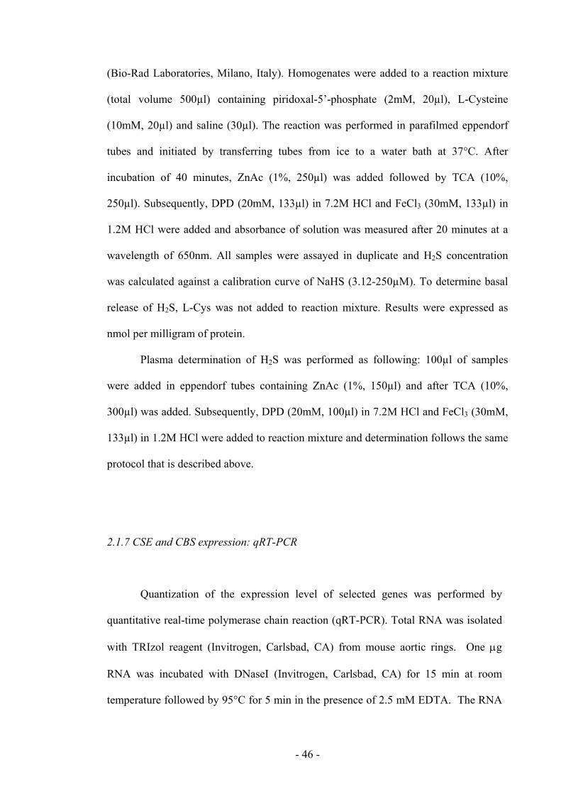
3.2 H2S AND VASCULAR SYSTEM: RELEASE AND MOLECULAR CHANGES
IN PRODUCTION
3.2.1 Impairment of CBS/CSE activity in NOD mice
Plasmatic concentration of H2S was between 50-65µM in NOR mice. In NOD
III mice there was a 50% reduction in H2S levels (fig. 4A; ** p<0.01 vs NOR). To
assess if the systemic H2S impaired production was reflected by an analogue
dysfunction at vascular level we evaluated the basal and the L-cysteine stimulated
release of H2S in aortic rings harvested by NOR and NOD mice. The basal production
of H2S in NOD mice aortas was not significantly reduced if compared to NOR mice
aortas (figure 4B). Conversely, the conversion of exogenous L-cysteine to H2S was
significantly impaired in aorta harvested from NOD II and NOD III mice (* p<0.05; ***
p<0.001; stimulated vs basal; # p<0.05; ## p<0.01 vs NOR). Although H2S levels
resulted progressively lowered during diabetes progression, both western blot (fig. 5A)
and qRT-PCR (fig. 5B) showed a significant upregulation of both CBS and CSE in
vessels harvested by NOD II and NOD III mice.
**
NOR
NOD-I
NOD-II
NOD-III
0
25
50
75
100
H2S
(µM
)
Fig 3.4 H2S plasmatic levels were
significantly reduced in NOD-III mice
(** p<0.01 vs NOR; n=5)
- 61 -
basalstimulated
NOR NOD I NOD III
0
300
600
900
1200
1500
1800 **** #
##
basalstimulated
NOR NOD I NOD III
0
300
600
900
1200
1500
1800 **** #
##
NOR NOD I NOD III
0
300
600
900
1200
1500
1800
NOR NOD I NOD III
0
300
600
900
1200
1500
1800 **** #
##
CBS
CSE
NOR I II III
NOD
NORNOD I
NOD II
NOD III
NORNOD I
NOD II
NOD III
0
5
10
15
CSE
200
400
600
800 CBS
mR
NA
2-dd
Ct
NORNOD I
NOD II
NOD III
NORNOD I
NOD II
NOD III
0
5
10
15
CSE
200
400
600
800 CBS
mR
NA
2-dd
Ct
Fig 3.5 a) Tissue H2S levels (basal) were not different in NOR and NOD mice, while H2S production
following L-cysteine challenge (stimulated) was significantly reduced in aorta harvested from NOD I or
NOD III mice (# p<0.05 vs NOR; ## p<0.01 vs NOR; *** p<0.001 vs basal; * p<0.05 vs basal). Western
blot (b) and qRT-PCR (c) analysis on aortic tissues harvested from NOR and NOR mice. Both western
blot (A) and qRT-PCR (B) showed a progressive increased expression of CBS and CSE.
a)
b)
c)

- 62 -
3.2.2 High glucose environment affects stimulated H2S production in BAEC
Western blot analysis showed that BAEC express CBS but not CSE. To assess
the specific role of endothelial cells we exposed BAEC to normal or high glucose
environment using a well established protocol (26). In normal glucose medium we
detected value of H2S of ∼70nmol/mg protein min-1, whereas on stimulation with L-
cysteine we measured values of H2S of ∼170nmol/mg protein min-1 with a 60% of
increase in the response. When cells are cultured in high glucose medium, the
unstimulated value was not affected, whereas L-cysteine-induced release was
significantly reduced (fig. 6; * p<0.05 stimulated vs basal).
Normal HighGlucose Glucose
H2S
(nm
ol/m
g pr
otei
n*m
in)
0
50
100
150
200
250
*
basalstimulated
Normal HighGlucose Glucose
H2S
(nm
ol/m
g pr
otei
n*m
in)
0
50
100
150
200
250
*
basalstimulated
CBS10 30 50 µg
Fig 3.6 Evaluation of H2S production in BAEC. a) Western blot analysis showed a specific
expression of CBS in BAEC. b) When BAEC were cultured in high glucose medium, basal H2S
production value was not affected, whereas L-cysteine-stimulated release of H2S was
significantly reduced (* p<0.05 vs basal)
a)
b)

- 63 -
3.3 H2S AND INFLAMMATION:
3.3.1 H2S donors decrease ASA-induced leukocyte adhesion through the activation of
KATP channels
Oral administration of aspirin (50 mg/kg) induced a significant time-dependent
increase in leukocyte adherence compared with rats that received vehicle (fig. 3.7).
Pretreatment of rats with Na2S dose-dependently decreased aspirin-induced leukocyte
adherence to the mesenteric microcirculation (ED50: 5.0 µmol/kg). The reduction of
leukocyte adherence by Na2S likely occurred through activation of K+ATP channels,
since pretreatment with an antagonist of those channels, glibenclamide, reversed the
effects of the H2S donor. Glibenclamide given to rats prior to vehicle or ASA did not
alter basal leukocyte adherence or that induced by aspirin (data not shown). NaHS (100
µmol/kg) also inhibited aspirin-induced leukocyte adherence.
Fig 3.7 Hydrogen sulfide inhibits aspirin induced leukocyte adherence in mesenteric venules through activation of K+
ATP channels. Na2S dose-dependently suppressed leukocyte adherence induced by intragastric aspirin (50mg/kg). The inhibition of aspirin-induced adherence by Na2S (100 µmol/kg) was abolished by pretreatment with glibenclamide (10 mg/kg), a K+
ATP channel antagonist. *P<0.05 vs. the corresponding vehicle-treated group. ψP< 0.05 vs. the corresponding group receiving aspirin alone. Data are expressed as mean ± SEM, n=5.

- 64 -
3.3.2.H2S donors inhibit fMLP-induced leukocyte adherence
Pretreatment with Na2S or NaHS (each at 100 µmol/kg) abolished fMLP-
induced leukocyte adherence to the mesenteric microcirculation (fig. 3.8). Lawesson’s
reagent also inhibited leukocyte adhesion when administered at a dose of 1 µmol/kg. At
a dose of 0.3 µmol/kg, Lawesson’s reagent did not affect fMLP-induced leukocyte
adherence (data not shown).
Fig 3.8 Hydrogen sulfide inhibits fMLP-induced leukocyte adherence in mesenteric venules. Superfusion of
the vessels with N-formylated-Met-Leu-Phe (fMLP; 10µM) induced a time-dependent increase in leukocyte
adherence. Hydrogen sulfide donors, given 30 min before fMLP, suppressed the increase in leukocyte
adherence to levels not significantly different from control levels of adherence (*P<0.05 vs. the group
receiving vehicle in place of fMLP). Na2S and NaHS were given at 100 µmol/kg, and Lawesson’s reagent
was given at 1 µmol/kg. The results are plotted as mean ± SEM, n=5.

- 65 -
3.3.3 H2S modulates oedema formation via effects on KATP channels
Injection of carrageenan into the hind footpads of rats resulted in a rapid and
marked increase in paw volume as a consequence of edema formation (fig. 3.9a). The
increase in paw volume could be significantly reduced by pretreatment with diclofenac
(an NSAID). Pretreatment with NaHS or Na2S similarly decreased carrageenan-induced
paw edema (ED50: 35 and 28 µmol/kg, respectively), as did pinacidil, a K+ATP channel
agonist. In contrast, suppression of endogenous H2S synthesis, through administration
of BCA, resulted in a significantly greater paw swelling response to carrageenan. The
reduction paw edema by either of the H2S donors (NaHS or Na2S) could be reversed by
pretreatment with glibenclamide (fig. 3.9b).
Fig 3.9 a) Injection of carrageenan into the rat hindpaw resulted in significant oedema formation as indicated by
the increase in paw volume. Pre-treatment with NaHS significantly reduced paw oedema (P<0.05), as pre-
treatment with diclofenac and pinacidil did. In contrast, administration of BCA significantly increased the
oedema formation induced by carrageenan (P<0.05). b) The increase in paw oedema occurring during the first
hour after carrageenan administration is shown. The reduction of paw oedema by both NaHS and Na2S was
abolished by pre-treatment with glibenclamide, whereas glibenclamide alone did not alter carrageenan-induced
oedema formation. Data are shown as mean ± SEM, n=5
a)
b)

- 66 -
3.3.4 H2S induced externalization of NaxA1 onto membrane surface
Neutrophils were isolated and treated with H2S donor NaHS at 0.1 and 1mM for
30 minutes at 37°C. Western blot analysis clearly showed that AnxA1, normally
resident in cytosolic compartment (C), mobilized towards cell membrane surface (M)
after treatment with NaHS. In addition, NaHS also induced release of AnxA1 in
extracellular environment (S) (fig. 3.10).
C M S C M S C M S
CTR
0.1 mM 1mMNaHS
33 kDa37 kDa
Annexin-1
C M S C M S C M S
CTR
0.1 mM 1mMNaHS
33 kDa37 kDa
C M S C M S C M S
CTR
0.1 mM 1mMNaHS
C M S C M S C M S
CTR
0.1 mM 1mMNaHS
33 kDa37 kDa
Annexin-1
Fig 3.10 Western blot analysis on neutrophils isolated from peripheral human blood. Cells
were treated and fractionated in cytosolic (C), membrane (M) and supernatant (S) fraction.
AnxA1 is shown in two bands of 33 (cleaved) and 37 kDa (uncleaved) respectively. Blot is
representative of three different experiments.

- 67 -
3.3.5 H2S reverted CSE, COX-2 and i-NOS over-expression in LPS treated BMDM
through AnxA1-dependent mechanism
qRT-PCR analysis on BMDM treated with LPS showed an increase in
expression of CSE (1.3 fold increase, *p<0.05, n=3) and a very high expression of
COX-2 and i-NOS (14 and 7 fold increase respectively, ***p<0,001, n=3). When
BMDM were treated with NaHS, the observed effect was the reversion of LPS-induced
increase in expression of CSE, COX-2 and i-NOS (# p<0.05, n=3; ### p<0.001, n=3; #
p<0.05, n=3 respectively) (fig. 3.11). Conversely, the effect of NaHS was completely
lost when the same experiment was carried out on BMDM from AnxA1-/- mice (fig.
3.12). These data may suggest that H2S anti-inflammatory effect could be partially due
to activation of AnxA1 signalling.
3.3.6 H2S production in AnxA1-/- mice
We speculated on the role of CBS and CSE in AnxA1-/- mice, performing qRT-
PCR analysis on several tissues. We found a marked increase in both CBS and CSE
expression in the aorta of knock-out mice compared to wild-type (21 and 60 fold
increase for CBS and CSE respectively). Similar results were observed in lymph nodes
(LN), even if the increase in expression in AnxA1-/- mice was not comparable to the one
found in the aorta (4 and 3 fold increase for CBS and CSE respectively). Liver, the most
important producer of H2S, did not show any significant change in the expression of
both enzymes between wild-type and knock-out. Conversely, in spleen and kidney we
found an upregulation only for CBS expression (10 fold increase) (fig. 3.13).

- 68 -
Furthermore, we dosed plasmatic levels of H2S and we found that LPS induced
an increase in H2S amount in AnxA+/+ mice compared to no-treated mice. In addition, is
noteworthy that plasmatic levels of H2S in no-treated AnxA1-/- mice were higher than
ones detected in no-treated AnxA1+/+ mice (fig. 3.13); these data fit with the higher
expression of CBS and CSE in AnxA1-/- mice compared to AnxA+/+ mice.
In addition to these data we have also performed H2S production assay on tissues
harvested from AnxA1+/+ and AnxA1-/- mice. We found that basal production of H2S in
aorta was increased in LPS treated animals. On the other hand, basal H2S produced in
non-treated AnxA1-/- mice was lowerd compared to wild-type animals. There were not
significant differences in production of H2S in liver, fitting with data not showing any
changes in CBS or CSE expression in liver. Conversely, in kidney we found a
significant increase in basal production of H2S in non-treated AnxA1-/- mice compared
with AnxA1+/+ mice (fig. 3.14).

- 69 -
CTRNaH
SLP
S
NaHS-L
PS
0
1
2
3
4
510
15
20COX-2
***
***###
expr
essi
on r
atio
(2-lo
g)
CTRNaH
SLPS
NaHS-LPS
0.0
0.5
1.0
1.5100
400
700
1000i-NOS
***
***
*
#
expr
essi
on r
atio
(2-lo
g)
Fig 3.11 qRT-PCR on BMDM isolated from AnxA1+/+ mice. NaHS pre-treatment reduces LPS-
induced overexpression of CSE (a), COX-2 (b) and i-NOS (c). (* p<0.05 vs CTR; *** p<0.001 vs
CTR; # p<0.05 vs LPS; ### p<0.001 vs LPS; n=3)
CTRNaH
SLPS
NaHS-LPS
0.0
0.5
1.0
1.5
2.0
2.5 CSE
***
*
#
expr
essi
on r
atio
(2-lo
g)
a)
b)
c)

- 70 -
CTRNaH
SLPS
NaHS-LPS
0
5
10
15
20COX-2
***
***
expr
essi
on r
atio
(2-lo
g)
CTRNaH
SLPS
NaHS-LPS
0
1
2
340
60
80 i-NOS
***
***
expr
essi
on r
atio
(2-lo
g)CTR
NaHS
LPS
NaHS-LPS
0
1
2
3 C S E
expr
essi
on r
atio
(2-lo
g)
Fig 3.12 qRT-PCR on BMDM isolated from AnxA1-/- mice. After genetic deletion of AnxA1,
the reducing effect of NaHS pre-treatment on expression of CSE (a), COX-2 (b) and i-NOS (c)
after LPS treatment was lost (n=3).
a)
b)
c)

- 71 -
$Aorta
CBS CSE
0.0
2.5
5.020
40
60
80
kowt
*
**
fold
incr
ease
(2-d
dCt)
Lymph Nodes
CBS CSE
0.0
2.5
5.0
7.5
kowt
*
*
fold
incr
ease
(2-d
dCt)
Kidney
CBS CSE
0
1
2
3
4
kowt
*
fold
incr
ease
(2-d
dCt)
Liver
CBS CSE
0.0
0.5
1.0
1.5
2.0
kowt
fold
incr
ease
(2-d
dCt)
Spleen
CBS CSE
0
2
4
kowt
5
10
15
20
*
fold
incr
ease
(2-d
dCt)
Fig 3.13 qRT-PCR analysis for CBS or CSE expression performed on aorta (a), liver (b), lymph
nodes (c), spleen (d) and kidney (e) harvested from AnxA1-/- mice. Data are expressed as mean
of fold increase ± SEM. e) Plasmatic H2S levels in AnxA1+/+ or AnxA1-/- mice under normal
condition and after stimulation with LPS
H2S plasma levels
wt ctr
wt LPS
ko ct
r
ko LP
S
0
10
20
30
40
H2S
(µM
)
a) b)
c) d)
e) f)

- 72 -
Aorta
wt ctr
wt LPS
ko ct
r
ko LP
S
0
500
1000
1500
2000
2500
3000
3500 basal stimulated
***
#
#H2S
(nm
ol/m
g pr
otei
n)
liver
wt ctr
wt LPS
ko ct
r
ko LPS
0
150
300
450 basal stimulated
(H2S
) nm
ol/m
g tis
sue*
min
kidney
wt ctr
wt LPS
ko ct
r
ko LPS
0
200
400
600
800 basal stimulated
(H2S
) nm
ol/m
g tis
sue*
min
Fig 3.14 H2S production in aorta (a), liver (b) and kidney (c) harvested from AnxA1+/+ and
AnxA1-/- mice under basal condition and after exogenous L-cysteine feeding (stimulated).
Animals were treated with LPS (10mg/Kg) and with saline (n=3).
a)
b)
c)

- 73 -
CHAPTER 4

- 74 -
4. DISCUSSION
The biological relevance of hydrogen sulphide has been recently addressed by
different research group and in many papers it has been shown that H2S participates to
various pathophysiological conditions.
Vascular studies have shown that H2S-induced vasorelaxation is not affected by
endothelium removal, suggesting that endothelium is not involved in H2S pathway at
vascular level (Zhao W et al., 2001; Wang R, 2002; Hosoki R et al., 1997). Mechanical
endothelium removal only partially mimics the pathological condition known as
endothelial dysfunction. Indeed, the alteration of endothelium functionality, in the
majority of the cases, is a gradual and progressive process and cannot be simply and
directly related to a mechanical non specific endothelium disruption. In the first part of
this work, we used NOD mice, a strain that spontaneously and gradually develops
diabetes associated to a progressive endothelial dysfunction, in order to assess the
involvement of H2S pathway in an endothelial progressive disorder and a possible
interplay with nitric oxide.
In physiological conditions, we have found by using NOR mice tissues that
administration of NaHS or L-cysteine, on pre-contracted aortic rings, induced a
concentration-dependent vasorelaxation. When we blocked the NO contribute by using
L-NAME, we found a marked inhibition of L-cysteine-induced relaxation, but not of
NaHS. A similar pattern was observed in NOD-I mice that have normal
glycosuria/glycaemia. Conversely, as expected, when an H2S donor is used, as
exogenous source of the gas, there are no changes in the vasodilatory pattern. Thus, it
appears that L-Arginine/NO pathway is involved in L-cysteine-induced vasorelaxation
in physiological condition.

- 75 -
In pathological conditions L-cysteine-induced vasodilatation was impaired.
Indeed, aortic vessels harvested from NOD II and NOD III mice showed a reduced
vasodilation that paralleled the disease progression and endothelial dysfunction. As we
have already demonstrated, NOD mice vessels display an impaired Ach-induced
vasorelaxation strictly related to a specific and progressive disorder in eNOS post-
translational activation (Bucci M et al., 2004). Thus, in pathological conditions, where
there is a reduced production of NO we mimicked the effect of L-NAME on normal
rings (physiological) further supporting the previous hypothesis that basal production
of NO is involved in the machinery necessary for endogenous production of H2S. This
hypothesis is further corroborated by the finding that NaHS-induced vasorelaxation was
significantly enhanced in NOD III mice but unaffected in NOD I or NOD II mice. The
enhanced response of NaHS in NOD III mice, where a strong impairment of Ach-
induced relaxation is present (Bucci M et al., 2004), could be explained by a reduced
direct interaction (chemical quenching) between basal NO and exogenous H2S .
Soluble guanylate cyclase is the primary target for NO, thus if the previous
hypothesis is valid, modulation of the enzyme should in turn modulate L-cysteine
induced response without affecting NaHS–induced vasodilatation. In order to assess this
theory we compared the vasodilatory response elicited by either NaHS or L-cysteine
following pharmacological modulation with either ODQ an inhibitor of soluble
guanylate cyclase or with SQ-22,536 an inhibitor of adenylate cyclase in NOR versus
NOD III aortic rings. As expected incubation with ODQ or SQ-22,536 did not affect
NaHS-induced vasorelaxation in both NOR and NOD III mice. Conversely, ODQ but
not SQ-22,536 caused a marked inhibition of L-cysteine-induced vasorelaxation in rings
harvested from both NOR or NOD III mice. This finding together with the previous data

- 76 -
strongly implies that endogenous H2S production through L-cysteine may be modulated
by nitric oxide through guanylate cyclase.
Recently, Jiang HL et al. have demonstrated a similar correlation between NO
and H2S in patients with coronary heart disease. Indeed, in these patients H2S levels
decrease parallels the disease severity and is further impaired if patients were smokers
and/or hypertensive patients as NO does (Jiang H et al., 2005). Interestingly similarly to
NO, H2S levels are inversely correlated to glycaemia as in our experimental model.
When we measured plasma levels we found that H2S concentration declined following
the disease severity as well as the glycosuria. In particular H2S levels were reduced of
about 50% in NOD III mice. Evaluation of H2S levels in aorta harvested from NOR,
NOD I, NOD II or NOD III mice was similar. However, when aorta tissues were fed
with exogenous L-cysteine, in order to assess the ability to convert the substrate, we
found a progressive reduction of H2S production that paralleled the disease progression.
Overall these data suggest a role for the endothelium and in particular of endothelial
nitric oxide in modulating endogenous production of H2S.
To further confirm this hypothesis we tested the direct effect of L-cysteine on
endothelial cells by using BAEC. Indeed, we have recently demonstrated that BAEC
incubated in high glucose environment display an impaired NO production as well as an
increased expression of caveolin-1, an endogenous inhibitory protein for eNOS
activation. The same increased caveolin-1 expression coupled to impaired NO
production was present in vessels harvested from NOD III mice (Bucci M et al., 2004).
Administration of L-cysteine to BAEC in normal glucose environment induced an
increase in H2S production, confirming the ability of endothelial cells to convert L-
cysteine to H2S. When L-cysteine was fed to BAEC grown in high glucose environment

- 77 -
there was a significant decrease in H2S production. This pattern paralleled the pattern
found in aortic tissue harvested from NOD III mice.
In conclusion these results show that endothelial dysfunction significantly
impairs endogenous H2S signalling without affecting the ability of vascular tissue to
react to exogenous H2S. Furthermore, these data suggest that endothelium has an active
role in modulating H2S endogenous production and that the reduction in L-cysteine-
induced relaxation may be originated by the lost ability by vascular tissue in converting
L-cysteine to H2S following eNOS impairment activity.
This part of this work has clearly demonstrated that endothelium is one of the
machinery involved in H2S signalling in vascular responses. Thus, we focussed our
attention on the role of endothelium in H2S-induced response in inflammatory
processes, since that endothelial cells have a critical role in both triggering and
resolving phases of inflammation.
Studies over the past 5 years have provided convincing evidence that H2S is an
important modulator of vascular tone and acts as a neuromodulator as well (Wang R,
2002; Abe K et al., 1996). The results coming from the second part of the present study
suggest that H2S also plays important roles in the context of inflammation. H2S is
generated at sites of inflammation and can influence the ability of neutrophils to cause
tissue injury (Whiteman et al., 2004); it was recently shown to reduce visceral pain
perception (Distrutti E et al., 2005). In the present study, we have demonstrated that
several H2S donors can suppress leukocyte adherence to the vascular endothelium and
can reduce leukocyte infiltration and oedema formation. These effects of H2S were seen
irrespective of the inflammatory stimulus used (carrageenan, aspirin, fMLP).
Suppression of endogenous H2S synthesis, through blockade of CSE, resulted in
enhanced leukocyte adhesion, leukocyte infiltration, and oedema formation. These

- 78 -
actions appeared to be mediated via K+ATP channels, as they were reversed by pre-
treatment with glibenclamide and mimicked by pinacidil, a K+ATP channels blocker and
a K+ATP channels opener respectively.
Our findings therefore suggest an important role for endogenous H2S as a
modulator of some of the key components of acute inflammatory responses, particularly
those occurring at the leukocyte-endothelial interface. As for other gaseous mediators
(carbon monoxide, NO), H2S was recognized for its toxicity long before its importance
in physiological processes was described. H2S is synthesized, primarily from L-
cysteine, through actions of the enzymes CSE and CBS. In rats, blood and plasma levels
of H2S are in the 10–100 µM range (Richardson CJ et al., 2000). In the present study,
we used three different H2S donors at doses that would approximate concentrations of
H2S that fall within this physiological range. Differences in the potency of Lawesson’s
reagent vs. Na2S and NaHS in suppressing leukocyte adherence/infiltration are
consistent with observed differences in their ability to elicit H2S-mediated vascular
smooth muscle relaxation. Moreover, the observation that suppression of endogenous
H2S synthesis with β-cyanoalanine led to increased leukocyte adherence and infiltration
is consistent with a role for this mediator as a tonic inhibitor of leukocyte
adherence/extravasation. Our observation that leukocyte rolling velocity decreased
sharply after administration of the CSE inhibitor is consistent with previous
observations that P-selectin expression can be regulated by H2S (Fiorucci S et al.,
2005). Since leukocyte expression/affinity of LFA-1 has also been shown to be
suppressed by H2S (Fiorucci S et al., 2005), it is possible to hypothesize that H2S by
modulating expression of proteins on both cell types regulates leukocyte migration.
It has been recently reported that NSAIDs suppress H2S synthesis by reducing
expression of CSE (Fiorucci S et al., 2005). The subsequent reduction of H2S synthesis

- 79 -
may contribute to the increase in leukocyte adherence that is seen after NSAID
administration (Asako H et al., 1992; Wallace JL et al., 1993), which has been shown to
contribute significantly to the gastric injury induced by this class of drugs (Wallace JL
et al., 1990; Wallace JLet al., 1991; Wallace JL et al., 1997). Indeed, co-administration
of an H2S donor with an NSAID resulted in inhibition of NSAID-induced leukocyte
adherence and reduction of the severity of gastric damage (Fiorucci S et al., 2005).
Pertinent to the present study, administration of an H2S donor prevented many of the
other “proinflammatory” effects of NSAIDs, including the elevation of ICAM-1 and
LFA-1 expression and the increase in mucosal TNFα expression (Fiorucci S et al.,
2005).
Among the four H2S donors used in the second part of this study, only N-
acetylcysteine requires metabolism in order for H2S to be released. N-acetylcysteine is a
precursor of Lcysteine (Fujii K et al., 2005), which is the substrate for H2S generation
via CSE and/or CBS. The observation that the anti-inflammatory actions of N-
acetylcysteine were reversed by an inhibitor of CSE (β-cyanoalanine) is consistent with
the effects being mediated by H2S.
Irreversible inhibitors of CSE and CBS have been reported to interfere with
other enzymes (Moore PK et al., 2003; Smith SB et al., 1977). As with any
pharmacological agent, we cannot exclude the possibility that the reversible inhibitor of
CSE, β-cyanoalanine, could exert non-specific effects. For these reasons we have
developed and studied four different H2S donors in order to adderss this issue.
Genetic deletion of CSE and CBS are lethal, ruling out the use of these
“knockouts”. CBS heterozygotes are viable, expressing half as much CBS as wild-type
(WT). When fed a diet high in homocysteine, CBS-/- mice have been shown to exhibit
increased leukocyte adherence, increased P-selectin expression, and increased vascular
- 80 -
permeability (in the brain) (Kamath AF et al., 2006), all consistent with a role for H2S in
mediating acute inflammation. However, use of these mice for direct studies of
inflammation is of questionable value, as they have drastically altered vascular
responsiveness to cholinergic agents and bradykinin (Weiss N et al., 2002).
A consistent finding in the various models used in the present study was that the
anti-inflammatory effects of H2S appeared to be mediated via activation of K+ATP
channels. Similarly, the analgesic effect have been ascribed to modulation of K+ATP
channels (Distrutti E et al., 2005). It is possible, therefore, that K+ATP channels represent
a novel target for anti-inflammatory and analgesic agents.
In addition to these data, hydrogen sulphide also plays a primary role in
resolving phase of inflammation. In fact, our results suggest that H2S is able to induce
an anti-inflammatory effect, likely relying on annexin-1-dependent responses, as we
have demonstrated in our experiments performed on human neutrophils, bone marrow-
derived macrophages and AnxA1-/- mice.
In summary, the results showed in the second part of the present study
demonstrates a role for endogenous H2S as a modulator of key inflammatory events
occurring at the interface of leukocytes and the vascular endothelium. H2S may act as a
tonic regulator of leukocyte adherence to the endothelium and of endothelial
permeability. The anti-inflammatory effects of H2S appear to be mediated via activation
of both K+ATP channels and annexin-1. These results, and recent reports that H2S donors
can down-regulate adhesion molecule and pro-inflammatory cytokine expression,
therefore identify H2S, the key enzymes responsible for H2S synthesis, and K+ATP
channels as potential targets for novel anti-inflammatory therapies.
Finally, we can conclude that H2S is a very complex modulator in despite of a
very simple chemical structure.

- 81 -
Indeed, it is involved in vascular homeostasis directly and also through
interaction with L-arginine/NO pathway; most likely through K+ATP channels and
annexin-1.
Furthermore, the ability of endothelial cells to generate H2S is very consistent
with the function that H2S exerts at the endothelium-blood interface. These findings
open a new horizon in the pharmacology of the gaseous mediators. Thus, the scenario
we have in front of us is far to be completely understood, since now there are three
mediators, NO, H2S and CO, all contributing to the complex phenomenon of
inflammation and still there is much to be understood on each single mediator as well as
on the possible cross talk among them.
Unravelling these issues will allow researchers in the future to develop new
therapeutic targets and design new and effective drugs.

- 82 -
CHAPTER 5

- 83 -
5. REFERENCES
1. Abe K & Kimura H. The possible role of hydrogen sulphide as an endogenous
neuromodulator. J Neurosci. 1996; 16: 1066–1071
2. Aplin AE, Howe A, Alahari SK & Juliano RL. Signal transduction and signal
modulation by cell adhesion receptors: the role of integrins, cadherins,
immunoglobulin-cell adhesion molecules, and selectins. Pharmacol Rev. 1998;
50: 197-263
3. Arribas M, Cutler DF. Weibel-Palade body membrane proteins exhibit
differential trafficking after exocytosis in endothelial cells. Traffic. 2000; 1:783-
793.
4. Asako H, Kubes P, Wallace JL, Wolf RE & Granger DN. Modulation of
leukocyte adhesion in rat mesentericvenules by aspirin and salicylate.
Gastroenterology 1992; 103: 146–152
5. Belardinelli MC, Chabli A, Chadefaux-Vekemans B & Kamoun P. Urinary
sulfur compounds in Down syndrome. Clin Chem. 2001; 47: 1500-1501
6. Bhatia M, Sidhapuriwala J, Moochhala SM & Moore PK. Hydrogen sulphide is
a mediator of carrageenan-induced hindpaw oedema in the rat. Br J Pharmacol.
2005; 145: 141-144
7. Braunstein AE & Azarkh RM. Participation of vitamin B6 in enzymic formation
of hydrogen sulfide from L-cysteine. Dokl Akad Nauk SSSR. 1950; 71 (1950) 93-
96
8. Braunstein AE & Azarkh RM. Phosphopyridoxal in aerobic deamination of
homoserine and serine. Dokl Akad Nauk SSSR. 1952; 85: 385-388

- 84 -
9. Braunstein AE, Goryachenkova EV, Tolosa EA, Willhardt IH & Yefremova LL.
Specificity and some other properties of liver serine sulphhydrase: evidence for
its identity with cystathionine -synthase. Biochim Biophys Acta. 1971; 242: 247-
260
10. Bucci M, Roviezzo F, Brancaleone V, Lin MI, Di Lorenzo A, Cicala C, Pinto A,
Sessa WC, Farneti S, Fiorucci S & Cirino G. Diabetic mouse angiopathy is
linked to progressive sympathetic receptor deletion coupled to an enhanced
caveolin-1 expression. Arterioscler Thromb Vasc Biol. 2004; 24: 721-726
11. Burns AR, Bowden RA, Abe Y, Walker DC, Simon SI, Entman ML, Smith CW.
P-selectin mediates neutrophil adhesion to endothelial cell borders. J Leukoc
Biol. 1999; 65: 299-306
12. Busse R & Fleming I. Nitric oxide, nitric oxide synthase, and hypertensive
vascular disease. Curr Hypertens Rep. 1999; 1: 88-95
13. Chen X, Jhee KH & Kruger WD. Production of the neuromodulator H2S by
cystathionine beta-synthase via the condensation of cysteine and homocysteine.
J Biol Chem. 2004; 279: 52082-52086
14. Cheng Y, Ndisang JF, Tang G, Cao K & Wang R. Hydrogen sulfide-induced
relaxation of resistance mesenteric artery beds of rats. Am J Physiol Heart Circ
Physiol. 2004; 287: H2316-2323
15. Cirino G, Fiorucci S & Sessa WC. Endothelial nitric oxide synthase: the
Cinderella of inflammation ? Trends Pharmacol Sci. 2003; 24: 91-95
16. Dello Russo C, Tringali G, Ragazzoni E, Maggiano N, Menini E, Vairano M,
Preziosi P & Navarra P. Evidence that hydrogen sulphide can modulate
hypothalamo-pituitary-adrenal axis function: in vitro and in vivo studies in the
rat. J Neuroendocrinol. 2000; 12: 225-233

- 85 -
17. Distrutti E, Sediari L, Mencarelli A, Renga B, Orlandi S, Antonelli E, Roviezzo
F, Morelli A, Wallace JL, Cirino G & Fiorucci S. Evidence that hydrogen
sulfide exerts antinociceptive effects in the gastrointestinal tract by activating
KATP channels. J Pharmacol. Exp. Ther. 2005; 316: 325–335
18. Eto K, Ogasawara M, Umemura K, Nagai Y & Kimura H. Hydrogen sulfide is
produced in response to neuronal excitation. J Neurosci. 2002; 22: 3386-3391
19. a Fiorucci S, Antonelli E, Distrutti E, Rizzo G, Mencarelli A, Orlandi S,
Zanardo R, Renga B, Di Sante M, Morelli A, Cirino G & Wallace JL. Inhibition
of hydrogen sulfide generation contributes to gastric injury caused by anti-
inflammatory nonsteroidal drugs. Gastroenterology. 2005; 129: 1210-1224
20. b Fiorucci S, Antonelli E, Mencarelli A, Orlandi S, Renga B, Rizzo G, Distrutti
E, Shah V & Morelli A. The third gas: H2S regulates perfusion pressure in both
the isolated and perfused normal rat liver and in cirrhosis. Hepatology. 2005; 42:
539-548
21. Flavin M & Segal A. Purification and properties of the cystathionine-γ-cleavage
enzyme of Neurospora. J Biol Chem. 9164; 239: 2220-2227
22. Flower RJ, Parente L, Persico P, Salmon JA. A comparison of the acute
inflammatory response in adrenalectomised and sham-operated rats. Br J
Pharmacol. 1986; 87: 57-62
23. Fujii K, Sakuragawa T, Kashiba M, Sugiura Y, Kondo M, Maruyama K, Goda
N, Nimura Y & Suematsu M. Hydrogen sulphide as an endogenous modulator
of biliary bicarbonate excretion in the rat liver. Antioxid Redox Signal. 2005; 7:
788–794
24. Garcia X & Stein F. Nitric oxide. Semin Pediatr Infect Dis. 2006; 17: 55-57

- 86 -
25. Giggenbach, W. Optical spectra of highly alkaline sulfide solutions and the
second dissociation constant of hydrogen sulphide. Inorg Chem. 1971; 10:
1333-1338
26. Hannon R, Croxtall JD, Getting SJ, Roviezzo F, Yona S, Paul-Clark MJ, Gavins
FN, Perretti M, Morris JF, Buckingham JC & Flower RJ. Aberrant inflammation
and resistance to glucocorticoids in annexin 1-/- mouse. FASEB J. 2003; 17: 253-
255
27. Hosoki R, Matsuki N & Kimura H. The possible role of hydrogen sulfide as an
endogenous smooth muscle relaxant in synergy with nitric oxide. Biochem
Biophys Res Commun. 1997; 237: 527-531
28. Hui Y, Du J, Tang C, Bin G & Jiang H. Changes in arterial hydrogen sulfide
(H2S) content during septic shock and endotoxin shock in rats. J Infect. 2003;
47: 155-160
29. Jiang HL, Wu HC, Li ZL, Geng B & Tang CS. Changes of the new gaseous
transmitter H2S in patients with coronary heart disease. Di Yi Jun Yi Da Xue Xue
Bao. 2005; 25: 951-954
30. Kamal AM, Flower RJ, Perretti M. An overview of the effects of annexin 1 on
cells involved in the inflammatory process. Mem Inst Oswaldo Cruz. 2005; 100
Suppl 1: 39-47
31. Kamath AF, Chauhan AK, Kisucka J, Dole VS, Loscalzo J, Handy DE &
Wagner DD. Elevated levels of homocysteine compromise blood-brain barrier
integrity in mice. Blood. 2006; 107: 591–593
32. Kery V, Bukovska G & Kraus JP. Transsulfuration depends on heme in addition
to pyridoxal 5'-phosphate. Cystathionine beta-synthase is a heme protein. J Biol
Chem. 1994; 269: 25283-25288

- 87 -
33. Kery V, Poneleit L, Meyer JD, Manning MC & Kraus JP. Binding of pyridoxal
5'-phosphate to the heme protein human cystathionine beta-synthase.
Biochemistry. 1999; 38: 2716-2724
34. Kimura H. Hydrogen sulfide induces cyclic AMP and modulates the NMDA
receptor. Biochem Biophys Res Commun. 2000; 267: 129-133
35. Kurzban GP, Chu L, Ebersole JL & Holt SC. Sulfhemoglobin formation in
human erythrocytes by cystalysin, an L-cysteine desulfhydrase from Treponema
denticola. Oral Microbiol Immunol. 1999; 14:153-164
36. Leeuwenberg JF, Smeets EF, Neefjes JJ, Shaffer MA, Cinek T, Jeunhomme TM,
Ahern TJ & Buurman WA. E-selectin and intercellular adhesion molecule-1 are
released by activated human endothelial cells in vitro. Immunology. 1992; 77:
543-549.
37. Levitt MD, Furne J, Springfield J, Suarez F & DeMaster E. Detoxification of
hydrogen sulfide and methanethiol in the cecal mucosa. J Clin Invest. 1999; 104:
1107-1114
38. Levonen AL, Lapatto R, Saksela M & Raivio KO. Human cystathionine
gamma-lyase: developmental and in vitro expression of two isoforms. Biochem
J. 2000; 347: 291-295
39. Li J, Zhou Z, Jiang DJ, Li D, Tan B, Liu H, Li YJ. Reduction of NO- and
EDHF-Mediated Vasodilatation in Hypertension: Role of Asymmetric
Dimethylarginine. Clin Exp Hypertens. 2007; 29: 489-501
40. Li L, Bhatia M, Zhu YZ, Zhu YC, Ramnath RD, Wang ZJ, Anuar FB,
Whiteman M, Salto-Tellez M & Moore PK. Hydrogen sulfide is a novel
mediator of lipopolysaccharide-induced inflammation in the mouse. FASEB J.
2005; 19: 1196-1198

- 88 -
41. Lu Y, O'Dowd BF, Orrego H & Israel Y. Cloning and nucleotide sequence of
human liver cDNA encoding for cystathionine gamma-lyase. Biochem Biophys
Res Commun. 1992; 189: 749-758
42. Lüscher TF & Barton M. Endothelins and endothelin receptor antagonists:
therapeutic considerations for a novel class of cardiovascular drugs. Circulation.
2000; 102: 2434-2440
43. Makino S, Kunimoto K, Muraoka Y, Mizushima Y, Katagiri K & Tochino Y.
Breeding of a non-obese, diabetic strain of mice. Jikken Dobutsu. 1980; 29: 1-13
44. Mariggiò MA, Pettini F & Fumarulo R. Sulfide influence on polymorphonuclear
functions: a possible role for Ca2+ involvement. Immunopharmacol
Immunotoxicol. 1997; 19: 393-404
45. Meier M, Janosik M, Kery V, Kraus JP & Burkhard P. Structure of human
cystathionine beta-synthase: a unique pyridoxal 5'-phosphate-dependent heme
protein. EMBO J. 2001; 20: 3910-3916
46. Mok YY, Atan MS, Yoke Ping C, Zhong Jing W, Bhatia M, Moochhala S &
Moore PK. Role of hydrogen sulphide in haemorrhagic shock in the rat:
protective effect of inhibitors of hydrogen sulphide biosynthesis. Br J
Pharmacol. 2004; 143: 881-9
47. Moncada S & Higgs EA. Endogenous nitric oxide: physiology, pathology and
clinical relevance. Eur J Clin Invest. 1991; 21: 361-374
48. Moore PK, Bhatia M & Moochhala S. Hydrogen sulfide: from the smell of the
past to the mediator of the future? Trends Pharmacol Sci. 2003; 12: 609–611
49. Myers, RJ. The new low value for the second dissociation constant for H2S: Its
history, its best value, and its impact on the teaching of sulfide equilibria.
Journal of Chemical Education. 1986; 63: 687-690

- 89 -
50. Nagareddy PR, Xia Z, McNeill JH & MacLeod KM. Increased expression of
iNOS is associated with endothelial dysfunction and impaired pressor
responsiveness in streptozotocin-induced diabetes. Am J Physiol Heart Circ
Physiol. 2005; 289: H2144-2152
51. Nakagawa H, Kimura H. Purification and properties of cystathionine synthetase
synthetase from rat liver: separation of cystathionine synthetase from serine
dehydratase. Biochem Biophys Res Commun. 1968; 32: 209-214
52. Palmer RM, Ashton DS & Moncada S. Vascular endothelial cells synthesize
nitric oxide from L-arginine. Nature. 1988; 333: 664-666
53. Pecháňová O & Simko F. The role of nitric oxide in the maintenance of
vasoactive balance. Physiol Res. 2007 Sep 5 (in press)
54. Perretti M, Becherucci C, Scapigliati G, Parente L. The effect of adrenalectomy
on interleukin-1 release in vitro and in vivo. Br J Pharmacol. 1989; 98:1137-
1142
55. Pfaffl MW, Horgan GW & Dempfle L. Relative expression software tool
(REST©) for group-wise comparison and statistical analysis of relative
expression results in real-time PCR. Nucleic Acids Res. 2002; 30: e36
56. Picton R, Eggo MC, Merrill GA, Langman MJ & Singh S. Mucosal protection
against sulphide: importance of the enzyme rhodanese. Gut. 2002; 50: 201-205
57. Pober JS. Endothelial activation: intracellular signalling pathways. Arthritis Res.
2002; 4 Suppl 3:S109-116
58. Ppova EN, Arkhipova DV, Kozlovskaia LV, Privalova EV, Bitsadze VO &
Kornev BM. Role of endothelial dysfunction and coagulation disorders in the
development of pulmonary fibrosis in patients with interstitial lung diseases.
Klin Med (Mosk). 2004; 82: 38-42

- 90 -
59. Punthakee Z, Werstuck GH, Gerstein HC. Diabetes and cardiovascular disease:
explaining the relationship. Rev Cardiovasc Med. 2007; 8: 145-153
60. Richardson CJ, Magee EA & Cummings JH. A new method for the
determination of sulphide in gastrointestinal contents and whole blood by
microdistillation and ion chromatography. Clin Chim Acta. 2000; 293: 115–125
61. Rubanyi GM & Polokoff MA. Endothelins: molecular biology, biochemistry,
pharmacology, physiology, and pathophysiology. Pharmacological Reviews.
1994; 46: 325-415.
62. Santiago-Delpin EA. The endothelium and early immune activation: new
perspective and interactions. Transplant Proc. 2004; 36: 1709-1713
63. Schlossmann K, Brueggemann J & Lynen F. Biosynthesis of cysteine. I.
Detection and isolation of serine sulfhydrase from baker's yeast. Biochem Z.
1962; 336: 258-273
64. Serhan CN & Savill J. Resolution of inflammation: the beginning programs the
end. Nat Immunol. 2005; 6: 1191-1197
65. Sheng L & Wharton RA. Functional characterization of two S-nitroso-L-
cysteine transporters, which mediate movement of NO equivalent into vascular
cells. Am J Physiol Cell Physiol. 2006; 292: C1263-C1271
66. Skovby F, Kraus JP, Rosenberg LE. Biosynthesis and proteolytic activation of
cystathionine beta-synthase in rat liver. J Biol Chem. 1984; 259: 588-593
67. Smith SB, Briggs S, Triebwasser KC & Freedland RA. Re-evaluation of amino-
oxyacetate as an inhibitor. Biochem J.1977; 162: 453–455
68. Stipanuk MH & Beck PW. Characterization of the enzymic capacity for cysteine
desulphhydration in liver and kidney of the rat. Biochem J. 1982; 206: 267-277.

- 91 -
69. Suzaki Y, Yoshizumi M, Yamashita Y, Abe S, Tsuchiya K, Satoh Y, Kuroda Y,
Horike K, Kano M, Fukata Y, Kitaichi T, Hori T, Masuda Y, Kitagawa T,
Minakuchi K & Tamaki T. Determination of plasma concentrations of
endothelin-1(1-31) and endothelin-1 in healthy subjects and patients with
atherosclerosis. J Cardiovasc Pharmacol. 2003; 41 Suppl 1: S83-87
70. Teague B, Asiedu S & Moore PK. The smooth muscle relaxant effect of
hydrogen sulphide in vitro: evidence for a physiological role to control intestinal
contractility. Br J Pharmacol. 2002; 137: 139-145
71. van der Molen EF, Hiipakka MJ, van Lith-Zanders H, Boers GH, van den
Heuvel LP, Monnens LA & Blom HJ. Homocysteine metabolism in endothelial
cells of a patient homozygous for cystathionine beta-synthase (CS) deficiency.
Thromb Haemost. 1997; 78: 827-833
72. Wallace JL, Arfors KE & McKnight GW. A monoclonal antibody against the
CD18 leukocyte adhesion molecule prevents indomethacin-induced gastric
damage in the rabbit. Gastroenterology. 1991; 100: 878–883
73. Wallace JL, Keenan CM & Granger DN. Gastric ulceration induced by
nonsteroidal anti-inflammatory drugs is a neutrophil-dependent process. Am J
Physiol. 1990; 259: G462–G467
74. Wallace JL, McKnight W, Miyasaka M, Tamatani T, Paulson J, Anderson DC,
Granger DN & Kubes P. Role of endothelial adhesion molecules in NSAID-
induced gastric mucosal injury. Am J Physiol. 1993; 265: G993-998
75. Wallace JL. Nonsteroidal anti-inflammatory drugs and gastroenteropathy: the
second hundred years. Gastroenterology. 1997; 112: 1000–1016
76. Wang R. Two's company, three's a crowd: can H2S be the third endogenous
gaseous transmitter ? FASEB J. 2002;16: 1792-1798

- 92 -
77. Weiss N, Heydrick S, Zhang YY, Bierl C, Cap A & Loscalzo J. Cellular redox
state and endothelial dysfunction in mildly hyperhomocysteinemic cystathione
beta-synthase deficient mice. Arterioscler Thromb Vasc Biol. 2002; 22: 34–41
78. Whiteman M, Armstrong J S, Chu SH, Siau JL, Wong BS, Cheung NS,
Halliwell B & Moore PK. The novel neuromodulator hydrogen sulphide: an
endogenous peroxynitrite ‘scavenger’ ? J Neurochem. 2004; 90: 765–768
79. Yan H, Du JB & Tang CS. Changes of endogenous hydrogen sulfide (H2S) in
hypertensive rats. Zhonghua Er Ke Za Zhi. 2004; 42: 172-175
80. Yap S, Naughten ER, Wilcken B, Wilcken DE & Boers GH. Vascular
complications of severe hyperhomocysteinemia in patients with homocystinuria
due to cystathionine beta-synthase deficiency: effects of homocysteine-lowering
therapy. Semin Thromb Hemost. 2000; 26: 335-340
81. Zanardo RC, Brancaleone V, Distrutti E, Fiorucci S, Cirino G & Wallace JL.
Hydrogen sulfide is an endogenous modulator of leukocyte-mediated
inflammation. FASEB J. 2006; 20: 2118-2120
82. Zhang Y & Hogg N. S-nitrosothiols: cellular formation and transport. Free
radical biology and medicine. 2005; 38: 831-838
83. Zhao W & Wang R. H2S-induced vasorelaxation and underlying cellular and
molecular mechanisms. Am J Physiol Heart Circ Physiol. 2002; 283: H474-480
84. Zhao W, Ndisang JF & Wang R. Modulation of endogenous production of H2S
in rat tissues. Can J Physiol Pharmacol. 2003; 81: 848-853
85. Zhao W, Zhang J, Lu Y & Wang R. The vasorelaxant effect of H2S as a novel
endogenous gaseous K(ATP) channel opener. EMBO J. 2001; 20: 6008-6016

- 93 -
86. Zhong G, Chen F, Cheng Y, Tang C & Du J. The role of hydrogen sulfide
generation in the pathogenesis of hypertension in rats induced by inhibition of
nitric oxide synthase. J Hypertens. 2003; 21: 1879-1885