diarrhea in early life - fedoa - universit degli studi di napoli
TRANSCRIPT

“FEDERICO II”
UNIVERSITY OF NAPLES
Faculty of Medicine and Surgery
Department of Pediatrics
PhD Program
“Human Reproduction, Development and Growth”
Director
Prof. Claudio Pignata
PhD Thesis
DIARRHEA IN EARLY LIFE:
PROGRESS IN DIAGNOSIS AND CONTROL OF DISEASE
Academic Years 2007-2010
Student
Dt. Gianluca Terrin
Tutor
Prof. Roberto Berni Canani

I
Section Page
Introduction 1
Acute diarrhea in early life 1
Chronic diarrhea in early life 2
Progress for diagnosis and control of early onset diarrheal diseases 3
Investigational plan 4
Objectives of PhD 7
Ethics 9
Projects 10
Research Area 1. Epidemiology and clinical features of diarrhea in early life 10
Epidemiology 10
Conclusive remarks 15
Future implications 17
Natural course of diarrhea associated with intestinal failure 18
Conclusive remarks 21
Future implications 23
Strategy for control of complications related to intestinal failure 24
Conclusive remarks 28
Future implications 30
INDEX

II
.
Section Page
Research Area 2. New approaches for control of acute diarrhea in early life 31
Efficacy of new ORS in acute gastroenteritis 31
Conclusive remarks 35
Future implications 37
Efficacy of different probiotic formulations 38
Conclusive remarks 42
Future implications 44
Research Area 3. Diagnosis and treatment of chronic diarrhea in early life 46
Genotype of children with congenital diarrhea 46
Conclusive remarks 49
Future implications 52
New therapeutic strategies for congenital diarrhea 54
Conclusive remarks 59
Future implications 62
Research Area 4. Prevention of diarrhea in early life 63
Prevention of infectious diarrhea determined by gastric acidity inhibitors 63
Conclusive remarks 67
Future implications 69
Primary prevention of food-induced diarrhea 70
Conclusive remarks 71
Future implications 72

III
Section Page
Conclusions 74
Tables 75
Figures 101
References 119
Acknowledgements 137

1
Diarrheal diseases occurring in early life (< 3 years) are an heterogeneous group of
abnormalities including gastrointestinal infections, food hypersensitivity and allergy,
immune dysregulation, and primary abnormalities of the enterocyte.
Diarrhea is a leading cause of illness and death in children younger than 3 years worldwide
(Figure 1), causing 1·3 million deaths every year (1). Despite defining etiology of diarrhea
is critical to decide therapy and prevention strategy, the overall prevalence of early onset
diarrhea was not clearly defined because of the large spectrum of etiologies and difficulties
in patient selection.
From a clinical point of view, diarrhea needs to be classified taking into account certain
characteristics such as trends over time (acute or chronic, using a limit of 4 week to separate
the two conditions) and the characteristics of the feces (2,3). Acute diarrheal diseases
occurring in early life, usually due to infectious agent or food allergy, were burdened by an
increased mobility and mortality, whereas chronic evolution of early onset diarrhea
frequently suggests a congenital disorder determined by genetic defects. Using this
classification a pediatrician can decide upon diagnosis and therapy more rationally.
However, acute diarrhea may be also a symptom of the onset of chronic organic or
functional disease.
Acute diarrhea in early life
In younger children acute diarrhea will lead to severe dehydration. Fluid loss and
dehydration are the cause of death in nearly all children with acute diarrhea. Over 3 decades
ago, the discovery of mechanisms of intestinal electrolytes transport, which was the basis
INTRODUCTION

2
for the development of oral treatment of dehydration, was hailed as the most important
medical advance of the 20th century (4). Complications can be prevented by the early and
adequate oral administration of a rehydration solution, by normal food for the child's age,
and through induction of benefical intestinal microbiota composition (4). The evidence-
based guidelines of the ESPGHAN and Cochrane analyses, based also on studies reported in
this manuscript, indicate zinc and probiotics as useful therapeutic aids for children younger
than 3 years with acute diarrhea (5-9).
Chronic diarrhea in early life
Chronic diarrhea in early life includes a group of rare chronic enteropathies characterized by
a heterogeneous etiology, which in most cases is related to an identified or to an as yet
unidentified genetic defect, generally inherited as an autosomal recessive trait. These
congenital diarrheal disorders (CDD, OMIM 251850) represent one of the most challenging
clinical conditions for pediatric gastroenterologists because of the severity of the clinical
picture and the broad range of conditions in its differential diagnosis (10). Early in life,
patients affected by CDD usually present with severe diarrhea that within a few hours leads
to a life-threatening condition secondary to massive dehydration and metabolic acidosis
(10). Consequently, children affected by CDD require a prompt diagnosis and assistance.
Clinical manifestations are variable from severe conditions leading to intestinal failure, to
milder forms with subtle clinical signs that may remain undiagnosed until adulthood, when
patients have just developed irreversible complications. Intestinal failure may lead to the
necessity of total parenteral nutrition with further complications for the health of the subject
affected by these disorders. The number of conditions included within the CDD group has
gradually increased over the years (10). Now it is clear that CDD depend on defects in the
structure and function of absorptive, enteroendocrine or inflammatory cells of the gut,
determined by mutations in genes expressed throughout the gastrointestinal tract involving
different segments and different cells. Therefore, as shown in Figure 2, we have proposed
that CDD could be classified into 4 groups: (i) Absorption and transport of nutrients and

3
electrolytes; (ii) Enterocyte differentiation and polarization; (iii) Enteroendocrine cell
differentiation; and (iv) Modulation of the intestinal immune response. In recent years,
many new genes have been identified. Such molecular techniques as positional candidate
genes and genome-wide linkage analysis have clarified the role of these genes in the
physiology of the gastrointestinal tract. Understanding the function of this genes may open
new diagnostic and therapeutic perspectives for chronic and acute forms of early onset
diarrhea.
Progress for diagnosis and control of early onset diarrheal diseases
The early postnatal period represents a critical window during which the evolving intestine
is programmed by intrinsic (genetic programming) and extrinsic factors (microbe, nutrition,
drugs). Intestinal content may modify pretranslational and post-translational gene
expression. These ‘‘epigenetic’’ mechanisms are involved in the development of gut
enzymes, hormones, transporters, and immunity. Occurrence of diarrheal diseases and
related treatments, during this temporarily window, may influence the entire body health
status in the short and long-term period (11,12) (Figure 3). In this scenario, accurate
identification and adequate control of diarrheal disorders occurring in early life are crucial
for reducing morbidity and mortality.
Finally, advances made in the pathophysiology of chronic early onset diarrheal disorders
could contributed to a better understanding the mechanisms also of the more common acute
diarrheal diseases and to identify novel strategy for disease control.
Starting from these considerations, we elaborate selected items focusing on the new
diagnostic, therapeutic and preventive strategies for diarrheal diseases occurring in early
life.

4
The PhD project was focused on 4 closely integrated research area:
Research Area 1. Epidemiology and clinical features of diarrhea in early life
A key to understanding the treatment and control of early onset diarrhea is establishment of
epidemiology and etiology. Early diagnosis and timely treatment are crucial because
diarrhea in the first phase of the life may rapidly lead to life-threatening dehydration and
malnutrition (15). Clinical and epidemiologic studies defining severity and etiology are
needed in order to improve diagnostic and therapeutic approaches for diarrhea in early life.
Frequently diarrhea is associated with intestinal failure during neonatal age, imposing
clinician to recur to artificial nutrition. Few studies have addressed the incidence, prognostic
factors, diagnosis, and nutritional aspects of intestinal failure occurring early in life (14).
Identification of the risk factors for dependence on parenteral nutrition is of critical
importance for rapid and appropriate preventive strategies application (15).
Planned studies. Two nationwide multicenter studies investigating (i) epidemiology,
etiology, clinical features, and management of diarrhea observed in hospitalized newborns,
and (ii) epidemiology and natural course of neonatal onset intestinal failure, were
performed.
INVESTIGATIONAL PLAN

5
Research Area 2. New approaches for control of acute diarrhea in early life
The most common causes of acute diarrhea involve gastrointestinal infection and food
allergy. Although diarrhea acts as a defense mechanism in the body, quickly eliminating
infective organisms and antigens, the most serious sequelae is dehydration. Oral rehydration
solution (ORS) is the first-line therapy for the management of children with acute
gastroenteritis worldwide (4). However, ORS neither reduces the severity nor the duration
of symptoms. Several substrates and substances acting on transepithelial fluid transport have
been proposed in order to enhance clinical efficacy of ORS (5-9). Such evidences indicated
that zinc-fortified ORS and probiotics could be effective in reducing diarrhea duration and
severity in children with acute gastroenteritis (4).
Planned studies: (i) Prospective study evaluating efficacy of new oral rehydration solution
in children with acute gastroenteritis; and (ii) multicenter study comparing the therapeutic
effects of different probiotics in children with infectious diarrhea, were performed.
Research Area 3. Diagnosis and treatment of chronic diarrhea in early life
Early diagnosis of this condition is necessary in infancy because hyponatremic episodes
may result in mental and psychomotor impairment and the chronic contraction of the
intravascular space leading to renal dysfunction and gout.
Recently, the role of amylase-resistant starch has been increasingly recognized for the
management of diarrheal diseases. On reaching the colon, amylase-resistant starch are
fermented by resident bacteria into the short-chain fatty acids (SCFA), including acetate,
propionate, and butyrate. As already shown, SCFA have a great capacity for stimulating ion
and water absorption; they provide energy and induce a trophic effect on both colonic and

6
small bowel mucosa (10). The important regulatory role of SCFA on fluid and electrolyte
absorption has led to the hypothesis that butyrate treatment could reduce diarrhea in patients
with such forms of chronic diarrhea occuring in early life.
Planned studies: Prospective studies (i) defining the genotype of children with early onset
chronic diarrhea, and (ii) evaluating the clinical efficacy of butyrate in children affected by
congenital diarrhea, were performed.
Research Area 4: Prevention of diarrhea in early life
In the previous century, more than 100 countries had established national programs for
control of diarrheal disease. In developed countries, these programs should be taken into
account that the first two causes of diarrhea in early life are infections and food allergy.
Gastric acidity inhibitors the mainstay of therapy of gastrointestinal acid-related disorders,
has been associated with increased risk of acute gastroenteritis (16). Reduction of the use of
GAI may reduce the risk of gastrointestinal infections occurring in early life. The
prevalence of food allergy has increased worldwide, especially in children (17). Data
suggest that the onset of food allergy may be prevented by nutritional intervention early in
life (18).
Planned studies: (i) prospective studies evaluating the risk of intestinal and extraintestinal
infections associated with the gastric acidity inhibitors use, and (ii) multicenter audit study
measuring the implementation of prevention program for control of food allergy, were
performed .

7
The objectives of PhD project were divided per research area:
1. Epidemiology and clinical features of diarrhea in early life
1. To study epidemiology and etiology of diarrhea occurring in early life
2. To define natural course of diarrhea associated with intestinal failure occurring in early
life and to verify the efficacy of strategies for control of complications related to early
onset intestinal failure
2. New approaches for control of acute diarrhea in early life
1. To evaluate efficacy of new oral rehydration solution in children with acute
gastroenteritis
2. To compare the therapeutic effects of different probiotic formulations in children with
infectious diarrhea
3. Diagnosis and treatment of chronic diarrhea in early life
1. To define the genotype of children with early onset chronic diarrhea
2. To evaluate the clinical efficacy of new therapeutic strategy by in vivo/in vitro approach
OBJECTIVES OF THE PhD

8
4. Prevention of diarrhea in early life
1. To prevent the risk of acute diarrhea reducing the use of gastric acidity inhibitors
2. To evaluate the efficacy of preventive strategy for control of early onset diarrhea due to
food allergy

9
Ethical Conduct of the Study
The study was conducted in accordance with the Declaration of Helsinki (6th
revision 2008)
and its amendments. All researchers involved into the study disclosed any financial and
personal relationships with organizations that could inappropriately influence their work.
Examples of financial conflicts included employment, consultancies, stock ownership,
honoraria, paid expert testimony, patent applications, and travel grants, all within at least 3
years of the beginning the study.
Patient Informed Consent
The parents of all eligible subjects invited to participate into the study received an
information sheet and consent form. Consent form was illustrated by researchers involved
into the care of the patient.
ETHICS

10
In this section were reported design and main results of the studies performed during the
PhD divided per research project section and aims. Results were discussed separately in
each section.
Research Area 1:
Epidemiology and clinical features of diarrhea in early life
Epidemiology
Results of this research were published on World Journal of Gastroenterology 2010; 16:2664-8 Web link
Diarrhea and neonatal age represent two major conditions responsible for pediatric mortality
worldwide (19,20). First weeks of the life are characterized by an increased susceptibility to
the complications related to diarrhea because of immaturity of the systems that regulate
fluid homeostasis and immunologic response (21). Early diagnosis and timely treatment are
crucial because diarrhea early in life may rapidly lead to life-threatening dehydration and
malnutrition (22,23). Clinical and epidemiologic studies defining severity and etiology are
needed in order to improve diagnostic and therapeutic approaches for early onset diarrhea.
Starting from these considerations, in collaboration with the Working Group on Intestinal
Infections of the Italian Society of Pediatric Gastroenterology Hepatology and Nutrition
(SIGENP), on behalf of the Italian Society of Neonatology (SIN), a nationwide study
aiming to investigate frequency, etiology, clinical features, nutritional management,
therapeutic approach, and outcomes of diarrhea observed in hospitalized newborns was
designed.
PROJECTS

11
STUDY DESIGN
A multicenter, retrospective study was planned. The study design and aims were presented
and discussed during two meetings of the SIGENP and of the SIN. We invited to participate
the chiefs of Neonatal Intensive Care Units (NICUs) of urban children’s hospitals,
university medical center or large community hospitals, observing at least 100 newborns per
year and having the following diagnostic facilities: determination of fecal electrolytes
concentration, full microbiological examination, food allergy tests, gastrointestinal
endoscopy and histology, metabolic tests, genetic counseling. The neonatologists operating
in participating centers were invited to review data of 3 consecutive years (i.e., 2000-2002).
Inclusion criteria were: a) age at hospitalization ≤ 28 days; b) gestational age at birth ≥ 24
weeks; c) clinical chart and hospital records available for review; d) presence of diarrhea,
defined on the basis of increased frequency and watery consistency of stool along with
dehydration. This definition that was adopted in previous study in neonatal age (22) is in
accordance with the traditional definition employed in pediatric gastroenterology (24) and
to the more recent guidelines for the management of acute gastroenteritis of the European
Society for Pediatric Gastroenterology Hepatology and Nutrition (ESPGHAN)/European
Society for Pediatric Infectious Diseases (ESPID) (4). Data were recorded in a specific
reporting form articulated in 5 sections: i. demographic characteristics: sex, gestational age,
birth weight, age at hospitalization and at diarrhea onset; ii. anamnesis: intrauterine growth
restriction, polyhydramnios, risk for allergy according to American Academy of Pediatrics
(AAP) guidelines (18), familiarity for chronic diseases including diarrhea, surgery, diet
before diarrhea onset; iii. characteristics of diarrhea: number of bowel movements, stool
consistency, presence of blood and mucus in the stools, severity of dehydration,
complications, development of chronic diseases, diarrhea duration and presence of other
intestinal or extra-intestinal symptoms; iv. etiology of diarrhea; v. nutritional and therapeutic
management, clinical outcomes.
To better define study population and to establish diarrhea occurrence rate, the number of
hospital admissions and the main demographic characteristic of all subjects observed during

12
the study period, in each participating NICU, were also evaluated. Diarrhea was classified
as acute or chronic if it was lasting <14 or > 14 days, respectively according to the
definition of World Health Organization (25,26). The study protocol was approved by
Ethics Committee of our institution. No competing interest was declared by the
investigators involved in the study.
Statistical analysis
Patients were classified according to the duration of diarrhea in acute and chronic group.
For continuous variables the t-test for equality of means was used. For categorical variables
the χ2
test and Fisher’s exact test were used. Possible correlations between the duration of
diarrhea with gestational age, birth weight, sex, number of bowel movements, antibiotic use,
age at diagnosis, severity of dehydration, were investigated by linear regression analysis.
The level of significance for all statistical tests was 2-sided p < 0.05. Statistical analysis was
performed by software SPSS (Version 14.0 for Windows, SPSS Inc, Chicago, IL).
RESULTS
Sixteen NICUs were invited and 7 accepted to participate into the study. The clinical charts
of 5801 were reviewed, and 39 cases of diarrhea were reported. Occurrence rate of diarrhea
was estimated in 6.72 per 1.000 hospitalized newborns. The number of cases observed
during the study period was: 12 in the first, 14 in the second and 13 in the third year of the
study. Diarrhea was the main cause of hospital admission in 14 out of 39 subjects (35.9%).
Diarrhea was classified as acute in 36 patients (92.3%) and chronic in 3 neonates (7.7%).
The etiology of diarrhea was identified in 29 out of 39 patients (74.3%) (Table 1).
A diagnosis of cow’s milk allergy (CMA) was reported in 8 cases. Five of them were to be
considered at risk for allergic diseases according to the AAP definitions (18). In patients
with CMA, diarrhea was associated with eczema or vomiting in 6 and in 5 subjects,
respectively. Specific IgE titers against milk proteins resulted positive in 6 out of 8 patients

13
(>5.0 KU/L) (27). In each case of CMA, the diagnosis was confirmed by observation that
antigen elimination diet with extensively hydrolyzed casein formula (Nutramigen, Mead-
Johnson Nutritionals, Italy) resulted in symptomatic improvement, and re-introduction of
cow's milk after 4 weeks caused symptoms reappearing. An open food challenge was
performed in 5 out of 8 subjects during hospitalization in consultation with a pediatric
allergy specialist, and in 3 out of 8 patients after the discharge, in a tertiary Center of
Pediatric Gastroenterology and Food Allergy.
Seven patients were classified as affected by intestinal infection according to the results of
microbiological analysis. The microorganisms responsible for diarrhea are reported in the
Table 1. One patient initially classified as affected by gastrointestinal infection had familiar
history of immunodeficiency and a clinical course characterized by growth delay, recurrent
opportunistic infections, lymphopenia, associated with defective cellular and humoral
immune responses. In this case, the molecular analysis confirmed the clinical diagnosis of
adenosine deaminase deficiency (OMIM 608958) (28).
Five babies presented antibiotic-associated diarrhea. The microbiological analysis, including
the search for Clostridium difficile, resulted negative in all these subjects.
According to clinical findings and the results of molecular analysis two patients received a
diagnosis of congenital diarrheal disorders (OMIM 251850): one with glucose-galactose
malabsorption (OMIM 182380); and one with congenital chloride diarrhea, (OMIM
214700) (3).
Neonatal withdrawal syndrome-induced diarrhea was reported in 2 subject birth from
mothers with a history of drug abuse (heroine and methadone) during pregnancy (29). One
case of Hirschsprung’s disease was diagnosed according to the clinical history (not passage
of meconium in the first 72 hours of life, bloating of the abdomen) and the result of
diagnostic tests, including rectal manometry, barium enema, and rectal biopsy (30). This
patient presented diarrhea as consequence of severe enterocolitis requiring broad-spectrum
of antibiotic therapy. Parenteral diarrhea induced by extraintestinal infection determined by
Klebsiella pneumonia-induced urinary infection was reported in one full term baby during

14
the first week of life. In this patient diarrhea started before antibiotic treatment, all
microbiological evaluation on stools resulted negative, and diarrhea improved rapidly when
urinary infection disappeared. For one subject presenting familiar history of cystic fibrosis
(CF), intrauterine growth retardation, and chronic diarrhea, a final diagnosis of CF was
achieved through sweat test and confirmed by identification of CFTR ∆F508 mutation, at
the age of 4 months (31). Urea defect cycle was diagnosed in one patient by the presence of
diarrhea together with metabolic acidosis, hyperammoniemia and protein load intolerance
(32).
Main anamnestic and demographic characteristics of neonates with diarrhea are provided in
Table 2. Symptoms associated with diarrhea are reported in Table 3. The newborns affected
by diarrhea with unknown origin showed a birth weight significantly lower (1920 g, IQR
1418 g) compared to subjects with an established etiologic diagnosis (2870 g, IQR 705 g)
(p<0.040). The mean number of bowel movements per day was 7.4 (95% CI 7.0-7.8)
without differences between acute and chronic diarrhea. The mean number of daily bowel
movements wasn’t influenced by the etiology. The mean duration of diarrhea was similar
for full-term (9.08 days, 95% CI 4.7-13.4) and preterm newborns (4.7 days, 95% CI 3.9-
5.6) (p = 0.953). Linear regression analysis using a stepwise method demonstrated that
diarrhea duration have a negative relationship to the age of symptoms onset (B -0.80; Beta -
0.48; p = 0.005). Inversely, birth weight, sex, gestational age, antibiotic use, severity of
dehydration, rehydration or re-feeding strategies, and assumption of breast milk not
correlated with duration of diarrhea.
Data on dehydration and rehydration strategies are reported in Table 4. As re-feeding
strategy in patients with acute diarrhea, extensive casein hydrolyzed formula (Nutramigen,
Mead-Johnson Nutritionals, Italy) was administered in all subjects with suspected CMA
diagnosis, and in only 6 out of 31 of remaining subjects (19.3%). Nine out of 39 subjects
presenting diarrhea were on breast milk, 3 of them received exclusive breast milk at the
diarrhea onset. After the diarrhea onset breast milk was continued in 3 subjects, temporally
withdrawal (24 hours) in 2, and definitively suspended in 4 out of 9 newborns.

15
The clinical outcomes of diarrhea are reported in the Table 4. Three deaths were reported
among the 39 subjects (7.7%). Patients affected by adenosine deaminase deficiency, CF,
and Hirschsprung’s disease died for complications related to fatal systemic infections at 1,
12 and 7 months of life, respectively.
CONCLUSIVE REMARKS
This is the first systematic study describing diarrhea in patients hospitalized in NICU in an
industrialized country and outside outbreak conditions. The results of our investigation
showed that, in this particular setting, diarrhea is a relatively uncommon but insidious
condition underlying a broad spectrum of illnesses. The list of diseases and mechanisms
responsible for diarrhea in neonatal age is large and the number of possible etiologies is
higher if compared to the other pediatric ages (3, 32-34). In the recent years, new diseases
have been described (i.e. enteric anendocrinosis), and more accurate knowledge about
neonatal enteropathies has been obtained (23,35). The pathophysiology of these conditions
is extremely variable and requires a complex diagnostic work up in order to rapidly adopt
adequate therapy. We believe that all these steps should be performed providing a tight
collaboration between neonatologist, pediatric gastroenterologist, immunologist, geneticist,
and nutritionist. The most common etiology identified in our patients was food allergy (FA).
Recently, the International Study of Asthma and Allergies in Childhood reported an
increase of allergic disease prevalence occurring in the younger pediatric age group(17).
The 20.5% of neonates in our population presented FA-related diarrhea, and none of these
were adherent to the recommendations of the American Academy of Pediatrics for primary
prevention of FA (18). These data suggest the importance of a high index of suspicion for
FA in neonates presenting diarrhea, and of the application of preventive strategies (18,
36,37). The most frequent infective etiology was Rotavirus that represents the leading cause
of infectious diarrhea in childhood (38). All patients acquired Rotavirus infection during
hospitalization. Considering that Rotavirus is implicated in up to 50% of nosocomial

16
pediatric diarrheal episodes, our data further support the importance of an accurate
surveillance against infection spreading in NICU (38).
In this study we reported a lack of a univocal rehydration and re-feeding strategy for
hospitalized newborn with diarrhea indicating the necessity of further prospective studies to
optimize the therapeutic approaches. The vast majority of newborn with diarrhea was
rehydrated by enteral route and received enteral feeding within 4-6 hour, without
complications. Despite it is difficult to assess the efficacy of rehydration and re-feeding
strategies in patients with such different disorders, our results suggest that the therapeutic
strategies for diarrhea that are commonly adopted in older subjects, could be successfully
used also in newborn hospitalized in NICU. This is of particular importance because the
available guidelines for diarrhea management are mainly focused on subjects of subsequent
pediatric ages (4,39,40).
To conclude, despite the limit deriving from the retrospective design, our study showed that
neonatal diarrheal diseases are challenging clinical conditions because of the heterogeneous
etiologies and possible severe outcomes. We believe that this study will help neonatologists
to prevent diarrhea from becoming a severe clinical condition, and to recognize and
correctly take in charge the rare cases that are chronic and need the assistance of specialized
team dedicated to their long-term treatment. Specific guidelines for the management of
diarrheal disorders in the neonatal age are advocate.
SUMMARY. To investigate frequency, etiology, and current management strategies for
diarrhea early in the life. Methods. Retrospective, nationwide study involving 5801 subjects
observed in neonatal intensive care units during 3 years. Results. 39 cases of diarrhea (36
acute, 3 chronic) were identified. Occurrence rate of diarrhea was 6.72 per 1000
hospitalized newborn. Etiology was defined in 29 out of 39 newborns (74.3%): food allergy
(20.5%), gastrointestinal infections (17.9%), antibiotic-associated diarrhea (12.8%), and
congenital defects of ion transport (5.1%), withdrawal syndrome (5.1%), Hirschsprung’s
disease (2.5%), parenteral diarrhea (2.5%), cystic fibrosis (2.5%), and metabolic disorders
(2.5%). Three patients died for complications related to diarrhea (7.7%). In 19 out of 39

17
patients (48.7%), the rehydration was performed exclusively by enteral route. Conclusions.
Diarrhea in the neonatal age is a challenging clinical condition because the possible
heterogeneous etiologies and severe outcomes. Specific guidelines are advocated in order to
optimize management of diarrhea in this particular setting.
Future implications. Our study showed that neonatal diarrheal diseases are challenging
clinical conditions due to their heterogeneous etiology and possible severe outcomes. The
list of diseases and mechanisms responsible for diarrhea in neonates is larger, and the
number of possible etiologies is higher compared with older pediatric patients. Specific
guidelines for the management of diarrheal disorders in neonates are advocated. The
research open the way to new investigation in the area of diarrheal diseases with onset in the
neonatal age. We believe that these studies will help neonatologists to prevent diarrhea from
becoming a severe clinical condition, and to recognize and correctly take in charge these
patients. Assistance by specialized team dedicated to their long-term treatment is advocated.

18
Natural course of diarrhea associated with intestinal failure
Results of this research were published on J Pediatrics 2008; 153:674-6 Web ink
The majority of cases of chronic diarrhea occurring in early life are associated with
intestinal failure. Intestinal failure in children can be defined as the critical reduction of
functional intestinal mass necessary to ensure adequate digestion and absorption for body
nutrient, fluid requirements, and growth (41). A diagnosis of intestinal failure is based on
the presence of a primary intestinal disease that induces the need of prolonged or persistent
parenteral nutrition (PN) (42). The intestinal failure can be transitory (short-term or
protracted) or permanent (irreversible), depending on the underlying cause (43). The cause
of intestinal failure includes 3 broad categories: (1) intestinal epithelial defects (ie, tufting
enteropathies, immune-mediated enteropathies); (2) motility disorders; and (3) short bowel
syndrome (SBS), of iatrogenic nature in many cases (44).The onset of intestinal failure is
most often during the neonatal period. Prompt diagnosis, adequate medical and surgical
treatment, and appropriate nutritional support are crucial to avoid complications and to
improve the prognosis (45). Unfortunately, few studies have addressed neonatal SBS, as
assessed the incidence, (46) prognostic factors,(47-49) surgical aspects, (50-51) prenatal
diagnosis, (52) or nutritional aspects of intestinal failure (53). Information regarding the
epidemiology, diagnostic tools available for prenatal and postnatal diagnosis, therapy, and
natural history of neonatal onset intestinal failure are lacking. Current information is derived
from single-institution case series and a small cohort of patients. Early identification of the
risk factors for long-lasting dependence on PN is of critical importance for rapid and
appropriate referral of children with intestinal failure to a coordinated interdisciplinary team
for management (54). We aimed to investigate the epidemiology, diagnostic, and
therapeutic approach and natural course of intestinal failure with neonatal onset through a
large nationwide multicenter study.

19
STUDY DESIGN
A multicenter, nationwide, retrospective study was planned in collaboration with the Italian
Society of Pediatric Gastroenterology Hepatology and Nutrition and with the Italian Society
of Neonatology. Clinical charts of all newborns observed in Italian tertiary center neonatal
intensive care units (NICU) during the years 2003 and 2004 were reviewed. Participating
centers were selected on the basis of reported characteristics: ability to take care of high-risk
pregnancy and of critically ill newborns (inborn and outborn), with direct contact with a
neonatal surgery service and tertiary care centers for the management of intestinal failure.
The diagnosis of intestinal failure was confirmed in all cases as a primary intestinal disease
that induced the need of total PN for more than 4 weeks or the need of partial PN for more
than 3 months (42). SBS was defined as residual small bowel length less than 25% of that
predicted for gestational age after intestinal resection or the need for PN for more than 42
days after intestinal surgery (47,48). Newborns were considered eligible for the study when
they fulfilled the following criteria: (1) gestational age >24 weeks; (2) postnatal age <28
days; (3) diagnosis of intestinal failure; and (4) parental consent. Data were recorded by
neonatologists operating in participating centers in a specific reporting form and included in
a national database expressed in the following sections: (1) demographic and clinical
characteristics; (2) intestinal failure diagnostic, nutritional and therapeutic management; and
(3) complications and clinical outcome. To establish intestinal failure occurrence rate, the
number of live births and hospitalizations during the study period was also provided by each
center. Newborns admitted to the NICU were considered as “high-risk infants.” Data
sources included the NICU patient chart and hospital records. The primary and tertiary care
physician and family of each subject with intestinal failure were contacted by phone to
obtain information about the long-term outcome. Written informed consent was obtained by
parents of each enrolled newborn. Ethical approval was conferred by the Institution Review
Board of the coordinator center after submission of the study protocol.

20
Statistical Analysis
For continuous variables the Mann-Whitney test was used. For categorical variables the×2
test and Fisher’s exact test were used. The level of significance for all statistical tests was 2-
sided P 0.05. Linear regression analysis with stepwise method was used to study the
possible influence of different variable on the cholestasis development. The Spearman rank
test was used to define coefficient of correlation (r). Statistical analysis was performed by
software SPSS (Version 14.0 for Windows, SPSS Inc, Chicago, Illinois).
RESULTS
The total number of live births in the 7 enrolled institutions was 30 353 newborns, and 5088
were admitted to the NICU. Twenty-six patients met the criteria for intestinal failure and
were included in the study (gestational age: 32.0 weeks, IQR 27.7-35.2; birth weight:
1865.0 g, IQR: 867.5-2262.5). The occurrence rate of intestinal failure was 0.1% (1:1077)
and 0.5% (1:196) considering all live-birth newborns and high-risk infants, respectively.
Main demographic and clinical characteristics are listed in Table 5. The causes of intestinal
failure are shown in the Figure 4. The length of follow-up was 34.5 months (IQR: 28.0-
41.0). Diagnosis of the disease leading to intestinal failure was suspected prenatally in 70%
of infants affected by congenital intestinal defects, on the basis of ultrasound pattern
suggestive of intestinal obstruction. The most common neonatal diseases that led to
intestinal failure were congenital intestinal defects, necrotizing enterocolitis, and intestinal
motility disorders. Twenty-two patients underwent intestinal surgery; SBS developed in
16.All 3 patients who underwent resection of the ileocecal valve were weaned off PN. After
a follow-up of 36 months, 84.6% of patients achieved intestinal competence, 1 patient
(3.8%) was still receiving home PN while awaiting intestinal transplantation, 1 patient
(3.8%) underwent transplantation, and 2 patients (7.7%) died (Table 6). The cause of death
was respiratory failure in a patient with congenital neuromuscular disease (died at 11.8
months of age) and liver failure in a patient (43 months old), who underwent intestinal
resection at birth for duodenal atresia. One patient with megacystis-
microcolonhypoperistalsis syndrome died at 31.6 months of age from complication of
intestinal transplantation. Among the infants weaned from PN, the median length of

21
intravenous nutrition was 55.5 days (IQR 34-85). Cholestatic liver disease was observed in
14 of 26 children. The linear regression analysis showed that development of cholestasis
was not influenced by birth weight, sex, or gestational age, but only by the diagnosis of SBS
(Beta 0.698, P < .005). Significant correlations between the development of cholestatic liver
disease and duration of total PN (r =0.449, P = .021) was observed. However, the difference
observed in the median duration of total PN between patients with (72.0 d; IQR 46.75) and
without cholestatic liver disease (40.0 d; IQR 37) as complication of intestinal failure was
not a statistically significant (P = .63). Twelve of 14 patients with cholestatic liver disease
were treated with ursodeoxycholic acid. Cholestasis improved in all the patients but one.
CONCLUSIVE REMARKS
The average incidence of intestinal failure is a 1 in 200 NICU admission. Although
improved technologies and expertise have led to better outcomes, complications of long-
term PN such as progressive liver disease, sepsis, and loss of venous access still represent a
major source of morbidity and death for PN-dependent infants. In this Italian cohort, the
major underlying disorders leading to intestinal failure by congenital intestinal defects that
require bowel surgery. Intestinal atresia and abdominal wall defects are more frequent in
intestinal failure neonatal patients than NEC; this contrasts with older case series available
in literature (49). The low incidence of NEC is due to advances in perinatal care (exogenous
surfactant availability, extensive use of antenatal steroids, centralization of high-risk
delivery at tertiary perinatal centers), despite a more prolonged survival of extremely
premature infants in recent years. An important finding of this study is that in certain cases
intestinal failure may be a transient condition. The longest duration of PN dependence was
observed in the group with severe motility disorders and those with congenital disease of
enterocyte development that commonly cause irreversible intestinal failure. An
understanding of the factors linked to poor outcomes allows an estimate of the need for
prolonged PN support and a multidisciplinary approach to the newborn with intestinal
failure (50). Intestinal failure-associated liver disease represents the major complication of
prolonged PN (51). Prolonged PN-associated cholestasis is the primary indication for

22
combined liver and intestinal transplantation in children and is responsible for significant
mortality rates (52-54). Our data confirm the high prevalence of cholestasis among PN-
treated infants. Treatment with ursodeoxycholic acid in our patients appeared to be of value;
however, the retrospective design of the study and the relatively small number of treated
subjects precludes definitive conclusion (55). A limitation is the retrospective design that
reduced our ability to assess actual incidence of intestinal failure. The multicenter approach
allowed the sharing of cases between national reference centers, thereby overcoming the
limitation of small case series that affects rare diseases.
Summary. To describe the natural course of intestinal failure with onset in the neonatal
period to provide data regarding the occurrence and to provide a population-based survey
regarding the spectrum of underlying diseases. Methods. We performed a retrospective chart
review including infants admitted to the neonatal intensive care unit of 7 Italian tertiary care
centers. Intestinal failure was defined as a primary intestinal disease that induces the need of
total parenteral nutrition (PN) for more than 4 weeks or the need of partial PN for more than
3 months. Results. The total number of live births during the study time within the enrolled
institutions was 30 353, and the number of newborns admitted to the neonatal intensive care
unit was 5088. Twenty-six patients satisfied the definition of intestinal failure; thus the
occurrence rate of intestinal failure was 0.1% among live-birth newborns and 0.5% among
infants at high risk. The main underlying diseases leading to intestinal failure in neonatal
age were congenital intestinal defects (42.3%), necrotizing enterocolitis (30.8%), severe
intestinal motility disorder (11.5%), intestinal obstruction (7.7%), structural enterocyte
defects (3.8%), and meconium peritonitis (3.8%). After a follow-up of 36 months, 84.6% of
patients achieved intestinal competence, 1 patient was still receiving home PN, 1 patient
underwent transplantation, and 2 patients died. Cholestatic liver disease was diagnosed in
54% of observed children. Conclusion. An understanding of the incidence, causes, and
natural history of intestinal failure would be helpful to appropriately allocate resources and
to plan clinical trials

23
Future implications. For conditions such as intestinal failure, the development of
nationwide databases would allow to advanced multidisciplinary support for the infants
affected by intestinal failure. Interpreting in advance the natural course of intestinal failure
occurring early in life may contribute to optimize treatment of the disease and prevention of
complications.

24
Strategy for control of complications related to intestinal failure
Results of this research were published on Acta Pediatr2009; 98:31-5 Web link
Providing a safe feeding approach and appropriate nutritional in the first phases of the life
remain a challenging aim in neonatal care (57). A major limit of enteral nutrition in these
subjects is represented by the concern for precipitating necrotizing enterocolitis (NEC)
(58,59). Parenteral nutrition (PN) is essential to meet many of the nutritional needs of
children with intestinal failure occuring early in the life. In particular, it is common in the
clinical practice to consider total PN as primary mean of nourishing premature infants when
they show the first signs of feeding intolerance in order to reduce the risk of developing
NEC (58-63). However, the basis for this practice is largely undefined. Clinical
manifestations of feeding intolerance may represent a physiological conditions related to a
late maturity of gut motility typical of many preterm newborns (60-62). In addition,
receiving nothing by enteral route (NBE) during feeding intolerance periods predisposes
neonate to the consequences of starvation and a prolonged duration of PN increases the risk
of infections (56-64). On the other hand, it has been demonstrated that also a small volume
of enteral feeding has several advantages when compared with total PN including
maintenance of intestinal barriers and decreased risk of infections in pediatric patients (65-
71). Thus we hypothesize that minimal enteral feeding (MEF) instead of NBE may alleviate
the side effect of parenteral nutrition in very low birth weight infants presenting feeding
intolerance without increasing risk for NEC. The aim of this study was to investigate MEF
efficacy and safety in VLBW infants presenting the first signs of feed intolerance.
STUDY DESIGN
A retrospective design using data reported in the clinical charts was adopted. Eligible
patients were: 1) consecutively observed in Neonatal Intensive Care Units (NICU) from
September 2001 to September 2003, 2) born with weigh <1500 g, 3) presenting at least one

25
episode of feed intolerance, defined by the presence of a gastric residual ≥ 3 ml/Kg
associated with abdominal distension (increase of abdominal circumference ≥ 2 cm) for at
least 2 consecutive feeds. All infants with the following conditions were excluded: 1)
Apgar score <3 at 5 min; 2) congenital heart diseases or malformations; 3) critical clinical
conditions, as indicated by a blood pH < 6.8, or by the presence of hypoxia with persistent
bradicardia; 4) immunodeficiency; 5) incomplete clinical data report.
Feeding protocol during study period
Enteral feeding was started on the first day of life at 10 ml/Kg/day divided in 12 feeds,
using preterm formula in all stable infants. Maternal unfortified-milk was administered
whenever available starting from the 24th
hour of life. Aspirate residual from orogastric tube
and abdominal circumference, were measured before every feed. Total amount of gastric
residual was calculated daily. The nutritional strategy changed during the 2 years of study
period for the patients presenting feed intolerance: in the first year of the study period when
subjects presented feeding intolerance they received only total PN and NBE for 24 hours,
whenever in the next study year these patients received PN plus MEF (10 ml/Kg/day) for 24
h. This change in feeding protocol derived from the increased acceptance of MEF in
neonatology clinical practice. All subjects were evaluated daily. The total amount of enteral
nutrition was increased of 20 ml/Kg/day in the absence of feed intolerance in the previous
24 hours. In the presence of erythematic abdominal wall, absence of bowel sounds, or
blood in the stools or in aspirates associated with radiological marker of NEC-Bell stage > I
(58,59) enteral nutrition was discontinued during both years of study period. Parenteral
nutrition was administered through a central vascular access in all subjects to maintain
adequate fluid, electrolytes and nutrients intake, until full enteral feeding (120 Kcal/Kg/day)
was reached. Fluids were started at 70-100 ml/kg/day and advanced by increments of 10-20
ml/Kg/day until 150-180 ml/kg/day.
Data collection and outcomes
Main demographic and clinical characteristics of the study population, together with the
critical respiratory index for babies (CRIB), were recorded in a specific reporting form.

26
Enrolled patients were grouped on the basis of two different nutritional strategies: 1) total
PN and NBE, 2) PN plus MEF, for at least 24 h. The efficacy outcome of the two feeding
strategies was determined primarily by the time to reach full enteral feeding (at least 120
Kcal/Kg/day by oral route) and by the incidence of late-onset culture proven sepsis (positive
blood culture obtained after 72 hours of life) (72), secondarily by the time to regain birth
weight and the length of hospital stay according to standardized criteria (73). The safety of
the two different feeding approaches was assessed determining the rate of subjects
presenting NEC Bell stage > II (58) and the rate of infants death. The risk factors associated
with NEC occurrence including time to start enteral feeding, assumption of breast milk, rate
of infants with umbilical catheter, patent ductus arteriosus (PDA), intraventricular
hemorrhages (IVH) and feeding intolerance characteristics (total gastric residual as a
percentage of total daily feed, maximum gastric residual volume, number of episodes of
feeding intolerance), were also collected. Clinical outcomes were systematically reviewed
by two independent investigators who were blinded to study aims and patient’s identity.
Any disagreement in opinion between investigators was subjected to a further review,
including a third investigator, and final decision was based on a consensus of opinion. The
study protocol followed the ethical standards of the responsible committee on human
experimentation and with the Helsinki Declaration of 1975, as revised in 1983 and it was
approved by the University “Federico II” Ethic Committee.
Statistical analysis
Statistical analysis was performed by a statistician blind to individual feeding strategy
adopted in the two groups of preterm infants. The chi-square test was applied for categorical
variables. Because of the non-Gaussian distribution of continuous variables data were
expressed as median and interquartile range (IQR) and analyzed with the Mann-Whitney U
test. Binary logistic regression analysis was used to predict the presence or absence of NEC
in each group based on values predictor variables: patient’s gestational age, birth weight,
sex, time to start enteral feeding, rate of infant with umbilical catheter, occurrence of PDA,
IVH, intake of breast milk at 14th
day of life. Kaplan-Meyer method was used to estimate
the probability of hospital discharge at 30 and 60 days in each study group, and the resulting

27
functions were compared with the log-rank test. Statistical analysis was performed with
SPSS Version 13.0 for Windows (SPSS Inc, Chicago, IL).
RESULTS
Two hundred and forty-two clinical charts were reviewed. One hundred and twelve
presented at least one episode of feed intolerance and were considered eligible for the study:
15 patients were excluded (8 cardiac or intestinal malformations, 7 incomplete clinical data)
and 97 were analyzed. Forty-nine subjects out of 97 were classified in the Group 1 (Total
PN and NBE), and 48 in the Group 2 (PN plus MEF). The study groups were comparable
for birth weight, gestational age, sex and CRIB score (Table 7); and for variables that may
influence NEC development (Table 8).
The amount of gastric residual was comparable in the 2 groups: the median total gastric
residual, as a percentage of total daily feed volume, was 32% in the Group 1 and 34% in
Group 2; the maximum median residual was 5.0 ml/Kg (IQR 4.0 ml/Kg) and 4.5 ml/Kg
(IQR 3.0 ml/Kg) in Group 1 and 2, respectively. Rate of patients that presents at least two
episodes of feeding intolerance was similar between the 2 groups (Table 9).
The neonates in Group 2 showed a shorter duration of central venous access and reached
full enteral intake earlier (Table 9). A significant difference between the 2 groups was
observed about the incidence of culture-proven late-onset sepsis (Table 9). Pathogens
identified were: Staphylococcus aureus (20%), Candida albicans (29%), Klebsiella
pneumoniae (38%), Serratia marcescens (12%), and Proteus mirabilis (10%). One patient
in Group 1 (septic shock) and 2 in Group 2 (disseminated intravasal coagulation) died
because of sepsis complications. A significant difference was observed between the 2
groups about the time to regained birth weight (Table 9). Finally, Kaplan-Meyer functions
showed a significant difference in the time to reach hospital discharge at day 40, 50 and 60
of life (Figure 5).
The NEC (Bell stage > II) incidence observed was similar in the 2 groups (Table 9). One
patient in Group 1 died because of severe NEC (stage IV) developed after 13 days of life. In
the Group 2, one newborn experienced severe NEC (stage III) at day 18, but a prompt

28
surgical therapy (resection of terminal ileum and of ileo-cecal valve) resulted in symptoms
resolution. Number of exitus was not significantly different between the 2 groups (Table 9).
CONCLUSIVE REMARKS
This is the first study investigating MEF utility in feed intolerant VLBW subjects. Our
findings suggest that MEF could be an efficacious and safe strategy also for VLBW infants
presenting feed intolerance. This nutritional approach could be able to reduce the incidence
of sepsis without increasing risk of NEC and death. We report also evidences that MEF
reduce the time of parenteral nutrition, promote regain of birth weight, and minimize
duration of hospital stay when adopted in preterm presenting feeding intolerance. In the last
years minimal enteral feeding (MEF) has gained a greater consensus in neonatology (70,71).
It has been demonstrated that an early introduction of MEF is helpful in VLBW infants
because their ability in promoting maturation of several intestinal and immune function
(64,70,71). Additionally to previous evidences we demonstrate a new role of MEF as a safe
nutritional strategy for feed intolerant VLBW infants.
Bloodstream infections are the most common severe complication of parenteral nutrition
(74). Numerous strategies have been attempted to prevent the risk of PN-related sepsis with
varying success (74,75). Prolonged duration of intravascular access for PN increases the risk
of sepsis (76,77). In addition, it has been demonstrated that total PN directly impairs
immune response to bacterial infections and that small volume of enteral feeding may
reverse this effect (78,79). Thus we speculate that the MEF effects observed in our study
could be related to the ability of small volume of enteral nutrition to reduce risks related to
parenteral nutrition.
The NEC is one of the most common gastrointestinal emergencies in neonatology (80).
Pathogenesis is still unproven, treatment is difficult, and no effective prevention strategy has
been agreed (58,59). The disease is especially poignant because it mainly affects VLBW
infants who have survived the early neonatal period and subsequently face a disease with
high morbidity and mortality (59,80,81). A recent reports (82-89) and a Cochrane meta-
analysis (90), including 9 randomized clinical trials on minimal enteral nutrition in

29
parenterally fed VLBW neonates, showed no convincing evidence for beneficial effects of
MEF. However, this meta-analysis was not designed to verify the MEF effect on NEC
occurrence (90). We showed a similar incidence of NEC in feed intolerant newborn
receiving total PN or MEF. Considering that impairment of intestinal microflora
composition has recently outlined in the development of NEC, we speculate that
introduction of MEF in feed intolerant infants may contribute to the growth of a balanced
intestinal microflora, which in turn could be protective for NEC (59,91). Additionally, we
speculate that the exposure of the neonatal gut to intraluminal nutrients may promote
maturation of intestinal motility, barrier function and immunity, thereby decreasing the
incidence of PN-induced mucosal atrophy and of bacterial overgrowth and translocation
(71,92-94). Further investigations are needed to address these interestingly hypothesis.
Because the mechanisms facilitating the development of NEC are not fully understood, the
identification of preventive strategy has been impaired. Clinical evidences derived by our
results suggest that suspension of enteral feeding on the basis of detection of first sign of
feeding intolerance would represent a cumulative risk for sepsis and not a protective
strategy versus NEC. Therefore, when making decision about suspending enteral nutrition
in feed intolerant preterm babies it should be remembered that diet has an important role in
intestinal development and systemic defense.
Finally, different gastric residual volume and abdominal circumference increase are usually
adopted by neonatologists in order to predict NEC development (58,61,62). In accordance
to previous report, our data suggest that a gastric residual volume up to 40% of total daily
feed could be considered safe and did not represent an increasing risk for NEC development
(63).
The MEF administration in feed intolerant VLBW infants results also in cost saving through
the reduction in duration of hospital care. In our country the cost of hospitalization is
estimated in about 750 Euro/day for VLBW infants. The difference in median duration of
hospitalization between the two groups was 10 days, resulting in a saving of about 7500
euro per patient when MEF strategy was adopted in infant presenting signs of feed
intolerance.

30
This study is limited because of its retrospective design. However, our data supporting the
feasibility and efficacy of MEF administration in feed intolerant VLBW patients could open
the way for future prospective trials.
Summary. To evaluate the efficacy and safety of minimal enteral feeding (MEF) nutritional
practice in parenteral nourished infants. Methods. Retrospective design using data reported
in the clinical charts including VLBW newborns consecutively observed in Neonatal
Intensive Care Units (NICU) that presents feed intolerance. During study period two
feeding strategies were adopted: total parenteral nutrition (PN) (Group 1) or PN plus MEF
(Group 2), for at least 24 hours. Outcomes were the time to reach full enteral feeding, the
occurrence of sepsis, the time to regain birth weight, the length of hospitalization, the
occurrence of NEC Bell stage > II and death. Results. 97 newborns were enrolled: 49 in the
Group 1, and 48 in the Group 2. Neonates in the Group 2 achieved full enteral nutrition
earlier (8 days, IQR 5) compared with subjects receiving total PN (11 days, IQR 5, p<
.0001). A reduction of sepsis episodes was observed in Group 2 (16.6%) compared to Group
1 (34.7%, p< 0.047). Additionally, subjects in the Group 2 regained their birth weight and
were discharged earlier. The occurrence of NEC and death were similar in the two groups.
Conclusions. MEF could be an efficacious and safe strategy for VLBW infants presenting
feed intolerance, reducing sepsis without increasing risk of NEC and death.
Future implications. Mechanisms trough minimal enteral feeding produce benefical effects
remain largely uninvestigated. Identification of nutrient that may contribute to growth and
maturation of normal intestinal functions represent the main research frontier in this field.

31
Research Area 2.
New approaches for control of acute diarrhea in early life
Efficacy of new oral rehydration solution in acute gastroenteritis
Results of this research were published on J Pediatr 2010; sep 7 (Epub ahead of print) Web link
Acute diarrhea, a major cause of childhood morbidity, is also a source of anxiety to families
of affected children, representing a heavy economic burden for families and for society as a
whole. Oral rehydration solution (ORS) is the first-line therapy for the treatment of children
with acute diarrhea worldwide (4,34,95,96). Currently available ORSs efficiently cure and
prevent dehydration, but are unable to reduce the duration and the severity of diarrhea.
Several substrates and substances that affect transepithelial fluid transport have been added
to ORS to limit diarrhea duration and severity, and the costs deriving from this condition,
but conclusive clinical data about their effect are scanty (4,97,98). Studies and meta-
analyses indicate that zinc fortified ORS reduces diarrhea duration and severity in children
with acute diarrhea (5, 99-104). Despite the evidence of benefit, there has been little
progress on widespread introduction of low osmolarity ORS and zinc for treatment of acute
diarrhea. In addition, most data came from studies of malnourished children living in
developing countries (99-104). Thus, at present there is not sufficient evidence to
recommend either in favor or against the addition of zinc to ORS in children living in
developed countries. Despite this, there is a large use of several formulations of ORS
containing such substances as zinc, prebiotics, probiotics, and glutamine on the market
without clear evidence of their efficacy in children living in developed countries
(104,105).The aim of this study was to investigate the efficacy of a new hypotonic ORS
containing zinc plus fructooligosaccharides (FOS) and xilooligosaccharides in the treatment
of children observed in the pediatric office for acute diarrhea.

32
STUDY DESIGN
We performed a prospective, randomized, single-blind controlled trial in collaboration with
family pediatricians, who care for children up to 14 years of age in the Italian Public Health
System. The study protocol was illustrated and discussed during 3 meetings. The study
protocol was reviewed and approved by the ethics committee of the University Federico II
of Naples. From November 2007 to March 2008, all children aged 3 to 36 months
consecutively observed in pediatrician offices with diarrhea lasting <24 hours with mild-
moderate dehydration were considered eligible for the study. Diarrhea was defined as >3
outputs of loose or liquid stools per day (7). At the enrollment, dehydration was assessed in
each patient by using standardized criteria, as previously described. Exclusion criteria were:
diarrhea lasting >24 hours; malnutrition as judged by a body weight/height ratio <5th
percentile; clinical signs of severe dehydration; clinical signs of a coexisting severe acute
systemic illness (meningitis, sepsis, pneumonia); immunodeficiency; underlying severe
chronic disease; malnutrition; cystic fibrosis; food allergy or other chronic gastrointestinal
diseases; endocrinopathy; use of prebiotics/probiotics in the previous 3 weeks; and use of
antibiotics or any antidiarrheal medication in the previous 3 weeks. Informed consent was
obtained from the parents of all enrolled children. Microbiologic and other laboratory
investigations were performed only when required for specific clinical reasons. Enrolled
patients were randomly allocated to standard hypotonic ORS (group 1) or super-hypotonic
ORS containing zinc and prebiotics (group 2). We used two commercial ORS preparations
available on the market as sachets, with similar cost and packaging. The composition of the
two ORSs is reported in Table 10. The parents were instructed to rehydrate their children
orally with ORS in 3 to 4 hours and then to administer ORS for dehydration prevention until
cessation of symptoms, and re-feed their child with a normal appropriate-for-age diet
including full strength lactose containing formula or cow’s milk. To circumvent the
problems in performing a blind study on commercially available products in a large
population, we used the third-part blind observer method to assess the efficacy of the ORS
preparations. Patients were allocated to each group according to a computer-generated
randomization list. The researchers responsible for enrolling patients allocated the next

33
available number on entry in the trial. To maintain the concealed randomization procedure,
each number of the randomization list corresponded to the number of a closed envelope
containing a written prescription of the name of the ORS product and instructions about
how it should be administered. The parents of enrolled children were instructed to record
daily on a specific form: (1) time and the number of fecal outputs; (2) amount of daily ORS
consumed by the child; (3) occurrence of adverse events; and (4) missed days work, hospital
admission, and use of other medications. To ensure unbiased efficacy assessment, the
investigators collecting the reporting forms completed by the parents were blind to the
patients’ treatment assignments, whereas the family pediatricians in charge of treatment
allocation were excluded from efficacy assessment. We previously used this procedure in a
study in children affected by acute diarrhea (7). The principal outcome measure of the study
was the rate of resolution of diarrhea 72 hours after starting oral rehydration therapy. We
selected this time point according to an earlier study that demonstrated an increased risk of
dehydration during this period and an effective use of zinc in reducing diarrhea after the first
72 hours of treatment (106). The latter finding was recently confirmed in a Cochrane meta-
analysis. Diarrhea was considered to have stopped after a patient had passed the last
abnormal (loose or liquid) stool preceding a normal stool output, as applied in an earlier
study (7). To obtain a power of the study of 80% (type 1 error = 0.05; 2-tailed test),
considering a difference of 25% (75% versus 50%) in the rate of resolution of diarrhea at 72
hours between the study groups, 57 patients in each group were required. This estimation
was based on our preliminary data and on earlier results obtained in children with acute
diarrhea treated with zinc (106). We decided to enroll 65 patients per group, considering a
possible dropout rate as high as 15%.
Statistical Analysis
A statistician blind to individual ORS preparations received performed statistical analysis
by children in the two groups. Continuous variables were expressed as means plus or minus
standard deviation. For categorical variables, the Pearson c2 test or Fisher exact test were
performed as appropriated. The two groups were compared for continuous variables with
the t test for equality of means. The Kaplan-Meier method was used to estimate the
34
probability of diarrhea at 72 hours in each study group, and the resulting functions were
compared with the log-rank test. Analyses were conducted on an intention-to-treat and per-
protocol basis. All tests of significance were two-sided. A P value <.05 was considered to be
significant. The statistical analysis was performed by using the SPSS software package for
Windows (release 16.0.0; SPSS Inc., Chicago, Illinois) and Stats Direct (release 2.6.6,
Altrincham, United Kingdom).
RESULTS
Figure 6 shows the flow of children through the study; 65 children in each group were
allocated to intervention. The baseline, demographic, and clinical characteristics were
similar in the 2 groups (Table 11). Resolution of diarrhea at 72 hours was observed in 30 of
60 children in group 1 (50.0%) and in 43 of 59 children in group 2 (72.9%, P = .010; Figure
7). The number of daily outputs was significantly reduced in group 2 compared with group
1 at 24 hours (4.5; 95% confidence interval [CI], 3.89-5.11 versus 5.9; 95% CI, 5.28-6.63; P
= .002), 48 hours (4.06; 95% CI, 3.46-4.66 versus 5.11; 95% CI, 4.29-5.94; P = .037), and
72 hours (2.88; 95% CI, 2.44-3.32 versus 3.89; 95% CI, 3.13-4.65; P = .020). The total ORS
intake in the first 24 hours of rehydration therapy was significantly lower in group 1 (22
mL/kg; 95% CI, 17-29) than in group 2 (50 mL/Kg; 95% CI, 41-59; P < .001). The number
of missed working days was significantly higher for parents of children enrolled in group 1
(1.45; 95% CI, 1.02-1.88 versus 0.39; 95% CI, 0.08-0.70; P < .001). The rate of parents who
missed at least one working day was significantly higher in group 1 (51.7% versus 15.3%, P
< .001). The rate of patients requiring hospitalization because of worsening of symptoms
was similar in the 2 groups (5.0% versus 1.7%). Adjunctive medications within the first 72
hours were not used by any patients in the two groups, whereas after the first 72 hours
additional treatments were used by 19 of 60 patients of group 1 and by 6 of 59 patients of
group 2 (P = .004). In particular, the medications used were probiotics (n = 12), diosmectite
(n = 4), racecadotril (n = 2), in group 1, and probiotics (n = 4), and domperidone ((n = 2) in
group 2.). No adverse events related to the use of the ORS were observed in the study
groups.

35
CONCLUSIVE REMARKS
In this trial, we investigated the therapeutic efficacy of a new commercially available
hypotonic ORS containing zinc and prebiotics in the treatment of acute diarrhea in children.
The positive clinical effect exerted by this new ORS on diarrhea could be related to a
synergistic effect between prebiotics and zinc. Prebiotics have been proposed for the
prevention and treatment of acute diarrhea, but efficacy data of FOS and
xilooligosaccharides in the treatment of acute diarrhea are still scant and conflicting (4,107-
114). Many of the effects attributed to prebiotics are related to the consequences of their use
on gut microbiota composition. The ability to target specific groups of organisms (ie,
bifidobacteria) in the large intestine by prebiotics is increasingly seen as being of significant
health value. Many studies have established that prebiotics increase bifidobacteria numbers
in infant stool to levels comparable with breast-fed infants (106-114). Several
investigations have demonstrated an increased sIgA response resulting from the use of
prebiotics (110,111). However, trials on diarrhea have been essentially limited to FOS, and
all except one have been carried out in animals. The exception is a promising study
involving 244 people at increased risk of acquiring traveler’s diarrhea. This investigation
showed that travelers who received FOS had a reduced incidence of diarrheal events
compared with the placebo group, although the reduction was not significant (113). A large
body of evidence supports the use of zinc in the treatment of acute diarrhea, and the
mechanisms of action of zinc are becoming clearer (5,104,116-118). Zinc is now included in
the World Health Organization essential medicine list for diarrhea treatment, and in the
2008 Copenhagen Consensus, a group of leading global economists ranked zinc
supplementation as the most effective intervention for advancing human development
(98,119). Clinical trials, reviews, and metaanalyses have demonstrated that zinc reduces
diarrhea duration, stool output, and stool frequency. In particular, a Cochrane meta-analysis
demonstrated that zinc is effective in reducing the duration of diarrhea at 72 hours (5). This
coincides with our finding that significantly fewer children who were treated with zinc-
containing ORS had diarrhea 72 hours after symptom onset versus the group treated with
standard hypotonic ORS. Although most studies reported positive effects elicited by zinc in
the treatment of childhood acute diarrhea, some negative results have recently been

36
published (120). This discrepancy could be caused by such factors as nutritional status, zinc
status, or both, age, race, sex, (121,122) and the causative pathogen (123,124).
Notwithstanding the discrepancy, zinc is widely used in the treatment of acute diarrhea in
developing countries, where it is responsible for saving >400 000 lives a year (121).
Moreover, a universal zinc containing super-ORS has been proposed by various authors
(125). These results will hopefully stimulate further investigation. Zinc supplementation
induces a therapeutic effect by stimulating water and electrolyte absorption across the
intestinal mucosa, thereby preventing villous atrophy and improving overall immunity (126-
128). We previously demonstrated that zinc induces a pro-absorptive effect on ion transport
in basal condition and inhibits the main intracellular pathways of intestinal ion secretion that
are involved in acute diarrhea by directly interacting with enterocytes. We have
demonstrated that zinc affects ion transport when used at concentrations (10-22 mmol/L)
that are within normal plasmatic ranges and very similar to the plasma concentrations
reported in clinical studies in patients with diarrhea treated with zinc. The ‘‘super-ORS’’
used in this study contains a zinc concentration of 3.75 mg/100 mL. This concentration
compares well with the United Nations Children Fund and World Health Organization
recommendations for the use of zinc as a universal treatment of children with acute diarrhea,
namely 10 to 20 mg zinc daily. The mean intake of 49.7 mL/kg corresponds to an average
daily intake between 10 and 20 mg. The positive therapeutic effects of this new ‘‘super-
ORS’’containing zinc and prebiotics are probably responsible for the reduction of drug use
and parental work days missed. The Italian Society of Pediatric Gastroenterology
Hepatology and Nutrition estimated an average cost of approximately 137.000 per episode
of acute diarrhea in ambulatory children aged <3 years, mostly related to drugs and to loss
of work days of parents (33). In this light, the use of this new ‘‘super-ORS’’ could be
responsible for a substantial reduction of the cost related to acute diarrhea. The results of
our trial suggest that a new hypotonic ORS containing zinc and prebiotics is useful in the
treatment of ambulatory children with acute diarrhea living in a developed country.
Summary. To evaluate the efficacy of a hypotonic oral rehydration solution (ORS)
containing zinc and prebiotics for treatment of acute diarrhea in early in the life. Methods.

37
We conducted a single-blind, prospective, controlled trial including children (age range, 3-
36 months) with acute diarrhea randomly assigned to standard hypotonic ORS (group 1) or
to new hypotonic ORS containing zinc and prebiotics (group 2). The main outcome was the
rate of resolution of diarrhea at 72 hours.
Results. A total of 60 children in group 1 (34male; mean age, 18.58months; 95%confidence
interval [CI], 15.5-21.6) and 59 in group 2 (36 male; mean age, 19.26 months; 95% CI,
15.9-22.6) completed the study protocol. The rate of diarrhea resolution at 72 hours was
higher in group 2 (50% versus 72.9%, P = .010). Total ORS intake in the first 24 hours was
higher in group 2 (50 mL/kg; 95% CI, 41-59 versus 22 mL/kg; 95% CI, 17-29; P < .001).
The mean number of missed working days by the parents of children in group 2 was lower
(0.39; 95% CI, 0.08-0.70 versus 1.45; 95% CI 1.02-1.88; P < .001). Fewer patients in group
2 needed adjunctive drugs for the treatment of diarrhea 6/59 versus 19/60, P = .004. No
adverse events were observed in either of the two groups.
Conclusion. The addition of zinc and prebiotics to ORS limits diarrhea duration in children.
Future implications. It is well known that ORS is largely underused because they are not
perceived as being effective against diarrhea. The addition of zinc would not only enhance
ORS efficacy, but also increase its use by introducing an active component capable of
reducing water loss, rather than relying on components that merely replace fluid loss, as
with standard ORS. This aspect of adding zinc to ORS could be even more important than
its ion proabsorbative/antisecretory effects. We believe that the use of zinc as adjunctive
therapy has the potential to improve the management of diarrhea and increase survival in
children. It has been estimated that the successful implementation of the UNICEF/WHO
recommendations on zinc use in the treatment of diarrhea could save nearly 400,000 lives
annually. Zinc may also improve child health in the public health setting (ie, growth and
development, respiratory infections, and malaria). There are several reasons to establish a
unique universal ORS, and the composition of ORS has undergone many changes in the last
30 years. Perhaps the time has come to consider adding zinc to a new universal ORS.

38
Efficacy of different probiotic formulations
Results of this research were published on BMJ 2007; 335:340 Web link
The search for agents that reduce diarrhea severity and duration started over 20 years ago
(129). Probiotics, defined as micro-organisms that exert beneficial effects on human health
when they colonize the bowel, have been proposed as adjunctive therapy in the treatment of
acute gastroenteritis (130). A number of micro-organisms have proven effective in reducing
the severity and duration of acute gastroenteritis in children: Lactobacillus rhamnosus
(formerly "Lactobacillus casei strain GG" or "Lactobacillus GG"), Lactobacillus plantarum,
several Bifidobacteria strains, Enterococcus faecium SF 68, the yeast Saccharomyces
boulardii and preparations containing a mix of strains (130-135). Several trials with
probiotic preparations have been conducted in different settings and with different
endpoints. Meta-analyses of probiotic efficacy, including a Cochrane review, are also
available (136-138). However, very few of these studies meet the criteria of properly
controlled trials (36). Probiotics were the most frequently prescribed medication in a recent
study of Italian children with diarrhea (33). With the increasing availability and widespread
use of probiotics, it is important to identify the most effective preparations. In the current
study we evaluated the efficacy of 5 probiotic preparations in the treatment of acute diarrhea
in children.
STUDY DESIGN
The study was performed in collaboration with family pediatricians, who in the Italian
Public Health System care for children up to 12 years of age. The study design was
discussed in 3 meetings with 6 family pediatricians. Diarrhea was defined as 3 or more
outputs of loose or liquid stools/day. Children 3–36 months of age seen in pediatricians’
offices from October 1999 to September 2000 because of diarrhea were eligible for the
study. Patients presenting diarrhea lasting less than 48 hours with informed consent gives

39
from parents were included in the study. Exclusion criteria were: (a) malnutrition as judged
by body weight/height ratio, (b) clinical signs of severe dehydration, (c) clinical signs of a
coexisting acute systemic illness (meningitis, sepsis, pneumonia), (d) immunodeficiency, (e)
underlying severe chronic diseases, (f) cystic fibrosis, (g) food allergy or other chronic
gastrointestinal diseases, (h) use of probiotics in the previous 3 weeks, (i) use of antibiotics
or any antidiarrheal medication in the previous 3 weeks and during study medication, (e)
poor compliance (defined by administration of at least four doses of study medication).
All children were rehydrated orally with 60 mMol Na+ ORS for 3–6 hours and then reefed
with full strength, lactose-containing formula or cow’s milk, depending on age (for
guidelines, see references 96,139-140). Microbiologic investigation was performed only if
required for specific clinical reasons. In addition to the above supportive treatment, children
were randomized to the following groups: 1. ORS alone; 2. Lactobacillus GG; 3.
Saccharomyces boulardii; 4. Bacillus clausii; 5. mix of Lactobacillus Delbrueckii var.
bulgaricus + Streptococcus thermophilus + Lactobacillus acidophilus + Bifidobacterium
bifidum; 6. Enterococcus faecium strain SF68. Probiotic preparations were prescribed for 5
days and administered by the oral route suspended in 20 ml of water according to the
manufacturers’ indications. The composition and administration schedules of probiotic
preparations included in the trial are listed in Table 12. A group of children receiving only
ORS served as controls.
Patients were allocated to each group according to a computer-generated randomization list.
Random allocation was made in blocks of six to obtain groups of similar size. The sequence
was concealed until treatments were assigned. The researchers responsible for enrolling the
patients allocated the next available number on entry into the trial, and the parent of each
child received a written prescription to use a specific product.
The primary outcome measures were the duration of diarrhea and its severity. Secondary
measures were the duration of vomiting and of fever, and hospital admission. Safety and
tolerability were also investigated.
The study was performed according to a multicenter, single-blind, and controlled design.
Due to the problems of performing a double-blind study of commercially available products
in a large population, we used the third-part blind observer method to assess the efficacy of

40
the probiotic preparations. To ensure unbiased efficacy assessment, the investigators
collecting the reporting forms were blinded to the patient's treatment assignment, whereas
the researchers in charge of treatment allocation were excluded from efficacy assessments.
The reporting forms were delivered to the coordinating Center at the Department of
Pediatrics for analysis.
A clinical history and physical examination was completed upon enrolment to identify the
patient, to determine the duration and severity of diarrhea, to assess associated clinical
features (fever, vomiting, dehydration) and to establish nutritional status and previous
therapy. The parents of enrolled children were instructed to record daily on a specific form,
the number of faucal outputs and their consistency, and the daily doses of the probiotic
preparation assumed by the child. This procedure was applied in previous studies of the
efficacy of anti-diarrheal treatments (141,142). Informed consent was obtained from the
parents of all enrolled children. The study protocol and consent form were approved by the
Ethics Committee of our Institution.
Outcome measures were divided into primary and secondary. The former were total
duration of diarrhea, number of stools per day and their consistency. Diarrhea duration was
defined as the time in hours from the first to the last abnormal (loose or liquid) stools
preceding a normal stool output. Stool consistency was evaluated through a score system, as
previously described (141), and faces were graded as 1 (normal), 2 (loose), 3 (semi liquid)
and 4 (liquid). Secondary outcome measures were the incidence and median duration of
vomiting, fever, defined as body temperature above 37.5°C, and the number of hospital
admissions in each group. Safety and tolerability were also investigated. Any
pharmaceutical company did not fund the research.
Estimate of sample size
Forty-five patients in each group were required to obtain a power of the study = 95%, type 1
error = 0.05, 2-tailed test. This estimate assumes that the mean difference in duration of
diarrhea is 24 h between treated and control children (corresponding to means of 120 h
versus 96 h) with a within-group standard deviation of 30 h. This computation was based on
the results of a preliminary open trial (141). To investigate the secondary outcome

41
parameters, we doubled the number of patients. Sample size estimation included a drop-out
as high as 10%.
Statistical analysis
Statistical analysis was performed by a statistician blind to individual probiotic preparations
received by the groups of children, as well as to the control group. The chi-square test was
applied for categorical variables. For continuous variables, differences between the 6 groups
were analyzed by the Kruskal-Wallis H test. All analyses were conducted on an intention-
to-treat (ITT) basis. Statistical analysis was performed with SPSS version 15.0.0 for
Windows (SPSS Inc, Chicago, IL).
RESULTS
Figure 8 shows the flow of children through the study. A total of 600 children with acute
gastroenteritis were considered eligible for the study: 29 were excluded, 571 were
randomized to receive intervention. Data from 571 subjects were considered for the
intention-to-treat analysis.
The baseline features of the patients enrolled in each group were similar (Table 13). The
total duration of diarrhea was significantly lower in children receiving Lactobacillus GG
(group 2) and in those receiving the bacterial strain mix (group 5) than in patients receiving
ORS alone (group 1) (Table 14). The other three probiotic preparations had no effect on
diarrhea, and the duration of diarrhea in groups 3, 4 and 6 was similar to that in the group
receiving only ORS (Table 14). Daily stool outputs were significantly lower (p < 0.001) in
groups 2 and 5 (Table 15), and a reduction occurred the day after the first probiotic
administration. Median stool outputs/day did not differ between groups 2 and 5 (Table 4).
Stool consistency, as judged by the scoring system, differed significantly (p<0.001) between
preparations 2 and 5 versus the other groups (Table 16). The median daily scores did not
differ between groups 2 and 5 (Table 16). Microbiological investigations were requested in
only few instances and the results did not provide useful information.
None of the secondary outcome measures evaluated in this study was significantly modified
in children receiving probiotic preparations or in the control group (Table 17). The probiotic

42
preparations included in the study were well received by the vast majority of the children,
and no adverse event was observed.
CONCLUSIVE REMARKS
Acute infectious gastroenteritis is still a major cause of childhood morbidity. Moreover, it is
a source of anxiety to families of affected children, and represents a heavy economic burden
for families and for society as a whole (143,144). Drugs that affect intestinal motility, ion
transport and adsorptive moieties, and living bacteria have been used in the attempt to
reduce diarrhea duration (144,145). Probiotics have progressively gained in credibility for
the treatment of diarrheal diseases (146,147). However, in most countries, micro-organisms
claiming probiotic properties are considered food additives, rather than drugs.
Consequently, only safety features, and not proof of efficacy, are required for marketing
(148). In addition, the term “probiotic” is often improperly used and information about
specific probiotic properties of the strains contained in the products is not exhaustive
(149,150). We did not conduct a qualitative and quantitative study of the microbial content
of the probiotic preparations in this trial because our aim was to carry out a field trial of the
clinical effectiveness of commercially available probiotic products.
In the only comparative trial reported previously, 3 preparations were tested in 46 children
(151). We evaluated the efficacy of 5 widely used preparations in 554 children, and found
substantial differences in their efficacy. Two preparations reduced diarrhea duration and its
severity, evaluated based on the number and consistency of stool outputs, whereas the other
3 had no significant effect. Recent Cochrane meta-analysis, including 23 randomized
controlled trials, found mild therapeutic benefit from probiotics that was generally
reproducible regardless of organism (138). On the contrary, our trial studied in depth this
issue demonstrating a clear therapeutic benefit only for selected probiotic preparations in the
treatment of acute gastroenteritis.
Lactobacillus GG was associated with a shorter diarrhea duration, which was not
unexpected because proof of efficacy of this strain has been obtained in various settings,
including hospitalized and outpatient children in both industrialized and developing
43
countries (130,141,152,153). The results that we obtained with Lactobacillus GG closely
resembled those obtained in a similar setting with the same strain (141). The other effective
preparation was a mix of 4 strains. Ours is the first trial with this preparation. However, a
formula with Streptococcus thermophilus and Bifidobacterium bifidum, 2 of the 4 bacterial
species in the effective preparation, protected against diarrhea in a ward with chronically
sick children below 24 months of age (133).
The other 3 preparations evaluated in our study had no or little clinical effect. This was
unexpected in the case of Saccharomyces boulardii, because in a controlled, open trial, it
was beneficial in children hospitalized for diarrhea (155), i.e., with a more severe condition
than the mild to moderate diarrhea in the children in our trial, which could explain the
different results obtained in the two studies. A previous trial with Streptococcus faecium
strain SF68 resulted in clinical improvement in children with diarrhea associated with
respiratory infection and treated with parenteral antibiotics (156). However, it had no effect
in adults with diarrhea (157). Finally, the Bacillus clausii preparation had no effect. To our
knowledge, ours is the first trial with this preparation. None of the preparations had a
significant effect on secondary outcomes. This was probably due to the relatively low
incidence of fever, vomiting and hospital admissions in our population of children with mild
to moderate gastroenteritis. No side effects were recorded.
Diarrhea in developed countries is usually self-limiting and active treatment is not generally
recommended. However, over-the-counter drugs or preparations are widely used to treat
children with gastroenteritis, and are often self-prescribed. We did not consider the etiology
of diarrhea. However, probiotics are generally prescribed without a specific etiology
indication. All the children enrolled in our study were outpatients and microbiological
investigations were performed only in a small number of subjects. Based on the findings of
a recent large study conducted in Italy (33), it is reasonable to assume that most of the
children were affected by viral gastroenteritis.
In conclusion, the efficacy of probiotic preparations for the treatment of childhood
gastroenteritis is related to individual bacterial strains. We believe that probiotic
preparations should be considered drugs and physicians should select preparations for which
evidence of efficacy, in a given clinical condition, is supported by solid data.

44
Summary. Probiotics are increasingly used adjunctive to oral rehydration solution to treat
childhood gastroenteritis. Numerous preparations are available, but clinical efficacy has not
been proven for many of them. We aimed to investigate the efficacy of 5 probiotic
preparations in the treatment of acute gastroenteritis in children. Methods. We conducted a
12-month randomized controlled clinical trial in collaboration with family pediatricians.
Children requiring an office visit for acute gastroenteritis were randomly assigned to one of
the following treatment groups: 1. oral rehydration solution (control group); 2. Lactobacillus
rhamnosus strain GG; 3. Saccharomyces boulardii; 4. Bacillus clausii; 5. mix of
Lactobacillus delbrueckii var. bulgaricus + Streptococcus thermophilus + Lactobacillus
acidophilus + Bifidobacterium bifidum; and 6. Enterococcus faecium SF68. Primary
outcomes were diarrhea duration, and daily stool number and consistency. Secondary
outcomes were vomiting and fever duration, and hospitalization rate. Safety and tolerability
were also recorded. Results. 571 children were allocated to intervention. Median diarrhea
duration was significantly shorter (p<0.001) in children receiving Lactobacillus GG (78
hours) and the mix of 4 bacterial strains (69 hours) than in children receiving oral
rehydration solution alone (115 hours). One day after the first probiotic administration, the
daily stool number was significantly lower (p<0.001) in children receiving Lactobacillus
GG and in those receiving the probiotic mix than in the other groups. The remaining
preparations did not affect primary outcomes. Secondary outcomes were similar in all
groups. Conclusions. Not all commercially available probiotic preparations are effective in
children with gastroenteritis. Pediatricians should choose bacterial preparations based on
efficacy data.
Future implications. The concept of beneficial interaction (symbiosis) between the
intestinal mucosa and the endogenous microflora is now firmly established. Any
disturbances in endogenous (host control mechanisms that are crucial for homeostatic and
symbiotic interaction) or exogenous (composition of the microflora during the colonization
process or later in life) factors may cause acute or chronic disorders. It is therefore logical to
develop new treatment strategies aimed at modifying the intestinal microflora. Those

45
modifications may enable re-equilibration of intestinal flora, for instance in response to a
pathogen, or after antibiotics treatment. However, changes in the intestinal microflora may
also be sensed by the intestinal mucosal immune system and give rise to specific or
nonspecific changes in endogenous inflammatory and immune responses. Probiotics are an
excellent tool with which to achieve controlled modification of the intestinal microflora. In
all studies on probiotics it is important to define the specific clinical setting (prevention vs.
treatment) and disease (infectious, immunoallergic, inflammatory, NEC) clearly. In this
context, it seems crucial to analyze the effect of a specific strain on a specific indication,
rather than to simplify the concept by considering probiotics as a whole and as a
multipurpose response to a variety of disorders or diseases. At present, the knowledge that
would enable selection of a specific strain for a specific condition remains fragmentary.
Various bifidobacteria, lactobacilli, and saccharomyces may be promising probiotics in
certain clinical contexts. The optimum dosages for specific probiotic interventions and the
optimum durations of treatment have yet to be defined. Therefore, many well-designed
comparative or placebo-controlled studies are required before clear recommendations can be
formulated. Given the particular microbial nature of probiotics, various delivery systems
may be envisaged, such as tablets or food additives, thus adding a completely different
dimension to the use of probiotics. Finally, given the fact that probiotics are living
microorganisms, particular quality standards are mandatory to ensure a safe and completely
harmless approach.
46
Research Area 3.
Diagnosis and treatment of chronic diarrhea early in life
Genotype of children with congenital diarrhea
Results of this research were published on J Pediatr Gastroenterol Nutr 2010;50:360-66
Among different form of early onset chronic diarrhea the PhD project was focused on
congenital chloride diarrhea, because of this condition could be considered a research model
useful for other forms of early onset diarrhea (Table 17 a-d).
Congenital chloride diarrhea (CLD, OMIM 214700) is caused by mutations in the gene
encoding the solute-linked carrier family 26 member A3 (SLC26A3) protein, which acts as
a plasma membrane anion exchanger for Cl- and HCO3
- (3,10). The main clinical symptom
is lifelong watery diarrhea with high Cl- content and low pH, which causes dehydration and
hypochloremic metabolic alkalosis (3,10). CLD may be fatal if not adequately treated.
Long-term prognosis is generally favorable, but complications such as renal disease,
hyperuricemia, inguinal hernias, spermatoceles and sub-fertility are possible. The clinical
picture of CLD varies from individual to individual. The SLC26A3 gene maps on
chromosome 7, in region q31, close to the Cystic Fibrosis Transmembrane Conductance
Regulator (CFTR) gene, and spans about 38 kb including 21 exons (3,10). In ethnic groups
where the disease is frequent, there is a single mutation: in Finns, the p.V317del mutation
affects up to 90% of CLD alleles; in Saudi Arabia and Kuwait, p.G187X is present in more
than 90% of altered chromosomes; in Poland 50% of CLD alleles carry the I675-676ins
mutation (official nomenclature: c.2022_2024dup – p.I675dup). Differently, a wide genetic
heterogeneity was found in about 100 CLD patients from ethnic groups where the disease is
sporadic (10). About 30 mutations have been identified so far and they involve a large
number of exons and several introns of the SLC26A3 gene. In addition, various types of
mutations have been reported, namely, point mutations (nonsense, frameshift and missense)

47
and small and large gene rearrangements. There are no reports on causing-disease mutations
in promoter or enhancer regions.
Before our studies the mechanism by which these mutations undermine function was largely
unknown. The C-terminal conserved domain called 'STAS' has various functions. This
domain ensures the correct location of the SLC26A3 protein on the apical membrane of
enterocytes. In addition, it interacts with the R-domain of the CFTR gene. Mutations in the
STAS domain cause CLD by reducing the levels of the protein at the plasma membrane by
at least two distinct mechanisms, both of which result in transporter miss-trafficking and
cytosol retention. Mutations p.I675dup and p.G702TfsX10 cause the STAS domain to
misfold so that the mutant transporters cannot reach the native state. In contrast, mutations
p.Y526_527del and p.I544N probably disrupt important intra-molecular interactions that are
critical for the formation of well folded, functional transporters. Moreover, these mutations
may affect other intermolecular interactions critical for correct folding. These aspects may
have important diagnostic and therapeutic implications.
STUDY DESIGN
Network for congenital diarrhea identification and treatment
Considering the low incidences of CLD we have founded an international network named
European Consortium for the Study of Congenital Diarrheal Diseases that has been crucial
for the enrolment of the patients with this extremely rare disease. The research involved the
following centers: Dipartimento di Pediatria Università degli Studi di Napoli Federico II;
Unità di Pediatria Università di Milano; Unità di Neonatologia Università di Cagliari;
Pediatric Gastroenterology, Hepatology and Nutrition Hôpital Necker-Enfants Malades,
Paris; Children's Hospital Medical Center, University of Bonn; Department of Pediatrics at
UCLA School of Medicine, Los Angeles; James Whitcomb Riley Hospital for Children
Indiana University School of Medicine, Indianapolis, IME; Division of Pediatric
Gastroenterology, Rhode Island Hospital, Providence, RI; Section of Gastroenterology,
University of Chicago Comer Children's Hospital, Chicago, IL, USA; Department of
Pediatrics Medical University, Innsbruck, Austria; Department of Pediatrics Academic
Medical Centre University of Amsterdam, Holland; Hospital for Children and Adolescents,

48
University of Helsinki, Finland; Department of Gastroenterology, Rigshospitalet,
Copenhagen, Denmark; Riyadh Military Hospital Riyadh Kingdom of Saudi Arabia. From
the September 2007 the European Consortium for the Study of Congenital Diarrheal
Diseases has observed 34 children with CLD. We enrolled in this study 24 subjects. In 20
out of 24 enrolled subjects diagnosis was confirmed by molecular analysis, as described
below.
Molecular analysis
In all children with clinical diagnosis of CLD a molecular analysis of whole coding region
of SLC26A3 was performed as described (158). DNA was extracted from an EDTA blood
sample using the Nucleon BACC2 kit (Amersham Biosciences, Piscataway, NJ, USA). We
used primers previously described. The touchdown PCR protocol that enables all exons to
be coamplified under the same PCR conditions is available on request. Sequencing analysis
was performed on both strands with an automated procedure using the 3100 Genetic
Analyzer (Applied Biosystem). All PCR fragments were sequenced using the same primers
used for PCR amplification. We screened exons 12, 13, 14, 15 and 17 (where point
mutations had been identified in patients 1, 3 and 6) in 100 unrelated healthy control
subjects using D-HPLC and the WAVE Nucleic Acid Fragment Analysis System 3500
(Transgenomic, Omaha, NE, USA). PCR and DHPLC protocols are available on request.
We used the Expand Long Template PCR System (Roche Molecular Biochemicals,
Mannheim, Germany) to verify deletion extension in patient II. We used the forward primer
of exon 17 and the reverse primer of exon 19, both known to be intact in exon-specific
assays. The expected fragment is about 6300 bp. The PCR conditions are available on
request. The walking analysis to identify the breakpoints was performed on both strands and
directions using the automated sequencing system 3100 Genetic Analyzer, starting from
exons 17 and 19. We aligned the sequences of the SLC26A3 proteins in Homo sapiens
(NP_000102), Pan Troglodytes (XP_527858), Rattus norvegicus (NP_446207), Mus
musculus (NP_067328), Canis familiaris (XP_540380), Gallus gallus (XP_415945),
Oryctolagus cuniculus (AAK00897), Xenopus laevis (AAO44922), Caenorhabditis elegans

49
(CAE75275) and Drosophila melanogaster (NP_649024) using the MegAlign 4.00 software
(DNASTAR Inc., Madison, WI, USA). This software is based on the Clustal V algorithm.
RESULTS
We identified 34 children with clinical diagnosis of CLD. Table 18 describes mutations of
24 out of 34 children with CLD that accept to participate in this study. We identify 12 new
mutations on the gene SLC26A3. The scanning of the whole coding region of the SCL26A3
gene did not reveal any other gene variant. The DHPLC analysis of 100 control subjects
(200 alleles) did not identify any of the mutations.
CONCLUSIVE REMARKS
Our study confirmed the genetic heterogeneity of sporadic CLD (159). Unlike other
autosomal recessive disorders, in which a few mutations are identified in a high percentage
of affected chromosomes, sporadic CLD is due to a large number of mutations spreading all
over the gene (159). Among the mutations identified in the present study 12 are novel.
These mutations involve different exons. Thus, molecular diagnosis in sporadic CLD
patients should be based on scanning techniques of all coding regions of SCL26A3 gene, as
the direct sequencing used in this study.
Several mutations identified in the present study clearly affect the synthesis of the protein.
The large deletion of 1752 bp excludes the whole exon 18 (55 bp), and creates a frameshift
(p.I670MfsX17). The novel deletion involves the nucleotides encoding “STAS-like” and
“PDZ” domains thus, no residual function is associated to the truncated protein (160-162).
There is only another report describing a large deletion of SLC26A3 gene in two Japanese
siblings bearing a 3502-bp homozygote deletion involving exons 7 and 8 (163,164). In our
cases, one of the breakpoints (c.2061+1546) lies within Alu sequence, as previously
described (163,164). The G187X nonsense mutation gives rise to an early truncated protein
with no residual activity. This mutation is present in most CLD patients from Saudi Arabia
and Kuwait, one of the three ethnic groups where CLD is frequent due to founder effect
(159) and to the high rate of consanguineous marriage. The novel c.1758delG, identified in
homozygosis, affects a CG dinucleotide, creating a frameshift and an early stop codon

50
(p.L586FfsX4) that interrupts the protein at the variable loop of the “STAS-like” domain.
The c.614delT gives rise to an early truncated protein at level of the 5th
transmembrane helix
(p.L205RfsX28), so none residual activity of the protein was present.
All missense mutations identified in this study are like to be disease-causing since: i) these
mutations have not been identified in 200 normal alleles; ii) for all patients, each of the
parents carried in heterozygosis one of the mutations identified in the proband; iii) no other
mutations were identified in none of the patients, with the exception of patient 3 that had a
complex CLD genotype (see below); iv) the involved aminoacids are highly conserved.
Mutations p.Q495H and p.A547E lie within the mutation-bearing hotspots characteristic of
most sporadic CLD cases (159). The mutation p.Q495H could prevent the correct folding of
the last transmembrane domain of the protein, altering the charge distribution. Otherwise,
the mutation could alter the sulphate transporter family domain (159). The mutation
p.A547E also involves the first sheet of the “STAS-like”. Glutammic acid is a strong alpha-
sheet interrupter and probably also induces a strong charge distribution change. Indeed,
mutations involving the “STAS-like” domain have been recently reported to be associated
to the absence of any residual activity of the protein (160-162). One patient bears a very
complex genotype. Novel mutation p.S654P and a putative gene variant i.e., p.T510M, are
present both in homozygosis in one patient. Both the mutations involve highly conserved
amino acids, and the first one involves an amino acid bearing to the “STAS-like”, sheet
structured, conserved loop. We suggest it could be the disease-causing mutation, since the
Proline is a strong sheet interrupter and mutations affecting amino acids within the “STAS-
like” domain have a high chance to be causative of the disease (160-162). Moreover, the
p.S654P mutation involves a palindromic sequence (c.GACTTTTCAG ->
c.GACTTTCCAG), which could be more susceptible to mutations. On the contrary, the
p.T510M mutation lies in a tract of the protein lacking of a specific function, and could be a
gene variant. The mutation p.S438P (patient n. 6), involves Serine 438, the first amino acid
of the 11th
transmembrane helix and Proline is a strong interrupter of helices. Otherwise, the
mutation could alter the sulphate transporter family domain (159). Also in this case, the
mutation falls in the hotspot region (159). The c.1234-11dupT, involves a poly T sequence.
It is like that this variant does not affect protein synthesis, even if sequence contraction of

51
the poly T locus in Cystic Fibrosis (165) induces the exon-skipping, but not the 7T to 9T
expansion. The second intronic variant is the c.1515-79delTGinsAAACTAACCAAA, a
very complex variant which involves a palindromic sequence.
All the four alterations identified in one African patient are present in heterozygosis in both
the parents. Familial anamnesis excluded consanguinity, furthermore, they bear to two
different ethnic groups; i.e., the mother is from Central Zimbabwe and belongs to
Gushungo, while father is from Northern Zimbabwe and belongs to Shamza. This is
surprising, and opens a possibility that the SLC26A3 mutations described in this subject
may be frequent among different ethnic groups in Southern Africa possibly due to some,
still unknown, protective effects.
All the cases studied show the prenatal evidence of polyhydramnios typical features of the
disease and prenatal evidence of polyhydramnios. This symptom should be recorded from
the gynecologist and must alert pediatricians who will have in charge the newborn. The age
at diagnosis is variable and this parameter is related to many variables such as knowledge of
the disease, experience of the pediatricians, and severity of CLD symptoms. This point out
to the crucial importance of molecular analysis for the earlier diagnosis of CLD. Correct
diagnosis allows the people involved both family members and health care personnel, to be
prepared to possible prematurity and lack of passing meconium such as to prevent
diselectrolytemia and severe dehydration. At present treatment involves the replacement of
continuous fluid electrolyte loses. Appropriate treatment from birth will have positive
influence on the entire life, correctly treated patients are likely to develop normally and
survive without complications (166). To our knowledge it is now possible to offer a more
reliable pre- and postnatal diagnostics for sporadic cases of CLD, improving genetic
counseling and clinical management.
Summary. Congenital Chloride Diarrhea (CLD) is an inherited disorder of intestinal
electrolyte transport transmitted by autosomal recessive manner caused by a defect in
SLC26A3/DRA gene that codifies for Cl-/HCO3
- exchanger. Patients affected by this
disorder present clinically with intestinal Cl- malabsorption that induce severe life-
threatening chronic diarrhea with neonatal onset. Early identification of this disorder is

52
essential because an early introduction of appropriate therapy could be life-saving for these
patients. Data on the prevalence and on molecular genetics of CLD are missing in many
countries. Aims. To describe the genotype of children from different countries affected by
CLD and to investigate genotype-phenotype correlations. Methods. Using automated direct
sequencing (310 Abi Prism, PE, Transgenomic), we screened the whole coding region of
SLC26A3 gene in 22 patients affected by CLD observed by the European Consortium for
Congenital Diarrheal Diseases (ECoCoDiS). Main clinical characteristics of each child
were recorded (nnumber of bowel movements per day; total fecal volume per day; stool
consistency according the following score: 1: formed, 2: loose, 3: semi liquid; 4: liquid;
urgency or incontinence; average doses of Cl- as mmol/kg in the substitution therapy).
Results. Molecular analysis revealed 12 novel mutations. Majority of mutations were
identified in STAS-like domain of the SLC26A3 gene. Not a clear correlation between
mutations and clinical characteristics was found. Conclusions. Molecular analysis can
contribute to rapid and specific diagnosis of CLD, and can be used to investigate the disease
prevalence in different ethnic groups. We confirm the strong genetic heterogeneity of CLD
in ethnic groups where the disease is sporadic, and conflicting genotype-phenotype
correlations in this condition.
Future implications. The identification of disease genes is a step forward in the diagnostic
approach to a patient in whom CCD is strongly suspected. However, it is conceivable that
faster, less expensive molecular procedures will, in the near future, become available. This
approach could spare the patient invasive procedures and limit complications associated
with a delay in diagnosis. It is also possible that a more widespread use of efficient
diagnostic tests may reveal a higher prevalence of the disorders classified within CDD.
Furthermore, carrier and prenatal molecular diagnosis may help pediatricians to better
manage the condition in the early stages of life. However, molecular diagnostics does not
mean only identifying or excluding gene mutations; in some cases, “second level”
approaches (including in vitro functional studies) are necessary to define the effect of a
mutation and to confirm that a novel variant is indeed disease-causing. Clinical laboratories
must be equipped for such studies. Nevertheless, proteomic studies may in the near future
53
predict the phenotype of congenital diarrhea, and guide physicians in the prescription of
treatment procedures. Thus, close collaboration between clinical laboratory professionals
and physicians may improve both diagnostics and research in the field of CCD, and may
also lead to verify the efficacy of novel therapeutic approaches.

54
New therapeutic strategy for congenital diarrhea
Results of this research were published on J Pediatr Gastroenterol Nutr 2010;50:360-66
In patients with CLD supplementation therapy with NaCl/KCl is essential in reducing the
risk of severe dehydration. Immediately after diagnosis, this therapy should be started and
accurately monitored to ensure that the increasing body weight requirements are being met.
Recently, the role of amylase-resistant starch has been increasingly recognized for the
management of diarrheal diseases. On reaching the colon, amylase-resistant starch are
fermented by resident bacteria into the short-chain fatty acids (SCFAs), including acetate,
propionate, and butyrate. As already shown, SCFAs have a great capacity for stimulating
ion and water absorption; they provide energy and induce a trophic effect on both colonic
and small bowel mucosa (158,167). The important regulatory role of SCFAs on fluid and
electrolyte absorption has led to the hypothesis that butyrate treatment could reduce diarrhea
in CLD patients. It has been demonstrated that butyrate could be effective in many cases of
patients with CLD (158,166,167).
The mechanism underlying this therapeutic effect is unclear, but it could be related, at least
in part, to stimulation of the Cl-/butyrate exchanger activity. It is also possible that butyrate
could reduce miss-trafficking or misfolding of the SLC26A3 protein. Alternatively, butyrate
may enhance expression of the SLC26A3 gene. We investigated the therapeutic effect of
butyrate in a series of patients with CLD. At the same time, studies evaluating effects of
butyrate on DRA expression in cell lines from patients with CLD, were performed.
STUDY DESIGN
Clinical trial
We performed a multicenter (20 Center), open crossover trial including children with a
diagnosis of CLD confirmed by molecular analysis of the SLC26A3. Enrolled children were

55
allocated into 2 different groups: Group 1 receiving only NaCl and KCl per os; Group 2
receiving salts plus butyrate for 1 week. After one week of treatment, a 1-week wash out
period was performed. After this, group assignments were inverted for each patient for
further 1 week. During the study period, parents were instructed to record daily in a special
form data regarding fecal volume, number of bowel movements, stool consistency, presence
of urgency and incontinence. At the end of each week the serum, urinary and fecal
electrolyte concentrations, together with hemogasanalysis, rennin and aldosteron were
determined.
We used as butyrate the product named SOBUTIR®, 1 g tablets, Promefarm, Milan, Italy.
In subject with body weight < 40 Kg or 88 Lb, butyrate treatment was administered at 100
mg/kg/day, orally in 2 doses. In subject with body weight > 40 Kg or 88 Lb, butyrate
treatment was administered at 4 g/day, orally in 2 doses.
The dosage of butyrate was established on the basis of previous report (158,166,167).
Maximum dosage of butyrate was 4 g/day. During the entire length of the study, all CLD
subjects were examined as outpatients and receive oral NaCl/KCl supplementation
according to the serum and urinary values and continue a normal diet.
Primary outcome of the study was the reduction of fecal electrolytes loss. Secondary
outcomes were number of bowel movements, stool consistency, fecal volume, presence of
incontinence, urgency, and serum and fecal electrolyte balance, number of hospital
admissions, and side events.
Enrolling 4 patients, the probability is 95% that the study detected a treatment difference at
a two sided 5% significance level, if the true difference between the groups in fecal loss of
sodium (from 80 ± 25 mmol/l to 25 ± 10 mmol/l) and chloride (from 145 ± 30 mmol/l to 90
± 30 mmol/l) was 55 mmol/l.
Cloning and mutagenesis studies
Prediction studies on both missense mutations were conducted using the software MUpro1.1
and Protean 4.00. Furthermore, the SLC26A3 cDNA was cloned into an expression vector
fused with GFP. Then, it was mutagenized to reproduce both missense mutations,
transfected into NIH3T3 cells and observed by fluorescence microscope. We isolated W.T.

56
cDNA from a cDNA healthy intestinal library (Gene Pool™ Human cDNA-Colon;
Invitrogen). cDNA was cloned into the pEGFP-C3 (Invitrogen) between XhoI and BamHI
restriction sites, to obtain a fused protein to track for the localization by fluorescence
microscopy. Fractions of the vector were mutagenized to replicate the selected mutations
observed in patients. The vectors were used to transfect a NIH3T3 cell line (murine
fibroblasts).
In brief we isolated, cloned and mutagenized the SLC26A3 cDNA. The cloning allowed us
to yield SLC26A3 proteins tagged with EGFP to highlight them and to study the
localization before and after the treatment of cells lines with rising concentrations of
butyrate. To isolate, clone and mutagenize the SLC26A3 cDNA we use the following
methods. The SLC26A3 cDNA was isolated from an intestinal cDNA library. To isolate the
cDNA we modified the primers previously described by Silberg et al., 1995, using BamHI
and XhoI restriction sites. The cDNA was amplified and cloned into the pEGFP-C3 vector.
The plasmid was amplified transforming an E. coli strain. Then, we used the site-specific
mutagenesis to obtain 6 SLC26A3 mutants (c.1181G>T, c.1312T>C, c.1484A>C,
c.1529C>T, c.1640C>T, c.1960T>C). All plasmids were checked by direct sequencing to
confirm the correct mutagenesis. We used these plasmids to transfect a NIH3T3 cell line
(mouse fibroblasts) growing on slides. Every experiment was duplicated using the kit
Attractene Tranfection reagent by Quiagen (Milano, Italy). Sixteen hours after transfection
the slides were treated to highlight the presence of tagged proteins. Cytoskeleton was
colored in red using phalloidine and nuclei were colored in blue with DAPI. The slides were
fixed and treated with an anti-fading agent (Vectashield®, Vector Laboratories, USA). The
slides were analyzed using a fluorescent microscope Leica DMI 4000B. Localization of
DRA before and after the incubation of NIH3T3 cells with butyrate at rising concentrations
was investigated.
To better understand the variations induced by mutations we used Protean 4.00 of Lasergene
suite (DNASTAR Inc.) to predict structural variations and Mupro: http://ics.uci.edu/
~baldig/mutation.htlm) to compute variations in Gibbs free energy.

57
Study of expression of DRA after butyrate stimulation
SLC26A3 expression was tested by quantitative RT-PCR on cultured nasal epithelial cells
of 2 enrolled patients before and after butyrate addition.
RESULTS
Clinical trial
In 22 subjects diagnosis was confirmed by molecular analysis. The firts 4 patients were
enrolled in the clinical trial. During butyrate therapy we observed a significant improvement
of the incontinence (44 ± 36% vs. 19 ± 32%, p<0.05) and a reduction of fecal concentrations
of sodium (83 ± 22 mmol/l vs. 24 ± 7 mmol/l, p<0.05) and chloride (147 ± 15 mmol/l vs.
119 ± 28 mmol/l, p<0.05). Not all subjects were clinical responders to butyrate therapy. A
reduction in the number of evacuations were observed only in 2 out of 4 patients (3.0 ± 1.4
vs. 2.0 ± 0.6, p<0.05, Figure 9), such as an improvement in stool consistency were assessed
in 3 out of 4 treated children (2.8±0.7 vs. 2.0± 0.3, p<0.05)(Figure 10). No difference was
observed before and after butyrate therapy regarding daily fecal volume.
Cloning and mutagenesis studies
Patient, in which the therapy caused a significant improvement of the incontinence and of
the stool pattern, is compound heterozygous for two missense mutations. These mutations
were both associated to a correct folding and topogenesis of the protein on the membrane
(Figure 11 and 12).
Mutation pS654P produce a DRA protein retained into the ER, pL205RfsX28 was the cause
of truncated no-functioning protein, whenever the other investigated mutations determine a
protein folded but with decrease conductance.
Studies of structural variations prediction and variations in Gibbs free energy were reported
below: p.Q495H mutation: the glutamine, a polar neutral amino acid, should be involved in
the 12th
transmembran helix. This protein tract is philogenetically well conserved. Its
substitution with histidine, a polar and basic amino acid, should greatly alter the charges
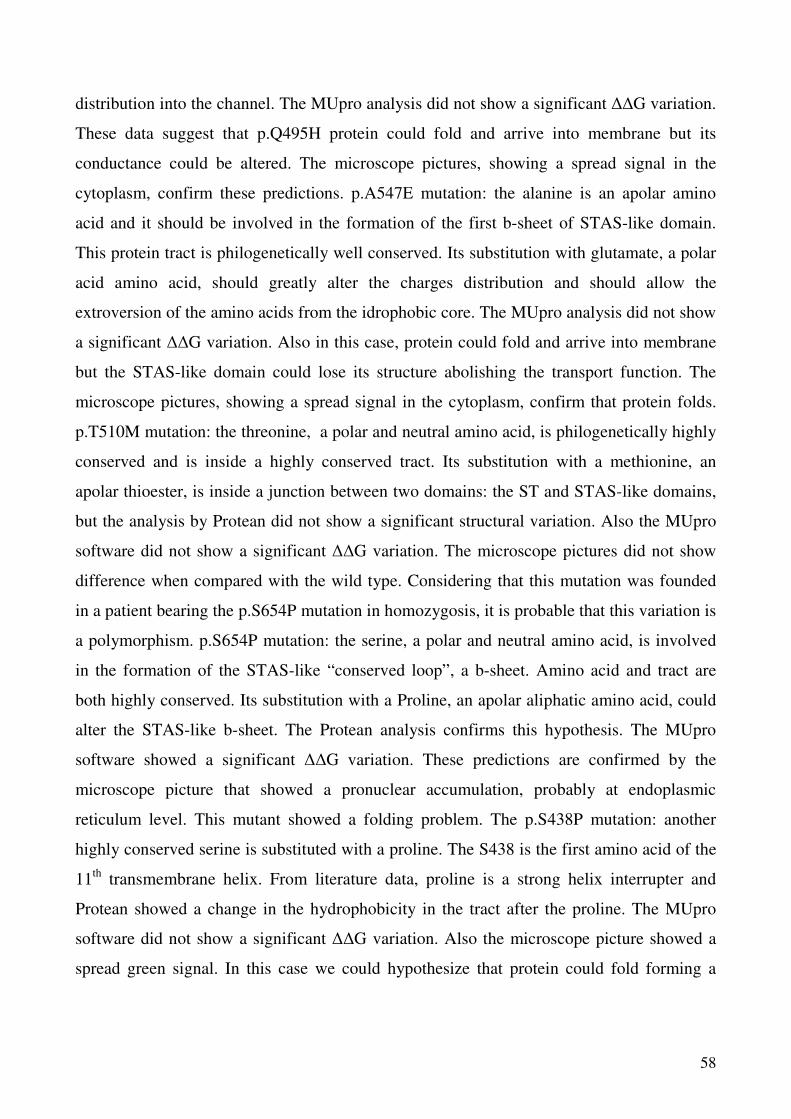
58
distribution into the channel. The MUpro analysis did not show a significant ∆∆G variation.
These data suggest that p.Q495H protein could fold and arrive into membrane but its
conductance could be altered. The microscope pictures, showing a spread signal in the
cytoplasm, confirm these predictions. p.A547E mutation: the alanine is an apolar amino
acid and it should be involved in the formation of the first b-sheet of STAS-like domain.
This protein tract is philogenetically well conserved. Its substitution with glutamate, a polar
acid amino acid, should greatly alter the charges distribution and should allow the
extroversion of the amino acids from the idrophobic core. The MUpro analysis did not show
a significant ∆∆G variation. Also in this case, protein could fold and arrive into membrane
but the STAS-like domain could lose its structure abolishing the transport function. The
microscope pictures, showing a spread signal in the cytoplasm, confirm that protein folds.
p.T510M mutation: the threonine, a polar and neutral amino acid, is philogenetically highly
conserved and is inside a highly conserved tract. Its substitution with a methionine, an
apolar thioester, is inside a junction between two domains: the ST and STAS-like domains,
but the analysis by Protean did not show a significant structural variation. Also the MUpro
software did not show a significant ∆∆G variation. The microscope pictures did not show
difference when compared with the wild type. Considering that this mutation was founded
in a patient bearing the p.S654P mutation in homozygosis, it is probable that this variation is
a polymorphism. p.S654P mutation: the serine, a polar and neutral amino acid, is involved
in the formation of the STAS-like “conserved loop”, a b-sheet. Amino acid and tract are
both highly conserved. Its substitution with a Proline, an apolar aliphatic amino acid, could
alter the STAS-like b-sheet. The Protean analysis confirms this hypothesis. The MUpro
software showed a significant ∆∆G variation. These predictions are confirmed by the
microscope picture that showed a pronuclear accumulation, probably at endoplasmic
reticulum level. This mutant showed a folding problem. The p.S438P mutation: another
highly conserved serine is substituted with a proline. The S438 is the first amino acid of the
11th
transmembrane helix. From literature data, proline is a strong helix interrupter and
Protean showed a change in the hydrophobicity in the tract after the proline. The MUpro
software did not show a significant ∆∆G variation. Also the microscope picture showed a
spread green signal. In this case we could hypothesize that protein could fold forming a

59
shorter 11th
helix: this allow the reaching of membrane but alter the conductance. p.S394I
mutation: another well serine into a well conserved tract of the 9th
transmembrane helix. In
this case the serine is changed with an isoleucine. The Protean analysis showed a reduction
in helix flexibility. The MUpro software did not show a significant ∆∆G variation. On the
basis of predictions and of microscope pictures, we could hypothesize that protein could
fold but its conduction is altered by a more unbending helix. Butyrate correct modifies the
conductance of the protein produced by pQ495H and pA547E.
Study of expression of DRA after butyrate stimulation
The effect of butyrate on DRA/SLC26A3 expression obtained in epithelial nasal cell from
the patients affected by CLD was reported in the Figures 13 and 14.
CONCLUSIVE REMARKS
Our research has made significant advances in the field of possible innovative therapeutic
strategies for Congenital Chloride Diarrhea (CLD). Before this study, the therapeutic
approach to children with CLD was based only on substitutive therapy with NaCl/KCl. This
traditional therapeutic approach is able to limit the risk of severe dehydration but is
ineffective to limit the severity of diarrhea and to improve the quality of life. Our studies
represent a classical model of an epigenetic modulation of intestinal functions. Epigenetics
is the study of inherited changes in phenotype (appearance) or gene expression caused by
mechanisms other than changes in the underlying DNA sequence, hence the name epi-
(Greek: επί- over, above) –genetics.
For the first time we have demonstrated the possibility to act efficiently on the expression
and function of the gene of the CLD limiting severity of diarrhea and fecal ion losses,
through the use of butyrate.
The efficacious collaboration with the others units involved in the research has provided the
possibility to obtain new advances also on the pathophysiology of this condition, on the

60
mechanisms of action of butyrate, and it has pointed out the importance of particular
genotypes in the clinical response to this therapeutic strategy.
We make light on the mechanism underlying the therapeutic effect of butyrate. Butyrate
could be responsible of activation of Cl-/butyrate exchanger activity (Figure 15). It is also
possible that butyrate could reduce miss-trafficking or misfolding of the SLC26A3 protein
(10). Alternatively, butyrate may enhance gene expression (Figure 15): the SLC26A3 gene
contains a 290-bp region between residues -398 and -688 that is crucial for high-level
transcriptional activation induced by butyrate. This might explain the variable response of
CLD patients to butyrate. In fact, depending on the patient's genotype, mutations in the
above-mentioned regulatory regions of the SLC26A3 gene could affect gene transcription
rate. It is also conceivable that other channels are involved in the therapeutic effect of
butyrate in CLD. SLC26A3, like other components of the SLC26 family, interacts with
CFTR. The interaction between CFTR and these components is mediated by binding of the
regulatory domain of CFTR to the STAS domain of SLC26. The interaction is enhanced by
phosphorylation of the regulatory domain by PKA (10) and is modulated by PDZ-binding
scaffold proteins. An important consequence of this interaction is that SLC26 anion
exchange activity is enhanced when CFTR is activated by phosphorylation. Moreover, the
two genes regulate each other, i.e. over expression of SLC26-A3 or -A6 causes up-
regulation of CFTR and vice versa (8). In patch-clamp experiments, PKA-stimulated CFTR
channel activity was six-fold higher in HEK293 cells co-expressing both SLC26 exchanger
and CFTR than in HEK293 cells expressing CFTR alone. Mutations may impair the
interactions between channels and thus reduce the effect of butyrate therapy. Interestingly, it
has recently been demonstrated that butyrate can act by different mechanisms in in vitro
models of cystic fibrosis (CF): i) it can increase the expression of the apical epithelial
membrane of the CFTR; and ii) it can act as a “chaperone-like” molecule, as shown in the
deltaF508del CFTR cell line (49, 50). Similar mechanisms could occur in CLD.
Summary. Congenital chloride diarrhea (CLD-OMIM 214700) is an extremely rare
inherited intestinal electrolyte transport disorder transmitted by an autosomal recessive
fashion due to mutations in the DRA/SLC26A3 gene. Clinical picture is characterized by

61
intestinal sodium and chloride malabsorption that lead to a persistent, life-long severe
diarrhea associated with incontinence and urgency. Thus, the quality of life of these children
is compromised. Unfortunately, oral substitution with NaCl and KCl replaces the intestinal
loss of salts, but it not limits severity of the diarrhea. Sporadic cases treated successfully
with Butyrate have been reported. The aims of the study were: 1) to evaluate the clinical
efficacy of butyrate in children affected by CLD with different genotype; 2) to establish the
mechanism of action of butyrate. Methods. Multicenter (20 Center), open crossover trial
including children with a diagnosis of CLD confirmed by molecular analysis of the
SLC26A3. Oral NaCl/KCl supplementation was administered in all patients. Enrolled
subjects received oral Butyrate (100 mg/Kg/die as 2 doses) for 1 week. Butyrate therapeutic
efficacy was assessed evaluating the daily stool electrolytes content, stool pattern (fecal
volume, number of evacuations, stool consistency evaluated through a scoring system:
normal = 1, loose = 2, liquid = 3), the presence of incontinence (expressed as number of
evacuation associated with incontinence/total daily evacuations), and the electrolytes
concentrations in serum, and urine. These parameters were compared with a baseline data
recorded 1 week before butyrate therapy in all enrolled subjects. Results. Four patients were
enrolled into the trial. During Butyrate therapy we observed a significant (p<0.05)
improvement of the incontinence (44 ± 36% vs. 19 ± 32%) and a reduction of fecal
concentrations of sodium (83 ± 22 mmol/l vs. 24 ± 7 mmol/l) and chloride (147 ± 15 mmol/l
vs. 119 ± 28 mmol/l). Four different mutations were observed in the 4 analyzed patients.
Our studies of mutagenesis and cloning demonstrated that only selected mutation may alter
folding of DRA on the apical membrane of the enterocyte and that butyrate efficacy depend
on the mutation causing diseases. Conclusions. Butyrate reduces the risk of severe
dehydration reducing fecal loss of sodium and chloride, and improves quality of life limiting
the incontinence. We hypothesize that the response to butyrate therapy could be variable
depending on different genetic profile.
Future implications. Studies on the efficacy of the butyrate in congenital chloride diarrhea
have make light on intestinal ions transport mechanisms and may open new therapeutic way
for more common forms of diarrhea occurring in early life. During PhD we also developed a

62
new more palatable formulation of Butyrate. Amidic derivates of butyrate has been obtained
by phenylalanine (Figure 16). This new butyric acids are less hygroscopic, easy to weight,
stable in acid and alkaline conditions. These features are crucial to obtain a good absorption
during intestinal transit. This formulation is odorless and tasteless thus more adaptable to
pediatric use. This new formulation has been patented in Italy (RM 2008, A 000214) and in
other countries (World Intellectual Property Organization n°WO 2009/130735). We tested
in vitro this new formulation obtaining positive effects on ions intestinal transport (Figure
17). Clinical trial to test this new formulation has been planned.

63
Research Area 4.
Prevention of early onset diarrhea
Prevention of infectious diarrhea determined by gastric acidity inhibitors
Results of this research were published on Current Opinion in Gastroenterology 2010;26:31-35
Gastric acid secretion is a philogenetically old function firstly developed about 500 million
years ago (168). Recent years have seen widespread use of potent acid- suppression in the
management of many upper gastrointestinal disorders, also in pediatric patients. The
efficacy of these drugs has continued to improve as more potent acid suppressants have
been introduced, but concerns have been raised with the respect to the effects of gastric
acidity inhibitors (GAI) on host defenses and thus, on the risk of infections. The concept of
“gastric microbicidal barrier” was introduced in 1925, subsequently it has been repeatedly
reported that the suppression of gastric acidity predisposes to infection by a variety of
pathogens, but only recently the increased risk of infection induced by GAI has been
investigated systematically, at clinical and laboratory level (168-170). The following is a
summary of the recent clinical studies performed in adult, children and neonates exploring
the possible association of GAI use with intestinal infections. Updated pathogenetic
hypotheses have also been reviewed.
Evidences from children
Gastric juice consists of HCl and pepsin and can kill bacteria within 15 min when the pH is
less than 3.0. If the pH is raised above 4.0, a state defined as hypochlorhydria, bacterial
overgrowth and infections are more common (169). Different bacterial pathogens have been
reported in the studies investigating the side effects of GAI use. Few studies have focused
on parasitic or fungal infections, and no data are available on viral intestinal infections. The
most investigated associations of selected pathogens with GAI use are reported in Table 20.

64
In this paragraph we will focus on more recent data on GAI-associated infections occurring
during different ages of life. For systematic reviews see references 1-3.
A recent case-control study of risk factors for Salmonella enteritidis in the Netherlands
showed an increased risk of gastroenteritis induced by these pathogens in children taking
GAI (OR 3.6, range 1.9-6.9) (171). A prospective study performed in pediatric patients
showing that the use of GAI is associated with an increased risk of acute gastroenteritis and
community-acquired pneumonia in GERD-affected children has been recently published by
our group (172). We obtained data in 186 subjects from 4 pediatric gastroenterology
centers: 95 healthy controls and 91 GAI users (47 on ranitidine, 44 on omeprazole). The two
groups were comparable for age, sex, weight, length and incidence of acute gastroenteritis
and pneumonia in the 4 month prior to enrollment. Rate of subjects presenting acute
gastroenteritis and community-acquired pneumonia was significantly increased in patients
treated with GAI compared to healthy controls (acute gastroenteritis: 47% vs. 20%,
p=0.001; pneumonia 12% vs. 2%, p=0.03) during the 4 month follow up period. In the GAI
treated group, the rate of subjects presenting acute gastroenteritis (20% vs. 47%, p<0.0001)
and community-acquired pneumonia (3% vs. 12%, p=0.02) was increased when comparing
the 4 month before and after enrollment. No differences were observed between ranitidine
and omeprazole users in acute gastroenteritis and pneumonia incidence in the previous 4
month and during the follow up period. On the contrary, in healthy controls, the incidence
of acute gastroenteritis and pneumonia remained stable. It could be interesting to underline
that we observed an increased incidence of intestinal and respiratory infections in otherwise
healthy children taking GAI for GERD treatment. On the contrary the majority of the
previous data showed that the patients most at risk for pneumonia were those with
significant co-morbid illnesses such as diabetes or immunodeficiency, and this point out to
the importance of gastric acidity suppression as a major risk factor for infections. The effect
on infection susceptibility seems to be sustained even after the end of therapy. We observed
a similar incidence of acute gastroenteritis and pneumonia during the use of GAI drugs and
in the 2 months following the stop of their use, resembling that previous observed in adult
patients (173). How much this effect can last remain to be defined in future studies.
Gastroesophageal reflux disease is commonly clinically diagnosed in children with many

65
nonspecific symptoms and frequently the treatment is empiric. These results could leave
pediatricians with some unanswered questions about how to handle GAI in selected patients
with severe neurological impairment or chronic lung diseases. These subjects appear to be at
increased risk for reflux and aspiration. Protecting these selected patients from aspiration
pneumonia secondary to untreated reflux is probably more convenient that the risk of
infection associated with GAI therapy. At the same time considering the high risk of
inappropriate treatment in children, the pediatrician needs to consider kindly the increased
risk of infection before prescribing a gastric acid blocker.
Evidences from newborn
Many drugs used in neonatal age are either not licensed for the use or are prescribed outside
the terms of their product license (off label prescribing) (174). The use of these drugs in
neonatal intensive care units (NICU) seems to be far greater than other pediatric settings. It
has been recently reported that at least 30% of neonates received treatment with GAI at the
moment of discharge from NICU (175). The European Parliament and the European
Medicines Agency (EMEA), aiming to increase the information available on the use of
medicinal products in pediatric population, indicates as priority safety studies on GAI use in
children and in newborns. These observations are particular relevant for very low birth
weight infants (VLBW) in which infections contribute significantly to morbidity and
mortality. Recently, Soll et al. examined the relationship between postnatal steroid exposure
and H2RA therapy (beginning at 2 weeks of age) and late-onset sepsis in VLBW infants into
a randomized, controlled trial (176). They observed that treatment with dexamethasone and
H2RA was associated with an increased risk of sepsis and meningitis). The increased risk of
infection in infants treated with H2RA before randomization to the study drug (steroid or
placebo) was postulated to be the result of a change in small-bowel colonization after the
development of hypochlorhydria. Similarly, in a prospective observational study, Beck-
Sague et al. reported that 12 (36%) of 33 VLBW neonates who received H2RA developed
bloodstream infections, whereas only 30 (9%) of 343 of those not treated with H2RA
developed bacteremia (177). In logistic-regression analysis, the risk of a bloodstream
infection was independently associated with lower birth weight, respiratory illness at the

66
time of admission, and receipt of H2RA. They postulated that gastric acidity may be a
protective mechanism against respiratory and gastrointestinal tract colonization by
nosocomial pathogens and subsequent bacteremia. More recently it has been observed in a
retrospective study an additional risk of infections and necrotizing enterocolitis (NEC) in
newborn treated with GAI (175). Unfortunately, in VLBW infants, the diagnosis of GERD
or of peptic disease is based on the evaluation of non-specific symptoms, and the empiric
treatment represents frequently the first diagnostic test. In addition, there is no clear
evidence of benefit of the use of H2RA in many clinical conditions typical of neonatal age.
These observations suggest the importance of a more careful use of GAI in these patients, in
particular if other risk factors for severe infections are present.
In this light, we performed a prospective study involving VLBW infants observed in
Neonatal Intensive Care Units (178). Rates of VLBW infants presenting infections were
calculated for both GAI-exposed and GAI-unexposed patients, during 8 weeks of
hospitalization after starting use of GAIs. Study population comprised 274 VLBW infants:
91 exposed to GAI (42 because treatment or prophylaxis of stress-induced peptic disease;
49 because of GERD) and 183 not exposed to GAI, similar for main clinical and
demographic characteristics. Rates of patients presenting infections in GAIs-exposed and
not-exposed were 34 out of 91 (37.4%) and 18 out of 183 (9.8%), respectively (p<0.001).
GAIs users VLBW infants had OR 5.47 (95% CI 2,89-10) fold increased risk of infections
compared with control subjects. Rate of subjects presenting sepsis (23 out of 91 vs. 16 out
of 183), pneumonia (2 out of 91 vs. 1 out of 183) or urinary tract infections (7 out of 91
vs.1 out of 183)and acute gastroenteritis (2 out of 91 vs. None out of 183) was significantly
increased in patients treated with GAIs compared to controls (p<0.0001) during
observational study period (Table 21). In conclusion, gastric acidity-inhibitors therapy is
associated with an increased risk of infections in VLBW infants. This effect seems to be
sustained even after the end of therapy. These results may be attributable to many factors,
including direct inhibitory effect of GA-inhibitors on leucocytes functions and qualitative
and quantitative gastrointestinal microflora modification.

67
CONCLUSIVE REMARKS
The mechanism of the deleterious effects on the risk of infections in subjects receiving GAI
is still not completely elucidated. A number of experimental and clinical evidences
suggested a multifactorial pathogenesis (Figure 18). The preservation of gastric acid
secretion during phylogenesis supports the biological importance of this highly energy
consuming system developed to inactivate ingested microorganisms. Gastric acid is
important in the killing of ingested acid sensitive organisms, in addition elevation in gastric
pH may also have other deleterious effects on gastrointestinal (GI) host defense including
delayed gastric empting , increased bacterial translocation, decreased gastric mucus
viscosity, and changes in the normal microbial flora (169). Impaired gastric acid secretion
has been shown to increase the colonization by several bacterial and parasitic agents. More
recently, two experimental studies of C. difficile infection showed that in a mouse model,
pretreatment with GAI before inoculation with C. difficile resulted in similar rates of
infection, toxin production, and colon injury to a group of mice pre-treated with ampicillin.
In addition spore germination was noted to be favored by elevated pH levels (pH = 6), and
by the presence of potassium chloride and inorganic phosphate. As the proton pumps in the
stomach exchange potassium for hydrogen ions, it is possible that their blockage, besides
resulting in higher gastric pH levels, may also result in increased intraluminal potassium
(169). Direct effects of GAI on several leukocyte functions have been demonstrated,
including decreased adhesion to endothelial cells, reduced bactericidal killing of microbes
and inhibition of neutrophils phagocitosis and phagosome acidification . This could be of
particularly importance in elderly and neonatal age when immunity is still largely immature.
A direct effect of histamine on intestinal immune response to selected pathogens has been
demonstrated in animal model. To test whether inhibition of gastric acid was involved in the
effects of H2 signaling on the mouse response to Y. enterocolitica, Handley et al. (179)
compared the effects of cimetidine with the proton pump inhibitor omeprazole in the orally
infected mouse model. The authors identified no effect of omeprazole under their
conditions, suggesting that gastric acid production is not involved in the effects of
cimetidine on Y. enterocolitica infection, but rather that the H2 antagonism likely plays a
role in regulating the innate immune response to infection at the level of the Payer’s patches

68
(179). Increased production of histamine is important for controlling the infection,
specifically through H2receptor. This increase in histamine could stimulate a significant
influx of mast cells or basophiles. After stimulation, the histamine, upon cellular release,
would bind to cells expressing H2. Activation of H2 has been shown to have a variety of
effects including altering the production of inflammatory cytokines and disrupting the Th1–
Th2 balance during the immune response. The Th1–Th2 balance is known to be important
for controlling such infection. A negative influence on the immune system could be the
consequence also of the modification of quantitative and qualitative composition of
intestinal microflora (179). A study of the numbers and type of bacteria in nasogastric tubes
of patients receiving GAI demonstrated an increased numbers of bacteria including beta
hemolytic Streptococcus, a known cause of community-acquired pneumonia (173).
Considering the information on the cross-talk between intestinal microflora and immune
system functions it is important that this aspect should be investigated in a more incisive
manner in the future. Concomitant risk factors including pre-existence of chronic diseases,
hospitalization, and antibiotic use could contribute to an increased risk of infections in
patients taking GAI (Figure 18). Finally, it has been demonstrated that when PPI are
prescribed to patients with H.pylori infection, the acid inhibition is more profound than in
patients without H.pylori infection (180). Patients with H.pylori infection may therefore be
at greater risk for infection.
Summary. Many studies and systematic reviews demonstrate an increased risk of bacterial
infection in adults taking acid suppressors. Little evidence is derived from the early in the
life period. The use of gastric acidity inhibitors has been associated with systemic
infections and necrotizing enterocolitis in preterm infants. Reduced gastric acidity, delayed
gastric emptying, increased gastric mucus viscosity, modification in microbiota, and
impairment of neutrophils functions, are all conditions determined by gastric acidity
blockers that potentially lead to an increased risk of gastrointestinal infections. A proper
utilization of these drugs, particularly for patients at high risk, is imperative in order to
reduce deleterious effects on infection risk and to optimize cost-effectiveness ratio.

69
Future implications. The GAI use accounts for significant cost expenditure in Western
Countries including over-the-counter and prescription formulations (170,181). With the
increasing use of GAI, the demonstration of an association with an increased risk of
intestinal and extraintestinal infections, and increasing concerns of multiresistant pathogens,
public and professional education is needed to stress the importance of appropriate use of
anti-secretary drugs. Because no drug is without side-effects, physicians have to satisfy
themselves that the benefits of treatment outweigh the potential risks. Further
pharmacovigilance studies exploring all the possible variables influencing the impact of
GAI therapy on the risk of infections are necessary, together with more basic and clinical
investigations on the mechanisms of these GAI-related problems and on possible
prophylactic measures to prevent them.

70
Primary prevention of food-induced diarrhea
Results of this research were published on Pediatr Allergy Immunol2010; 21:889-91 Web link
The prevalence of atopic manifestations has increased worldwide, especially in children
(182-184). It is now recognized that early childhood events, including diet, are probably
important in the development of atopic diseases. Various preventive measures have been
proposed including definition of high-risk infants, maternal dietary restrictions during
pregnancy, breastfeeding, dietary restriction while breastfeeding, the use of hypoallergenic
formulas, and delays in the introduction of certain foods into the infant’s diet (185). Family
history is the best indicator for population screening (185). Current evidence does not
support a major role for maternal dietary restrictions during pregnancy or lactation but there
is evidence that breastfeeding for at least 4 months, compared with feeding formula made
with intact cow’s milk protein, prevents or delays the occurrence of atopic dermatitis, food
allergy, and wheezing in early childhood (186). Data suggest for infants at high risk of
atopy and who were not exclusively breastfed for 4–6 months that the onset of atopic
disease may be delayed or prevented by hydrolyzed formulas compared with formula made
with intact cow’s milk protein, particularly for atopic dermatitis (18,186). The Committees
on Nutrition and Section on Allergy and immunology of American Academy of Pediatrics
(AAP) has recently published recommendations on early nutritional interventions on the
development of atopic diseases in infants and children (18). Recommendations for primary
prevention of atopy disorders should be applied early in the perinatal period to have a
chance of success. The first care givers in contact with high-risk subjects are the
neonatologists, but little is known about their awareness of and adherence to dietary
management for the prevention of atopic diseases. Thus, we assessed adherence to the
dietary management recommendations of the AAP for the prevention of atopic diseases in
neonatal age through an audit study.

71
STUDY DESIGN
We invited by e-mail, the chiefs of 30 maternity units (MU) of the national public health
system with more than 1500 live births/yr operating in South Italy to report the policy
applied in their MU and to complete a questionnaire constituted by three simple items: (i)
identification of high risk of developing allergy newborns, (ii) dietary restrictions for
lactating women, (iii) use of hypoallergenic formula for bottle-fed high-risk infants.
RESULTS
Thirty MU were invited to participate representative of about 135,000 newborn/yr. Twenty-
two MU returned the questionnaire. Results are reported in Table 22. Only 2/22 MU has a
policy in complete agreement with the early nutritional intervention on development of
atopic disease proposed by the AAP (18).
CONCLUSIVE REMARKS
Our study suggests scarce knowledge and adherence to dietary recommendations for
primary prevention of atopic disease in neonatology clinical practice. These results could be
influenced by a suboptimal awareness of atopic disorders by neonatologists and by the
belief of little clinical benefit that these recommendations could obtain. However,
considering the dramatic increase of incidence and prevalence of atopic disorders and the
possibility to prevent these conditions by, at least in part, a nutritional approach, we believe
that an increased awareness of the matter by neonatologists is important. This audit
highlights the need for an awareness campaign to promote the use of primary prevention
strategies for atopic disease among neonatologists.
Summary. The prevalence and severity of atopic manifestations in children are increasing
in western countries in the last decades. Specific nutritional intervention may prevent or
delay the onset of atopic diseases in infants at high risk of developing allergy. These
nutritional interventions should be applied early in the perinatal period to have a chance of

72
success. Thus, we assessed adherence to the dietary management recommendations of the
Committee on Nutrition and Section on Allergy and Immunology of the American Academy
of Pediatrics (AAP) for the prevention of atopic diseases in neonatal age through an audit
study. Questionnaire was administered to the chiefs of 30 maternity units (MU) with more
than 1500 live births/yr to report the policy applied in their MU. Twenty-twoMU returned
the questionnaire. Identification of high-risk newborns was routinely performed only in 7/22
MU (31.8%). High-risk newborns were identified by the presence of at least two or one
first-degree relative (parent or sibling) with documented allergic disease by 18.2% and
45.5% of MU, respectively. Specific maternal dietary restrictions during lactation were
adopted in 7/22 MU (31.8%).Extensively or partially hydrolyzed formula was prescribed for
bottle-fed high-risk infants in 22.7% of MU. Only 2/22 MU have a policy in complete
agreement with the nutritional intervention proposed by the AAP. Our studies suggest a
poor adherence to dietary recommendations for primary prevention of atopic disease in
neonatology clinical practice. Further efforts should be planned to improve the knowledge
and the application of these preventive strategies.
Future implications. In recent years, much attention has been given to oral tolerance and
gut flora. Oral tolerance mechanisms develop postnatal mainly in response to the gut flora
and activation of specific Toll-like receptors on regulatory cells. The pivotal role of the
luminal bacteria is highlighted by the impaired tolerance in germ-free mice, the different
intestinal flora in populations that will later develop atopy, the immunomodulatory
properties of specific probiotics, and the promising results of interventional studies. Allergic
infants showed, even before the appearance of symptoms, a significant higher prevalence of
Clostridia, coliforms, and Staphylococcus aureus versus lactobacilli and bifidobacteria
(bifidum). Manipulation of the gut flora as early as in the first days or months of life may
influence subsequent colonization and expression of regulatory cytokines through
microenvironmental modification and competition. Specific probiotics, including LGG, may
induce production of anti-inflammatory IL-10 and transforming growth factor, inducing a
tolerogenic effect before sensitization occurs. According to two trials, using
supplementation of LGG or E. coli in the perinatal period may reduce some non-IgE-

73
mediated allergic manifestations. Supplementation of a cow’s milk-based formula with
prebiotics has a bifidogenic effect, but a prospective beneficial effect on food allergy has up
to now only been suggested with a specific prebiotics mixture.

74
During the PhD project, using a bench to bedside approach, several progresses in the field
of diarrhea occurring in early life have been obtained. Epidemiology and etiology of
diarrhea have been clarified, new therapeutic and preventive approaches have been
developed for control of acute and chronic diarrhea occurring in early life. In addition, a
novel model for the study of congenital diarrheal disease has been set. This model could be
usefull to reduce the distance from in vitro to in vivo research on these rare conditions, and
to identify new therapeutic targets for more common diarrheal disease occurring early in
life.
The main future target was the intestinal ecosystem (i.e., epithelium, immunity, microflora,
nutrients), a dynamic network that could be modulated by different therapeutic and
nutritional approaches. Epigenetic modulation of gene expression in early life elicited by
nutrients, in order to modify the natural course of the disease, represents the new research
frontier.
Focusing on prompt identification and control of diarrheal diseases in early life, PhD project
has outlined innovative approaches usefull to reduce immediate complications and influence
body health in subsequent age of life.
CONCLUSIONS

75
Table 1. Etiology of diarrhea in early life
Acute Chronic Overall
Number of patients 36 3 39
Unknown etiology 10 - 10
Cow’s milk allergy 8 - 8
Infections 7 - 7
Rotavirus 4 -
Salmonella parathyphi 1 -
Shigella flexneri 1 -
Enterotoxigenic E.coli 1 -
Antibiotics-associated diarrhea 5 - 5
Neonatal withdrawal syndrome 2 - 2
Congenital diarrheal disorders - 2 2
Glucose-galactose malabsorption - 1 1
Congenital chloride diarrhea - 1 1
Parenteral diarrhea
Urinary tract infection 1 - 1
Hirschsprung’s disease 1 - 1
Cystic fibrosis - 1 1
Immunodeficiency
Adenosine deaminase deficiency 1 - 1
Metabolic disorders
Urea cycle defect 1 - 1

76
Table 2. Main demographic and anamnestic data of the subjects with diarrhea in early life
Acute Chronic
Demographic data
Number of patients 36 3
Demographic data
Male, n (%) 15 (41.7) 1 (33.3)
Birth weight, g 2504 ± 778 2560 ± 664
Gestational age, weeks 36.4 ± 3.8 37.7 ± 1.5
Age at diarrhea onset, d 8.3 ± 6.6 5.7 ± 4.4
Age at hospitalization, d* 6.2 ± 8.5 19.7 ± 10.1
Anamnestic data
Intrauterine growth restriction, n (%) 6 (16.7) 1 (33.3)
Polyhydramnios, n (%)* 1 (2.8) 2 (66.7)
Premature birth, n (%) 12 (33.3) 1 (33.3)
Familiarity for chronic diarrhea, n (%)* 1 (2.8) 2 (66.7)
Newborns at high risk of developing allergy, n (%) 12 (33.3) -
Exclusive breast milk, n (%) 3 (8.33) -
Data are expressed as mean ± Standard Deviation (SD) when not specified.
*p<0.05

77
Table 3. Symptoms associated with diarrhea occurring early in life
Data are expressed as mean ± Standard Deviation (SD) when not specified.
*p<0.05
Acute Chronic
Gastrointestinal, n (%)
Vomiting 18 (50.0) 1 (33.3)
Mucus in the stools 13 (36.1) 1 (33.3)
Blood in the stools 9 (25.0) -
Abdominal distension 24 (66.7) 2 (66.7)
Systemic, n (%)
Fever 4 (11.1) -
Hypovolemia* 1 (2.8) 1 (33.3)
Eczema 7 (19.4) -
Seizures* 3 (8.3) 1 (33.3)
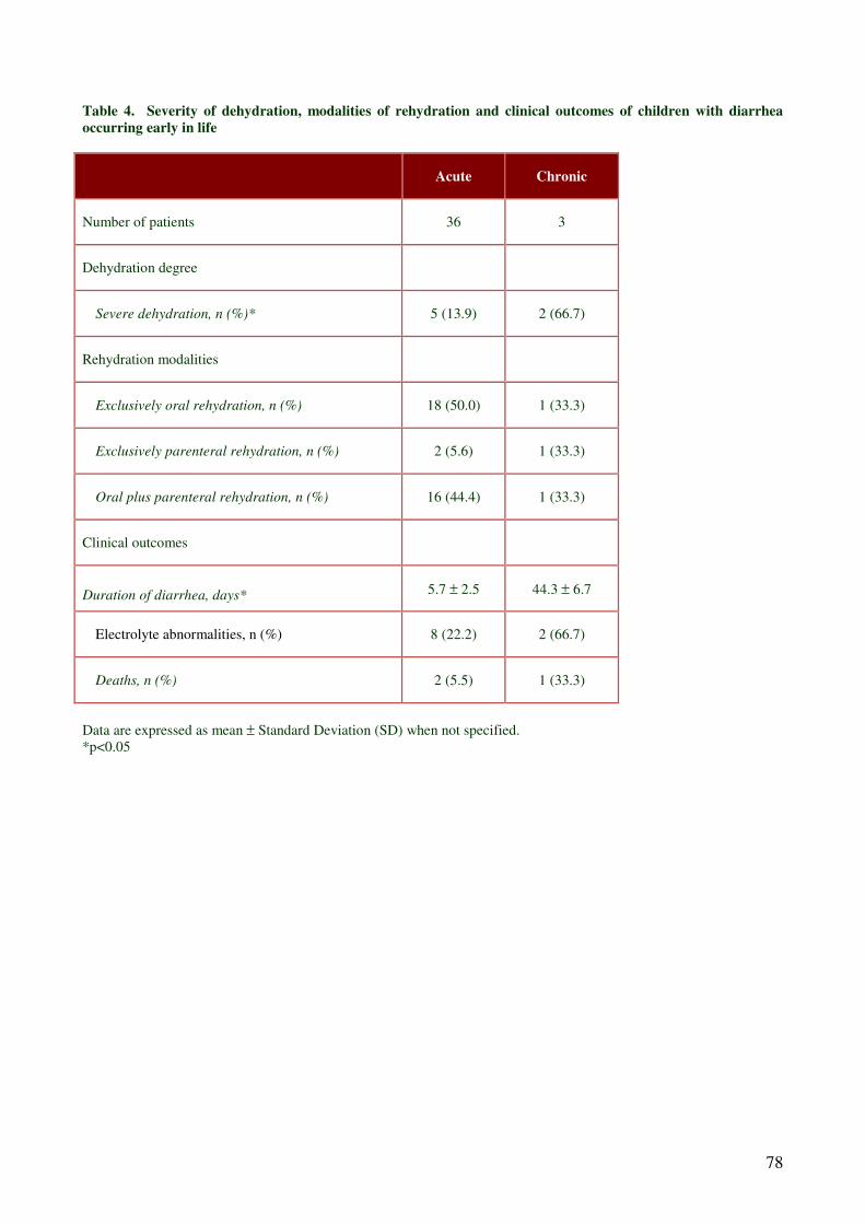
78
Table 4. Severity of dehydration, modalities of rehydration and clinical outcomes of children with diarrhea
occurring early in life
Acute Chronic
Number of patients 36 3
Dehydration degree
Severe dehydration, n (%)* 5 (13.9) 2 (66.7)
Rehydration modalities
Exclusively oral rehydration, n (%) 18 (50.0) 1 (33.3)
Exclusively parenteral rehydration, n (%) 2 (5.6) 1 (33.3)
Oral plus parenteral rehydration, n (%) 16 (44.4) 1 (33.3)
Clinical outcomes
Duration of diarrhea, days* 5.7 ± 2.5 44.3 ± 6.7
Electrolyte abnormalities, n (%) 8 (22.2) 2 (66.7)
Deaths, n (%) 2 (5.5) 1 (33.3)
Data are expressed as mean ± Standard Deviation (SD) when not specified.
*p<0.05

79
Table 5. Subjects with intestinal failure in early life
Pt GA (w) BW (Kg) Sex Causes of intestinal failure Age at the
onset (d)
Follow-up
length (m)
1 34 2.4 F Abdominal wall defect 0 40
2 38 2.2 M Abdominal wall defect 0 25
3 26 0.8 M Congenital CMV infection 7 27
4 36 2.5 M Congenital neuromuscular disease 5 33
5 34 2.1 F Intestinal atresia 0 38
6 35 2.1 M Intestinal atresia 0 41
7 34 1.9 F Intestinal atresia 0 36
8 36 2.2 F Intestinal atresia 0 43
9 35 2.1 M Intestinal atresia 0 33
10 30 0.9 M Intestinal atresia 0 42
11 37 2.5 F Intestinal atresia 0 26
12 26 0.9 F Intestinal atresia 0 40
13 26 0.9 M Intestinal atresia 0 27
14 27 0.6 F Intestinal obstruction 0 36
15 33 1.8 F Intestinal obstruction 0 28
16 26 0.6 F Meconium peritonitis 0 34
17 36 4.4 F Megacystis Microcolon
Hypoperistalsis syndrome 0 48
18 31 1.4 F Necrotizing enterocolitis 12 48
19 27 0.9 F Necrotizing enterocolitis 27 33
20 33 2.2 M Necrotizing enterocolitis 10 50
21 28 0.6 F Necrotizing enterocolitis 0 44
22 30 1.8 F Necrotizing enterocolitis 0 34
23 30 1.3 M Necrotizing enterocolitis 20 35
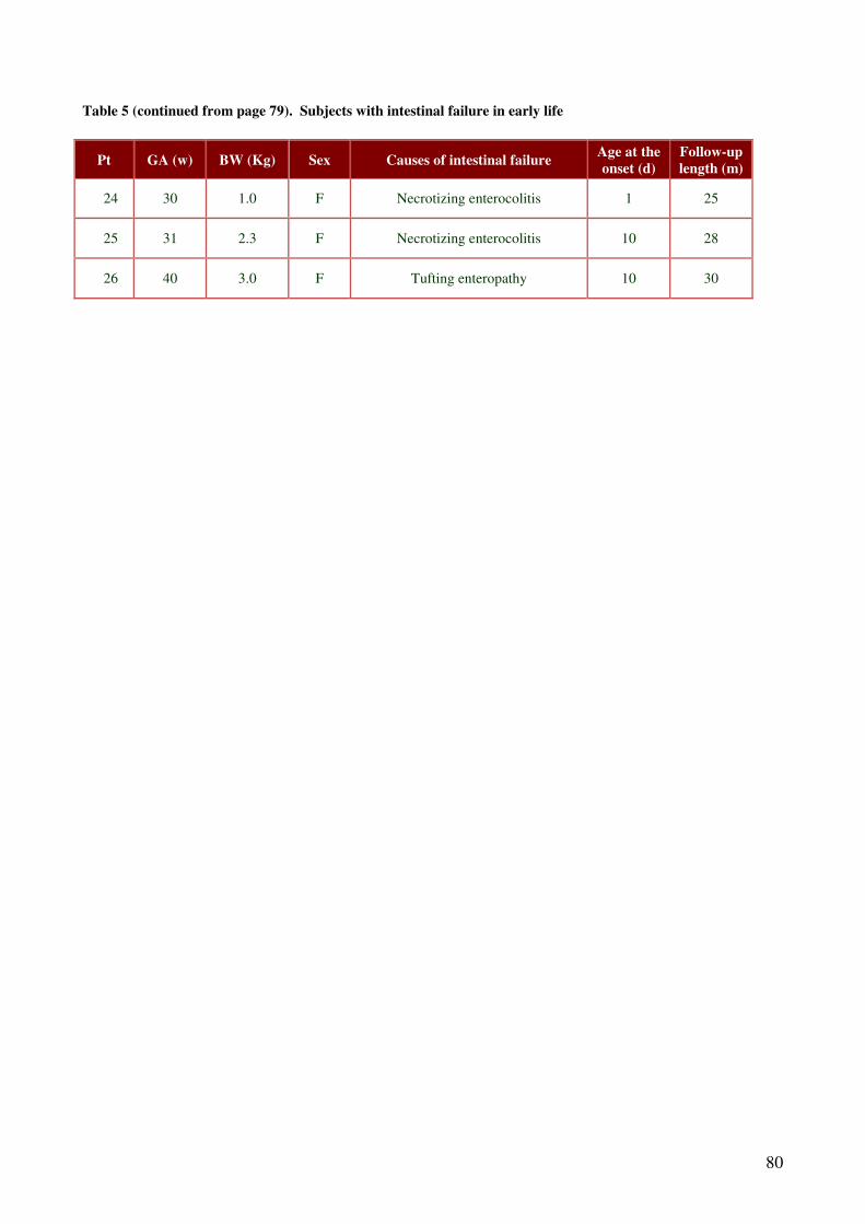
80
Table 5 (continued from page 79). Subjects with intestinal failure in early life
Pt GA (w) BW (Kg) Sex Causes of intestinal failure Age at the
onset (d)
Follow-up
length (m)
24 30 1.0 F Necrotizing enterocolitis 1 25
25 31 2.3 F Necrotizing enterocolitis 10 28
26 40 3.0 F Tufting enteropathy 10 30

81
Table 6. Natural history of subjects with intestinal failure occurring early in life
Cause % Successfully PN (%) Duration of PN (d) Death (%)
Congenital intestinal defects 42 91 50 9
Necrotizing enterocolitis 31 100 59
Motility disorders 11 33 445 33*
Intestinal obstruction 8 100 60
Ultrastructural defects 4 Waiting for IT* 920
Meconium peritonitis 4 100 65
*Due to complications of IT (intestinal transplantation)

82
Table 7. Main features of subjects dependent from parenteral nutrition treated with 2 different nutritional
strategy
Group 1
(Total PN)
Group 2
(PN + MEF)
Male, n (%) 26 (53.1) 21 (43.8)
Birth weight, g (IQR) 1100 (415) 1095 (405)
Gestational age, weeks (IQR) 29 (3) 29 (3)
CRIB score 1 (2) 1 (2)
Age at the first episode of feeding intolerance, d (IQR) 6 (8) 7 (7)
Note. Data expressed as Median (IQR) when not specified.
Abbreviations. PN: parenteral nutrition MEF: minimal enteral feeding. IQR: interquartile range. CRIB: critical
respiratory index for babies.

83
Table 8. Risk factors associated with NEC development in subjects dependent from parenteral nutrition treated
with 2 different nutritional strategy
Group 1
(Total PN)
Group 2
(PN + MEF)
Time to start enteral nutrition, h 11 (4) 8 (2)
Umbilical catheter, n (%) 38 (78) 38 (79)
Patent ductus arteriosus, n (%) 7 (14) 8 (17)
Intraventricular hemorrhage stage III-IV, n (%) 7 (14) 7 (15)
BM/total enteral feeding at day 14 of life 0.4 (0.3) 0.5 (0.3)
BM/total enteral feeding at discharge 0.3 (0.2) 0.3 (0.2)
Note. Data expressed as Median (IQR) when not specified.
Abbreviations. NEC: necrotizing enterocolitis. PN: parenteral nutrition. MEF: minimal enteral feeding. BM: breast
milk. IQR: interquartile range.

84
Table 9. Time to reach full enteral feeding, occurrence of NEC and sepsis in subjects dependent from parenteral
nutrition treated with 2 different nutritional strategy
Group 1
(Total PN)
Group 2
(PN + MEF) p
Patients with NEC, n (%) 1 (2) 1 (2.1) 0.747
Central vascular access duration, d 12 (5) 7 (4) <0.001
Time to reach full enteral feeding, d 11 (5) 8 (5) <.0001
Patients with ≥ 2 episodes of fed intolerance, n (%) 14 (28.6) 14 (29.2) 0.948
Patients with late onset sepsis, n (%) 17 (34.7) 8 (16.6) 0.047
Time to regained birth weight, d 12 (4) 9 (1.5) <.0001
Death, n (%) 2 (4.1) 3 (6.3) 0.490
Note. Data expressed as Median (IQR) when not specified.
Abbreviations. NBE: nothing by enteral route. MEF: minimal enteral feeding. NEC: necrotizing enterocolitis. IQR:
interquartile. range.

85
Table 10. Composition of the two oral rehydration solution used in a comparative study involving children with
acute diarrhea occurring in early life
Standard ORS Super ORS
Commercial brand name Reidrax® Prereid®
Assigned group Group 1 Group 2
Osmolarity (mOsm/L) 225 200
Na+
(mmol/L) 60 50
K+ (mmol/L) 20 20
Cl- (mmol/L) 60 40
Glucose (mmol/L) 75 77
Citrate (mmol/L) 10 10
Zn2+
(mmol/L) 0 1
Fructooligosaccharides (g/L) 0 0.35
Xilooligosaccharides (g/L) 0 0.35
Note. Reidrax is a brand name of the EG SpA, Milan Italy. Prereid is a
brand name of the Milte Italia SpA, Milan, Italy

86
Table 11. Comparative study on ORS involving children with acute diarrhea in early life: baseline main
demographic and clinical characteristics
Group 1 Group 2 p
N. 60 59 -
Age, m* 18.58 (15.5-21.6) 19.26 (15.9-22.6) 0.765
Body weight, kg* 10.68 (9.79-11.58) 11.25 (9.79-12.72) 0.474
Male, n (%) 34 (56.7) 36 (61.0) 0.630
Duration of symptoms before treatment, h* 9.0 (8.3-9.9) 10.0 (9.3-10.8) 0.083
Presence of vomiting, n (%) 14 (23.3) 22 (37.3) 0.098
*Mean (95% confidence interval) when not specified

87
Table 12. Comparative study on probiotics involving children with acute diarrhea in early life: microorganism
load, administration, and main characteristics of preparations
Groups Microorganisms Strains Dosage
2 Lactobacillus casei Rhamnosus GG 6 x 10⁹ CFU/dose (bid)
3 Saccharomyces boulardii Sb It 5 x 10
9 live micro-
organisms/dose (bid)
4 Bacillus clausii O/C84, N/R84, T84,
SIN84 10
9 CFU/dose (bid)
5
L. delbrueckii var. bulgaricus +
L. acidophilus +
S. thermophilus +
B. bifidum
LMG-P17550 + LMG-P
17549 + LMG-P 17503
+ LMG-P 17500
109 CFU+
109 CFU+
109 CFU+
5 x 10⁸ CFU/dose (bid)
6 Enterococcus faecium SF 68 7.5 x 10⁷ CFU/dose (bid)

88
Table 13. Comparative study on probiotics involving children with acute diarrhea: Baseline features
Groups 1
(n=92)
2
(n=100)
3
(n=91)
4
(n=100)
5
(n=97)
6
(n=91)
Male (%) 41 (44%) 60 (60%) 44 (48%) 49 (49%) 49 (50%) 39 (42%)
Median age in months (IQR) 17.0 (17.2) 20.0 (12.0) 18.0 (17.0) 19.0 (14.0) 16.5 (18.5) 15.0 (14.0)
Median weight in grams (IQR) 11925
(2805.0)
12100
(4072.5)
11450
(4700.0)
12300
(4000.0)
12700
(4625.0)
12234
(3150.0)
Feeding (%)
Breast milk
Formula
Cow’s milk
13.5
55.8
30.8
11.7
55.0
33.3
13.3
55.6
31.1
10.0
60.2
29.8
13.4
46.2
40.5
17.9
43.6
38.5
Median diarrhoea duration
before treatment in hours (IQR) 9 (9.0) 10 (13.0) 11 (12.0) 10 (11.0) 9 (12.0) 10 (11.0)
IQR: Interquartile range.

89
Table 14. Comparative study on probiotics involving children with acute diarrhea: duration of diarrhoea (in
hours) in the study groups
Groups Treatment Median
hours IQR
Estimated difference
(95% CI)* P value †
1 ORS alone 115 35 — —
2 Lactobacillus casei subsp.
rhamnosus GG 78 46 -32 (-41 to -24) <0.001
3 Saccharomyces boulardii 103 43 -6 (-15 to 4) 0.262
4 Bacillus clausii 118 33 3 (-3 to 11) 0.311
5
L. delbrueckii var. bulgaricus +
L. acidophilus +
S. thermophilus +
B. bifidum
69 50 -38 (-47 to -27) <0.001
6 Enterococcus faecium SF 68 115 41 2 (-7 to 11) 0.670
IQR: Interquartile range.
* vs. ORS alone.
† Kruskal-Wallis H test with Dunn test for multiple comparisons. P vs. ORS alone
90
Table 15. Comparative study on probiotics involving children with acute diarrhea: daily stool outputs
Group Treatment Day
1 2 3 4 5 6 7
1 ORS alone 5 (2) 5 (3) 4 (2) 4 (2) 3 (2) 2 (1) 2 (2)
2
Lactobacillus
casei subsp.
rhamnosus GG
Estimated
difference (95%
CI)
6 (2)
—
4 (2)*
-1 (-1 to 0)
3 (2) †
-1 (-1 to 0)
3 (2)*
-1(-1 to -1)
2 (1) †
-1 (-1 to 0)
2 (1)
—
2 (1)
—
3 Saccharomyces
boulardii 5(2) 5 (3) 4 (2.25) 4 (2) 3 (2) 2 (1) 2 (2)
4 Bacillus clausii 6 (2) 5 (3) 4 (2) 4 (2) 3 (2) 2 (1) 2 (1)
5
L. delbrueckii
var. bulgaricus +
L. acidophilus +
S. thermophilus +
B. bifidum
Estimated
difference (95%
CI)
6 (2)
—
4 (2)*
-1 (-1 to 0)
3 (1.50)*
-1 (-1 to 0)
3 (2)*
-1(-1 to -1)
2 (1) †
-1 (-1 to 0)
2 (1)
—
2 (1)
—
6 Enterococcus
faecium SF 68 5 (2) 5 (2.75) 4 (2) 4 (2) 3 (2) 2 (1) 2 (1)
Day 1 is the day of the first probiotic administration.
Data are expressed as median (IQR, interquartile range).
*P < 0.001 or †P=0.001 vs. ORS alone at the same time point by Kruskal-Wallis H test with Dunn test for multiple
comparisons.
The estimated differences are vs. ORS alone.

91
Table 16. Comparative study on probiotics involving children with acute diarrhea: daily stool consistency score
Groups Treatment Day
1 2 3 4 5 6 7
1 ORS alone 3 (0) 3 (1) 2 (1) 2 (2) 2 (1) 1 (0) 1 (0)
2
Lactobacillus
casei subsp.
rhamnosus GG
Estimated
difference
(95% CI)
3 (0)
—
2 (0.50)*
0 (-1 to
0)
1 (1)*
-1 (-1 to
0)
1 (1)*
-1 (-1 to
0)
1 (0)*
0 (-1 to
0)
1 (0)
—
1 (0)
—
3 Saccharomyces
boulardii 3 (0) 3 (1) 2 (1) 2 (1) 2 (1) 1 (0.25) 1 (0)
4 Bacillus clausii 3 (0) 3 (1) 2 (1) 2 (1) 2 (1) 1 (1) 1 (0)
5
L. delbrueckii
var bulgaricus
+
L. acidophilus +
S. thermophilus
+
B. bifidum
Estimated
difference
(95% CI)
3 (0)
—
2 (1) †
0 (-1 to
0)
1 (1)*
-1 (-1 to
0)
1 (1)*
-1 (-1 to
0)
1 (0)*
0 (-1 to
0)
1 (0)
—
1 (0)
—
6 Enterococcus
faecium SF 68 3 (0) 3 (1) 2 (1) 2 (1.75) 2 (1) 1 (0) 1 (0)
Day 1 is the day of the first probiotic administration.
Stool consistency score system (see ref. 15): 1, normal; 2, loose; 3, semi liquid; 4, liquid.
Data are expressed as median (IQR, interquartile range).
*P < 0.001 or †P=0.001 vs. ORS alone at the same time point by Kruskal-Wallis H test with Dunn test for multiple
comparisons.
The estimated differences are vs. ORS alone.

92
Table 17a. Molecular basis of the main forms of congenital diarrheal diseases: defects of digestion, absorption
and transport of nutrients and electrolytes
Disease Gene Location Function
Disaccharidase deficiency
Congenital lactase deficiency LCT 2q21 Lactase-phlorizin hydrolase
activity
Sucrase-isomaltase deficiency EC 3.2.1.48 3q25-q26 Isomaltase-sucrase
Maltase-glucoamylase deficiency MGAM 7q34 Maltase-glucoamylase activity
Ion and nutrient transport defects
Glucose-galactose malabsorption SGLT1 22q13.1 Na+/glucose cotransporter
Fructose malabsorption GLUT5 1p36 Fructose transporter
Fanconi-Bickel syndrome GLUT2 3q26 Basolateral glucose transporter
Cystic fibrosis CFTR 7q31.2 cAMP-dependent Cl- channel
Acrodermatitis enteropatica SLC39A4 8q24.3 Zn2+
transporter
Congenital chloride diarrhea DRA 7q22-q31.1 Cl-/base exchanger
Congenital sodium diarrhea SPINT2* 19q13.1 Serine-protease inhibitor
Lysinuric protein intolerance SLC7A7 14q11 Hydrolyzes endo-/exopeptidases
AA basolateral transport
Congenital bile acid diarrhea ABAT 13q3 Ileal Na+/ bile salt transporter
Pancreatic insufficiency
Enterokinase deficiency PRSS7 21q21 Proenterokinase
Trypsinogen deficiency PRSS1 7q35 Trypsinogen synthesis
Pancreatic lipase deficiency PNLIP 10q26.1 Hydrolyzes triglycerides to fatty
acids
Lipid trafficking
Abetalipoproteinemia MTP 4q22 Transfer lipids to apolipoprotein
B
Hypobetalipoproteinemia APOB 2p24 Apolipoprotein that forms
chylomicrons
Chylomicron retention disease SAR1B 5q31.1 Intracellular chylomicron
trafficking
*This mutation has been associated to the syndromatic form of congenital sodium diarrhea.

93
Table 17b. Molecular basis of the main forms of congenital diarrheal diseases: defects of enterocyte
differentiation and polarization
Disease Gene Location Function
Microvillous inclusion disease EpCAM 18q21 Intracellular protein trafficking
Congenital tufting enteropathy Unknown 2p21 Cell-cell interaction
Syndromic diarrhea Unknown Unknown Unknown
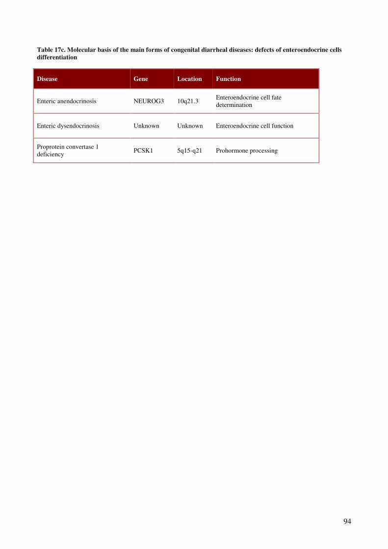
94
Table 17c. Molecular basis of the main forms of congenital diarrheal diseases: defects of enteroendocrine cells
differentiation
Disease Gene Location Function
Enteric anendocrinosis NEUROG3 10q21.3 Enteroendocrine cell fate
determination
Enteric dysendocrinosis Unknown Unknown Enteroendocrine cell function
Proprotein convertase 1
deficiency PCSK1 5q15-q21 Prohormone processing

95
Table 17d. Molecular basis of the main forms of congenital diarrheal diseases: defects of modulation of intestinal
immune response.
Disease Gene Location Function
IPEX (Immunodysregulation,
polyendocrinopathy, enteropathy, X-linked)
syndrome
FOXP3 Xp11.23-q13.3 Transcription factor
IPEX-like syndrome Unknown Unknown Unknown
Immunodeficiency-associated autoimmune
enteropathy Unknown Unknown Unknown
Autoimmune polyglandular syndrome-1
(APS-1) AIRE 21p22.3
Regulation gene
transcription
Autoimmune enteropathy with colitis-
generalized autoimmune gut disorder
(GAGD)
Unknown Unknown Unknown

96
Table 18. Baseline demographic and clinical characteristics of children with Congenital Chloride Diarrhea
Number 22
Age at diagnosis, month 39 ± 42
Fecal volume, ml/Kg/day 42.3 ± 19.3
Evacuations, n/day 3.6 ± 1.8
Fecal Na concentration, mmol/l 83 ± 22
Fecal Cl concentration, mmol/l 147 ± 15
Data are expressed as Mean ± SD when not specified

97
Table 19. Molecular basis and protein functions of enrolled patients with congenital chloride diarrhea
Pts Mutation Nucleotide Amino Acid Exon Protein
Product Hypothesis on DRA function
1
Missense c.1960T>C p.S654P 17 Unfolded
protein Protein retained in ER
Missense c.1529C>T p.T510M 14 Folded
protein Decreased conductance
2 Missense c.1312T>C p.S438P 12
Folded
protein Decreased conductance
Frameshift c.614delT p.L205RfsX28 6 Truncated No function
3
Missense c.1484A>C p.Q495H 13 Folded
protein Decreased conductance
Missense c.1640C>A p.A547E 15 Folded
protein Decreased conductance
4
Missense c.1181G>T p.S394I 10 Folded
protein Decreased conductance
Missense c.1181G>T p.S394I 10 Folded
protein Decreased conductance
5 Nonsense c.2132T>G p.L711X 19 Unfolded
protein Protein retained in ER
6 Nonsense c.559G>T p.G187X 5 Truncated No function
7 Nonsense c.559G>T p.G187X 5 Truncated No function
8 Nonsense c.559G>T p.G187X 5 Truncated No function
9 Nonsense c.559G>T p.G187X 5 Truncated No function
10 Nonsense c.559G>T p.G187X 5 Truncated No function
11 Nonsense c.559G>T p.G187X 5 Truncated No function
12 Nonsense c.559G>T p.G187X 5 Truncated No function
13 Nonsense c.559G>T p.G187X 5 Truncated No function
14
Frameshift c.614delT p.L205RfsX28 6 Truncated No function
Missense c.358G>A p.G120S 4 Not
determined Not determined
15 Frameshift c.1758delG p.L586FfsX4 16 Truncated No function
16 Exon
Deletion
c.2008-
151_2061+
1546del
Not
predictable 18 Truncated No function

98
Table 19 (continued from page 97) . Molecular basis and protein functions of enrolled patients with congenital
chloride diarrhea
Pts Mutation Nucleotide Amino Acid Exon Protein Product Hypothesis on DRA function
17
Abolished
Splicing
Acceptor
site
c.1408-
1G>C
Not
predictable 13 Truncated No function
18
Abolished
Splicing
Acceptor
site
c.1312-
1G>T
Not
predictable 12 Truncated No function
19 Frameshift c.2022_202
4dup p. I675dup 18 Unfolded protein* Protein retained in ER*
20 Missense c.1522T>C p.C508R 14 Not tested Not tested
Nonsense c.1735C>T p.R579X 16 Unfolded protein Protein retained in ER
21 Exon
Deletion Unknown Unknown
Unkn
own Unfolded protein Protein retained in ER
22 Macrodeleti
on Unknown Unknown
Unkn
own Not predictable Not predictable

99
Table 20. Pathogens possibly responsible for gastrointestinal infections in patients treated with gastric acidity
inhibitors
Pathogen Drugs° Strength of evidences*
Bacteria
Clostridium difficile H2RA / PPI I
Non-typhoid Salmonella spp H2RA / PPI I
Campylobacter jejuni H2RA / PPI I
Brucella spp H2RA / PPI Vb
Vibrio cholera PPI Vb
Parasites
Giardia lamblia H2RA Vb
Strongyloides stercoralis H2RA Vb
Fungi
Candida albicans. H2RA / PPI Vb
°H2RA: histamine 2 receptor antagonists. PPI: proton pump inhibitors
*Strength of evidences based on : Muir Gray JA. Evidences-based health care: how to make health policy and
management decisions. London: Churchill Livingstone; 1997.

100
Table 21. Intestinal and extraintestinal infections observed during treatment with gastric acidity inhibitors in
early life
Not -exposed to GAI
(n=183)
Exposed to GAI
(n=91) p
Overall Infections (%) 18 (9.8) 34 (37.4) <0.0001
Sepsis (%) 16 (8.7) 23 (25.3) <0.0001
Pneumonia (%) 1 (0.5) 2 (2.2) <0.0001
Urinary Tract Infections (%) 1 (0.5) 7 (7.7) <0.0001
Acute Gastroenteritis (%) 0 2 (2.2) <0.0001

101
Figure 1. Death due to diarrhea occurring in early life (data expressed per 100 000 children)

102
Figure 2. Mechanisms involved in the pathogenesis of chronic diarrhea occurring in early life

103
Figure 3. Interplay between the major components of the intestinal ecosystem and the
expression of genetic program.
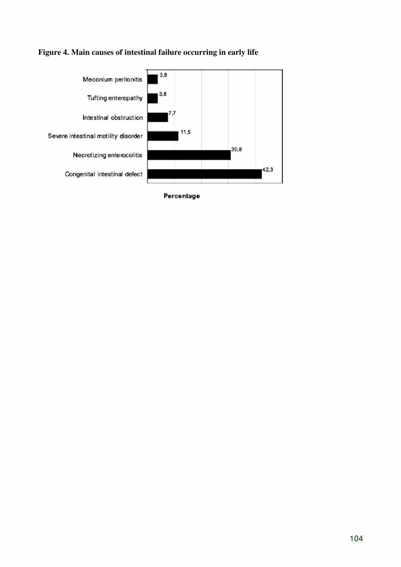
104
Figure 4. Main causes of intestinal failure occurring in early life

105
Figure 5. Duration of hospitalization in subjects dependent from parenteral nutrition treated
with 2 different nutritional strategy in early life
Kaplan-Meyer analysis shows a significant difference between VLBW feeding intolerant infants
administered with total parenteral nutrition (PN) (Group 1) and with PN plus minimal enteral
feeding (MEF)(Group 2) at day 40, 50 and 60 of life in percentage of patients discharged from
Neonatal Intensive Care Unit.

106
Figure 6. Comparative study on ORS involving children with acute diarrhea
Group 1
Enrollment (n=130)
Excluded: refused to partecipate (n=2)
Assessed for eligibility (n=132)
Analyzed (n= 60) Analyzed (n=59)
Lost to follow-up (n=1)
Discontinued intervention (n=1,
hospitalization for worsening
symptoms)
Group 2
Discontinued intervention (n=3,
hospitalization for worsening
symptoms)
Allocated to intervention (n= 65)
Received allocated intervention (n= 63)
Did not receive allocated intervention (n = 2,
ORS not available)
Allocated to intervention (n= 65)
Received allocated intervention (n= 61)
Did not receive allocated intervention (n = 4,
ORS not available)

107
Figure 7. Comparative study on ORS involving children with acute diarrhea: probability of
persistence of diarrhea in first 72 hours of treatment
Time from the enrolment (h)
Cu
mu
lati
ve
pro
bab
ilit
y o
f d
iarr
hea
(%
)
Group 1
Group 2
Log-rank test (p) 0.032

108
Figure 8. Comparative study on probiotics involving children with acute diarrhea: flow of
patients

109
Figure 9. Efficacy of butyrate in children with congenital chloride diarrhea
Stool consistency of children with congenital chloride diarrhea during a trial with oral butyrate

110
Figure 10. Efficacy of butyrate in children with congenital chloride diarrhea
Number of daily bowel movements of children with congenital chloride diarrhea during a trial with
oral butyrate
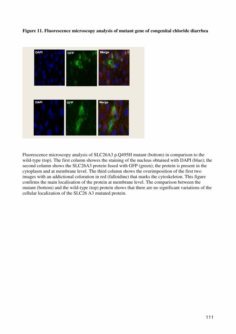
111
Figure 11. Fluorescence microscopy analysis of mutant gene of congenital chloride diarrhea
Fluorescence microscopy analysis of SLC26A3 p.Q495H mutant (bottom) in comparison to the
wild-type (top). The first column showes the staining of the nucleus obtained with DAPI (blue); the
second column shows the SLC26A3 protein fused with GFP (green); the protein is present in the
cytoplasm and at membrane level. The third column shows the overimposition of the first two
images with an addictional coloration in red (falloidine) that marks the cytoskeleton. This figure
confirms the main localisation of the protein at membrane level. The comparison between the
mutant (bottom) and the wild-type (top) protein shows that there are no significant variations of the
cellular localization of the SLC26 A3 mutated protein.

112
Figure 12. Fluorescence microscopy analysis of mutant gene of congenital chloride diarrhea
Fluorescence microscopy analysis of SLC26A3 p.A547E mutant (bottom) in comparison to the
wild-type (top). The first column showes the staining of the nucleus obtained with DAPI (blue); the
second column shows the SLC26A3 protein fused with GFP (green); the protein is present in the
cytoplasm and at membrane level. The third column shows the overimposition of the first two
images with an addictional coloration in red (falloidine) that marks the cytoskeleton. This figure
confirms the main localisation of the protein at membrane level. The comparison between the
mutant (bottom) and the wild-type (top) protein shows that there are no significant variations of the
cellular localization of the SLC26 A3 mutated protein.

113
Figure 13. In vivo butyrate modulation of the gene of congenital chloride diarrhea
Effect of butyrate on DRA/SLC26A3 expression obtained in epithelial nasal cell of a children
(missence mutation) with congenital chloride diarrhea and their parents

114
Figure 14. In vivo butyrate modulation of the gene of congenital chloride diarrhea
.
Effect of butyrate on DRA/SLC26A3 expression obtained in epithelial nasal cell of a children
(mutation: exon deletion) with congenital chloride diarrhea and mother

115
Figure 15. Mechanisms of action of butyrate

116
Figure 16. New butyrate formulation
CH2 CH z
XY
R
R1
R3Y
CH2CH
2CH
3
R2
W

117
Figure 17. Effects of a new butyrate formulation on intestinal ions transport

118
Figure 18. The multifactorial pathogenesis of the increased risk of gastrointestinal infections
in patients treated with gastric acidity inhibitors

119
References
1. Santosham M, Chandran A, Fitzwater S,Fischer-Walker C, Baqui AH, Black R.
Progress and barriers for the control of diarrhoeal disease. Lancet 2010; 376:63-67.
2. Berni Canani R, Passariello A, Terrin G. La diarrea acuta. In Gastroenterologia
pediatrica. A cura di Armido Rubino. Ed. Pacini 2008, 148-58.
3. Berni Canani R, Cirillo P, Terrin G. Chronic and intractabile diarrhea. In: Guandalini
S. Ed. Essential Pediatric Gastroenterology Hepatology, and Nutrition. Mc Graw-Hill
Mediacla Publishing Division Chicago 2005: 25-47.
4. Guarino A, Albano F, Ashkenazi S, Gendrel D, Hoekstra JH, Shamir R, Szajewska H
ESPGHAN/ESPID Evidence-Based Guidelines for the Management of Acute
Gastroenteritis in Children in Europe Expert Working Group. European Society for
Paediatric Gastroenterology, Hepatology, and Nutrition/European Society for
Paediatric Infectious Diseases evidence-based guidelines for the management of
acute gastroenteritis in children in Europe: executive summary. J Pediatr
Gastroenterol Nutr. 2008; 46: S81-122.
5. Lazzerini M, Ronfani L. Oral zinc for treating diarrhoea in children. Cochrane
Database Syst Rev. 2008; 16:CD005436.
6. Szajewska H, Dziechciarz P. Gastrointestinal infections in the pediatric population.
Curr Opin Gastroenterol. 2010; 26:36-44.
7. Berni Canani R, Cirillo P, Terrin G, Cesarano L, Spagnolo MI, De Vincenzo A,
Albano F, Passariello A, De Marco G, Manguso F, Guarino A. Probiotics for the
treatment of acute diarrhea: a randomized single-blind trial with five different
preparations. BMJ 2007; 335: 340-6.
8. Passariello A, Terrin G, De Marco G, Cecere G, Ruotolo S, Marino A, Cosenza L,
Tardi M, Nocerino R, Berni Canani R. Efficacy of a New Hypotonic Oral
Rehydration Solution Containing Zinc and Prebiotics in the Treatment of Childhood
Acute Diarrhea: A Randomized Controlled Trial. J Pediatr. 2010 Sep 7. [Epub ahead
of print].

120
9. Johnston BC, Shamseer L, da Costa BR, Tsuyuki RT, Vohra S. Measurement issues
in trials of pediatric acute diarrheal diseases: a systematic review. Pediatrics 2010;
126:222-231.
10. Berni Canani R, Terrin G, Cardillo G, Tomaiuolo R, Castaldo G. Congenital
diarrheal disorders: improved understanding of gene defects is leading to advances in
intestinal physiology and clinical management.J Pediatr Gastroenterol Nutr. 2010;
50:360-366.
11. Berni Canani R, Passariello A, Buccigrossi V, Terrin G, Guarino A. The nutritional
modulation of the evolving intestine. J Clin Gastroenterol 2008; 42; suppl3: S197-
200.
12. Salvatore S, Hauser B, Devreker T, Arrigo S, Vandenplas Y. Chronic enteropathy
and feeding in children: An update Nutrition 2008; 24:1205–1216.
13. Passariello A, Terrin G, Baldassarre ME, De Curtis M, Paludetto R, Berni Canani R.
Diarrhea in neonatal intensive care unit. World J Gastroenterol. 2010;16:2664-2668.
14. Salvia G, Guarino A, Terrin G, Cascioli C, Paludetto R, Indrio F, Lega L, Fanaro S,
Stronati M, Corvaglia L, Tagliabue P, De Curtis M. Neonatal onset intestinal failure:
an Italian Multicenter study. J Pediatr 2008; 153:674-676.
15. Terrin G, Passariello A, Berni Canani R, Manguso F, Paludetto R, Cascioli C.
Minimal enteral feeding reduces the risk of sepsis in feed-intolerant very low birth
weight newborns. Acta Paediatr. 2009; 98:31-35.
16. Berni Canani R, Terrin G. Gastric acidity inhibitors and the risk of intestinal
infections. Curr Opin Gastroenterol 2010; 26:31-35.
17. Asher MI, Montefort S, Biorkstén B, Lai CK, Strachan DP, Weiland SK, Williams
H; ISAAC Phase Three Study Group. Worldwide time trends in the prevalence of
symptoms of asthma, allergic rhinoconjunctivitis, and eczema in childhood: ISAAC
Phase One and Three repeat multicountry cross-sectional surveys. Lancet 2006;
368:733-743.
18. Greer FR, Sicherer SH, Burks AW and the Committee on Nutrition and Section on
Allergy and Immunology. Effects of Early Nutritional Interventions on the
Development of Atopic Disease in Infants and Children: The Role of Maternal

121
Dietary Restriction, Breastfeeding, Timing of Introduction of Complementary Foods,
and Hydrolyzed Formulas. Pediatrics 2008; 121:183-191.
19. Jones G, Steketee RW, Black RE, Bhutta ZA, Morris SS and Bellagio Child Survival
Study Group. How many child deaths can we prevent this year? Lancet 2003;
362:65-71.
20. Kosek M, Bern C, Guerrent RL. The global burden of diarrhoeal disease as estimated
from studies published between 1992 and 2000. Bull World Health Organ 2003;
81:197-204.
21. Fanaroff AA, Stoll BJ, Wright LL, Carlo WA, Ehrenkranz RA, Stark AR, Bauer CR,
Donovan EF, Korones SB, Laptook AR, Lemons JA, Oh W, Papile LA, Shankaran S,
Stevenson DK, Tyson JE, Poole WK; NICHD Neonatal Research Network. Trends in
neonatal morbidity and mortality for very low birth weight infants. Am J Obstet
Gynecol 2007;196:147e1-147e8.
22. Parkash J, Das N. Pattern of admissions to neonatal unit. J Coll Physicians Surg Pak
2005; 15:341-344.
23. Sherman PM, Mitchell DJ, Cutz E. Neonatal enteropathies: defining the causes of
protracted diarrhoea of infancy. J Pediatr Gastroenterol Nutr 2004; 38:16-26.
24. Baqui AH, Black RE, Yunus M. Methodological issues in diarrhoeal diseases
epidemiology: definition of diarroeal episodes. Int J Epidemiol 1991; 20:1057-1063.
25. WHO, DDC Persistent diarrhea in children. Geneva: WHO; 1985.
26. WHO, The treatment of diarrhea. A manual for physicians and other senior health
workers. Geneva: WHO; 2005. P. WHO/CDD/SER/80.2.
27. Miceli Sopo S, Radzik D, Calvani M. The predictive value of specific
immunoglobulin E levels for the first diagnosis of cow’s milk allergy. A critical
analysis of pediatric literature. Pediatr Allergy Immunol 2007; 18:575-582.
28. Nyhan WL. Disorders of purine and pyrimidine metabolism. Mol Genet Metab 2005;
86:25-33.
29. Kuschel C. Managing drug withdrawal in the newborn infant. Semin Fetal Neonatal
Med 2007; 12:127-133.

122
30. de Lorijn F, Boeckstaens Ge, Benninga MA. Symptomatology, pathophysiology,
diagnostic work-up, and treatment of Hirschsprung disease in infancy and childhood.
Curr Gastroenterol Rep 2007; 9:245-253.
31. Minasian C, Mccullagh A, Bush A. Cystic fibrosis in neonates and infants. Early
Hum Dev 2005; 81:997-1004.
32. Brosilow SW, Horwich AL. Urea cycle disorders. In: The metabolic and molecular
bases of inherited disease. Eds. Scriver CR, Beaudet AL, Sly WS, Vale DT, New
York: McGraw-Hill; 2001 pp 1909-63.
33. Fontana M, Zuin G, Pancheri P, Fusco FC, Lambertini A, Berni Canani R for the
SIGEP Working Group on Intestinal Infections (2004). Costs associated with
outpatient diarrhoea in infants and toddlers: a nationwide study of the Italian Society
of Paediatric Gastroenterology and Hepatology (SIGEP). Dig Liver Dis 2004;
36:523-527.
34. Guandalini S Acute Diarrhea. In Essential Pediatric Gastroenterology Hepatology
and Nutrition. Ed. Guandalini S. New York: McGraw-Hill; 2005; pp 15-23.
35. Wang J, Cortina G, Wu SV, Tran R, Cho JH, Tsai MJ, Bailey TJ, Jamrich M, Ament
ME, Treem WR, Hill ID, Vargas JH, Gershman G, Farmer DG, Reyen L, Martín
MG. Mutant neurogenin-3 in congenital malabsorptive diarrhea. N Engl J Med 2006;
355:270-280.
36. Host A, Koletzko B, Dreborg S, Muraro A, Wahn U, Aggett P, Bresson JL, Hernell
O, Lafeber H, Michaelsen KF, Micheli JL, Rigo J, Weaver L, Heymans H, Strobel S,
Vandenplas Y. Joint STATEMENT of the European Society for Paediatric
Allergology and Clinical Immunology (ESPACI) Committee on Hypoallergenic
Formulas and the European Society for Paediatric Gastroenterology, Hepatology and
Nutrition (ESPGHAN) Committee on Nutrition. Dietary products used in infants for
treatment and prevention of food allergy. Arch Dis Child 1999; 81:80-84.
37. Zuppa AA, Cota F, Barberi S, De Luca D, Visintini F, Tortorolo G. Alimentary
strategies in the neonatal period in the prevention of allergies. Pediatr Med Chir
2002; 24:45-52.
123
38. Gleizes O, Desselberger U, Tatochenko V, Rodrigo C, Salman N, Mezner Z,
Giaquinto C, Grimprel E. Nosocomial Rotavirus infection in European countries: a
review of the epidemiology, severity and economic burden of hospital-acquired
rotavirus disease. Pediatr Infect Dis J 2006; 25: S12-21.
39. Elliot EJ Acute gastroenteritis in children. BMJ 2008; 334:35-40.
40. Thomas PD, Forbes A, Green J, Howdle P, Long R, Playford R, Sheridan M, Stevens
R, Valori R, Walters J, Addison GM, Hill P, Brydon G. Guidelines for the
investigation of chronic diarrhea, 2end edition. Gut 2003; 52: 1-15.
41. Goulet O, Ruemmele F. Causes and management of intestinal failure in children.
Gastroenterology 2006;130(Suppl 1):S16-28.
42. Guarino A, De Marco G. Natural history of intestinal failure, investigated through a
national network-based approach. J Pediatr Gastroenterol Nutr 2003;37:136-141.
43. Goulet O, Ruemmele F, Lacaille F, Colomb V. Irreversible intestinal failure. J
Pediatr Gastroenterol Nutr 2004;38:250-269.
44. Gupte GL, Beath SV, Kelly DA, Millar AJ, Booth IW. Current issues in the
management of intestinal failure. Arch Dis Child 2006;91:259-264.
45. Sondheimer JM, Asturias E, Cadnapaphornchai M. Infection and cholestasis in
neonates with intestinal resection and long-term parenteral nutrition. J Pediatr
Gastroenterol Nutr 1998;27:131-137.
46. Wales PW, de Silva N, Kim J, Lecce L, To T, Moore A. Neonatal short bowel
syndrome: population-based estimates of incidence and mortality rates. J Pediatr
Surg 2004;39:690-695.
47. Sondheimer JM, Cadnapaphornchai M, Sontag M, Zerbe GO. Predicting the duration
of dependence on parenteral nutrition after neonatal intestinal resection. J Pediatr
1998;132:80-84.
48. Spencer AU, Neaga A, West B, Safran J, Brown P, Btaiche I, et al. Pediatric short
bowel syndrome: redefining predictors of success. Ann Surg 2005;242:403-409;
discussion 409-412.

124
49. Goulet O, Baglin-Gobet S, Talbotec C, Fourcade L, Colomb V, Sauvat F, et al.
Outcome and long-term growth after extensive small bowel resection in the neonatal
period: a survey of 87 children. Eur J Pediatr Surg 2005;15:95-101.
50. Andorsky DJ, Lund DP, Lillehei CW, Jaksic T, Dicanzio J, Richardson DS, et al.
Nutritional and other postoperative management of neonates with short bowel
syndrome correlates with clinical outcomes. J Pediatr 2001;139:27-33.
51. Balistreri WF, Heubi JE, Suchy FJ. Immaturity of the enterohepatic circulation in
early life: factors predisposing to “physiologic” maldigestion and cholestasis. J
Pediatr Gastroenterol Nutr 1983;2:346-354.
52. Kelly DA. Intestinal failure-associated liver disease: what do we know today?
Gastroenterology 2006;130(Suppl 1):S70-S77.
53. Muiesan P, Dhawan A, Novelli M, Mieli-Vergani G, Rela M, Heaton ND. Isolated
liver transplant and sequential small bowel transplantation for intestinal failure and
related liver disease in children. Transplantation 2000;69:2323-2326.
54. Colomb V, Dabbas-Tyan M, Taupin P, Talbotec C, Revillon Y, Jan D, et al. Long-
term outcome of children receiving home parenteral nutrition: a 20-year single-center
experience in 302 patients. J Pediatr Gastroenterol Nutr 2007;44:347-353.
55. De Marco G, Sordino D, Bruzzese E, Di Caro S, Mambretti D, Tramontano A, et al.
Early treatment with ursodeoxycholic acid for cholestasis in children on parenteral
nutrition because of primary intestinal failure. Aliment Pharmacol Ther 2006;24:387-
394.
56. McGuire W, Henderson G, Fowlie PW. Feeding the preterm infant. BMJ 2004;329:
1227-1230.
57. Fletcher AB. Nutrition. In Avery GB, ed. Neonatology. 4th ed. Philadelphia, PA: JB.
Lippincott; 1994: 330–356.
58. Lin PW, Stoll BJ. Necrotizing enterocolitis. Lancet 2006;368: 1271-1283.
59. Hunter CJ, Upperman JS, Ford HR, Camerini V. Understanding the susceptibility of
yhe premature infant to necrotizing enterocolitis (NEC). Pediatr Res 2008; 63:117-
123.

125
60. Mihatsch WA, von Schoenaich P, Fahnenstich H, Dehne N, Ebbecke H, Plath C et al.
The significance of gastric residuals in the early enteral feeding advancement of
extremely low birth weight infants. Pediatrics 2002;109: 457-459.
61. LaGamma, EF, Ostertag, SG, Birenbaum H. Failure of delayed oral feedings to
prevent necrotizing enterocolitis. Am J Dis Child 1985;139: 385-389.
62. Cobb BA, Carlo WA, Ambalavanan N. Gastric residuals and their relationship to
necrotizing enterocolitis in very low birth weight infants. Pediatrics 2004;113: 50-53.
63. Dunn L, Hulman S, Weiner J, Kliegman R. Benefical effects of early hypocaloric
enteral feeding on neonatal gastrointestinal function: preliminary report of a
randomized trial. J Pediatr 1988;112: 622-629.
64. Greer FR. Feeding the premature infant in the 20th
century. J Nutr 2001;131: 426S-
430S.
65. Saito H, Trocki O, Alexander JW, Kopcha R, Heyd T, Joffe SN. The effect of route
of nutrient administration on the nutritional state, catabolic hormone secretion and
gut mucosal integrity after burn injury. JPEN 1987; 11:1-7.
66. Illig KA, Ryan CK, Hardy DJ, Rhodes J, Locke W, Sax HC. Total parenteral
nutrition induced changes in gut mucosal function: atrophy alone is not the issue.
Surgery 1992; 112:631-637.
67. Moore FA, Moore EE, Jones TN, McCroskey BL, Peterson VM. TEN versus TPN
following major abdominal trauma: reduced septic morbidity. J Trauma 1989;
29:916-923.
68. Moore FA, Feliciano DV, Andrassy RJ, McArdle AH, Booth FV, Morgenstein-
Wagner TB, et al. Early enteral feeding, compare with parenteral, reduces
postoperative septic complications: the results of meta-analysis. Ann Surg 1992;
216:172–183.
69. Fletcher JP and Little JM. A comparison of parenteral nutrition and early
postoperative enteral feeding on the nitrogen balance after major abdominal surgery.
Surgery 1986; 100: 21–24.

126
70. Owens L, Burrin DG, Berseth CL. Minimal enteral feeding induces maturation of
intestinal motor function but not mucosal growth in neonatal dogs. J Nutr 2002;132:
2717-2722.
71. Berseth CL, Nordyke C. Enteral nutrients promote postnatal maturation of intestinal
motor activity in preterm infants. Am J Physiol 1993;264: G1046 –G1051.
72. Stoll BJ, Hansen N, Fanaroff AA, Wright LL, Carlo WA, Ehrenkranz RA, et al. Late-
onset sepsis in very low birth weight neonates: the experience of the NICHD
neonatal research network. Pediatrics 2002;110: 285-291.
73. American Academy of Pediatrics, Committee on Fetus and Newborn. Hospital
Discharge of the hight-risck neonate-proposed guidelines. Pediatrics 1998;102: 411-
417.
74. Stool BJ, Hansen N. Infections in VLBW infants: studies from the NICHD Neonatal
Research Network. Semin Perinatol 2003;27: 293-301.
75. Fanaroff AA, Hack M, Walsh MC. The NICHD neonatal research network: changes
in practice and outcomes during the first 15 years. Semin Perinatol 2003;27: 281-
287.
76. Wesley JR, Coran AG. Intravenous nutrition for the pediatric patient. Sem Pediatr
Surg 1992; 1:212-230.
77. Bos AP, Tibboel D, Hazebroek FW, Bergmeijer JH, van Kalsbeek EJ, Molenaar JC.
Total parenteral nutrition associated cholestasis: a predisposing factor for sepsis in
surgical neonates ? Eur J Pediatr 1990; 149:351-353.
78. Okada Y, Klein N, van Saene HKF, Pierro A. Small volumes of enteral feedings
normalise immune function in infants receiving parenteral nutrition. J Pediatr Surg
1998; 33:16-19.
79. Okada Y, Papp E, Klein NJ, Pierro A. Total parenteral nutrition directly impairs
cytokine production after bacterial challenge. J Pediatr Surg 1999; 34:277-280.
80. Stoll BJ. Epidemiology of necrotizing enetrocolitis. Clinical Perinatol 1994;21: 205-
218.
127
81. Berseth CL. Effect of early enteral feeding on maturation of the preterm infants small
intestine. J Pediatr 1992;120: 947–953.
82. McClure RJ, Newell SJ. Randomised controlled study of clinical outcome following
trophic feeding. Arch Dis Child Fetal Neonatal Ed 2000;82: F29–33.
83. Slagle TA, Gross SJ. Effect of early low-volume enteral substrate on subsequent
feeding tolerance in very low birth weight infants. J Pediatr 1988;113: 526-531.
84. Tyson JE, Kennedy KA. Minimal enteral nutrition for promoting feeding tolerance
and preventing morbidity in parenterally fed infants. Cochrane Database Syst Rev
2004;2: CD000504.
85. de la Cochetiere MF, Piloquet H, des Robert C, Darmaun D, Galmiche JP, Roze JC.
Early intestinal bacterial colonization and necrotizing enterocolitis in premature
infants: the putative role of Clostridium. Pediatr Res 2004;56: 366-370.
86. Strodbeck F. The role of early enteral nutrition in protecting premature infants from
sepsis. Crit Care Nurs Clin North Am 2003;15: 79-87.
87. Niinikoski H, Stoll B, Guan X, Kansagra K, Lambert BD, Stephens J, et al. Onset of
small intestinal atrophy is associated with reduced intestinal blood flow in TPN-fed
neonatal piglets. J Nutr 2004;134:1467-1474.
88. Dominguez JA. Use of Minimal Enteral Nutrition to Prevent Necrotizing
Enterocolitis: Got Milk? JPEN J Parenter Enteral Nutr. 2010; 20. [Epub ahead of
print].
89. McHale SM, McCarthy R, O'Donnell CP. How minimal is "minimal" enteral
feeding? Arch Dis Child Fetal Neonatal Ed. 2010; 95(2):F149-50.
90. Oste M, Van Haver E, Thymann T, Sangild P, Weyns A, Van Ginneken CJ. Formula
induces intestinal apoptosis in preterm pigs within a few hours of feeding. JPEN J
Parenter Enteral Nutr. 2010;34:271-279.
91. Donini LM, Savina C, Ricciardi LM, Coletti C, Paolini M, Scavone L, De Felice
MR, Laviano A, Rossi Fanelli F, Cannella C. Predicting the outcome of artificial
nutrition by clinical and functional indices. Nutrition. 2009; 25(1):11-19.

128
92. Hay WW Jr. Strategies for feeding the preterm infant. Neonatology. 2008;94(4):245-
54.
93. Duro D, Kamin D, Duggan C. Overview of pediatric short bowel syndrome. J Pediatr
Gastroenterol Nutr. 2008; 47 Suppl 1:S33-S36.
94. Mosqueda E, Sapiegiene L, Glynn L, Wilson-Costello D, Weiss M. The early use of
minimal enteral nutrition in extremely low birth weight newborns. J Perinatol. 2008;
28: 264-9.
95. Khan AM, Sarker SA, Alam NH, Hossain MS, Fuchs GJ, Salam MA. Low osmolar
oral rehydration salts solution in the treatment of acute watery diarrhoea in neonates
and young infants: a randomized, controlled clinical trial. J Health Popul Nutr 2005;
23:52-57.
96. Recommendations for composition of oral rehydration solutions for the children of
Europe. Report of an ESPGAN Working Group. J Pediatr Gastroenterol Nutr 1992;
14:113-115.
97. Fischer Walker CL, Fontaine O, Young MW, Black RE. Zinc and low osmolarity
oral rehydration salts for diarrhea: a renewed call to action. Bull World Health Organ
2009; 87:780-786.
98. WHO/UNICEF joint statement: clinical management of acute diarrhoea. New York,
NY and Geneva, Switzerland: The United Nations children’s Fund/World Health
Organization, 2004:1-8.
99. Patro B, Golicki D, Szajewska H. Meta-analysis: zinc supplementation for acute
gastroenteritis in children. Aliment Pharmacol Ther 2008; 28:713-723.
100. Lukacik M, Thomas R L, Aranda JV. A meta-analysis of the effects of oral zinc in
the treatment of acute and persistent diarrhea. Pediatrics 2008; 121:326-336.
101. INCLEN Childnet zinc effectiveness for diarrhea (IC-ZED) Group. J Pediatr
Gastroenterol Nutr 2006; 42:300-305.
102. Bhandari N, Mazumder S, Taneja S, Dube B, Agarwal RC, Mahalanabis D, et al.
Effectiveness of zinc supplementation plus oral rehydration salts compared with oral

129
rehydration salts alone as a treatment for acute diarrhea in a primary care setting: a
cluster randomized trial. Pediatrics 2008; 121:e1279-1285.
103. Roy SK, Hossain MJ, Khatun W, Chakraborty B, Chowdhury S, Begum A et al. Zinc
supplementation in children with cholera in Bangladesh: randomised controlled trial.
BMJ 2008; 336:266-268.
104. Shamir R, Makhoul IR, Etzioni A, Shehadeh N. Evaluation of a diet containing
probiotics and zinc for the treatment of mild diarrheal illness in children younger
than one year of age. J Am Coll Nutr 2005; 24:370-375.
105. Molleston J, Alonso E, Oliva-Hemker MO. Zinc for infectious diarrhea in developed
countries: should we be sprinkling our own lawns? J Pediatr Gastroenterol Nutr
2008; 46:484-485.
106. Polat TB, Uysalol M, Cetinkaya F. Efficacy of zinc supplementation on the severity
and duration of diarrhea in malnourished Turkish children. Pediatr Int 2003; 45:555-
559.
107. Hoestra JH, Szajewska H, Zikri MA, Micetic-Turk D, Weizman Z, Papadopoulou A
et al. Oral rehydration solution containing a misture of non-digestible carbohydrates
in the treatment of acute diarrhea: a multicenter randomized placebo controlled study
on behalf of the ESPGHAN Working Group on Intestinal Infections. J Pediatr
Gastroenterol Nutr 2004;39:239-245.
108. Knol J, Boehm G, Lidestri M, Negretti F, Jelinek J, Agosti M et al. Increase of
faecal bifidobacteria due to dietary oligosaccharides induces a reduction of clinically
relevant pathogen grems in the faeces of formula-fed preterm infants. Acta Paediatr
2005; 94:31-33.
109. Arslanoglu S, Moro GE, Boehm G. Early supplementation of prebiotic
oligosaccharides protects formula-fed infants against infections during the first 6
months of life. J Nutr 2007; 2420-2424.
110. Bakker-Zierikzee AM, Tol EA, Kroes H, Alles MS, Kok FJ, Bindels JG. Faecal
SIgA secretion in infants fed on pre- or probiotic infant formula. Pediatr Allergy
Immunol 2006; 17:134-140.

130
111. Scholtens PA, Alliet P, Raes M, Alles MS, Kroes H, Boehm G, et al. Fecal secretory
immunoglobulin A is increased in healthy infants who receive a formula with short-
chain galacto-oligosaccharides and long-chain fructooligosaccharides J Nutr 2008;
138:1141-1147.
112. Ten Bruggencate SJ, Bovee-Oudenhoven IM, Lettink-Wissink ML, Van der Meer R.
Dietary fructooligosaccharides dose-dependently increase translocation of salmonella
in rats. J Nutr 2003; 133:2313-2318.
113. Cummings JH, Christie S, Cole TJ. A study of fructo oligosaccharides in the
prevention of travellers diarrhoea. Aliment Pharmacol Ther 2001; 15:1139-1145.
114. Scholtens PA, Alles MS, Bindels JG, van der Linde EG, Tolboom JJ, Knol J.
Bifidogenic effects of solid weaning foods with added prebiotic oligosaccharides: a
randomized controlled clinical trial. J Pediatr Gastroenterol Nutr 2006; 42:553-559.
115. Macfarlane GT, Steed H, Macfarlene S. Bacterial metabolism and health-related
effects of galacto-oligosaccharides and other prebiotics. J Appl Microbiol 2008;
104:305-344.
116. Berni Canani R, Cirillo P, Buccigrossi V, Ruotolo S, Passariello A, De Luca P et al.
Zinc inhibits cholera toxin-induced, but not Escherichia coli heat-stable enterotoxin-
induced, ion secretion in human enterocytes. J Infect Dis 2005; 191:1072-1077.
117. Berni Canani R, Secondo A, Passariello A, Buccigrossi V, Canzoniero LM, Ruotolo
S et al. Zinc inhibits calcium-mediated and nitric oxide-mediated ion secretion in
human enterocytes. Eur J Pharmacol 2010; 626:266-270.
118. Altaf W, Perveen S, Rehman KU, Teichberg S, Vancurova I, Harper RG, et al. Zinc
supplementation in oral rehydration solutions: experimental assessment and
mechanism of action. J Am Coll Nutr 2002; 21:26-33.
119. Copenhagen Consensus 2008-Results. Frederiksberg: Copenhagen Consensus
Center; 2008. Avalaible from: http://www.copenhagenconsensus.com
120. Patel A, Dibley MJ, Mamtani M, Badhoniya N, Kulkarni H. Zinc and copper
supplementation in acute diarrhea in children: a double-blind randomized controlled
trial. BMC Medicine 2009; 7:22.

131
121. Fischer Walker CL, Ezzati M, Black RE. Global and regional child mortality and
burden of disease attributable to zinc deficiency. Eur J Clin Nutr 2009; 63:591-597.
122. Garenne M, Becher H, YE Y, Kouyate B, Müller O. Sex-specific responses to zinc
supplementation in Nouna, Burkina Faso. J Pediatr Gastroenterol Nutr 2007; 44:619-
628.
123. Bhatnagar S, Bahl R, Sharma P, Kumar GT, Saxena SK, Bhan MK. Zinc with oral
rehydration therapy reduces stool output duration of diarrhea in hospitalized children:
a randomized controlled trial. J Pediatr Gastroenterol Nutr 2004; 38:34-40.
124. Surjawidjaja JE, Hidayat A, Lesmana M. Growth Inhibition of Enteric Pathogens by
Zinc Sulfate: an in vitro Study. Med Princ Pract 2004; 13:286-289.
125. Berni Canani R, Ruotolo S. The dawning of the "zinc era" in the treatment of
pediatric acute gastroenteritis worldwide? J Pediatr Gastroenterol Nutr 2006; 42:
253-255.
126. Cousins RJ, Liuzzi JP, Lichten LA. Mammalian zinc transport, trafficking, and
signals. J Biol Chem 2006; 281:24085-24089.
127. Ibs KH, Rink L. Zinc-altered immune function. J Nutr 2003; 133:1452S-1456S.
128. Rink L, Haase H. Zinc homeostasis and immunity. Trends Immunol 2007; 28:1-4.
129. Alam NH, Ashraf H. Treatment of infectious diarrhea in children. Paediatr Drugs
2003;5:151-165.
130. Szajewska H, Setty M, Mrukowicz J, Guandalini S. Probiotics in gastrointestinal
diseases in children: hard and not-so-hard evidence of efficacy. J Pediatr
Gastroenterol Nutr 2006;42:454-475.
131. Isolauri E. Probiotics for infectious diarrhoea. Gut 2003;52:436-437.
132. Davidson GP, Butler RN. Probiotics in intestinal disorders. Curr Opin Pediatr
2000;12:477-481.
133. Saavedra JM, Bauman NA, Oung I, Perman JA, Yolken RH. Feeding of
Bifidobacterium bifidum and Streptococcus thermophilus to infants in hospital for
prevention of diarrhoea and shedding of rotavirus. Lancet 1994;344:1046-1049.

132
134. Tankanow RM, Ross MB, Ertel IJ, Dickinson DG, McCormick LS, Garfinkel JF. A
double-blind, placebo controlled study of the efficacy of Lactinex in the prophylaxis
of amoxicillin-induced diarrhea. DICP 1990;24:382-384.
135. Elmer GW, Surawicz CM, McFarland LV. Biotherapeutic agents: a neglected
modality for the treatment and prevention of selected intestinal and vaginal
infections. JAMA 1996;275:870-876.
136. Huang JS, Bousvaros A, Lee JW, Diaz A, Davidson EJ. Efficacy of probiotic use in
acute diarrhea in children: a meta-analysis. Dig Dis Sci 2002;47:2625-2634.
137. Van Niel CW, Feudtner C, Garrison MM, Cristakis DA. Lactobacillus therapy for
acute infectious diarrhea in children: a meta-analysis. Pediatrics 2002;109:678-684.
138. Allen SJ,Okoko B, Martinez E, Gregorio G,Dans LF. Probiotics for treating
infectious diarrhoea. Cochrane Database Syst Rev 2004;(2): CD003048.
139. Walker-Smith JA, Sandhu BK, Isolauri E, Banchini G, van Caillie-Bertrand M, Dias
JA, et al. Guidelines prepared by ESPGAN working group on acute diarrhoea.
Recommendations for feeding in childhood gastroenteritis. J Pediatr Gastroenterol
Nutr 1997;24:619-620.
140. Guarino A, Albano F. Guidelines for the approach to outpatient children with acute
diarrhoea. Acta Paediatr 2001;90:1096-1098.
141. Guarino A, Berni Canani R, Spagnuolo MI, Albano F, Di Benedetto L. Oral bacterial
therapy reduces the duration of symptoms and of viral excretion in children with mild
diarrhea. J Pediatr Gastroenterol Nutr 1997;25:516-519.
142. Guarino A, Canani RB, Russo S, Albano F, Canani MB, Ruggeri FM, et al. Oral
immunoglobulins for treatment of acute rotaviral gastroenteritis. Pediatrics
1994;93:12-16.
143. Majamaa H, Isolauri E, Saxelin M, Vesikari T. Lactic acid bacteria in the treatment
of acute rotavirus gastroenteritis. J Pediatr Gastroenterol Nutr 1995;20:333-338.
144. Guandalini S. Treatment of acute diarrhea in the new millennium. J Pediatr
Gastroenterol Nutr 2000;30:486-489.

133
145. Zimmerman CM, Bresee JS, Parashar UD, Riggs TL, Holman RC, Glass RI. Cost of
diarrhea-associated hospitalizations and outpatient visits in an insured population of
young children in the United States. Pediatr Infect Dis J 2001;20:14-19.
146. Farthing MJG. Novel targets for the pharmacotherapy of diarrhoea: a view for the
millennium. J Gastroenterol Hepatol 2000;15:G38-45.
147. Reid G, Jass J, Sebulsky MT, McCormick JK. Potential uses of probiotics in clinical
practice. Clin Microbiol Rev 2003;16:658-672.
148. Szajewska H, Hoekstra JH, Sandhu B, the working group on acute diarrhoea of the
European Society for Paediatric Gastroenterology, Hepatology and Nutrition.
Management of acute gastroenteritis in Europe and the impact of the new
recommendations: a multicenter study. J Pediatr Gastroenterol Nutr 2000;30:522-
527.
149. Young J. European Market developments in prebiotic- and probioticcontaining
foodstuffs. Br J Nutr 1998;80:S231-S233.
150. Drago L, De Vecchi E, Nicola L, Colombo A, GismondoMR. Microbiological
evaluation of commercial probiotic products available in Italy. J Chemother
2004;16:463-467.
151. Oberhelman RA, Gilman RH, Sheen P, Taylor DN, Black RE, Cabrera L, et al. A
placebo-controlled trial of Lactobacillus GG to prevent diarrhea in undernourished
Peruvian children. J Pediatr 1999;134:15-20.
152. Saxelin M. Lactobacillus GG a human probiotic strain with thorough clinical
documentation. Food Rev Int 1997;13:293-313.
153. Guandalini S, Pensabene L, Zikri MA, Dias JA, Casali LG, Hoekstra H, et al.
Lactobacillus GG administered in oral rehydration solution to children with acute
diarrhea: a multicenter European trial. J Pediatr Gastroenterol Nutr 2000;30:54-60.
154. Kurugol Z, Koturoglu G. Effects of Saccharomyces boulardii in children with acute
diarrhoea. Acta Paediatr 2005;94:44-47.

134
155. Bellomo G, Mangiagli A, Nicastro L, Frigerio G. A controlled double blind study of
SF68 strain as a new biological preparation for the treatment of diarrhoea in
paediatrics. Curr Ther Res 1980;27:927-936.
156. Mitra AK, Rabbani GH. A double-blind, controlled trial of Bioflorin (Streptococcus
faecium SF68) in adults with acute diarrhea due to Vibrio cholerae and
enterotoxigenic Escherichia coli.Gastroenterology 1990;99:1149-1152.
157. Caprioli A, Pezzella C,Morelli R,Giammanco A, Arista S, Crotti D, et al.
Enteropathogens associatedwith childhood diarrhea in Italy. Pediatr Infect Dis J
1996;15:876-883.
158. Berni Canani R, Terrin G, Cirillo P, et al. Butyrate as an effective treatment of
congenital chloride diarrhea. Gastroenterology 2004;127: 630–634.
159. Makela S, Kere J, Holmberg C, Hoglund P. SLC26A3 mutations in congenital
chloride diarrhea. Hum Mutat. 2002; 20:425-438.
160. Chernova MN, Jiang L, Shmukler BE, et al. Acute regulation of the SLC26A3
congenital chloride diarrhoea anion exchanger (DRA) expressed in Xenopus oocytes.
J Physiol 2003;549:3–19.
161. Gray MA Bicarbonate secretion: it takes two to tango. Nat Cell Biol 2004; 6:292-
294.
162. Ko SB, Zeng W, Dorwart MR, et al. Gating of CFTR by the STAS domain of SLC26
transporters. Nat Cell Biol 2004;6:343-350.
163. Ho¨glund P, Holmberg C, Sherman P, et al. Distinct outcomes of chloride diarrhoea
in two siblings with identical genetic background of the disease: implications for
early diagnosis and treatment. Gut 2001;48:724-727.
164. Ho¨glund P, Sormaala M, Haila S, et al. Identification of seven novel mutations
including the first two genomic rearrangements in SLC26A3 mutated in congenital
chloride diarrhea. Hum Mutat 2001;18:233-242.
165. Pagani F, Raponi M, Baralle FE. Synonymous mutations in CFTR exon 12 affect
splicing and are not neutral in evolution. Proc Natl Acad Sci U S A. 2005 May
3;102(18):6368-6372.

135
166. Wedenoja S, Hoglund P, Holmberg C. Review article: the clinical management of
congenital chloride diarrhoea. Aliment Pharmacol Ther 2010; 31:477-485.
167. Wedenoja S, Holmberg C, Ho¨glund P. Oral butyrate in treatment of congenital
chloride diarrhea. Am J Gastroenterol 2008;103:252-254.
168. Martinsen TC, Bergh K, Waldum HL. Gastric juice: a barrier against infectious
diseases. Basic Clin Pharm Toxicol 2005; 96: 94–102.
169. Dial MS. Proton Pump Inhibitor use and enteric infections. Am J Gastroenterol
2009;104:S10-S16.
170. Leonard J, Marshall JK, Moayyedi P. Systematic review of the risk of enteric
infection in patients taking acid suppression. Am J Gastroenterol 2007;102:2047-
2056.
171. Doorduyn Y, Van Den Bradhof WE, Van Duynhoven YTHP, et al. Risk factors for
Samonella Enteritidis and Typhimurium (DT104 and non-DT104) infections in the
Netherlands: predominant roles for raw eggs in Enteritidis and sandboxes in
Typhimurium infections. Epidemiol Infect 2006;134:617-626.
172. Berni Canani R , Cirillo P , Roggero P, et al. Therapy with gastric acidity inhibitors
increases the risk of acute gastroenteritis and community-acquired pneumonia in
children . Pediatrics 2006 ; 117:e817-820.
173. Vakil N. Acid inhibition and infections outside the gastrointestinal tract. Am J
Gastroenterol 2009;104:S17-S20.
174. Conroy S, McIntyre J. The use of unlicensed and off-label medicines in the neonate.
Semin Fetal Neonatal Med. 2005; 10:115-122.
175. Guillet R, Stoll BJ, Cotton CM, et al. For members of the National Institute of Child
Health and Human Development Neonatal Research Network association of H2-
Blocker Therapy and Higher Incidence of Necrotizing Enterocolitis in Very Low
Birth Weight Infants. Pediatrics 2006;117;137-142.
176. Stoll BJ, Temprosa M, Tyson JE, et al. Dexamethasone therapy increases infection in
very low birth weight infants. Pediatrics 1999;104:e63.

136
177. Beck-Sague CM, Aximi P, Fonseca SN, et al. Bloodstream infections in neonatal
intensive care unit patients: results of multicenter study. Pediatr Infect Dis J 1994;
13:1110–1116.
178. Terrin G, Ruggieri A, Salvia G, Lega L, Cascioli CF, Messina F, Passariello A, Berni
Canani R. Gastric acidity inhibitors therapy increses the risk of infection in
newborns. J Pediatr Gastroenterol Nutr 2006;42:G2-03.PG3-08.
179. Handley SA, Dube PH, Miller VL. Histamine signaling through the H2 receptor in
the Peyer’s patch is important for controlling Yersinia enterocolitica infection. PNAS
2006; 103:9269-9273.
180. Schubert ML, Peura DA. Control of gastric acid secretion in health and disease.
Gastroenterology 2008; 134:1842-1860.
181. IMS Health National Sale Prescription Web site. 2004 Year-End US Prescription and
Sales Information and Commentary. Available at:
http://www.imshealthcanada.com/htmen/3_2_28.htm. Accessed November 21, 2005.
182. Zeiger RS. Prevention of food allergy in infants and children. Immunol Allergy Clin
North Am 1999: 19: 619-646.
183. Berni Canani R, Ruotolo S, Discepolo V, Troncone R. The diagnosis of food allergy
in children. Curr Opin Pediatr 2008: 20: 584-589.
184. Asher MI, Montefort S, Bjorksten B, et al., ISAAC Phase Three Study Group.
Worldwide time trends in the prevalence of symptoms of asthma, allergic
rhinoconjunctivitis, and eczema in childhood: ISAAC phases one and three repeat
multicountry cross-sectional survey. Lancet 2006: 368: 733-743.
185. Zeiger RS. Food allergen avoidance in the prevention of food allergy in infants and
children. Pediatrics 2003: 111: 1662-1671.
186. Osborn DA, Sinn J. Formulas containing hydrolysed protein for prevention of allergy
and food intolerance in infants. Cochrane Database Syst Rev 2006: 18: CD003664.

137
Acknowledgements
Thanks to the Department of Pediatrics of University of Naples Federico II it was possible
to realize all the studies of the PhD project. The prestigious collaboration with the CEINGE,
the Department of Biochemistry and the Department of Pharmacology of University of
Naples Federico II has been crucial for studies on congenital diarrhea. The researches on
congenital chloride diarrhea were supported by the AIFA (Italian Agency for Drugs) grant
n° FARM6FJ728.