universita’ degli studi di milano-bicocca facoltà di ... · scuola di dottorato di scienze...
TRANSCRIPT

UNIVERSITA’ DEGLI STUDI DI MILANO-BICOCCA
Facoltà di Scienze Matematiche, Fisiche e Naturali
Scuola di Dottorato di Scienze
Dottorato di Ricerca in Biotecnologie Industriali
XXIV Ciclo
Evolution of copper tolerance in yeast cells
Giusy Manuela Adamo
Tutor: Dott. ssa Stefania Brocca
Anno Accademico 2011

Dottorato di Ricerca in Biotecnologie Industriali
XXIV Ciclo
Dottorando: Giusy Manuela Adamo
Matricola: 045269
Tutor: Dott. ssa Stefania Brocca
Coordinatore: Prof. Marco Vanoni
Università degli Studi di Milano-Bicocca
Piazza dell’Ateneo Nuovo, 1, 20126, Milano
Dipartimento di Biotecnologie e Bioscienze
Piazza della Scienza 2, 20126, Milano

Table of contents
Abstract p. 4
Riassunto p.6
1. Introduction p. 9
1.1 The role of copper in cell biology p.9
1.2 Yeast as a model organism to study copper metabolism p. 13
1.3 Antioxidant and prooxidant properties of copper p. 15
1.4 Protection against copper toxicity p. 18
1.5 Yeast as a new food source of essential copper. Is it possible? p. 21
1.6 Biotechnological tools to obtain copper enriched yeast biomass p. 23
1.7 Evolutionary engineering p. 24
2. Experimental work p. 31
2.1 Laboratory evolution of copper tolerant yeast strains
GM Adamo, S Brocca, S Passolunghi, B Salvato and M Lotti
(Microbial Cell Factories 2012, 11:1) p. 35
2.2 Evolution of copper tolerance in Saccharomyces cerevisiae relies on
amplification of the CUP1 gene
GM Adamo, M Lotti, MJ Tamás and S Brocca (Manuscript) p 69
2.3 Proteomic analysis of natural and copper-adapted cells of the yeast
Candida humilis
GM Adamo, S Brocca and M Lotti (Preliminary results) p 91
References p 109
Acknowledgements p 118


4
Abstract
For all living organisms, copper (Cu) is an essential micronutrient
taking part, with its redox chemistry, to several metabolic and
regulatory cellular events. However, the same redox properties that
make Cu essential are responsible for its toxicity. Indeed, Cu
participates in reactions that generate Reactive Oxygen Species
(ROS). ROS target main cellular macromolecules (proteins, lipids, DNA
and RNA), leading to cellular dysfunctions and in the extreme case, to
cell death. All living organisms evolved molecular mechanisms for Cu
homeostasis. Indeed, uptake, transport and detoxification systems
that actively prevent both Cu deficiency and poisoning are well
conserved along the phylogenetic tree. Among eukaryotes, these
mechanisms have been mainly investigated in the yeast
Saccharomyces cerevisiae used as a model organism.
Evolutionary engineering is a rational approach that uses the
evolutionary principles to direct the selection of organisms with a
desired set of phenotypes, allowing for the improvement of microbial
properties. This approach can be exploited to obtain Cu-tolerant and
Cu-accumulating yeast cells, with potential application in
nutraceutics, as nutritional supplements, as well as in bioremediation,
for the removal or recovery of metal ions. At the same time,
evolutionary engineering is a valuable strategy to gain more insight
into the molecular aspects of Cu tolerance in microbial cells.
In the present work is described an evolutionary engineering strategy
to improve Cu tolerance of natural yeasts. Strains of Saccharomyces
cerevisiae and of Candida humilis originally endowed with different
sensitivity and tolerance toward Cu have been exposed to increasing
concentrations of Cu during cell cultivation in liquid medium. This
treatment stably improved Cu tolerance of all strains. One evolved

5
strain for each yeast species was then chosen to analyze in detail the
physiological response to Cu. Compared with the original Cu-sensitive
strains the two evolved strains showed improved cell viability and
attenuated production of ROS. A reshaping of the profile of
antioxidant enzymes and Cu-binding proteins was observed in both
strains as a specific response to copper.
Further investigations carried out on S. cerevisiae strains demonstrated
a pivotal role of the CUP1 gene, encoding for a metallothionein. A 7-
fold amplification of this gene was found associated with evolution of
Cu tolerance.
Finally, Cu tolerance in C. humilis cells was studied by proteomic
analyses. Changes were observed in the levels of several proteins
involved in the oxidative stress response (such as glycolytic enzymes),
heat shock proteins, proteins involved in protein synthesis and energy
production, proteins with a role in phospholipids synthesis. Cu
exposure resulted in differential protein expression, in both non-
evolved and Cu evolved cells. In general, changes in protein levels
detected in evolved cells were smaller. On this basis, it was
hypothesized that in the evolved cells copper tolerance relies only
partly on the molecular mechanisms associated with the oxidative
stress response. This work shows once again that evolutionary
engineering is a powerful strategy to drive the gain of stable
phenotypic traits. The evolved strains might found direct application
in several biotechnological fields, and provide a kind of “molecular
platform” for the investigation on the mechanisms of stress tolerance.
The availability of data about the S. cerevisiae genome allowed a
focused investigation on the molecular actors involved in Cu
tolerance. In the case of C. humilis, the use of a proteomic approach
allowed to compensate for the poor information available on the
determinants of Cu tolerance.

6
Riassunto
Il rame (Cu) è un micronutriente essenziale per tutti gli organismi
viventi; questo metallo prende parte a numerose reazioni redox e
rappresenta un cofattore essenziale per proteine coinvolte in
numerosi pathways metabolici e processi di regolazione cellulare.
Tuttavia le stesse proprietà redox che rendono il rame essenziale sono
responsabili della tossicità di questo metallo. Il rame infatti prende
parte a reazioni che generano le cosiddette specie reattive
dell’ossigeno (Reactive Oxygen Species, ROS). Queste interagiscono
con le principali macromolecole cellulari (proteine, lipidi, DNA ed
RNA) causando malfunzionamento e morte cellulare. Tutti gli
organismi hanno evoluto sistemi strettamente regolati per l’uptake, il
trasporto e la detossificazione del rame. Questi sistemi di omeostasi
risultano essere altamente conservati lungo tutto l’albero filogenetico
e tra le cellule eucariote l’omeostasi del rame è stata principalmente
studiata nel lievito Saccharomyces cerevisiae.
L’ingegneria evolutiva (evolutionary engineering) si basa sui principi
dell’evoluzione per selezionare organismi con determinate
caratteristiche fenotipiche.
Questo approccio può essere sfruttato per migliorare la robustness dei
ceppi microbici ed ottenere cellule di lievito resistenti al rame e con
migliori capacità di accumulo. Questi ceppi modificati potrebbero
trovare applicazione come integratori alimentari e nel settore del
biorisanamento. Allo stesso tempo, l’evolutionary engineering
rappresenta un valido approccio per lo studio della tolleranza al
rame, sia negli aspetti fisiologici che molecolari.
In questo lavoro viene descritta una strategia di evolutionary
engineering mirata al miglioramento della tolleranza al rame di ceppi
naturali di Saccharomyces cerevisiae e Candida humilis caratterizzati

7
da una diversa sensibilità di base a tale metallo. L’approccio ha
previsto l’esposizione delle cellule di lievito a concentrazioni crescenti
di rame aggiunto nel terreno di coltura ed ha permesso l’acquisizione
di una stabile tolleranza al rame. Le due specie di lievito evolute
hanno mostrato in generale un miglioramento della vitalità cellulare e
diminuita produzione di ROS, oltre che specifici adattamenti
nell’attività di alcuni enzimi antiossidanti e nel profilo di proteine che
legano il rame.
Studi condotti sul ceppo di S. cerevisiae hanno dimostrato che
l’amplificazione del gene CUP1 ha un ruolo centrale nell’evoluzione
della tolleranza al rame. Il gene CUP1 codifica per una
metallotioneina. Nel ceppo evoluto, l’amplificazione di 7 volte di
questo locus genico porta ad un forte aumento dell’espressione
genica che è stata messa in relazione con l’attenuazione dello stress
ossidativo che generalmente si osserva esponendo le cellule al rame.
Infine, sono presentati risultati riguardanti la tolleranza al rame del
ceppo di C. humilis. Da analisi di proteomica, realizzate mediante
elettroforesi bidimensionale, sono emersi cambiamenti nella
regolazione di proteine coinvolte nella risposta da stress ossidativo tra
cui enzimi glicolitici, heat shock proteins, proteine coinvolte nella
sintesi proteica e nella produzione di energia, proteine coinvolte nella
sintesi di fosfolipidi. E’ stato osservato che proprio nel ceppo naturale
– non-evoluto - di C. humilis l’esposizione al rame causa cambiamenti
più accentuati nel livello di espressione di molte proteine deputate
alla difesa da stress ossidativo . Ciò ha fatto ipotizzare che viceversa
nel ceppo evoluto la risposta all’esposizione al rame sia solo
parzialmente affidata ai sistemi di difesa da stress ossidativo.
In generale questo lavoro sottolinea la validità delle strategie di
evolutionary engineering per ottenere ceppi con proprietà di
resistenza al rame stabilmente acquisite. I ceppi evoluti potrebbero
trovare applicazione diretta in diversi settori delle biotecnologie ed
8
allo stresso tempo rappresentare una “piattaforma molecolare” per
ulteriori indagini sui meccanismi di resistenza allo stress.
Le conoscenze sulla genetica di S. cerevisiae hanno permesso di
valutare in maniera mirata il coinvolgimento di specifiche proteine
nella resistenza al rame. D’altro canto l’approccio di proteomica
utilizzato per C. humilis, resosi necessario per l’esiguità di informazioni
disponibili sulla risposta a stress, ha permesso di individuare specifiche
proteine che possano render conto dell’evoluzione della tolleranza al
rame in questo lievito.

9
1. Introduction
1.1 The role of copper in cell biology
It is widely recognized that Copper (Cu), like other transition metals
such as Zinc (Zn), Iron (Fe) and Manganese (Mn), is an essential
micronutrient required for the survival of all living organisms, from
bacteria to humans (1). Cu, cycling between the oxidized (Cu2+) and
the reduced (Cu1+) state, is an important catalytic and structural
cofactor for several biochemical processes essential for life. Cu has
been found as a co-factor of several proteins, where coordination to
amino acidic residues occurs with the aid of numerous ligands (such
as sulphur, nitrogen and oxygen). The contribution of Cu atoms to
structural and functional features/properties of proteins has been
already well documented and proved to be involved also in
important metabolic and regulatory events (Table 1)(2). Its redox
chemistry confers to Cu a central role in electron transport in the
respiratory chain of biological systems (3). Deficiency or alterations in
the activities of Cu-requiring proteins is often associated with diseases
and/or with physiological dysfunctions (4). The redox properties of Cu
ions are also responsible for their toxicity. Indeed Cu participates in
reactions that generate Reactive Oxygen Species (ROS) (5) that are
major contributing factors in cancer, diseases of the nervous system
and aging (6). An example of the essential and toxic role of Cu is
provided by two genetic conditions characterized by the inability to
properly distribute Cu to cells and tissues: Menkes and Wilson’s
diseases (7-9). The first one results in Cu deficiency (10), while the
second results in cirrhosis due to Cu-induced oxidative damage in
liver and other tissues (11, 12). Moreover, dysfunctions in Cu

10
metabolism are associated with neurodegenerative diseases as
familiar amyotrophic sclerosis (FALS), Alzheimer’s disease and prion
disease (13) and some studies suggest that a diet low in Cu can
contribute to an increased risk of ischemic heart disease (14).
According to the ESSADI (Estimated Safe and Adequate Daily Dietary
Intake), the intake of Cu by humans ranges between 0.5-1.18 mg/day
(15). The Cu content in the diet is variable and dependent on the
nature of the foodstuffs. Cu-rich foods include organ meat (liver),
shellfish (oysters), nuts and seeds (16). Drinking water can also be a
source of Cu, but its concentration depends on several factors such
as natural mineral content, pH and the type of plumbing system (17).
Table 2 lists the relative Cu concentration in foodstuffs and water (18).
Being copper both essential and toxic, maintaining of its balance is
critical. Therefore, living organisms evolved complex mechanisms for
the uptake, distribution and detoxification of this metal that are finely
regulated and conserved through the evolution.

11
Table 1. Cu-requiring proteins and their biological function. From (2).
Protein Function
Amyloid precursor protein
(APP)
Protein involved in neuronal development and
potentially Cu metabolism; cleavage leads to
generation of Aβ peptide that aggregates in plaque
associated with Alzheimer’s disease
Atox1 Metallochaperone that delivers Cu to ATP7A and ATP7B
Cu1+ transporters
ATP7A Cu1+-transporting P-type ATPase expressed in all tissues
except liver
ATP7B Cu1+-transporting P-type ATPase expressed primarily in
the liver
Carbon monoxide
dehydrogenase to acetyl-
CoA synthase
M. thermoacetica bifunctional enzyme; reduces CO2 to
CO with subsequent assembly of acetyl-CoA
Ceruloplasmin Serum ferroxidase that functions in Fe3+ loading onto
transferrin
CCS Metallochaperone that delivers Cu to Cu/Zn SOD
CopZ A. fulgidus [2Fe-2S] and Zn2+-containing Cu chaperone
Cox17 Metallochaperone that transfers Cu to Sco1 and Cox11
for cytochrome oxidase Cu loading in mitochondria
Ctr1 High-affinity Cu1+transporter involved in cellular Cu
uptake
Cu/Zn SOD (SOD1) Antioxidant enzyme, catalizes the disproportionation of
superoxide to hydrogen peroxide and dioxygen
Cytochrome c oxidase Terminal enzyme in the mitochondrial respiratory chain,
catalyzes the reduction of dioxygen to water
Dopamine β-hydroxylase
(DBH) Oxygenase, converts dopamine to norepinephrine
Ethylene receptor (ETR1) Member of a plant receptor family that uses a Cu
cofactor for ethylene binding and signaling
Hemocyanin Oxygen transport protein found in the hemolymph of
many invertebrates such as arthropods and molluscs
Hephaestin Transmembrane multi-Cu ferroxidase; involved in iron
efflux from enterocytes and macrophages
Glucose oxidase
Pentose phosphate pathway oxidoreductase that
catalyzes the oxidation of D-glucose into D-glucono-1,5-
lactone and hydrogen peroxide
Laccase Phenol oxidase involved in melanin production
Lysyl oxidase Catalyzes formation of aldehydes from lysine in collagen
and elastin precursor for connective tissue maturation
Metallothionein Cystein-rich small-molecular weight metal-binding and
detoxification protein
Peptidylglycine-α-
amidating mono-
oxygenase (PAM)
Catalyzes conversion of peptidylglycine substrate into α-
amidated products; neuropeptide maturation
Prion protein (PrP) Protein whose function is unclear but binds Cu via N-
terminal octapeptide repeats
Steap proteins/Fre1/Fre2 Family of metalloreductase involved in Fe3+ and Cu2+
reduction
Tyrosinase Monophenol mono-oxygenase; melanin synthesis

12
Content
µg/g
Foods
Muscle meats 0.9-1.0
Fish
Sea 2-3
Freshwater 0.3-3
Shellfish 12-37
Poultry 0.5-3
Liver 4.6-6.2
Nuts 6-37
Grains and seeds 3-8
Bran 15
Germ 6
White flour 0.6
Legumes 3-7
Vegetables 0.3-3
Fruit 0.4-1.5
Potatoes 2.1
Soils (nutritionally adequate)
Inorganic ≥4
Organic 20-30
Fresh water 0.0001-0.001
Sea water
Surface 0.001
-1000 m 0.1
Algae 0.05-0.29
Yeast 8
Animals 1.5-2.5
Humans 1.7
Table 2. Typical Cu content of food, soils, waters and organisms. From (17).
13
1.2 Yeast as a model organism to study copper metabolism
To maintain Cu homeostasis, all organisms, from prokaryotes to
eukaryotes, possess conserved pathways strictly regulated for metal
uptake and detoxification (4, 19, 20). Among eukaryotes, Cu
homeostasis has been studied in-depth in the yeast Saccharomyces
cerevisiae (4, 21, 22). The use of yeast as model system is mainly due
to broad and exhaustive studies on its genetics and proteomics, that
led to the complete sequencing of its genome, and to the availability
of wide libraries of single gene deletion and overexpression mutants
(23). An overview of Cu metabolism in S. cerevisiae is reported in
Figure 1 (24). Extracellular Cu can be transported only in the reduced
state, Cu(I). This requires the activity of ferric/cupric reductases Fre1
and Fre2 (25, 26) located at the cell surface. Reduced Cu is imported
into yeast cells by the plasma membrane high-affinity transporters
Ctr1 and Ctr3(27). Expression of the genes involved in high-affinity
transport depends on Cu availability in the medium and is regulated
by the transcription factor Mac1. During Cu starvation, transcription is
enhanced, enabling Cu uptake. On the contrary, Cu overload results
in down-regulation, decreasing Cu uptake (28). Once inside the cell,
Cu is routed in three different trafficking pathways that require the
action of specific Cu chaperones. These include: Cu delivery to the
secretory pathway for the activation of cell surface enzymes; Cu
trafficking to Cu/Zn superoxide dismutase (Sod1) in the cytosol; and
Cu delivery to the mitochondria where it activates cytochrome
oxidase (29).
Cu excess results in the expression of the metallothioneins Cup1 and
Crs5, that bind free cytosolic Cu (30, 31), and the Zn/Cu superoxide
dismutase Sod1 that is involved in ROS scavenging (32). The expression

14
of the genes encoding these proteins is regulated by the transcription
factor Ace1(33).
More recently, new proteins with a role in Cu metabolism and
homeostasis were found: i) Ctr2, a vacuolar low-affinity Cu transporter,
deputed to the mobilization of Cu stores to cytosolic chaperones (34);
ii) the Heat Shock Protein Hsp12 and the siderophore-iron transporter
Arn1 that protect cells against Cu toxicity (24); iii) proteins involved in
Cu import or regulation like a protein of the respiratory chain Cyc1
and Ccr1, a putative glycosidase of the cell wall (24); and iv) proteins
with vacuolar functions as Pep12 involved in endosome-to-vacuole
fusion and the vacuolar H+-ATPase Vma13 (24).
In yeast, like in other organisms, the metabolism of Cu and Fe are
tightly linked. Indeed proteins deputed to Fe uptake contain Cu ions
(19, 35). This implies that defects in Cu metabolism have negative
consequence on Fe homeostasis too (36).
As previously anticipated, specific proteins and complete pathways
of Cu metabolism are conserved from S. cerevisiae to humans (37).
The elucidation of Cu metabolism in yeast led not only to important
insight in mammalian metabolic pathways, but has per se applicative
revenues such as the design of new strategy of bioremediation of
metal contaminated sites, improving dietary availability of Cu and
treating metals related human disorders.

15
Figure 1. Cu metabolism in S. cerevisiae. From (24).
1.3 Antioxidant and prooxidant properties of copper
As already mentioned, one of the functions of Cu ions is the
maintenance of protein activity. A classic example of protein whose
activity depends on proper coordination of Cu ions in the prosthetic
group is the Cu/Zn superoxide dismutase. This enzyme catalyzes the
breakdown of the ROS superoxide anions constantly produced during
the aerobic metabolism of cells (38). Cu, together with Zn and Se, is
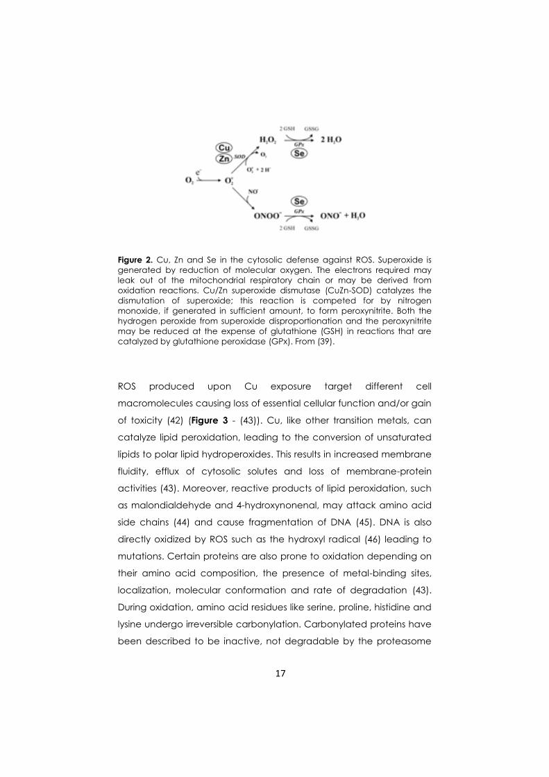
involved in cytosolic antioxidant defense as illustrated in Figure 2 (39):
hydroperoxides, including H2O2, are reduced to the respective

16
alcohols and water by the selenoenzyme glutathione peroxidase
(GPx) (40, 41).
Notwithstanding Cu is involved in cellular defence against ROS, its
redox chemistry makes it also responsible for ROS production.
Interaction of Cu ions with thiols such as glutathione – GSH – and
oxygen generates superoxide and hydrogen peroxide (see reactions i
and ii, where the latter is the sum of reactions iia and iib):
2Cu2+ + 2R-SH → 2Cu+ + R-SS-R + 2H+ (i)
2Cu+ + O2 + 2H+ → 2Cu2+ + H2O2 (ii)
2Cu+ + 2O2 → 2Cu2+ + 2O2-• (iia)
2O2-• + 2H+ → O2 + H2O2 (iib)
After its generation, Cu+ may further interact with hydroperoxides to
generate hydroxyl or alkoxyl radicals in Fenton-type reactions (see
reaction iii):
ROOH + Cu+ → RO•+ OH- + Cu2+ (iii)
Cu-induced ROS formation could be the reason why cells evolved
defense mechanisms that both depend on Cu and scavenge ROS
(39).
17
Figure 2. Cu, Zn and Se in the cytosolic defense against ROS. Superoxide is
generated by reduction of molecular oxygen. The electrons required may
leak out of the mitochondrial respiratory chain or may be derived from
oxidation reactions. Cu/Zn superoxide dismutase (CuZn-SOD) catalyzes the
dismutation of superoxide; this reaction is competed for by nitrogen
monoxide, if generated in sufficient amount, to form peroxynitrite. Both the
hydrogen peroxide from superoxide disproportionation and the peroxynitrite
may be reduced at the expense of glutathione (GSH) in reactions that are
catalyzed by glutathione peroxidase (GPx). From (39).
ROS produced upon Cu exposure target different cell
macromolecules causing loss of essential cellular function and/or gain
of toxicity (42) (Figure 3 - (43)). Cu, like other transition metals, can
catalyze lipid peroxidation, leading to the conversion of unsaturated
lipids to polar lipid hydroperoxides. This results in increased membrane
fluidity, efflux of cytosolic solutes and loss of membrane-protein
activities (43). Moreover, reactive products of lipid peroxidation, such
as malondialdehyde and 4-hydroxynonenal, may attack amino acid
side chains (44) and cause fragmentation of DNA (45). DNA is also
directly oxidized by ROS such as the hydroxyl radical (46) leading to
mutations. Certain proteins are also prone to oxidation depending on
their amino acid composition, the presence of metal-binding sites,
localization, molecular conformation and rate of degradation (43).
During oxidation, amino acid residues like serine, proline, histidine and
lysine undergo irreversible carbonylation. Carbonylated proteins have
been described to be inactive, not degradable by the proteasome

18
and to form toxic aggregated species impairing cell viability, as
recently reviewed by Avery (43). Beside this direct modification of
already synthesized proteins, ROS can affect the translation
machinery (43). Finally, intracellular ROS can modulate several
signaling pathways triggering apoptotic cell death (43, 47, 48).
Figure 3. Major routes of ROS action in cells. From (42).
1.4 Protection against copper toxicity
To cope with the toxic effects of Cu and of other heavy metals, cells
rely on “protective” proteins such as metallothioneins, that directly
bind free cytosolic metal ions and on free radical scavengers.
Metallothioneins (MTs) are a family of cystein-rich, low molecular
weight polypeptides (49-51). MTs bind metal ions since they contain
multiple sequences motifs like Cys-X-Cys or Cys-X-X-Cys and have a
low content of hydrophobic residues (49, 52, 53). Three major features
19
allow MTs to be effective in metal ions detoxification: (a) the strength
of metal ions binding within polymetallic clusters (49, 50, 52, 53); (b)
the ease of metal exchange reactions - resulting in the sequestration
of potentially toxic ions (54); (c) the metalloregulated gene expression
– allowing to modulate MTs concentration in response to the
intracellular concentration of a certain metal (55, 56). Many
eukaryotic organisms contain a family of MTs genes. At least nine MTs
genes are expressed in humans and have been classified in four
isoform classes on the basis of their role in metal ion detoxification (57-
61).
In S. cerevisiae the MTs gene family is composed by two loci: CUP1
and CRS5, encoding two polypeptides unrelated in sequence.
Multiple copies of CUP1 are present in the yeast genome (50, 61-64),
whereas only a single copy gene was found for CRS5 (65). Both genes
are transcriptionally activated by Cu ions (56, 65, 66), and their basal
and induced expression depends on the transcription factor Ace1
(33, 56, 65-69). The CRS5 promoter contains only a single recognition
sequence for Ace1 while CUP1 has four recognition sites, resulting in a
different induction of the two genes (65, 66, 70, 71). In addition, Cup1
and Crs5 bind a different number of metal ions. In particular, Cup1
binds 7 Cu(I) and 4 Cd(II) ions (71-73), whereas a greater binding
capacity was demonstrated for Crs5: 11-12 Cu(I) and 6 Cd(II) (30).
However, Cup1-Cu(I) complexes are kinetically more stable than Crs5-
Cu(I) complexes (30). Thus, multiple gene copies, greater
responsiveness of the promoter to Cu ions and strongest Cu avidity
contribute altogether to the dominant role of Cup1 in Cu buffering in
S. cerevisiae. Indeed, yeast cells defective in CUP1 gene are
hypersensitive to Cu salt (63, 74); on the contrary deletion in CRS5
gene leads to only reduced sensitivity to Cu (65). Whereas Cup1
expression is established as a primary defense to counteract Cu ions
20
toxicity, Jensen and collaborators (30) – on the basis of metal-binding
studies - proposed for Crs5 a role in Cu bioavailability and utilization.
Besides the promoters of CUP1 and CRS5, the Cu-dependent
transcription factor Ace1 recognizes also the promoter of SOD1. This
gene encodes Sod1, a Cu/Zn superoxide dismutase, and is
transcribed upon binding of Ace1 to a single binding site (32). Sod1
protects cells from oxidative damages by the disproportionation of
superoxide anions to oxygen and hydrogen peroxide (38). In S.
cerevisiae – as well as in mammals – Sod1 is mainly located in the
cytosol, with a smaller fraction in the intermembrane space of
mitochondria (75, 76) The sequence and structure of Sod1 is
conserved from prokaryotes to eukaryotes (77). The protein associates
to form a dimer (78) stabilized by an intramolecular disulfide bond.
Each subunit contains an active site with one Cu ion, acting as the
catalyst for superoxide disproportionation, and one Zn ion, that
participates in proper folding (79). Insertion of Cu into the active site is
facilitated by the metallochaperone Ccs1 that docks with and
transfers the metal ion into the disulfide-reduced apo-Sod1 (80, 81).
In S. cerevisiae metallothioneins and Cu/Zn superoxide dismutase are
directly induced by Cu exposure and protect cells either by buffering
Cu ions or by detoxifying Cu-derived ROS. In the cell, among proteins
participating in direct ROS detoxification or in redox balance of
protein thiols there are also catalases and peroxidases (82). Catalases
reduce hydrogen peroxide using the redox properties of a haem
group complexed to the polypeptide (83) and peroxidases
(glutathione peroxidases and peroxiredoxins) reduce peroxides to the
corresponding alcohols using active site cysteine thiols (84, 85).
In addition to enzymatic antioxidant systems, cells possess also non
enzymatic defences against metal-induced oxidative stress, the most
important of which is the tripeptide glutathione – GSH. In S. cerevisiae
GSH may account for 1% of the cell dry weight. This small molecule is

21
a storage form of endogenous sulphur and nitrogen as well as a
metal-ion detoxifying agent (86). In the cytosol, GSH has a role of
cellular redox buffer, since it is maintained in the reduced state by a
NADPH-dependent glutathione reductase (87).
While in Saccharomyces cells metallothioneins act as primary
defenses against toxic levels of Cu, in other yeasts Cu resistance can
be achieved by other mechanisms. For example, in the fission yeast
Schizosaccharomyces pombe peptides named phytochelatins
sequester Cu (88) and in Candida albicans a plasma membrane
efflux pump can extrude Cu ions by a mechanism described also in
some prokaryotic cells (89, 90).
1.5 Yeast as a new food source of essential copper. Is it
possible?
Beside its use as a model of eukaryotic cells, yeast is an ante litteram
biotechnological tool, traditionally used by mankind for thousands
years for fermenting food and beverages (91). Starting from ancient
times, domestication of naturally occurring yeasts by human selection
led to the diffusion of strains optimized for specific fermentation
procedures (92). Moreover, yeast can be used in human diet due to
its composition in proteins (35-55% dry weight), carbohydrates (25-
40%) and fats (up to 3%). Yeast has a high content of total nitrogen (7-
9%), 18-20% of which is non-protein nitrogen and is a rich source of
soluble vitamins (B group) and calciferol (D group) (93, 94). In
addition, yeast biomass contains different trace elements (TE) –
specifically bound to the proteic component - such as Cr, Se, I, Fe, Zn,
Cu and Mn, even if their amounts are not sufficient to satisfy human
requirements (94). However, if properly modified and/or cultured,
yeast cells can accumulate significative amounts of TE. Such a
22
feature makes yeast a valuable food source of TE to compensate
deficiencies in specific people categories (95, 96). Another important
issue is the chemical form by which TE are supplied: the organic form,
present in the majority of foods, is preferred because of its easier
uptake and the reduced risk of detrimental side effects compared to
inorganic salts and oxides (97). In this context, yeast biomass could be
a safe (it is a “GRAS” organism) and good quality source of TE.
A number of literature data and patents report on the preparation
and the application of TE-enriched yeasts and, among them, also Cu-
enriched yeasts (98-101), for example a patent concerning protocols
for the Cu-enrichment of S. cerevisiae biomass and its use for
preparation of pro-biotic, dietary and nutraceutic products (101). Cu-
enriched yeasts are commercially available, for example
Lalmin®Cu1000 (Lallemand Health Ingredients, Copenhagen,
Denmark) containing 1000-1400 µg Cu/g biomass. Vinson and
collaborators (102) compared the bioavailability of different forms of
Cu - Lalmin®Cu1000 and Cu-gluconate - as dietary supplement. Cu is
present in the body only bound to proteins, but not as a free ion or
salt. It has been demonstrated that Cu administered as Cu-enriched
yeast, is more bioavailable than Cu-gluconate in rats liver. It has been
hypothesized that Cu from yeasts is bound to proteins and/or
glutathione and therefore is more easily absorbed into the body from
the gastrointestinal tract and then transported to the liver. On the
other hand, Cu-gluconate requires proteins as carriers for absorption
and, unless associated with a protein-rich diet, this salt form of Cu
supplementation is not effective. In a similar study (103), the relative
absorption of Cu in animals fed with diets containing Cu-enriched
yeast was statistically higher if compared with those fed with diets
containing Cu sulphate. However, though these studies support the
usefulness of Cu-enriched yeasts, according to the European Food
Safety Authority (EFSA) available information for Cu-enriched yeasts is

23
still not sufficient to assess the safety of their use for nutritional
purposes.
1.6 Biotechnological tools to obtain copper-enriched yeast
biomass
The use of metal-enriched microbial biomasses, especially S.
cerevisiae cultures, has been proposed for bioremediation, removal
or recovery of metal ions and for analytical purposes (104-107). In this
context, based on the understanding of metal uptake mechanisms,
genetic technologies have been applied to improve
bioaccumulation properties of biomasses (108-110). For example,
Kuroda and collaborators (109, 111) obtained a cell-surface modified
S. cerevisiae strain with an histidine hexa-peptide exposed on the cell
surface. This strain displayed improved ability of Cu chelation and self-
aggregation properties. Using cell-surface engineering Kuroda and
Ueda (112, 113) also constructed yeast cells exposing tandem repeats
of metallothioneins leading to an enhanced adsorption of Cu and Cd
ions, as well as increased tolerance toward toxic metal
concentrations.
Beside the use of engineered cells, the simplest way to obtain metal-
enriched and tolerant cells is to exploit the intrinsic capacity of
microbial cells to adjust themselves to changing environments. This
ability represents the pivotal principle on which experimental designs
of evolutionary engineering are based on.

24
1.7 Evolutionary engineering
The term evolutionary engineering was introduced for the first time by
Butler and collaborators (114) by analogy with metabolic engineering
(115) to indicate an approach that uses the evolutionary principles to
direct the selection of organisms with a desired set of phenotypes. This
approach allows improvement of microbial properties by simple
random mutation and natural evolution but also by recombination
and continuous evolution of large populations over many generations
(116).
According to the basic rules of evolution, species evolve through
random variations (via mutation, recombination, or other operators);
the next step consists in natural selection in which the fittest survive
and reproduce themselves, propagating their genetic material to the
next generations. In addition, novel metabolic functions can be
acquired by mutational activation of cryptic genes, which represent
a versatile tool to enhance the adaptive potential of a species. These
cryptic genes are phenotypically silent DNA sequences, not
expressed under normal conditions, that are supposed to have
played a role in natural evolution (117). In this context, another
important set of genes are the evolution genes, that act in DNA repair
for the benefit of evolution itself, generating and modulating
spontaneous mutations (118, 119).
Evolutionary adaptation of species to changing environments is a
process occurring in all simplest cultivation systems. For example the
wild-type laboratory strains are the outcome of an evolutionary
domestication process (like in the case of S. cerevisiae, largely
exploited for baking and alcohol production (116)).
A schematic representation of the population dynamics during
adaptative evolution of a population is provided in Figure 4 (116).

25
Figure 4. Scheme of the population dynamics during adaptive evolution of an
asexual population. The gray line at the bottom represents the abundance of
neutral mutants (at a linear scale). The other lines indicate periodic selection
of two consecutively evolving advantageous mutants (at a logarithmic
scale). From (102) inspired by a similar scheme (106).
In a culture inoculated from a single clone, a new advantageous
mutation generally occurs in the much larger population that does
not carry neutral mutation. Subsequently, the adaptive mutant
replaces the existing population (including the fraction of neutral
mutants) at the log linear rate of selection. Neutral mutation will
continue to arise at the same linear frequency in the adaptive
mutant, until another advantageous mutation occurs, again in the still
predominant population without the neutral marker phenotype. The
abundance of the neutral marker phenotype drops again and the
cycle is repeated (116, 120).
Adaptive evolution can be achieved in a number of ways.

26
Selection on solid media offers the advantage to screen a great
number of mutants with a certain phenotype by direct read-out of
the progress of the evolutionary adaptation, especially when it is
unclear to what extent improvement is possible (121). However this
protocol of selection is inefficient for complex phenotypes that require
multiple mutations (116).
Adaptation by selection in liquid media allows the evolution of fitter
variants over time, with consequent replacement of the parental
population. During selection in batch cultures, cells are subjected to
changes in environmental conditions and alternate periods of growth
and stasis upon serial transfers (116). An exemplar experiment of
selection in batch culture was performed by Lensky and coworkers
(122-124). They analyzed the fitness of 12 independent E. coli
populations derived from a single ancestor. These populations were
propagated for about 10000 generations by daily serial transfer in
shaking flasks containing glucose-supplemented minimal medium.
After 10000 generations, the mean fitness of the derived clonal
variants was increased by 50% with respect to the common ancestor.
Although in the 12 populations the phenotypic changes were
consistent, they showed a great genetic diversity. Over time, the
evolved genomes became more and more different from the
ancestor and from each other. In the evolved populations point
mutations occurred rarely, meaning that the accumulated genomic –
and phenotypic – changes were mostly a consequence of
chromosomal rearrangements. Fundamental mutations shared
among all members of a given population represent good
candidates for phenotypically relevant mutations. This example
illustrates how evolution of adaptative performance is very
reproducible, although the same phenotypic adaptation can be
achieved by different genotypes (116). Moreover, Lensky and
collaborators (123) demonstrated that about half of the phenotypic

27
improvement occurred within the first 2000 generations, thus the rate
of fitness obtained in microbial population assumes an hyperbolic
profile, decreasing over time.
In batch culture, cells are subjected to changes in several
environmental parameters and the effects of evolutionary adaptation
could be attributed to any of the different phases of growth that cells
undergo. Conversely, continuous culture systems – in chemostat -
providing a constant environment, are a good tool to modulate
selective pressure toward a phenotype of interest (120, 125). However,
a disadvantage of selection in continuous systems is the strictly
sequential appearance and fixation of adaptive mutations. As a
consequence, a newly appearing variant may compete only with its
immediate one or few predecessor, if the older variants were
previously counter-selected. It may happen that new variants exhibit
lower fitness compared with the more distant predecessor (126).
Therefore, the combination of adaptive mutations during continuous
experiments may result in misadapted clones, which may have never
directly competed with the later occurring variants (116). Conversely,
during selection in batch culture a steady, although hyperbolic
improvement in fitness is observed (123).
Evolutionary engineering has been used in biotechnology to improve
simple as well as complex cellular subsystems, as illustrated in Table 3
(116).

28
Evolved phenotype Selection system
Novel catabolic activities
Utilization of carbon substrates
(coryneform bacteria)
Plates (with limiting amount of yeast
extract)
Utilization of pentoses (E. coli) Plates (non-growing cells)
Novel esterase activities (P. putida) Plates (non-growing cells)
Galactitol dehydrogenase
(Rhodobacter)
Chemostat (glucose-limited, excess
galactitol)
PTS-independent uptake Chemostat
Improved enzymes properties
Functionality (E. coli mutator strain) Batch (increasing antibiotic
concentrations)
Improved plasmid functions
Stability (Gram positive, yeast) Chemostat (antibiotic and auxotrophic
marker selection)
Stable host-plasmid combinations (E.
coli)
Chemostat
Improved stress resistance
Multiple stress resistance (yeast) Chemostat and batch (with stress
challenges)
Membrane protein overexpression (E.
coli)
Plates
Improved production properties
Endo-enzyme overexpression Chemostat
Protein secretion (Streptomyces) Chemostat (different selection schemes)
Biomass yield (yeast, E. coli) Chemostat (carbon-limited)
Table 3. Recent examples of evolutionary engineering. From (116).
Recent experimental works report on the use of evolutionary
engineering to evolve microorganisms resistant to metals, i.e. Cobalt
and Boron (127, 128). In these works, selection under continuous
exposure to gradually increasing metal concentrations allowed the
isolation of hyper-resistant mutant cells. Evolved cells showed
acquired cross-resistance to other stress conditions (such as exposure
to other metals, heat shock and oxidizing agents). Literature also
refers the effect of evolutionary adaptation on different yeast cells

29
growing in presence of Cu and their properties of bioaccumulation
(129, 130).
In conclusion, in vivo evolutionary engineering of whole organisms
represents a valuable alternative to metabolic engineering, often
limited by the great complexity of dynamic interactions in cellular
systems (116). In a flow chart taken from Sauer’s review (Figure 5)
(116), it is clearly illustrated how evolutionary engineering well fits in
the routes exploited by biotechnology to develop new strains.
Strains obtained with evolutionary engineering can be used as hosts
for further rational improvements by metabolic engineering or a
desired phenotype can be transferred to a production host – an
approach known as inverse metabolic engineering (131). In this latter
case, characterization of the molecular bases of evolved phenotypes
is required. In this context proteomics and transcriptomics, DNA chips
and metabolic flux analysis are precious tools for the identification of
genes beneficial or detrimental for a specific selected phenotype
(132-134). Altogether – and combined with bioinformatics tools for
modeling cellular functions and activities (116) – these methodologies
contribute to make evolutionary engineering an attractive approach
in the field of microbial biotechnologies.

30
Figure 5. Flow chart for future development of biotechnological strains. From
(116).

31
2. Experimental work
In this section three experimental works are presented:
2.1 Laboratory evolution of copper tolerant yeast
strains
GM Adamo, S Brocca, S Passolunghi, B Salvato and M
Lotti ( Microbial Cell Factories 2012, 11:1)
2.2 Evolution of copper tolerance in Saccharomyces
cerevisiae relies on amplification of the CUP1
gene
GM Adamo, M Lotti, MJ Tamás and S Brocca
(Manuscript)
2.3 Proteomic analysis of natural and copper-
adapted cells of the yeast Candida humilis
GM Adamo, S Brocca and M Lotti
(Preliminary results)
32
As already mentioned in the introduction, evolutionary
engineering is a valuable strategy to obtain strains with a desired
phenotype. In the first work (2.1), we described an evolutionary
engineering strategy to obtain yeast strains endowed with
tolerance toward high concentration of Cu ions, supplied as
CuSO4. The starting yeast populations were natural strains of S.
cerevisiae and C. humilis, characterized by different basal Cu
tolerance, being the former Cu sensitive and the latter Cu
tolerant. These strains were step-wise evolved through continuous
cultivation in presence of increasing concentration of copper
salt. As a result, cells improved their Cu tolerance up to 2.5 g/L
CuSO4 and were able to accumulate high amounts of metal ions.
The acquired tolerance was permanently maintained, underlying
the effectiveness of evolutionary engineering in the acquisition of
stably phenotypic traits. A preliminary characterization of Cu-
evolved Saccharomyces and Candida cells revealed common
features associated with Cu tolerance: improvement of cell
vitality and reduction of ROS production. At the same time,
differences in the response of antioxidant enzymes and Cu-
binding proteins were found between the two yeasts. An
intriguing issue emerging from this work is that, although for both
strains evolution of Cu tolerance moves in the same direction
(increase in strain robustness), S. cerevisiae, naturally more
sensitive to Cu, improved its robustness at higher extent than C.
humilis, characterized by a natural Cu tolerance. This is likely to be
associated with different genetic backgrounds of the two strains.
This finding prompted us to further investigate the molecular
bases of Cu tolerance in evolved yeast cells.
In the second work (2.2), we focused our attention on evolved S.
cerevisiae cells to evaluate the role of the metallothionein Cup1.
This work was based on literature data demonstrating the pivotal

33
role of Cup1 in mediating Cu tolerance of natural S. cerevisiae
cells (61). Briefly, in the S. cerevisiae evolved strain we found out
an amplification of CUP1 gene copy number, triggering a strong
increase in gene expression. We ascribed to the over expression
of Cup1 the reduction of oxidative stress observed in the evolved
strain grown in copper medium. At the same time, we found that
other factors, such as the metallothionein Crs5 and the radical
scavenger Sod1, play a secondary role in protection against Cu-
induced oxidative stress. Altogether these results, show the
predominant role of CUP1 amplification in the evolution of Cu
tolerance.
A different approach was applied to investigate on Cu tolerance
of evolved C. humilis cells; preliminary results are presented in the
last work (2.3). Due to the scarce information available about the
C. humilis genome, we used a proteomic approach to gain
insight into the molecular changes occurring during Cu exposure
in non-evolved and evolved cells (obtained as described in the
work 2.1). The differential analysis revealed for both strains a
comparable response, in terms of number and type of proteins
up- or down- regulated by copper. Identified proteins belong to
distinct groups such as glycolytic enzymes, heat shock proteins,
proteins involved in protein synthesis and energy production,
proteins involved in phospholipids synthesis. All these proteins are
involved in the protection against oxidative stress and were
previously identified in works performed on S. cerevisiae cells (135-
138). This is to our knowledge the first proteomic analysis of
Candida cells during oxidative stress. As a matter of fact, while
copper induce changes in the level of proteins of both evolved
and natural cells, we cannot ascribe the evolution of copper
tolerance to the differential expression of one (or few) specific
protein. However, any observed change in protein level was less

34
marked in the evolved strain. This observation suggests that the
tolerance to copper in evolved strain is only partially sustained by
molecular mechanisms involved in the oxidative stress response
and that other mechanisms can be active in preventing Cu
toxicity in the evolved strain.

35

36
Laboratory evolution of copper tolerant yeast strains
Microbial Cell Factories 2012,11:1
Published 3 January 2012
Giusy Manuela Adamo1, Stefania Brocca
1, Simone Passolunghi
1,
Benedetto Salvato2 and Marina Lotti
1*
Address: 1Dipartimento di Biotecnologie e Bioscienze, Università degli Studi di
Milano-Bicocca, Piazza della Scienza 2, 20126 Milano, Italy and 2Dipartimento di
Biologia, Università di Padova, Via Ugo Bassi, 58/B, 35121 Padova, Italy
Email: Giusy M. Adamo: [email protected]; Stefania Brocca:
[email protected]; Simone Passolunghi: [email protected];
Benedetto Salvato [email protected]; Marina Lotti :
* Corresponding author

37
Background
Yeast strains endowed with robustness towards copper and/or enriched in
intracellular Cu might find application in biotechnology processes, among others in
the production of functional foods. Moreover, they can contribute to the study of
human diseases related to impairments of copper metabolism. In this study, we
investigated the molecular and physiological factors that confer copper tolerance to
strains of baker’s yeasts.
Results
We characterized the effects elicited in natural strains of Candida humilis and
Saccharomyces cerevisiae by the exposure to copper in the culture broth. We
observed that, whereas the growth of Saccharomyces cells was inhibited already at
low Cu concentration, C. humilis was naturally robust and tolerated up to 1 g · L-1
CuSO4 in the medium. This resistant strain accumulated over 7 mg of Cu per gram of
biomass and escaped severe oxidative stress thanks to high constitutive levels of
superoxide dismutase and catalase. Both yeasts were then “evolved” to obtain hyper-
resistant cells able to proliferate in high copper medium. While in S. cerevisiae the
evolution of robustness towards Cu was paralleled by the increase of antioxidative
enzymes, these same activities decreased in evolved hyper-resistant Candida cells.
We also characterized in some detail changes in the profile of copper binding
proteins, that appeared to be modified by evolution but, again, in a different way in
the two yeasts.
Conclusions
Following evolution, both Candida and Saccharomyces cells were able to proliferate
up to 2.5 g · L-1
CuSO4 and to accumulate high amounts of intracellular copper. The
comparison of yeasts differing in their robustness, allowed highlighting
physiological and molecular determinants of natural and acquired copper tolerance.
We observed that different mechanisms contribute to confer metal tolerance: the
control of copper uptake, changes in the levels of enzymes involved in oxidative
stress response and changes in the copper-binding proteome. However, copper
elicits different physiological and molecular reactions in yeasts with different
backgrounds.

38
Keywords: yeast, copper, adaptation, evolutionary engineering, oxidative stress
response, micronutrients

39
Background
Metal ions like copper, manganese, zinc and iron are essential micronutrients for
living organisms and play a central role in the cell metabolism being the cofactors of
a large number of enzymes and electron transport proteins [1]. The metabolism of
copper and the mechanisms that control its intracellular concentration are the targets
of intense studies since impairments in Cu level, transport and localization have
been associated with several human diseases [2, 3]. In fact, while copper deficiency
impacts the function of key cell enzymes, Cu overload can generate highly reactive
oxygen species (ROS) which produce peroxidation of membrane lipids,
displacement of other metal cofactors from their natural ligands in signalling
proteins [4], oxidation of proteins and cleavage of DNA and RNA molecules [5]
resulting in general cellular damage. Moreover, ROS are thought to play a major
role in cancer development and in aging [6]. To cope with such strict constraints, all
organisms have developed complex regulatory mechanisms to maintain copper
homeostasis.
Yeast cells are a good tool both for the investigation and the manipulation of
copper metabolism. Studies on the accumulation of metals in edible microorganisms
are of relevance for the production of functional foods enriched in micronutrients
(for example the ones about the inclusion of iron, cobalt, copper and manganese in
yeast cells [7]) and the industrial production of Saccharomyces cerevisiae biomass
highly enriched with organic forms of selenium [7, 8]. Yeast cells resistant to and
accumulating intracellular copper have been recently patented for cleaning copper
from extracellular solutions [9] and use in pro-biotic, cosmetic, dietary and
nutraceuticals products [10].
It has been reported that microorganisms can acquire stress tolerance and novel
metabolic abilities when exposed to the appropriate selection pressure. This
approach is often reported as “evolutionary engineering”, a term introduced by
Butler and collaborators in 1996 [11], since it uses evolutionary principles based on
the selection of random mutants arising in the microbial population either
spontaneously or upon mutagenesis and has been applied for improving complex
physiological properties whose genetic and physiological basis is not fully
understood [12]. For example, microbial cells were recently evolved to improve their

40
resistance towards multiple stresses [13], cobalt [14], iron- and sulfur-compounds
[15], alcohols [16]; and to gain the ability to ferment xylose [17] and lactose [18].
S. cerevisiae is a powerful model organism to investigate copper metabolism and
homeostasis in Eukaryotes. As a consequence, a large body of knowledge is
available about Cu uptake, intracellular transport and functional role in yeast cells
[19], as well as about non-enzymatic and enzymatic mechanisms of protection from
ROS and oxidative stress [20].
In this study, yeast strains endowed with different natural robustness towards
copper were compared with strains evolved by stepwise adaptation to tolerate high
metal concentrations. We report that different and overlapping physiological and
molecular responses are elicited in cells with different backgrounds to allow them to
tolerate challenging conditions.
Results
Tolerance towards copper of one Candida and three Saccharomyces strains was
first assessed by a drop test on minimal or rich (YPD) solid medium supplemented
with increasing concentrations of copper salt (CuSO4) (Fig. 1). In good agreement
with results from other laboratories showing that the composition of the culture
medium and the growth conditions affect copper sensitivity of yeast cells [21, 22],
we observed that on minimal medium, 0.5 g · L-1
CuSO4 was sufficient to inhibit the
growth of all yeast strains, whereas on YPD plates all of them tolerated up to 1 g · L-
1 CuSO4. However, above this concentration only C. humilis cells still proliferated,
suggesting low copper tolerance in all strains assayed but C. humilis which showed a
higher tolerance.
The tolerance of cells to metals is relevant both for understanding the
mechanisms of defence towards stress and for the production of microorganisms
enriched in a given micronutrient for biotechnological applications. We applied an
evolution-based approach to improve robustness towards copper in all strains,
independently from their natural background. The experimental protocol relied on
the stepwise cultivation of cells in media supplemented with progressively higher
concentrations of copper sulphate. At each step the culture was grown for 72 hours
before withdrawing aliquots to be inoculated at a higher CuSO4 concentration. The

41
starting condition was YPD + 1 g · L-1
CuSO4, which is permissive for all strains.
The following steps were in YPD + 1.5 g · L-1
CuSO4, YPD + 2 g · L-1
CuSO4 and
YPD + 2.5 g · L-1
CuSO4. This last one was the highest concentration applied, since
above it copper salts led to acidification of the medium resulting in the precipitation
of its protein components. Single colonies isolated after the last step of adaptation
displayed relatively high rates of growth when directly re-inoculated in YPD + 2.5 g
· L-1
CuSO4. These cells are defined in the following as “evolved”.
Figure 2 compares the growth kinetics of non-evolved and evolved cells in YPD
+ 2.5 g ·L-1
CuSO4 (for simplicity we will refer to this condition as “copper
medium”). Among the natural strains (in the following “non-evolved”), only C.
humilis AL5 proliferated under this condition, even though growth started after a
prolonged lag phase and a very low final cell density was achieved (Fig. 2 a). On the
contrary, all evolved strains proliferated in copper medium and reached final
biomass densities close to those observed in YPD medium, although with longer
duplication times (Additional file 1). Cells subjected to 10 cycles of growth/re-
inoculation in YPD without Cu (referred to as “de-adapted”), retained their ability to
proliferate if re-inoculated in copper medium, showing only negligible differences
when compared with the corresponding evolved strain (Fig. 2 and Additional file 1).
This observation suggests that copper tolerance is maintained also in absence of
selective pressure. To gain more insight into the behaviour of the copper-sensitive S.
cerevisiae strains, we compared the kinetics of growth of evolved and non-evolved
cells also at 1, 1.5 and 2 g ·L-1
CuSO4 (Additional file 2), highlighting a progressive
decrease of the proliferation ability of natural cells at increasing copper
concentrations. As expected, the same conditions were permissive for evolved
Saccharomyces cells.
Analysis of the Cu content of Candida cell samples from the same cultures
shown in Figure 2 revealed relatively high amounts of intracellular copper, i.e. 6.5
and 7.6 mg · g-1
biomass in evolved and non-evolved cells, respectively. The
kinetics of bioaccumulation was faster in non-evolved cells where Cu measured
after 24 hours of growth was three times higher than in the evolved ones (Fig. 3 a).
In copper medium non-evolved Candida grew poorly and contained high copper
concentration from the very beginning of the experiment, while Cu was lower in
evolved cells – which proliferated at the same rate as in YPD broth. In both cases,

42
intracellular copper kept increasing up to 48 hours and then reached a plateau. The
amount of copper measured in evolved and de-adapted cells grown in copper
medium was always comparable, supporting once more the hypothesis that
evolutionary engineering produced stable effects.
The increase of intracellular copper in evolved S. cerevisiae was slower and at
the end of the experiments we measured 2.1 to 4.2 mg Cu · g-1
biomass (Fig. 3 b, c,
d). The Cu content of non-evolved and evolved Saccharomyces cells was compared
also in a milder condition, (1 g ·L-1
CuSO4). Whereas both kinds of BL7 cells
displayed the same kinetics of copper accumulation (Additional file 3 a), non-
evolved EL1 and GL6 showed a faster kinetic of bioaccumulation (Additional file 3
b, c) associated to a growth kinetic slower than in their evolved counterpart
(Additional file 2). As in Candida samples, also in this case the behaviour of
evolved and de-adapted cells was similar.
Results obtained up to this point suggested that all Saccharomyces strains are
rather homogeneous in their response to copper, but different from the Candida one.
Therefore, subsequent experiments aimed at highlighting possible adaptive changes
were focused on a more in-depth comparison between Candida humilis and the only
Saccharomyces cells strain BL7.
Initially, the effect of copper on cell viability was evaluated by citofluorimetry
(Fig. 4), that allows to get this information at the single cell level [23]. Samples of
C. humilis and S. cerevisiae cultures were harvested during exponential growth in
copper and stained with propidium iodide, an intercalating agent excluded by viable
cells, that can instead permeate the surface of seriously injured/dead cells [24]. In
both evolved strains the percentage of propidium-positive cells was lower (8.5 % for
C. humilis and 11 % for S. cerevisiae) than in their non-evolved counterpart (28 %
for C. humilis and 60 % for S. cerevisiae). The percentage of propidium-positive
cells grown in absence of copper (used as a control) was around 2 % (data not
shown). Altogether, these results confirm that evolution confers robustness -
although not complete insensitivity - to copper. Copper was more detrimental for S.
cerevisiae than C. humilis natural cells, while this difference fainted after adaptive
evolution, in good agreement with the kinetics of growth reported in Figure 2.
Since copper is a strong oxidizing agent, we measured the activities of
superoxide dismutase, peroxidase, glutathione peroxidase and catalase - all involved
43
in the so-called copper-dependent oxidative stress response – in evolved and non-
evolved C. humilis and in evolved S. cerevisiae cells harvested from copper medium
in the exponential phase of growth. As a control, basal enzymatic activities were
determined in YPD-grown cells (Table 1). In S. cerevisiae, exposure to copper
resulted in the increase of superoxide dismutase and catalase activities, while
peroxidase and glutathione peroxidase activities were only marginally affected. The
picture emerging from the analysis of the copper tolerant C. humilis strain is
different and more exhaustive, since the data set can also include the response of
non-evolved cells in copper medium. In all cultivation broths, all enzymatic
activities tested in natural Candida cells were 2 to 8 fold higher than in S. cerevisiae.
Interestingly enough, we detected high constitutive superoxide dismutase and
catalase activities in non-evolved cells, independently from the composition of the
culture broth, while peroxidase and glutathione peroxidase activities were induced
only by growth in copper medium. Evolution resulted in lower levels of all tested
activities in both YPD and copper medium, with the most remarkable effect on
superoxide dismutase. Catalase activity that was high in YPD-grown cells (both
kinds of cells), strongly decreased in evolved cells grown in copper medium.
We then evaluated the production of reactive oxygen species (ROS) staining
cells with dihydroethidium (Fig. 5). In presence of superoxide anions in the
cytosolic space, this probe is oxidized to the fluorescent product ethidium.
Therefore, fluorescence intensity reports on oxidative stress. While in YPD medium
the amount of ROS was low in both Candida and Saccharomyces cells, copper
exposure clearly triggered oxidative stress, though with milder effects in the evolved
cells. Moreover, in agreement with growth and cytofluorimetric data (Fig 2 and 4),
the effect on S. cerevisiae was stronger than on Candida with ROS production three-
fold higher. Evaluation of oxidative stress in S. cerevisiae at intermediate metal
concentration (1, 1.5 and 2 g ·L-1
CuSO4) showed that while ROS production
increases with copper concentration in the non-evolved sample, it remains low in the
evolved cells (Additional file 4).
Finally we performed a preliminary analysis of the copper-binding proteins
extracted from non-evolved and evolved cells grown either in YPD or in copper
medium. Samples were enriched by affinity chromatography as described in
Materials and Methods and then analysed by SDS-PAGE. Equal volumes of elution

44
fractions obtained from the same amount (800 µg) of proteins applied on the column
were loaded for the electrophoretic run, therefore assuring that differences detected
in the gel reflect changes in composition and content of copper-binding proteins in
the starting samples. Figure 6 shows the electrophoretic analysis and Table 2 lists
proteins identified by tandem mass spectrometry. The exposure of evolved S.
cerevisiae cells to copper elicited the induction of several proteins (Fig. 6a, lane 2
and 3) involved in different biochemical and metabolic functions, i.e. the pentose
phosphate pathway (band 1), amino acid and sulphur metabolism (bands 2, 3 and
10), glucose metabolism (bands 4 and 7), redox reactions (bands 6 and 8), the
translation machinery (bands 5 and 9) and isomerization reactions (bands 1 and 9).
The same trend was detected at lower CuSO4 concentration (data not shown). The
picture relevant to Candida is completely different (Fig. 6 b). Non-evolved Candida
cells react to copper repressing a number of Cu-binding proteins that would be
otherwise expressed during growth in YPD (Fig. 6b, lane 1 and lane 3). Among the
down-regulated proteins we could identify ribosomal proteins and components of
the protein translation apparatus (band 11, 13 and 16). The profile of Cu-binding
proteins extracted from evolved Candida cells (Fig. 6, lane 2 and 4) showed a
massive enrichment of a protein of ~35 kDa (band 14), identified as glyceraldehyde-
3-posphate dehydrogenase 3 (GAPDH). We further observed the increase of a
protein of ~ 22 kDa (band 15) identified as peroxiredoxin and of a protein inducible
by oxidative stress (band 12).
Discussion
In this work, we obtained copper-enriched and copper-tolerant yeasts through a
number of generations smaller than reported in similar recent works [13, 14, 25].
This might be explained on the basis of a different experimental set-up in which
relatively few rounds of selection were applied, but with longer cultivation times (72
h) and wider intervals of metal concentration (increases of 0.5 g . L
-1 at each round).
The effectiveness of the method of evolution is substantiated by the preservation of
metal tolerance in absence of selective pressure (de-adapted cells), meaning that
stable molecular changes occurred. This view is corroborated by the amplification of
the CUP1 gene, which encodes for a metallothionein, detected in evolved S.

45
cerevisiae cells (Adamo et al., in preparation). However, it is possible that other
adaptive (transient) modifications contribute to the increased resistance.
In the frame of this complex picture, the comparison of yeasts differing in their
robustness towards copper allowed us to investigate physiological differences
involved in natural and acquired copper tolerance and to obtain some preliminary
information about the molecular determinants of this trait. Taken together, our
results hint at the concurrence of different mechanisms that we briefly summarize
and discuss in the following.
i) Copper uptake. C. humilis cells can grow in copper medium due to their natural
tolerance that can be further improved via evolutionary engineering. This feature
allowed to compare the bioaccumulation of intracellular Cu in non-evolved and
evolved cells at the highest copper concentration used (2.5 g ·L-1
CuSO4), showing
that copper is lower in these latter. On this basis we hypothesize that one of the
mechanisms of robustness might rely on hindering metal uptake. Consistently, the
toxicity of Cu incorporated in the non-evolved strain (mainly in the first hours of
growth) might account for the growth impairment and the increase in the rate of
propidium positive cells we observed. In this light, the variability in copper
sensitivity between Candida and Saccharomyces cells might depend (at least partly)
on a different ability to limit copper uptake and its overload. Such a mechanism has
been reported to protect S. cerevisiae cells from copper [26] cadmium [27] and
cobalt [14] and points to a central role of the plasma membrane [28-30] and of the
cell wall [31] in the onset of tolerance to heavy metals.
ii) Antioxidative enzymes and ROS production. Our results indicate that the
biochemical bases of copper resistance can be deeply different among yeast species.
In S. cerevisiae, evolution of copper tolerance is associated with the increase of
antioxidative activities, as already documented by others [32], and with a reduced
production of ROS. On the contrary, non-evolved cells suffer severe oxidative
stress, as showed by the complete inhibition of their growth and the huge percentage
of propidium positive cells. Basal activities of most detoxifying enzymes are higher
in C. humilis than in S. cerevisiae cells, fact that could partly explain the natural
copper tolerance of Candida cells and the reduced ROS production. The response of
evolved Candida cells to copper is intriguing since our results show a reduction in
the production of ROS, a generalised decrease of the antioxidative activities, and
46
non-responsiveness to copper (compare values reported for evolved cells in YPD
and copper medium). Indeed, in evolved cells superoxide dismutase is always lower
than in the original strain, independently from the presence of copper. We would
assume that the different activity profiles and their re-shaping upon evolution might
reflect different and peculiar defence mechanisms responsible for both natural and
acquired copper tolerance in Saccharomyces and Candida strains.
iii) Cu-binding proteins. The hypothesis above is further corroborated by the
observation that the amounts of soluble Cu-binding proteins extracted from non-
evolved and evolved cells are different too. Over-expression of Cu-binding proteins
is consistent with their role in the primary response to copper exposure and results in
copper tolerance [33]. We are aware that the enrichment procedure used might lead
to overestimation of some proteins that carry modifications such as thiolation [34,
35] or sequence motives that increase their affinity for the chromatographic resin or
to underestimation of low-affinity proteins. Nevertheless, the differences detected
between the two yeasts are marked and relevant proteins identified are in agreement
with literature data reported by others. Among Cu-binding proteins, enzymes
involved in sulphur metabolism (i.e. cystathionine-γ-lyase and aspartate-
semialdehyde dehydrogenase) were over-expressed by evolved S. cerevisiae cells
during growth in copper medium, suggesting that copper might redirect the
metabolic flux towards the production of GSH to balance the redox equilibrium. The
over-expression of glycolytic enzymes such as triose phosphate isomerase and
GAPDH3 might be a consequence of the reconfiguration of the glycolytic flux, a
mechanism reported to regulate the response to oxidative stress in human [36], plant
[37] and yeast [38] cells. Furthermore, the increase of GAPDH3 is consistent with
the role of this protein as a sensor of oxidative stress in DNA repair [39] and
apoptosis [40, 41]. GAPDH3 increase is also the most remarkable effect triggered by
copper in evolved C. humilis. Besides, these cells are enriched in the peroxiredoxin
Tsa1 [42, 43], known to act as an antioxidant against ROS [44] and to protect
actively translating ribosomes from stress conditions [45]. On this basis we propose
that in evolved Candida cells the inhibition of protein synthesis associated with
oxidative stress [46] occurs in an attenuated form in comparison to non-evolved
cells, that show a strong decrease of Cu-binding proteins, without any remarkable

47
down regulation of the ribosomal proteins and other components of the protein
translation apparatus.
Conclusion
The comparison between yeast cells naturally resistant or experimentally evolved
to tolerate high copper concentration reported in this work supports the view that
copper tolerance is due to multiple responses relying on different physiological and
macromolecular changes. Yeasts endowed with copper tolerance and able to
accumulate metal ions can find application in the biotechnology field for example
for bioremediation or as dietary supplement, being these “GRAS organisms”
valuable sources of microelements in organic form. Moreover, the comprehension of
physiological and molecular responses of microorganisms to metal stress and of the
mechanisms triggered during evolution of tolerance could help in the identification
of biomarkers for ecotoxicological studies.
Methods
Yeast strains and growth conditions
Yeasts used in this study were isolated from sourdough. Strains were identified by
Random Amplification of Polymorphic DNA-PCR (RAPD-PCR) and designated as
Candida humilis AL5, and Saccharomyces cerevisiae BL7, EL1 and GL6 (Veneto
Agricoltura - Istituto per la Qualità e le Tecnologie Agroalimentari – VI). Growth
was on YPD medium [2% (w/v) glucose, 1% (w/v) yeast extract, 2% (w/v) tryptone]
or on minimal medium [2% (w/v) glucose, 0.67% (v/v) yeast nitrogen base]. Solid
media contained 2% (w/v) agar.
Copper tolerance was tested on cells grown shaking over night in 3 mL of liquid
YPD at 30°C and subjected to serial dilutions in physiological solution [0.9% (w/v)
NaCl]. 5 L aliquots were spotted on either YPD or minimal medium plates
containing CuSO4 and incubated at 30° C for 2 days.
Adaptative evolution was performed stepwise starting from cells taken from a fresh
culture on agarized YPD and grown overnight at 30°C in 3 mL of liquid YPD + 1 g
48
· L-1
CuSO4. In the subsequent steps, 5 × 10⁵ cells from stationary cultures were
inoculated and cultivated for 72 hours in fresh medium containing each time
increasing concentrations of copper (1.5 - 2 - 2.5 g · L-1
). Single colonies were
isolated from the culture on YPD + 2.5 g · L-1
CuSO4 by plating on solid YPD and
re-inoculated in liquid YPD + 2.5 g · L-1
CuSO4. To assess the endurance of metal
tolerance, evolved cells were subjected to 10 cycles of inoculation-growth in fresh
YPD medium without copper (de-adaptation) prior to be cultivated again in YPD +
2.5 g · L-1
CuSO4.
Growth of non-evolved and evolved S. cerevisiae cells (BL7, EL1 and GL6) was
assayed in liquid YPD medium supplemented with intermediate copper
concentrations (1, 1.5, 2 g, · L-1
CuSO4).
Growth was determined as the increase of cells number using a cells counter
(Particle Count Size Analyzer, Beckman Coulter).
Copper determination
Cells were harvested from 2 mL culture by centrifugation at 10,000 g for 10 min,
washed twice with de-ionized water and at least four times with 10 mM citric acid in
0.5% (w/v) NaCl to remove copper ions adsorbed on the cell surface. The biomass
was dried by Max-Dry Iyo (Heto) for 30 min, re-suspended in 300 L of 20% (w/v)
trichloroacetic acid (TCA), transferred to a 2 mL screw cap tube containing 100 μL
of glass microbeads and subjected to mechanical lysis by three cycles of 20 sec at
maximum speed with a Fast Prep - FP120 (Bio101-Savant). The crude extract was
clarified by centrifugation at 10,000 g for 10 min and the supernatant transferred to a
new tube. Copper was quantified according to Brenner and Harris [47] adapted as
follow. The clarified crude extract was diluted in 500 L of de-ionized water and
added to 100 L of 1% (w/v) ascorbic acid and 400 L of “BCA reagent” [0.006 %
(w/v) 2,2’-biquinoline-4,4’-dicarboxylic acid disodium salt hydrate (Fluka), 3.6%
(w/v) NaOH, 15.6% (w/v) Hepes]. After 2 min incubation at room temperature,
absorbance was recorded at 354 nm with a spectrophotometer Ultraspec 1000
(Pharmacia Biotec). The amount of copper is referred as mg Cu · g-1
dry biomass.

49
Flow cytometry analysis
Samples containing 107
cells were withdrawn during the exponential growth phase.
Cells were harvested by centrifugation, washed twice with deionized water and four
times with 10 mM citric acid in 0.5% (w/v) NaCl. Cells were washed with 1 mL of
PBS (3.3 mM NaH2PO4, 6.7 mM Na2HPO4, 127 mM NaCl, 0.2 mM EDTA) and
stained with 1 mL of the fluorescent probe propidium iodide (5 ng . mL
-1). Stained
cells were sonicated for 15 sec and then analyzed using a Cell Lab QuantaTM
SC
flow cytometer (Beckman Coulter) equipped with a diode laser (excitation
wavelength 488 nm, laser power 22 mW). The fluorescence emission was measured
through a 670 nm long pass filter (FL3 parameter) in logarithmic mode for
propidium iodide (PI) signal. Not stained and ethanol-treated samples were used as
controls. The sample flow rate during analysis did not exceed 500 cells sec-1
. A total
of 2 × 10⁴ cells were measured for each sample. Data analysis was performed with
WinMDI 2.8 software, build#13 01-19-2000 (Purdue University, Cytometry
Laboratories [http://facs.scripps.edu/software.html]).
Preparation of cell-free extracts
Cells from exponential cultures were harvested by centrifugation at 4,900 g for 10
min and washed twice with cold deionised water and four times with cold 10 mM
citric acid in 0.5% (w/v) NaCl. The cell pellet was finally re-suspended in 0.5 M
Tris-Cl pH 8.5, 0.25 M EDTA pH 8.4 added with protease inhibitor cocktail (Sigma)
and mechanically disrupted using glass microbeads. Cell debris was removed by
centrifugation at 700 g for 10 min and the clarified crude extract was used for
enzymatic analyses. The protein concentration in cell-free extracts was estimated
according to Bradford [48] using bovine serum albumin as the reference.
Enzyme assays
Enzyme activities were measured on cell-free extracts by spectrophotometric assays.
Activities were expressed as Units · mg-1
proteins. Catalase activity was determined
according to Bergmeyer [49] monitoring hydrogen peroxide decrease at 240 nm.
Superoxide dismutase activity was measured as the inhibition of the rate of
reduction of cytochrome c by the superoxide radical, observed at 550 nm [50].
Peroxidase activity was measured following the oxidation of pyrogallol at 420 nm

50
[51]. The activity of glutathione peroxidase was determined monitoring NADPH
oxidation at 340 nm [52].
Purification of copper-binding proteins
Supernatants obtained by ultracentrifugation (18,000 g, 45 min) of cell extracts were
heated at 65°C for 10 min to enrich thermostable proteins, since thermostability is
common to several copper-binding proteins [53]. Samples were then centrifugated at
10,000 g for 10 min and supernatants purified by affinity chromatography on a
Sepharose Chelating resin (Sigma) loaded with 0.2 M CuSO4 (copper resin).
Samples (ca. 800 µg protein) were incubated for 20 min with 0.5 mL of copper resin
previously equilibrated with 1 mL of binding buffer (0.02 M Na2HPO4, 0.5 M NaCl,
pH 7.2). Unbound proteins were removed by gravity flow and the column washed
three times with 0.5 mL of binding buffer. Bound proteins were eluted first with
0.02 M Na2HPO4, 0.5 M NaCl, pH 3.5 and then with 0.02 M Na2HPO4, 0.5 M NaCl,
0.05 M EDTA, pH 7.2. Proteins from 200 µL of each elution fraction were
precipitated with 60 µl of 20% (w/v) TCA, resuspended in 50 µL of SDS-Sample
buffer (0.25 M Tris-Cl pH 6.8, 50% (v/v) glycerol, 10% (w/v) sodium dodecyl
sulphate, 5% (v/v) β-mercaptoethanol, 0.25% (w/v) bromophenol blue), heated at
99°C for 5 min and applied to 18% (w/v) polyacrylamide gels. Electrophoresis in
denaturing conditions (SDS-PAGE) was carried out according to Laemmli [54].
Gels were stained by GelCode Blue (Pierce).
Mass spectrometry
Bands were excised from the polyacrylamide gels, cut into small pieces and de-
stained by repeated washing cycles alternating 50 mM ammonium hydrogen
carbonate and pure acetonitrile. After complete destaining, gel particles were
dehydrated by acetonitrile, covered with trypsin solution (12.5 ng/mL in 50 mM
ammonium hydrogen carbonate, pH 8.0) and incubated 1 h on ice. Excess liquid was
removed and the gel pieces covered with a solution of 50 mM ammonium hydrogen
carbonate (pH 8.0) and incubated overnight at 37°C. Tryptic peptides were extracted
by alternating incubation in pure acetonitrile and 1% formic acid. Samples were
lyophilised, resuspended in 1% formic acid, and desalted by ZipTip (Millipore)
before ESI-MS analysis.

51
ESI-MS experiments were performed with a hybrid Quadrupole-Time-of-Flight (q-
TOF) mass spectrometer (QSTAR ELITE, Applied Biosystems) equipped with a
nano-electrospray ionisation sample source. Metal-coated borosilicate capillaries
(Proxeon, Denmark) with medium-length emitter tip of 1-
used for off-line analysis. The instrument was calibrated with standard solution
Renin (MH2+ 879.97 Da and its fragment MH+ 110.07 Da, Applied Biosystems).
Peptide identification was performed using the MASCOT software with the
following parameters: 2 missed cleavages, peptide tolerance 0.6 Da, MS/MS
tolerance 0.6 Da, peptide charges 2+ and 3+. Only monoisotopic masses were
considered as precursor ions.
Spectra of tryptic peptides were acquired in the 400-1,500 m/z range, with 1.0 sec
accumulation time, ion-spray voltage 1,3000 V, declustering potential 60 V, with
active Information Dependent Acquisition (IDA), using rolling collision energy to
fragment peptides for MS/MS analysis.
Determination of Reactive Oxygen Species
Samples containing 5 × 10⁶
cells were withdrawn from exponential cultures. Cells
were harvested by centrifugation, washed twice with de-ionized water, four times
with 10 mM citric acid in 0.5% (w/v) NaCl, once with 1 mL of PBS and finally
incubated with 0.5 mL of 5 µM (v/v) dihydroethidium (Sigma, stock solution 30
mM in DMSO) for 30 min in the dark. Stained cells were washed three times with
PBS and sonicated for 15 sec. The fluorescence signal was detected using a Cary
Eclipse spectrofluorimeter (Varian, CA, USA) at excitation wavelength of 518 nm
and emission wavelength of 605 nm. Fluorescence values were normalized against
those of not stained cells.
Competing interests
The author(s) declare that they have no competing interests.

52
Authors’ contributions
GMA performed most of the physiological and biochemical characterization of
yeasts. SB participated in the design of experiments and in the analysis of results. SP
designed and carried out the experiments of cytofluorimetry. BS drew attention on
the importance of Cu tolerance in yeast and suggested the experimental strategy of
adaptation. ML conceived this study, contributed to the interpretation of results and
drafted the paper. All authors read and approved the final manuscript.
Acknowledgements
Authors wish to thank Angiolella Lombardi of Veneto Agricoltura - Istituto per la
Qualità e le Tecnologie Agroalimentari (VI) – for providing the yeast strains and
Maria Šamalíková for technical support in MS analysis. G.M.A. is the recipient of a
PhD fellowship of the University of Milano-Bicocca.

53
References
1. Gadd GM: Metals and microorganisms: a problem of definition. FEMS Microbiol Lett
1992, 79:197-203.
2. Waggoner DJ, Bartnikas TB, Gitlin JD: The role of copper in neurodegenerative disease.
Neurobiol Dis 1999, 6:221-230.
3. De Freitas J, Wintz H, Kim JH, Poynton H, Fox T, Vulpe C: Yeast, a model organism for
iron and copper metabolism studies. Biometals 2003, 16:185-197.
4. Pena MMO, Koch KA, Thiele DJ: Dynamic regulation of copper uptake and detoxification
genes in Saccharomyces cerevisiae. Mol Cell Biol 1998, 18:2514-2523.
5. Halliwell B, Gutteridge JM: Oxygen toxicity, oxygen radicals, transition metals and
disease. Biochem J 1984, 219:1-14.
6. Halliwell B, Gutteridge JM: Role of free radicals and catalytic metal ions in human
disease: an overview. Methods Enzymol 1990, 186:1-85.
7. Bass-Shakhdan HF: On opportunity of yeast to increase food value. In: Problems of healthy
and sick human nutrition AN Larvian SSR Edition 1960:107-115.
8. Zolotov PA, Tutelyan VA, Knyazhev VA: A metod of baker's yeast production. Russian
Federation Patent 2103352 1998.
9. Abe F, Horikoshi K: Copper tolerant yeast and pectinases produced by the yeast In: US
Patent PCT/JP2001/002255. 2001.
10. Manzoni M, Rollini SM, Benedetti A: Copper-enriched biomass, method for the
preparation thereof and pro-biotic cosmetic, dietary and neutraceutic products
comprising the same. In: U S Patent PCT/IB2009/053072. 2009.
11. Butler PR, Brown M, Oliver SG: Improvement of antibiotic titers from Streptomyces
bacteria by interactive continuous selection. Biotechnol Bioeng 1996, 49:185-196.
12. Sauer U: Evolutionary engineering of industrially important microbial phenotypes. Adv
Biochem Eng Biotechnol 2001, 73:129-169.
13. Cakar ZP, Seker UO, Tamerler C, Sonderegger M, Sauer U: Evolutionary engineering of
multiple-stress resistant Saccharomyces cerevisiae. FEMS Yeast Res 2005, 5:569-578.
14. Cakar ZP, Alkim C, Turanli B, Tokman N, Akman S, Sarikaya M, Tamerler C, Benbadis L,
Francois JM: Isolation of cobalt hyper-resistant mutants of Saccharomyces cerevisiae by
in vivo evolutionary engineering approach. J Biotechnol 2009, 143:130-138.
15. Rawlings DE: Characteristics and adaptability of iron- and sulfur-oxidizing
microorganisms used for the recovery of metals form metals and their concentrates.
Microbial cell factories 2005, 4.
16. Minty JJ, Lesnefsky AA, Lin F, Chen Y, Zaroff TA, Veloso AB, Xie B, McConnell CA, Ward
RJ, Schwartz DR et al: Evolution combined with genomic study elucidates genetic bases of
isobutanol tolerance in Escherichia coli. Microb Cell Fact 2011, 10:18.
17. van Maris AJ, Winkler AA, Kuyper M, de Laat WT, van Dijken JP, Pronk JT: Development
of efficient xylose fermentation in Saccharomyces cerevisiae: xylose isomerase as a key
component. Adv Biochem Eng Biotechnol 2007, 108:179-204.
18. Guimaraes PM, Francois J, Parrou JL, Teixeira JA, Domingues L: Adaptive evolution of a
lactose-consuming Saccharomyces cerevisiae recombinant. Appl Environ Microbiol 2008,
74:1748-1756.
19. Labbe S, Thiele DJ: Pipes and wiring: the regulation of copper uptake and distribution in
yeast. Trends Microbiol 1999, 7:500-505.
20. Jamienson DJ: Oxidative stress response in the yeast Saccharomyces cerevisiae. Yeast
1998, 14:1511-1527.
21. Sarais I, Manzano M, De Bertoldi M, Romandini P, Beltramini M, Salvato B, Rocco GP:
Adaptation of a Saccharomyces cerevisiae strain to high copper concentrations.
Biometals 1994, 7:221-226.

54
22. Mrvcic J, Stanzer D, Stehlik-Tomas V, Skevin D, Grba S: Optimization of bioprocess for
production of copper-enriched biomass of industrially important microorganism
Saccharomyces cerevisiae. J Biosci Bioeng 2007, 103:331-337.
23. Hewitt CJ, Nebe-Von-Caron G: The application of multi-parameter flow cytometry to
monitor individual microbial cell physiological state. Adv Biochem Eng Biotechnol 2004,
89:197-223.
24. Deere D, Shen J, Vesey G, Bell P, Bissinger P, Veal D: Flow cytometry and cell sorting for
yeast viability assessment and cell selection. Yeast 1998, 14:147-160.
25. Sen M, Yilmaz U, Baysal A, Akman S, Cakar ZP: In vivo evolutionary engineering of a
boron-resistant bacterium: Bacillus boroniphilus. Antonie Van Leeuwenhoek 2011, 99:825-
835.
26. White C, Gadd GM: Uptake and cellular distribution of copper, cobalt and cadmium in
strains of Saccharomyces cerevisiae cultured on elevated concentrations of these metals.
FEMS Microbiol Ecol 1989, 38:277-283.
27. Joho M, Fujioka Y, Murayama T: Further studies on the subcellular distribution of Cd2+
in Cd-sensitive and Cd-resistant strains of Saccharomyces cerevisiae. J Gen Microbiol
1985, 131:3185-3191.
28. Avery SV, Howlett NG, Radice S: Copper toxicity towards Saccharomyces cerevisiae:
dependence on plasma membrane fatty acid composition. Appl Environ Microbiol 1996,
62:3960-3966.
29. Fernandes AR, Sa-Correia I: The activity of plasma membrane H(+)-ATPase is strongly
stimulated during Saccharomyces cerevisiae adaptation to growth under high copper
stress, accompanying intracellular acidification. Yeast 2001, 18:511-521.
30. Vagabov VM, Ivanov AY, Kulakovskaya TV, Kulakovskaya EV, Petrov VV, Kulaev IS:
Efflux of potassium ions from cells and spheroplasts of Saccharomyces cerevisiae yeast
treated with silver and copper ions. Biochemistry (Mosc) 2008, 73:1224-1227.
31. Abbott DA, Knijnenburg TA, de Poorter LM, Reinders MJ, Pronk JT, van Maris AJ: Generic
and specific transcriptional responses to different weak organic acids in anaerobic
chemostat cultures of Saccharomyces cerevisiae. FEMS Yeast Res 2007, 7:819-833.
32. Perrone GG, Tan SX, Dawes IW: Reactive oxygen species and yeast apoptosis. Biochim
Biophys Acta 2008, 1783:1354-1368.
33. Fogel S, Welch JW: Tandem gene amplification mediates copper resistance in yeast. Proc
Natl Acad Sci U S A 1982, 79:5342-5346.
34. Thomas JA, Poland B, Honzatko R: Protein sulfhydryls and their role in the antioxidant
function of protein S-thiolation. Arch Biochem Biophys 1995, 319:1-9.
35. Grant CM, Quinn KA, Dawes IW: Differential protein S-thiolation of glyceraldehyde-3-
phosphate dehydrogenase isoenzymes influences sensitivity to oxidative stress. Mol Cell
Biol 1999, 19:2650-2656.
36. Duan X, Kelsen SG, Merali S: Proteomic analysis of oxidative stress-responsive proteins in
human pneumocytes: insight into the regulation of DJ-1 expression. J Proteome Res 2008,
7:4955-4961.
37. Contreras L, Moenne A, Gaillard F, Potin P, Correa JA: Proteomic analysis and
identification of copper stress-regulated proteins in the marine alga Scytosiphon gracilis
(Phaeophyceae). Aquat Toxicol, 96:85-89.
38. Morigasaki S, Shimada K, Ikner A, Yanagida M, Shiozaki K: Glycolytic enzyme GAPDH
promotes peroxide stress signaling through multistep phosphorelay to a MAPK cascade.
Mol Cell 2008, 30:108-113.
39. Krynetski EY, Krynetskaia NF, Gallo AE, Murti KG, Evans WE: A novel protein complex
distinct from mismatch repair binds thioguanylated DNA. Mol Pharmacol 2001, 59:367-
374.
40. Ishitani R, Sunaga K, Hirano A, Saunders P, Katsube N, Chuang DM: Evidence that
glyceraldehyde-3-phosphate dehydrogenase is involved in age-induced apoptosis in
mature cerebellar neurons in culture. J Neurochem 1996, 66:928-935.

55
41. Hara MR, Agrawal N, Kim SF, Cascio MB, Fujimuro M, Ozeki Y, Takahashi M, Cheah JH,
Tankou SK, Hester LD et al: S-nitrosylated GAPDH initiates apoptotic cell death by
nuclear translocation following Siah1 binding. Nat Cell Biol 2005, 7:665-674.
42. Park SG, Cha MK, Jeong W, Kim IH: Distinct physiological functions of thiol peroxidase
isoenzymes in Saccharomyces cerevisiae. J Biol Chem 2000, 275:5723-5732.
43. Gomez-Pastor R, Perez-Torrado R, Cabiscol E, Ros J, Matallana E: Reduction of oxidative
cellular damage by overexpression of the thioredoxin TRX2 gene improves yield and
quality of wine yeast dry active biomass. Microb Cell Fact, 9:9.
44. Garrido EO, Grant CM: Role of thioredoxins in the response of Saccharomyces cerevisiae
to oxidative stress induced by hydroperoxides. Mol Microbiol 2002, 43:993-1003.
45. Trotter EW, Rand JD, Vickerstaff J, Grant CM: The yeast Tsa1 peroxiredoxin is a
ribosome-associated antioxidant. Biochem J 2008, 412:73-80.
46. Clemens MJ: Initiation factor eIF2 alpha phosphorylation in stress responses and
apoptosis. Prog Mol Subcell Biol 2001, 27:57-89.
47. Brenner AJ, Harris ED: A quantitative test for copper using bicinchoninic acid. Anal
Biochem 1995, 226:80-84.
48. Bradford MM: A rapid and sensitive method for the quantitation of microgram quantities
of protein utilizing the principle of protein-dye binding. Anal Biochem 1976, 72:248-254.
49. Bergmeyer HU: Methods of enzymatic analysis (3rd ed) Verlag Chemie, Weinheim 1983, 3
50. Mc Cord JM, Fridovich I: Superoxide dismutase: an enzymatic function for
erythrocuprein chemocuprein. J Biol Chem 1969, 244:6049-6050.
51. Chance B, Maehly AC: Assay of catalase and peroxidase. Met Enzymol 1955, 2:773-775.
52. Wendel A: Glutathione peroxidase. Enzym Bas Detox Jakoby W B (Ed) Acad Press NY 1980,
1:333.
53. Wang SH, Shih YH, Lin LY: Yeast consensus initiator sequence markedly enhances the
synthesis of metallothionein III in Saccharomyces cerevisiae. Biotech Lett 1998, 20:9-13.
54. Laemmli UK: Cleavage of structural proteins during the assembly of the head of
bacteriophage T4 Nature 1970, 5259:680-685.
56
Legends
Figure 1. Assay of copper tolerance in yeast strains. Five l of 1:10 serial
dilutions were plated on minimal medium without or with 0.5 g · L-1
CuSO4 (a) or
YPD medium containing 0-2.5 g · L-1
CuSO4 (b). Plates were incubated two days at
30°C. AL5 is the C. humilis strain; BL7, EL1 and GL6 are different S. cerevisiae
strains.
Figure 2. Growth of yeast cells in YPD + 2.5 g · L-1
CuSO4. Evolved (black
cirles), non-evolved (black triangles) and de-adapted (black squares) cells of C.
humilis AL5 (a), S. cerevisiae BL7 (b), S. cerevisiae EL1 (c), S. cerevisiae GL6 (d).
The values reported are averages of three replicates. Calculated standard deviations
are ≤ 0.6, making error bars not appreciable.
Figure 3. Intracellular copper measured during growth in YPD + 2.5 g · L-1
CuSO4. C. humilis AL5 (a); S. cerevisiae BL7 (b); S. cerevisiae EL1 (c) and S.
cerevisiae GL6 (d). White bars: evolved cells; black bars: de-adapted cells; grey
bars: non-evolved cells. The amount of Cu is reported as mg · g-1
biomass. Values
are the average of three replicates. Note the change of scale in (a).
Figure 4. Cytofluorimetric analysis. Evolved and non-evolved C. humilis (a) and
S. cerevisiae (b) cells. Cells were collected during exponential growth in YPD + 2.5
g . L
-1 CuSO4 and stained with propidium iodide. The x- and y-axes of the histogram
display the log fluorescence intensity (PI) and the number of collected cells (events)
per sample, respectively. Propidium positive cells are on the right side of the
distribution whereas viable cells are on the left side. Fluorescence distributions are
representative of three replicates obtained in independent experiments. This analysis
was carried out with S. cerevisiae cells despite their extreme poor growth in copper
medium thanks to the very small number of cells required.
Figure 5. Fluorimetric analysis of superoxide anion (OH-•) production. Evolved
and non-evolved C. humilis (a) and S. cerevisiae (b) cells exponentially growing in
YPD (white bars) and in YPD + 2.5 g . L
-1 CuSO4 (grey bars). OH
-• formation is
expressed as fluorescence intensity of ethidium in arbitrary units. Data presented are
the mean of at least three independent analyses.

57
Figure 6. SDS-PAGE of copper-binding proteins. (a) S. cerevisiae. Lane 1:
proteins from non-evolved cells grown in YPD; lane 2: proteins from evolved cells
grown in YPD; lane 3: proteins from evolved cells grown in YPD + CuSO4 2.5 g ·
L-1
. (b) C. humilis. Lane 1: proteins from non-evolved cells grown in YPD; lane 2:
proteins from evolved cells grown in YPD; lane 3: proteins from non-evolved cells
grown in YPD + CuSO4 2.5 g · L-1
; lane 4: proteins from evolved cells grown in
YPD + CuSO4 2.5 g · L-1
.

58
Table 1. Antioxidant enzyme activities in S. cerevisiae and C. humilis cells
grown in YPD and in YPD + CuSO4 2.5 g · L-1
(Cu).
Activities measured were superoxide dismutase (SOD), catalase, peroxidase and
glutathione peroxidase (GPO). Values, reported as U · mg-1
of proteins, are average
of three independent measurements. n.d.: not determined due to lack of viable cells.

59
Table 2. Identification of copper-binding proteins
a ESI-MS indicates that bands 1, 2, 3, 9 and 14 contain more than one protein
b number of peptides analyzed by ESI-MS

60
Additional file 1
Table. Specific growth rate (h-1
) and final cell density (cells · mL-1
) . 10
7 of non-
evolved, evolved and de-adapted cells from C. humilis AL5, and S. cerevisiae BL7,
EL1 and GL6 strains grown on YPD and/or YPD + 2.5 g · L-1
CuSO4(Cu). n.d.: not
determined.
Additional file 2
Growth of yeast cells in YPD supplemented with 1, 1.5 and 2 g · L-1
CuSO4.
Evolved (black cirles), non-evolved (black triangles) cells of S. cerevisiae BL7 (a),
S. cerevisiae EL1 (b), S. cerevisiae GL6 (c). The values reported are averages of
three replicates. For values of standard deviations ≤ 0.6 error bars are not
appreciable.
Additional file 3
Intracellular copper measured during growth in YPD + 1 g · L-1
CuSO4. S.
cerevisiae BL7 (a); S. cerevisiae EL1 (b) and S. cerevisiae GL6 (c). White bars:
evolved cells; grey bars: non-evolved cells. The amount of Cu is reported as mg · g-1
biomass. Values are the mean of three replicates.
Additional file 4
Fluorimetric analysis of superoxide anion (OH-•) production in S. cerevisiae
BL7 growing at different CuSO4 concentration. Detection of OH-• was carried out
after growth in YPD and in YPD supplemented with 1, 1.5 and 2 g · L-1
CuSO4.
White bars: evolved cells; grey bars: non-evolved cells. OH-•
formation is expressed
as fluorescence intensity of ethidium in arbitrary units. Data presented are the mean
of at least three independent analyses.

61
Figure 1
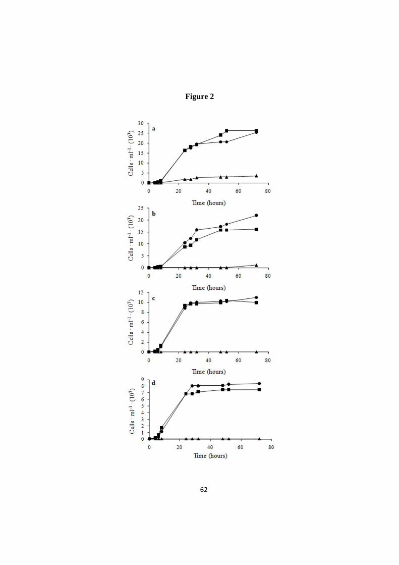
62
Figure 2
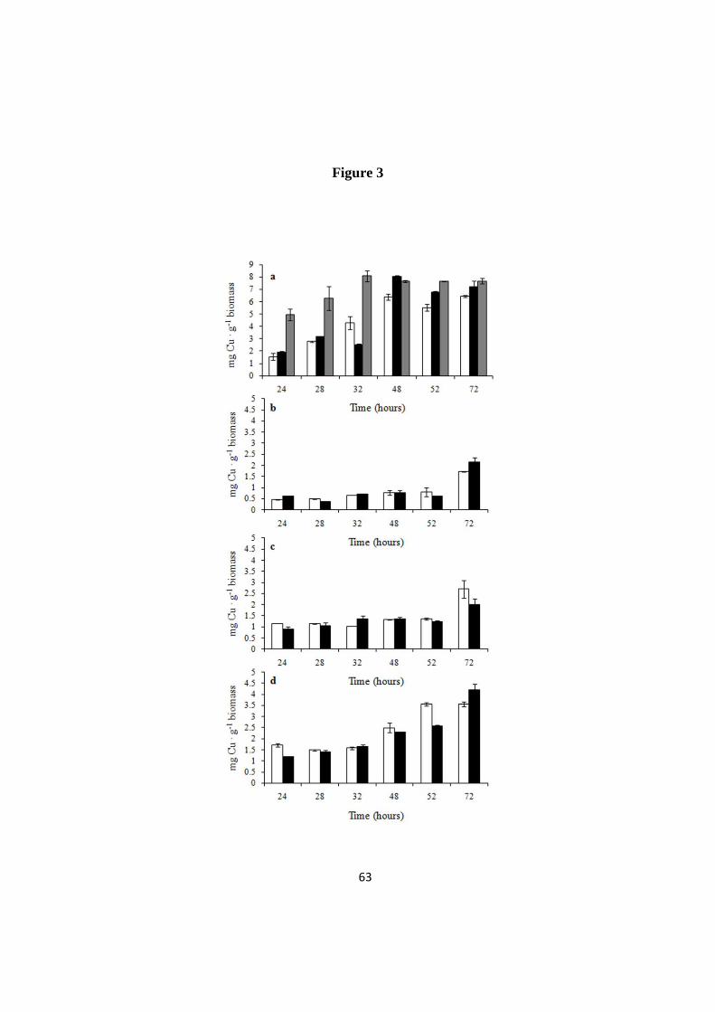
63
Figure 3

64
Figure 4

65
Figure 5
Figure 6

66
Additional file 1

67
Additional file 2

68
Additional file 3

69
Additional file 4
F

70
Evolution of copper tolerance in Saccharomyces
cerevisiae relies on amplification of the CUP1 gene
Manuscript
Giusy Manuela Adamo1, Marina Lotti
1, Markus J. Tamás
2*, Stefania Brocca
1*
1 Department of Biotechnology and Biosciences, University of Milano-Bicocca,
Milan, Italy
2 Department of Cell and Molecular Biology, University of Gothenburg,
Gothenburg, Sweden
*To whom correspondence should be addressed.
E-mail: [email protected], Tel. +390264483518
[email protected],Tel. +46317862548
71
Abstract
Background
In living organisms, copper (Cu) contributes to essential functions but may elicit
also toxic effects that inhibit the ability of microbial cells to proliferate in the
presence of high metal concentrations. Copper-tolerant yeast strains are of relevance
both for application in industrial biotechnology and in the study of physiological and
molecular mechanisms leading to stress resistance. One way to obtain tolerant
strains is to exploit experimental methods that rely on the principles of natural
evolution (evolutionary engineering) and allow for the development in microbial cell
populations of complex phenotypic traits such as stress resistance and new
metabolic abilities. However, in most cases the molecular and physiological bases of
the phenotypic changes produced are far to be unraveled.
Results
We investigated the determinants of Cu resistance in a Saccharomyces cerevisiae
strain evolved to tolerate up to 2.5 g . L
-1 CuSO4 in the culture medium. We found
that intracellular copper content and the expression level of SOD1, encoding for the
superoxide dismutase, were similar in the Cu tolerant (evolved) and in the Cu
sensitive (non-evolved) strain. The major difference detected in the two strains was
the copy number of the gene CUP1 that encodes a metallothionein, with a 7-fold
gene amplification observed in the Cu-tolerant cells. Accordingly, the expression of
CUP1 in evolved cells exposed to Cu was strongly and steadily increased.
Conclusions
Overall our results highlight a central role of CUP1 in the protection of the
evolved S. cerevisiae cells against Cu toxicity. In these cells, robustness towards
copper was stably inheritable and could be reproducibly selected by controlling
environmental conditions. This finding corroborates the effectiveness of laboratory
evolution of whole cells as a tool to develop microbial strains for biotechnological
application.
Keywords: Evolutionary engineering, Cu tolerance, gene amplification,
oxidative stress, Saccharomyces cerevisiae

72
Background
Copper (Cu) acts as the cofactor in a variety of enzymes involved in electron
transfer and is therefore essential in trace amounts for most living beings. However,
similarly to other heavy metals, excess Cu is toxic to cells, because of several
interconnected effects, e. g. the formation of coordination complexes with cellular
components such as phosphates, purines, porphyrins, cysteinyl and histidyl side
chains of proteins [1]. Moreover, Cu ions can elicit oxidative damage either directly
through their redox cycling activities that produce the highly reactive OH• radical [2]
or indirectly, by depleting free radical scavengers such as glutathione and protein-
bound sulphydryl groups [3]. Therefore, a tight regulation of Cu homeostasis is
required for cell survival. Accordingly, it was reported that in yeast free copper is
restricted to less than one atom per cell [4].
Copper homeostasis has been investigated in depth in the yeast Saccharomyces
cerevisiae, and complex mechanisms protecting cells from excess Cu have been
identified, such as the induction of metallothioneins [5-7] and of antioxidant
enzymes [8], and the activation of vacuolar functions [9]. Metallothioneins (MTs)
form a family of cystein-rich, low molecular weight polypeptides common in
eukaryotic organisms [10, 11], whose main function is to bind metal ions thereby
buffering their intracellular concentration. In S. cerevisiae, MTs are encoded by two
genes, CUP1 and CRS5, whose expression is regulated at the transcriptional level,
and, in the case of CUP1, also via gene amplification [12]. The transcription of both
MTs is regulated by Cu through the action of the transcription factor Ace1 [6, 13].
Upon Cu exposure, Ace1 induces also the transcription of the SOD1 gene [14]
encoding a cytosolic copper-zinc superoxide dismutase that breaks down superoxide
ions to dioxygen and hydrogen peroxide [15, 16].
It has been reported that microorganisms can be experimentally endowed with
tolerance to different stress conditions through evolutionary engineering. The term
“evolutionary engineering” was introduced for the first time by Butler and
collaborators [17] to indicate an experimental approach that uses the principles of
adaptive evolution to drive the selection of organisms with desired phenotypic traits.
While “metabolic engineering” implies the rational modification of the cellular

73
metabolic pathways [18], this approach relies on the onset of spontaneous or
experimentally induced random mutations followed by recombination and
continuous evolution of large populations over many generations [19]. For example,
strains of S. cerevisiae were recently evolved to gain resistance towards multiple
stresses [20] and to cobalt [21], and the ability to ferment xylose [22] and lactose
[23]. Evolutionary engineering of whole organisms offers a valuable alternative to
metabolic engineering, that is often limited by the great complexity of dynamic
interactions in cellular systems [19]. “Evolved” strains can be used as hosts for
further rational improvements by metabolic engineering, or a desired phenotype can
be transferred to a production host – an approach known as “inverse metabolic
engineering” [24].
In a previous work, we exploited evolutionary engineering to obtain yeast cells
tolerant to the presence of high Cu concentrations (up to 2.5 g. L
-1) in the growth
medium [25]. Here we report that in one evolved S. cerevisiae strain enhanced
tolerance is associated to an increased copy number of the CUP1 gene resulting in
its strong and stable expression. Increase in the amount of this metallothionein is
proposed to be the major factor of protection against Cu-induced oxidative stress.
Results
Effects of copper on yeast cells growth
The kinetics of growth of non-evolved (Cu sensitive) and evolved (Cu tolerant) S.
cerevisiae cells were compared and are presented in Figure 1. While in YPD
medium the growth of the two strains was very similar, the addition of CuSO4 at the
final concentration of 2.5 g .
L-1
to cultures in the exponential phase strongly
impaired the growth of non-evolved cells, without perturbing the proliferation of the
evolved ones (Fig. 1a). Non-evolved cells exposed to Cu for one hour did not resume
multiplicating when plated on YPD (without Cu); on the contrary, in the same
conditions evolved cells better recovered (Fig. 1b).

74
Expression levels of CUP1 and CRS5
To gain insight into the determinants of Cu tolerance in evolved cells, we
evaluated the expression levels of the genes CUP1 and CRS5, both encoding for
metallothioneins. RNA was extracted from cells growing exponentially in absence
of Cu or at different hours after the addition of Cu, mRNA was retrotranscribed to
the corresponding cDNAs and the expression of the two genes was evaluated by
quantitative PCR. In absence of Cu, expression of CUP1 was hardly detectable in
both non-evolved and evolved cells (Fig. 2 – see 0 h). In response to copper, the
level of CUP1 in the evolved strain was rapidly and strongly induced, keeping high
along the monitored period of 24 hours (Fig. 2 – white histograms). Copper
triggered the expression of CUP1 also in non-evolved cells, although at lower extent
(Fig. 2 – grey histograms). Under the same conditions, CRS5 was expressed at very
low level in both evolved and non-evolved strains (data not shown). Such different
responsiveness is well in agreement with the hypothesis of a primary role of Cup1
induction in the response to copper stress [26, 27].
Copy number of CUP1 and CRS5
It has been reported that different yeast strains may bear different copies of the
CUP1 gene [28, 29], whereas CRS5 is always present in single copy [6]. To
investigate whether the copy number of these metallothionein genes could contribute
to copper tolerance, we determined by RT-qPCR the number of copies of the CUP1
and CRS5 genes in the evolved strain and in two “intermediates” of the adaptation
process, namely cells tolerant to 1.5 and 2 g . L
-1 CuSO4 (referred to as int 1.5 and int
2) [25]. As shown in Figure 3, the CUP1 copy number did not change in int 1.5,
whereas a sudden 7-fold increase was noticed in int 2. No further amplification was
detected in cells tolerant to the highest Cu concentration used in this work (2.5 g .L
-
1). No amplification of the CRS5 copy number was observed in the evolved strain
(Fig. 3).
Expression level of SOD1
Besides metallothioneins, yeast cells respond to excess of Cu by increasing the
level of Sod1, a cytosolic Cu,Zn-superoxide dismutase that participates in the
scavenging of superoxide anions resulting from Cu redox metabolism. Therefore, we
evaluated the expression of SOD1 in evolved and non-evolved cells, by quantifying
75
its mRNA, in the same conditions described above (Fig. 4). For both strains, one
hour of Cu exposure resulted in a two-fold increase of SOD1 expression level. In the
evolved cells, this value did not further increase with time, whereas in the non-
evolved strain a marked increase was appreciable just after prolonged cultivation in
copper medium.
Copper-induced carbonylation of proteins
ROS produced upon copper stress are known to promote damages to different
cell components, including proteins [30] that might undergo carbonylation of amino
acids side chain [31]. Therefore, we used an immunoblot assay to detect oxidized
proteins, assuming the carbonylation of proteins as indicative of oxidative stress.
Total proteins were extracted from cells exponentially growing either in absence of
Cu or at different hours after Cu addition. Extracted proteins were derivatized with
DNPH that specifically reacts with carbonyl groups. We observed a comparable
amount of carbonylated proteins in the non-evolved and the evolved populations
cultivated without copper (Fig. 5 – see 0h in both strains). However, carbonylation
increased in samples of non-evolved cells at already one hour after copper exposure,
and the signal became stronger after prolonged Cu exposure. On the contrary, in
evolved cells the signal associated to oxidized proteins increased only after several
hours of growth in copper medium, although carbonylation was still much lower
than in non-evolved cells (Fig. 5, compare 24h in both strains).
Quantification of intracellular copper
We next determined the levels of copper accumulated by yeast cells. Within the
first hour of Cu exposure, intracellular copper was comparable in both yeast samples
(Fig. 6). These data seem to bring into question the previous findings showing that
the evolved strain has growth advantages and encounters a milder oxidative stress in
presence of Cu than the natural strain. However, taking into account that the
biochemical assay used for Cu quantification [25] does not discriminate between
toxic Cu ions (e.g. those participating in reactions generating ROS or improperly
bound to cellular proteins) and non-toxic Cu ions (e.g. those bound to buffering
molecules as metallothioneins), it is reasonable to hypothesize that although we
detected a similar quantity of Cu, the toxicity of Cu ions might vary in the two
strains.

76
Discussion
Adaptive evolution is a central biological process underlying several phenomena,
from the gain of antibiotic resistance in microbial populations to the niche
specialization. An intriguing question is what kind of genomic variations are
associated with evolution of improved phenotypes and how reproducible the process
is [32]. Adaptive evolution in microbial populations can typically occur through
relatively few mutations that confer large benefits, rather than through countless
mutations with minor benefits. The most extreme beneficial mutations are greatly
overrepresented owing to selection [33-35]. Adaptive evolution can be
experimentally attained at the laboratory scale [19]. In this work, we showed that the
evolution of increased Cu tolerance in baker’s yeasts is mainly mediated by the
amplification of the CUP1 gene, encoding the metallothionein Cup1. These results
describe for the first time the bases of Cu tolerance in an industrial strain of S.
cerevisiae deliberately subjected to a strategy of evolutionary engineering, and are in
good agreement with findings by others on genetically modified yeast strains from
both laboratory [5, 36, 37] and industrial [28] sources. Compared with its non-
evolved counterpart, the evolved strain encountered a 7-fold amplification of CUP1
gene, resulting in high expression during growth on Cu medium. Our results suggest
that in evolved cells the Cup1 dosage provides effective protection against
intracellular Cu overload, avoiding toxicity and resulting in growth advantage. On
the contrary, a comparable amount of copper internalized by non-evolved cells is
highly toxic and causes growth impairment.
Looking at the amplification of CUP1 gene to monitor the whole progress of
evolution, we found that a 7-fold amplification arises not only in the hyper-resistant
evolved strain, but earlier and suddenly in the int 2, an intermediate of the
evolutionary engineering procedure. It has been proved that adaptation to selection
conditions requires first a small number of mutations that exert large positive
effects, then a higher number of mutations with small effects [38-40]. We can
conceive that CUP1 gene amplification found in int 2 corresponds to a major,
primary mutation featuring a cell population that attains a local optimum of the
fitness landscape. Other secondary mutations that improve copper tolerance from 2
to 2.5 g . L
-1 CuSO4 might contribute to the final adaptation of the evolved strain,

77
presumably placing it closer to a global fitness optimum in the conditions of
selection.
How could the evolution of copper tolerance have occurred? According to a neo-
Darwinist theory, the overall CUP1 expression in the natural, non-evolved S.
cerevisiae cultures might result from stochastic differences in gene dosage among
individuals and could average low expression by the large majority of cells, and a
high expression by a small subgroup of cells bearing a higher CUP1 copy number.
Indeed, according to the concept of “quasispecies”, the result of evolution consists in
a distribution of related variants that occupy a distinct region in sequence space [41].
Exposure of the population to high copper might reduce the original heterogeneity of
the culture (number of variants) by selecting amplification mutants of the CUP1
locus since this beneficial mutation contributes to their fitness. On the other hand,
we cannot exclude that adaptive mutations, and specifically amplification of the
CUP1 locus, could have occurred during the prolonged cultivation in the presence of
Cu [42]. In this scenario, the stresses applied to the cell population might change the
genome evolvability by activating mutation mechanisms already well documented in
prokaryotic organisms [43], and that appear shared by eukaryotic cells too [44-47].
In the evolved strain the expression of CRS5 was not significantly increased. This
can be ascribed both to its lower gene dosage and to its lower responsiveness to Cu
mediated by the transcription factor Ace1 [6, 48, 49]. The role of CUP1 in
determining Cu tolerance is dominant also with respect to SOD1. Our data, in
agreement with results by Gralla and collaborators [14], show that in the evolved
strain, induction of SOD1 occurs just in the first hour of Cu exposure. Although the
evolved and non-evolved strains express SOD1 at similar level and accumulate the
same amount of Cu, oxidative stress is stronger in non-evolved cells. This finding
further corroborates the view that over-expression of Cup1 might allow for the
effective buffering of cytosolic Cu. This effect would be sufficient to prevent ROS
overproduction and to limit the need for free radical scavengers. On the other hand,
upon prolonged Cu exposure, the non-evolved strain compensates the overload of
dangerous Cu ions with a strong induction of SOD1 expression. Nevertheless, the
late (24h) time point measured (Fig. 4) should be considered with some care due to
the low viability of the non-evolved strain at that Cu concentration.

78
Literature data on evolutionary engineering of microbial strains highlight the
potential of this approach in selecting strains with specific phenotypes. Moreover,
acquisition of cross-resistance to different stresses has been already well
documented [50-52]. Recent studies demonstrate that evolution of tolerance to a
given metal triggers cross-resistance to other metals and/or other stress conditions
[21, 53]. For instance, Çakar and collaborators [21] found that cobalt-resistant S.
cerevisiae cells can also evolve tolerance to metals that are in the proximity of
cobalt in the Periodic Table of the Elements, like iron, manganese, zinc and nickel
but not copper and chromium. However, cross-resistance to other metals was not
detected in evolved Cu–tolerant S .cerevisiae cells used in this work (data not
shown), underscoring the specificity of the Cu tolerance acquired during evolution.
Finally, we can observe that amplification of MTs genes cannot be considered a
general rule and it has not been encountered in the development of other heavy
metal-resistant cells, such as cobalt-resistant S. cerevisiae [54]. Therefore, it is likely
that metal tolerance may arise from both general and specific molecular mechanisms
to be elucidated case by case.
Conclusions
In conclusion, this work demonstrates that the phenotypic traits of a Cu tolerant
yeast obtained by evolutionary engineering are mainly associated with the
amplification of the locus CUP1. Overall, the application of evolutionary
engineering emerges as a powerful tool to stably improve the robustness of
microbial cells. The evolved strains can find direct applications in several processes
of biotechnological interest and provide a starting point for further rational
improvements, provided that molecular determinants of evolved phenotypes are
disclosed.

79
Methods
Strains and growth conditions
S. cerevisiae cells used in this work derive from a natural Cu-sensitive strain whose
growth is inhibited at 1 g . L
-1 CuSO4 in the culture medium. This parental strain is
quoted in the text as “non-evolved”. In a previous work [25], Cu tolerance was step-
wise evolved in parental cells by exposure to increasing Cu concentrations. The
strain tolerant to the highest Cu concentration (2.5 g . L
-1 CuSO4) was defined as
“evolved”, while two strains isolated as intermediates of the evolutionary
engineering procedures, and tolerant to 1.5 and 2 g . L
-1 CuSO4 [25] are indicated in
the text as int 1.5 and int2, respectively.
Cells from a fresh culture grown on solid YPD medium [2% (w/v) glucose, 1%
(w/v) yeast extract, 2% (w/v) peptone, 2% (w/v) agar] were pre-cultured overnight
in liquid YPD at 30°C under shaking at 160 rpm and then inoculated at optical
density (OD600) of 0.05 in shaking flasks containing fresh medium. A solution of
CuSO4 was added to cultures exponentially growing in YPD medium. The final
concentration of CuSO4 in the culture broth was 2.5 g .
L-1
if not differently
indicated.
RNA extraction and cDNA production
Total RNA for gene expression analysis was extracted from cells either growing in
YPD or at different hours after Cu addition using the RNeasy Mini Kit (Quiagen).
The quality of extracted RNA was assessed on 1.5 % (w/v) agarose gel and RNA
was quantified by NanoDrop® (ND-1000 v 3.3.2 – Saveen Wernev). A dilution of
0.320 µg. µL
-1 of RNA was used to produce the corresponding cDNA according to
the following protocol: a mix composed of 8.5 µL RNase-free water, 2.5 µL RNA
and 1 µL of anchored oligo-dT Primer (ABgene) was incubated at 65°C for 5 min in
a thermocycler (T3000 - Biometra®). After 1 min of incubation on ice, the mix was
added of 4 µL 5× first-strand buffer (Invitrogen), 2 µl 0.1 M DTT, 1 µL 10 mM
dNTPs and incubated at 42°C for 2 min. Retrotranscription was performed adding 1
µL SuperScript™ II RT (Retro Transcriptase) followed by incubation at 40°C for 50
min and 70°C for 15 min. The obtained cDNA was diluted by adding 80 µL RNase-
free water, and 5 µL of the dilution were used for gene expression analysis.

80
Genomic DNA extraction
Genomic DNA was extracted from cells grown overnight at 30°C in 5 mL of YPD
medium. Cells were centrifuged at 4,900 × g for 5 min, re-suspended in 200 µL of
breaking buffer [2% (v/v) Triton X-100, 1% (w/v) sodium dodecyl sulphate (SDS)],
100 mM NaCl, 10 mM Trizma-HCl pH 8.0, 1 mM ethylene-diamine tetra acetic acid
(EDTA) pH 8.0, 200 µL PCI (phenol/chlorophorm/isoamylalchool at 25/24/1) and
transferred to a screw cap tube containing 200 µL of acid-washed glass beads (425-
600 µm – Sigma). Cells were subjected to mechanical lysis by three cycles of 20 sec
at maximum speed with a Fast Prep - FP120 (Bio101-Savant). Two hundred µL of
10 mM TE (Trizma and EDTA) pH 8.0 were added to the crude extract and, after
centrifugation at 10,000 g for 5 min, the aqueous layer was transferred to a new
tube. After addition of 1 mL 100% ethanol and centrifugation, the pellet was dried
and RNA was removed by incubation for 15 min at 37°C in 400 µL TE containing
2.5 µL RNase A (20 mg. mL
-1). Ten µL of 4 M ammonium acetate and 1 mL 100%
ethanol were added to the sample and, after centrifugation, the DNA pellet was dried
and re-suspended in 100 µL TE. The obtained DNA was checked on 1.5 % (w/v)
agarose gel and quantified by NanoDrop® (ND-1000 v 3.3.2 – Saveen Wernev).
Gene expression analysis and determination of gene copy number by RT-qPCR
Relative and absolute quantifications were performed by quantitative real time PCR
(RT-qPCR) to determine gene expression and gene copy number, respectively. RT-
qPCR was performed using the IQ5 Multicolor real time PCR detection system
(Bio-Rad).
For gene expression analysis, cDNA gene sequences of CUP1, CRS5, SOD1 and
ACT1 (Table 1) were retrieved from the Saccharomyces Genome Database
(www.yeastgenome.org) and the internet-based interface Primer-3 [55] was used to
design PCR oligonucleotides. Individual RT-qPCR mixtures were prepared in a 96-
well plate (Bioplastic), each 20-µL reaction containing 5 µL cDNA and 15 µL
SYBR green master mix [SYBR Green® Supermix (Bio-Rad), 10 µM forward and
reverse oligonucleotides and water]. Amplification conditions were: 3 min at 95°C
followed by 40 cycles at 95°C for 10 sec and at 58°C for 30 sec and 81 cycles at
55°C for 8 sec. Quantification of the target sequence in the samples was estimated

81
generating a standard curve with known concentrations of DNA. Values were
normalized against the housekeeping gene ACT1.
Five µL of genomic DNA were used to calculate the copy number variation in
comparison with the non-evolved strain, used as a control. Copy number was
calculated according to the 2ΔΔCt
method [56, 57]. For standardization, the results
were expressed as ratios of the amount of the target gene (CUP1 or CRS5) in
evolved and non-evolved strains, the reference gene being the housekeeping gene
ACT1.
Protein extraction and electrophoresis in denaturing conditions
Fifty mL samples were withdrawn from cultures at different times after Cu addition
and processed for protein extraction. Biomass was re-suspended in 2 mL of 2 M
NaOH, incubated for 2 min at room temperature and then 2 mL of 50% (w/v)
trichloroacetic acid (TCA) were added. After centrifugation for 3 min at 10,000 × g,
pellets were re-suspended in 1 mL of 1 M Tris-HCl pH 8.0 and centrifuged again.
Proteins were re-suspended in 250 µL 1× SDS sample buffer (0.0625 M Tris-Cl pH
6.8, 10% (v/v) glycerol, 3% (w/v) SDS, 5% (v/v) β-mercaptoethanol, 0.004% (w/v)
bromophenol blue) and heated at 99°C for 5 min. The protein concentration in cell-
free extracts was estimated by the RC-DC™ Protein Assay (Bio-Rad) using bovine
serum albumin as the reference. Twenty µg of proteins (derivatized to detect
carbonyl groups – see below) were applied to 12% (w/v) polyacrylamide gel. SDS-
polyacrylamide gel electrophoresis (PAGE) was carried out according to Laemmli
[58].
Detection of carbonyl groups in proteins
Analyses of carbonylated proteins were performed using the chemical and
immunological reagents of an OxyBlot oxidized protein detection kit (Millipore).
The carbonyl groups in the protein side chains were derivatized to 2,4-dinitrophenyl-
hydrazone (DNP-hydrazone) by reaction with 2,4-dinitrophenyl hydrazine (DNPH).
The DNP-derivatized proteins were analyzed immunochemically by Western blot
using rabbit anti-DNP as primary antibody (Sigma-Aldrich). A goat anti-Hog1
antibody (Santa Cruz – Biotechnology) was used as a control. Donkey anti-Rabbit
and donkey anti-Goat antibodies Odyssey® (Li-Cor – Bioscience) were used as
82
secondary antibody. Visualization of carbonylated proteins was performed using
Odyssey®- Infrared Imaging System 2.1 (Li-COR – Bioscience).
Copper determination
Intracellular Cu in non-evolved and evolved cells was measured as previously
described [25], within the first hour of Cu exposure. The amount of Cu is referred as
mg Cu. g
-1dry biomass.
Acknowledgements
This study was supported by a grant FAR (Fondo Ateneo per la Ricerca) of the
Università Milano-Bicocca to M.L. and S.B., by the Swedish Research Council to
M.J.T. and by a doctoral fellowship of MIUR (Ministero dell’Istruzione,
dell’Università e della Ricerca Scientifica) to G.M.A.

83
References
1. Gadd GM: Interaction of fungi with toxic metals. New Phytol 1993, 124:25-60. 2. Halliwell B, Gutteridge JM: Oxygen toxicity, oxygen radicals, transition metals and
disease. Biochem J 1984, 219(1):1-14.
3. Stohs SJ, Bagchi D: Oxidative mechanisms in the toxicity of metal ions. Free Radic Biol Med 1995, 18(2):321-336.
4. Rae TD, Schmidt PJ, Pufahl RA, Culotta VC, O'Halloran TV: Undetectable intracellular
free copper: the requirement of a copper chaperone for superoxide dismutase. Science 1999, 284(5415):805-808.
5. Karin M, Najarian R, Haslinger A, Valenzuela P, Welch J, Fogel S: Primary structure and
transcription of an amplified genetic locus: the CUP1 locus of yeast. Proc Natl Acad Sci U S A 1984, 81(2):337-341.
6. Culotta VC, Howard WR, Liu XF: CRS5 encodes a metallothionein-like protein in
Saccharomyces cerevisiae. J Biol Chem 1994, 269(41):25295-25302. 7. Gross C, Kelleher M, Iyer VR, Brown PO, Winge DR: Identification of the copper regulon
in Saccharomyces cerevisiae by DNA microarrays. J Biol Chem 2000, 275(41):32310-
32316. 8. Romandini P, Tallandini L, Beltramini M, Salvato B, Manzano M, de Bertoldi M, Rocco GP:
Effects of copper and cadmium on growth, superoxide dismutase and catalase activities
in different yeast strains. Comp Biochem Physiol C 1992, 103(2):255-262. 9. Szczypka MS, Zhu Z, Silar P, Thiele DJ: Saccharomyces cerevisiae mutants altered in
vacuole function are defective in copper detoxification and iron-responsive gene
transcription. Yeast 1997, 13(15):1423-1435. 10. Kagi JH: Overview of metallothionein. Methods Enzymol 1991, 205:613-626.
11. Waalkes MP, Goering PL: Metallothionein and other cadmium-binding proteins: recent
developments. Chem Res Toxicol 1990, 3(4):281-288. 12. Butt TR, Ecker DJ: Yeast metallothionein and applications in biotechnology. Microbiol
Rev 1987, 51(3):351-364.
13. Buchman C, Skroch P, Welch J, Fogel S, Karin M: The CUP2 gene product, regulator of
yeast metallothionein expression, is a copper-activated DNA-binding protein. Mol Cell
Biol 1989, 9(9):4091-4095.
14. Gralla EB, Thiele DJ, Silar P, Valentine JS: ACE1, a copper-dependent transcription
factor, activates expression of the yeast copper, zinc superoxide dismutase gene. Proc
Natl Acad Sci U S A 1991, 88(19):8558-8562.
15. Fridovich I: Superoxide dismutases. Adv Enzymol Relat Areas Mol Biol 1986, 58:61-97. 16. Bannister JV, Bannister WH, Rotilio G: Aspects of the structure, function, and applications
of superoxide dismutase. CRC Crit Rev Biochem 1987, 22(2):111-180.
17. Butler P, Brown M, Oliver S: Improvement of antibiotic titers form Streptomyces bacteria
by interactive continuous selection. Biotech Bioeng 1996, 49:185-196.
18. Bailey JE: Toward a science of metabolic engineering. Science 1991, 252(5013):1668-1675.
19. Sauer U: Evolutionary engineering of industrially important microbial phenotypes. Adv Biochem Eng Biotechnol 2001, 73:129-169.
20. Cakar ZP, Seker UO, Tamerler C, Sonderegger M, Sauer U: Evolutionary engineering of
multiple-stress resistant Saccharomyces cerevisiae. FEMS Yeast Res 2005, 5(6-7):569-578. 21. Cakar ZP, Alkim C, Turanli B, Tokman N, Akman S, Sarikaya M, Tamerler C, Benbadis L,
Francois JM: Isolation of cobalt hyper-resistant mutants of Saccharomyces cerevisiae by
in vivo evolutionary engineering approach. J Biotechnol 2009, 143(2):130-138. 22. van Maris AJ, Winkler AA, Kuyper M, de Laat WT, van Dijken JP, Pronk JT: Development
of efficient xylose fermentation in Saccharomyces cerevisiae: xylose isomerase as a key
component. Adv Biochem Eng Biotechnol 2007, 108:179-204. 23. Guimaraes PM, Francois J, Parrou JL, Teixeira JA, Domingues L: Adaptive evolution of a
lactose-consuming Saccharomyces cerevisiae recombinant. Appl Environ Microbiol 2008, 74(6):1748-1756.
24. Bailey JE, Sburlati A, Hatzimanikatis V, Lee K, Renner WA, Tsai PS: Inverse metabolic
engineering: A strategy for directed genetic engineering of useful phenotypes. Biotechnol Bioeng 1996, 52(1):109-121.
25. Adamo GM, Brocca S, Passolunghi S, Salvato B, Lotti M: Laboratory evolution of copper
tolerant yeast strains. Microbial Cell Factories 2012, 11:1.

84
26. Hamer DH, Thiele DJ, Lemontt JE: Function and autoregulation of yeast copperthionein. Science 1985, 228(4700):685-690.
27. Ecker DJ, Butt TR, Sternberg EJ, Neeper MP, Debouck C, Gorman JA, Crooke ST: Yeast
metallothionein function in metal ion detoxification. J Biol Chem 1986, 261(36):16895-16900.
28. Welch JW, Fogel S, Cathala G, Karin M: Industrial yeasts display tandem gene iteration at
the CUP1 region. Mol Cell Biol 1983, 3(8):1353-1361. 29. Liti G, Carter DM, Moses AM, Warringer J, Parts L, James SA, Davey RP, Roberts IN, Burt
A, Koufopanou V et al: Population genomics of domestic and wild yeasts. Nature 2009,
458(7236):337-341. 30. Davies KJ: Protein damage and degradation by oxygen radicals. I. general aspects. J Biol
Chem 1987, 262(20):9895-9901.
31. Stadtman ER, Levine RL: Chemical modification of proteins by reactive oxygen species. In: Redox proteomics: from protein modifications to cellular dysfunctions and diseases. Edited
by Dalle Donne I, Scaloni A, Butterfield DA. Hoboken, NJ: John Wiley and Sons; 2006: p. 3-
23. 32. Gresham D, Desai MM, Tucker CM, Jenq HT, Pai DA, Ward A, DeSevo CG, Botstein D,
Dunham MJ: The repertoire and dynamics of evolutionary adaptations to controlled
nutrient-limited environments in yeast. PLoS Genet 2008, 4(12):e1000303. 33. Rozen DE, de Visser JA, Gerrish PJ: Fitness effects of fixed beneficial mutations in
microbial populations. Curr Biol 2002, 12(12):1040-1045.
34. Burch CL, Chao L: Evolution by small steps and rugged landscapes in the RNA virus
phi6. Genetics 1999, 151(3):921-927.
35. Gerrish PJ, Lenski RE: The fate of competing beneficial mutations in an asexual
population. Genetica 1998, 102-103(1-6):127-144. 36. Fogel S, Welch JW: Tandem gene amplification mediates copper resistance in yeast. Proc
Natl Acad Sci U S A 1982, 79(17):5342-5346.
37. Fogel S, Welch JW, Cathala G, Karin M: Gene amplification in yeasts. Development of
copper resistance is mediated by an increase in CUP1 gene copy number. Current Biology
1983:1-9.
38. Lenski RE, Mongold JA, Sniegowski PD, Travisano M, Vasi F, Gerrish PJ, Schmidt TM:
Evolution of competitive fitness in experimental populations of E. coli: what makes one
genotype a better competitor than another? Antonie Van Leeuwenhoek 1998, 73(1):35-47.
39. Orr HA: The evolutionary genetics of adaptation: a simulation study. Genet Res 1999, 74(3):207-214.
40. Orr HA: The population genetics of adaptation: the distribution of factors fixed during
adaptive evolution. Evolution 1998, 52:935-949. 41. Biebricher CK, Eigen M: What is a quasispecies? Curr Top Microbiol Immunol 2006, 299:1-
31.
42. Hersh MN, Ponder RG, Hastings PJ, Rosenberg SM: Adaptive mutation and amplification
in Escherichia coli: two pathways of genome adaptation under stress. Res Microbiol 2004,
155(5):352-359.
43. Hastings PJ, Lupski JR, Rosenberg SM, Ira G: Mechanisms of change in gene copy number. Nat Rev Genet 2009, 10(8):551-564.
44. Finch CE, Goodman MF: Relevance of 'adaptive' mutations arising in non-dividing cells
of microorganisms to age-related changes in mutant phenotypes of neurons. Trends Neurosci 1997, 20(11):501-507.
45. Hall BG: Adaptive mutations in Escherichia coli as a model for the multiple mutational
origins of tumors. Proc Natl Acad Sci U S A 1995, 92(12):5669-5673. 46. Harris RS, Longerich S, Rosenberg SM: Recombination in adaptive mutation. Science
1994, 264(5156):258-260.
47. Strauss BS: The origin of point mutations in human tumor cells. Cancer Res 1992, 52(2):249-253.
48. Furst P, Hu S, Hackett R, Hamer D: Copper activates metallothionein gene transcription
by altering the conformation of a specific DNA binding protein. Cell 1988, 55(4):705-717.
49. Thiele DJ, Hamer DH: Tandemly duplicated upstream control sequences mediate copper-
induced transcription of the Saccharomyces cerevisiae copper-metallothionein gene. Mol Cell Biol 1986, 6(4):1158-1163.
50. Lewis JG, Learmonth RP, Watson K: Induction of heat, freezing and salt tolerance by heat
and salt shock in Saccharomyces cerevisiae. Microbiology 1995, 141 ( Pt 3):687-694.
85
51. Park JI, Grant CM, Attfield PV, Dawes IW: The freeze-thaw stress response of the yeast
Saccharomyces cerevisiae is growth phase specific and is controlled by nutritional state
via the RAS-cyclic AMP signal transduction pathway. Appl Environ Microbiol 1997,
63(10):3818-3824. 52. Hohmann S: Osmotic stress signaling and osmoadaptation in yeasts. Microbiol Mol Biol
Rev 2002, 66(2):300-372.
53. Sen M, Yilmaz U, Baysal A, Akman S, Cakar ZP: In vivo evolutionary engineering of a
boron-resistant bacterium: Bacillus boroniphilus. Antonie Van Leeuwenhoek 2011,
99(4):825-835.
54. Conklin DS, McMaster JA, Culbertson MR, Kung C: COT1, a gene involved in cobalt
accumulation in Saccharomyces cerevisiae. Mol Cell Biol 1992, 12(9):3678-3688.
55. Rozen DE, Skaletski HJ: Primer on the WWW for general users and for biologist
programmers In: Bioinformatics methods and protocols: methods in molecular biology. Edited by Misener SKaS, vol. 132. Totowa, NJ.: Humana Press; 2000: 365-386.
56. Livak KJ, Flood SJ, Marmaro J, Giusti W, Deetz K: Oligonucleotides with fluorescent dyes
at opposite ends provide a quenched probe system useful for detecting PCR product and
nucleic acid hybridization. PCR Methods Appl 1995, 4(6):357-362.
57. Livak KJ, Schmittgen TD: Analysis of relative gene expression data using real-time
quantitative PCR and the 2(-Delta Delta C(T)) Method. Methods 2001, 25(4):402-408. 58. Laemmli UK: Cleavage of structural proteins during the assembly of the head of
bacteriophage T4. Nature 1970, 227(5259):680-685.

86
Table 1. Oligonucleotides used for quantitative RT-PCR

87
Legends
Figure 1. Effect of Cu on cells growth. a) Growth kinetics of non-evolved (black
circles) and evolved (black squares) S. cerevisiae cells. Addition of Cu (supplied as
CuSO4 to a final concentration of 2.5 g . L
-1) to cells exponentially growing on YPD
medium is indicated by an arrow. Values are the mean of three biological replica. b)
Drop test of non-evolved and evolved strains harvested before (0h) and after one
hour (1h) of copper exposure. Five l of 1:10 serial dilutions were plated on YPD
and plates were incubated for two days at 30°C.
Figure 2. Relative expression of CUP1. Non-evolved (grey bars) and evolved (white
bars) cells were harvested at different hours after the addition of copper (0h-24h).
Gene expression was quantified by RT-qPCR and normalized for ACT1 expression.
Values are the mean of two biological replica performed in triplicate.
Figure 3. Determination of the gene copy number in evolved cells. Copy number of
the CRS5 gene was determined on the evolved strain tolerant to 2.5 g .L
-1 of CuSO4
(CRS5 evolved); for CUP1 gene analysis was carried out on different “intermediates
of evolution”-strains evolved to tolerate 1.5 g . L
-1 of CuSO4(CUP1 int1.5), 2 g
. L
-1
of CuSO4 (CUP1 int2) - and on the evolved strain tolerant to 2.5 g .
L-1
of
CuSO4(CUP1 evolved). Estimations were made according to the 2ΔΔCt
method.
Values are the mean of two biological replica performed in triplicate.
Figure 4. Relative expression of SOD1. Non-evolved (grey bars) and evolved (white
bars) cells were harvested at different hours after Cu addition (0h-24h). Gene
expression was quantified by RT-qPCR and normalized for ACT1 expression.
Values are the mean of two biological replica performed in triplicate.
Figure 5. Protein carbonylation upon Cu exposure. Non-evolved and evolved cells
were harvested at different hours after Cu addition (0h-24h). Equal amounts of
proteins were assayed after derivatization with DNPH and western blotting with an
anti-DNP-hydrazone antibody. Cntr-: not-derivatized protein samples. The
expression level of Hog1 is included as a control of protein amount. The picture
represents one of three independent experiments.

88
Figure 6. Measurement of intracellular Cu. Intracellular Cu was measured in non-
evolved (grey bars) and evolved (white bars) cells at 0, 5, 10, 15, 20, 60 min after Cu
addition. The amount of Cu is reported as mg. g
-1dry biomass. Values are the mean
of three biological replica.

89
Figure 1
Figure 2

90
Figure 3
Figure 4

91
Figure 5
Figure 6

92
Proteomic analysis of natural and copper-adapted cells
of the yeast Candida humilis
Preliminary results
Giusy Manuela Adamo, Stefania Brocca and Marina Lotti
Background
Copper (Cu), like other transition metals, is an essential micronutrient required
for the survival of all living organisms (1). Indeed, cycling between the oxidized
(Cu2+
) and the reduced (Cu1+
) state, copper acts as an important catalytic and
structural cofactor for several biochemical processes essential for life (2). However,
the redox properties of Cu are also responsible for its toxicity, since Cu participates
in reactions that generate Reactive Oxygen Species (ROS) (3). ROS target different
cell macromolecules, such as proteins, lipids, DNA and RNA, causing loss of
essential cellular functions and/or gain of toxicity (4). In microbial cells, tolerance
toward toxic concentrations of copper, as well as toward other stress conditions, can
be experimentally improved by “evolutionary engineering” that is the application of
the principles of natural evolution to drive the selection of organisms with a desired
set of phenotypes (5). These evolved strains may find a direct application in several
areas, from the industrial sector to bioremediation and allow gaining insight in the
processes of adaptive evolution at the basis of the specific properties acquired by the
evolved strains.
Beside Saccharomyces cerevisiae, extensively studied in its Cu metabolism (6,
7), other yeasts are attractive for their marked natural tolerance to high copper, but
still poorly investigated.
In a previous work, starting from a strain of Candida humilis endowed with
some natural resistance to the presence of copper, we exploited a strategy based on
step-wise adaptation obtaining cells able to proliferate at high metal concentration

93
(8). In copper medium, the evolved strain featured growth advantage and decreased
production of ROS in comparison with the natural strain (non-evolved). In the frame
of a broader research program aimed at the characterization of the molecular
determinants of copper robustness in yeasts, we performed a differential proteomic
analysis on non-evolved and evolved C. humilis cells grown in high Cu
concentration.
Methods
Strains and growth conditions
In this work were used evolved and non-evolved C. humilis strains respectively
tolerant and partly sensitive to copper sulphate (CuSO4). The evolved strain was
obtained from the non-evolved one through evolutionary engineering as previously
described (8). Cells from a fresh culture on agarized YPD medium were pre-cultured
overnight in liquid YPD medium [2% (w/v) glucose, 1% (w/v) yeast extract, 2%
(w/v) peptone] at 30°C under orbital shaking (140 rpm) and then inoculated at initial
OD of 0.05 in shaking flasks containing YPD or YPD + CuSO4 2.5 g/L.
Extraction and quantification of total proteins
Two grams of cells from exponential cultures were harvested by centrifugation at
4,900 g for 10 min at 4°C. The cell pellet was washed twice in cold deionised water
and re-suspended in a breaking buffer containing 8 ml of deionised water, 200 µl of
0.5 M Tris-Cl pH 8.5, 200 µl of 0.25 M ethylenediaminetetraacetic acid pH 8.4
added of protease inhibitor cocktail (Sigma). Cells were subjected to mechanical
lysis with glass microbeads by four cycles of 20 sec at maximum speed with a Fast
Prep - FP120 (Bio101-Savant). Each lysis cycle was followed by two minutes of
incubation on ice. Cell debris was removed by centrifugation at 700 g for 10 min at
4°C. Total proteins were precipitated from the crude extract with an excess of
acetone maintaining samples for two hours at -20°C. Samples were centrifuged
again for 20 min, acetone was discarded and when the pellet of precipitated proteins
was completely dried, proteins were re-suspended in a solubilization buffer
containing 7 M urea, 2 M thiourea, 4% (w/v) 3-[(3-

94
Cholamidopropyl)dimethylammonium]-1-propanesulfonate (CHAPS), 60 mM
dithiothritol (DTT), 20 mM iodacetamide (IAA).
Protein concentration was estimated according to Bradford (9) using protein bovine
serum albumin as the reference.
Two-dimensional gel electrophoresis
Six hundred µg of proteins were diluted in 300 µL of sample buffer containing 60
mM DTT, 60 mM IAA, 1% (v/v) carrier ampholytes and traces of bromophenol
blue. For separation in the first dimension, samples were applied on a 17-cm pH 4-7
Immobilized pH Gradient (IPG) strip (BioRad) and isoelectrofocusing was
performed with the use of a isoelectrofocusing apparatus (BioRad PROTEAN IEF
Cell) under the following conditions: passive rehydratation with sample (1h), 200 V
(1 h), 3500 V (2.5 h), 3500 V (3 h), 8000 V (4 h), 8000 V (6 h) and 500 V h (hold).
The focused IGP strips were first equilibrated in equilibration buffer (6 M urea, 30%
(v/v) glycerol, 2% (w/v) sodium dodecyl sulphate (SDS) in 0.05 M Tris-HCl pH 6.8)
containing 2% (v/v) DTT for 20 min and then in equilibration buffer containing 2.5
% (w/v) IAA for 20 min. Gel strips were adjusted on the top of a 12% SDS-
polyacrylamide gel and incorporated with 0.5% (w/v) agarose. Electrophoresis was
carried out in a refrigerated system (EttanDALTsix electrophoresis unit, Amersham
Biosciences) at 15-30 mA/gel. Gels were finally stained with GelCode Blue Stain
Reagent (Pierce).
Spot matching
Images of the stained gels were analyzed using DECODON Delta 2D software
(http://www.decodon.com). The differential analysis was performed for pairs of
image: in each pair a reference image was warped with an image of interest first
using automatic warping and later setting specific landmarks manually. The
software provides the creation of a fused image were spots can be detected and
transferred back to all analyzed gels. The intensity of each protein spot is normalized
to the total intensity of all valid spots detected on each gel.
Proteins were considered to be differentially expressed if there was at least 1.5-fold
absolute difference in the intensity of corresponding spots between the reference and
the sample of interest. Expression changes were considered significant if the p value

95
of the corresponding spot provided by the program (running an Anova test) was
lower or close to 0.05.
Mass spectrometry (MS)
Spots of interest were excised from the polyacrylamide gels, cut into small pieces
and destained by repeated washing cycles alternating 50 mM ammonium hydrogen
carbonate and pure acetonitrile. After complete destaining, gel particles were
dehydrated by acetonitrile, covered with trypsin solution (12.5 ng/mL in 50 mM
ammonium hydrogen carbonate, pH 8.0) and incubated 1 h on ice. Excess liquid was
removed and the gel pieces covered with a solution of 50 mM ammonium hydrogen
carbonate (pH 8.0) and incubated overnight at 37°C. Tryptic peptides were extracted
by alternating incubation in pure acetonitrile and 1% (v/v) formic acid. Samples
were lyophilised, re-suspended in 1% (v/v) formic acid, and desalted by ZipTip
(Millipore) before Electro Spray Ionization-Mass Spectrometry (ESI-MS) analysis.
ESI-MS experiments were performed with a hybrid Quadrupole-Time-of-Flight (q-
TOF) mass spectrometer (QSTAR ELITE, Applied Biosystems) equipped with a
nano-electrospray ionisation sample source. Metal-coated borosilicate capillaries
(Proxeon, Denmark) with medium-length emitter tip of 1-µm internal diameter were
used for off-line analysis. The instrument was calibrated with standard solution
Renin (MH2+ 879.97 Da and its fragment MH+ 110.07 Da, Applied Biosystems).
Peptide identification was performed using the MASCOT software with following
parameters: 2 missed cleavages, peptide tolerance 0.6 Da, MS/MS tolerance 0.6 Da,
peptide charges 2+ and 3+. Only monoisotopic masses were considered as precursor
ions. Spectra of tryptic peptides were acquired in the 400-1,500 m/z range, with 1.0
sec accumulation time, ion-spray voltage 1,300 V, declustering potential 60 V, with
active Information Dependent Acquisition (IDA) using rolling collision energy to
fragment peptides for MS/MS analysis.
Determination of thiobarbituric acid reactive substances (TBARS)
Determination of TBARS to evaluate lipids peroxidation was carried out following
the procedure previously described by (10). Cells exponentially growing on either
YPD or YPD + 2.5 g/L CuSO4 were harvested, mechanically disrupted and the crude
extract mixed with a solution containing 15% (w/v) TCA, 0.375% (w/v)
96
Thiobarbituric acid and 0.25 (v/v) HCl. After boiling and cooling, samples were
centrifuged and the presence in the supernatants of malondialdehide (MDA) – a
product of lipids peroxidation - was monitored spectrophotometrically at 532 nm.
Results were expressed as mM MDA/g biomass.
Results
In a previous work (8) through prolonged cultivation of C. humilis cells in
presence of increasing amounts of copper sulphate, we obtained cells (hereafter
referred to as evolved) that, compared with the natural strain (hereafter non-evolved)
exhibited an increased robustness toward copper toxicity. Such a robustness lied in
marked growth advantage and diminished rate of mortality during cultivation in
copper medium and was accompanied by a negligible production of ROS. These
features of copper tolerance acquired by the evolved strain resulted stably
maintained.
Proteomic analysis
To characterize more in-depth changes induced by adaptation, we compared the
proteomes of non-evolved and evolved cells cultured in both YPD and YPD + 2.5
g/L CuSO4. Total proteins were extracted from cells collected at the exponential
phase of growth and resolved in the pI and the molecular mass ranges of 4-7 and 11-
245 kDa, respectively. With the aim to detect both intra- and inter- strain
differences, we performed the differential analysis comparing the proteomic profile
of the two strains in different conditions in the following “comparison couples”
(i) non-evolved and evolved strain grown in YPD
(ii) non-evolved strain grown in YPD and in YPD + 2.5 g/L CuSO4
(iii) evolved strain grown in YPD and in YPD + 2.5 g/L CuSO4
(iv) non-evolved and evolved strain grown in YPD + 2.5 g/L CuSO4
For the couples (i) and (iv) the protein pattern of the non-evolved strain was used
as the reference, whereas for the couples (ii) and (iii) the reference was the protein
pattern obtained during growth in YPD. Figure 1 shows the proteomic profiles in
four of the above-described conditions, whereas Figure 2 presents one representative
gel in which relevant spots are indicated.

97
No statistically relevant differences were found between proteins of the couple
(i), hence we have no indication that non-evolved and evolved cells may feature
different physiology during growth in permissive conditions. Instead, several spots
differed in the couple (ii), that considers changes induced by copper in the natural
strain. In this case, we found that 9 proteins were up-regulated and 4 down-regulated
by the presence of the metal. Out of these proteins, 4 were differentially represented
also in the evolved cells challenged by copper (couple iii). The direction of
regulation –either up or down – was the same detected in the non evolved
population. Finally, no major changes were highlighted in the comparison couple
(iv), suggesting that the improved Cu tolerance in the evolved strain cannot be
ascribed to a sharp up- or down-regulation of a few group of proteins. Taken
together these data suggest that the major variations in the cells proteome are
associated with copper exposure rather than with the evolution of Cu tolerance.
The differentially regulated proteins were successfully identified by ESI-MS
analysis followed by MASCOT database searches. Table 1 lists the proteins
identified in comparison couples (ii) and (iii) and the corresponding statistically
significative fold change. In table 1 proteins are classified according to their
biochemical functions in: heat shock response (spot n° 6, 7, 9, 11, 13), protein
translation (spot n° 8, 10), energy production (spot n° 16), phospholipids synthesis
and sulphur aminoacids metabolism (spot n° 14), glycolysis (spot n° 2, 4) and
alcoholic fermentation enzymes (spot n° 1-3). Out of them, all heat shock proteins,
S-adenosylmethionine synthase (a protein participating in phospholipids synthesis
and sulphur aminoacid metabolism), ATP synthase and proteins involved in protein
synthesis were up-regulated during copper exposure. The glycolytic enzymes
pyruvate kinase and glyceraldehydes-3-phosphate dehydrogenase as well as alcohol
dehydrogenase were found to be down-regulated On Two-dimensional gels this last
protein was identified in two different spots (n°1 and n°3 – see Figure 2) with
different isoelectric points, suggesting the occurrence of post translational
modifications.
Lipids peroxidation
As already mentioned, one of the targets of ROS are membrane lipids that can be
converted into polar lipids hydroperoxides, causing increase in membrane fluidity,
98
efflux of cytosolic solutes and loss of membrane-protein activities (4). Moreover,
reactive products of lipids peroxidation may attack amino acid side chains (11) and
cause fragmentation of DNA (12). Determination of TBARS revealed a reduced
extent of lipids peroxidation in the evolved than in the natural strain grown in the
presence of copper (Figure 3).
Discussion
This work is intended as the first step in the molecular characterization of
evolved tolerance to copper in Candida humilis cells. Cu is a strong oxidizing agent
that promotes the formation of ROS (13). To cope with the effects triggered by
ROS, cells have evolved several defence and repair mechanisms highly conserved
from unicellular organisms to multicellular eukaryotes. In bacteria and yeasts the
mechanisms of protection against oxidation damages partly depend on changes in
gene expression (14, 15). However, a recent study demonstrated that, under certain
stress condition, changes in the microbial transcriptome and proteome are
surprisingly small (16). A similar conclusion can be drawn from the data reported in
this work. Indeed, the protein expression profiles of evolved and non-evolved cells
are very similar, with only a few proteins produced at different amount in response
to copper. In the following, these results are commented with some more detail for
the different groups of proteins
Heat shock proteins
Changes in the expression of heat shock proteins (HSPs) were elicited by copper
both in non-evolved and in evolved cells. Both cytosolic and mitochondrial-
localized HSPs were up-regulated in response to Cu. The activation of Heat shock
genes in response to oxidizing agents (17, 18) as well as to other stress conditions
has been already reported (19-23). The biological meaning of this response can be
appreciated in the frame of a general homeostasis strategy to cope with oxidative
stress injuries acting through the protein quality control (24), the resolubilization of
protein aggregates (24, 25), the control of mitochondrial membrane integrity and of
the cellular redox state (26). We have observed that C. humilis strains respond to
Cu-induced oxidative stress increasing the level of HSPs, with a more pronounced
99
up-regulation in the non-evolved strain, which suggests a more stringent need to
activate the defence systems for the sensitive strain.
ATP synthase
ATP synthase is a mitochondrial protein involved in the energy production (27).
A greater amount of the β subunit of this protein was found to in the non-evolved
strain during growth in copper medium. This observation is in agreement with
literature data demonstrating the induction of ATP synthase in response to osmotic
and oxidative stress conditions in plants (28, 29) and in yeast cells (30).
S-adenosylmethionine synthase
S-adenosylmethioninesynthetase (Sam) amount was reduced in non-evolved cells
exposed to copper. This enzyme catalyzes the production of S-adenosylmethionine,
AdoMet (31-33), a cell metabolite with antistress properties. Malakar and co-
workers (34, 35) demonstrated the protective role of AdoMet in S. cerevisiae cells
exposed to acids. The authors proposed that AdoMet promotes the production of
phosphatidilcoline, required to repair the damages induced in the cell membrane by
the exposure to toxic inorganic acids, maintaining plasma membrane integrity and
thus preserving the activity of integral proteins, such as H+-ATPase, that counteract
acidic stress (36). Since plasma membrane lipids are one of the targets of oxidative
stress as well, up-regulation of Sam in C. humilis could reflect the activation of a
cell surface repair system. Being AdoMet also involved in glutathione production
(37), the increased amount of Cu-dependent Sam could indicate the involvement of
sulphur amino acid metabolism in protecting cells from Cu-induced ROS (38).
Glycolytic and fermentative enzymes
Recent studies carried out on S. cerevisiae demonstrated that glycolytic enzymes
are among the major targets of oxidative stress (39), and metabolic reconfiguration
of the carbohydrate flux is a key strategy to counteract oxidative stress (40-42).
According to our data, in C. humilis exposure to Cu down-regulates pyruvate kinase
and glyceraldheyde-3-phosphate dehydrogenase, with a stronger effect in non-
evolved cells. Glyceraldehyde-3-phosphate dehydrogenase catalyzes the conversion
of glyceraldehydes-3-phosphate to 1,3-biphosphoglycerate and pyruvate kinase
catalyzes the conversion of phosphoenolpyruvate to pyruvate in one of the check-

100
point reactions of the glycolytic pathway (43). A recent work demonstrated that in
yeast cells, the reduction of pyruvate kinase activity triggers a metabolic feedback
loop that reduces ROS production (40) re-routing the carbohydrate source to the
pentose phosphate pathway with production of NADPH. This latter provides the
redox power for known antioxidant systems (44, 45). Under oxidative stress, down-
regulation of the glyceraldehyde-3-phosphate dehydrogenase also contributes to
balance the intracellular redox equilibrium In response to various oxidants, this
enzyme is inactivated and transported into the nucleus. Moreover, it can undergo
post-translational modifications (such as S-nitrosylation, S-thionylation, S-
glutathionylation, carbonylation) that alter its activity (46-50). In summary, we can
conclude that the observed down-regulation of the pyruvate kinase and
glyceraldheyde-3-phosphate dehydrogenase in C. humilis cells is well in agreement
with the need to produce reducing equivalents to cope with the formation of ROS
triggered by copper. Down-regulation of alcohol dehydrogenase in both non-evolved
and evolved cells exposed to Cu is consistent with the hypothesis that the alteration
of metabolites homeostasis contributes to balance the redox equilibrium in cells
subjected to oxidative stress. Since alcohol dehydrogenase is involved in ethanol
production (51), one could also hypothesize that the reduced activity of pyruvate
kinase results in a decreased availability of the substrates for alcoholic fermentation,
with consequent decrease of the related enzymes.
In general, our results indicate a major effect of Cu on proteins involved in the
oxidative stress response. This effect is more prominent in the non-evolved strain,
suggesting that in the evolved yeast are active additional defence mechanisms to
counteract copper toxicity and that reduce therefore the need to trigger the oxidative
stress response. The hypothesis that different defence mechanisms could have been
implemented by adaptation is supported also by the lower degree of lipids
peroxidation detected in the evolved strain. Moreover, a preliminary characterization
(8) of the evolved strain showed a lower ROS production, a non-conventional
responsiveness of antioxidant enzymes and lower intracellular level of Cu. However,
the determinant of Cu tolerance in the evolved strain are still largely unknown and
further experiments aimed to their disclosure are required.
101
References
1. Linder, M., Biochemistry of copper, Plenum Press, New York (1991). 2. Kim, B.-E., Nevitt, T., and Thiele, D., Mechanisms for copper acquisition, distribution and
regulation, Nature Chem Biol, 4, 176 (2008).
3. Halliwell, B., and Gutteridge, J. M. C., Oxygen-Toxicity, Oxygen Radicals, Transition-Metals and Disease, Biochemical Journal, 219, 1 (1984).
4. Avery, S., Molecular targets of oxidative stress, Biochem J, 434, 201 (2011).
5. Sauer, U., Evolutionary engineering of industrially important microbial phenotypes, Adv Biochem Eng Biotech, 73, 129 (2001).
6. De Freitas, J., Wintz, H., Kim, J. H., Poynton, H., Fox, T., and Vulpe, C., Yeast, a model
organism for iron and copper metabolism studies, Biometals, 16, 185 (2003). 7. van Bakel, H., Strengman, E., Wijmenga, C., and Holstege, F. C. P., Gene expression profiling
and phenotype analyses of S-cerevisiae in response to changing copper reveals six genes with
new roles in copper and iron metabolism, Physiological Genomics, 22, 356 (2005). 8. Adamo, G., Brocca, S., Passolunghi, S., Salvato, B., and Lotti, M., Laboratory evolution of
copper tolerant yeast strains, Microbial Cell Factories11:1 (2012).
9. Bradford, M., A rapid and sensitive method for the quantitation of microgram quantities of proteins utilizing the principle of protein-dye binding, Anal Biochem, 72, 248 (1976).
10. Lef'evre, G., and al, e., Evaluation of lipid peroxidation by measuring thiobarbituric acid
substances, Annal Biol Clin (Paris), 56, 305 (1998). 11. Raquena, J., Levine, R., and Stadtman, E., Recents advances in the analysis of oxidized
proteins, Amino Acids, 25, 221 (2003).
12. Wang, G., Hong, Y., Johnson, M. K., and Maier, R. J., Lipid peroxidation as a source of oxidative damage in Helicobacter pylori: Protective roles of peroxiredoxins, Biochimica Et
Biophysica Acta-General Subjects, 1760, 1596 (2006). 13. Avery, S., Metal toxicity in yeast and the role of oxidative stress, Adv Appl Microbiol, 49, 111
(2001).
14. Godon, C., Lagniel, G., Lee, J., Buhler, J. M., Kieffer, S., Perrot, R., Boucherie, H., Toledano, M. B., and Labarre, J., The H(2)O(2) stimulon in Saccharomyces cerevisiae, Journal of
Biological Chemistry, 273, 22480 (1998).
15. Demple, B., and Amabile-Cuevas, C., Redox redux: the control of oxidative stress responses,
Cell, 67, 837 (1991).
16. Ishii, N., Nakahigashi, K., Baba, T., Robert, M., Soga, T., Kanai, A., Hirasawa, T., Naba, M.,
Hirai, K., Hoque, A., Ho, P. Y., Kakazu, Y., Sugawara, K., Igarashi, S., Harada, S., Masuda, T., Sugiyama, N., Togashi, T., Hasegawa, M., Takai, Y., Yugi, K., Arakawa, K., Iwata, N.,
Toya, Y., Nakayama, Y., Nishioka, T., Shimizu, K., Mori, H., and Tomita, M., Multiple high-
throughput analyses monitor the response of E-coli to perturbations, Science, 316, 593 (2007). 17. Raitt, D., Johnson, A., Erkine, A., Makino, K., Morgan, B., Gross, D., and Johnston, L., The
Skn7 response regulator of saccharomyces cerevisiae interacts with Hsf1 in vivo and is
required for the induction of heat shock genes by oxidative stress, Mol Biol Cell, 11, 2335 (2000).
18. Yamamoto, A., Ueda, J., Yamamoto, N., Hashikawa, N., and Sakurai, H., Role of heat shock
transcription factor in Saccharomyces cerevisiae oxidative stress response, Eukaryotic Cell, 6, 1373 (2007).
19. Quan, X., Rassadi, R., Rabie, B., Matusiewicz, N., and Stochaj, U., Regulated nuclear
accumulation of the yeast hsp70 Ssa4p in ethanol-stressed cells is mediated by the N-terminal domain, requires the nuclear carrier Nmd5p and protein kinase C, FASEB J (2004).
20. Pacheco, A., Pereira, C., Almeida, M. J., and Sousa, M. J., Small heat-shock protein Hsp12
contributes to yeast tolerance to freezing stress, Microbiology-Sgm, 155, 2021 (2009). 21. Praekelt, U. M., and Meacock, P. A., Hsp12, a New Small Heat-Shock Gene of
Saccharomyces-Cerevisiae - Analysis of Structure, Regulation and Function, Molecular &
General Genetics, 223, 97 (1990). 22. Motshwene, P., Karreman, R., Kgari, G., Brandt, W., and Lindsey, G., LEA (late embryonic
abundant)-like protein Hsp 12 (heat-shock protein 12) is present in the cell wall and enhances
the barotolerance of the yeast Saccharomyces cerevisiae, Biochemical Journal, 377, 769 (2004).
23. Stone, R. L., Matarese, V., Magee, B. B., Magee, P. T., and Bernlohr, D. A., Cloning,
Sequencing and Chromosomal Assignment of a Gene from Saccharomyces-Cerevisiae Which Is Negatively Regulated by Glucose and Positively by Lipids, Gene, 96, 171 (1990).

102
24. Persell, D., and Lindquist, S., The function of heat-shock proteins in stress tolerance: degradation and reactivation of damages proteins, Annu Rev Genets, 27, 437 (1993).
25. Persell, D., Sanchez, Y., Stitzel, J., and Lindquist, S., Hsp104 is a highly conserved protein
with two essential nucleotide-binding sites, Nature, 353, 270 (1991). 26. Lee, S. H., Kim, M., Yoon, B. W., Kim, Y. J., Ma, S. J., Roh, J. K., Lee, J. S., and Seo, J. S.,
Targeted hsp70.1 disruption increases infarction volume after focal cerebral ischemia in mice,
Stroke, 32, 2905 (2001). 27. Boyer, P., The ATP synthase, a splendid molecular machine, Annu Rev Biochem, 66, 717
(1997).
28. Christine, P., Allen, G., and JT, G., Induction of vacuola ATPase and mitochondrial ATP synthase by aluminium in an aluminium-resistant cultivar of wheat, Plant Physiol, 125 (2001).
29. Zhang, X., Takano, T., and Liu, S., Identification of a mitochondrial ATP synthase small
subunit gene (RMtATP6) expressed in response to salt and osmotic stress in rice (Oryza sativa L), J Exp Bot, 57, 193 (2006).
30. Hamilton, C., Taylor, G., and Good, A., Vacuolar H+-ATPase, but not mitochondrial F1F0-
ATPase, is required for NaCl tolerance in Saccharomyces cerevisiae, FEMS Microbiol Lett, 208, 227 (2002).
31. Mato, J., Corrales, F., Lu, S., and Avila, M., S-Adanosylmethionine: a control switch that
regulates liver function, FASEB J, 16, 15 (2002). 32. Fuso, A., Seminara, L., Cavallaro, R. A., D'Anselmi, F., and Scarpa, S., S
adenosylmethionine/homocysteine cycle alterations modify DNA methylation status with
consequent deregulation of PS1 and BACE and beta-amyloid production, Molecular and Cellular Neuroscience, 28, 195 (2005).
33. Mischoulon, D., and Fava, M., Role of S-adenosyl-L-methionine in the treatment of
depression: a review of the evidence, American Journal of Clinical Nutrition, 76, 1158s (2002).
34. Malakar, D., Dey, A., and Ghosh, A. K., Protective role of S-adenosyl-L-methionine against
hydrochloric acid stress in Saccharomyces cerevisiae, Biochimica Et Biophysica Acta-General Subjects, 1760, 1298 (2006).
35. Malakar, D., Dey, A., Basu, A., and Ghosh, A. K., Antiapoptotic role of S-adenosyl-L-
methionine against hydrochloric acid induced cell death in Saccharomyces cerevisiae, Biochimica Et Biophysica Acta-General Subjects, 1780, 937 (2008).
36. Serrano, R., Genome dynamics, Protein synthesis and Energetics, In: RJ Broach, RJ Pringle,
E Jones (Eds), Molecular and cellular biology of the yeast Saccharomyces cerevisiae, Cold Spring Harbor Laboratory Press, Cold Spring Harbor, New York, USA, 523 (1991).
37. Thomas, D., and Surdin-Kerjan, Y., Metabolism of sulfur amino acids in Saccharomyces
cerevisiae, Microbiol Mol Biol Rev, 61, 503 (1997). 38. Jamieson, D., Oxidative stress response of the yeast Saccharomyces cerevisiae, Yeast, 14,
1511 (1998).
39. Shanmuganathan, A., Avery, S., Willetts, S., and Houghton, J., Copper-induced oxidative stress in Saccharomyces cerevisiae targets enzymes of the glycolytic pathway, FEBS Lett, 556,
253 (2004).
40. Gruning, N.-M., Rinnerthaler, M., Bluemlein, K., Mulleder, M., Wamelink, M., Lehrach, H., Jakobs, C., Breitenbach, M., and Ralser, M., Pyruvate kinase triggers a metabolic feedback
loop that controls redox metabolism in respiring cells, Cell Metabolism, 14, 415 (2011).
41. Grant, C., Metabolic reconfiguration is a regulated response to oxidative stress, J Biol, 7 (2008).
42. Ralser, M., Wamelink, M., Kowald, A., Gerish, B., Heeren, G., Struys, E., Klipp, E., Jakobs,
C., Breitenbach, M., Lehrach, H., and Krobitsch, S., Dynamic rerouting of the carbohydrate flux is key to counteracting oxidative stress, J Biol, 6 (2007).
43. Bodner, G., Metabolism; Part I, Glycolysis, or the Embden-Meyerhoff Pathway, J Chem Ed,
63, 566 (1986). 44. Nogae, I., and Johnston, M., Isolation and Characterization of the Zwf1 Gene of
Saccharomyces-Cerevisiae, Encoding Glucose-6-Phosphate-Dehydrogenase, Gene, 96, 161 (1990).
45. Pollak, N., Dolle, C., and Ziegler, M., The power to reduce: pyridine nucleotides - small
molecules with a multitude of functions, Biochemical Journal, 402, 205 (2007). 46. Newman, S., Sultana, R., Perlugi, M., Coccina, R., Cai, J., Pierce, W., Klein, J., Turner, D.,
and Butterfield, D., An increase in S-glutathionilated proteins in the Alzheimer's disease
inferior parietal lobule, a proteomics approach, J Neurosci Res, 85, 1506 (2007).

103
47. Shenton, D., and Grant, C., Protein S-thionilation targets glycolysis and protein synthesis in response to oxidative stress in the yeast Saccharomyces cerevisiae, Biochem J, 374, 513
(2003).
48. Colussi, C., Albertini, M., Coppola, S., Rovidati, S., Galli, F., and Ghibelli, L., H2O2-induced block of glycolysis as an active ADP-ribosylation reaction protecting cells from apoptosis,
FASEB J, 14, 2266 (2000).
49. Chuang, D., Hough, C., and Senatorov, V., Glyceraldehyde-3-phosphate dehydrogenase, apoptosis, and neurodegenerative diseases, Annu Rev Pharmacol Toxicol, 45, 269 (2005).
50. Ravichandran, V., Seres, T., Moriguchi, T., Thomas, J. A., and Johnston, R. B., S-Thiolation
of Glyceraldehyde-3-Phosphate Dehydrogenase Induced by the Phagocytosis-Associated Respiratory Burst in Blood Monocytes, Journal of Biological Chemistry, 269, 25010 (1994).
51. Boyer, P., The Enzymes, New York: Academic Press, 5-9 (1972).

104
Legends
Figure 1. Electrophoretic separation of total proteins from the non-evolved and
evolved C. humilis cells grown either in YPD or YPD + 2.5 g/L CuSO4. Proteins
were resolved in the pI range of 4-7 and the molecular mass range of 11-245 kDa on
a 12% polyacrilamide gel.
Figure 2. Total proteins of non-evolved C. humilis cells grown in copper medium
with relevant spots circled. Numbers of spots refer to proteins in Table 1.
Figure 3. TBARS production in non-evolved and evolved C. humilis cells during
growth in YPD (white bars) and YPD + 2.5 g/L CuSO4 (grey bars). Data represent
the average of three biological replica and are expressed as mM MDA/g biomass.

105
Table 1. Proteins indentified as significantly up- or down- regulated in non-evolved
and evolved C. humilis cells upon Cu exposure.
(a) Nominal MW (in Dalton) (b) Calculated pI (c) Absolute differences in the intensity of corresponding spots between the
reference and the sample of interest
(d) Fold change of proteins of the non-evolved strain expressed in
YPD+CuSO4 2.5 g/L compared to the expression in YPD (reference) (e) Fold change of proteins of the evolved strain expressed in YPD+CuSO4
2.5 g/L compared to the expression in YPD (reference)

106
Figure 1

107
Figure 2

108
Figure 3
109
References
1. Linder, M., Biochemistry of copper, Plenum Press, New York (1991).
2. Kim, B.-E., Nevitt, T., and Thiele, D., Mechanisms for copper
acquisition, distribution and regulation, Nature Chem Biol, 4 (3), 176-
85 (2008).
3. Valko, M., Morris, H., and Cronin, M. T. D., Metals, toxicity and
oxidative stress, Curr Med Chem, 12(10), 1161-208 (2005).
4. Pena, M., Lee, J., and Thiele, D., A delicate balance: homenostatic
control of copper uptake and distribution, J Nutr, 129(7), 1251-60
(1999).
5. Halliwell, B., and Gutteridge, J. M. C., Oxygen-Toxicity, Oxygen
Radicals, Transition-Metals and Disease, Biochem J, 219(1), 1-14
(1984).
6. Halliwell, B., and Gutteridge, J. M. C., Role of Free-Radicals and
Catalytic Metal-Ions in Human-Disease - an Overview, Meth Enzymol,
186, 1-85 (1990).
7. Bull, P., and DW, C., Wilson disease and Menkes Disease: new handles
on heavy-metal transport, Trend Genet, 10(7), 246-52 (1994).
8. Harrison, M., and Dameron, C., Molecular mechanisms of copper
metabolism and the role of Menkes disease protein, J Biochem Mol
Toxicol, 13(2), 93-106 (1999).
9. Mercer, J., The molecular basis of copper-transport diseases, Trends
Mol Med, 7(2), 64-9 (2001).
10. Mercer, J., Livingston, J., Hall, B., Paynter, J., Lockhart, P., Grimes, A.,
Bhave, M., Siemieniak, D., et al., Isolation of a partial candidate gene
for Menkes disease by positional cloning, Nat Genet, 3(1), 20-5 (1993).
11. Yamaguchi, Y., Heiny, M. E., and Gitlin, J. D., Isolation and
Characterization of a Human Liver cDNA as a Candidate Gene for
Wilson Disease, Biochem Biophys Res Comm, 197(1), 271-7 (1993).
12. Bull, P., Thomas, G., Rommens, J., Forbes, J., and Cox, D., The Wilson
disease gene is a putative transporting P-type ATPase similar to the
Menkes gene, Nat Genet, 5(4), 327-37 (1993).
13. Waggoner, D., Bartnikas, T., and Gitlin, J. D., The role of copper in
neurodegenerative disease, Neurobiol Dis, 6(4), 221-30 (1999).
14. Kelvay, L., Cu dietery and risk of coronary heart disease, Am J Clin
Nutr, 71(5)1213-4 (2000).
15. Wright, H., Guthrie, H., M, Q. W., and Bernardo, V., The 1987-88
nationwide food consumption survey: an update of the nutrient
intake of the respondents, Nutr Today, 26, 21-27 (1991).
16. Pennington, J., and Calloway, D., Copper content of foods. Factors
affecting reported values, J Am Diet Assoc, 63(2), 143-53 (1973).
17. C. N. Research, Copper in drinking water, Washington, DC: Natiolìnal
Academy Press (2000).
18. Linder, M., and Hazegh-Azam, M., Copper biochemistry and
molecular biology, Am J Clin Nutr, 63(5), 797S-811S (1996).

110
19. O'Halloran, T., and Culotta, V., Metallochaperones, an intracellular
shuttle service for metal ions, J Biol Chem, 275(33), 25057-60 (2000).
20. van Bakel, H., Huynen, M., and Wijmenga, C., Prokaryotic diversity of
the Saccharomyces cerevisiae Atx1p-mediated copper pathway,
Bioinf, 20(16), 2644-55 (2004).
21. Culotta, V., Lin, S., Schmidt, P., Klomp, L., Casareno, R., and Gitlin, J.
D., Intracellular pathways of copper trafficking in yeast and humans,
Adv Exp Med Biol, 488, 247-54 (1999).
22. De Freitas, J., Wintz, H., Kim, J. H., Poynton, H., Fox, T., and Vulpe, C.,
Yeast, a model organism for iron and copper metabolism studies,
Biometals, 16(1), 185-97 (2003).
23. Suter, B., Auerbach, D., and Stagljar, I., Yeast-based functional
genomics and proteomics technologies: the first 15 years and
beyond, Biotechniques, 40(4), 625-44 (2006).
24. van Bakel, H., Strengman, E., Wijmenga, C., and Holstege, F. C. P.,
Gene expression profiling and phenotype analyses of S. cerevisiae in
response to changing copper reveals six genes with new roles in
copper and iron metabolism, Physiol Genom, 22(3), 356-67 (2005).
25. Georgatsou, E., Mavrogiannis, L. A., Fragiadakis, G. S., and
Alexandraki, D., The yeast Fre1p/Fre2p cupric reductases facilitate
copper uptake and are regulated by the copper-modulated Mac1p
activator, J Biol Chem, 272(21), 13786-92 (1997).
26. Dancis, A., Roman, D. G., Anderson, G. J., Hinnebusch, A. G., and
Klausner, R. D., Ferric Reductase of Saccharomyces cerevisiae -
Molecular Characterization, Role in Iron Uptake, and Transcriptional
Control by Iron, PNAS USA, 89(9), 3869-73 (1992).
27. Kampfenkel, K., Kushnir, S., Babiychuk, E., Inze, D., and Vanmontagu,
M., Molecular Characterization of a Putative Arabidopsis thaliana
Copper Transporter and Its Yeast Homolog, J Biol Chem, 270(47),
28479-96 (1995).
28. Yamaguchi-Iwai, Y., Serpe, M., Haile, D., Yang, W., Kosman, D.,
Klausner, R. D., and Dancis, A., Homeostatic regulation of copper
uptake in yeast via direct binding of MAC1 protein to upstream
regulatory sequences of FRE1 and CTR1., J Biol Chem, 272(28), 17711-
8 (1997).
29. Field, L., E, L., and Culotta, V., Copper chaperones: personal escorts
for metal ions, J Bioen Biomembr, 34(5), 373-9 (2002).
30. Jensen, L., Howard, W., Strain, J., Winge, D., and Culotta, V.,
Enhanced effectiveness of copper ion buffering by CUP1
metallothionein compared with CRS5 metallothionein in
Saccharomyces cerevisiae, J Biol Chem, 271(31), 18514-9 (1996).
31. Winge, D., Nielson, K., Gray, W., and Hamer, D., Yeast metallothionein.
Sequence and metal binding proteins, J Biol Chem, 260(27), 14464-70
(1985).
32. Gralla, E., Thiele, D., Silar, P., and Valentine, J., ACE1, a copper-
dependent transcription factor, acrivates expression of the yeast
copper, zinc superoxide dismutase gene., PNAS USA, 88(19), 32310-62
(1991).
33. Buchmann, C., Skroch, O., Welch, J., Fogel, S., and Karin, M., The
CUP2 gene product, regulator of yeast metallothionein expression, is

111
a copper-activated DNA-binding protein, Mol Cell Biol, 9(9), 4091-5
(1989).
34. Rees, E., Lee, J., and Thiele, D., Mobilization of intracellular copper
stores by the Ctr2 vacuolar copper transporter, J Biol Chem, 279(52),
54221-9 (2004).
35. Lin, S., Pufahl, R., Dancis, A., O'Halloran, T. V., and Culotta, V. C., A
role for the Saccharomyces cerevisiae ATX1 gene in copper
trafficking and iron transport, J Biol Chem, 272(14), 9215-20 (1997).
36. Yuan, D., Stearman, R., Dancis, A., Dunn, T., Beeler, T., and Klausner, R.
D., The Menkes Wilson gene homologue in yeast provides copper
ceruloplasmin-Like oxidase required for iron uptake, PNAS USA, 92(7),
2632-6 (1995).
37. Valentine, J., and Gralla, E., Delivering copper inside yeast and
human cells, Science, 278(5339), 817-8 (1997).
38. McCord, J., and Fridovich, I., Superoxide dismutase. An enzymic
function for erythrocuprein (hemocuprein), J Biol Chem, 244(22),
6049-55 (1969).
39. Klotz, L.-O., Kroncke, K.-D., Buchczyk, D., and Sies, H., Role of copper,
zinc, selenium and tellurium in the cellular defense against oxidative
and nitrosative stress, J Nutr, 133(%), 1448S-51S (2003).
40. Flohé, L., Gunzler, W., and Schock, H., Glutathione peroxidase: a
selenoenzyme, FEBS Lett, 32(1), 132-4 (1973).
41. Rotruck, J., Pope, A., Ganther, H., Swanson, A., Hafeman, D., and
Hoekstra, W., Selenium, biochemical role as a component of
glutathione peroxidase, Science, 179(4073), 588-90 (1973).
42. Avery, S., Metal toxicity in yeast and the role of oxidative stress, Adv
Appl Microbiol, 49, 111-42 (2001).
43. Avery, S., Molecular targets of oxidative stress, Biochem J, 434(2), 201-
10 (2011).
44. Raquena, J., Levine, R., and Stadtman, E., Recents advances in the
analysis of oxidized proteins, Amino Acids, 25(3-4), 221-6 (2003).
45. Wang, G., Hong, Y., Johnson, M. K., and Maier, R. J., Lipid
peroxidation as a source of oxidative damage in Helicobacter pylori:
Protective roles of peroxiredoxins, Biochim Biophys Acta-General
Subjects, 1760(11), 1596-603 (2006).
46. Dawes, I., Stress response. In: The metabolism and molecular
physiology of Saccharomyces cerevisiae, Dickinson, JR & Schweizer,
M eds. Taylor & Francis Ltd, London, 227-36 (1999).
47. Chan, H., Chou, H., Duran, M., Gruenewald, J., Waterfield, M., Ridley,
A., and Timms, J., Major role of epidermal growth factor and SRC
kinases in promoting oxidative stress-dependent loss of adhesion and
apoptosis in epithelial cells, J Biol Chem, 285(7), 4307-18 (2010).
48. Noguchi, T., Takeda, K., Matsuzawa, A., Saegusa, K., Nakano, H.,
Gohda, J., Inoue, J., and Ichijo, H., Recruitment of tumor necrosis
factor receptor-associated factor family proteins to apoptosis signal-
regulating kinase 1 signalosome is essential for oxidative stress-
induced cell death, J Biol Chem, 280(44), 37033-40 (2005).
49. Kagi, J., Overview od metallothionein, Meth Enzymol, 205, 613-26
(1991).
50. Hamer, D.,Metallothionein, Annu Rev Biochem, 55, 913-51 (1986).

112
51. Waalkes, M., and Goering, P., Metallothionein and other cadmium-
binding proteins: recent developments, Chem Res Toxicol, 3(4), 281-8
(1990).
52. Kagi, J., and Schaffer, A., Biochemistry of metallothionein, Biochem,
27(23), 8509-15 (1988).
53. Kille, P., Hemmings, A., and Lunney, E. A., Memories of
Metallothionein, Biochim Biophys Acta-Protein Structure and
Molecular Enzymology, 1205(2), 151-61 (1994).
54. Otvos, J., Liu, X., Li, H., Shen, G., and Basti, M., In: Metallothionein III
(Suziki, KT, Imura, N and Kimiura, M, eds.), 57-74 (1992).
55. Petering, D., and Fowler, B., Roles of metallothionein and related
proteins in metal metabolism and toxicity: problems and
perspectives, Environ Health Perspect, 65, 217-24 (1986).
56. Thiele, D., Toxicol, 15, 1 (1995).
57. Michalska, A., and Choo, K., Targeting and germ-line transmission of a
null mutation at the metallothionein I and II loci in mouse, PNAS USA,
90(17), 8088-92 (1993).
58. Masters, B., Kelly, E., Quaife, C., Brinster, R., and Palmiter, R., Targeted
disruption of metallothionein I and II genes increases sensitivity to
cadmium, PNAS USA, 91(2), 584-8 (1994).
59. Palmiter, R., Findley, S., Whitmore, T., and Durnam, D., MT-III, a brain
specific member of the metallothionein gene family, PNAS USA,
89(14), 6333-7 (1992).
60. Quaife, C., Findley, S., Erickson, J., Froelick, G., Kelly, E., Zambrowicz,
B., and Palmiter, R., Induction of a new metallothionein isoform (MT-
IV) occurs during differentiation of stratified squamous epithelia,
Biochem, 33(23), 7250-9 (1994).
61. Fogel, S., and Welch, J., Tandem gene amplification mediates
copper resistance in yeast, PNAS USA, 79(17), 5342-6 (1982).
62. Butt, T., Sternberg, J., Gorman, J., Clark, P., Hamer, D., Rosenberg, M.,
and Crooke, S., Copper metallothionein of yeast, structure of the
gene, and regulation of expression, PNAS USA, 81(11), 3332-6 (1984).
63. Hamer, D., Thiele, D., and Lemontt, J., Function and autoregulation of
yeast copperthionein, Science, 288(4700), 685-90 (1985).
64. Karin, M., Najarian, R., Haslinger, A., Valenzuela, P., Welch, J., and
Fogel, S., Primary Structure and Transcription of an Amplified Genetic-
Locus - the Cup1 Locus of Yeast, PNAS USA-Biological Sciences, 81(2),
337-41 (1984).
65. Culotta, V., Howard, W., and Liu, X., CRS5 encodes a metallothionein-
like protein in Saccharomyces cerevisiae, J Biol Chem, 269(41), 25295-
302 (1994).
66. Furst, P., Hu, S., Hackett, R., and Hamer, D., Copper Activates
Metallothionein Gene-Transcription by Altering the Conformation of a
Specific DNA-Binding Protein, Cell, 55(4), 705-17 (1988).
67. Thiele, D., ACE1 regulates expression of the Saccharomyces
cerevisiae metalllothionein gene, Mol Cell Biol, 8(7), 2745-52 (1988).
68. Welch, J., Fogel, S., Buchmann, C., and Karin, M., The CUP2 gene
product regulates the expression of the CUP1 gene, encoding for
yeast metallothionein, EMBO J, 8(1), 255-60 (1989).
69. Szczypka, M., Zhu, Z., Silar, P., and Thiele, D., Saccharomyces
cerevisiae mutants altered in vacuole function are defective in
113
copper detoxification and iron-responsive gene transcription, Yeast,
13(15), 1423-35 (1997).
70. Thiele, D., and Hamer, D., Tandem duplicated upstream control
sequences mediate copper-induced transcription of the
Saccharomyces cerevisiae copper-metallothionein gene, Mol Cell
Biol, 6(4), 1158-63 (1986).
71. Winge, D., Nielson, K., Gray, W., and Hamer, D., Yeast metallothionein.
Sequence and metal-binding properties, J Biol Chem, 260(27), 14464-
70 (1985).
72. George, G., Byrd, J., and Winge, D., X-ray absorption studies of yeast
copper metallothionein, J Biol Chem, 263(17), 8199-203 (1988).
73. Narula, S., Winge, D., and Armitage, I., Copper- and silver- substituted
yeast metallothioneins: sequential 1H NMR assignments reflecting
conformational heterogeneity at the C terminus, Biochem, 32(26),
6773-87 (1993).
74. Hecker, D., Butt, T., Sternberg, J., Neeper, M., Debouck, C., Gorman,
J., and Crooke, S., Yeast metallothionein function in metal ion
detoxification, J Biol Chem, 261(36), 16895-900 (1986).
75. Lindenau, J., Noack, H., Possel, H., Asayama, K., and Wolf, G., Cellular
distribution of superoxide dismutases in the rat CNS, Glia, 29(1), 25-34
(2000).
76. Sturtz, L., Diekert, K., Jensen, L., Lill, R., and Culotta, V., A fraction of
yeast Cu,Zn-superoxide dismutase and its metallochaperone, CCS,
localize to the intermembrane space of mitochondria - A
physiological role for SOD1 in guarding against mitochondrial
oxidative damage, J Biol Chem, 276, 38084-89 (2001).
77. Bordo, D., Djinovic, K., and Bolognesi, M., Conserved Patterns in the
Cu,Zn Superoxide-Dismutase Family, J Mol Biol, 238(3), 366-86 (1994).
78. Khare, S., Caplow, M., and Dokholyan, N., The rate and equilibrium
constant for a multistep reaction sequence for the aggregation of
superoxide dismutase in amyotrophic lateral sclerosis, PNAS USA, 101,
15094-99 (2004).
79. Fukurawa, T., Torres, A., and O'Halloran, T., Oxygen-induced
maturation of SOD1; a key role for disulfide formation by the copper
chaperone CCS1, EMBO J, 23(14), 2872-81 (2004).
80. Rae, T., Torres, A., Pufahl, R., and O'Halloran, T., Mechanism of Cu,Zn-
superoxide dismutase activation by the human metallochaperone
hCCS, J Biol Chem, 276(7), 5166-76 (2001).
81. Culotta, V., Klomp, L., Strain, J., Casareno, R., Krems, J., and Gitlin, J.
D., The copper chaperone for superoxide dismutase, J Biol Chem,
272(38), 23469-72 (1997).
82. Herrero, E., Ros, J., Belli, G., and Cabiscol, E., Redox control and
oxidative stress in yeast cells, Biochim Biophys Acta-General Subjects,
1780(11), 1217-35 (2008).
83. Izawa, S., Inoue, Y., and Kimura, A., Importance of catalase in the
adaptive response to hydrogen peroxide: Analysis of acatalasaemic
Saccharomyces cerevisiae, Biochem J, 320(Pt 1), 61-7 (1996).
84. Brihelius-Flohe, R., Glutathione peroxidases and redox-regulated
transcription factors, Biol Chem, 387(10-11), 1329-35 (2006).

114
85. Rhee, S., Chae, H., and Kim, K., Peroxiredoxins: a historical overview
and speculative preview of novel mechanisms and emerging
concepts in cell signalling, Free Rad Biol Med, 38(12), 1543-52 (2005).
86. Gharieb, M., and Gadd, G., Role of glutathione in detoxification of
metal(loid)s by Saccharomyces cerevisiae, Biometals, 17(2), 183-8
(2004).
87. Meister, A., and Anderson, M. E., Glutathione, Ann Rev Biochem, 52,
711-60 (1983).
88. Clemens, S., Kim, E. J., Neumann, D., and Schroeder, J. I., Tolerance to
toxic metals by a gene family of phytochelatin synthases from plants
and yeast, Embo J, 18(12), 3325-33 (1999).
89. Weissman, Z., Berdicevsky, I., Cavari, B. Z., and Kornitzer, D., The high
copper tolerance of Candida albicans is mediated by a P-type
ATPase, PNAS USA, 97(7), 3520-5 (2000).
90. Riggle, P., and Kumamoto, C., Role of a Candida albicans P1-Type
ATPase in resistance to copper and silver ion toxicity, J Bacteriol,
182(17), 4899-905 (2000).
91. Tamang, J., and Fleet, G., Yeast diversity in fermented foods and
beverages, in T, Kunze Satyanarayana (Ed), Yeast biotechnology:
Diversity and application, Springer, Netherlands, (2009).
92. Sicard, D., and Legras, J. L., Bread, beer and wine: Yeast
domestication in the Saccharomyces sensu stricto complex, Comptes
Rendus Biologies, 334(3), 229-36 (2011).
93. Janzso, B., Suhajda, A., and Pais, I., Yeast enriched with micro-
elements. In: Turner, A (Ed), Food technology International Europe.
Sterling Publications Ltd., Hong Kong, 173-77 (1993).
94. Mazo, V., Gmoshinsky, I., and Zorin, S., New food sources of essential
trace elements produced by biotechnology facilities, Biotech J, 2(10),
1297-305 (2007).
95. Mazo, V., Skalny, A., and Gmoshinsky, I., Essential trace elements in
nutrition, Physician, 5, 34-36 (2003).
96. World Health Organization, Trace elements in human nutrition and
health. Geneva, 28 (1996).
97. Olin, S., Between a rock and a hard place: methods for setting
dietary allowances and exposure limits for essential minerals, J Nutr,
128 (Suppl 2), 364S-67S (1998).
98. Sarais, I., Manzano, M., Debertoldi, M., Romandini, P., Beltramini, M.,
Salvato, B., and Rocco, G. P., Adaptation of a Saccharomyces
cerevisiae Strain to High Copper Concentrations, Biometals, 7(3), 221-
6 (1994).
99. Mrvcic, J., Stanzer, D., Stehlik-Tomas, V., Skevin, D., and Grba, S.,
Optimization of bioprocess for production of copper-enriched
biomass of industrially important microorganism Saccharomyces
cerevisiae, J Biosc Bioeng, 103(4), 331-7 (2007).
100. Abe, F., and Horikoshi, K., Copper tolerant yeast and pectinases
produced by the yeast, In: US Patent PCT/JP2001/002255 (2001).
101. Manzoni, M., Rollini, S., and Benedetti, A., Copper-enriched biomass.
Method for the preparation thereof and probiotic, cosmetic, dietary
and neutraceutics products comprising the same, In: US Patent
PCT/IB2009/053072 (2009).
115
102. Vinson, J., Tompkins, T., and Agbor, G., Comparative bioavailability of
mineral-enriched gluconates and yeast in rat liver after depletion-
repletion feeding, Biol Trace Elem Res, 118(2), 104-10 (2007).
103. Korniewicz, D., Dobrzansky, Z., Chojnacka, K., Korniewicz, A., and
Kolacz, R., Effect of dietary yeasts enriched with Cu, Fe and Mn on
digestibility of main nutrients and absorption of minerals by growing
pigs, Am J Agric Biol Sci, 2, 267-75 (2007).
104. Brady, D., Stoll, A., and Duncan, J. R., Biosorption of Heavy-Metal
Cations by Nonviable Yeast Biomass, Environm Technol, 15, 429-38
(1994).
105. Schott, E., and Gardner, R., Aluminium-sensitive mutants of
Saccharomyces, Mol Gen Genet, 154(1), 63-72 (1997).
106. Madrid, Y., and Camara, C., Biological substrates for metal
preconcentration and speciation, Trac-Trends in Analytical Chemistry,
16, 36-44 (1997).
107. PerezCorona, T., Madrid, Y., and Camara, C., Evaluation of selective
uptake of selenium (Se(IV) and Se(VI)) and antimony (Sb(III) and
Sb(V)) species by baker's yeast cells (Saccharomyces cerevisiae),
Analyt Chim Acta, 345, 249-55 (1997).
108. Bae, W., Wu, C. H., Kostal, J., Mulchandani, A., and Chen, W.,
Enhanced mercury biosorption by bacterial cells with surface-
displayed MerR, Appl Env Microbiol, 69(6), 3176-80 (2003).
109. Kuroda, K., Ueda, M., Shibasaki, S., and Tanaka, A., Cell surface-
engineered yeast with ability to bind, and self-aggregate in response
to, copper ion, Appl Microbiol Biotechnol, 59(2-3), 259-64 (2002).
110. Wang, J., Microbial cell-surface display and the application in
bioremediation of contaminated environment, Chin Biotechnol, 25,
112-17 (2005).
111. Kuroda, K., Shibasaki, S., Ueda, M., and Tanaka, A., Cell surface-
engineered yeast displaying a histidine oligopeptide (hexa-His) has
enhanced adsorption of and tolerance to heavy metal ions, Appl
Microbiol Biotechnol, 57(5-6), 697-701 (2001).
112. Kuroda, K., and Ueda, M., Bioadsorption of cadmium ion by cell
surface-engineered yeasts displaying metallothionein and hexa-His,
Appl Microbiol Biotechnol, 63(2), 182-6 (2003).
113. Kuroda, K., and Ueda, M., Effective display of metallothionein
tandem repeats on the bioadsorption of cadmium ion, Appl
Microbiol Biotechnol, 70(4), 458-63 (2006).
114. Butler, P., Brown, M., and Oliver, S., Improvement of antibiotic titers
form Streptomyces bacteria by interactive continuous selection,
Biotech Bioeng, 49(2), 185-96 (1996).
115. Bailey, J., Toward a science of metabolic engineering, Science,
252(5013), 1668-75 (1991).
116. Sauer, U., Evolutionary engineering of industrially important microbial
phenotypes, Adv Biochem Eng Biotech, 73, 129-69 (2001).
117. Hall, B., Yokohama, S., and Calhoun, D., Role of cryptic genes in
microbial evolution, Mol Biol Evol, 1(1), 109-24 (1983).
118. Arber, W., Genetic variation: molecular mechanisms and impact on
microbial evolution, FEMS Microbiol Rev, 24(1), 1-7 (2000)
119. Radman, M., Mutation - Enzymes of evolutionary change, Nature,
401(6756), 866-69 (1999).

116
120. Dykhuizen, D., Experimental studies of natural selection in bacteria,
Annu Rev Ecol Syst, 21, 373-98(1990).
121. Walker, J., and Miroux, B., Selection of Escherichia coli hists that are
optimized for the expression of proteins. In: Demain, AL, Davies, JE
(eds), Manual of industrial microbiology and biotechnology. ASM
Press, Washington, DC, 575 (1999).
122. Papadopulos, D., Schneider, D., Meier-Eiss, J., Arber, W., Lensky, R.,
and Blot, M., Genomic evolution during a 10,000-generation
experiment with bacteria PNAS USA, 96(7), 3807-12 (1999).
123. Lensky, R., Mongold, J., Sniegowsky, P., Travisano, M., Vasi, F., Gerrish,
P., and Schmidt, T., Evolution of competitive fitness in experimental
population of E. coli: what makes one genotype a better competitor
than another?, Ant Leeuwenhoek, 73(1), 35-47 (1998).
124. Lensky, R., and Travisano, M., Dynamic adaptation and
diversification: a 10,000-generation experiment with bacterial
populations, PNAS USA, 91(15), 6808-14 (1994).
125. Dykhuizen, D., and Hartl, D., Selection in chemostat, Microbiol Rev,
47(2), 150-68 (1983).
126. Paquin, C., and Adams, J., Relative fitness can decrease in evolving
asexual populations of S. cerevisiae, Nature, 306(5941), 368-70 (1983).
127. Cazar, Z., Alkim, X., Turanli, B., Tokman, N., Akman, S., Sarikaya, M.,
Tamerler, C., Benbadis, L., and Francois, J., Isolation of cobalt hyper-
resistant mutants of Saccharomyces cerevisiae by in vivo evolutionary
engineering approach, J Biotechnol, 143(2), 130-8 (2009).
128. Sen, M., Yilmaz, U., Baysal, A., Akman, S., and Cazar, Z., In vivo
evolutionary engineering of a boron-resistant bacterium: Bacillus
boroniphilus, Antonie van Leeuwenhoek, 99(4), 825-35 (2011).
129. Donmez, G., and Aksu, Z., The effect of copper(II) ions on the growth
and bioaccumulation properties of some yeasts, Proc Biochem, 35,
135-42 (1999).
130. Donmez, G., and Aksu, Z., Bioaccumulation of copper(II) and nickel(II)
by the non-adapted and adapted growing Candida sp., Water Res,
35(6), 1425-34 (2001).
131. Bailey, J., Sburlati, A., Hatzimanikatis, V., Lee, K., Renner, W., and Tsai,
P., Inverse metabolic engineering: A strategy for directed genetic
engineering of useful phenotypes, Biotech Bioeng, 52(1), 109-21
(1996).
132. James, P., Protein identification in the post-genome era: the rapid rise
of proteomics, Quarterly Rev Biophys, 30(4), 279-31 (1997).
133. Gerhold, D., Rushmore, T., and Caskey, C. T., DNA chips: promising
toys have become powerful tools, Trends Biochem Sci, 24(5), 168-73
(1999).
134. Sauer, U., Lasko, D. R., Fiaux, J., Hochuli, M., Glaser, R., Szyperski, T.,
Wuthrich, K., and Bailey, J. E., Metabolic flux ratio analysis of genetic
and environmental modulations of Escherichia coli central carbon
metabolism, J Bacteriol, 181(21), 6679-88 (1999).
135. Shanmuganathan, A., Avery, S., Willetts, S., and Houghton, J.,
Copper-induced oxidative stress in Saccharomyces cerevisiae targets
enzymes of the glycolytic pathway, FEBS Lett, 556(1-3), 253-9 (2004).
136. Ralser, M., Wamelink, M., Kowald, A., Gerish, B., Heeren, G., Struys, E.,
Klipp, E., Jakobs, C., Breitenbach, M., Lehrach, H., and Krobitsch, S.,

117
Dynamic rerouting of the carbohydrate flux is key to counteracting
oxidative stress, J Biol, 6(4), 10 (2007).
137. Gruning, N.-M., Rinnerthaler, M., Bluemlein, K., Mulleder, M., Wamelink,
M., Lehrach, H., Jakobs, C., Breitenbach, M., and Ralser, M., Pyruvate
kinase triggers a metabolic feedback loop that controls redox
metabolism in respiring cells, Cell Metab, 14(3), 415-27 (2011).
138. Yamamoto, A., Ueda, J., Yamamoto, N., Hashikawa, N., and Sakurai,
H., Role of heat shock transcription factor in Saccharomyces
cerevisiae oxidative stress response, Euk Cell, 6(8), 1373-9 (2007).

118
Aknowledgements
Thanks to Professor Marina Lotti, that offered me the opportunity to be part
of her team, to grow up as “young scientist” and that taught me to deal with
all the scientific challenges with professionalism and optimism.
A special thank goes to Stefania to whom I owe everything! Thanks for the
time devoted to thousands trivial issues, for all the teaching (not only the
scientific one....), for the endless affection and for the “zest” she put in the
daily lab. life!!!!!
Thanks to Simone, my unreplaceable cytometrist, for the patience and the
friendship.
Thanks to Lorenzo, Francesco, Fede, Chiara, Federico, Cristian, Stefano,
Marco, Electra, Piè, Riccardo and last but not least Ilaria (my special travel
mate). Each of them helped me to understand the meaning to work in a team
and the value of friendship.
Thanks to Professor Markus J. Tamás, for his helpfulness, kindness and for
the welcome in Gӧteborg. It was a precious experience from whom I learned
a lot. Thanks to all swedish and not swedish people: Clara, Therese, Roja,
Jenny, Michael, Raul, Dorihanne, Loubna, Peter and Sebastian.
Thanks to my family that now as always in my life supports my choices.
Everyone I met during these three years contributed to make me who I am
today and to face with determination with this important stage of my life!
