dottorato di ricerca in biotecnologie cellulari e molecolari
TRANSCRIPT
ALMA MATER STUDIORUM
UNIVERSITÀ DEGLI STUDI DI BOLOGNA
Dottorato di Ricerca in Biotecnologie Cellulari e Molecolari
XX CICLO
Settore scientifico disciplinare BIO14
GENOMIC AND NON GENOMIC EFFECTS OF ELEVATED CONCENTRATION OF ANABOLIC
STEROIDS IN HUMAN NEURONAL CELLS
Presentata da:
Dr. Goffredo Guarino
Coordinatore Chiar.mo Prof. Relatore Chiar.mo Prof.
Lanfranco Masotti Santi Spampinato
ANNO ACCADEMICO 2006-2007
2
INDEX
Abstract
Chapter 1 – Anabolic androgenic steroid
AAS abuse pag. 7
Testosterone structural modification pag. 9
Nandrolone pag.12
Synthesis and Metabolism pag.12
Mechanism of action pag.14
Androgen receptor pag.15
Physiological effects of AAS pag.18
Therapeutic use of AAS pag.19
Side effect of AAS pag.21
Androgen and Brain pag.22
Neurosteroids pag.23
In vivo and in vitro studies pag.23
References pag.27
Chapter 2. – Mu opioid receptor
Opioid receptor ..pag.33
Receptor structure and function .pag.37
Physiological and Pharmacological action of opioid system .pag.40
Opioid endogenous peptides .pag.40
3
Analgesia .pag.41
Tolerance and dependence .pag.43
Side effects of acute opioid application pag.45
References pag.47
Chapter - 3. Neuronal cell death
Apoptosis pag.50
Apoptosis in neurodegeneration pag.53
Androgen steroids and neuronal cell death pag.57
Sigma receptors and apoptosis pag.59
References pag.60
Aim of the Research
Chapter - 4. Materials and Methods .
Cell Cultures. pag.66
Semiquantitative real-time polymerase chain reaction pag.67
Western blotting pag.69
Radioligand Binding assay pag.70
Cell viability assays pag.71
Plasmid costruction pag.71
Cell trasfection and report gene assay pag.72
Immunocitochemistry pag.72
Caspase-3 activity pag.73
4
Statistical Analysis pag.73
References pag.74
Chapter - 5. Results
Nandrolone downregulates steady-state levels of MOPr mRNA and the density of MOPr
binding sites in SH-SY5Y pag.75
Nandrolone reduces MOPr mRNA levels through a post-transcriptional effect
pag.77
Supra-physiological concentration of androgen steroids induce toxicity in SH-SY5Y cells
pag.79
mechanisms mediated toxic effect of supra-physiological concentration of AAS
pag.82
Discussion

Abstract Nandrolone and other anabolic androgenic steroids (AAS) at elevated concentration can alter
the expression and function of neurotransmitter systems and contribute to neuronal cell death.
This effect can explain the behavioural changes, drug dependence and neuro degeneration
observed in steroid abuser.
Nandrolone treatment (10-8
M–10-5
M) caused a time- and concentration-dependent
downregulation of mu opioid receptor (MOPr) transcripts in SH-SY5Y human neuroblastoma
cells. This effect was prevented by the androgen receptor (AR) antagonist hydroxyflutamide.
Receptor binding assays confirmed a decrease in MOPr of approximately 40% in nandrolone-
treated cells. Treatment with actinomycin D (10-5
M), a transcription inhibitor, revealed that
nandrolone may regulate MOPr mRNA stability. In SH-SY5Y cells transfected with a human
MOPr luciferase promoter/reporter construct, nandrolone did not alter the rate of gene
transcription. These results suggest that nandrolone may regulate MOPr expression through
post-transcriptional mechanisms requiring the AR.
Cito-toxicity assays demonstrated a time- and concentration dependent decrease of
cells viability in SH-SY5Y cells exposed to steroids (10-6
M–10-4
M). This toxic effects is
independent of activation of AR and sigma-2 receptor. An increased of caspase-3 activity was
observed in cells treated with Nandrolone 10-6
M for 48h.
Collectively, these data support the existence of two cellular mechanisms that might
explain the neurological syndromes observed in steroids abuser.
Chapter 1 -ANABOLIC ANDROGENIC STEROIDS
The anabolic-androgenic steroids (AAS) are all synthetic derivates of testosterone, the natural
male hormone produced primarily by the testes. Women also produce testosterone, but lower
amount than do men. The hormone is responsible for androgenic, or masculinising, and
anabolic, or tissue building, effect noted during male adolescence and adulthood.
On June 1, 1889, Charles Edouard Brown-Sequard, a French physiologist, announced that he
had devised a rejuvenating therapy for body and mind. He explained that he inject himself
with a liquid extract derived from the testicles of dogs and guinea pigs and observed an
increased in his physical strength and intellectual energy. In the years later there were an
increase of studies about the effect of organotherapy as possible therapy of a very broad range
of disorder. The use of animal extract as treatment to hypogonadism and impotence continued
until 1935 when testosterone was synthesize. Three research teams, subsidized by competing
pharmaceutical companies, raced to isolate the hormone and publish their result in this year.
Karoly Gyula David and Ernst Laqueur submitted a paper entitle “On crystalline male
hormone from testicle (testosterone)”. Butenandt and Hanish backed by Shering Corporation
in Berlin, published a paper describing “a method for preparing testosterone from
cholesterol”. The Ciba scientists Ruzicka and Wettstein wrote a studies entitle “On the
artificial preparation of testicular hormone testosterone (androsten-3-one-17-ol). Butenandt
and Ruzicka shared the 1939 Noble Prize for Chemistry for this discovery. The possibility to
obtain a high quantity of steroids lead the scientists to investigate directly in clinical trial the
efficacy of this compound. This studies employing injections of testosterone propionate, a
slow-release derivate, as well as oral doses of methyl testosterone. These experiment were
initially as hapharzard and unregulated as the more primitive methods involving testicular
extract or transplants. In its early phase synthetic testosterone therapy was reserved primarily
for treating man with hipogonadism and impotence, however, the used of this hormones by
elite athletes and bodybuilders show up since 1940. During 40’s scientist discovered that
7
testosterone could facilitate the growth of muscle tissue. This findings were popularized by
the writer Paul de Kruif. His book , “The Male Hormone”, published in 1945 may have
helped promote testosterone use among athletes. For the past 25 year the use of testosterone
and his derivates by healthy people has been officially proscribed.
AAS abuse
AAS are commonly used as sport performance enhancers in athletes (weightlifters,
runners), as “muscle volume enhancer” for cosmetic purpose (bodybuilder), as performance
enhancer for occupational purpose (security), and as possible “fountain of youth” in aging (
Di luigi et al., 2005). The AAS used for non-therapeutic purpose are: androstenedione,
DHEA; 17-βesters of testosterone (cypionate, enanthate et al.) 17-α-alkyl derivates of
testosterone (methyltestosterone), 19-nortestosterone (nandrolone), tetrahydrogestrinone.
More than 100 different AAS have been developed, with most of them being used illegally,
synthesized in clandestine laboratories, commercialized without medical prescription or safety
controls, and sometimes unknown in the scientific world. Many natural products with
anabolic action are freely marketed and used to aging people. These products are weak
androgenic steroids and are used as anti-obesity, anti-anging and their positive effects on
sexual performance and libido.
Illicit anabolic-androgenic steroids (AAS) use is a public health problem in many
country. Since 1950s and 1960s use of AAS has broadened beyond athletes and body-builder
to include adolescent male seeking and idealised appearance and adolescent associated with
multiple drug use. Anabolic-androgenic steroid users are often polysubstance abusers, using
either traditional recreational drugs or misusing prescription drugs. Even those who avoid
traditional recreational drugs are still enveloped in the drug culture to obtain their steroids (eg,
suppliers or pushers), to find ways to administer them (eg, finding large-gauge needles), and
to develop the means to continue to use (eg, hiding and paying for their AAS). This
8
immersion in the drug culture often leads to the abuse of other substances. Studies looking at
AAS as a gateway drug have found that 29% of people who abuse both steroids and opioids
started with steroids and were later introduced to opioids by the person who supplied them
with the AAS. DuRant et al found that 25% of AAS abusers shared needles to inject drugs
and that a positive correlation existed between AAS abuse and the use and abuse of cocaine,
injectable drugs, and marijuana. For those who use steroids to enhance “bodily health,” the
use of other drugs to further enhance the AAS or decrease AAS side effects is common. Other
drugs that are frequently abused as adjuncts include human growth hormone, which acts
synergistically with AAS; human chorionic gonadotropin to block the testicular side effects of
the anabolic steroids; diuretics to prevent water retention and improve visual muscle
appearance (rippled effect); and antiestrogens such as tamoxifen to block gynecomastia. To
help hide the fact that steroids are being used, some AAS abusers will take antibiotics and
antiacne medications to help prevent testosterone- induced acne, which often involves the
face, neck, and torso. AAS use was associated with such problem behaviour as marijuana
(cannabis) involvement and overt nondestruction (e.g., aggressive-type conduct problems)
and, to some extent, with involvement in power sports and disordered eating (Wichstrøm L) It
is interesting to note that AAS users sometimes report a syndrome called “reverse anorexia
nervosa” or “muscle dysmorphia” ( Pope at al., 2005) characterized by 1) preoccupations that
they look small when they are actually muscular 2) giving up social and occupational
opportunities because of need to work out and 3) avoiding situations where their bodies might
be seen in public. Many AAS users first developed the full syndrome of muscle dysmorphia
after their first AAS use, which suggests that using AAS and gaining muscle did not
necessarily resolve these men’s insecurities about size ( Kanyama at al., 2006).
The major differences between medically used AAS and the recreational abuse of these drugs
are the dosage and schedule of administration used by illegal users. Usually, medical usage is
at a physiologic replacement level (eg, hypogonadism 6 to 10 mg/d), on a continuous basis,
9
and with regular intervals of use. Recreational users generally develop complicated multidrug
regimens (using oral and intramuscular preparations) that progressively increase in dose until
40 to 100 times physiologic levels are reached. This practice is referred to as “stacking.” It is
not uncommon for users to take multiple forms of AAS (five different drugs on average) from
multiple classes of steroids to take advantage of the different pharmacokinetic properties of
these drugs. The perceived physiologic basis for stacking is to maximize AAS receptor
binding and to activate multiple steroid receptor sites. ( Hall et al, 2005).
Testosterone structural modification
Testosterone molecule has been modified to obtain anabolic and androgenic effects,
changing in metabolism, release and catabolism. The structural modification can be group in
three principal categories
1. alkylation on 17α
2. Steroidal rings modification
3. Esterification on 17β idrossile group
17a alkylation involves the addition of an alkyl group (methyl or ethyl) to the alpha position
of the 17 carbon of the steroid backbone. The alkylation at this position prevents the major
route of androgen deactivaton – oxidation to a 17-keto steroid - from taking place. This allows
a large part of the steroid to avoid liver first pass metabolic degradation. Examples of 17a
alkylated steroids are methyltestosterone and Norethandrolone, these compounds are orally
active. There is one more class of anabolic androgenic steroids that are orally active, these
have unique structural modifications in the steroid A ring. This modification do is help
preserving the steroid 17beta hydroxyl group, and minimize oxidation to the inactive 17-keto
form. The most common A-ring modifications that shift the 17beta hydroxyl / 17-keto
equilibrium to the left are methylation at the 1alpha position, and unsaturation (double bond)
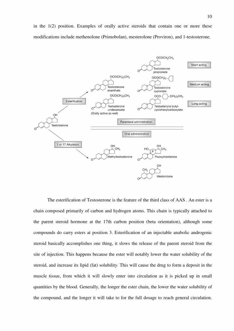
10
in the 1(2) position. Examples of orally active steroids that contain one or more these
modifications include methenolone (Primobolan), mesterolone (Proviron), and 1-testosterone.
The esterification of Testosterone is the feature of the third class of AAS . An ester is a
chain composed primarily of carbon and hydrogen atoms. This chain is typically attached to
the parent steroid hormone at the 17th carbon position (beta orientation), although some
compounds do carry esters at position 3. Esterification of an injectable anabolic androgenic
steroid basically accomplishes one thing, it slows the release of the parent steroid from the
site of injection. This happens because the ester will notably lower the water solubility of the
steroid, and increase its lipid (fat) solubility. This will cause the drug to form a deposit in the
muscle tissue, from which it will slowly enter into circulation as it is picked up in small
quantities by the blood. Generally, the longer the ester chain, the lower the water solubility of
the compound, and the longer it will take to for the full dosage to reach general circulation.
11
Slowing the release of the parent steroid is a great benefit in steroid medicine, as free
testosterone (or other steroid hormones) previously would remain active in the body for a very
short period of time (typically hours). This would necessitate an unpleasant daily injection
schedule if one wished to maintain a continuous elevation of testosterone (the goal of
testosterone replacement therapy). By adding an ester, the patient can visit the doctor as
infrequently as once per month for his injection, instead of having to constantly re-administer
the drug to achieve a therapeutic effect. Clearly without the use of an ester, therapy with an
injectable anabolic/androgen would be much more difficult. Esterification temporarily
deactivates the steroid molecule. With a chain blocking the 17th beta position, binding to the
androgen receptor is not possible (it can exert no activity in the body). In order for the
compound to become active the ester must therefore first be removed. This automatically
occurs once the compound has filtered into blood circulation, where esterase enzymes quickly
cleave off (hydrolyze) the ester chain. This will restore the necessary hydroxyl (OH) group at
the 17th beta position, enabling the drug to attach to the appropriate receptor. In summary this
modification achieve a number of goals, including a) slow metabolism; b) enhanced
affinity
for the androgen receptor (19-nortestosterone); c) resistance to aromatization to estradiol
(fluoxymesterone, 19 nortestosterone); and d) decreased binding of metabolites to
androgen
receptor (5 -reduced metabolites of 19-nortestosterone, 7 -19- nortestosterone). Agents such
as fluoxymesterone and 19-nortestosterone (nandrolone) that resist aromatization
lack the
feminizing side effects of testosterone. 19-nortestosterone possesses another characteristic that
increases its anabolic activity because its 5 -reduced metabolite has poor affinity
for the
androgen receptor. Similarly, alpha-methyl-19-nortestosterone
is not a substrate for 5
reductase (Sundaram et al., 1995).
12
Nandrolone
The drug known as nandrolone (also known commercially as Deca-Durabolin) has the IUPAC
name 17b-hydroxy-19-nor-4-andro-sten-3-one, is an anabolic steroid which occurs naturally
in the human body, but only in tiny quantities. Nandrolone is a 19-Nor steroid (i.e. it lacks a
carbon atom at the 19 position on the steroid molecule) and is derived from the male
hormone, testosterone. Nandrolone may have some legitimate medical uses, such as the
treatment of major burns, malnutrition and osteoporosis. However, nandrolone has been
implicated in relation to doping (especially in sports, where muscle mass and strength are
deciding factors) and was
banned by the International Olympic Committee Medical
Commission in 1974. Athletes who resort to nandrolone do so for various reasons. The drug,
which needs to be administered via intra-muscular injection to be effective, is used to increase
muscle mass. However, its reputation for easing the pain and strain caused by intensive
training, or hasten recovery from injury, is not scientifically based.(ASADA, 2007)
Nandrolone decanoate is the compound used for clinical and no-clinical purpose. This
molecule is an ester synthesize for intra-muscle injection of the anabolic-androgenic steroid
nandrolone. The ester modification make the molecule hydrophobic and extends its duration
of action. The latter is dependent from slow release of the hydrophobic nandrolone ester from
the muscle. A single intra-muscle injection of 50–150 mg nandrolone decanoate in healthy
young men reached a peak serum levels after 2–3 days, and
the maximum serum level ranged
from 2.14 ng/ml in the 50-mg group to 5.16 ng/ml in the 150-mg group (Wilma et al., 2005)
Synthesis and Metabolism
Androgenic steroids are produced by the testes, adrenals and ovaries from cholesterol. In men
testosterone is the principal secreted androgen. The leyding cells synthesize the majority of
testosterone. In women testosterone is synthesize both in the corpus luteum and the adrenal
cortex. The synthesis pathway is similar between man and women. This metabolic pathway
13
have a unique precursor , cholesterol, but it can have two routes. The first one (the ∆5
pathway) involving pregnolone and dehydroepiandrosterone (DHEA) and the other (the ∆4
pathway) involving progesterone and androstenedione. The ∆5 pathway appears to
predominate in the testis of men. Both pathways are functional in adrenals and ovaries. The
precursors androstenedione and deydroepiandrosterone are weak androgens.
Testosterone can be produced by metabolism of certain steroids, such as 4-androstenedione in
organs beside the testis, but this route contributes to less then 5% of plasma testosterone in
men. The leyding cell produce the majority of testosterone. The production of testosterone by
the leyding cells of the testis is controlled by blood levels of leuteinizing hormone (LH)
release from anterior pituitary.
The adrenal synthesizes and secretes dehydroepiandrosterone and dehydroepiandrosterone
sulphate, both have the potential to serve as precursors of synthesis of more potent androgens,
but only 1% of the testosterone derive from this route. The secretion rate for these androgenic
steroids are similar in men and women , and the rates of secretion are insufficient to produce
virilisation. However , condition producing adrenal hyperplasia often lead to increase
secretion of androgens and their precursor, causing virilization. About 50 percent of plasma
testosterone in normal women is derived from peripheral conversion of 4-androstenedione to
testosterone, and a equal amount comes from direct secretion of ovary. Secretion of
androgenic steroids from by the ovary varies with the stage of the menstrual cycle and is
regulated by LH and FSH, witch control steroid synthesis by various cell type in the ovary.
The metabolism of testosterone have a pivotal rule in their function. Testosterone is
metabolize by the enzyme 5α-reductase to 5α-dihydrotestosterone and by aromatase to
estradiol. Some effects of testosterone appear to be mediated by testosterone itself, some by
dihydrotestosterone, and some by estradiol. 5α-dihydrotestosterone have a high affinity for
the androgen receptor and activate gene expression more efficiently, in the tissues expressed
the 5α-reductase. Two forms of 5α-reductase have been identified, type I is found
14
predominantly in nongenital skin and the liver, and type II is found in urogenital tissue in men
and genital skin in both men and women. The conversion of testosterone to estrogens occurs
predominantly in adipose tissue, with lesser amount produce in muscle, kidney, liver and
hypothalamus.
Testosterone is metabolized in the liver to androsterone and etiocholanolone, which are
biologically inactive. Dihydrotestosterone is metabolized to androsterone, androstanedione
and androstanediol.
Mechanism of action
Testosterone and derivates can work through genomic and non-genomic mechanism.
Testosterone enter a cell by passive diffusion through cell membrane. The genomic
mechanism involve the activation of specific androgen receptor (AR). AR is a member of
superfamily of nuclear receptors, which includes steroid hormone receptors, thyroid receptors
and orphan receptors. Testosterone and dihydrotestosterone bind the hormone-binding domain
of the androgen receptor allowing the ligand–receptor complex to bind, via the DNA-binding
domain of receptor, to certain responsive element. The ligand-receptor complex acts as a
transcription factor complex and stimulates expression of specific genes. Androgen receptor
interaction with co-activators and co-repressors transcription factors lead the tissue specific
gene regulation.
The non-genomic mechanism involve the AR or a membrane androgen binding sites that are
discussed to be responsible for rapid androgen signaling have been described in several
tissues or
Cells. But of late, the nature of these binding sites has been controversial. The sex hormone
binding globulin (SHBG) is considered to play a role in permitting certain steroids to act
without entering the cell. Upon ligand binding, the SHBG interacts with a high-affinity
membrane receptor. This SHBG-receptor complex has been described to cause a rapid
15
generation of cAMP after exposure to the respective steroid. For example, in prostate cell
membranes, an increase in intracellular cAMP in response to both androgens and estradiol,
via binding to a membrane SHBG receptor, has been shown. Recently, binding sites for
testosterone on the surface of T cells and IC-21 macrophages have been detected with
testosterone-BSA-FITC that are likely to be involved in the context of nongenomic
testosterone action on [Ca2_]i (Benten et al., 1999). A study lead on pheochromocytoma rat
cells PC-12 has showed a high affinity membrane testosterone binding sites. This specific
androgen membrane receptor regulate cell survival. The NGF-induce neuronal differentiation
of PC12 cells resulted in the suppression of the number of membrane testosterone sites
(Alexaki et al., 2006).
Androgen Receptor
Androgen receptor (AR) modulate the androgen steroids action at level of gene
expression directly by interacting with specific elements in the regulatory regions of target
genes or indirectly by activating various growth factor signalling pathways. AR gene was
cloned in 1988 (Chang et al., 1988 and Lubahn et al., 1988). Like other members of the
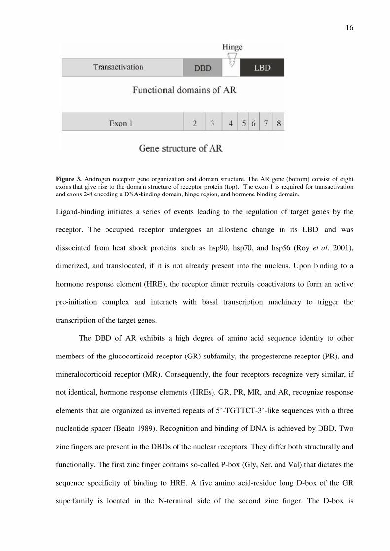
nuclear receptor superfamily, AR has four major functional regions (Fig. 3): the N-terminal
transactivation domain (TAD), a central DNA-binding domain (DBD), a C-terminal ligand-
binding domain (LBD), and a hinge region connecting the DBD and LBD. Two autonomous
transactivation functions, a constitutively active activation function (AF-1) originating in the
N-terminal and a ligand-dependent activation function (AF-2) arising in the LBD, are
responsible for the transcriptional activity of nuclear receptors.
16
Figure 3. Androgen receptor gene organization and domain structure. The AR gene (bottom) consist of eight
exons that give rise to the domain structure of receptor protein (top). The exon 1 is required for transactivation
and exons 2-8 encoding a DNA-binding domain, hinge region, and hormone binding domain.
Ligand-binding initiates a series of events leading to the regulation of target genes by the
receptor. The occupied receptor undergoes an allosteric change in its LBD, and was
dissociated from heat shock proteins, such as hsp90, hsp70, and hsp56 (Roy et al. 2001),
dimerized, and translocated, if it is not already present into the nucleus. Upon binding to a
hormone response element (HRE), the receptor dimer recruits coactivators to form an active
pre-initiation complex and interacts with basal transcription machinery to trigger the
transcription of the target genes.
The DBD of AR exhibits a high degree of amino acid sequence identity to other
members of the glucocorticoid receptor (GR) subfamily, the progesterone receptor (PR), and
mineralocorticoid receptor (MR). Consequently, the four receptors recognize very similar, if
not identical, hormone response elements (HREs). GR, PR, MR, and AR, recognize response
elements that are organized as inverted repeats of 5’-TGTTCT-3’-like sequences with a three
nucleotide spacer (Beato 1989). Recognition and binding of DNA is achieved by DBD. Two
zinc fingers are present in the DBDs of the nuclear receptors. They differ both structurally and
functionally. The first zinc finger contains so-called P-box (Gly, Ser, and Val) that dictates the
sequence specificity of binding to HRE. A five amino acid-residue long D-box of the GR
superfamily is located in the N-terminal side of the second zinc finger. The D-box is
17
important in specifying the half-site spacing requisite at the HRE. Conformation changes
resulting from the binding of androgens to the LBD located at the C-terminal end of the
molecule are responsible for activating the androgen response. Receptors with a deletion of
their LBD are constitutively active, suggesting that the AF-1 is ligand-independent. AF-2 was
demonstrated in LBDs of AR but this action seem to be weak and it is not been described to
be ligand independent (McEwan, 2004). AF-2 in the LBD of AR forms a hydrophobic cleft,
which core is present in helix 12, that binds the LXXLL motif of the p160 family of
transcriptional coactivators , which are associated with histone acetyl transferase activity and
can recruit coactivitors required for chromatin modification. The transcriptional activity of
AR is affected by coregulators that influence a number of functional properties of AR,
including ligand selectivity and DNA binding capacity. AR coregulators participate in DNA
modification of target genes, either directly through modification of histones or indirectly by
the recruitment of chromatin-modifying complexes, as well as functioning in the recruitment
of the basal transcriptional machinery. Some of the better characterized coregulators are
members of the p160 family, ARA70, ARA55, ARA54, ARA267-α, Smad-3, and AIB1.
ARA55 and ARA70 both allow the activation of AR by 17β -estradiol (E2), with ARA70
being the most effective coactivator for conferring androgenic activity to E2. Furthermore,
both ARA55 and Smad-3 have been suggested to function as bridges for cross-talk between
transforming growth factor-β signalling pathway and androgen/AR action (Kang et al., 2001).
Aberrant AR coregulator activity due to mutation or altered expression levels may be a
contributing factor in the progression of diseases related to AR activity, such as prostate
cancer (Heinlein & Chang 2002).
Nuclear receptors (NRs) may also be activated by signalling pathways that originated
at the cell surface. NRs, along with other transcription factors, are regulated by reversible
phosphorylation . Kinase-mediated signal transduction pathways could affect the activity of
NRs. The consensus phosphorylation sites found in AR indicate that AR could be a substrate
18
for the DNA-dependent protein kinase, protein kinase A, protein kinase C, mitogen-activited
kinase, and casein kinase II . Indeed, AR could be activated in an androgen-independent way
by growth factor or cytokine signalling pathways, like those initiated by epidermal growth
factor (EGF), insulin-like growth factor-1 (IGF-1), keratinocyte growth factor and IL-6,
which would elicit AR-mediated transcriptional activation (Wu et al., 2006). Receptor
distribution and hormone metabolism can explain part of the steroid-specificity in vivo. Other
ways of imposing selectivity have been proposed at the chromatin level (Beato et al. 1995)
and at the level of cooperativity of receptors with other transcription factors. A recent study
demonstrated that selective DNA binding by AR could be a mechanism for hormone-specific
gene regulation (Schoenmakers et al. 2000).
Physiological effects of AAS
Testosterone and DHT play a critical roles in male sexual differentiation during
embryogenesis and in the development of secondary sexual characteristics. Testosterone
secreted by the fetal testis is responsible during the embryogenesis for differentiation of the
Wolffian ducts into the epididymides, vas deferentia and seminal vesicles. Instead, the
virilization of external genitalia is dependent from 5α-reductatse which convert testosterone
into DHT. The androgen secretion lead the complete genital growth until shortly after the
birth, when declining production of testosterone by testis cause a cessation of androgen-
dependent development until puberty.
At puberty, synthesis and secretion of testosterone by the testis increases and blood
level of testosterone gradually rise over 4-5-years period until adult levels are reached. The
increase of secretion of FSH and LH from the gonadotroph cell, in turn stimulate by increased
secretion of GnRH from the hypothalamus, stimulate the testis and consequently the
testosterone release. Increased secretion of testosterone into the systemic circulation affect
19
many tissues and the changes in most of them occur gradually during the course of several
years. The skin becomes coarser and oilier due to increased sebum production, with
contributes to the development of acne. Sexual hair begins to grow, initially pubic and
axillary hair, the air in the lower legs, and finally other body hair and facial hair. Muscle mass
and strength, especially of the shoulder girdle, increased, and subcutaneous fat decreases.
Epiphyseal bone growth accelerates, resulting in the pubertal growth spurt, but epiphyseal
maturation leads eventually to a slowing and then cessation of growth. The androgenic effect
on bone growth may involve conversion of testosterone to estradiol, which alone or in
conjunction with testosterone stimulates the synthesis and secretion of growth hormone and
insulin-like growth factor I. The increased in muscle mass and bone result in a pronounced
increase in weight. The larynx thickens, resulting in a lower voice. In the senescence period
the testosterone concentration gradually declines. The fall in serum concentration could
contribute to several changes that occur with increasing age in men, including decreases in
energy, libido, muscle mass and bone mineral density.
Therapeutic use of AAS
The AAS have a limited therapeutic use. Androgen replacement therapy is prescribe to
patients with a androgen deficiencies. Androgens are administered to these individuals to
restore a normal steroids physiological function. In male is used to restore the development of
male secondary sexual characteristics, as well as to promote the effect of androgen on somatic
growth. Androgen therapy also is used to normalize male sexual behaviour. In the case of
hypogonadism developed prior to the normal time period for puberty the androgen replace
therapy can bring about the series of changes that usually take at puberty. The exposure to a
adult levels of testosterone can lead to premature closure of epiphysis, this effects is used in
individuals with abnormal growth. The use of androgen therapy in aging male is submit at
clinical trial evaluation. Symptoms and findings of T deficiency are similar to those
20
associated with aging. They include loss of energy, depressed mood, decreased libido, erectile
dysfunction, decreased muscle mass and strength, increased fat mass, frailty, osteopenia, and
osteoporosis. Several studies suggested that some symptoms and signs of andropause may be
improved by use of androgen steroids, but the benefit/risk ratio of testosterone replacement
therapy in aging men is not known, until now (Hijazi at al., 2005). The women can be affect
by androgen deficiencies that cause an impaired sexual function, lessened well being, loss of
energy and negative effects on bone mass. The testosterone replacement therapy result a
optimal treatment for women with post-menopausal problem and premature ovarian failure.
The side effects are rare when the testosterone levels are close to normal reproductive range
for women (Davis et al., 1999)
Anabolic steroids have been used in a variety of catabolic states such as those
involving acute and chronic illnesses, surgical trauma. The rationale for the use of androgen
therapy in these case depend from two process. The first one is the ability of androgens to
promote a positive nitrogen balance and overall protein synthesis, a second process is the anti-
cortisol activity. The Latter decreases the catabolic effects of cortisol but not alter its
protective anti-inflammatory response. These mechanisms have been shown to increase
muscle and bone growth and body weight. (Demling et al., 2005). Androgen therapy receiving
renewed interest in treatment of patients with chronic kidney disease. In this subjects has been
observed a reduction of skeletal muscles mass that affect the physical function. A nandrolone
therapy in this patients increase muscle mass, enhance quality of life and reduce mortality
(McDonald et al., 2007).
Androgens enhance erythropoiesis by stimulating the production of erythropoietin.
Because of this effect, androgens have been used in treatment of some haematological
disorder, such as the anemia associate with bone marrow and renal failure, and with
myelofibrosis. After the introduction of recombinant human erythropoietin (rHuEPO) during
the late 1980s the use of these compounds has been reduced. The used of androgens as
21
adjuvant of rHuEPO therapy could be useful. Androgens not regulate the EPO levels but
increased the erythroid progenitors to EPO. Mechanism of this action may be mediated by
triggering the pluripotential stem cells from G0 phase or prolonged G1 phase into a G1
interval responsive to EPO. It can explain why a positive response to androgen therapy may
depend on the severity of the anemia and that the presence of stem cells in bone marrow
favourably influence the response and survivability of patients (Navarro et al., 2001).
Side effect of AAS
Androgen therapy in case of hypogonadism and androgen deficiencies aging related
have been showed to be quite safety. No large-scale, long-term studies have yet been initiated
to assess the risks of testosterone-replacement therapy in men. The side effects of AAS
depend from the sex and health of patients and administration routes. Unwanted effects can
occur in women and prepubertal boys. In these patients virilization, acne, hair growth, weight
gain, gynaecomastia and male-pattern hair loss may be observed, and should be managed
symptomatically. Hirutism, excessive hair growth in androgen-sensitive areas, is the major
side effect of exogenous androgen administration in women (Braunstein 2007).
Several AAS-induced adverse cardiovascular effects have been reported, including
hypertension, left ventricular hypertrophy (LVH), impaired diastolic filling, arrhythmia,
erythrocytosis, altered lipoprotein profile, and thrombosis (Dickerman et al., 1996 and 1997 ).
Steroids abuser have shown psychiatric side effects. Studies compare steroids abuser with not
steroid user described a higher incidence of behavioral changes as irritability, aggressiveness,
euphoria, hyperactivity (Hall et al., 2005). In same case it was shown acute psychoses,
exacerbation of tics and depression, and development acute confusional/delirious states. In
subject that use low-dose of steroids a minimal risk of adverse psychological effects was
described (Yates et al., 1999). This side effects occur, particularly, among the body-builders
and athletes who are steroids abuser.
22
Androgens and Brain
Androgens have an outstanding part in the brain functions. It was showed a
neuroprotective and neuromodulation actions of AAS during brain development but also
during brain aging and the important of androgens on the systems involved in cognitive
function, mood disturbances and central drive of sexuality. Androgen receptors are found in
brain regions that are crucial for learning and memory including the hypothalamus, the
hippocampus, prefrontal cortex, and amygdala, but are not found in other cortical regions of
the brain. In these areas the androgens acting on specific nuclear receptors or throughout the
metabolism into neuroactive compounds. The hormone are important during pre- and
perinatal brain development for the formation of sex-specific behavioral. The brain areas
involved in pre and perinatal androgens effects are the same that show functional loss with
aging. Bioavailable testosterone levels decline with age in men and women and numerous
studies suggest a link between testosterone and cognition in men, particularly with aging.
Testosterone supplementation improves spatial cognition and working memory in healthy
older men. Visual-spatial cognition improves in older men with testosterone replacement in a
dose-dependent manner, but similar effects are not found in young men. Testosterone
replacement has few effects in men with low or no testosterone throughout life (congenital
hypogonadism). Lower testosterone levels are associated with a higher risk for Alzheimer's
disease, some studies suggest that low testosterone is associated with increased beta amyloid
deposition in men (Almeida et al., 2004).
The effects of hormone in brain function is observable in healthy people. It has been
suggested that effects within the central nervous system (CNS) contribute to AAS effects on
strength because AAS user feel more energetic and therefore train harder. The androgens have
a positive ( elevate mood) and negative (psychotic symptoms) effects on user behavioral.
23
These hormones can be consider a drug of abuse. The effect of androgens in central nervous
system could cause dependence state and activation of rewarding system (Kuhn, 2002).
Neurosteroids
The brain is one of tissues where the androgens are synthesized. The steroids produce
in various regions of the central and peripheral nervous system are called neurosteroids.
Dehydroepiandrosterone sulfate (DHEAS), was the first steroids found at high levels in the
brain long after gonadectomy and adrenalectomy, and later shown to be synthesized by the
brain. Later, androstenedione, pregnenolone, their sulfates and lipid derivatives, as well as
tetrahydrometabolites of progesterone (P) and deoxycorticosterone (DOC) were identified as
neurosteroids. There are differences between steroid synthesis in the brain and in the adrenals.
Corticosteroid synthesis involves converting deoxycorticosterone (DOC) to either aldosterone
by aldosterone synthase or to corticosterone by 11β-hydroxylase. In the adrenals the enzymes
are never expressed in the same cell. But in the brain, the enzymes co-express not only in the
same region, but even within the same cell, therefore aldosterone synthase and 11β-
hydroxylase must compete for DOC . In the brain, the pregnanes are metabolized in sequence
by the enzymes 5α-reductase and 3α hydroxysteroid dehydrogenase (HSD). Progesterone (P)
to tetrahydroprogesterone (THP), deoxycorticosterone (DOC) to
tetrahydrodeoxycorticosterone (THDOC) and testosterone to androstanediol (Dubrovsky ,
2006). The function attributed to specific neurosteroids include of GABAA, NMDA and
sigma receptor function , regulation of myelinization, neuroprotection and growth of axon and
dendrites.
In vivo and in vitro studies
A number of excellent studies, using animal models, was performed to evaluate the
effects of AAS on neural circuits that underlie the behavioral effects. The in vivo studies
differentiate in treatment regiment and hormonal state, sex and age of animals.
24
The in vivo experiments drove to study the aggression showed a sex – species and compound
specificity. The administration of testosterone propionate for a long period of time enhanced
aggression in intact male rats (Breuer et al., 2001) . It was observed that aggression increase
is provoked by physical stimuli (tail-pich) and in social and environmental context that do not
provoke aggression in control. Other AAS failed to stimulate aggression (nandrolone) or
inhibited the display of aggression (stanozolol). Testosterone and stanozolol effects on
aggression appear to be dependent upon the continued presence of the AAS. In addiction, the
withdrawal from testosterone propionate did not itself induce aggressive behaviour (McGinnis
et al.,2002). The different effects of steroids on aggression may reflect differences in the
ability of these compounds to act to androgen and estrogen receptors and resulting differences
in the balance of estrogen and androgen receptor-mediated signaling. In female rats treated
with AAS it was observed a striking effects on aggression (Bronson et al., 1996). It was
demonstrated that decreased serotonergic tone is pivotal to the ability of the AAS to increase
aggression. Treatment of Fischer rats with testosterone propionate significantly decreased
both 5-HT and the 5-HT metabolite, 5-hydroxyindoleacetic acid, in the hippocampus. The
serotonergic agonist, quipazine, reduced the testosterone-induced dominance in a dose-
dependent fashion (Bonson et al., 1992). Moreover, the effects of quizapine were themselves
antagonized by the 5-HT1A and 5-HT1B receptor antagonist, pindolol, demonstrating that the
actions of quipazine in reducing androgen-induced aggression were specific for 5-HT-
mediated transmission
The Studies that investigate the effect of AAS on the sexual behaviour of intact male
rodents, showed two different classes of AAS. Stanozolol, oxymetholone and 17α-
methyltestosterone eliminate the display of male sexual behaviour. These compounds acting
on sexual behavioral suppressing the secretion of testosterone. Methandrostenolone,
nandrolone and testosterone had minimal effects on intact male animals while maintain male
sexual behaviour in gonadectomised male rats (Clarck et al., 1997). Experiments led on
25
female rats suggest that AAS act in the brain to interfere with events necessary for the
estrogen-dependent induction of female sexual behaviour and the regulation of the
neuroendrocrine events required for reproductive cyclicyty.
The number of experiments that have tested the effects of AAS on anxiety behaviour is
limited but the results obtained showed an anxiety-reduction a time course and dose-response
dependent (Britan et al., 1993). This effects of AAS is supposed to be mediated by the
GABAA receptor. AAS has been shown to be a expression and a allosteric modulator of
GABAA receptor when they are given chronically or acutely.
An effects on brain reward has been described in rats exposed to AAS. To evaluate the
rewarding properties of drugs is used, extensively, the conditioned place preference task. It
has reported that testosterone induce a conditioned place preference in male rats. This effect is
mediate by its metabolites 3α-androstanediol (Frye, 2007). Animal treated with androgens
may have an indirect effects on brain reward. It was showed a potentiated rewarding effects of
amphetamine on rats exposed to AAS (Clark et al., 1996) . In these experiments it was found
that nandrolone influence morphine rewarding an the somatic expression of withdrawal when
it was chronically administered on rats before the start of opioid treatment (Celelier et al.,
2003). In this experiments has been describe a suppression of morphine reward and increased
withdrawal. Similar results was obtain in rats treated with nandrolone and cannabinoids
(Celelier et al., 2006). Nandrolone induce changes that may reflect long-term modifications in
the brain reward circuits leading to a progressive decrease in the basal hedonic level, which
would result in an unpleasant state facilitating the development of an addictive process (Koob
and Le Moal, 2001). In hamsters was observed a self-administer effects of testosterone
(Wood, 2002). This data confirm the idea about the abuse potential of AAS (Wood, 2004)
Nandrolone decanoate altered the levels of both D1 and D2 dopamine receptors in the
mesocorticolimbic system of Sprague–Dawley rats (Kindlundh et al., 2001). Studies to date
suggest that chronic exposure to high doses of AAS alters both dopamine and dopamine
26
receptor expression. In rats exposed to nandrolone it was showed a down-regulation of
expression of D(1)-receptor and an up-regulation of D(2)-receptors (Kindlundh et al., 2003).
Enhanced activity of the mesocorticolimbic dopaminergic system is critical for the acute
rewarding effects of cocaine and amphetamines (Thiblin et al., 1999).
Opioids and opioid receptors are highly expressed in brain regions that mediate reward.
Studies carried out by Nyberg and colleagues showed a modulation of opioid peptide Met-
enkephalin-Arg-Phe, a µ and δ opioid receptor agonist, in n the hypothalamus, striatum and in
the periaqueductal gray (Johansson et al., 2000). Experiments carried out in Hamster showed
that self-administration of testosterone in the presence of naltrexone, an opioid antagonist,
was blocked. Naltrexone inhibit, also, the onset of testosterone intoxication in hamster treated
with high dose of hormone (Peter and Wood, 2004). A modulation of expression of δ opioid
receptor (DOR) is described in an in-vitro study. Cell lines exposed to nandrolone showed a
down-regulation of expression of DOR, mRNA and DOR binding sites. These changes in δ
opioid receptor levels of mRNA and protein were not blocked by coincubation with the
androgen receptor-specific antagonist, flutamide, indicating that this effect of nandrolone is
independent of androgen receptor activation (Pasquariello et al., 2000).
Learning and memory are highly dependent on synaptic plasticity, which involves
structural changes in neurons and synapses. The glutamate receptor N-methyl-d-aspartate
subtype (NMDAR) plays a crucial role in synaptic plasticity. Le Greves and colleague have
reported that repeated administration of AAS affect the gene regulation of NMDAR subunits
in different brain areas. the drug produced a significant decrease in the mRNA expression of
the NR2A and NR2B receptors subunit, while the NR1 subunit was not affect. Instead, the
combination of AAS nandrolone and cocaine bring about a significant decrease in the NR1
mRNA (Le Greves et al., 1997 and 2002). A single high dose of nandrolone mediate the
phosphorylation of NMDA receptor subunits and ERKs. These effects were not seen after a
2-week treatment period, indicating adaptation to high steroid levels (Rossbach et al., 2007).
27
References
• A. Pasquariello, R. Di Toro, F. Nyberg and S. Spampinato, Down-regulation of delta opioid receptor
mRNA by an anabolic steroid in neuronal hybrid cells. NeuroReport 11 4 (2000), pp. 863–867
• A.M. Kindlundh, J. Lindblom, L. Bergström, J.E. Wikberg and F. Nyberg, The anabolic-
androgenic steroid nandrolone decanoate affects the density of dopamine receptors in the male rat brain.
Eur J Neurosci 13 (2001), pp. 291–296.
• Alexaki VI, Dermitzaki E, Charalampopoulos I, Kampa M, Nifli AP, Gravanis A, Margioris AN,
Castanas E. Neuronal differentiation of PC12 cells abolishes the expression of membrane androgen
receptors.Exp Cell Res. 2006 Sep 10;312(15):2745-56. Epub 2006 May 16
• Almeida et al., One year follow-up study of the association between chemical castration, sex
hormones, beta-amyloid, memory and depression in men, Psychoneuroendocrinology 29 (2004), pp.
1071–1081
• Ann J Conway, David J Handelsman, Douglas W Lording, Bronwyn Stuckey, Jeffrey D Zajac
:Use, misuse and abuse of androgens The Endocrine Society of Australia consensus guidelines for
androgen prescribing
• ASADA- Australian driving force for pure performance in sport- Fact about substance:
Nandrolone. 2007
• Beato M (1989) Gene regulation by steroid hormones. Cell 56: 335-344.
• Beato M, Herrlich P & Schütz G (1995) Steroid hormone receptors: Many actors in search of a plot.
Cell 83: 851-857.
• Benten WP,Lieberherr M, Stamm O, Wrehlew C, Guo Z, Wunderlich F. Testosterone signaling
through internalizable surface receptors in androgen receptor-free macrophages. Mol Biol Cell 10:
3113–3123, 1999.
• Berg JM (1989) DNA binding specificity of steroid receptors. Cell 57: 1165-1168.
• Bitran D, Kellogg CK, Hilvers RJ. Treatment with an anabolic-androgenic steroid affects anxiety-
related behavior and alters the sensitivity of cortical GABAA receptors in the rat. Horm Behav. 1993
Dec;27(4):568-83.
• Braunstein GD. Safety of testosterone treatment in postmenopausal women. Fertil Steril. 2007
Jul;88(1):1-17. Epub 2007 May 10
28
• Breuer ME, McGinnis MY, Lumia AR, Possidente BP. Aggression in male rats receiving anabolic
androgenic steroids: effects of social and environmental provocation. Horm Behav. 2001
Nov;40(3):409-18
• Bronson FH, Nguyen KQ, De La Rosa J. Effect of anabolic steroids on behavior and physiological
characteristics of female mice. Physiol Behav. 1996 Jan;59(1):49-55.
• Célérier E, Ahdepil T, Wikander H, Berrendero F, Nyberg F, Maldonado R. Influence of the
anabolic-androgenic steroid nandrolone on cannabinoid dependence. Neuropharmacology. 2006
Jun;50(7):788-806. Epub 2006 Jan 27
• Clark AS, Harrold EV. Comparison of the effects of stanozolol, oxymetholone, and testosterone
cypionate on the sexual behavior of castrated male rats. Behav Neurosci. 1997 Dec;111(6):1368-74
• Clark AS, Lindenfeld RC, Gibbons CH. Anabolic-androgenic steroids and brain reward. Pharmacol
Biochem Behav. 1996 Mar;53(3):741-5
• David A. Gruenewald, MD, Alvin M. Matsumoto Testosterone Supplementation Therapy for Older
Men: Potential Benefits and Risks
• Davis S.R. : The therapeutic use of androgens in women. Journal of Steroid Biochemistry and
Molecular Biology 69 (1999) 177±184
• Demling RH. The role of anabolic hormones for wound healing in catabolic states. J Burns Wounds.
2005 Jan 17;4:e2.
• Di Luigi L., Romanelli F., Lenzi A.: Androgenic-anabolic steroids abuse in males. J. Endocrinol.
Invest. 28 (Suppl. to no 3): 81-84, 2005
• Dickerman RD, McConathy WJ, Schaller F, Zachariah NY.Cardiovascular complications and
anabolic steroids.Eur Heart J. 1996 Dec;17(12):1912
• Dickerman RD, McConathy WJ, Zachariah NY.Testosterone, sex hormone-binding globulin,
lipoproteins, and vascular disease risk. J Cardiovasc Risk. 1997 Oct-Dec;4(5-6):363-6.
• Dubrovsky B. Neurosteroids, neuroactive steroids, and symptoms of affective disorders. Pharmacol
Biochem Behav. 2006 Aug;84(4):644-55. Epub 2006 Sep 7
• DuRant RH, Rickert VI, Ashworth CS, et al. Use of multiple drugs among adolescents who use
anabolic steroids. N Engl J Med 1993;328: 922–926.
• E. Célérier, M.T. Yazdi, A. Castane, S. Ghozland, F. Nyberg and R. Maldonado, Effects of
nandrolone on acute morphine responses, tolerance and dependence in mice, Eur. J. Pharmacol. 465
(2003), pp. 69–81

29
• Frye CA. Some rewarding effects of androgens may be mediated by actions of its 5alpha-reduced
metabolite 3alpha-androstanediol. Pharmacol Biochem Behav. 2007 Feb;
• G.F. Koob and M. Le Moal, Drug addiction, dysregulation of reward, and allostasis,
Neuropsychopharmacology 24 (2001), pp. 97–129
• Hall RC, Hall RC, Chapman MJ. Psychiatric complications of anabolic steroid abuse.
Psychosomatics. 2005 Jul-Aug;46(4):285-90
• Heinlein CA, Chang C. The roles of androgen receptors and androgen-binding proteins in nongenomic
androgen actions. Mol Endocrinol. 2002 Oct;16(10):2181-7
• Hijazi RA, Cunningham GR .Andropause: is androgen replacement therapy indicated for the aging
male? Annu Rev Med. 2005;56:117-37. Review.
• Hoberman, Yesalis: The History of Synthetic Testosterone; February 1995;
• I. Thiblin, A. Finn, S.B. Ross and C. Stenfors, Increased dopaminergic and 5-hydroxytryptaminergic
activities in male rat brain following long-term treatment with anabolic androgenic steroids. Br J
Pharmacol 126 (1999), pp. 1301–1306
• Janowsky JS. The role of androgens in cognition and brain aging in men. Neuroscience.
2006;138(3):1015-20. Epub 2005 Nov 28.
• Janowsky JS. Thinking with your gonads: testosterone and cognition. Trends Cogn Sci. 2006
Feb;10(2):77-82. Epub 2006 Jan 4
• K.R. Bonson and J.C. Winter, Reversal of testosterone-induced dominance by the serotonergic
agonist quipazine. Pharmacol Biochem Behav 42 (1992), pp. 809–813
• Kanayama G, Barry S, Hudson JI, Pope HG Jr. Body image and attitudes toward male roles in
anabolic-androgenic steroid users. Am J Psychiatry. 2006 Apr;163(4):697-703.
• Kang HY, Lin HK, Hu YC, Yeh S, Huang KE, Chang C. From transforming growth factor-beta
signaling to androgen action: identification of Smad3 as an androgen receptor coregulator in prostate
cancer cells. Proc Natl Acad Sci U S A. 2001 Mar 13;98(6):3018-23
• Kindlundh AM, Lindblom J, Nyberg F. Chronic administration with nandrolone decanoate induces
alterations in the gene-transcript content of dopamine D(1)- and D(2)-receptors in the rat brain. Brain
Res. 2003 Jul 25;979(1-2):37-42
• Kuhn CM. Anabolic steroids. Recent Prog Horm Res. 2002;57:411-34
30
• Le Grevès P, Huang W, Johansson P, Thörnwall M, Zhou Q, Nyberg F. Effects of an anabolic-
androgenic steroid on the regulation of the NMDA receptor NR1, NR2A and NR2B subunit mRNAs in
brain regions of the male rat. Neurosci Lett. 1997
• Le Grevès P, Zhou Q, Huang W, Nyberg F. Effect of combined treatment with nandrolone and
cocaine on the NMDA receptor gene expression in the rat nucleus accumbens and periaqueductal gray.
Acta Psychiatr Scand Suppl. 2002;(412):129-32.
• Macdonald JH, Marcora SM, Jibani MM, Kumwenda MJ, Ahmed W, Lemmey AB.Nandrolone
decanoate as anabolic therapy in chronic kidney disease: a randomized phase II dose-finding study.
Nephron Clin Pract. 2007;106(3):c125-35. Epub 2007 May 22.
• Matias PM, Donner P, Coelho R, Thomaz M, Peixoto C, Macedo S, Otto N, Joschko S, Scholz P,
Wegg A, Basler S, Schafer M, Egner U & Carrondo MA 2000 Structural evidence for ligand
specificity in the binding domain of the human androgen receptor. Implications for pathogenic gene
mutations. Journal of Biological Chemistry 275 26164–26171
• McEwan IJ. Molecular mechanisms of androgen receptor-mediated gene regulation: structure-function
analysis of the AF-1 domain. Endocr Relat Cancer. 2004 Jun;11(2):281-93
• McGinnis MY, Lumia AR, Possidente BP. Effects of withdrawal from anabolic androgenic steroids
on aggression in adult male rats. Physiol Behav. 2002 Apr 1;75(4):541-9
• Navarro JF, Mora C. Androgen therapy for anemia in elderly uremic patients. Int Urol Nephrol.
2001;32(4):549-57. Review.
• P. Johansson, M. Hallberg, A. Kindlundh and F. Nyberg, The effect on opioid peptides in the rat
brain, after chronic treatment with the anabolic androgenic steroid, nandrolone decanoate. Brain Res
Bull 51 5 (2000), pp. 413–418
• Patchev VK, Schroeder J, Goetz F, Rohde W, Patchev AV.Neurotropic action of androgens:
principles, mechanisms and novel targets. Exp Gerontol. 2004 Nov-Dec;39(11-12):1651-60.
• Peters KD, Wood RI. Androgen dependence in hamsters: overdose, tolerance, and potential
opioidergic mechanisms. Neuroscience. 2005;130(4):971-81
• Pope CG, Pope HG, Menard W, Fay C, Olivardia R, Phillips KA. Clinical features of muscle
dysmorphia among males with body dysmorphic disorder.Body Image. 2005 Dec;2(4):395-400
• Rossbach UL, Steensland P, Nyberg F, Le Grevès P. Nandrolone-induced hippocampal
phosphorylation of NMDA receptor subunits and ERKs. Biochem Biophys Res Commun. 2007 Jun
15;357(4):1028-33. Epub 2007 Apr 17
31
• Schoenmakers E, Verrijdt G, Peeters B, Verhoeven G, Rombauts W, Claessens F. Differences in
DNA binding characteristics of the androgen and glucocorticoid receptors can determine hormone-
specific responses. J Biol Chem. 2000 Apr 21;275(16):12290-7
• Sundaram K, Kumar N, Monder C, Bardin CW 1995 Different patterns of metabolism determine
the relative anabolic activity of 19-norandrogens. J Steroid Biochem Mol Biol 53: 253–257
• Wichstrøm L, Pedersen W. : Use of anabolic-androgenic steroids in adolescence: winning, looking
good or being bad?; Scientific American Magazine;
• Wilma M. Bagchus, Jean M. W. Smeets, Herman A. M. Verheul, Suzanne M. De Jager-Van Der
Veen, Andreas Port and T. B. Paul Geurts: Pharmacokinetic Evaluation of Three Different
Intramuscular Doses of Nandrolone Decanoate: Analysis of Serum and Urine Samples in Healthy Men.
The Jou. Cl. Endo. & Meta. Vol. 90,No.5,2624-2630,2005
• Wood RI. Oral testosterone self-administration in male hamsters: dose-response, voluntary exercise,
and individual differences. Horm Behav. 2002 May;41(3):247-58
• Wood RI. Reinforcing aspects of androgens. Physiol Behav. 2004 Nov 15;83(2):279-89
• Wu JD, Haugk K, Woodke L, Nelson P, Coleman I, Plymate SR. Interaction of IGF signaling and
the androgen receptor in prostate cancer progression. J Cell Biochem. 2006 Oct 1;99(2):392-401
• Yates WR, Perry PJ, MacIndoe J, Holman T, Ellingrod V. Psychosexual effects of three doses of
testosterone cycling in normal men. Biol Psychiatry. 1999 Feb 1;45(3):254-60
32
Chapter 2- Mu Opioid Receptor
Opioid systems are responsible for a variety of processes in organisms, it controls pain,
reward and addictive behaviours. Opioids exert their pharmacological actions through three
opioid receptors, mu, delta and kappa whose genes have been cloned (Oprm, Oprd1 and
Oprk1, respectively). Neurons release a family of endogenous peptides like enkephalins,
dynorphins and endorphin, which bind the opioid receptors in the brain. Opioid receptors are,
also, activated exogenously by alkaloids of the opium poppy plant Papaver somniferum, the
prototype of which is morphine. Morphine was first isolated from opium in 1805 by a German
pharmacist, Wilhelm Sertürner. Sertürner described it as the Principium Somniferum. He
named it morphium - after Morpheus, the Greek god of dreams. Alkaloids opiaces have been
known to relieve pain and alter mood since the advent of recorded history. For centuries, these
agents have been integrated into medical practice with varying efficacy. Morphine was first
used medicinally as a painkiller. During the American Civil War it was used as a surgical
anaesthetic and was sent home with many wounded soldiers for relief of pain. At the end of
the war, over 400,000 people had the "army disease," morphine addiction. The Franco-
Prussian War in Europe had a similar effect. In 1906 the Pure Food and Drug Act required
accurate labelling of patent medicines and tonics. Various laws restricting the importation of
opium were enacted, and the Harrison Narcotics Act (1914) prohibited possession of narcotics
unless properly prescribed by a physician. The morphine positive (pain relieve) and negative
(change behavioral and addiction ) effects are carried out primarily thought the mu opioid
receptor (MOPr).
33
Opioid receptor
The opioid receptors were discovered in the brain by binding studies using radiolabeled
opioid ligands, in the early 197Os. The first evidence about being of different opioid receptors
was reported by Martin and colleague in 1976. They deduced the existence of three distinct
opioid receptors from the different pharmacologic effects of various opioid agonists and
antagonists that selectively induce or inhibit different physiologic responses and named them
µ for the morphine group, κ for the ketocyclazocine group, and σ for N-allylnormetazocine
(SKF10047). In addition to these three types of receptors in the 1977 was found a high-
affinity receptor for enkephalins in the mouse vas deferens and named it the δ receptor (Lord
et al., 1977). All these receptors are members of the G protein-coupled family of receptors
and show significant amino acid sequence homologies. Multiple receptor subtypes have been
proposed based on pharmacologic criteria. However, genes encoding only one subtype from
each of the µ, κ and δ receptor families have been isolated and characterized thus far.
The first opioid receptor cloning was δ. Kieffer and her colleague isolated the cDNA
of δ opioid receptor (DOPr) from a expression cDNA library. The plasmid bore the cDNA
encodes the 371 amino-acid residues of DOPr was cloned into COS cells and screened for it
ability to binding the ligand 3H-labeled Tyr-D-Thr-Gly-Phe-Leu-Thr. Thereafter, µ and κ
opioid receptor cDNAs were cloned based upon their homology to the cloned δ -opioid
receptor. The human µ opioid receptor (hMOPr) cDNA has been identified from a cerebral
cortical cDNA library using sequences from the rat µ opiate receptor cDNA. hMOPr shares
95% amino acid identity with the rat sequence. The human µ , δ and κ opioid receptor genes
are located on chromosomes 6q24-25, 1p34.3-36.1 and 8q11.2 respectively. The three opioid
receptor genes share a common genomic structure and the coding region is divided into three
major exons. The promoter region of three opioid receptors genes in mouse and rat share
several common feature. All three promoters lack a TATA box, are highly G/C rich and share
several common transcription factors, including SP-1/2, Ikaros (IK), E-box factors and
34
AP1/AP2. However, each promoter has its own distinct transcription factor. The MOPr gene
in both mouse and rat contain a proximal and a distal promoter (Wei and Loh, 2002). A
similar structure was described for the human MOPr gene, Carr and Xu discovered a distal
promoter (-827) and a proximal promoter (-252). The analysis conduct with luciferase
reporter vectors bore different sequences of promoter showed a strong activity of proximal
promoter in MOPr-expressing cells (SK-N-SH) and in no-expressing cells (Hela). DNA
sequence analysis indicated that the hMOR proximal promoter lacked a consensus TATA
box, a consensus initiator, and GC-rich sequences ( Xu and Carr, 2001a). The distal promoter,
instead , had a weakly activity in both cell lines. It was also identified two cis-acting elements
that allow positive and negative regulation of proximal promoter activity in SK-N-SH. A
fragment of 40bp (-540 to -501) containing a GCC core in MOPr gene has been described to
enhance hMOPr transcription. A protein complex was described binding this fragment but
remain to identified which transcription factor is. A sequence of 34-bp (-694 to 660) was
discovered in the MOPr gene, this fragment have a negative effect on transcription.
Comparison of the 35bp element with sequences deposited in the transcription factor
databases revealed several interesting putative binding motifs, including Ikaro-2, MZF1, NFY
and C/EBPβ . Whether these factor are functional in hMOPr gene regulation remains to be
determined ( Xu and Carr, 2001b ). Analysis of -500 to 292 region of hMOPr promoter
showed that Sp1 and Sp3 bound to the CCCTCCTCCC motif in this region. STAT1 and
STAT3 transcription factors binding site was observed in -1583 to 1575 region of MOPr
promoter. The interlukine-6 up-regulate the expression of mRNA of MOPr in SH-SY5Y cells
via STAT1 and STAT3 activation (Börner C et al., 2004). Neuronal and immune cells express
MOPr mRNA, also, under control of nuclear factor- B. NF B recognise three cis-active
elements on the µ-opioid receptor gene promoter, at nt -2174, -557, and -207 (Kraus et al.,
2003). Loh and colleague showed that Poly-C binding proteins (PCBPs) bound from -317 to -
304 region in hMOPr proximal promoter (Kim et al., 2007). This poly-C elements is recognise
35
by different regulator factor whit a positive (PCBPs) or negative (alpha CP3) effects (Choi et
al., 2007). In mouse was discover a sequence located at 10kb upstream the exon1 coding to a
new exon called exon11. This exon have a upstream promoter that control the expression of
exon11-associated variants of MOPr (Pan YX, 2002). Mouse exon11 promoter differs from
mouse exon1 promoter in several aspects. First, the exon11 promoter contains a TATA box
that is absent in the exon1 promoter. Second, the exon 11 promoter has one major
transcription start point (tsp), while the exon 1 promoter contains multiple tsp. The exon 1
promoter contains several GC-rich cis-acting elements like Sp1 and AP-2 that are missing in
the exon 11 promoter. The exon 11 promoter appears to be a typical eukaryote class II
promoter associated with RNA polymerase II, while the exon 1 promoter favors a
“housekeeping” gene mode .These preliminary results indicated a complex control tissues and
spatial specific expression of hMOPr gene modulate by positive and negative regulatory
element.
The opioid receptor expression are regulate by multiple promoters and alternative pre-
mRNA splicing mechanism. In rat, mouse and human MOPr gene was discovered a high
number of splicing variants. Early after the cloning of mouse MOR-1 (mMOR-1) was isolate
the first two splice variants mMOR-1A and mMOR1-B. Using a modified 3’RACE strategy
combined with a nested PCR approach, Pan and colleague have isolated 10 additional splice
variants, mMOR-1B2, mMOR-1B3, mMOR-1B4, mMOR- 1B5, mMOR-1C, mMOR-1D,
mMOR-1E, mMOR-1F, mMOR-1O, and mMOR-1P. These mRNA share the first three exons
and differ at 3’ of the molecule utilizing different exons. These MOPr variants exhibit very
similar ligand binding profile compare with the wild-type MOR-1 in transfected cells. All
these variants have the same protein structure predicted from exons 1–3, which includes the
N-terminus, seven trans-membrane (TM) domains, three extracellular loops, three
intracellular loops, and part of the intracellular carboxyl terminus; but these variants have
different carboxyl terminal tips encoded by the different downstream exons. Involvement of
36
these C-termini in receptor phosphorylation, internalization and desensitization in response to
mu agonist has been reported. In human was identified ten MOPr splicing variants that all
differ on carboxyl terminal sequence as mouse variants. It was, also, discovered a variants
encoding for a single trans-membrane protein in which exon 1 was spliced to exon 4 and
called hMOR-S (Du et al, 1997). Another variants of human MOPr is mu3. This contain exon
2, 3 and 4 and it is translate in a receptor with six TM. Mu3 expressed in CHO cells displayed
a selectivity for the opioid alkaloids but is insensitive to opioids peptides (Cadet et al., 2003).
In mouse was discovered MOPr variants produce by 5’ splicing. These are the exon11
associate variants. They are under control of the specific promoter of exon11 and they might
translate in seven TM (wild type), six TM or one TM protein. All the variants except the
single one TM contain exon 2 and 3. These parts constitute the ligand binding pocket and G
protein coupling, and are mainly conserved within the opioid receptor family. The generation
of mRNA variants encoding similar protein products subserves a regulatory purpose, such as
to control the expression level, tissue-specificity or receptor dimerization.
The fact that was discovered many MOPr splicing variants which differ in the 5’ or 3’
portion suggest that such regions may contain physiological-relevant element for regulation of
receptor expression. The exons involved in this alternative splicing mechanism can have
coding or non-coding function, these exons can act as coding sequence in one transcript and
in other like a non-coding. The non-coding sequence in the 5’ and 3’ of mRNA molecules
have a pivotal rule in modulation of mRNA stability, control of mRNA poly-A tail length,
influence on mRNA localization and regulation of translation initiation and efficiency.
Surratt and colleague carried out a study about the 5’ and 3’ noncoding regions of MOPr
transcript. A plasmid containing a 2162-bp human MOPr cDNA, with 212 bp of 5’ noncoding
region, a 1200 bp coding region, and 750 bp of 3’ noncoding region, was deleted to built
different truncated construct and transfected in COS-7 cells. This experiment revealed that
the non-coding region in the 5’ and 3’ of MOPr mRNA influence receptor expression levels.
37
The simultaneous deletion of 5’ and 3’ non coding regions up-regulate the expression in
comparison to wild type. The disruption of RNA secondary structure and a cis-element with
negative effects predicted in 3’non-coding sequence of MOPr mRNA might explain the
increase of expression of truncate MOPr mRNA (Zöllner et al., 2000). In the 3’region of
hMOR1 variant was found numerous AU-reach elements (AREs). This cis-element affecting
in a negative way the stability of mRNA, suggesting low stability of hMOR-1 mRNA ( Kasai
et al., 2006). A study conducted on KOPr mRNA showed the existent of two transcript
variants with different mRNA stability and transcription efficiency. These two mRNAs are
differentially regulate by retinoic acid (Hu et al., 2002). These preliminary results about the
post-transcriptional mechanisms regulating the opioids receptor expression confirm the
biological and pharmacological significance of generating mRNA variants. The expression of
opioids receptors in different cells and at different physiological states is regulate by complex
transcriptional and post-transcriptional mechanism.
Receptor structure and function
All are members of the G protein-coupled family of receptors and show significant Amino
acid sequence homologies. opioid receptors form a family of proteins that physically couple
to G proteins and through this interaction affect ion channel gating, modulate intracellular
Ca2+ disposition, and alter protein phosphorylation. The opioids have two well-established
direct actions on neurons: (1) they close voltage-gated Ca2+ channels on presynaptic nerve
terminals and thereby reduce transmitter release and (2) they hyperpolarize and thus inhibit
postsynaptic neurons by opening K+ channels. Opioid receptors belong to a G protein–
coupled
receptor (GPCR) super family (Smith and Lee 2003)
characterized by seven
hydrophobic transmembrane (TM) helices (TM1–7) connected by alternating intracellular
(ICL1–3) and extracellular loops (ECL1–3).
The N terminus is located on the extracellular
side of the membrane, whereas the C terminus is on the intracellular
side.

38
Figure 1: Mu opioid receptor (MOPr)
The GPCR acts as a link between the extracellular ligand and the intracellular G
protein. Binding of an agonist to the inactive receptor leads to a structural change in the
receptor primarily involving movement of helices III, VI, and VII. The active state of the
receptor can then couple to a G protein via interactions with the intracellular
loops (and the C-
terminal in some receptors), which then initiates
the subsequent intracellular signaling
cascade. Opioids receptors are prototypal Gi/Go-coupled receptor. After opioid agonist bind
to the receptor, dissociation of the trimeric G protein complex into Gα and Gβγ subunits can
subsequently lead to inhibition of cyclin 3’5’ adenylyl cyclase (cAMP) and/or to direct
interaction with K+ and Ca
2+. The effects on ion channels is mediate by Gβγ subunit. The
opioid suppress Ca2+
influx and so inhibit the excitation and neurotramitter release. The
regulation of K+
take place on the postsynaptic membrane where opioid mediate
hyperpolarization that prevent excitation or propagation of action potentials. Another ion
channels regulate from opioid are NMDA in the central nervous system and potential
vanilloid type 1 receptor (TRPV1) mainly express in peripheral sensory neuron involved in
thermosensation and nociception. (Koyama et al., 2008 and Endres-Becker et al., 2007).
39
Opioid activate the phospholipase Cβ(PLCβ) pathway via Gβγ-subunits or Gq proteins
(Rubovithc et al, 2003). This action stimulate the open of calcium channels in cell membrane.
This effect is dependent by phosphokinase C (PKC) activate by PLC. The Ca2+
into the cell
stimulate the production of cAMP. Another , pathway involved in opioid action is the
mitogen-activate protein kinase (MAPK) cascade, also known as extracellular signa-regulated
kinase (ERK) (Shultz et al., 2003). The µ agonist,
[D-ala2,mephe
4,gly-ol
5]enkephalin
(DAMGO), induces a transient activation of ERK that dissipates within 30 min. DAMGO
induces the release of calmodulin from this receptor. Calmodulin may then activate PLC,
generating diacylglycerol (DAG) that binds to PKC , leading to its phosphorylation. PKC can
then signal to a metalloprotease, which can cleave membrane-anchored EGF-type ligands,
thereby initiating
EGF receptor transactivation and ultimately activation of the
MAPK
phosphorylation cascade.
All three opioid receptors ( , , and µORs) have been shown
to undergo
homodimerization, and both – and –µ
heterodimers have been demonstrated by
coimmunoprecipitation (George et al., 2000) or BRET (Ramsay et al., 2002),
whereas –µ
heterodimers have not been observed.. That the interaction between µ- and -opioid receptors
resulted in the creation of a unique binding site is evident from the pharmacological profile of
the coexpressed receptors. This interaction is present at the cell surface, as indicated by the
identical radioligand binding parameters in whole cell binding as in membranes.
This would
suggest that distinct conformational changes occurred, altering the original binding pockets of
the µ- and -receptors and even altering the conformation of the G protein-interacting
intracellular domains. The finding that blockade of one receptor with a selective antagonist
did not restore binding of the other suggests that the binding site is indeed novel, rather than
occurring as a result of altered cooperativity between ligand-binding sites
on adjacent µ- and
-receptors. Agonist treatment of the coexpressed µ- and -receptors revealed significant
differences compared with µ- or -receptors expressed alone. In the combined presence of µ-
40
and -opioid receptors,
there was resistance to desensitization and internalization upon
exposure to DPDPE, although exposure to DAMGO resulted in an accentuated loss of
receptors from the membrane. The agonist affinities for the µ · -receptor were not affected by
the addition of guanine nucleotide or by pertussis toxin treatment of the cells, suggesting that
the agonist-detected binding
sites may not result from coupling to a G protein or,
alternatively, may result from coupling to a pertussis toxin-insensitive G protein. he ability of
the µ · complex to inhibit adenylyl cyclase activity following pertussis toxin treatment
suggests coupling to a G protein that is pertussis toxin-insensitive, such as Gz,
which has been
shown to link to inhibition of adenylyl cyclase activity. The involvement of other effector
pathways by the µ · complex remains to be
established.
Physiological and Pharmacological action of opioid system
Opioid systems are responsible for a variety of processes in organisms. It control analgesia,
the most well characterized effect, mood and behaviour, respiration, cardiovascular,
gastrointestinal and neuroendocine function. Brain opioid peptide systems are known to play
an important role in motivation, emotion, attachment behaviour, the response to stress and
pain, and the control of food intake. The central nervous system in mammals is able to react to
painful stimuli at many levels that are involved in transmission, modulation and sensation of
pain. Endogenous opioid peptides and their receptors are located at key points in pain
pathways, and response to pain can be modulated by local application of opioids at many
sites.
Opioids endogenous peptides
Three families of endogenous opioid peptides have been identified: the enkephalin,
endorphins and dynorphins. These endogenous opioid peptides are derived from three
precursor proteins: prepro-opiomelanocortin (POMC), preproenkephalin (proenkephalin A),
41
and preprodynorphin (proenkephalin B) and has a characteristic anatomical distribution. Each
precursor is subject to complex cleavage and post-translational modification resulting in the
synthesis of multiple active peptides. The best-characterized of the opioid peptides possessing
analgesic activity are the pentapeptides methionine-enkephalin (met-enkephalin) and leucine-
enkephalin (leu enkephalin) both specific DOPr ligans. the Endogenous peptides
endomorphin-1 and endomorphin-2, that bind MOPr, have been found to possess many of the
properties of opioid peptides, notably analgesia and high affinity binding to the receptor
(Fichna J et al., 2007).
Pro-enkephalin peptides are present in areas of the CNS that are presumed to be
related to the perception of pain, to the modulation of effective behaviour, to the modulation
of motor control and the regulation , and the regulation of the autonomic nervous system and
neuro-endocrinological functions. Evidence suggests that they can be released during stressful
conditions such as pain or the anticipation of pain to diminish the sensation of noxious
stimuli. Endogenous opioid peptides are released from neurons and axon terminal by
depolarization and can exert pre and post synaptic effects. In addition endogenous opioids are
produced in many non-neuronal tissues, notably also in lymphocytes, monocytes and
granulocytes in inflamed tissue (Rittner et al., 2001)
Analgesia
Opioids are the most broadly effective analgesics and are used in both acute and
chronic pain. The analgesic effects of opioids arise from their ability to inhibit directly the
ascending transmission of nociceptive information. Local circuits in the spinal dorsal horn
play a critical role in processing nociceptive afferent input and in mediating the actions of
descending pain modulating systems. Opioid receptors are present both on spinal cord pain
transmission neurons and on the primary afferents that relay the pain message to them. Opioid
agonists inhibit the release of excitatory transmitters from these primary afferents, and they
directly inhibit the dorsal horn pain transmission neuron. Thus, opioids exert a powerful
42
analgesic effect directly upon the spinal cord. The depression of neuronal firing is the result of
the opioid receptor activation. The inhibition Ca2+ influx and the subsequent release of
glutamate and neuropeptides ( substance P, calcitonin gene-related peptide) at presynaptically
sites and the K+ increasing conductance that cause a hyperpolarisation of ascending neurons.
Different combinations of opioid receptors are found in the supraspinal regions implicated in
pain transmission and modulation. The opioid binding sites in pain-modulating descending
pathways, including the rostral ventral medulla, the locus ceruleus, and the midbrain
periaqueductal gray area, have a particular importance. At these sites as at others, opioids
directly inhibit neurons, yet neurons that send processes to the spinal cord and inhibit pain
transmission neurons are activated by the drugs. This activation has been shown to result from
the inhibition of inhibitory neurons in several locations. When pain-relieving opioid drugs are
given systemically, they presumably act upon brain circuits normally regulated by
endogenous opioid peptides. Part of the pain-relieving action of exogenous opioids involves
the release of endogenous opioid peptides. An exogenous opioid agonist (eg, morphine) may
act primarily and directly at the µ receptor, but this action may evoke the release of
endogenous opioids that additionally act at δ and κ receptors. Thus, even a receptor-selective
ligand can initiate a complex sequence of events involving multiple synapses, transmitters,
and receptor types. The analgesic effect of opioids is arisen from their binding and activation
of µ,δ and κ receptor. The MOPr have a pivotal rule in this mechanism but as supported by
the study of genetic knockouts of the opioid receptors and genes in mice, also DOPr and
KOPr are involved. Delta receptor agonists retain analgesic properties in receptor knockout
mice. The distribution of opioid receptors in descending pain control circuits indicates a
overlap between MOPr and KOPr. While the MOPr agonist action are invariably analgesic,
whereas those of KOPr can be either analgesic or antianalgesic. The antinalgesic actions of
the KOPr agonists appear to be mediated by functional antagonism of the action of MOPr
agonist. Opioids are clinical used in both acute and chronic pain. In intra-operative, post-
43
operative and post-traumatic pain, acute pain situations, opioids are used pre-emptively or
therapeutically. The use of opioids in chronic pain, malignat (cancer-related) or non-
malignant (neuropathic and imflammatory) is controversial because is not clear how well they
work.
Tolerance and dependence
The major limitation to opioid long-term use is the development of physiological
tolerance, a profound decrease in analgesic effect observed in during prolonged
administration of these drugs. The development of opioid tolerance in humans varies
depending on the route of administration and on the disease state for which the opioids are
prescribed. For example, tolerance is usually not a problem with short-term postoperative
epidural or intrathecal opioids but rather presents itself following chronic epidural or
intrathecal opioid usage. Multiple hypotheses exist to try to explain morphine tolerance.
Several manipulations of multiple mechanisms have been observed to effect the development
of morphine tolerance: receptor down-regulation, receptor desensitization and cAMP
superactivation. These mechanisms modulation is an adaptive response to nonphysiological
doses of morphine. The downregulation of opioid receptors would lead to tolerance by
reducing the number of receptors available for drug-mediated actions. A number of studies
have been described a down-regulation of MOPr following chronic exposure to agonist in
vitro but these data are not comparable with the in vivo time course of development of
tolerance. Taken together, the available data suggest that it is unlikely that receptor
downregulation is solely responsible for the development of morphine tolerance. This view
has lead to the idea that rather than becoming downregulated, MOPrs may instead become
desensitized or, more precisely, uncoupled from downstream signaling pathways. Several
studies have examined whether there is functional desensitization of MOPrs in tolerant
animals. In these reports, differences in opioid receptor G-protein coupling were measured in
different brain regions and in the spinal cord before and after chronic opioid exposure in rats.
44
These detailed studies demonstrate that the extent of MOPr desensitization is highly
dependent upon the brain region examined. The most substantial changes in
desensitization/coupling were observed in brain areas that mediate nociception and where
tolerance to the analgesic and respiratory depressant effects of morphine were observed. (Sim-
Selley et al., 2007). Following chronic morphine treatment, cellular levels of cAMP are
elevated, a phenomenon termed cAMP superactivation. Instead the elevated cAMP levels
reflect cellular adaptive changes, which include increased expression of certain types of
adenylyl cyclase, protein kinase A (PKA), and cAMP response element binding protein
(CREB). These changes are long term and difficult to reverse.
The chronic use of opioids also causes physiological dependence. Dependence is
reflected by a need for continued administration of increasing doses of drug to prevent the
development of opioid withdrawal. The physiological drug-dependent state is revealed
following cessation of the drug, manifested with the classic withdrawal syndrome. The signs
and symptoms of withdrawal include rhinorrhea, lacrimation, yawning, chills, gooseflesh
(piloerection), hyperventilation, hyperthermia, mydriasis, muscular aches, vomiting, diarrhea,
anxiety, and hostility. Administration of an opioid at this time suppresses abstinence signs and
symptoms almost immediately. Neuronal adaptations resulting from the long-term use of
opioid drugs were first hypothesized to explain the development of drug dependence.
Tolerance and dependence may share a common mechanism because the severity of
withdrawal signs and the extent of the development of tolerance correlate well in vivo and in
vitro. The cAMP superactivation has been suggested as a common mechanism contributing to
both tolerance and dependence. However, experiment rule out in modified mice have been
showed that tolerance and dependence can be dissociated. In DOPr deficient mice that not
develop morphine tolerance, was observed withdrawal behaviours (Nitsche et al., 2002).
Another phenomena associated with opioids is the addiction, describe as the compulsive
seeking and taking of the drug, and the persistent risk of relapse even after years of
45
abstinence. A specific neurocicuits mediate the transition from occasional, controlled drug use
and the loss of behavioral control over drug seeking and drug taking that defines chronic
addiction. Opioids are a classic drug of addiction that develop tolerance and escalation of
intake, as well as profound dysphoria, physical discomfort and somatic withdrawal signs
during abstinence. The neurocicuits involved in the addiction effect are the mesocorticolimbic
dopamine system, which play a key role in positive aspect of opioid addiction, and the LC and
the periaqueductal gray (PAG) in negative aspect. The effects of chronic morphine treatment
on the activity of dopamine cells on mesolimbic system are made up of both pre- and
postsynaptic adaptations. Presynaptic adaptations involve cAMP-dependent and
-independent
mechanisms that include the regulation of both GABA and glutamate release. Many of the
postsynaptic adaptations may
result from the direct activation of -opioid receptors or
indirectly
from altered afferent input. The primary result is a long-lasting
decrease in
dopamine cell activity. This decreased activity results in part because of an increase in
GABA-mediated inhibition and
possibly augmented presynaptic inhibition of glutamate
release. The similar inhibitor regulation mechanism has been shown in PAG and LC, which
form part of a descending (and ascending) network that plays a core role in the generation and
expression of negative aspects of opioid withdrawal with diverse influences on somatic,
autonomic, and aversive components of the phenomenon.
Side Effects of acute opioid application
In addiction to tolerance, dependence and addiction opioids arise other side effects
dependent on distribution of their receptor. The opioids receptors MOP and DOP are located
on the pre-Boetzinger complex (PBC) of the brainstem. The activation of these receptors
inhibit the respiratory rhythm regulate in this brain region. Opioids produce a dose-dependent
depression of ventilatory response to hypercarbia and hypoxia. It has been shown a possible
involvement of serotonin 4(a) (5-HT4(a) ) receptors expressed in PBC and regulate by MOPr.
46
Treatment of rats with a 5-HT4 receptor-specific agonist overcame opioids induced
respiratory depression and re-established stable respiratory rhythm without loss of opioids
analgesic effect (Manzke et al., 2003). Another central nervous system side effect is sedation,
sometimes reported as fatigue and tiredness by patients. Opioids stimulate nausea and
vomiting by a direct effect on the chemoreceptor trigger zone in the area postrema in the
brainstem. Cough suppression effects take place on medullary cough centers (raphe nuclei).
This effect is produce by dextroisomers of opioids that do not have analgesic activity. The
increase of parasympatic tone in the autonomic nucleus of oculomotor nerve mediate the pupil
constriction caused by opioid use. Opioid receptors are found throughout the enteric nervous
system, in the nervous plexus of the bowel, in the sacral plexus, along the biliary tree, and in
ureters and bladder. Opioids stimulate tonic contraction of smooth muscle at all of these sites,
while reducing normal propulsive activity. Inhibition of normal intestinal secretions and
peristalsis can lead to increased water absorption and constipation. This effect is mediated by
the blockade of presynaptic release of acetylcholine.
Chronic morphine use and abuse has been documented to result in severe immune
consequence. Acute and chronic opoids treatment affect innate and adaptive immunity.
Opioids has been shown to mediate these effects by acting directly both on receptors present
on the immune cells and on centrally mediated pathways. MOPr gene expression is modulate
in both immune cell and neuronal cell by immune-cells derived cytokines. Kraus and
colleague shown that IL-4 induce MOPr transcripts in human primary blood cells (T cells and
polymorphonuclear leukocytes), immune cell lines (U-937, and HMEC-1), and dendritic cells
(Kraus et al., 2001) This effect was mediated through the STAT6 family of transcription
factors (Bonner et al., 2004). The indirect mechanism involving opioid receptors in the CNS,
cause the release of catecholamines and steroids, which then affect the functioning of immune
cells.
47
References
• Börner C, Kraus J, Schröder H, Ammer H, Höllt V. Transcriptional regulation of the human mu-
opioid receptor gene by interleukin-6. Mol Pharmacol. 2004 Dec;66(6):1719-26. Epub 2004 Sep 24
• Cadet P., Mantione K.J., Stefano G.B. (2003). Molecular identification and functional expression of
mu 3, a novel alternatively spliced variant of the human mu opiate receptor gene. J. Immunol. 170,
5118–5123.
• Choi HS, Kim CS, Hwang CK, Song KY, Law PY, Wei LN, Loh HH. Novel function of the
poly(C)-binding protein alpha CP3 as a transcriptional repressor of the mu opioid receptor gene.
FASEB J. 2007 Dec;21(14):3963-73. Epub 2007 Jul 11
• Du Y.-L., Elliot K., Pan Y.-X., Pasternak G.W., Inturrisi C.E. (1997). A splice variant of the mu
opioid receptor is present in human SHSY-5Y cells. Soc. Neurosci. Asbtr. 23, 1206.
• Endres-Becker J, Heppenstall PA, Mousa SA, Labuz D, Oksche A, Schäfer M, Stein C, Zöllner C.
Mu-opioid receptor activation modulates transient receptor potential vanilloid 1 (TRPV1) currents in
sensory neurons in a model of inflammatory pain. Mol Pharmacol. 2007 Jan;71(1):12-8. Epub 2006 Sep
27
• Fichna J, Janecka A, Costentin J, Do Rego JC. The endomorphin system and its evolving
neurophysiological role. Pharmacol Rev. 2007 Mar;59(1):88-123
• George SR, Fan T, Xie Z, Tse R, Tam V, Varghese G, O’Dowd BF. Oligomerization of µ- and -
opioid receptors: generation of novel functional properties. J Biol Chem. 2000; 275: 26128–26135
• Hu X, Bi J, Loh HH, Wei LN. Regulation of mouse kappa opioid receptor gene expression by
different 3'-untranslated regions and the effect of retinoic acid. Mol Pharmacol. 2002 Oct;62(4):881-7
• Kasai S, Han W, Ide S, Hata H, Takamatsu Y, Yamamoto H, Uhl GR, Sora I, Ikeda K.
Involvement of the 3' non-coding region of the mu opioid receptor gene in morphine induced analgesia.
Psychiatry Clin Neurosci. 2006 Apr;60 Suppl 1:S11-7
• Kim SS, Pandey KK, Choi HS, Kim SY, Law PY, Wei LN, Loh HH. Poly(C) binding protein family
is a transcription factor in mu-opioid receptor gene expression. Mol Pharmacol. 2005 Sep;68(3):729-36.
Epub 2005 Jun 2
• Koob GF, Le Moal M. Addiction and the Brain Antireward System. Annu Rev Psychol. 2008 Jan
10;59:29-53
48
• Koyama S, Akaike N. Activation of mu-opioid receptor selectively potentiates NMDA-induced
outward currents in rat locus coeruleus neurons. Neurosci Res. 2008 Jan;60(1):22-8. Epub 2007 Sep 14
• Kraus J, Borner C, Giannini E, Hickfang K, Braun H, Mayer P, Hoehe MR, Ambrosch A, Konig
W, Hollt V (2001) Regulation of µ-opioid receptor gene transcription by interleukin-4 and influence of
an allelic variation within a STAT6 transcription factor binding site. J Biol Chem 276:43901–43908
• Kraus J, Börner C, Giannini E, Höllt V. The role of nuclear factor kappaB in tumor necrosis factor-
regulated transcription of the human mu-opioid receptor gene. Mol Pharmacol. 2003 Oct;64(4):876-84
• Manzke T, Guenther U, Ponimaskin EG, Haller M, Dutschmann M, Schwarzacher S, Richter
DW. 5-HT4(a) receptors avert opioid-induced breathing depression without loss of analgesia. Science.
2003 Jul 11;301(5630):226-9
• Martin, W. R., Eades, C. G., Thompson, J. A., Huppler, R. E. and Gilbert, P. E. (1976) The effects
of morphine and nalorphine-like drugs in the nondependent and morphine-dependent chronic spinal
dog. J. Pharmacol. Exp. Ther. 197: 517-532.
• Nitsche JF, Schuller AG, King MA, Zengh M, Pasternak GW, Pintar JE. Genetic dissociation of
opiate tolerance and physical dependence in delta-opioid receptor-1 and preproenkephalin knock-out
mice. J Neurosci. 2002 Dec 15;22(24):10906-13
• Pan YX. Identification and characterization of a novel promoter of the mouse mu opioid receptor gene
(Oprm) that generates eight splice variants. Gene. 2002;295:97–108
• Ramsay D, Kellett E, McVey M, Rees M, Milligan G. Homo- and hetero-oligomeric interactions
between G-protein–coupled receptors in living cells monitored by two variants of bioluminescence
resonance energy transfer (BRET): hetero-oligomers between receptor subtypes form more efficiently
that between less closely related sequences. Biochem J. 2002; 365: 429–440
• Rittner HL, Brack A, Machelska H, Mousa SA, Bauer M, Schäfer M, Stein C. Opioid peptide-
expressing leukocytes: identification, recruitment, and simultaneously increasing inhibition of
inflammatory pain. Anesthesiology. 2001 Aug;95(2):500-8
• Roy S, Wang J, Kelschenbach J, Koodie L, Martin J.Modulation of immune function by morphine:
implications for susceptibility to infection. J Neuroimmune Pharmacol. 2006 Mar;1(1):77-89
• Rubovitch V, Gafni M, Sarne Y. The mu opioid agonist DAMGO stimulates cAMP production in SK-
N-SH cells through a PLC-PKC-Ca++ pathway. Brain Res Mol Brain Res. 2003 Feb 20;110(2):261-6
• Schulz R, Eisinger DA, Wehmeyer A. Opioid control of MAP kinase cascade. Eur J Pharmacol. 2004
Oct 1;500(1-3):487-97
49
• Sim-Selley LJ, Scoggins KL, Cassidy MP, Smith LA, Dewey WL, Smith FL, Selley DE. Region-
dependent attenuation of mu opioid receptor-mediated G-protein activation in mouse CNS as a function
of morphine tolerance. Br J Pharmacol. 2007 Aug;151(8):1324-33
• Smith AP, Lee NM. Opioid receptor interactions: local and nonlocal, symmetric and asymmetric,
physical and functional. Life Sci. 2003 Aug 29;73(15):1873-93. Review
• Waldhoer M, Bartlett SE, Whistler JL. Opioid receptors. Annu Rev Biochem. 2004;73:953-90
• Williams JT, Christie MJ, Manzoni O. Cellular and synaptic adaptations mediating opioid
dependence. Physiol Rev. 2001 Jan;81(1):299-343
• Xu Y, Carr LG. Functional characterization of the promoter region of the human mu opioid receptor
(hMOR) gene: identification of activating and inhibitory regions. Cell Mol Biol (Noisy-le-grand).
2001;47 Online Pub:OL29-38
• Xu Y, Carr LG. Transcriptional regulation of the human mu opioid receptor (hMOR) gene: evidence
of positive and negative cis-acting elements in the proximal promoter and presence of a distal promoter.
DNA Cell Biol. 2001 Jul;20(7):391-402.
• Zöllner C, Johnson PS, Bei Wang J, Roy AJ Jr, Layton KM, Min Wu J, Surratt CK. Control of
mu opioid receptor expression by modification of cDNA 5'- and 3'-noncoding regions. Brain Res Mol
Brain Res. 2000 Jun 23;79(1-2):159-62
Chapter 3- Neuronal cells death
Cell death does not only account for the maintenance of a constant size and cell number in
proliferative tissues (skin, intestinal mucosa or the immune system), but also plays a crucial
role during the development of the peripheral and central nervous system. Moreover,
degeneration of one or more nerve cell populations is involved in acute and chronic
neurological diseases. In neuronal cells was observed three forms of cell death : Apoptosis,
necrosis and autophagy (Levine et al., 2005). This chapter is focus on apoptotic mechanism in
neuronal cell death and neurodegeneration.
Apoptosis
In 1972, Kerr coined the term apoptosis, after the Greek word meaning leaves falling from a
tree, to describe an intrinsic cell suicide program involved in the normal turnover of
hepatocytes (Kerr et al 1972). Apoptosis is characterized by cell shrinkage, chromatin
condensation, nuclear membrane breakdown and the formation of apoptotic bodies, which are
small membrane-bound vesicles phagocytosed by neighboring cells. Apoptotic signals, both
intracellular and extracellular, converge to activate a group of apoptotic-specific cysteine
proteases termed caspases that cleave their substrates (Thornberry and Lazebnik, 1998). Two
apoptotic pathways have been identified: The extrinsic pathway, which involves the activation
of a death receptor upon binding of its ligand, recruitment of specific protein at death domain
and downstream signaling throught a cascade of protein-protein interaction and the intrinsic
pathways involves mitochondria and the release of pro-apoptotic factors into the cytosol with
subsequent activation of executioner caspases.
The death receptor transmit the signal from the cell surface to intracellular pathway.
The best characterized death receptor are Fas (CD95 or APO1), TNF receptor 1, TNF-related
apoptosis inducing ligand receptor 1 and 2 (TRAIL-R1 and TRAIL-R2). The death ligands
are constitutively homotrimeric; so binding to their receptors leads to the formation of a
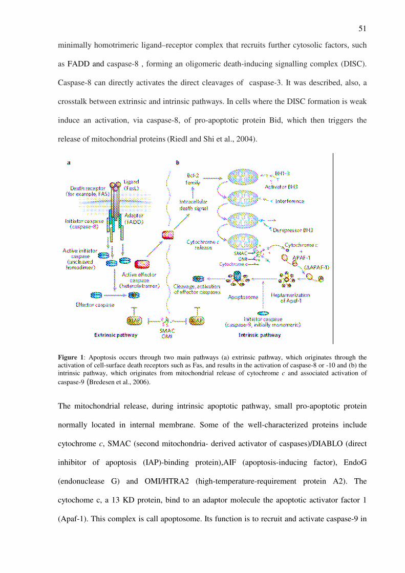
51
minimally homotrimeric ligand–receptor complex that recruits further cytosolic factors, such
as FADD and caspase-8 , forming an oligomeric death-inducing signalling complex (DISC).
Caspase-8 can directly activates the direct cleavages of caspase-3. It was described, also, a
crosstalk between extrinsic and intrinsic pathways. In cells where the DISC formation is weak
induce an activation, via caspase-8, of pro-apoptotic protein Bid, which then triggers the
release of mitochondrial proteins (Riedl and Shi et al., 2004).
Figure 1: Apoptosis occurs through two main pathways (a) extrinsic pathway, which originates through the
activation of cell-surface death receptors such as Fas, and results in the activation of caspase-8 or -10 and (b) the
intrinsic pathway, which originates from mitochondrial release of cytochrome c and associated activation of
caspase-9 (Bredesen et al., 2006).
The mitochondrial release, during intrinsic apoptotic pathway, small pro-apoptotic protein
normally located in internal membrane. Some of the well-characterized proteins include
cytochrome c, SMAC (second mitochondria- derived activator of caspases)/DIABLO (direct
inhibitor of apoptosis (IAP)-binding protein),AIF (apoptosis-inducing factor), EndoG
(endonuclease G) and OMI/HTRA2 (high-temperature-requirement protein A2). The
cytochome c, a 13 KD protein, bind to an adaptor molecule the apoptotic activator factor 1
(Apaf-1). This complex is call apoptosome. Its function is to recruit and activate caspase-9 in
52
the presence of ATP. Caspase-9 activate pro-caspase 3 or 7 which process substrates, like
caspase activated DNase (ICAD) or PARP, and leads to DNA fragmentation. Caspase-3 is
also activate by SMAC/DIABLO – IAP way. SMAC/DIABLO protein is release from
mitochondria and inhibit the IAP function. IAP is an inhibitor of caspase-3 cleavage, so the
effect of SMAC/DIABLO is to block the caspase-3 cleavage inhibition. OMI/HTRA2 protein
is another inhibitor of IAPs that have a pro-apoptotic effect. OMI/HTRA2 can induce cell
death also via its serine protease activity what may in turn induce caspase activation. AIF and
EndoG are involved in the caspase independent apoptotic pathway. The both protein interact
with DNA and or RNA to cause chromatin condensation and large scale DNA fragmentation
(Lorenzo and Susin, 2007). AIF does not display intrinsic endonuclease properties and its
DNA-degrading activity certainly depends on the recruitment of downstream nucleases. A
cooperation between AIF and EndoG, which is a mithocondrial DNAse, was observed
(Niikura et al, 2007). The mitochondrial release of these protein hinge on the membrane
permeabilization regulation. Several mechanisms about membrane permeabilization have
been propose. The first involve the opening of permeability transition pore (PTP), which
cause swelling and rupture of membrane and the later release of the pro-apoptotic protein. A
second mechanism is mediated by the Bcl-2 pro – anti apoptotic proteins. It has been
hypothesized that PTP consist of the voltage-dependent anion channel (VDAC: outer
membrane channel), the adenine nucleotide translocator (ANT: inner membrane channel),
cyclophilin D (Cyp D). The role of these three component of the pore in the MPT
mechanisms is not well know. Cyp D might be involved in the MPT Ca2+
-dependent events
and in the cyclosporin A (CsA) sensitive MPT. The Ca2+
- induce MPT mechanism might
activate the VDAC component of PTP.
Bcl-2 family proteins show a range of bioactivities, from inhibition to promotion of
apoptosis. In mammals, there are at least 12 core BCL-2 family proteins, including BCL-2
itself and proteins that have either three-dimensional (3D) structural similarity. BCL-2 family
53
members have classically been grouped into three classes. One class inhibits apoptosis (BCL-
2, BCL-XL, BCL-W, MCL1, BCL-B), whereas a second class promotes apoptosis (BAX,
BAK and BOK). A third divergent class of BH3-only proteins (BAD, BIK, BID, HRK , BIM,
BMF, NOXA and PUMA) have a conserved BH3 domain that can bind and regulate the anti-
apoptotic BCL-2 proteins to promote apoptosis. BAX and BAK proteins act as pro-apoptotic
factor inducing a conformation change and permeabilize the outer mitochondrial membrane.
This proteins appear to be able to form membrane channels and it might be a possible
mechanism of action . However, the biochemical nature of this putative BAX/BAK pore, such
as the number of molecules of BAX that comprise the pore, remains unknown. BAX/BAK
interact with the VDAC pore component (Antosson et al., 1997). Bcl-2 might directly inhibit
a component of the PTP complex. In fact, Bcl-2 (Bcl-xL) is capable of blocking VDAC
activity and ANT activity in liposome systems and consequently inhibit the MPT (Shimizu
et al., 1998). In summary, the experiments carried out until now has been shown as the
formation of homo and/or hetero dimers and the equilibrium shift between pro- and anti-
apoptotic members of the Bcl- 2 family may determine the sensitivity of a cell to apoptotic
stimuli.
Apoptosis in neurodegeneration
Chronic neurodegenerative diseases are characterized by a selective loss of specific subsets of
neuronal populations over a period of years or even decades (Okouchi et al, 2007). Recent
findings indicate that components of both the extrinsic (death receptors or their ligands) and
intrinsic (Bcl-2 family members) death pathways are involved during neurodegeneration or
neuronal injury in vivo. The induction of death pathway is mediate by different mechanism.
The reactive oxygen and nitrogen species are involved in the neuronal cell death activation. In
neuronal cells exist an equilibrium between the reactive oxygen species (ROS) flux and the
antioxidant system. A deficit of antioxidant defenses, that occurred in aging, and a ROS
overload leads to oxidative stress. In Alzheimer disease (AD) has been shown that β-amyloid
54
production activate the generation of H2O2 and HNE, ROS species, which induce JNK and
p38 pathways. JNK was shown to down-regulate anti-apoptotic Bcl-xL and Bcl-w expression
and this effect potentiated the release of Smac and the consequent activation of caspase-9
(Yao et al., 2005). Similar mechanisms involves JNK pathways and mitochondrial released
protein is been described in the Huntington’s disease (HD) neuronal cells loss. This pathology
cause a neuronal death in the striatum and cerebral cortex, cause motor impairment,
personality change and dementia. The expansion of CAG repeats in exon 1 of the
IT15(huntingtin) gene are translate into an expanded polyglutamine tract near the N-terminus
of the Huntingtin protein. Huntingtin modify protein provoke the JNK activation and the
release of Smac/Diablo protein form outer mitochondrial membrane. Parkinson disease (PD),
the second common neurodegenerative disorder after AD, has been demonstrated to
degenerate the dopaminergic neuron via a JNK/p38 pathways. The substantia nigra (SN) pars
compacta, which is the primary area of the brain that is affected by PD, contain dopaminergic
neuron that exhibit high levels of basal oxidative stress, rending this area vulnerably to a toxic
insult. In addiction to oxidative challenge, the loss of essential growth factors ( NGF and
BDGF) critical for survivals of neurons also contribute to the degeneration of this cells. The
familiar Parkinson disease is associated with the genetic mutation of a parkin a protein
associated with outer mitochondrial membrane. Over expression of parkin wild-type has been
shown protective effect on neuroblastoma cells (Jiang et al., 2004). Another PD associated
gene DJ-1, even it involved in mitochondrial function, have an anti-apoptotic effect.
In the amyotrophic lateral sclerosis (ALS) has been described the activation of
Fas/NOS pathaways as mediator of motoneuronal death. NO species are product by specific
neuronal NO synthase (nNOS). In the Brain, nNOS, whose expression is regulated by both
physiological and pathophysiological stimuli, accounts for most NO activity. NO reacts
rapidly with to form peroxynitrite (ONOO––
) which is the most reactive species. Fas
signalling activates two principal pathways that act synergistically: one involves Fas-
55
associated death domain protein (FADD)-mediated activation of procaspase-8 and
cytochrome c release, and the other, which appears to be specific to motoneurons, involves
the activation of p38 mitogen-activated protein kinase (MAPK) and neuronal nitric oxide
synthase (nNOS). An increased sensitivity to Fas-activation and nNOS pathway has been
shown in patients with a mutate superoxide dismutase-1 (SOD1). SOD1 mutation is
associated with ASL disease. This mutate protein have a toxic effect for the motoneurons, a
hypothesis to this effect is that SOD can directly promote the generation of active oxygen and
nitrogen species. Mutate SOD1 is purpoted to down-regulate Nrf2, an inhibitor of Fas
pathways (Raoul et al., 2005).
Degeneration of α-motor neurons of spinal cord is the molecular mechanism of spinal
muscular atrophy (SMA). The genetic cause of this disease is the mutation of survival motor
neuron (SMN1) gene. SMN1 interact with Bcl-2, attenuating Bax and Fas mediated
apoptosis, and with p53. the decreases in the ability of mutate protein to bind Bcl-2 and p53 is
associated with disease severity. The neuronal apoptosis inhibitory protein (NAIP) is another
gene correlated with SMA. NAIP acts as an inhibitor of caspase-3 and 7.
The lost of brain function observed in diabetics patients are associated with a neuronal
apoptosis. Diabetic encephalopathy is a diabetic complication which causes alteration in
cognition, behaviour and neuropsychology. The neuronal death seems to be dependent of
impaired insulin/IGF axis and signaling. It has been documented a possible rule of
insulin/IGF axis in the regulation of β-amyloid levels (Steen et al., 2005). Another
contributing pathogenic factor of diabetic encephalopathy is C-peptide. This peptide has an
insulin mimetic effect on insulin receptor. Deficiency in C-peptide is associated with neuronal
apoptosis (Sima and Li, 2005). The hyperglycemia associated with diabetes induce an
oxidative stress and a consequent susceptibility of the hippocampus and cortical regions of
brain.
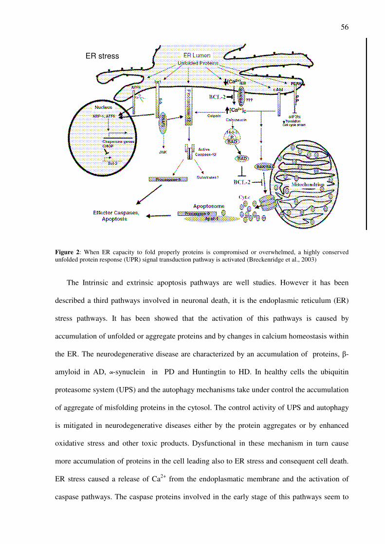
56
Figure 2: When ER capacity to fold properly proteins is compromised or overwhelmed, a highly conserved
unfolded protein response (UPR) signal transduction pathway is activated (Breckenridge et al., 2003)
The Intrinsic and extrinsic apoptosis pathways are well studies. However it has been
described a third pathways involved in neuronal death, it is the endoplasmic reticulum (ER)
stress pathways. It has been showed that the activation of this pathways is caused by
accumulation of unfolded or aggregate proteins and by changes in calcium homeostasis within
the ER. The neurodegenerative disease are characterized by an accumulation of proteins, β-
amyloid in AD, -synuclein in PD and Huntingtin to HD. In healthy cells the ubiquitin
proteasome system (UPS) and the autophagy mechanisms take under control the accumulation
of aggregate of misfolding proteins in the cytosol. The control activity of UPS and autophagy
is mitigated in neurodegenerative diseases either by the protein aggregates or by enhanced
oxidative stress and other toxic products. Dysfunctional in these mechanism in turn cause
more accumulation of proteins in the cell leading also to ER stress and consequent cell death.
ER stress caused a release of Ca2+
from the endoplasmatic membrane and the activation of
caspase pathways. The caspase proteins involved in the early stage of this pathways seem to
ER stress
57
be caspase-12, caspase-4 and capsase-2, the later specific of neuronal cells (Dahmer , 2005).
It has been recognized that exists an important crosstalk between the ER and mitochondria in
the execution of cell death. It has been suggested that the Bcl-2 family proteins play a crucial
role in ER–mitochondria interactions, and Bcl-2 and Bcl-xL associate with mitochondria and
with the ER membrane. Mitochondrial membrane permealitation is modulate by Ca2+
concentration in the cytosol and it is, another, point of crosstalk between mitochondria and
ER (Breckenridge DG et al., 2003). In the PD the expression of Parkin can restore proteasome
function and promote survival, while loss of function of Parkin causes ER stress with
accumulation of cytotoxic fibrils and protein aggregates in cells (Imai et al., 2004). ER stress
is associated with the AD neuronal degeneration. ER is involved in the cleavage of β-amyloid.
The PS1 and PS2 proteins, ER transmembrane proteins, are parts of the multiprotein -
secretase complex that mediates the intramembranous cleavage of APP. Mutations in these
proteins cause changes in the pattern of APP processing in the cell and increases the amount
of the more toxic A -42 peptide (Wilquet et al., 2004). The activation of caspase-12 and
caspase-4 may contribute to neuronal death in AD as described in different studies (Nakagawa
et al., 2000 and Hitomi et al., 2004).
Androgen Steroids and neuronal cell death
Androgen steroids, testosterone and its derivates, can regulate the survival and death of
neuronal cells through genomic and nongenomic mechanism of actions. It has been well
described the neuro-protective role of androgens in AD (Driscoll and Resnick, 2007).
Androgens inhibit the accumulation of β-amyloid through two different pathways. The
aromatization of AS to estradiol and activation of estrogen pathways is the first mechanism. A
second pathways, estrogen independent, was observed in a series of rodent studies using the
testosterone metabolite Dihydrotestosterone (DHT). It has been found that testosterone
provides partial protection against Aβ toxicity , by activating a rapid, non-genomic pathway
that involves MAPK signaling (Nguyen et al., 2005). This testosterone neuroprotection is
58
mimicked by DHT but not 3α-diol and is not attenuated by antagonists to estrogen receptors
(Pike, 2001). In cerebellar granule cells treated with micromolar concentration of testosterone
is observed a neuro- protective effect of steroids against the oxidative stress (Ahlbom et al.,
2001).
A central nervous system toxicity is assume to be a possible factor of syndrome call
steroid dementia. This is a side effect observed in some patients treated with glucocorticoid
and it was assumed to be caused by a neuronal cell death (Wolkowitz et al., 2007). Similar
effects of androgenic steroids are not yet described in clinical studies. Although, neurotoxic
effect has been observed in vivo and in vitro studies. In a cellular model of spinal and bulbar
muscular atrophy (SBMA) was demonstrated pivotal rule of androgen receptors with a
repeated glutamine residues in the amino terminus region. The cells expressing this modified
receptor are more susceptible to cell death when treated with androgens (Grierson et al.,
2001). The cells SH-SY5Y treated with supra-physiological concentration of testosterone
showed an apoptotic effects through the activation of inositol-1-4-5-trisphosphate receptor
mediated intracellular Ca2+
release (Estrada et al., 2006). In a mouse cortical mixed cultures
treated with steroids was observed an amplification of excitotoxicity induce by NMDA
(Orlando et al., 2007). This effects was observed at micromolar concentration of testosterone,
while nanomolar concentration is neuroprotective. Nandrolone, stanozolol and genestrione are
toxic at nanomolar concentration. Testosterone effect is regulate by aromatase activity. In
fact, when it was combine with anastrozole, an aromatase inihibitor, nanomolar concentration
was no longer neuroprotective. Androgen receptor seems to be not involved in this pro-
apoptotic effect of testosterone as show in experiment carried out with flutamide, an AR
specific inhibitor. Instead, nandrolone acting via AR activation. The neurotoxic effect of
steroids would be mediate by a specific membrane receptor. Studies carried out in primary
cortical astrocytes and PC12 cell line shown a binding of dihydrotestosterone (DHT) or
testosterone on membrane receptor. DHT conjugate with BSA enhances the toxic effect of
59
IAA, an inhibitor of glycolysis and a promoter of oxidative stress, and the cell death in
astrocytes cells (Gatson and Singh , 2007). The PC12 cells not-differentiated express a
androgen steroids membrane specific receptor. Its activation increased cytosolic Ca(2+) and
induced apoptosis (Alexaki et al., 2006).
Sigma receptor and apoptosis
Sigma receptors (Walker at al., 1990) are non opiate and non phencyclidine binding
sites that mediate the psychotomimetic actions of certain opioid derivatives. Sigma receptors
consist of two subtypes, sigma-1 and sigma-2, that are distinguishable by biochemical and
pharmacological means (Quirion et al., 1992). Sigma receptor 1 (σR1) is the best
characterized. It mediates immunosuppressant, antipsychotic, and neuroprotective effects.
Sigma receptor-1 ligands have demonstrated neuroprotective properties including inhibition
of ischemia-induced glutamate release, attenuation of postsynaptic glutamate-evoked calcium
influx, and reduced NO production. Studies anatomical distribution of σ1 subtype showed that
in the central nervous system (CNS) the regions with high levels are the areas involved in
motor, sensory, and endocrine functions, and memory. In peripheral tissues, they are present
in placenta, liver, immune cells, and gastrointestinal tract. The high expression of σR1
receptors in steroid-producing tissues and in CNS suggests their possible role in functions of
neuroendocrine and central neuroactive steroid system. Additional involvement of σR1
receptors has been postulated in psychosis with modulation of synthesis and release of
neurotransmitters such as acetylcholine and dopamine.
Sigma receptor 2 (σR2) subtypes have a different pattern of anatomical distributions in
CNS and in peripheral tissues. A very high concentration of σR2 receptors has been
discovered in neuronal and non-neuronal tumor cell lines and this provides evidence of a
possible role in cell proliferation and viability (Vilner et al., 1995). In fact, putative σR2
agonists, such as ibogaine and CB-64D, induce apoptotic death by a mechanism involving the
60
increase of intracellular Ca++
level by release from endoplasmic reticulum and subsequently
from mitochondrial stores (Vilner and Bowen, 2000). These effects are inhibited by putative
σR2 antagonists BD1047 and BD1063 (Bowen 2000). In a breast tumor cell line MCF-7 was
observed a caspase-3 independent apoptosis pathways activate by σR2 (Crawford and Bowen,
2002). Bowen and colleagues showed in neuroblastoma cells SK-N-SH a caspase-10
dependent apoptotic pathway activated by CB-64D and associated with Bid cleavage and
endonuclease G mitochondrial release (Wang and Bowen, 2006). Sigma-2 receptor activated,
also, a lipid dependent apoptotic pathways. In MCF-7 was found an increase in ceramide
synthesis associated with the induction of apoptosis ( Crawford et al., 2002).
References
• Ahlbom E, Prins GS, Ceccatelli S. Testosterone protects cerebellar granule cells from oxidative stress-
induced cell death through a receptor mediated mechanism. Brain Res. 2001 Feb 23;892(2):255-62
• Alexaki VI, Dermitzaki E, Charalampopoulos I, Kampa M, Nifli AP, Gravanis A, Margioris AN,
Castanas E. Neuronal differentiation of PC12 cells abolishes the expression of membrane androgen
receptors. Exp Cell Res. 2006 Sep 10;312(15):2745-56.
• Antonsson, B. Conti F, Ciavatta A, Montessuit S, Lewis S, Martinou I, Bernasconi L, et al.
Inhibition of Bax channel-forming activity by Bcl-2. Science 277, 370–372 (1997)
• Bowen WD. Sigma receptors: recent advances and new clinical potentials. Pharm Acta Helv. 2000;
74(2-3):211-218.
• Breckenridge DG, Germain M, Mathai JP, Nguyen M and Shore GC Regulation of apoptosis by
endoplasmic reticulum pathways. Oncogene (2003) 22: 8608–8618
• Crawford KW, Bowen WD. Sigma-2 receptor agonists activate a novel apoptotic pathway and
potentiate antineoplastic drugs in breast tumor cell lines. Cancer Res. 2002 Jan 1;62(1):313-22
• Crawford KW, Coop A, Bowen WD. sigma(2) Receptors regulate changes in sphingolipid levels in
breast tumor cells. Eur J Pharmacol. 2002 May 17;443(1-3):207-9
• Dahmer MK. Caspases-2, -3, and -7 are involved in thapsigargin-induced apoptosis of SH-SY5Y
neuroblastoma cells. J Neurosci Res. 2005 May 15;80(4):576-83
61
• Driscoll I, Resnick SM. Testosterone and cognition in normal aging and Alzheimer's disease: an
update. Curr Alzheimer Res. 2007 Feb;4(1):33-45
• Estrada M, Varshney A, Ehrlich BE. Elevated testosterone induces apoptosis in neuronal cells. J Biol
Chem. 2006 Sep 1;281(35):25492-501
• Gatson JW, Singh M. Activation of a membrane-associated androgen receptor promotes cell death in
primary cortical astrocytes. Endocrinology. 2007 May;148(5):2458-64
• Grierson AJ, Shaw CE, Miller CC. Androgen induced cell death in SHSY5Y neuroblastoma cells
expressing wild-type and spinal bulbar muscular atrophy mutant androgen receptors. Biochim Biophys
Acta. 2001 Apr 30;1536(1):13-20
• Heidenreich KA. Molecular mechanisms of neuronal cell death. Ann N Y Acad Sci. 2003
Jun;991:237-50
• Hitomi J, Katayama T, Eguchi Y, Kudo T, Taniguchi M, Koyama Y, Manabe T, Yamagishi S,
Bando Y, Imaizumi K, Tsujimoto Y and Tohyama M. (2004) Involvement of caspase-4 in
endoplasmic reticulum stress-induced apoptosis and Abeta-induced cell death. J. Cell Biol. 165: 347–
356
• Imai Y and Takahashi R (2004) How do Parkin mutations result in neurodegeneration? Curr. Opin.
Neurobiol. 14: 384–389
• Jiang H, Ren Y, Zhao J, and Feng J. Parkin protects human dopaminergic neuroblastoma cells
against dopamine-induced apoptosis. Hum Mol Genet 13: 1745–1754, 2004.
• Kerr, J. F. F., Wylie, A. H. & Currie, A. R. Apoptosis: a basic biological phenomenon with wide-
ranging implications in tissue kinetics. Br. J. Cancer 26, 239–257 (1972)
• Kitamura, Y., et al. 1998. Alteration of proteins regulating apoptosis, Bcl-2, Bcl-x, Bax, Bak, Bad,
ICH-1 and CPP32, in Alzheimer's disease. Brain Res. 780: 260-269
• Levine B and Yuan Y. Autophagy in cell death: an innocent convict? J Clin Invest 115: 2679–2688,
2005.
• Lorenzo HK, Susin SA. Therapeutic potential of AIF-mediated caspase-independent programmed cell
death. Drug Resist Updat. 2007 Dec;10(6):235-55
• Nakagawa T, Zhu H, Morishima N, Li E, Xu J, Yankner BA and Yuan J (2000) Caspase-12
mediates endoplasmic-reticulum-specific apoptosis and cytotoxicity by amyloid-beta. Nature 403: 98–
103
62
• Nguyen, T.V., et al., 2005. Androgens activate mitogen-activated protein kinase signaling: role in
neuroprotection. J. Neurochem. 94, 1639–1651.
• Okouchi M, Ekshyyan O, Maracine M, Aw TY. Neuronal apoptosis in neurodegeneration. Antioxid
Redox Signal. 2007 Aug;9(8):1059-96
• Orlando R, Caruso A, Molinaro G, Motolese M, Matrisciano F, Togna G, Melchiorri D, Nicoletti
F, Bruno V. Nanomolar concentrations of anabolic-androgenic steroids amplify excitotoxic neuronal
death in mixed mouse cortical cultures. Brain Res. 2007 Aug 24;1165:21-9.
• Pike, C.J., 2001. Testosterone attenuates beta-amyloid toxicity in cultured hippocampal neurons. Brain
Res. 919, 160–165.
• R. Quirion, W.D. Bowen, Y.T. Itzhak, J.L. Junien, J.M. Musacchio, R.B. Rothman, T.-P. Su, S.W.
Tam, D.P. Taylor, A proposal for the classification of sigma binding sites, Trends Pharmacol. Sci. 13
(1992) 85– 86.
• Raoul C, Barthelemy C, Couzinet A, Hancock D, Pettmann B, Hueber AO. Expression of a
dominant negative form of Daxx in vivo rescues motoneurons from Fas (CD95)-induced cell death. J
Neurobiol. 2005 Feb 5;62(2):178-88
• Riedl SJ, Shi Y. Molecular mechanisms of caspase regulation during apoptosis. Nat Rev Mol Cell Biol.
2004 Nov;5(11):897-907
• Shimizu S, Eguchi Y, Kamiike W, Funahashi Y, Mignon A, Lacronique V, Matsuda H, Tsujimoto
Y (1998) Bcl-2 prevents apoptotic mitochondrial dysfunction by regulating proton flux. Proc Natl Acad
Sci USA 95:1455–1459
• Sima AA, Li ZG. The effect of C-peptide on cognitive dysfunction and hippocampal apoptosis in type
1 diabetic rats. Diabetes. 2005 May;54(5):1497-505
• Steen E, Terry BM, Rivera EJ, Cannon JL, Neely TR, Tavares R, Xu XJ, Wands JR, de la Monte
SM. Impaired insulin and insulin-like growth factor expression and signaling mechanisms in
Alzheimer's disease--is this type 3 diabetes? J Alzheimers Dis. 2005 Feb;7(1):63-80
• Thornberry NA, Lazebnik Y. 1998. Caspases: enemies within. Science 281: 1312– 16
• Vilner BJ, Bowen WD. Modulation of cellular calcium by sigma-2 receptors: release from intracellular
stores in human SK-N-SH neuroblastoma cells.J Pharmacol Exp Ther. 2000 Mar;292(3):900-11
• Vilner BJ, John CS, Bowen WD. Sigma-1 and sigma-2 receptors are expressed in a wide variety of
human and rodent tumor cell lines. Cancer Res. 1995 Jan 15;55(2):408-13
63
• Walker JM, Bowen WD, Walker FO, Matsumoto RR, De Costa B, Rice KC. Sigma receptors:
biology and function. Pharmacol Rev. 1990 Dec;42(4):355-402. Review
• Wang X. , Bowen WD. Sigma-2 receptors mediates apoptosis in SK-N-SH neuroblastoma cells via
caspase-10 dependent Bid cleavage and mithocondrial release of endonuclease G and apoptotic
inducing factor. Neuroscience 2006
• Wilquet V and De Strooper B (2004) Amyloid-beta precursor protein processing in
neurodegeneration. Curr. Opin. Neurobiol. 14: 582–584
• Wolkowitz OM, Lupien SJ, Bigler ED. The "steroid dementia syndrome": a possible model of human
glucocorticoid neurotoxicity. Neurocase. 2007 Jun;13(3):189-200
• Y. Niikura, A. Dixit, R. Scott, G. Perkins and K. Kitagawa, BUB1 mediation of caspase-independent
mitotic death determines cell fate, J. Cell Biol. 178 (2007), pp. 283–296
• Yao M, Nguyen TV, and Pike CJ. Beta-amyloid-induced neuronal apoptosis involves c-Jun N-
terminal kinase-dependent downregulation of Bcl-w. J Neurosci 25: 1149–1158, 2005.
64
Aim of the research
Epidemiological studies and in vivo experiment has been shown that administration of large
doses of anabolic androgenic steroids (AAS) are correlated with neurobehavioral changes like
hyperexcitability, supra-aggressive nature and suicidal tendencies. These behavioral changes
could be mediated by the regulation of neurotransmission or to be the outward manifestation
of neuronal damage resulting from exposure to high concentrations of AAS.
Nandrolone regulation of opioid pathway has been shown in vitro and in vivo studies.
In the first part of my thesis I study the possible effects of nandrolone on MOPr expression at
both the transcriptional and post-transcriptional levels in the neuroblastoma cell line SH-
SY5Y, which expresses this receptor constitutively and serves as a common model for
neuronal cells. AAS may modulate neural transmission via genomic and non genomic
mechanisms by binding directly to the intracellular androgen receptor (AR) or acting through
other signalling pathways. MOPr expression can be regulate by a direct effect on MOPr
promoter or by post-transcriptional mechanism occur at mRNA and protein levels. In fact ,
changes in mRNA stability, differences in translation efficiency, alterations in mRNA
transport, and modification of the receptor are among the possible mechanisms involved in
modulation of MOPr mRNA expression. I explore the possible rule of AR activation in the
MOPr regulation mediated by nandrolone with Hydroxyflutamide, a specific inhibitor of AR.
A luciferase reporter vector bear the sequence of MOPr promoter and the inhibitor of
transcription , Actinomycin D were used to elucidated the transcriptional and post-
transcriptional mechanisms .
The neuronal cell death caused by supra-phisiological concentration of anabolic
androgenic steroids has been evaluated with three cito-toxic assays : MTT, LDH and Trypan
blue exclusion assay. The neuronal cells SH-SY5Y were exposed to nandrolone, testosterone
and stanozolol and a time and concentration dependent neurotoxicity has been observed. To

65
explore the involvement, in neurotoxicity testosterone and nandrolone dependent, of androgen
receptor and sigma-2 receptor was carried out MTT experiments with Hydroxyflutamide and
a competitive radioligand binding assay between testosterone and RHM-1, selective sigma-2
ligand. In vitro studies performed with steroids conjugate with bovine serum albumin (BSA)
was observed a specific steroid membrane receptor, which activation lead cell death
progression. To evaluate the possible activation of this pathways in my neuronal cells, I
exposed the SH-SY5Y with testosterone conjugate with bovine serum albumin.
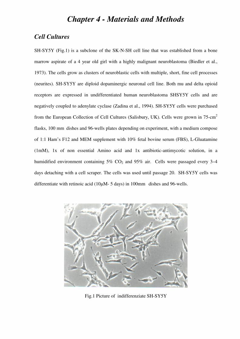
Chapter 4 - Materials and Methods
Cell Cultures
SH-SY5Y (Fig.1) is a subclone of the SK-N-SH cell line that was established from a bone
marrow aspirate of a 4 year old girl with a highly malignant neuroblastoma (Biedler et al.,
1973). The cells grow as clusters of neuroblastic cells with multiple, short, fine cell processes
(neurites). SH-SY5Y are diploid dopaminergic neuronal cell line. Both mu and delta opioid
receptors are expressed in undifferentiated human neuroblastoma SHSY5Y cells and are
negatively coupled to adenylate cyclase (Zadina et al., 1994). SH-SY5Y cells were purchased
from the European Collection of Cell Cultures (Salisbury, UK). Cells were grown in 75-cm2
flasks, 100 mm dishes and 96-wells plates depending on experiment, with a medium compose
of 1:1 Ham’s F12 and MEM supplement with 10% fetal bovine serum (FBS), L-Gluatamine
(1mM), 1x of non essential Amino acid and 1x antibiotic-antimycotic solution, in a
humidified environment containing 5% CO2 and 95% air. Cells were passaged every 3–4
days detaching with a cell scraper. The cells was used until passage 20. SH-SY5Y cells was
differentiate with retinoic acid (10µM- 5 days) in 100mm dishes and 96-wells.
Fig.1 Picture of indifferenziate SH-SY5Y
67
MCF7 cells was established from the pleural effusion from a 69 year female
Caucasian suffering from a breast adenocarcinoma. MCF-7 cells are a well-characterized
estrogen receptor (ER) positive control cell line (cells are positive for cytoplasmic estrogen
receptors) and therefore are a useful in vitro model of breast cancer to study the role of
estrogen in breast cancer. This cell express high quantity of androgen receptor (Buchanan et
al., 2005). MCF-7 was grown in 75-cm2 flasks with a medium composed of RPMI medium
supplemented with 10% (v/v) FCS.
Semiquantitative real-time polymerase chain reaction
For reverse transcription-polymerase chain reaction (PCR) experiments, cells were collected
from tissue culture flasks, centrifuged (500 g for 5 min) and rinsed with phosphate-buffered
saline (PBS). Total cellular RNA was extracted using Trizol® reagent (Invitrogen). RNA was
quantify with spectrophotometere and a 2-µg aliquot was digested with RNAse-free DNAse
(Invitrogen) for 15 min at 25 °C and according to the manufacturer's instructions. A 2-µg
sample was reverse-transcribed using the Transcriptor First Strand cDNA Synthesis Kit
(Roche Diagnostics, Milan, Italy) according to the manufacturer's instructions. A real-time
PCR was employed for relative quantification of REST transcripts using the Light Cycler
Instrument (Roche Diagnostics, Milan, Italy) and the SYBR Premix Ex Taq (Takara Bio
Europe S.A., Gennevilliers, France). This 'hot start' reaction mix contains Takara Ex TaqHS
DNA polymerase. This ready-to-use ‘hot start’ reaction mix contains Taq DNA polymerase,
dNTP mix and the fluorescent dye SYBR Green I for real-time detection of double-stranded
DNA. Reactions were set up in 20 µL including 10 ng of target DNA. To amplify the hMOPr
cDNA, a sense primer (5'-CTGGGTCAACTTGTCCCACT-3') and an antisense primer (5'-
TGGAGTAGAGGGCCATGATC-3') were used at 2.5 nM final concentration for amplifying
a 146-bp fragment (327 to 472 bp) from GenBank™ Accession no. NM_000914. As a
control, a 169-bp fragment of the human L19 ribosomal proteingene was amplified with a
68
sense primer (5'-CTAGTGTCCTCCGCTGTGG-3') and an antisense primer (5'-
AAGGTGTTTTTCCGGCATC3') at 5 nM final concentration, producing a fragment (62 to
230 bp) from GenBank™ Accession no. BC062709. Amplification was as follows: 95°C for
10 s followed by 40 cycles of 95°C for 5 s, 62°C for 20 s and 72°C for 6 s. After that, the
temperature was lowered to 40°C for 30 s and the specificity of the reaction was verified by
analysis of the melting curve once the appropriate double-stranded DNA melting temperature
had been reached.
Relative expression of the reverse transcription-PCR products was determined using
the ∆∆Ct method (Livak and Schmittgen, 2001). This method calculates relative expression
using the equation: normalized relative quantities (NRQs) = E–∆∆Ct
(Hellemans et al., 2007).
The corresponding real-time PCR efficiency (E) of one cycle in the exponential phase was
calculated according to the equation: E = 10[–1/slope]
(Pfaffl, 2001), optimal and identical real-
time amplification efficiencies of target and reference gene of E = 2 are presumed, so the final
equation is NRQs = 2–∆∆Ct
. Ct is the threshold cycle, i.e. the cycle number at which the
sample's relative fluorescence rises above the background fluorescence and ∆∆Ct = [Ct gene
of interest (unknown sample) − CtL19 (unknown sample)] − [Ct gene of interest (calibrator
sample) − CtL19 (calibrator sample)]. The control samples was chosen as the calibrator
sample and used in each PCR. Each sample was run in duplicate and the mean Ct was used in
the ∆∆Ct equation. L19 was chosen for normalization because this gene showed consistent
expression relative to other housekeeping genes among the treatment groups in our
experiments. The data was express as per cent of the 2–∆∆Ct
, respect to the control assume as
100% (Fig.2)
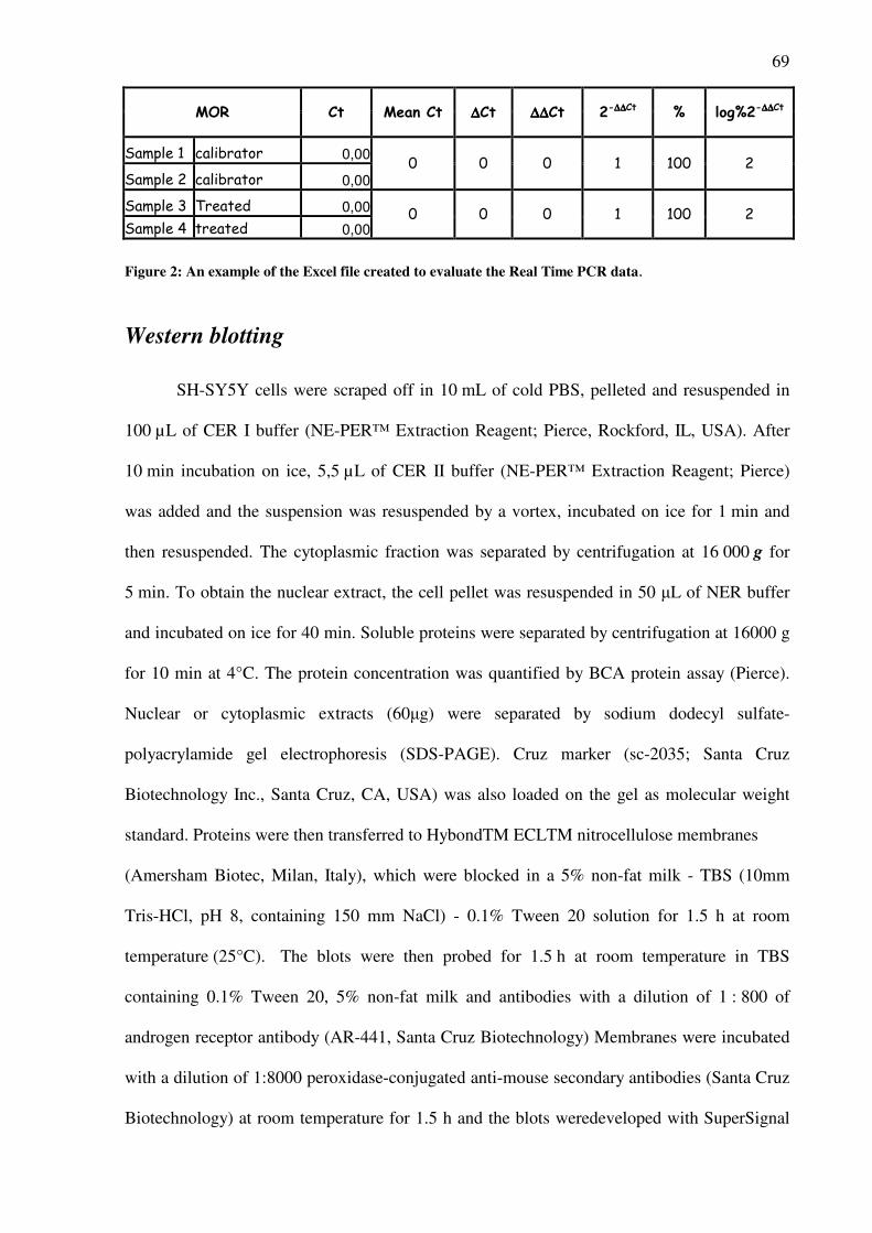
69
MOR Ct Mean Ct ∆∆∆∆Ct ∆∆∆∆∆∆∆∆Ct 2-∆∆∆∆∆∆∆∆Ct % log%2-∆∆∆∆∆∆∆∆Ct
Sample 1 calibrator 0,00
Sample 2 calibrator 0,000 0 0 1 100 2
Sample 3 Treated 0,00
Sample 4 treated 0,000 0 0 1 100 2
Figure 2: An example of the Excel file created to evaluate the Real Time PCR data.
Western blotting
SH-SY5Y cells were scraped off in 10 mL of cold PBS, pelleted and resuspended in
100 µL of CER I buffer (NE-PER™ Extraction Reagent; Pierce, Rockford, IL, USA). After
10 min incubation on ice, 5,5 µL of CER II buffer (NE-PER™ Extraction Reagent; Pierce)
was added and the suspension was resuspended by a vortex, incubated on ice for 1 min and
then resuspended. The cytoplasmic fraction was separated by centrifugation at 16 000 g for
5 min. To obtain the nuclear extract, the cell pellet was resuspended in 50 µL of NER buffer
and incubated on ice for 40 min. Soluble proteins were separated by centrifugation at 16000 g
for 10 min at 4°C. The protein concentration was quantified by BCA protein assay (Pierce).
Nuclear or cytoplasmic extracts (60µg) were separated by sodium dodecyl sulfate-
polyacrylamide gel electrophoresis (SDS-PAGE). Cruz marker (sc-2035; Santa Cruz
Biotechnology Inc., Santa Cruz, CA, USA) was also loaded on the gel as molecular weight
standard. Proteins were then transferred to HybondTM ECLTM nitrocellulose membranes
(Amersham Biotec, Milan, Italy), which were blocked in a 5% non-fat milk - TBS (10mm
Tris-HCl, pH 8, containing 150 mm NaCl) - 0.1% Tween 20 solution for 1.5 h at room
temperature (25°C). The blots were then probed for 1.5 h at room temperature in TBS
containing 0.1% Tween 20, 5% non-fat milk and antibodies with a dilution of 1 : 800 of
androgen receptor antibody (AR-441, Santa Cruz Biotechnology) Membranes were incubated
with a dilution of 1:8000 peroxidase-conjugated anti-mouse secondary antibodies (Santa Cruz
Biotechnology) at room temperature for 1.5 h and the blots weredeveloped with SuperSignal
70
West Pico chemiluminescent substrate according to themanufacturer’s protocol (Pierce). Blot
images were digitally acquired by an LAS3000Imager (Fujifilm Corporation, Stamford, CT,
USA). Protein expression was analyzedsemiquantitatively using AIDA (Raytest
Isotopenmessgeräte GmbH, Mannheim, Germany).
Radioligand binding assay
SH-SY5Y cell membranes were prepared by homogenizing cells in 50 mM Tris- HCl buffer
(pH 7.4) containing 1 mM EDTA, 1 mM dithiothreitol and 1 mM benzamidine, with a
Polytron homogenizer. After centrifugation (1000 x g for 10 min at 4°C), supernatants were
centrifuged (18000 x g for 30 min at 4°C) and the pellet was resuspended in 50 mM Tris-HCl
buffer (pH 7.4) containing 5 mM MgCl2. Protein concentration was determined by BCA
assay (Pierce). For saturation binding experiments, cell membranes (100 µg/assay tube) were
incubated in 100 mM Tris-HCl (pH 7.4) containing 0.3% BSA with increasing concentrations
of [3H]-DAMGO (0.1-5 nM). Non-specific binding was determined in the presence of
DAMGO (10 µM). After 90 min incubation at 25°C, bound ligand was isolated by rapid
filtration on Whatman GF/B filters (Schleicher & Schuell). Filters were washed with 20 ml of
ice-cold 50 mM Tris-HCl buffer (pH 7.4) and left in scintillation fluid for 8 h before counting.
Data were fitted by nonlinear least-square regression and the LIGAND program (Munson and
Rodbard 1980) was used to calculate receptor density (Bmax), Hill slopes and ligand affinity
(Kd). Data are expressed as fmol of [3H]-DAMGO bound and normalized to cell protein
content.
Competitive binding assay was performed incubating cell membranes (100 µg/assay
tube) in 100 mM Tris-HCl (pH 7.4) containing 0.3% BSA with N-[4-(3,4-dihydro-6,7-
dimethoxyisoquinolin-2(1H)-yl)butyl]-2-methoxy-5-methyl-benzamide (RHM-1) radiolabeled
with tritium (8nM) ( Xu et al., 2005) alone or with increasing concentration of Testosterone (
10-7
-10-5
)

71
Cell viability Assays
The cell viability was evaluated with different methods: MTT, LDH and trypan blue.
The yellow tetrazolium salt (3-(4,5-dimethylthiazol-2-yl)-2,5-diphenyltetrazolium bromide
(MTT) is reduced in metabolically active cells to form insoluble purple formazan crystals,
which are solubilized by the addition of a detergent. The color can then be quantified by
spectrophotometric means. SH-SY5Y cells were seeded in 96-well plates at a density of
0.5 × 10⁶cells/well in 100 µl of medium. The cells were treated with steroids and after 24 h,
48 h or 72 h 10 µl of MTT (5 mg/ml in phosphate buffered saline) was added at in each wells.
The plates were incubated for 4h and later 100 µl of solubilization mixture (20% sodium
dodecyl sulfate and 0.01 N HCl) were added and after an overnight incubation, absorbance
was measured with a microplate reader at 570 nm.
To evaluate the release into the medium of lactate dehydrogenase from the cells was
used the CytoTox 96 NonRadioactive Cytotoxicity Assay (Promega) 50 µl of medium from
each wells were pipet in a new plate. An equal volume (50 µl ) of substrate solution was
added in each well. The plate is incubate 30 minute in the dark, after was added the stop
buffer solution and analyzed the colorimetric changes with a microplate reader at 490 nm.
For trypan blue exclusion assay the cells were trypsinized, resuspended
in 1ml PBS, and 10 µl
of suspension was mixed with 10µl trypan blue dye solution (Sigma) before being counted
with a hemacytometer.
Plasmid constructs
The plasmids used in this study are based on the pGL3-Basic vector system (Promega,
Madison, WI) containing the luciferase reporter gene. Firstly, a fragment of the human MOPr
promoter, ranging from –2447 to +8 (designated +1 at translation start codon, GenBankTM
accession number AF153500) was amplified by PCR using genomic DNA obtained from SH-
72
SY5Y cells as template.The PCR was run using a forward primer (5’-
GTACGTCGACTCCCTCCAAAGCAACTAT-3’) and a reverse primer (5’-
GTATCTCGAGCTGTCCATGGTACTGACG-3’) bearing the BspHI site at its 3’ terminus.
This PCR product, named mu, was cloned into pCR-Blunt-II-TOPO (Invitrogen), sequenced
(BMR Genomics, Padova, Italy), digested with SacI/XhoI and cloned into the pGL3-Basic
vector or pGL3-SV40, vector containing the sequence of SV40 promoter between the multi
cloning site, where the MOPr promoter is inserted, and luciferase coding sequence.
Cell Transfection and Reporter gene assays.
SH-SY5Y were plated in 24-well dishes and at 50-60% confluence were transiently
transfected with pGL3-Basic, pGL3-Basic/mu or pGL3-SV40/mu reporter plasmids (1,5 mg
/well) and pSV-ßGal (0,5 mg/well; Promega) by using EXGEN 500 Transfection Reagent
(Fermentas, Hanover, MD). Cells were subsequently cultured in serum-free medium for 16-
18 h and then exposed to nandrolone or left untreated for 48 h. Finally, cells were lysed in
Reporter Lysis Buffer (Promega) and samples collected. Luciferase and beta-galactosidase
activities of cell lysates were measured by Bright-GloTM
Luciferase Assay System and Beta-
GloTM
Assay System (Promega) respectively, according to manufacturer instructions.
Immunocytochemistry of activated caspase-3 Undifferentiated SH-SY5Y cells were exposed to nandrolone and testosterone (10
-
4M) for 48 h. After exposure the cells were washed with pre-warmed PBS and fixed in
formalin (4% in PBS, pH 7.4) for 10 min, scraped into one tube per treatment, and pelleted by
centrifugation (300 g, 5 min). The cells were washed one time with PBS and re-centrifuged.
The cells were incubated with agarose (3% in PBS), 1 h, at 50°C. Subsequently, the cells were
cast on ice and dehydrated in ethanol (70% overnight, 95% for 4 h, 99.5% for 3 h and xylene
73
for 30 min) and embedded in paraffin. The cells were sectioned (4 µm) on a microtome
(Microm Heidelberg HM350, Germany).
The paraffin sections were deparaffinized in xylene and rehydrated in ethanol series
(2 × 5 min in 99.5%, 95% and 70%), washed in PBS and PBS with 0.3% triton (PBST), and
treated with 1% hydrogen peroxide in PBST (30 min). The sections were washed three times
in PBST and PBS, and antigen retrieval was performed using microwaves. The sections were
then washed three times in PBS followed by 1 h pre-incubation with 4% BSA for 1 h. The
sections were then incubated with the antibody anti-cleaved caspase-3 (Cell Signaling, USA)
overnight (at 5°C). After 10 min rinsing in PBS, the sections were incubated with biotinylated
second antibody (30 min) and then with ABComplex/HRP (30 min) at room temperature.
Immunoreactions were visualized using Vector VIP substrate kit for peroxidase. (Ostergren A
et al., 2005)
Caspase-3 activity
The protein cytoplasmatic fraction of SH-SY5Y was extracted as described in previously
pages in “Western blotting”. For colorimetric measurement of caspase activity, Ac-DEVD-
pNA (BioMol, USA) was added at a final concentration of 200 µM, in a reaction buffer (
40mM Hepes, 20mM NaCl, 2mM EDTA and 20% glycerol), to 100 µg of protein generated
from cells under different experimental conditions. Past an incubation of 1h an 30m, caspase
activity was monitored as optical absorbance at 405 nm.
Statistical analysis
In reporter gene experiments, each sample was assayed in triplicate, and each RT-PCR
sample was amplified in duplicate. All data are presented as mean SEM for the number of
experiments indicated. Statistical significance was determined by Newman-Keuls test after
74
ANOVA using GraphPad Prism, version 3.0 (GraphPad Software, Inc., San Diego, CA,
USA). P values <0.05 were considered significant.
References
• Biedler JL, Helson L, Spengler BA. Morphology and growth, tumorigenicity, and cytogenetics of
human neuroblastoma cells in continuous culture. Cancer Res. 1973 Nov;33(11):2643-52
• Buchanan G, Birrell SN, Peters AA, Bianco-Miotto T, Ramsay K, Cops EJ, Yang M, Harris JM,
Simila HA, Moore NL, Bentel JM, Ricciardelli C, Horsfall DJ, Butler LM, Tilley WD. Decreased
androgen receptor levels and receptor function in breast cancer contribute to the failure of response to
medroxyprogesterone acetate. Cancer Res. 2005 Sep 15;65(18):8487-96
• Hellemans J, Mortier G, De Paepe A, Speleman F, Vandesompele J. qBase relative quantification
framework and software for management and automated analysis of real-time quantitative PCR data.
Genome Biol. 2007;8(2)
• Livak KJ, Schmittgen TD. Analysis of relative gene expression data using real-time quantitative PCR
and the 2(-Delta Delta C(T)) Method. Methods. 2001 Dec;25(4):402-8
• Ostergren A, Svensson AL, Lindquist NG, Brittebo EB. Dopamine melanin-loaded PC12 cells: a
model for studies on pigmented neurons. Pigment Cell Res. 2005 Aug;18(4):306-14.
• Påhlman S, Ruusala AI, Abrahamsson L, Mattsson ME, Esscher T. Retinoic acid-induced
differentiation of cultured human neuroblastoma cells: a comparison with phorbolester-induced
differentiation. Cell Differ. 1984 Jun;14(2):135-44
• Pfaffl MW 2001 A new mathematical model for relative quantification in real-time RT-PCR. Nucleic
Acids Research 29 E45
• Voigt A, Zintl F. Effects of retinoic acid on proliferation, apoptosis, cytotoxicity, migration, and
invasion of neuroblastoma cells. Med Pediatr Oncol. 2003 Apr;40(4):205-13
• Zadina JE, Harrison LM, Ge LJ, Kastin AJ, Chang SL. Differential regulation of mu and delta
opiate receptors by morphine, selective agonists and antagonists and differentiating agents in SH-SY5Y
human neuroblastoma cells. J Pharmacol Exp Ther. 1994 Sep;270(3):1086-96
• Xu J, Tu Z, Jones LA, Vangveravong S, Wheeler KT, Mach RH. [3H]N-[4-(3,4-dihydro-6,7-
dimethoxyisoquinolin-2(1H)-yl)butyl]-2-methoxy-5-methylbenzamide: a novel sigma-2 receptor probe.
Eur J Pharmacol. 2005 Nov 21;525(1-3):8-17.
75
RESULTS
Nandrolone downregulates steady-state levels of MOPr mRNA and
the density of MOPr binding sites in SH-SY5Y
SH-SY5Y cells were exposed to nandrolone (10-8
M-10-5
M) for 48 h and the
endogenous MOPr transcripts were evaluated using a real time RT-PCR technique. A
significant concentration-dependent (Fig. 1B) and time-dependent (Fig. 1A) downregulation
of MOPr transcripts was observed. The maximum effect was at 10-5
M and it was present in
cells exposed to this agent at least 48 h (Fig. 1B). Cells were treated, also, with testosterone
(10-6
M) and stanozolol (10-6
M) for 48h and these experiments showed a downregulation of
MOPr transcripts (Fig. 1C).
As show in Fig. 1D, we confirmed that in cytoplasmatic fractions of SH-SY5Y cells is
present the AR showing the seem molecular weight of the AR occurring in MCF-7 cells a
breast cancer cell line which express it. Therefore, it could be suggested an interaction
between nandrolone and the AR expressed in SH-SY5Y.
To evaluate the potential involvement of the AR on this effect of nandrolone, the cells
were exposed to hydroxyflutamide (10-5
M; 48 h), an androgen antagonist. This compound
prevented the nandrolone-induced (10-6
M) reduction of MOPr transcripts (Fig. 1A) and when
administered alone did not cause any change of MOPr mRNA.
A saturation binding assays was set up to evaluate MOPr density in cell cultures
treated with 10-6
M nandrolone (48 h). The assay showed a significant decrease in the density
of MOPr (Bmax) on cell membranes (Table 1).
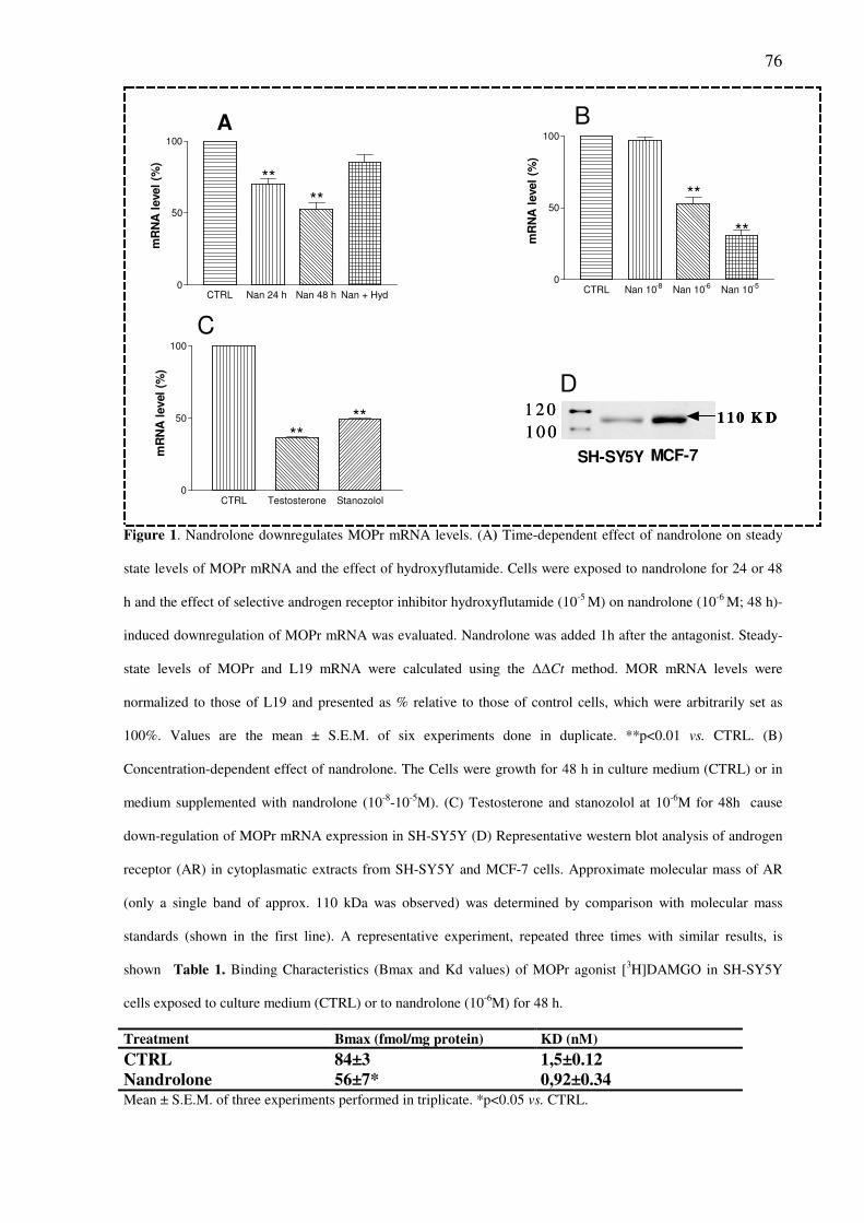
76
CTRL Nan 24 h Nan 48 h Nan + Hyd0
50
100
**
**
mR
NA
level
(%)
CTRL Nan 10-8 Nan 10-6 Nan 10-50
50
100
**
**
mR
NA
level
(%)
CTRL Testosterone Stanozolol0
50
100
****
mR
NA
level
(%)
D
MCF-7SH-SY5Y
1 2 0
1 0 01 1 0 K D
1 2 0
1 0 01 1 0 K D1 1 0 K D
A
C
B
Figure 1. Nandrolone downregulates MOPr mRNA levels. (A) Time-dependent effect of nandrolone on steady
state levels of MOPr mRNA and the effect of hydroxyflutamide. Cells were exposed to nandrolone for 24 or 48
h and the effect of selective androgen receptor inhibitor hydroxyflutamide (10-5
M) on nandrolone (10-6
M; 48 h)-
induced downregulation of MOPr mRNA was evaluated. Nandrolone was added 1h after the antagonist. Steady-
state levels of MOPr and L19 mRNA were calculated using the ∆∆Ct method. MOR mRNA levels were
normalized to those of L19 and presented as % relative to those of control cells, which were arbitrarily set as
100%. Values are the mean ± S.E.M. of six experiments done in duplicate. **p<0.01 vs. CTRL. (B)
Concentration-dependent effect of nandrolone. The Cells were growth for 48 h in culture medium (CTRL) or in
medium supplemented with nandrolone (10-8
-10-5
M). (C) Testosterone and stanozolol at 10-6
M for 48h cause
down-regulation of MOPr mRNA expression in SH-SY5Y (D) Representative western blot analysis of androgen
receptor (AR) in cytoplasmatic extracts from SH-SY5Y and MCF-7 cells. Approximate molecular mass of AR
(only a single band of approx. 110 kDa was observed) was determined by comparison with molecular mass
standards (shown in the first line). A representative experiment, repeated three times with similar results, is
shown Table 1. Binding Characteristics (Bmax and Kd values) of MOPr agonist [3H]DAMGO in SH-SY5Y
cells exposed to culture medium (CTRL) or to nandrolone (10-6
M) for 48 h.
Treatment Bmax (fmol/mg protein) KD (nM)
CTRL 84±3 1,5±0.12 Nandrolone 56±7* 0,92±0.34 Mean ± S.E.M. of three experiments performed in triplicate. *p<0.05 vs. CTRL.
77
Nandrolone reduces MOPr mRNA levels through a post-
transcriptional effect
To evaluate the effects of nandrolone on MOPr mRNA stability, cells were treated
with the transcription inhibitor actinomycin D. By blocking transcription, we were able to
determine whether nandrolone affects mRNA stability of MOPr transcripts. The cells were
pre-treated with actinomycin D (1µg/ml) for 30 minutes and then exposed to nandrolone (10-6
M) for 48 hours. Real-time RT-PCR showed that the rate of MOPr mRNA decay was
significantly increased. Therefore, this steroid seems to reduce the stability of MOPr mRNA
(Fig. 2).
To investigate the effects of nandrolone on MOPr promoter transcriptional activity, a
DNA construct containing a portion of the human MOPr promoter (from -2447 to +8 bp)
including a GRE responsive element was generated. This cDNA was cloned into a pGL3-
Basic plasmid, which contains a luciferase reporter, or into a pGL3-SV40 plasmid, containing
the luciferase gene under the positive control of the SV40 promoter. The transcriptional
activity of these plasmids was evaluated in transient transfection assays. Luciferase is express
in response to promoter activation and will catalyzes the oxidation of substrate luciferin
(added during the assay), resulting in photon production. Light output is directly proportional
to amount of luciferase present in each sample. As show in Fig. 2B, nandrolone treatment (10-
6 M; 48h) did not influence MOPr transcription in SH-SY5Y cells transfected with the pGL3-
Basic/mu plasmid or with the pGL3-SV40/mu plasmid. As consequence of SV40-positive
promoter activity, a more pronounced elevation of MOPr transcripts was observed in cells
transfected with the pGL3-SV40/mu plasmid; however, nandrolone treatment did not modify
this transcriptional activity.
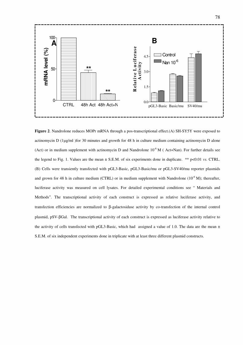
78
CTRL 48h Act 48h Act+Nan0
50
100
**
**
mR
NA
level
(%)
pGL3-Basic Basic/mu SV40/mu0.0
1.5
3.0
4.5 Control
Nan 10-6
Re
lati
ve
Lu
cif
er
as
eA
cti
vit
y
A B
Figure 2. Nandrolone reduces MOPr mRNA through a pos-transcriptional effect.(A) SH-SY5Y were exposed to
actinomycin D (1µg/ml )for 30 minutes and growth for 48 h in culture medium containing actinomycin D alone
(Act) or in medium supplement with actinomycin D and Nandrolone 10-6
M ( Act+Nan). For further details see
the legend to Fig. 1. Values are the mean ± S.E.M. of six experiments done in duplicate. ** p<0.01 vs. CTRL.
(B) Cells were transiently transfected with pGL3-Basic, pGL3-Basic/mu or pGL3-SV40/mu reporter plasmids
and grown for 48 h in culture medium (CTRL) or in medium supplement with Nandrolone (10-6
M); thereafter,
luciferase activity was measured on cell lysates. For detailed experimental conditions see “ Materials and
Methods”. The transcriptional activity of each construct is expressed as relative luciferase activity, and
transfection efficiencies are normalized to β-galactosidase activity by co-transfection of the internal control
plasmid, pSV-βGal. The transcriptional activity of each construct is expressed as luciferase activity relative to
the activity of cells transfected with pGL3-Basic, which had assigned a value of 1.0. The data are the mean ±
S.E.M. of six independent experiments done in triplicate with at least three different plasmid constructs.
79
Supra-physiological concentration of androgen steroids induce
toxicity in SH-SY5Y cells
To examine the effect of androgen steroids supra-physiological concentration, we set
up a MTT assay. Reduction of yellow methyltiazoletetazolium (MTT) into blue insoluble
formazan crystals is drived by mitochondrial of living cells. The viability of SH-SY5Y cells
seeded in 96-wells plates were evaluated after to be exposed to nandrolone, testosterone and
stanozolol (10-4
M-10-6
M) for 48h and 72h (Figure 3). The data collected show a toxic effect
concentration and time dependent for the three androgen steroids. Nandrolone have a weak
toxic effect at 10-5
M and more high effects at 10-4
M but no effects at 10-6
M. Testosterone
shown a similar result but a highest potency as demonstrated by EC50 value while stanozolol
has the comparable dose respond curves and EC50 values with nandrolone. (Figure 4)
Toxic effects of AS nandrolone and testosterone are evaluated , also, with the LDH
test. The measurement of lactate dehydrogenase release from damaged cells is correlated with
cell death. The medium of each samples was collected and analysed to evaluate the enzymatic
activity of LDH release from the cells This assay performed on SH-SY5Y cells exposed to
nandrolone and testosterone (10-4
M-10-5
M) for 72h, confirm the toxic effect time- and
concentration dependent of AAS (Figure 5)
The trypan blue exclusion test is another experiment carried out to confirm the toxicity
of supra-physiological concentration of AS. A decreased in the living cells number was
observed in SH-SY5Y cells treated with nandrolone and testosterone (10-4
M and 10-5
M) for
48h in comparison with control cells not treated (Figure 5).
SH-SY5Y cells are differentiated with retinoic acid to evaluated a possible changes in
cell death hormones dependent. Cells exposed with retinoic acid for 5 days were after treated
with the three steroids (10-4
M-10-5
M) for 48h. Data collected did not show any remarkable
different in neurotoxicity compare with undifferentiated cells (Figure 3).
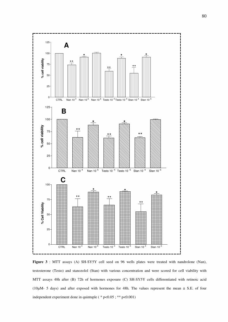
80
CTRL Nan 10-4 Nan 10-5 Nan 10-6 Testo 10 -4 Testo 10 -5 Stan 10 -4 Stan 10 -50
25
50
75
100
125
**
** **
*
% c
ell v
iab
ilit
y
* *
CTRL Nan 10 -4 Nan 10 -5 Testo 10 -4 Testo 10 -5 Stan 10 -4 Stan 10 -50
25
50
75
100
125
**** **
* *
% c
ell v
iab
ilit
y
CTRL Nan 10-4
Nan 10-5
Testo 10-4
Testo 10-5
Stan 10-4
Stan 10-5
0
25
50
75
100
% C
ell V
iab
ilit
y
** ****
* **
A
B
C
Figure 3 : MTT assays (A) SH-SY5Y cell seed on 96 wells plates were treated with nandrolone (Nan),
testosterone (Testo) and stanozolol (Stan) with various concentration and were scored for cell viability with
MTT assays 48h after (B) 72h of hormones exposure (C) SH-SY5Y cells differentiated with retinoic acid
(10µM- 5 days) and after exposed with hormones for 48h. The values represent the mean ± S.E. of four
independent experiment done in quintuple ( * p<0.05 ; ** p<0.001)
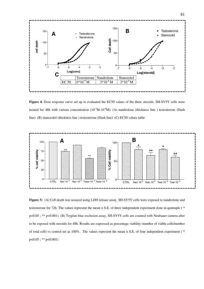
81
-7 -6 -5 -4 -3 -20
50
100
150Testosterone
Nandrolone
Log[conc]
cell
death
-7 -6 -5 -4 -3 -20
50
100
150Testosterone
Stanozolol
Log[steroid]
Cell
death
AB
C Testosterone Nandrolone Stanozolol
EC 50 6*10-5 M 3*10-4 M 3*10-4 M
Figure 4: Dose response curve set up to evaluated the EC50 values of the three steroids. SH-SY5Y cells were
treated for 48h with various concentration (10-3
M-10-6
M). (A) nandrolone (thickness line ) testosterone (Dash
line) (B) stanozolol (thickness line ) testosterone (Dash line) (C) EC50 values table
CTRL Nan 10-5 Nan 10-4 Test 10 -5 Test 10 -40
50
100
* ***
% C
ell
via
bil
ity
**
CTRL Nan 10-4
Nan 10-5
Testo 10-4
Testo 10-5
0
25
50
75
100
% c
ell
via
bil
ity **
**
A B
Figure 5: (A) Cell death was assayed using LDH release assay. SH-SY5Y cells were exposed to nandrolone and
testosterone for 72h. The values represent the mean ± S.E. of three independent experiment done in quintuple ( *
p<0.05 ; ** p<0.001) (B) Tryplan blue exclusion assay. SH-SY5Y cells are counted with Neubauer camera after
to be exposed with steroids for 48h. Results are expressed as percentage viability (number of viable cells/number
of total cell) vs control set as 100%. The values represent the mean ± S.E. of four independent experiment ( *
p<0.05 ; ** p<0.001)
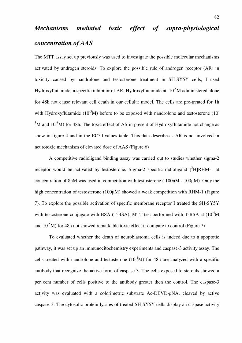
82
Mechanisms mediated toxic effect of supra-physiological
concentration of AAS
The MTT assay set up previously was used to investigate the possible molecular mechanisms
activated by androgen steroids. To explore the possible rule of androgen receptor (AR) in
toxicity caused by nandrolone and testosterone treatment in SH-SY5Y cells, I used
Hydroxyflutamide, a specific inhibitor of AR. Hydroxyflutamide at 10-5
M administered alone
for 48h not cause relevant cell death in our cellular model. The cells are pre-treated for 1h
with Hydroxyflutamide (10-5
M) before to be exposed with nandrolone and testosterone (10-
3M and 10
-6M) for 48h. The toxic effect of AS in present of Hydroxyflutamide not change as
show in figure 4 and in the EC50 values table. This data describe as AR is not involved in
neurotoxic mechanism of elevated dose of AAS (Figure 6)
A competitive radioligand binding assay was carried out to studies whether sigma-2
receptor would be activated by testosterone. Sigma-2 specific radioligand [3H]RHM-1 at
concentration of 8nM was used in competition with testosterone ( 100nM - 100µM). Only the
high concentration of testosterone (100µM) showed a weak competition with RHM-1 (Figure
7). To explore the possible activation of specific membrane receptor I treated the SH-SY5Y
with testosterone conjugate with BSA (T-BSA). MTT test performed with T-BSA at (10-4
M
and 10-5
M) for 48h not showed remarkable toxic effect if compare to control (Figure 7)
To evaluated whether the death of neuroblastoma cells is indeed due to a apoptotic
pathway, it was set up an immunocitochemistry experiments and caspase-3 activity assay. The
cells treated with nandrolone and testosterone (10-4
M) for 48h are analyzed with a specific
antibody that recognize the active form of caspase-3. The cells exposed to steroids showed a
per cent number of cells positive to the antibody greater then the control. The caspase-3
activity was evaluated with a colorimetric substrate Ac-DEVD-pNA, cleaved by active
caspase-3. The cytosolic protein lysates of treated SH-SY5Y cells display an caspase activity
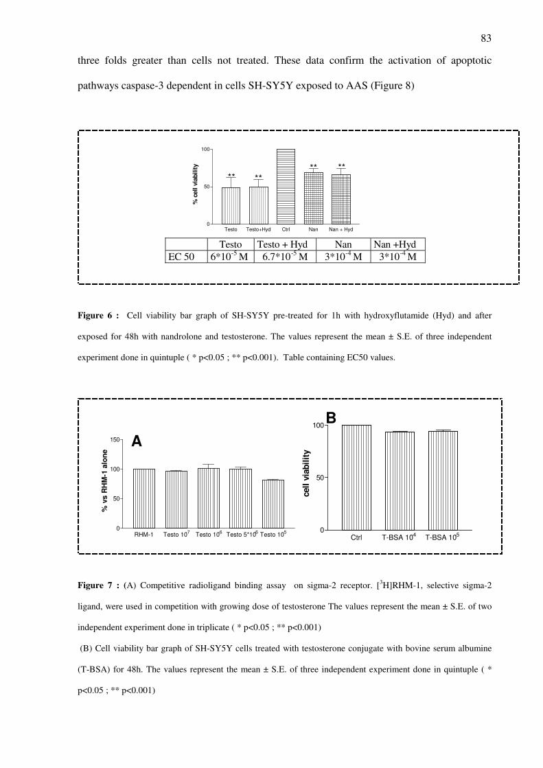
83
three folds greater than cells not treated. These data confirm the activation of apoptotic
pathways caspase-3 dependent in cells SH-SY5Y exposed to AAS (Figure 8)
Testo Testo+Hyd Ctrl Nan Nan + Hyd0
50
100
% c
ell v
iab
ilit
y
** **** **
Testo Testo + Hyd Nan Nan +Hyd
EC 50 6*10-5
M 6.7*10-5
M 3*10-4
M 3*10-4
M
Figure 6 : Cell viability bar graph of SH-SY5Y pre-treated for 1h with hydroxyflutamide (Hyd) and after
exposed for 48h with nandrolone and testosterone. The values represent the mean ± S.E. of three independent
experiment done in quintuple ( * p<0.05 ; ** p<0.001). Table containing EC50 values.
RHM-1 Testo 10-7
Testo 10-6
Testo 5*10-6
Testo 10-5
0
50
100
150
% v
s R
HM
-1 a
lon
e
A
B
Ctrl T-BSA 10-4 T-BSA 10-50
50
100
cell
via
bil
ity
Figure 7 : (A) Competitive radioligand binding assay on sigma-2 receptor. [3H]RHM-1, selective sigma-2
ligand, were used in competition with growing dose of testosterone The values represent the mean ± S.E. of two
independent experiment done in triplicate ( * p<0.05 ; ** p<0.001)
(B) Cell viability bar graph of SH-SY5Y cells treated with testosterone conjugate with bovine serum albumine
(T-BSA) for 48h. The values represent the mean ± S.E. of three independent experiment done in quintuple ( *
p<0.05 ; ** p<0.001)
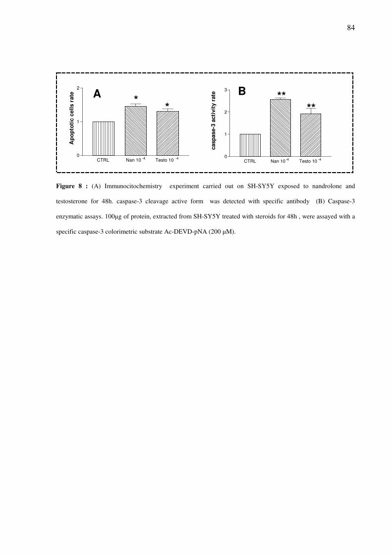
84
*****
*
CTRL Nan 10-4
Testo 10-4
0
1
2
3
casp
ase-3
acti
vit
y r
ate
CTRL Nan 10 -4 Testo 10 -40
1
2
Ap
op
toti
c c
ell
s r
ate A B
Figure 8 : (A) Immunocitochemistry experiment carried out on SH-SY5Y exposed to nandrolone and
testosterone for 48h. caspase-3 cleavage active form was detected with specific antibody (B) Caspase-3
enzymatic assays. 100µg of protein, extracted from SH-SY5Y treated with steroids for 48h , were assayed with a
specific caspase-3 colorimetric substrate Ac-DEVD-pNA (200 µM).
Discussion
Anabolic androgenic steroids (AAS) cause numerous side effects at both the central
and peripheral nervous system, including increased aggression, irritability, mania, depression,
and suicidal tendencies. Changes elicited by AAS have been observed for the
neurotransmitters GABAA (γ-aminobutyric acid) (Britan et al., 1993) and 5-HT (serotonin)
(Kindlundh et al., 2004) as well as for the neurotransmitter receptors dopamine D1 and D2
(Kindlundh et al., 2003), NK-1 (neurokinin-1) (Hallberg et al, 2005), and NMDA (N-methyl-
D-aspartic acid) (Rossbach et al., 2007). AAS exposure has been proposed as a risk factor for
other drugs of abuse. Animal studies, in fact, have confirmed that AAS evoke
neurobiochemical alterations related to behavioral responses and affect the endogenous opioid
and dopamine systems (Clark et al, 1993). A possible explanation for this effect is that
chronic nandrolone treatment results in long-term changes in reward brain circuits and leads
to a progressive decrease in the basal hedonic level, which results in an unpleasant state that
renders the organism susceptible to the development of an addictive process (Celierer et al.,
2003). Based on these data, AAS have been proposed to serve as ‘‘gateway’’ drugs to opioid
dependence (Arvary et al., 2000). Nyberg and colleagues have determined the molecular
mechanisms of the rewarding effects of nandrolone on different opioid peptides (Magnusson
et al, 2007). Moreover, the effects of nandrolone on delta opioid receptor (DOPr) expression
have been evaluated in neuronal cell lines (Pasquariello et al., 2000) . In this study,
downregulation of DOPr in cell exposed to nandrolone was shown to be independent of AR.
I found, in this thesis, that treatment of SH-SY5Y neuronal cells with nandrolone at a
supra-physiological concentration (10-6
M), which may mimic abuse regimes (Clark et al.,
1993), decreased the expression of MOPr mRNA. Exposure to nandrolone also significantly
decreased the number of MOPr binding sites. This finding suggests that a prolonged
exposure to nandrolone affects MOPr function.
Opioid
receptor expression is regulated by multiple mechanisms,
including
transcriptional and post-transcriptional events (Law et al., 2004). MOPr promoter activity is
86
under the control of various transcription factors (Kim et al., 2006) that interact with DNA
responsive elements mainly located in the 5’ upstream region of this promoter. Interestingly
the human MOPr promoter contains a glucocorticoid responsive element GRE (from -2215 to
-2195 bp), which can bind the glucocorticoid receptor or the AR (Takai et al., 2007). Post-
transcriptional regulation occurs at the mRNA or protein level via control of mRNA stability,
alterations in translation efficiency, alterations of mRNA transport, and covalent modification
of receptor molecules. Recent studies have indicated that the 5’ and 3' untranslated regions
(UTR) of MOPr mRNA affect the overall activity of the transcripts (Law et al., 2004). Thus,
post-transcriptional regulation of the MOPr gene may play an important role in the spatial and
temporal expression of the receptor protein. A correlation between cis elements in the 5’ and
3’ UTR regions and the alteration in MOPr expression has been described (Xu and Carr et al.,
2001). In addition, the 5' UTR in the MOPr mRNA is a key site of post-transcriptional
regulation. This mechanism is mediated by uAUG/uORF (open reading frame) sequences that
control translation initiation. Recent discoveries in hormone action emphasize that regulation
of gene expression is not restricted to alterations in the rate of gene transcription. On the
contrary, hormonal effects on the stability of a specific mRNA can profoundly alter its steady-
state concentration (Gies et al., 1997). The down-regulation of MOPr mRNA nandrolone
dependent may be due to a decrease in the stability of mRNA since no change was observed
in the rate of MOPr promoter/luciferase transcription in a gene reporter system containing a
sequence of 2455 bp of the 5’upstream region of the human MOPr promoter. Until now, the
identities of the cellular proteins that are involved in the post-transcriptional mechanisms that
regulate MOPr and are modulated by nandrolone are unknown.
Nandrolone may regulate MOPr mRNA levels through mechanisms involving the
classic intracellular AR. This notion is supported by the finding that nandrolone-induced
downregulation of MOPr mRNA levels in SH-SY5Y cells was blocked by hydroxyflutamide,
an androgen antagonist that binds to the AR and is expressed in the SH-SY5Y cell line. In
87
previous studies, was showed that nandrolone modulates DOPr expression in neuronal cells
but that this action is independent from AR involvement (Pasquariello et al., 2001).
Therefore, the present data demonstrate that nandrolone activates different pathways to
modulate the expression of MOPr and DOPr in neuronal cells. A down-regulation of MOPr
expression is observed also in cells treated with testosterone and stanozolol (10-6
M) for 48h.
This in vitro observation need to be investigate deeply and correlate with the stacking
administration regime common in AS abusers.
The second part of my studies was focus on the explanation of androgen steroids toxic
effect at supra-physiological concentrations. The neurotoxicity of steroids on neuronal cells
can negatively affect the behaviour of user and might induce a degenerative process. The
“steroid dementia” syndrome found in some patients using high concentration of steroids
could be caused by neuro-toxic consequence (Wolkowitz et al., 2007). This thesis show , with
different cito-toxic assays and condition, the toxic effect of supra-physiological concentration
of testosterone, nandrolone and stanozolol in SH-SY5Y neuroblastoma cells. These steroids
show this effect at micromolar concentration after 48h and 72h of exposure. A study lead in
PC12 cell line exposing to testosterone showed opposite result between differentiate and
undifferentiated cells. Instead, SH-SY5Y cells differentiated with retinoic acid show similar
susceptibly cell death when treated with steroids in confront with undifferentiated cells.
Studies carried out in different cellular model, PC12 (Alexaki et al., 2006), cortical astrocytes
(Gatson and Singh, 2007), and mouse cortical cells (Orlando et al., 2007), described that AS
can promote cell death. This effect is time- and concentration-dependent and various
mechanisms might be involved. In mouse cortical cells was described an AR dependent action
of nandrolone that promotes the NMDA toxic effect, in the same models testosterone effects
are not mediated by AR but it is regulated by aromatase conversion of testosterone to
estradiol. At nanomolar concentration testosterone is completely converted in 17β-Estradiol, a
molecule with a protective effect on neuronal cells. Nanomolar concentration of testosterone
88
became neurotoxic in the present of aromatase inhibitor. This indicates that testosterone is
intrinsically toxic to cortical neurons and high dose of steroid overwhelmed the
neuroprotection effect aromatase dependent and showed a toxic effect. I investigated the
possible rule of AR in testosterone and nandrolone toxic effect in SH-SY5Y and I found that
AR seems not mediated this mechanism. In neuroblastoma cells model, used in this thesis, is
not yet know whether the metabolism of steroids , aromatization of testosterone or 5α-
reductase conversion of nandrolone, regulate the cito-toxicity of this two steroids.
Another possibility, described in various studies, associated with neuronal cell death
dependent from steroids, is the activation of membrane specific androgen receptor. Recently,
it was demonstrated that testosterone induces intracellular Ca2+
oscillation in the cytosol and
nucleus, wich are an important mediator of downstream events as apoptosis (Estrada et al.,
2006). Sigma-2 receptor, which activate an apoptotic pathways, has been described to activate
Ca2+
oscillation in SK-N-SY cells, progenitor of SH-SY5Y cells (Vilner and Bowen, 2000).
The competitive radio binding assay set up to evaluated a possible activation of this apoptosis
pathways by testosterone not show a positive result. Testosterone not seems to compete with
specific sigma-2 ligand RHM-1 to binding the receptor. To elucidate the hypothesis of
involvement of a membrane receptor in the toxic effects of steroids on SH-SY5Y cells. I
made an experiment with testosterone-BSA. The results obtained did not show any direct
toxic effect at micromolar concentration after 48h of exposure. In PC12 and astrocytes was
hypothesized to exist a receptor on the cytoplamatic membrane of this cells recognize
specifically by steroids and that steroids binding on this receptor activate a pro-apoptotic
pathways. This receptor seems to have a low affinity to AS and it could explain the high-
concentration need to observe the toxic effect. Until now it is not clear which is this receptor.
In astrocytes exposed to DHT-BSA and iodoacetic acid (IAA) was observed an increase of
the toxic effect provoke by IAA alone. DHT-BSA alone not show any toxic effect but it
makes the cells more susceptible to oxidative stress. My data seems not to be comparable to
89
PC12 and astrocytes results. Therefore, it might be hypothesized a mechanism that involved
membrane fluidity. It was observe the intercalation of the lipophilic steroids into membrane of
target tissues, resulting in perturbation of lipid-lipid interactions that may, in turn, alter the
function of membrane proteins (Withing et al., 2000).
In a in vitro cellular model of neurodegenerative disease, spinal bulbar muscular
atrophy (SBMA), was detected cleavage of caspase-3 upon androgen stimulation, suggesting
that this caspase is involved in the cell death induced by androgen treatment (Grierson et al.,
2001). In SH-SY5Y cells exposed to testosterone was obersved caspase-3 activation (Estrada
et al., 2006). I found that the androgen steroids nandrolone induce toxic effect throught
activation of caspase-3 pathway in my time and concentration experimental condition (10-
4M;48h). The immunocitochemistry and caspase-3 activity assays results showed the
activation of this pathway.
In summary, in this thesis I observed genomic and non-genomic effects of high dose
of anabolic androgenic steroids in a neuronal cellular model. Development of neurological
syndromes in steroids abuser might be explain by a complex molecular mechanisms.
Together, my results described two mechanisms which could be correlate with behavioural
changes and neuro degeneration observed in steroids abuser. I showed as steroids regulated in
SH-SY5Y the opioid neuronal circuit through the control of MOPr expression via androgen
receptor (AR) activation and mRNA stability inhibition and a neurotoxic effect, AR
independent, associated with elevate dose of steroids. The beneficial effects of AAS are
carefully regulated over a narrow (nanomolar) range of concentration, which is crucial for
normal neuronal functions. Elucidation of androgens effects in neuronal cells is, also,
important to define the safety of androgen supplementation therapy.
90
Reference
• Alexaki VI, Dermitzaki E, Charalampopoulos I, Kampa M, Nifli AP, Gravanis A, Margioris AN,
Castanas E. Neuronal differentiation of PC12 cells abolishes the expression of membrane androgen
receptors. Exp Cell Res. 2006 Sep 10;312(15):2745-56
• Arvary D, Pope HG Jr. Anabolic-androgenic steroids as a gateway to opioid dependence. N Engl J
Med. 2000; 342: 1532.
• Bitran D., Kellog CK, Hilvers RJ : Treatment with anabolic androgenic steroid affects anxiety-related
behavior and alters the sensitivity of cortical GABAA receptors in the rat. Hormons Behavor 1993; 27:
568-83.
• Celerier E, Yazdi MT, Castane A, Ghozland S, Nyberg F, Maldonado R.Effects of nandrolone on
acute morphine responses, tolerance and dependence in mice. Eur J Pharmacol. 2003; 465: 69-81.
• Clark AS, Lindenfeld R, Gibbons C. : Anabolic-androgenic steroids and brain reward. Pharmacology
Biochemical Behavoral 1996; 53: 741-5.
• Estrada M, Varshney A, Ehrlich BE. Elevated testosterone induces apoptosis in neuronal cells. J Biol
Chem. 2006 Sep 1;281(35):25492-501
• Gatson JW, Singh M. Activation of a membrane-associated androgen receptor promotes cell death in
primary cortical astrocytes. Endocrinology. 2007 May;148(5):2458-64
• Gies EK, Peters DM, Gelb CR, Knag KM, Peterfreund RA. Regulation of mu opioid receptor
mRNA levels by activation of protein kinase C in human SH-SY5Y neuroblastoma cells.
Anesthesiology. 1997; 87: 1127-38.
• Grierson AJ, Shaw CE, Miller CC. Androgen induced cell death in SHSY5Y neuroblastoma cells
expressing wild-type and spinal bulbar muscular atrophy mutant androgen receptors. Biochim Biophys
Acta. 2001 Apr 30;1536(1):13-20
• Hallberg M, Kindlundh AM, Nyberg F.: The impact of chronic nandrolone decanoate administration
on the NK1 receptor density in rat brain as determined by autoradiography. Peptides. 2005; 26: 1228-
34.
• Kim CS, Choi HS, Hwang CK, Law PY, Wei LN, Loh HH. Et al.Evidence of the neuron-restrictive
silencer factor (NRSF) interaction with Sp3 and its synergic repression to the mu opioid receptor
(MOR) gene. Nucleic Acids Res. 2006; 34: 6392-403 .
91
• Kindlundh AM, Lindblom J, Nyberg F. Chronic administration with nandrolone decanoate induces
alterations in the gene-transcript content of dopamine D(1)- and D(2)-receptors in the rat brain Brain
Res. 2003; 979: 37-42.
• Kindlundh AM, Rahman S, Lindblom J, Nyberg F.: Increased dopamine transporter density in the
male rat brain following chronic nandrolone decanoate administration. Neurosci Lett. 2004; 356: 131-4.
• Law PY, Loh HH, Wei LN. Insights into the receptor transcription and signaling: implications in
opioid tolerance and dependence. Neuropharmacology. 2004;47 (Suppl 1): 300-11.
• Magnusson K, Hallberg M, Bergquist J, Nyberg F. Enzymatic conversion of dynorphin A in the rat
brain is affected by administration of nandrolone decanoate. Peptides. 2007; 28: 851-8.
• Orlando R, Caruso A, Molinaro G, Motolese M, Matrisciano F, Togna G, Melchiorri D, Nicoletti
F, Bruno V. Nanomolar concentrations of anabolic-androgenic steroids amplify excitotoxic neuronal
death in mixed mouse cortical cultures. Brain Res. 2007 Aug 24;1165:21-9.
• Pasquariello A, Di Toro R, Nyberg F, Spampinato S. Down-regulation of delta opioid receptor
mRNA by an anabolic steroid in neuronal hybrid cells. Neuroreport. 2000; 11: 863-7.
• Rossbach UL, Steensland P, Nyberg F, Le Greves P: Nandrolone-induced hippocampal
phosphorylation of NMDA receptor subunits and ERKs. Biochem Biophys Res Commun. 2007; 357:
1028-33.
• Takai H, Nakayama Y, Kim DS, Arai M, Araki S, Mezawa M, et al. Androgen receptor stimulates
bone sialoprotein (BSP) gene transcription via cAMP response element and activator protein
1/glucocorticoid response elements. J Cell Biochem. 2007; 102: 240-51.
• Vilner BJ, Bowen WD. Modulation of cellular calcium by sigma-2 receptors: release from intracellular
stores in human SK-N-SH neuroblastoma cells.J Pharmacol Exp Ther. 2000 Mar;292(3):900-11
• Whiting KP, Restall CJ, Brain PF. Steroid hormone-induced effects on membrane fluidity and their
potential roles in non-genomic mechanisms. Life Sci. 2000 Jul 7;67(7):743-57
• Wolkowitz OM, Lupien SJ, Bigler ED. The "steroid dementia syndrome": a possible model of human
glucocorticoid neurotoxicity. Neurocase. 2007 Jun;13(3):189-200
• XU Y and Carr LG : Functional characterization of the promoter region of human µ opioid receptor
(hMOR) gene: identification of activating and inhibitory regions. Cellular and Molecular Biology
2001; 47: 29-38.

92