università degli studi di milano scuola di dottorato in medicina … · nei tumori con...
TRANSCRIPT

Università degli Studi di Milano
Scuola di Dottorato in Medicina Molecolare
Direttore della Scuola: Ch.mo Prof. Mario CLERICI
Tutore: Prof. Alberto MALESCI
Correlatore: Dott. Luigi LAGHI
Molecular Heterogeneity and Prognostic
Implications of Synchronous Advanced Colorectal
Neoplasia
Curriculum di Oncologia Molecolare
Ciclo XXV
Anno Accademico 2011/2012
Dottorando: Gianluca BASSO
Laboratorio di Gastroenterologia Molecolare
Istituto clinico Humanitas, Rozzano, Milano

I
ABSTRACT
Background and Aim. It is uncertain whether synchronous colorectal cancers (S-
CRC) develop through a preferential carcinogenetic pathway and whether they
have a poorer prognosis. Since microsatellite unstable tumors (MSI) might act as a
bias when assessing methylation and outcome of S-CRC, we analysed for
synchronous neoplasia and for disease-specific survival (DSS) a large series of
CRC systematically characterized by BRAF and MS status.
Patients and Methods. 881 patients consecutively resected for CRC and
perioperatively investigated with complete colonoscopy, were included in the study.
For each patient, demographic and clinico-pathological records were retrieved and
DSS was retrospectively assessed. All cancers were screened for MSI by BAT
26/BAT25 mononucleotide analysis. MSI tumors had the MMR protein defect
identified at immunohistochemistry (IHC) and were tested for germline mutation of
the corresponding gene. All tumors were also analysed for BRAF c.1799T>A
mutation.
Results. S-CRC was associated with stage IV (p=0.01) and with HNPCC
(p<0.001), but only MSS S-CRC accounted for the association with metastatic
CRC (p=0.001). BRAF mutation was associated with sporadic MSI CRC (p<0.001)
but not with S-CRC (p=0.96). S-CRC did not affect DSS in patients with MSI CRC
(HR 0.74; 95%CI 0.09-5.75; p=0.77). Conversely, S-CRC (HR 1.82; 95%CI 1.15-
2.87;p=0.01), as well as synchronous advanced adenoma (HR 1.81; 95%CI 1.27-
2.58; p=0.001), and BRAF c.1799T>A
mutation (HR 2.16; 95%CI 1.25-3.73; p=0.01)
were stage-independent predictors of death related to MSS CRC.
Conclusions. Microsatellite-stable CRC have a worse prognosis if S-CRC, or even
a synchronous advanced adenoma, is diagnosed. Neither the occurrence of
multiple MSS advanced neoplasia nor their enhanced aggressiveness are
mediated by an epigenetic field effect. Surveillance and therapeutic protocols for
MSS CRC should take into account the prognostic burden of synchronous
advanced colorectal neoplasms.

II
SOMMARIO
Introduzione e scopo. In letteratura molte ipotesi sono state formulate
sull’insorgenza dei tumori sincroni del colon retto (S-CRC). Infatti, non è ancora del
tutto chiaro se questo tipo di malattia sviluppa attraverso un particolare pathway
molecolare legato ad una elevata suscettibilità della mucosa colica a divenire
neoplastica e se a livello clinico evidenzia una prognosi peggiore. Dal momento
che i tumori con instabilità dei microsatelliti (MSI) possono costituire un bias nel
valutare la presenza di metilazione e l’andamento clinico dei S-CRC, abbiamo
analizzato per neoplasia sincrona e per disease-specific survival (DSS), una ampia
casistica di CRC caratterizzata per la mutazione di BRAF e per lo stato dei
microsatelliti. Pazienti e Metodi. Nello studio sono stati arruolati 881 pazienti
consecutivi, i quali sono stati sottoposti a resezione del cancro del colon e hanno
eseguito a livello perioperatorio una colonscopia completa. Per ogni paziente, i dati
demografici e clinico-patologici sono stati recuperati e il DSS è stato valutato in
modo retrospettivo. Tutti i tumori sono stati sottoposti a screening per MSI, per
identificare pazienti con HNPCC, mediante l’analisi di marcatori mononucleotidici
come BAT 26 e BAT25. Nei tumori con instabilità dei microsatelliti, il difetto
proteico è stato individuato mediante immunoistochimica (IHC) e successivamente
sono stati testati per la mutazione germinale del gene corrispondente. Inoltre, tutti i
tumori sono stati anche analizzati per la mutazione di BRAF c.1799T> A
. Risultati. Il
S-CRC è stato associato con lo stadio IV (p = 0.01) e con l’HNPCC (p <0.001), ma
solo i casi MSS S-CRC sono più frequentemente metastatici (p = 0.001). La
mutazione di BRAF c.1799T> A
era associata con gli MSI CRC sporadici (p <0.001),
ma non con i S-CRC (p = 0.96). I cancri sincroni non cambiano il miglior DSS dei
pazienti con MSI CRC (HR 0.74, 95% CI 0.09-5.75, p = 0.77). Viceversa, i MSS S-
CRC (HR 1.82, 95% CI 1.15-2.87, p = 0.01), così come un tumore MSS e la
presenza di un adenoma avanzato (sincrono adenoma avanzato) (HR 1.81, 95%
CI 1.27-2.58, p = 0.001), e la mutazione di BRAF c.1799T> A
(HR 2.16, 95% CI 1.25-
3.73, p = 0.01) risultano essere variabili predittori di un aumentato rischio di morte
indipendenti dallo stadio di malattia. Conclusioni. I MSS S-CRC evidenziano una
prognosi peggiore, e lo stesso vale per la presenza di un sincrono adenoma
avanzato. Dallo studio emerge che né la presenza di multiple avanzate neoplasie
in tumori MSS né la loro maggiore aggressività sono mediati da un effetto
epigenetico. Per cui la sorveglianza e i protocolli terapeutici per i pazienti con un
MSS CRC dovrebbero tener conto del peso prognostico dato dalla presenza di
neoplasie sincrone avanzate del colon-retto.
III
INDEX
1. INTRODUCTION ............................................................................................... 1
1.1 Colorectal Cancer ......................................................................................... 1
1.1.1 Epidemiology ........................................................................................... 1
1.1.2 Prognostic Factors ................................................................................... 3
1.1.3 Polyps ...................................................................................................... 7
1.1.4 Molecular basis ...................................................................................... 10
1.1.4.1 Chromosomal instability (CIN or MSS) ............................................... 10
1.1.4.2 Microsatellite instability (MSI) ............................................................. 12
1.1.4.3 CpG Island Methylator Phenotype (CIMP) ......................................... 16
1.1.5 Clinical and Molecular Implication ......................................................... 17
1.2 Synchronous Colorectal Cancer ............................................................. 25
1.2.1 Epidemiology ......................................................................................... 26
1.2.2 Molecular basis ...................................................................................... 28
2. AIM OF THE WORK ........................................................................................... 32
3. MATERIALS AND METHODS ........................................................................... 33
3.1 Study Population and CRC Subgroup by Synchronous Neoplasia .... 33
3.2 Tumor Molecular Subtyping ...................................................................... 34
3.2.1 Assessment of MSI ................................................................................ 34
3.2.2 Immunohistochemistry ........................................................................... 34
3.2.3 MLH1, MSH2 and MYH sequencing ...................................................... 35
3.2.4 Multiple ligation probe amplification analysis (MLPA) ........................... 35
3.2.5 TaqMan SNP Genotyping ...................................................................... 36
3.3 Statistical Analysis ..................................................................................... 39
4. RESULTS ........................................................................................................... 40
5. DISCUSSION ..................................................................................................... 49
6. BIBLIOGRAPHY ................................................................................................ 52
7 SCIENTIFIC PRODUCTS ........................................................................................ 64
IV
ABBREVIATIONS
APC= Adenomatous Polyposis Coli
CIMP= CpG Island Methylator Phenotype
CIN= Cromosomal Instability
CRC= Colorectal Cancer
FAP= Familial Adenomatous Polyposis
HNPCC= Hereditary Non Polyposis Colorectal Cancer
HP= Hyperplastic polyp
LOH= Loss of Heterozygosity
MAP= MYH-Associated Polyposis
MSI= Microsatellite Instability
MSS= Microsatellite Stability
TSA= Traditional Serrated Adenoma(
S-CRC= Synchronous Colorectal Cancer
SSA= Sessile Serrated Adenoma

1
1. INTRODUCTION
1.1 Colorectal Cancer
1.1.1 Epidemiology
Colorectal cancer (CRC) is the second leading cause of cancer death in Western
countries [2-4], with a mortality rate of 30 cases per 100,000 inhabitants in Europe
each year [5]. More than 90% of CRC new cases are diagnosed in people over the
age of 50 [6]. In men, CRC is the most common tumor second only to
adenocarcinoma prostate with an average age of onset of about 68 years [1].
Meanwhile in women is the third most frequent tumor after breast and uterus
cancers [1]. with an average onset age of about 72 years [1].
More than one million new cases are diagnosed worldwide each year [7], and in
Europe the incidence is about 58 cases per 100,000 people [5]. Over the past
twenty years a decline in CRC incidence and mortality was observed [3, 6]. In
particular from 1990 to 2005 the death rate was significantly reduced (-31.8% for
men and -28% for women) [3], from 50% in 2000 [6] to 33% in 2010 [7]. The
reduction in the incidence rate may depend not only from the intensification of
screening methods use [6], but also from increased aspirin and other FANS
assumption, and postmenopausal hormone replacement therapy introduction in
women, both protective factors for the occurrence of CRC [3].
The frequency of the CRC is very variable depending on the cohort analyzed.
Indeed it have been demonstrated that geographical CRC incidence differences
exist [9]. A possible explanations include the distribution of some regional or local
characteristics of patients (race, ethnicity, culture), modifiable lifestyle (smoking,
diet, exercise) and theoretically the genetic risk factors of CRC [8, 9]. In particular,
African American population shows a higher incidence and mortality [8]. Moreover,
in literature it has been suggested that the socio-economic status could influence
CRC risk. Therefore, the measure of poverty status of a geographical area may
explain the regional variations in the incidence and survival of cancer [8].
Regarding the location of the tumor, in the 80s it has been observed a reduction in
the left colon (descending, sigmoid colon, rectum) CRC incidence, which are also
more frequent in the under 70 years, but not in CRC in the right side (ascending
colon, caecum).

2
This was attributed to the use of endoscopic screening by recto-sigmoidoscopy
(RSS) and colonoscopy (PCS). Indeed, the full PCS is strongly associated with a
decrease in distal tumors mortality (OR 0.33, but not in proximal tumors death (OR
0.99), probably due to biological differences between the proximal and distal colon
and to a higher number of lesions not identified in the right colon side [2].

3
1.1.2 Prognostic Factors
In many types of tumors the extent or stage of cancer at the time of diagnosis is a
key factor that defines prognosis and it is a critical element in determining
appropriate treatment [11]. The most clinically useful and important staging system
is the Tumor Node Metastasis (TNM) system.
This system is the result of close collaboration between the International Union
Against Cancer (UICC-TNM) and the American Joint Committee on Cancer
(AJCC). TNM system was initially developed only to predict prognosis, but its
application was expanded also to establish the suitable treatment and to select the
patients to include in for clinical trials [12].
The TNM system classifies cancers by the primary tumor size and extention (T),
the involvement of regional lymph node (N), and the presence or absence of
distant metastases (M). However, the criteria for defining anatomic extent of
disease are specific for the tumors with different anatomic sites and different
histological types [11].
The TNM staging classification include:
- clinical TNM (cTNM) is defined by information obtained from symptoms;
physical examination; endoscopic examinations; imaging studies of the
tumor, regional lymph nodes, and metastases; biopsies of the primary
tumor; and surgical exploration without resection.
- pathological TNM (pTNM) is defined by the same diagnostic studies used
for clinical staging supplemented by findings from surgical resection and
histologic examination of the surgically removed tissues.
- post therapy or post neoadjuvant therapy TNM (yTNM) includes systemic
or radiation treatment prior to surgical resection.
In CRC, the TNM staging remains the gold standard of prognostic factor and play
an important role in determining the 5-years survival rate in these patients [5].
Despite many editions of TNM have been proposed by the years, those after the
fifth edition did not provide significant and adequate advantage in CRC staging
[13].
Staging of CRC is based on the depth of tumor penetration through the bowel wall
or adherence to adjacent organs or structures (T), degree of presence of regional
lymph node involvement (N) and presence or absence of distant metastasis (M)
(Figure 1).

4
Figure 1 The different components of colic mucosa involved during tumor invasion.
According to the fifth edition of the TNM staging system, the depth of tumor
invasion is classified by:
pT is refers to the presence of malignant cells confined above the basement
membrane or lamina propria;
pT1 tumor invades submucosa;
pT2 tumor invades muscularis propria;
pT3 tumor invades through the muscularis propria into the subserosa or into
nonperitonealized pericolic tissues;
pT4 tumor directly invades other organs or structures and/or perforates visceral
peritoneum;
The lymph node involvement is classified by:
N0 no regional lymph node metastasis;
N1 metastasis to one to three regional lymph nodes;
N2 metastasis to four or more regional lymph nodes;
The presence or absence of distant metastasis is classified by
M0 no distant metastasis;
M1 distant metastasis;

5
Patients with a similar prognosis, T, N and M are grouped into so-called anatomic
stage/prognostic groups, commonly referred to as stage and this classification with
respect to the fifth edition of the American Joint Committee on Cancer (AJCC)
system [14] in the (Table I).
Stage Local Invasion Nodal Invasion Distant Metastasis
I T1
T2
N0
N0
M0
M0
II T3
T4
N0
N0
M0
M0
III Any T N1 M0
IV Any T Any N M1
Table I CRC stages classified according to the fifth edition of the American Joint
Committee on Cancer (AJCC) system.
In addition to TNM stage, other tumor-related features such as venous and
lymphatic invasion, tumor grade, and tumor budding have been identified as
essential or important prognostic factors [5].
Venous and lymphatic invasion represents crucial steps in the formation of micro-
metastases and eventually macroscopic tumor growth at a secondary site.
Tumor grade is based on the percentage of gland formation.
- well-differentiated (G1) adenoma with more than 95% of glands within the
tumor;
- moderately differentiated (G2) with 50 to 95% of glandular structure;
- poorly differentiated (G3) with 5 to 50% of glandular structure;
- undifferentiated (G4) carcinoma with less than 5% of glandular structure
[15].

6
Tumor budding is described as a transition from glandular structures to single cells
or clusters of up to four cells at the invasive margin of CRC with malignant
potential. It has been show that the presence of “buds” at the tumor invasive front
represents an independent predictor of lymph node metastasis in patients with sub-
mucosal invasive or early pT1 CRCs and probably the frequency of tumor budding
increases with more advanced TNM stage [16]. Therefore, molecular and
immunohistochemical analysis have been proposed to improve the prognostic
value, although at present the tumor stage is still the most important prognostic
factor in CRC.

7
1.1.3 Polyps
Polyps are most common in the colon but may occur in the esophagus, stomach,
and small intestine.
Intestinal polyps can be classified as non-neoplastic or neoplastic in nature. The
common neoplastic polyp is adenoma while the non-neoplastic polyps can be
further classified as inflammatory, hamartomatous, or hyperplastic [17].
Many colorectal cancer develops from adenomas, the precursor lesions, which are
benign intraepithelial neoplasms composed of dysplastic cells with malignant
potential. The risk of cancer developing within an adenoma increases with size,
grade of dysplasia and villosity. These features, together with polyp numbers, are
also predictive of metachronous neoplasia and may therefore influence the
decision to offer follow-up endoscopic surveillance [17].
The presence of one or more colorectal adenomas in the different populations
depends on age, gender and family history. In fact, the prevalence rates in
asymptomatic individuals ≥ 50 years, determined by colonoscopy, range from 24%
to 50% while the presence of advanced adenomas (≥ 1 cm in size with villous
features, and/or with high grade dysplasia) varies from 3.4 up to 9.5% [18].
The incidence of adenomas at intervals ranging from 6 months to 5 years in post-
polypectomy surveillance colonoscopy studies varies from 20 to 50%. The most
incident polyps are small, and a higher incidence has been associated with multiple
adenomas at the index colonoscopy, larger size of the index adenoma, older age,
and a family history of a parent with colorectal cancer. A large studies have
demonstrated that regular surveillance colonoscopy and polyps removal reduce the
incidence of colorectal adenocarcinoma [18].
Adenomas are characterized according to macroscopic and histological features.
Gross features: pedunculated; sessile and flat [15].
- Pedunculated adenomas appear as exophytic, mucosal protrusions with a
lobulated head and a stalk covered by normal mucosa. The adenomatous
epithelium remains confined to the mucosa of the head of the polyp. The stalk
consists of normal mucosa, including the muscularis mucosae and
submucosal tissue, in continuity with the major part of the bowel wall.
- Sessile adenomas attach to the mucosa by a broad base. They are often less
well circumscribed than pedunculated ones. Because of their ill-defined
edges, they are difficult to delineate, and have a greater tendency to recur
following local excision.
- Flat adenomas are lesions that lack an exophytic polypoid configuration.
They consist of slightly elevated dysplastic mucosal plaques that are never

8
- greater than twice the thickness of the surrounding normal colonic mucosa.
They constitute a special subgroup of adenomas with a greater potential for
malignant transformation, while still being smaller than exophytic adenomas.
Histological features: tubular; villous; tubulo-villous and grade of dysplasia [15].
- Tubular adenomas maintain the original crypt architecture, but adenomatous
epithelium replaces the normal colonic epithelium in lining the crypts. This is
the most common type of adenoma (about 68% to 87%). Tubular lesions are
those that contain greater than the 80% of tubular component. Tubular
adenomas consist of closely packed branching tubules separated by varying
amounts of lamina propria. The tubule may be relatively regular, or when the
adenomatous tubules grow, they may branch and show considerable
irregularity.
- Villous adenomas (approximately 20%) have villi with cores of lamina propria
covered by a single layer of adenomatous epithelium. Villous lesions are
those that contain greater than 80% of a villous component. Villous
adenomas fall into three types: flat, carpet-like masses; lobulated, bulky,
sessile masses and pedunculated lesions with short, broad pedicles.
- Tubulo-villous adenomas contain a mixture of both tubular and villous
patterns, or have broad villi containing short tubular structures. Tubulo-villous
lesions are those that contain from 20% to 79% villous components. They
tend to be larger than tubular adenomas, with a mean diameter of 19 mm
and, usually, a villous component is present in 35% to 75% of all adenomas
measuring more than 1 cm in largest diameter.
Grade of dysplasia: low-grade-dysplasia (LGD) and high-grade dysplasia (HGD)
[18].
- Low grade dysplasia is characterized by tubules which are lined from the top
to the bottom by epithelium which is morphologically similar to the normal
basal proliferative zone. The nuclei are enlarged, oval, hyperchromatic, and
have normal orientation. There is a slight excess of mitotic figures but the
architecture is not disrupted.
- High grade dysplasia is characterized by large vesicular nuclei, irregular and
conspicuous nucleoli, scalloped nuclear membranes, and increased nuclear
to cytoplasmic ratio. Nuclear polarity is disrupted and marked cellular
pleomorphism and both numerous and aberrant mitoses are present.
Structural alterations include budding and branching tubules, back-to-back
arrangement of glands, and cribriform growth of epithelial cells in clusters and
sheets. This dysplasia encompasses the histological changes called
carcinoma in situ and intramucosal carcinoma.

9
In addition, another group of polyps are represented by serrated polyps. In the
past, all serrated polyps were classified simply as hyperplastic polyps and were
considered to have no malignant potential. This group of polyps is comprised not
only of hyperplastic polyps, but also of sessile serrated adenomas, traditional
serrated adenomas and mixed polyps, showing serrated and classical
adenomatous features [19].
- Hyperplastic polyp (HP) are by far the most common serrated polyps (80–
90%). They occur most often in the distal part of the colon and rectum.
Grossly, these are slightly elevated lesions with a diameter of usually less
than 5 mm. Microscopically, hyperplastic polyps are characterized by
elongated crypts with serrated architecture in the upper half of the crypts and
sometimes, these changes can be detected only in the upper third and on the
surface of the crypts. These polyps do not show cytological atypia or
intraepithelial neoplasia. and structural or architectural changes but can be
present genetic alterations.
- The traditional serrated adenoma (TSA) is the rarest variant of serrated
lesions (1–6%). Grossly, TSAs are pedunculated or villous polyps, which are
more common in the left side (60%) than in the right side of the colon in
mostly elderly patients. Microscopically, TSA is characterized by 90% of low-
grade-dysplasia and 10% of high-grade-dysplasia.
- The sessile serrated adenoma (SSA) is the second most common form of
serrated polyps. Grossly, SSAs are flat or slightly elevated lesions typically
>5 mm in diameter and localized in the right part of the colon. The
microscopic characteristic of the SSA is hyperserration and dilatation of the
crypts (with reduced stroma and back-to-back positioning of the dilated
crypts) with T- and L-shaped branching at the crypt base.
- Mixed polyps are combinations of conventional (tubular, tubule-villous, and
villous) adenomas with different grades of dysplasia and serrated lesions.
There are different forms of mixed polyps according to their components:
SSA and TSA, SSA and conventional adenoma, TSA and conventional
adenoma, and rare combination as HP and conventional adenoma [19].

10
1.1.4 Molecular basis
The colorectal cancer develop through a “adenoma-carcinoma sequence” is
characterized by a stepwise accumulation of genetic and epigenetic alterations,
leading to the transformation of a normal colonic mucosa to an adenomatous
intermediate and then ultimately adenocarcinoma [20].
Identification of different molecular pathways such as the chromosomal instability
(CIN), the microsatellite instability (MSI), and the CpG Island Methylator Phenotype
(CIMP) have demonstrated the heterogeneous nature of CRC.
1.1.4.1 Chromosomal instability (CIN or MSS)
CIN has been the first model of colorectal carcinogenesis proposed by Fearon and
Vogelstein in 1990. In this multistep genetic model different genes are involved
during onset, growth and development of CRC. Recent genome-wide sequencing
efforts have calculated that more than 80 genes are mutated in colorectal tumors,
but only a smaller group of mutations (<15) have been considered to be the true
“drivers” of tumorigenesis.
CIN is observed in 80%-85% of sporadic colorectal cancers; and this type of the
genomic instability shows an accelerated rate of gains or losses of whole or large
portions of chromosomes that results in karyotypic variability from cell to cell. The
consequence of CIN is an allelic imbalance at several chromosomal loci (including
5q, 8p, 17p, and 18q), subchromosomal genomic amplifications, and a high
frequency of loss of heterozygosity (LOH) [20].
Figure 2 Multistep genetic model of colorectal carcinogenesis. (The Chromosomal
instability in Colorectal cancer, Gastroenterology, 2010)

11
The first step is the inactivation of the adenomatous polyposis coli (APC) gene.
APC mutations are observed in 5% of dysplastic aberrant crypt foci, 30%-70% of
sporadic adenomas, and in as many as 72% of sporadic tumors, indicating that
functional loss of APC is an early event in tumor initiation (Figure 2).
The APC gene product is a large protein with multiple functional domains that
regulates differentiation, adhesion, polarity, migration, development, apoptosis, and
even chromosomal segregation. Generally, APC binds to β-catenin, glycogen
synthase kinase-3β, and casein kinase 1α/ε on an axin-conductin scaffold. The
subsequent phosphorylation of β-catenin by glycogen synthase kinase-3β leads to
proteasome-dependent degradation and suppression of the Wnt signal. The
inactivation of APC leds to disruption of complex formation and to the increased
cytoplasmic levels of β-catenin that can translocate to the nucleus, where it drives
the transcription of multiple genes implicated in tumor growth and invasion through
its interaction with the T-cell factor/lymphoid enhancer factor family of transcription
factors [20].
The growth of adenoma is characterized by mutations in KRAS gene. KRAS is
mutated in 30%-50% of CRCs and in 60%-95% of non-dysplastic or hyperplastic
aberrant crypt foci, indicating that these microscopic lesions are unlikely precursors
of adenomas and cancer [21-22]. The major single nucleotide point mutations
occur in codons 12 and 13, and to a lesser extent in codon 61. Mutations in each of
these three codons compromise the ability of GTPase-activating proteins leading to
constitutive activation of RAS downstream signaling.
Activated RAS regulates multiple cellular functions through well-described
effectors. The best characterized effector is the Raf–mitogen-activated protein
kinase kinase (MEK)-extracellular signal-regulated kinase (ERK) pathway. The Raf
family includes 3 serine/ threonine kinases (ARAF, BRAF, and RAF1) that activate
MEK1 and MEK2, which in turn phosphorylate ERK1 and ERK2. ERK then
phosphorylates cytosolic and nuclear substrates, including JUN and ELK1, which
regulate enzymes such as cyclin D1, which is involved in the control of cell cycle
progression [23].
The growth progression and the adenomato-carcinoma transition is characterized
by genetic alterations (mainly deletions) to genes on chromosome 18q, biallelic
loss or inactivation of TP53 and in small proportion to mutational activation of
PIK3CA.
Allelic loss at chromosome 18q has been identified in more than 70% of primary
colorectal tumors, particularly in advanced stages and involved mainly in tumor
suppressor genes such as SMAD2 and SMAD4.

12
This genes are intracellular mediators of the transforming growth factor-β pathway
that are involved in the regulation of cell growth, differentiation, and apoptosis [24-
25].
TP53 tumor suppressor gene is located on the short arm of chromosome 17 and
the loss of function has been reported in 4%-26% of adenomas, 50% of adenomas
with invasive foci, and in 50%-75% of CRC [26].
This gene encodes a 393 amino acid transcription factor that plays a central role in
the controls of the transcription of genes involved in DNA metabolism, apoptosis,
cell cycle regulation, senescence, angiogenesis, immune response, cell
differentiation, motility, and migration [27-28].
1.1.4.2 Microsatellite instability (MSI)
MSI is a hypermutable phenotype caused by the loss of DNA mismatch repair
system (MMR) activity. It is detected in about 15% of all colorectal cancers, the 3%
are associated with Lynch syndrome also known as Hereditary Non-Polyposis
Colorectal Cancer (HNPCC) and the other 12% are caused by hypermethylation of
the promoter of the MLH1 gene, which occurs in sporadic tumors with the CpG
island methylator phenotype [29].
Microsatellites are repetitive sequences of DNA distributed throughout the human
genome that consist of mono-, di-, trinucleotide or higher-order nucleotide repeats
such as (A)n , (CA)n. [30]. DNA polymerase are error-prone enzymes, especially, in
these sequences and inappropriate base insertion or DNA replication slippage at
these sites results in insertion or deletion loops consisting of multiples of the
nucleotide repeat sequence. These are normally repaired, but in absence of
efficient MMR function, these loops become “fixed” and the next round of
replication, result in alleles of different size [31] (Figure 3).
Instability of microsatellites is a reflection of the inability of the MMR system to
correct these errors.
The MMR system is highly conserved from bacteria to humans and are responsible
for maintaining genetic stability by repairing base to base mismatches and
insertion/deletion loops (IDLs) that arise during S phase of DNA replication. In
eukaryotic cells, MMR system include MutS and MutL proteins complex.
MutS family is characterized by MutSα (MSH2-MSH6) and MutSβ (MSH2-MSH3).
These heterodimers have different relative abilities to bind to DNA mismatches and
have a broader ability to recognize and repair different types of DNA
misincorporation. In fact, MutSα has a higher affinity for recognizing single base-
pair mismatches, while, MutSβ recognized with increased ability the IDLs [29].

13
Figure 3 Mechanism of microsatellite instability. A) normal function of DNA mismatch
repair the error is repaired; B) absence function of DNA mismatch repair the error is
incorporated in the new strand of DNA. (Microsatellite instability in colorectal cancer,
British journal of Surgery, 2006)

14
MutL family is characterized by MutLα (MLH1-PMS2), MutLβ (MLH1-PMS1), and
MutLγ (MLH1-MLH3). MutLα mediates the interaction between the MutS proteins
and enzymes involved in long-patch excision in post- replication mismatch repair,
while, MutLβ suppresses mutagenesis in yeast but has an uncertain function in
humans and MutLγ helps suppress IDL mutations and functions during meiosis in
yeast, but its function in humans is unknown [29].
MMR process include other factors that help correct the errors and play a role in
the initiation, DNA re-synthesis steps of the mismatch repair such as proliferating
cell nuclear antigen (PCNA), single-strand DNA-binding protein (RPA), replication
factor C (RF-C), exonuclease1 (EXO1), and DNA polymerase (Figure 4)
PCNA interacts with MSH2 and MLH1 and is thought to play roles in the initiation
and DNA re-synthesis steps of MMR. PCNA also interacts with MSH6 and MSH3
via a conserved PCNA interaction motif termed the PIP box. It has been proposed
that PCNA may help localize MutSα and MutSβ to mispairs in newly replicated
DNA. Although PCNA is absolutely required during 3′ nick-directed MMR, it is not
essential during 5′ nick-directed MMR. This observation might be explained by the
fact that EXO1, a 5′→3′ exonuclease, is involved in both 5′ and 3′ directed MMR.
Like PCNA, EXO1 also interacts with MSH2 and MLH1. While EXO1 can readily
carry out 5′ directed mismatch excision in the presence of MutSα or MutSβ and
single-strand DNA-binding protein (RPA), its role in catalyzing 3′ nick-directed
excision requires the MutLα endonuclease, which is activated by PCNA and RFC.
Although it has been suggested that EXO1 possesses a cryptic 3′→5′ exonuclease
activity, current data do not support that hypothesis. Finally, the last steps consist
in resynthesis of the excised strand, which requires the polymerases δ and DNA
ligase I [32].

15
Figure 4 The DNA MMR system functions through a series of steps. A) MSH2–MSH6
(MutSα) recognizes single base-pair mismatches, in which the DNA polymerase has
matched the wrong base (G) with the T on the template (shown on left), and creates a
sliding clamp around the DNA. This step that requires the exchange of adenosine
triphoshpate (ATP) for adenosine diphosphate (ADP) (by MSH2, but not MSH6 or
MSH3). The complex diffuses away from the mismatch site, which is then bound by
the MLH1-PMS2 (MutLα) complex (right). This “matchmaker” complex moves along
the new DNA chain until it encounters the DNA polymerase complex. B) The DNA
MMR protein sliding clamp interacts with exonuclease-1, proliferating cell nuclear
antigen (PCNA), and DNA polymerase. This complex excises the daughter strand
back to the site of the mismatch (shown on left). Eventually, the complex falls off the
DNA and resynthesis occurs, correcting the error. C) Variations on the DNA MMR
theme. Whereas MSH2–MSH6 recognizes single pair mismatches and small IDLs,
MSH2–MSH3 (MutSβ) complements this by also recognizing larger IDLs (shown on
left). The right side shows the possible interactions with different MutL dimers, as
MLH1 can dimerize with PMS2, PMS1, or MLH3. The preferred interaction with MSH2–
MSH3 is MLH1–MLH3 (MutLγ), but the precise roles of the other MutL heterodimers in
this reaction are not entirely understood. (Microsatellite Instability in Colorectal
cancer, Gastroenterology, 2010)

16
1.1.4.3 CpG Island Methylator Phenotype (CIMP)
Epigenetic alterations refer to changes in gene expression or function without
changing the DNA sequence of that particular gene. In 1982, aberrant epigenetic
alterations were first discovered in CRC. Since that time, research has revealed an
‘epigenetic landscape’ consisting of a complex array of epigenetic regulatory
mechanisms that control gene expression in both normal tissue and cancerous
tissue [33-34]. The epigenetic landscape is largely a reflection of factors that
determine the condensation state of chromatin, which determines whether the DNA
is accessible to proteins that control gene transcription. A relaxed or ‘open’
chromatin state allows for gene transcription, whereas a condensed or ‘closed’
chromatin state prevents gene transcription [35]. The epigenetic mechanisms
currently believed to have a role in cancer development include: DNA methylation
of cytosine bases in CG-rich sequences, called CpG islands; post-translational
modification of histones (proteins that form the nucleosomes), which regulate the
packaging structure of DNA (called chromatin); microRNAs and noncoding RNAs;
and nucleosome positioning [35] (Figure 5).
Figure 5 CpG island methylation leads to gene silencing. A)The gene promoter and
coding regions of an active gene are indicated in red and blue, respectively. B) The
aberrant addition of methyl groups (CH3) to CpG sites in the promoter region
interferes with gene transcription, resulting in silencing. (Role of the Serrated
Pathway in Colorectal Cancer Pathogenesis, Gastroenterology, 2010)

17
1.1.5 Clinical and Molecular Implication
The cornerstone of treatment for non-metastatic CRC is the surgical resection of
the primary tumor. However, following surgical resection, there is a considerable
risk for tumor recurrence in patients with stage III and high-risk stage II CRC. In the
absence of post-operative or adjuvant therapy, about 50% of such patients who
undergo potentially curative surgery ultimately relapse and die of metastatic
disease. The introduction of 5-fluorouracil (5-FU) over 50 years ago, as adjuvant
therapy for CRC, has been used to diminish the risk of metastasis. Adjuvant
chemotherapy has since evolved to 5-FU in combination with leucovorin and
oxaliplatin (FOLFOX), a regimen that is associated with a higher 5-year disease-
free and overall survival compared with 5-FU alone in stage III CRC patients. In
addition, FOLFOX has been shown to significantly reduce recurrence rates and
increase overall survival in high-risk stage II CRC patients [36-37].
In patients with stage IV or metastatic CRC (mCRC), treatment goals are mainly
palliative and the 5-year survival rate is less than 10% [14]. With 5-FU adjuvant
treatment, overall survival has been shown to be around 12 months. However, the
addition of cytotoxic drugs such as irinotecan and oxaliplatin with 5-FU and
leucovorin, has significantly improved overall survival to about 20 months [38].
The most important development in molecular markers for metastatic colorectal
cancer has been the validation of KRAS mutation status as predictive of non-
response to EGFR targeted drugs [7]. However, not all 60% of patients with wild-
type KRAS will respond to treatment. Additional factors, such as amphiregulin and
epiregulin, might contribute to treatment response; and mutation of BRAF or
NRAS, or loss of PTEN or PIK3CA activation might contribute to resistance to
EGFR-targeted monoclonal antibodies [39].
BRAF is the main downstream effector of KRAS. Mutations that activate BRAF
arise in 8–10% of metastatic colorectal cancers and are mutually exclusive of
KRAS mutations. BRAF mutation was associated with poor prognosis (reduced
progression-free and overall survival) in patients with metastatic colorectal cancer
given anti-EGFR antibodies [40].
Recent studies have suggested an association between loss of heterozygosity at
chromosome 18q with poor prognosis in patients with colorectal cancer [41]. Allelic
imbalance at 18q had a negative effect on overall survival in patients with stage III
microsatellite stable CRCs, but this result was not replicated in other studies [41-
44].

18
Microsatellite instability (MSI) is the molecular fingerprint of the deficient mismatch
repair (MMR) system, which characterizes 15% of colorectal cancers. MSI
develops as a result of germline mutations in MMR genes or, more commonly, from
epigenetic silencing of MLH1 in sporadic tumors occurring in a background of
methylation of CpG islands in gene promoter regions and in tumors that frequently
show hotspot mutations in the BRAF oncogene [45] (Figure 6).
Figure 6 Schematic pathways to mismatch repair deficiency in colorectal cancer.
(Microsatellite Instability in Colorectal Cancer: Prognostic, Predictive, and
Therapeutic Implications, Clinical Cancer Research, 2012)

19
Germline MMR mutations give rise to Lynch syndrome or HNPCC, an autosomal
dominant disorder that accounts for 3% of all CRC.
Patients with HNPCC develop CRC at a younger age than the general population.
They mainly develop proximal colon cancers (70-85% of colon cancers are right
sided), and they present a higher risk for synchronous CRCs [46]. Moreover,
patients show a higher risk of developing extra-colonic tumors including
endometrial, ovarian, gastric, small bowel, pancreatic, hepatobiliary, skin, brain,
and urethral tumors. The cumulative lifetime risk of an extra-colonic malignancy in
females and males is 47% and 27%, respectively [47]. Histologically, CRCs are
often poorly differentiated, mucinous, and have large numbers of tumor-infiltrating
lymphocytes [48].
In clinical diagnosis several tool are available to help clinicians to identify patients
at risk of HNPCC, including analyses of family histories, tumor testing, mutation
prediction models and genetic testing. It is important to obtain a detailed personal
and family history. Specific factors indicate patients that have a high-risk CRC
condition and should be referred for genetic counseling. The Amsterdam criteria I
(Table II) were originally developed for research purposes to identify families likely
to have Lynch syndrome. More than 50% of families with Lynch syndrome,
however, fail to meet these criteria. To increase sensitivity, the Amsterdam criteria
II and the Bethesda guidelines were developed. The Amsterdam criteria and
revised Bethesda guidelines are used in clinical practice to identify individuals at
risk for Lynch syndrome who require further evaluation [48]. One approach to
identify patients with Lynch syndrome is to perform genetic testing when individuals
meet the personal and/or family history criteria. Because germline mutations in
hMLH1 and hMSH2 account for the majority of cases, genetic testing typically
starts with analysis of these genes. Limitations of this strategy include high costs
and a reduced sensitivity compared to other diagnostic approaches. A second
approach to identify Lynch syndrome, which has shown to be cost-effective, is to
perform tumor testing when any of the Bethesda guidelines are identified. Various
tumor testing strategies exist, most of which begin with MSI and/or
immunohistochemistry (IHC) analysis of colorectal tumors [48]. Today, the panel
for identification of MSI consist of five mononucleotide repeat markers (BAT25,
BAT26, NR21, NR24, and NR27) (figure 7). This panel allows accurate evaluation
of tumor MSI status of DNA with 100% sensitivity and specificity without the need
to match normal DNA [49]. MSI can be classified in MSI-Low defines by instability
at only one of the five reference markers, while, MSI-High is defined by instability of
at least two markers. Although CRC classified as MSI-Low were phenotypically
indistinguishable from microsatellite stable (MSS) CRC, defined by the absence of

20
allelic shifts in any of the five markers, this terminology is still in use by some
authors [50]. Tumor testing with IHC utilizes four antibodies specific for MLH1,
MSH2, MSH6, and PMS2 proteins to evaluate tumors for MMR deficiency. The
sensitivity of IHC is comparable to that of MSI analysis. However, IHC analysis can
direct genetic testing to the appropriate MMR gene when loss of MMR protein
expression is identified [48]. Additional tumor testing, including BRAF mutation and
hMLH1 promoter methylation analyses, can be helpful in differentiating sporadic vs
Lynch syndrome-associated CRCs. Finally, a number of models have recently
been developed to help facilitate the diagnosis of Lynch syndrome, including but
not limited to PREMM, MMRpro, and MMRpredict. These models utilize personal
and family history to estimate the probability that an individual carries a MMR gene
mutation [48].
Table II Criteria used in clinical practice to identify individuals at risk for Lynch
syndrome. (Review of the Lynch syndrome: history, molecular genetics, screening,
differential diagnosis, and medico legal ramifications, Clinical Genetics, 2009)
AC-I
AC-II
3. At least one of the syndrome-associated cancers should be diagnosed at <50 years of age;
e Criteria 4 and 5 have been rew orded to clarify the Revised Bethesda Guidelines.
5. Colorectal cancer or syndrome-associated tumora diagnosed at any age in two first- or second-degree relativese.
Amsterdam Criteria I and II (AC-I and II) and Bethesda Guidelines
a Syndrome-associated tumors include colorectal, endometrial, stomach, ovarian, pancreas, ureter or renal pelvis, biliary tract, and brain (usually
glioblastoma as seen in Turcot syndrome) tumors, sebaceous gland adenomas and keratoacanthomas in Muir–Torre syndrome, and carcinoma of the
small bow el.
b MSI-H = microsatellite instability-high in tumors refers to changes in tw o or more of the f ive National Cancer Institute-recommended panels of
microsatellite markers.
c Presence of tumor infiltrating lymphocytes, Crohn disease-like lymphocytic reaction, mucinous/signet-ring differentiation, or medullary grow th pattern.
d There w as no consensus among the Workshop participants on w hether to include the age criteria in guideline 3 above; participants voted to keep <60
years of age in the guidelines.
2. Presence of synchronous or metachronous colorectal, or other syndrome-associated tumorsa regardless of age.
3. Colorectal cancer with microsatellite instability-high (MSI-H)b histology
c diagnosed in a patient who is <60 years of age
d.
4. Colorectal cancer or syndrome-associated tumor a diagnosed under age 50 years in at least one first-degree relative
e.
1. Colorectal cancer diagnosed in a patient who is <50 years of age.
• At least three relatives with histologically verified colorectal cancer:
Bethesda Guidelines for testing of colorectal tumors for microsatellite instability (MSI)
1. One is a first-degree relative of the other two;
2. At least two successive generations affected;
4. FAP should be excluded in any colorectal cancer cases;
5. Tumors should be verified whenever possible.
• At least three relatives with an hereditary nonpolyposis colorectal cancer (HNPCC)
associated cancer [colorectal cancer, endometrial, stomach, ovary, ureter/renal pelvis,
brain, small bowel, hepatobiliary tract, and skin (sebaceous tumors):
1. One is a first-degree relative of the other two;
2. At least two successive generations affected;
3. At least one of the relatives with colorectal cancer diagnosed at <50 years of age;
4. Familial adenomatous polyposis (FAP) has been excluded.

21
Figure 7 Electropherograms of the analysis of mononucleotide microsatellite
markers BAT26, BAT25, NR-21, Nr-24 and NR-27. From top to bottom, an example of
MS-Stable CRC, and MSI CRC. (Irrelevance of Microsatellite Instability in the
Epidemiology of Sporadic Pancreatic Ductal Adenocarcinoma, Plos One 2012)
Patients with MSI are more common in stage II (~22%) compared to stage III
(~12%) CRC, while retain a low prevalence among stage IV CRC (~4%) [51] and
numerous studies have been performed to assess the relationship between MSI
and MSS CRC-related prognosis. In a retrospective analysis of five randomized
clinical trials of patients with stage II and III colon cancer, Ribic et al. [52]
demonstrated that patients with MSI-H tumors had greater 5-year overall survival
compared with patients with MSS tumors (hazard ratio [HR] = 0.31; 95%
confidence interval [CI] = 0.14–0.72, P=0.004). In addition, Popat et al. [53]
showed that patients with MSI-H CRC had improved disease-free and overall
survival, irrespective of disease stage, compared to patients with MSS CRC, in a
meta-analysis of 32 trials. Recently, the PETACC III trial confirmed these
retrospective findings by demonstrating prospectively that MSI-H is a strong
prognostic factor for relapse-free and overall survival in patients with stage II and III
CRC. A subgroup analysis suggested a stronger association of MSI-H with survival
among patients with stage II than in stage III CRC. In another recent prospective
study, encompassing five randomized clinical trials, Sargent et al. showed that
defective MMR (dMMR) or MSI-H was associated with improved disease-free (HR
= 0.51; 95% CI = 0.29–0.89, P=0.009) and overall survival (HR = 0.47; 95% CI =
0.26–0.83, P=0.004) compared to proficient MMR (pMMR) or MSS in untreated
stage II and III CRC patients [54].

22
Hence, microsatellite-instability status in addition to be a significant prognostic
marker in CRC, might be predictive factor to decide which patients should not be
treated with adjuvant fluorouracil [7].
Other common hereditary CRC syndromes are Familial Adenomatous Polyposis
(FAP) and MYH-Associated Polyposis (MAP).
- FAP is a highly penetrant autosomal dominant disorder caused by germline
mutations of the Adenomatous Polyposis Coli (APC) gene and accounts for
less than 1% of all CRC [55-57]. Clinically, patients with FAP present with
hundreds to thousands of colorectal adenomatous polyps, usually in the
second decade of life. The life time risk of CRC approaches 100% and
patients with FAP are also at risk of extra-colonic manifestations such as
cutaneous lesions, osteomas, dental anomalies, congenital hypertrophy of
the retinal pigment epithelium, desmoid tumors, and extracolonic cancers
(liver, pancreas, gastric and small bowel, periampullary, thyroid, and central
nervous system) [58]. A distinct variant and less aggressive form of FAP is a
Attenuated FAP (AFAP). It is characterized by delayed age of onset and
fewer colorectal adenomatous polyps. Extra-colonic manifestations are less
common in attenuated FAP [59].
- MAP is characterized by the presence of colorectal adenomatous polyps and
an increased risk of CRC. It is an autosomal recessive disorder caused by bi-
allelic mutations in the MYH gene [48]. The MYH gene is located on
chromosome 1p35 and is a base excision repair (BER) gene primarily
targeting oxidative DNA damage [60]. The MAP carcinogenesis pathway
appears to be distinct from CIN or MSI. It involves a high frequency of
somatic APC mutations, a low frequency of loss of heterozygosity (LOH), and
the tumors are usually microsatellite stable [61]. Clinically, patients with MAP
have multiple adenomatous polyps, with varying numbers (ranging from 10 to
more than 100). CRC develop in about 65% of patients, and usually presents
at an older age than classic FAP [62]. One third of patients with MAP could
have upper gastrointestinal lesions, but other extra-colonic manifestations are
less common than classic FAP [62]. Phenotypically, MAP can be
indistinguishable from FAP or attenuated FAP, and therefore genetic testing
for MYH mutations should be performed in patients with suspected FAP or
attenuated FAP and negative APC germline mutations.

23
The CIMP pathway provides the epigenetic instability necessary for sporadic
cancers to methylate the promoter regions of, and thus epigenetically inactivate the
expression of mismatch repair gene, such as, MLH1.
In 1999, Toyota et al. proposed the term CIMP to describe a subset of CRCs that
consistently show widespread CpG island hypermethylation at seven different loci
defined methylated in tumors (MINT) [63]. Subsequently, methylation at least three
MINT loci has strongly been correlated with CDKN2A (p16) and MLH1 methylation
constituting the so-called “classic panel” and providing a simplified approach to
CIMP definition [64-66]. Using these markers, CIMP positive tumors are more
frequently associated with MSI-CRCs than the MSS counterpart and localized to
the right colon (up to 40%) than left colon and rectum (3–12%). The CIMP
phenotype is, however, uncommon in HNPCC that exhibits MSI, suggesting distinct
underlying molecular processes [65-67]. The existence of such a phenotype has
largely been debated and a consensus on which markers should be used for its
definition has not been reached yet. To overcome this difficulty and support the
CIMP phenotype as a distinct CRC molecular trait, Curtin et al. proposed
alternative markers (CACNA1G, IGF2, NEUROG1, RUNX3 and SOCS1) to the
classic list of genes [67]. Using this panel, there is a strong association between
CIMP-positive cancers and BRAF mutation, while do not have any relationship with
KRAS mutations[66-67].
Currently, CIMP-positive CRCs are defined by a panel of CpG island methylation
markers, that are classified as having or not having DNA methylation on the basis
of certain thresholds. The CIMP panel of genes and or markers is analogous to the
panel of microsatellites used to determine microsatellite status [68]. However,
some investigators, rightly advocate for the further refinement of the CIMP-positive
group into CIMP-low (or CIMP2) and CIMP-high (or CIMP1) categories [69].
Patients with CIMP positive tumors are characterized by a well-defined cluster of
clinic-pathological features, including proximal location and a gender and age bias
for the development of CIMP in older women [70-72]. In addition, the CIMP-positive
CRCs that are MSI-H share MSI-H characteristics, specifically the relative good
prognosis, but in the absence of MSI-H, the CIMP-positive phenotype is
characterized by more advanced pathology, poorer clinical outcome and an
absence of tumor-infiltrating lymphocytes [72].
Different findings from a large study suggest that CIMP-high is associated with a
favorable prognosis in colorectal cancer patients, independently of MSI and BRAF
mutation status [73], while studies that examined the predictive utility of CIMP for 5-
FU–based therapy were inconclusive [74-75]. In addition, a recent analysis of a
population based cohort of patients with stage II and III colon cancers showed that

24
patients with CIMP-positive tumors did not benefit from adjuvant 5-FU, whereas
patients with CIMP negative tumors treated with 5-FU showed improved survival
[76]. Of importance, the discrepancies among these studies may be related to the
different methylation markers and definitions of CIMP used [63, 68, 73].
Recently, the serrated pathway has been described as a distinct model to
colorectal carcinogenesis [77]. It is now evident that what was previously classified
as hyperplastic polyps actually encompasses a heterogeneous group of polyps
which are now classified as traditional serrated adenomas (TSA), sessile serrated
adenomas (SSA) [78]. Many studies suggest that the TSA and SSA progress via
different pathways. Indeed, KRAS mutation have been found in almost 80% of
TSA, but are very rare in SSA [79], while BRAF mutations and high level of CIMP
have been described in 75-83% of SSA [70, 79-81].
Finally, the classification of CRC according to presence of MSI and CIMP has been
proposed by Jeremy Jass. Five molecular subtypes, each with a different molecular
profile and clinico-pathological features [72].
These are:
- CIMP high/MSI high (12% of CRC); originates in serrated adenomas and is
characterized by BRAF mutation and MLH1 methylation.
- CIMP high/MSI low or microsatellite stable (8%); originates in serrated
adenomas and is characterized by BRAF mutation and methylation of
multiple genes.
- CIMP low/MSI low or microsatellite stable (20%); originates in tubular,
tubulovillus, or serrated adenomas and is characterized by chromosomal
instability (CIN), KRAS as mutation, and methylguanine methyltransferase
(MGMT) methylation.
- CIMP negative/microsatellite stable (57%); originates in traditional adenoma
and is characterized by CIN.
- Hereditary Non Polyposis Colorectal Cancer (HNPCC); CIMP negative/MSI
high; negative for BRAF mutations.

25
1.2 Synchronous Colorectal Cancer
The synchronous colorectal cancers (S-CRCs) are defined according to the
modified criteria of Moertel [82] as:
- two or more invasive tumors (at least submucosal invasion, pT1);
- lesions clearly separated from each other by at least 5 cm of normal
mucosa;
- lesions found in a patient simultaneously or within 6 months of each other
(a tumor diagnosed after 6 months can be defined metachronous).
Using the system AJCC / TNM; the most invading lesion (greatest pT) was taken
as reference lesion or, called index lesion.
Early diagnosis of S-CRC is important step because, if undetected, will be
presented as metachronous tumors, and so, increasing complications related to a
second surgical procedure [83-84].
In the literature, S-CRC appears to be preferentially localized in the proximal colon
compared to the solitary CRC. Therefore, the recognition of S-CRC as a clinical
entity has enhanced the awareness that an accurate perioperative exploration of
the entire colon is mandatory in patients undergoing CRC resection.
Although there have been major improvements in techniques available for
detecting S-CRC e.g., barium enema, colonoscopy and the more recent CT
colonography, the result is still not satisfactory enough.
The barium enema may fail to achieve diagnosis because visualization of the
tumour may be obscured by bleeding and inadequate bowel preparation not
identifies the 70% of the lesions [85-86]. An annular carcinoma may interfere with
cleansing and passing of barium through the lesion to demonstrate a more
proximal one. The quality of colonoscopy is better in most cases, because faeces
can be removed more easily, but good preparation of the large bowel is still
essential. Finally, it has been suggested that all patients should have pre- or
postoperative colonoscopy because impalpable synchronous tumors have been
reported in up to 34% of resected cases [85]. Nowadays, virtual colonoscopy has
become a viable alternative method for the evaluation of the whole colon. CT
colonography is above all of value in those patients with stenosis or colon
elongation that leads to incomplete colonoscopy. It is not only useful in the
evaluation of the proximal bowel, but can also provide surgeons with accurate
information about staging and tumor localization [87-88]. Other technical advances,
such as magnetic resonance colonography [89] and the combination of CT
colonography with PET [90], have been reported as useful tools for the
preoperative evaluation of S-CRCs.

26
These changes in diagnostic modalities may cause a variation in tumor detection
and the reported incidence of S-CRCs. This was also suggested by Latournerie et
al., who showed an increased use of colonoscopy and an increased incidence of
S-CRC in the period 1976 up to 2004 [91]. In fact, since the early 1990s, several
guidelines, like those of the American Society of Clinical Oncology (ASCO),
recommend a full colonoscopy to ensure a cancer-free and polyp-free colon in the
preoperative or perioperative setting in all CRC patients [92].
S-CRC shows a curiosity aspect related to its molecular origin, onset and
localization site. In fact these tumors tend strongly to co-localization in the same
site, with a concordance rate ranging 67-71% [85, 93, 94], although on the other
hand this feature is not confirmed by other studies [95]. However, it is uncertain
whether S-CRC is the result of a stochastic oncogenic event or , alternatively, of an
increased susceptibility of colonic mucosa to neoplastic transformation, as
supported by the established association of S-CRC with metachronous CRC [96].
1.2.1 Epidemiology
In prospective and retrospective studies in patients with sporadic colorectal cancer
have established that the frequency of S-CRC is approximately 3.5%. This
frequency increases to 10–20% in patients with syndromes of hereditary
predisposition to CRC as familial adenomatous polyposis (FAP), hereditary non-
polyposis colorectal cancer (HNPCC) and patients with inflammatory bowel
diseases (IBD) and ulcerative colitis [85]. HNPCC patients show a high risk (RR =
34) to develop S-CRC and for this reason in 2003 was included in the panel of the
revised Bethesda criteria [97]. The incidence figures range from 1 to 7% [98-99],
though in earlier studies, the concepts of ‘synchronous’ and ‘metachronous’ had
not yet been fully distinguished, and as a result the two were often mixed together
in the analysis, causing an overestimation of the synchronous incidence [100].
Several studies analyzed the distribution and survival of the S-CRC in the
population, and identify risk factors associated with their occurrence. The main risk
factors are represented by gender, age, site and presence of adenomas.
However, S-CRC occurs more frequently in male patients [83-84, 91, 96, 101],
although some evidence indicates that there are not significantly differences
between men and women [85-86, 94-95, 98, 102-103], and S-CRC occurs in
patients of advanced age [83, 91, 94-96, 102], although many studies do not
confirm this finding [84-86, 98, 103].

27
In addition, different authors such as Mulder et al. found a significantly higher risk
of having S-CRCs in patients diagnosed with an index tumor in the colon,
compared with those located in the rectum, Pinol et al. showed that a proximal
localization of the primary CRC was a risk factor for synchronous colorectal
neoplasms, although this also included synchronous adenomas and Passman et
al. showed that synchronous CRCs (index and second CRC) were more often
localized in the right colon, compared with single CRCs [96].
Finally most studies have reported that the incidence of concurrent or presence of
adenomas was significantly higher in patients with S-CRCs than in those with
solitary cancer. Latournerie et al. reported that 34.1% of patients with S-CRCs had
adenomas compared with 19.1% of the single cancers. They also showed that
patients with S-CRCs, in which both tumors were in the right colon, were likely to
have concurrent adenomas in the right side. Similarly, if both tumors were in the
left colon, the adenomas tended to occur in the left colon. Evers et al. also found
that in S-CRCs, 52% had adenoma remnants; this result was similar to the findings
of Latournerie et al. in a pathological examination, which revealed adenomatous
remnants in 24.3% of S-CRCs but only 12.7% of single cancers [100].
Another controversial issue is the expected 5-year survival rates of patients
affected by S-CRCs. One study found a higher survival in patients with S-CRCs
[104], others have demonstrated that patients with solitary colon primary tumors
are more likely to survive [105]. In the first prospective study carried out to
eliminate sources of considerable bias that were inevitably present in retrospective
case-control studies, Nosho et al. showed that S-CRC cases were significantly
associated with poor prognosis. Poor prognosis of S-CRCs is thought to be due
mainly to the relatively frequent distant metastasis that occur in synchronous
cases. However, even more studies have shown that there is no difference in
survival between S-CRCs and solitary CRC when the pathological stages of tumors
were identical and the resections were curative [85-86, 91, 95, 106-108].
Nowadays, it is clear that various case-control studies examining characteristic and
outcome of patients with S-CRC have provided discrepant results and that, male
gender, older age, coexisting adenomas, and worse survival were associated with
S-CRC in some series but not in others. All these contradiction likely reflect a bias
in selection of index cases as well as in the recruitment of control with solitary
CRC. In particular, different series may have variable prevalence of cancers with
microsatellite instability, which are associated with multiple lesions but also with
better prognosis.

28
1.2.2 Molecular basis
Regarding the molecular carcinogenesis, CRC represents a heterogeneous
disease. S-CRC reflects this intrinsic characteristic, indeed, the occurrence of S-
CRC is a natural experiment, which tests whether the molecular events of CRCs
are stochastic or if there is a field effect due to unique genetic and/or environmental
factors in an individual. S-CRC would be a common risk factor in individuals with
inherited predisposition, such as HNPCC, familial adenomatous polyposis (FAP),
including attenuated familial adenomatous polyposis (AFAP) and MYH-associated
polyposis (MAP). For this reason, the presence of synchronous or metachronous
cancers or other HNPCC-associated tumors regardless of age are included in the
Bethesda guidelines as an important indicator for MSI testing. On the other hand,
the molecular mechanism involved in the onset of sporadic S-CRC, without an
apparent familial predisposition, is unclear. In the past, for understand the cause to
underlie the multicentricity of cancer in many patients who have multiple tumors in
the same organ, Slaughter et al in 1953 introduced the concept of the field effect or
field cancerization in the context of oral squamous cell carcinoma. The
development of modern molecular technologies has extended the field effect
concept by exploring the molecular abnormalities in tissues that appear
histologically normal and two popular hypotheses have been proposed. One
hypothesis implicates genetic alterations that occur in a stepwise fashion (initiation,
promotion, and progression); a clone gains growth advantage and acquires more
genetic alterations, which eventually result in cancer. A second hypothesis focuses
on epigenetic alterations, which include hypermethylation of the DNA promoter of
certain tumor suppressor genes, leading to down-regulation of these genes [109].
In colorectal cancer, the field effect is usually characterized by the simultaneous
occurrence of multiple but distinct tumors. In the multistep carcinogenesis model
proposed by Fearon and Vogelstein, genetic alterations occur in a stepwise fashion
such that a clone with growth advantage proliferates, acquires more genetic
alterations, and undergoes another selection for survival and growth, eventually
resulting in cancer. According to this model, precancerous cells that are in
proximity to cancer cells should have some, but not all, of the genetic alterations
that are present in the fully developed cancer. At least three steps are suggested in
tumorigenesis: initiation, promotion, and progression [110]. Initiation starts when a
loss of specific chromosomal regions occurs, which is frequent in colorectal
neoplasia. The common region of loss on chromosome 17p in colorectal tumors
has been identified and contains the p53 gene [111]. The second most common
region of allelic loss is chromosome 18q, which includes another tumor suppressor
gene, Deleted in Colorectal Carcinoma (DCC), encoding a protein with significant

29
homology to the cell adhesion family of molecules [110]. Other regions, including
chromosomes 5q, 1q, 4p, etc., may also be involved. When a stem cell acquires
one or more genetic alteration, it forms a patch with genetically altered daughter
cells. As a result of subsequent genetic alterations, the stem cell escapes normal
growth control, gains growth advantage, and develops into an expanding clone of
tumor initiating stem cells or tumor propagating cells. The lesion gradually
becomes a field, which displaces the normal epithelium. As the lesion becomes
larger, additional genetic hits give rise to various subclones within the field and
diverge at a certain time point [112].
The second hypothesis is related to the role of epigenetic. Some studies have
established that epigenetic alterations could be indicators of field cancerization in
the colon. In support of the role of methylated genes as effectors of the earliest
steps in cancer formation, studies of primary human mammary epithelial cells have
shown that the aberrant methylation of genes is associated with immortalization as
well as with subsequent steps in the malignant progression of these cells [113]. It
has also been proposed that the aberrant methylation of genes might lock stem
cells in an undifferentiated state, predisposing them to malignant transformation
[114]. Shen et al. showed that 50% of tumor-adjacent histologically normal tissue
carried detectable methylated MGMT (O6-methylguanine methyltransferase) when
the primary tumor had methylated MGMT, whereas only 12% of histologically
normal tissue from cancer-free control patients had MGMT promoter methylation.
In cases in which the primary tumor did not have methylated MGMT, only 6% of the
tumor-adjacent normal tissue carried methylated MGMT [115].
S-CRCs have been shown to have similar epigenetic events through concordant
DNA methylation patterns in multiple genes thought to be important in colorectal
carcinogenesis [116-117]. A major question related to field cancerization is what
underlying factor mediates this process. Although some studies have shown a
correlation with folate exposure, most studies have not identified an association
with folate exposure and the methylation state of genes in the normal colonic
mucosa [118].
Of interest, the methylation state of DNA repair genes, such as MGMT and MLH1,
in the adjacent normal mucosa can also be shown to correlate with specific
mutations in the tumor DNA, such as KRAS mutations or microsatellite instability,
respectively, which are predicted to result from loss of function of these DNA repair
genes [119-120]. This finding suggests that epigenetic alterations create a
predisposition for specific cancer-related mutations [119-120]. Furthermore,
hypermethylation and downregulation of SFRP genes are present in monoclonal
aberrant crypt foci lacking APC mutations.

30
This step is thought to contribute to constitutive WNT ligand signaling and
decreased apoptosis, which may cancerize the field and make those cells
particularly sensitive to further activating events in the WNT pathway [121]. The
methylation patterns of SFRP genes, which regulate the WNT signaling pathway,
could be useful for predicting the risk of developing colon cancer [122]. However,
the relationship between aberrant gene methylation in the normal colonic mucosa
and priming of the mucosa to undergo cancer formation seems to be complex, as
there are also data suggesting that the hypomethylation of genes that undergo
age-related DNA methylation correlates with the presence of colorectal neoplasms
in the CIMP molecular class [123]. At this time, it still remains to be determined
whether the methylated genes detected in the normal mucosa are truly indicative of
a field effect in the colon or are a marker of an associated phenomenon, such as
folate status or tobacco exposure [124]. It is also clear that the methylation state of
the right and left colon varies, and that this aspect of normal mucosa sampling
needs to be taken into consideration when measuring the methylation state of
genes in normal colonic mucosa [125-126].
Recently, one more hypothesis was formulated about the occurrence of S-CRC.
This hypothesis is related to the role of serrated neoplastic pathway [77]. In
aberrant crypt foci, BRAF activation induces synthesis and secretion of insulin-like
growth factor binding protein 7 (IGFBP7), an important mediator of p53-induced
senescence. The via CIMP silencing of IGFBP7 and p16INK4a allows the escape
from senescence and the progression to SSA. Subsequently, in the sequence SSA
to cancer the WNT pathway is implicated. The mechanism of WNT pathway
activation is likely to be different from conventional adenomas where APC mutation
is common. Methylation-induced silencing of DCC might have an important role in
pathogenesis of serrated polyps. DCC suppresses WNT signaling by directly
interacting with β-catenin. Silencing of DCC by promoter hypermethylation
correlates with BRAF mutation, CIMP, and p16INK4a methylation and it is common
in serrated lesions, including hyperplastic polyps, sessile serrated adenomas, and
traditional serrated adenomas [77].

31
Thus, different pathways of colorectal cancer have been proposed to be involved in
the occurrence of S-CRC (figure 8).
Figure 8 Relative contributions of different pathways of colorectal carcinogenesis to
the occurrence of synchronous cancer. Block pink to red shading represents the
relative genetic contribution and the red dots depict superimposed patchy
environmental events and the typical distribution of pathology. CIN, chromosomal
instability; MSI, microsatellite instability; CIMP, CpG island methylator phenotype;
MSS, microsatellite stable; DALM, dysplasia-associated lesion or mass.
(Synchronous Colorectal Cancer: Not Just Bad Luck?, Gastroenterology 2009)

32
2. AIM OF THE WORK
Different studies examining characteristics and outcome of patients with S-CRC
have provided discrepant results. Male gender, older age, coexisting adenomas,
and worse survival were associated with S-CRC in some series but not in others.
These inconsistencies likely reflect a bias in the selection of index cases as well as
in the recruitment of controls with solitary CRC. In particular, different series may
have variable prevalence of cancers with microsatellite instability, which are
associated with multiple lesions but also with a better prognosis.
It is also uncertain whether S-CRC is the result of a stochastic oncogenic event or,
alternatively, of an increased susceptibility of the colonic mucosa to neoplastic
transformation, as supported by the established association of S-CRC with
metachronous CRC. Beside the controversial association of S-CRC with
conventional adenomas, evidence also exists that S-CRC might be more frequent
among cancers of the serrated neoplastic pathway, which is characterized by a
CpG island methylation phenotype, as opposed to tumors of the chromosomal
instability pathway.
Given the complex interactions between outcome and distinct molecular profiles, it
is obvious that the prognostic significance of S-CRC can be safely assessed only
through the analysis of single molecular subgroups of CRC.
To better understand the molecular events occurring in the S-CRC onset and the
molecular profiles involved in the prognosis, we underwent retrospective study in a
well characterized cohort. These patients performed a perioperative colonoscopy
and were classified in molecular subclasses according to microsatellite and BRAF
status. Finally, we assessed clinico-pathological features and outcome of patients
with synchronous advanced neoplasia.

33
3. MATERIALS AND METHODS
3.1 Study Population and CRC Subgrouping by Synchronous
Neoplasia
The study population originally included 1,000 consecutive patients who had
undergone resective surgery for CRC at the Humanitas Clinical and Research
Center between February 2, 1998 and April 6, 2006. Exclusion criteria were limited
to: 1) absence of submucosal invasion at pathology; 2) recurrence of a previously
resected colorectal tumor; 3) diagnosis of familial adenomatous polyposis; 4) CRC
associated to inflammatory bowel disease. The protocol was approved by the
Ethical Committee of the Institution and the informed consent of patients regarding
the treatment of their personal data was obtained by the referring physician or by
other clinicians involved in the study. At preliminary analysis of records, 119
patients (none of which with S-CRC), were excluded because of incomplete or
poor-quality perioperative colonoscopy, this leading to a final study population of
881 subjects.
Demographic and clinico-pathological records were obtained for each patient from
the hospital’s intranet system. S-CRCs was defined as the simultaneous detection
of 2 or more invasive (at least pT1) tumors, separated by at least 5 cm of normal
colorectal mucosa, at the time of diagnosis or within 6 months for obstructing
tumors. The most invading lesion (greatest pT) was taken as the reference lesion
for pathological and molecular classification of S-CRC. By combining macroscopic
and histological findings, the following CRC subsets were defined: I) no
synchronous neoplasia (n=548); II) synchronous not-advanced adenoma (n=177);
III) synchronous advanced adenoma (tubular adenoma 10 mm or greater in
diameter, and/or >25% villous component, and/or high-grade dysplasia), (n=106);
IV) S-CRC (n=50). To define stage IV disease, pathological reports were combined
with surgical findings and with perioperative imaging. The disease-specific survival
(DSS) was calculated from diagnosis until death, or until data were censored, as of
September 30, 2011. At this date, each patient was confirmed to be alive by direct
phone call or by formal inquiry at the local registry of vital statistics.

34
3.2 Tumor Molecular Subtyping
3.2.1 Assessment of MSI
Tumor samples from all patients were screened for microsatellite instability using
the BAT-26 mononucleotide marker. In patients fulfilling the Amsterdam Criteria II
and/or the Bethesda Criteria (n=279), tumor samples were also tested for the BAT-
25 mononucleotide marker. DNA was obtained from paraffin-embedded sections of
tumors containing at least 50% tumor cells or from tumor micro-dissections. Tissue
sections were deparaffinized for 5 min with 600ul of Xilene and then centrifuged at
13,000rpm for 10 min. Following centrifugation, the Xilene was removed and the
tissue pellet was washed with 600ul of 100% ethanol. After a centrifugation at
13,000rpm for 10 min the ethanol was removed and the tissue pellet was putted to
dry in termoblock to 80°C for 20 min. Finally, the tissue pellet was suspended with
300ul of proteinase-K digestion to 56°C over-night.
After, BAT-25 and BAT-26 loci were amplified by fluoresceinated primers.
PCR was carried out in 25ul reactions containing 1X PCR buffer, 1.5 mM MgCl2,
0.2 mM deoxynucleotide triphosphates, 0.4 μM of each primer, 0.5 units of Taq
polymerase (Genespin), and 100 ng genomic DNA. The cycles were as follows: 4
min at 94°C, then 35 cycles of 94°C for 15 seconds, 55°C for 20 seconds, 72°C for
30 seconds, and a final extension at 72°C for 10 minutes. Finally, the products
were analyzed by capillary gel electrophoresis (ABI PRISM 310 DNA Sequencer,
Perkin-Elmer) (Table III).
3.2.2 Immunohistochemistry
In all MSI CRC, a defect in mismatch repair (MMR) protein was assessed by the
lack of nuclear expression of hMLH1 (clone G-168-15, 1:50, BD Bio sciences),
hMSH2 (clone FE11, 1:200, Calbiochem), or hMSH6 (clone 44, 1:200, BD Bio
sciences). MMR protein expression was also checked in microsatellite-stable
(MSS) tumors from patients fulfilling the Amsterdam Criteria (n=10).
CRC specimens of 3 µm thick sections were cut and processed for
immunohistochemistry for MMR protein. After, deparaffining and rehydration, the
sections were immersed in an antigen retrieval solution (Diva Decloaker, Biocare
Medical) and incubated in the Decloaking Chamber pressure system for 3 min at
125°C and then 5 min at 90°C. Subsequently, two reagents were used Peroxidase-
1 and Background Sniper (Biocare Medical) for 10 min. The first was used to
quench endogenous peroxidase activity, and the second was used to performed
non-specific block. The slides were treated for one hour at room temperature with
primary antibodies and then, the MACH 4 Universal HRP-Polymer

35
(Biocare Medical) was used for 30 min. This detection kit uses a specific probe to
detect mouse primary antibodies and is then followed by a horseradish peroxidase
polymer (HRP) that binds to the probe. Finally, 3,3-diaminobenzidine
tetrahydrochloride (Dako) was used as a chromogen to yield brown reaction
products. The nuclei were lightly counterstained with hematoxylin solution (DAKO).
MMR protein staining was considered negative when all of the tumor cell nuclei
failed to react with the antibody. Adjacent normal tissue served as an internal
control for positive staining in almost all tumor tissue sections (>99%).
3.2.3 MLH1, MSH2 and MYH sequencing
Constitutional mutations in MMR genes were searched on DNA extracted from
blood lymphocytes and all exons of MYH gene were analyzed in all patients with
MSS S-CRC through direct sequencing. Each exon was amplified and sequenced.
PCRs were performed in 50μl volumes containing 100 ng genomic DNA, 1X PCR
buffer, 1.5mM MgCl2, 0.2 mM each dATP, dCTP and dTTP, 0.2 μM each primer,
and 0.5 U Taq Finenzyme (Thermoscientific). PCR products were purified with
ExoSap-it (USB® Products, Affymetrics Inc.) following the manufacturer's
instructions. In the sequencing reaction cycle, 1μl of purified DNA fragment was
blended with each primer (0.1 μM) in a Terminator Ready Reaction Mix containing
Big Dye Terminators 1.1 (Applied Biosystems, Foster City, California, USA),
denatured at 96°C for 5 min and submitted to 30 cycles at 96°C for 30 s, 50°C for
15 s, and 60°C for 4 min. A second purification with DyeEx 2.0 Spin Kit (Qiagen)
was performed to remove Big Dye. Finally, 10µl of purified single strand DNA were
submitted to sequencing analysis on the ABI PRISM 310 Genetic Analyser
(Applied Biosystems) (Table III).
3.2.4 Multiple ligation probe amplification analysis (MLPA)
Multiple ligation probe amplification analysis (SALSA MLPA P003 MLH1-MSH2
probemix, P248 MLH1-MSH2 probemix, P072 MSH6 probemix, Medical Research
Council-Holland, Amsterdam, the Netherland) was performed in mutation negative
patients. MLPA reaction was performed following the manufacturer's instructions,
characterized by a 4 step protocol: DNA denaturation, Hybridisation reaction,
Ligation reaction and PCR reaction. Finally, the products were analyzed by
capillary gel electrophoresis (ABI PRISM 310 DNA Sequencer, Perkin-Elmer).

36
3.2.5 TaqMan SNP Genotyping
All CRC samples were screened for BRAF c.1799T>A
mutation by Real-Time PCR
using a TaqMan SNP Genotyping Assay (Applied Biosystem). TaqMan MGB
probes were designed using the Custom TaqMan Assay Design Tool (Applied
Biosystem). The chosen reporter fluorophores were VIC to detect the wild type
allele and FAM for the mutant allele (Figure 9).
Figure 9 Schematic work-flow of allelic discrimination by the selective annealing of
TaqMan® MGB probes.

37
Table III Primer sequences used for fragment and sequencing analysis
Gene/Exon Forward Primer Reverse Primer
BAT 25 GCCTCCAAGAATGTAAGTGGGAg TGCTTTTGGTTACCACACTTCAA
BAT 26 TGACTACTTTTGACTTCAGCC AACCATTCAACATTTTTAACCC
MLH1 EXON 1 GAGGTGATTGGCTGAAGGCACTTC GTGTCCGCGCCATTGAGTGAC
MLH1 EXON 2 GGAGTTTGTTATCATTGCTTGGCTCATA CTGACTCTTCCATGAAGCGCACAA
MLH1 EXON 3 CCTGGATTAAATCAAGAAAATGGGAATTC TGACAGACAATGTCATCACAGGAGGAT
MLH1 EXON 4 GTGACAGTGGGTGACCCAGCAGT ACACTGGTGTTGAGACAGGATTACTCTGAG
MLH1 EXON 5 TGATTTTCTCTTTTCCCCTTGGGATT ATATCTTGGGACCTCCATTAACTAGTGCAA
MLH1 EXON 6 AGACCTCGCTTTTGCCAGGACA TTCACCATCTAGCTCAGCAACTGTTCA
MLH1 EXON 7 AAAAGGGGGCTCTGACATCTAGTGTG TCATGGCTGAGACTGAAACATCATAACC
MLH1 EXON 8 AGGTTATGATGTTTCAGTCTCAGCCATG CTGTGTATTTGACTAAAGCAAACTCTTAACACAC
MLH1 EXON 9 GGAGGACCTCAAATGGACCAAGTC GTGGGTGTTTCCTGTGAGTGGATTT
MLH1 EXON 10 AATGTACACCTGTGACCTCACCCCTC GCATGCTCATCTCTTTCAAAGAGGAGA
MLH1 EXON 11 TACACCATATGTGGGCTTTTTCTCCC AGGCAAAAATCTGGGCTCTCACG
MLH1 EXON 12.1 TTAGTACTGCTCCATTTGGGGACCTG TGGACAGGGGTTTGCTCAGAGG
MLH1 EXON 12.2 GGAAGTAGTGATAAGGTCTATGCCCACC GGCAGAGAGAAGATGCAAGTGATTCA
MLH1 EXON 13 TGCAACCCACAAAATTTGGCTAAGT TTTCCAAAACCTTGGCAGTTGAGG
MLH1 EXON 14 TGCCTGGTGCTTTGGTCAATGA TTTTGTGCCTGTGCTCCCTGG
MLH1 EXON 15 CTTCTCCCATTTTGTCCCAACTGG GTGGAGAGCTACTATTTTCAGAAACGATCA
MLH1 EXON 16 CAGGCTTCATTTGGATGCTCCG CACCCGGCTGGAAATTTTATTTGA
MLH1 EXON 17 GGGAAAGCACTGGAGAAATGGGAT TCATTCCAGATCAAAGGGTGGTCATT
MLH1 EXON 18 GTCTGTGATCTCCGTTTAGAATGAGAATGT ATCTCCTAAAGATTGTATGAGGTCCTGTCC
MLH1 EXON 19 ACATCCCATCAGCCAGGACACC CACACTTTGATACAACACTTTGTATCGGA

38
Gene/Exon Forward Primer Reverse Primer
MYH EXON 1 TGAAGGCTACCTCTGGGAAG GACGTCTGAACGGAAGTTCG
MYH EXON 2 TCATTGTGACTGACTGCTTTG GGCCCTTAGTAAGTCTCTTAATGT
MYH EXON 3 CTGATGCACAGCCTGTGCA CCCACTGTCCCTGCTCCTC
MYH EXON 4 CCTCCACCCTAACTCCTCATC GGTTGGCATGAGGACACTG
MYH EXON 5 GTAGGGGCAGGTCAGCAGT GAGGCTCTCATCTGGGGTCT
MYH EXON 6 TTGGGGTGGGTGTAGAGAAG TCACCCGTCAGTCCCTCTAT
MYH EXON 7 ATAGAGGGACTGACGGGTGA CCAAGACTCCTGGGTTCCTA
MYH EXON 8 CCAGGAGTCTTGGGTGTCTT CTGGGCACGCACAAAGTG
MYH EXON 9 CAGCCCAGGCTAACTCTTTG AGCAGAGCTCCTTTGCAGAC
MYH EXON 10 CTGCTTCACAGCAGTGTTCC GAGGCACAGGGTTGAGTGTC
MYH EXON 11 GTGACTCTGCCCTATGACACTC AGGTTAGAGGAAGAACTGGAATG
MYH EXON 12 CTAAAGCCCTCTTGGCTTGAGTAG CACGCCCAGTATCCAGGTA
MYH EXON 13 TAACAAGAGAGAATGGAGGGAATC AGCCAACATCCTTGGCTATTC
MYH EXON 14 TCCACAGGCCTATTTGAACC GGAAACACAAGGAAGTACAACAAA
MYH EXON 15 CCCTCACCTCCCTGTCTTCT TGAAGCCTGGAGTGGAGAAT
MYH EXON 16 GGGAAAGGGAGAGAGGACAA ACAGGATTCTCAGGGAATGG
Gene/Exon Forward Primer Reverse Primer
MSH2 EXON 1 CAGCTTAGTGGGTGTGGGGTCG CACTCTCTGAGGCGGGAAAGGAG
MSH2 EXON 2 CAGCATGAAGTCCAGCTAATACAGTGCT TGCTAATTGCTATTAAAGTGTCTCAAACCA
MSH2 EXON 3 GTTCATAGAGTTTGGATTTTTCCTTTTTGC GCCTGGAATCTCCTCTATCACTAGACTCA
MSH2 EXON 4 TCATTTTTGCTTTTCTTATTCCTTTTC TCATTGATACACAGTTTAGGTTTTGAGATA
MSH2 EXON 5 GAGGGACTTCAGAATTTATTTTCATTTTGC CATTTTTTAACCATTCAACATTTTTAACCC
MSH2 EXON 6 TTGTTCCTCTGTTTTTCATGGCGTAG TCATGTGGGTAACTGCAGGTTACATAAAAC
MSH2 EXON 7 TGAGCTGATTTAGTTGAGACTTACGTGCTT TTTATGAGGACAGCACATTGCCAAGT
MSH2 EXON 8 CCTTTTGGATCAAATGATGCTTGTTTATC CAAACTTTCTTAAAGTGGCCTTTGCTTT
MSH2 EXON 9 TGAAAACAGTAAAATTTAAGTGGGAGGAAA GAAGTCATCATCTTGGGGACAGGG
MSH2 EXON 10 TTTAGAATTACATTGAAAAATGGTAGTAGG AAAACTTATCATAGAACATTCACATCATG
MSH2 EXON 11 TTTGGATATGTTTCACGTAGTACACATTGC CTTCTGTTACCAAAAGCCAGGTGACA
MSH2 EXON 12 TTCCCAAATGGGGGGATTAAATGT CCACAAAGCCCAAAAACCAGGTT
MSH2 EXON 13 AGCAGAAAGAAGTTTAAAATCTTGCTTTCT TCTGCAAATATACTTTTCCTTCTCACAGG
MSH2 EXON 14 TGTGGCATATCCTTCCCAATGTATTG TTCAAGGGTAGTAAGTTTCCCATTACCAAG
MSH2 EXON 15 TGACAAGGTGAGAAGGATAAATTCCATTT CAACAACAAAAAACCTTCATCTTAGTGTCC
MSH2 EXON 16 ATGAAACAATTTGTCACTGTCTAACATGAC TATTACCTTCATTCCATTACTGGGATTTT

39
3.3 Statistical analysis
Associations between synchronous neoplasia and clinico-pathological or molecular
features of the index CRC, were tested using Chi-square test or, if appropriate,
Fisher’s Exact test for categorical variables and by Student’s t-test for continuous
variables. Pathological and molecular factors significantly associated with S-CRCs
at univariate analysis were entered into a multivariate logistic regression analysis.
Survival curves were drawn according to the Kaplan-Meyer method to
comparatively evaluate the disease-specific survival of patients with synchronous
colorectal neoplasia. To better assess the prognostic role of S-CRCs, as well as of
synchronous adenomas, and of tumor MS/BRAF status, Cox proportional-hazard
models were also used. For all statistical tests p < 0.05 was considered statistically
significant.
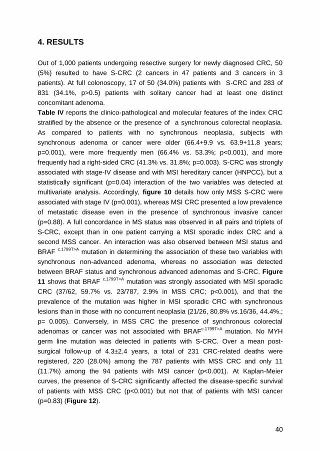
40
4. RESULTS
Out of 1,000 patients undergoing resective surgery for newly diagnosed CRC, 50
(5%) resulted to have S-CRC (2 cancers in 47 patients and 3 cancers in 3
patients). At full colonoscopy, 17 of 50 (34.0%) patients with S-CRC and 283 of
831 (34.1%, p>0.5) patients with solitary cancer had at least one distinct
concomitant adenoma.
Table IV reports the clinico-pathological and molecular features of the index CRC
stratified by the absence or the presence of a synchronous colorectal neoplasia.
As compared to patients with no synchronous neoplasia, subjects with
synchronous adenoma or cancer were older (66.4+9.9 vs. 63.9+11.8 years;
p=0.001), were more frequently men (66.4% vs. 53.3%; p<0.001), and more
frequently had a right-sided CRC (41.3% vs. 31.8%; p=0.003). S-CRC was strongly
associated with stage-IV disease and with MSI hereditary cancer (HNPCC), but a
statistically significant (p=0.04) interaction of the two variables was detected at
multivariate analysis. Accordingly, figure 10 details how only MSS S-CRC were
associated with stage IV (p=0.001), whereas MSI CRC presented a low prevalence
of metastatic disease even in the presence of synchronous invasive cancer
(p=0.88). A full concordance in MS status was observed in all pairs and triplets of
S-CRC, except than in one patient carrying a MSI sporadic index CRC and a
second MSS cancer. An interaction was also observed between MSI status and
BRAF c.1799T>A
mutation in determining the association of these two variables with
synchronous non-advanced adenoma, whereas no association was detected
between BRAF status and synchronous advanced adenomas and S-CRC. Figure
11 shows that BRAF c.1799T>A
mutation was strongly associated with MSI sporadic
CRC (37/62, 59.7% vs. 23/787, 2.9% in MSS CRC; p<0.001), and that the
prevalence of the mutation was higher in MSI sporadic CRC with synchronous
lesions than in those with no concurrent neoplasia (21/26, 80.8% vs.16/36, 44.4%.;
p= 0.005). Conversely, in MSS CRC the presence of synchronous colorectal
adenomas or cancer was not associated with BRAFc.1799T>A
mutation. No MYH
germ line mutation was detected in patients with S-CRC. Over a mean post-
surgical follow-up of 4.3±2.4 years, a total of 231 CRC-related deaths were
registered, 220 (28.0%) among the 787 patients with MSS CRC and only 11
(11.7%) among the 94 patients with MSI cancer (p<0.001). At Kaplan-Meier
curves, the presence of S-CRC significantly affected the disease-specific survival
of patients with MSS CRC (p<0.001) but not that of patients with MSI cancer
(p=0.83) (Figure 12).

41
Table IV. Demographics, Pathology and Molecular Features of CRC with Synchronous
Colorectal Neoplasia.
a “no adenoma” b “low-grade-dysplasia < 10mm tubular adenoma c 95 patients with low-grade-dysplasia adenoma (10 mm or greater in diameter or with villous component greater than 25%), and 59 with high-grade-dysplasia adenoma d Pathological and molecular characteristics of the most advanced cancer (“index” lesion, by pT) were to be inserted. Of 23 pairs with identical pT (no “index lesion assessable), 22 had fully concordant pathological and molecular features, whereas 1 pair with discordant tumor site was excluded from the analysis of this variable e not assessed in 48 cases (34, 6, 7, and 1 in the four subclasses, respectively) * at Fisher’s exact test Interactions at multivariate analysis (logistic regression): ♦ “Stage” * “MS status” , p=0.03 ● “BRAF status” * MS-status” (excluding HNPCC), p=0.04
Synchronous Colorectal Neoplasia
None a
Not Advanced
Adenoma b
Advanced
Adenoma c
Invasive
Cancer d
n=548, ref. n=177 P* n=106 P* n=50 P*
Age
years, mean ± SD 63.9±11.8 66.8±9.7 0.003 66.6±10.0 0.02 64.6±10.8 0.70
Gender
Male 292 (53.3) 115 (65.0) 77 (72.6) 29 (53.4)
Female 256 (46.7) 62 (35.0) 0.006 29 (27.4) <0.001 21 (46.6) 0.52
Site
Distal 374 (68.2) 101 (57.1) 64 (60.4) 30 (61.2)
Proximal 174 (31.8) 76 (42.9) 0.006 42 (39.6) 0.11 19 (38.8) 0.31
Stage
I-to-III 419 (76.5) 139 (78.5) 80 (75.5) 30 (60.0)
IV 129 (23.5) 38 (21.5) 0.57 26 (24.5) 0.83 20 (40.0) 0.01
Histotype
Adenoca. 506 (92.3) 162 (91.5) 100 (94.3) 48 (96.0)
Variant 42 ( 7.7) 15 (8.5) 0.73 6 (5.7) 0.47 2 (4.0) 0.34
Grade e
G1-G2 406 (79.3) 143 (83.6) 78 (77.2) 38 (77.6)
G3 106 (20.7) 28 (16.4) 0.22 23 (22.8) 0.64 11 (22.4) 0.77
Vein Invasion
No 423 (77.2) 140 (79.1) 82 (77.4) 36 (72.0)
Yes 125 (22.8) 37 (20.9) 0.60 24 (22.6) 0.97 14 (28.0) 0.40
MS Status
MSS 495 (90.3) 153 (86.4) 100 (94.3) 39 (78.0)
MSI-Sporadic 36 (6.6) 19 (10.7) 0.04 ●
5 (4.7) 0.44 2 (4.0) 0.64
HNPCC 17 (3.1) 5 (2.8) 0.92 1 (0.9) 0.20 9 (18.0) <0.001
BRAF
BRAF WT 516 (94.2) 158 (89.3) 100 (94.3) 47 (94.0)
BRAF c.1799T>A
32 (5.8) 19 (10.7) 0.02 ●
6 (5.7) 0.94 3 (6.0) 0.96

42
Figure 10 Interaction of CRC microsatellite-status and of synchronous colorectal
malignancy in determining the association of these two variables with TNM stage.
Stage-distribution of synchronous CRC is compared to that of single CRC, stratifying
by microsatellite-status. Stage IV was significantly associated with synchronous CRC
in patients with microsatellite-stable (MSS) cancer (19/39, 48.7% vs.186/748, 24.9%;
p=0.001). The frequency of stage IV disease was not different in MSS CRC patients
with no concomitant adenoma (124/495, 25.1%, ref.), with synchronous not advanced
adenoma (36/153, 23.5%; p=0.70), and with synchronous advanced adenoma (26/100;
26.0%; p=0.84). The frequency of stage IV disease, in patients with MSI CRC, was not
associated with S-CRC (7/83, 8.4% vs. 1/11, 9.1%; p=0.88).
* p values at Chi-square or Fisher’s exact test, as appropriate (stage IV vs. others)

43
Figure 11 Rate of BRAF c.1799T>A
mutation in microsatellite-stable (MSS) and –unstable
(MSI) sporadic CRC, by synchronous colorectal neoplasia. BRAF c.1799T>A
mutation
was significantly (p<0.001) more frequent in MSI-Sporadic (37/62, 59.7%) than in MSS
CRC (23/787, 2.9%). In MSS CRC, no association was found between the occurrence
of BRAF mutation in the index CRC and the presence of synchronous neoplasia.
Conversely, the mutation in MSI-sporadic CRC was less frequent in the absence of
synchronous neoplasia (16/36, 44.4%) than in a) any synchronous neoplasia (21/26,
80.8%, p=0.005), b) synchronous not-advanced adenoma (15/19, 78.9%, p=0.01), c)
synchronous advanced adenoma or CRC (6/7, 85.7%, p=0.05). HNPCC, which
invariably carry no BRAF mutation, were excluded from analysis. P values are from
Chi-square or Fisher’s exact test, as appropriate .

44
Figure 12 Disease-specific survival of patients with CRC by MS status and by
synchronous invasive cancer. Synchronous CRC significantly affected disease-
specific survival of patients with microsatellite-stable (MSS) cancer but not of those
with microsatellite-unstable (MSI) tumor (Kaplan-Meier curves, Log-rank test).

45
MSS CRC had a poorer prognosis also in the presence of a synchronous
advanced adenoma (p=0.02), but not in the presence of a not-advanced adenoma
(p=0.29) (Figure 13). The negative prognostic effect of S-CRC or synchronous
advanced adenoma was limited to MSS cancers with no BRAF c.1799T>A
mutation
(p<0.001), whereas BRAF-mutated MSS CRC had a much poorer outcome
independently of the presence of a synchronous advanced neoplasia (p=0.98)
(Figure 14). At Cox proportional hazard models (Table V), the presence of
synchronous advanced neoplasia was confirmed to be associated with a worse
outcome of MSS CRC (S-CRC: HR 2.66; 95% CI, 1.69-4.19; p<0.001;
synchronous advanced adenoma: HR 1.59; 95% CI, 1.11-2.26; p=0.01).
Figure 13 Disease-specific survival of patients with microsatellite-stable (MSS) CRC
stratified by synchronous colorectal neoplasia. The presence of synchronous
advanced neoplasia, but not that of synchronous not advanced adenoma, negatively
affected the survival of CRC patients, (Kaplan-Meier curves, Log-rank test).

46
Figure 14 Disease-specific survival of patients with microsatellite-stable (MSS) CRC
stratified by tumor BRAF-status and by synchronous advanced colorectal neoplasia.
The presence of synchronous advanced neoplasia negatively affected the survival of
patients with BRAF-WT CRC, whereas patients with BRAF-mutated CRC had a poorer
survival regardless of rarely accompanying synchronous neoplasia (Kaplan-Meier
curves, Log-rank test).

47
Table V Synchronous Advanced Colorectal Neoplasia and BRAF c.1799T>A
Mutation as Predictors of Death from CRC (Cox proportional-hazard models).
HR, Hazard ratios < 1.00 represent a decreased risk of death. whereas HR >1.00 represent an increased risk of death NA, not applicable * no HNPCC exhibited the BRAF
c.1799T>A mutation
Death Univariate Stage-adjusted
Multivariate
No Yes HR (95% C.I.) P HR (95% C.I.) P
A. MSS CRC
Synchronous Advanced Neoplasia
None 487 161 1.00 Ref. 1.00 Ref.
Advanced Adenoma 62 38 1.59 (1.11-2.26) 0.01 1.81 (1.27-2.58) 0.001
Invasive Cancer 18 21 2.66 (1.69-4.19) < 0.001 1.82 (1.15-2.87) 0.01
BRAF-Status
WT 558 206 1.00 Ref. 1.00 Ref.
BRAF c.1799T>A
9 14 3.29 (1.91-5.70) < 0.001 2.16 (1.25-3.73) 0.01
B. MSI CRC
Synchronous Advanced Neoplasia
None 67 10 1.00 Ref.
Advanced Adenoma 6 0 NA 0.45
Invasive Cancer 10 1 0.74 (0.09-5.75) 0.77
BRAF-Status by Sporadic/HNPCC *
BRAF WT - Sporadic CRC 23 2 1.00 Ref.
BRAF WT - HNPCC 30 2 0.75 (0.11-5.33) 0.77
BRAF c.1799T>A
- Sporadic CRC 30 7 2.68 (0.55-12.9) 0.22

48
Given the higher prevalence of stage IV disease in patients with MSS S-CRC but
not in those with synchronous advanced adenoma (see Figure 10 and its legend),
the incremental risk of death associated with synchronous advanced adenoma (HR
1.81; 95% CI, 1.27-2.58; p=0.001) and that conferred by the presence of
synchronous invasive cancer (HR 1.82; 95% CI, 1.15-2.87; p=0.01) were almost
identical at stage-adjusted multivariate analysis. The occurrence of BRAF c.1799T>A
mutation in the index cancer also predicted a higher risk of death from MSS CRC,
independently of the presence of a synchronous advanced neoplasia and of TNM
stage (HR 2.16; 95%CI 1.25-3.73; p=0.01). On the contrary, neither the presence
of synchronous advanced neoplasia nor the BRAF-status of the tumor significantly
affected the disease-specific survival of patients with MSI CRC.

49
5. DISCUSSION
In this large, hospital-based study, patients with MSS CRC had a significantly
poorer outcome if originally diagnosed with a synchronous invasive cancer (S-
CRC) or even with a synchronous advanced adenoma. The finding is important in
that it contributes to a highly controversial issue generated by the fact that most
studies failed to recognize any association between S-CRC and poor prognosis
[85-86, 91, 95], whereas the only prospective study reported a higher mortality in
patients with multiple primary cancers [94]. Notably, our study is unique in having
investigated the prognostic role of S-CRC in molecularly defined subgroups of
CRC, so to avoid the confounding effect of MSI cancers which more likely develop
synchronous malignancies but also have an overall better prognosis. In addition,
BRAF c.1799T>A
mutation, which is an established marker of CpG island methylation
and of poor prognosis [68, 127], was not associated with MSS S-CRC, indicating
that neither the occurrence nor the outcome of chromosomal-unstable synchronous
cancers are likely due to an epigenetic field effect. Several studies have
documented the association between MSI and S-CRC [94, 102, 128]. The strong
concordance in MSI status among synchronous cancers also led to the concept
that, for genetic and/or environmental reasons, some individuals may be prone to
develop multiple cancers through the genetic pathway of microsatellite instability
secondary to widespread CpG island methylation and to silencing of the mismatch
repair gene MMR MLH1 [28]. This concept was mainly based on the assumption
that the majority of MSI S-CRC were sporadic tumors, as suggested by the
typically old age of patients with synchronous colorectal malignancies and by the
established association between MSI-sporadic tumors and older age [94, 129]. As
a matter of fact, no previous study addressing the issue of synchronous cancers,
systematically screened patients with MSI S-CCR for germ-line mutations in MMR
genes. Therefore, it was a novel, and somehow unexpected finding of our series to
see that HNPCC largely accounted for MSI S-CRC (9 of 11, 82%) and that about
1/5 of all S-CRC were diagnosed in patients with hereditary cancer. Consistently,
BRAF c.1799T>A
mutation was associated only with sporadic MSI CRC, while no
mutation was detected in any HNPCC. Overall, data cannot exclude the existence
of an epigenetic field effect favoring the development of multiple neoplasia in
patients with sporadic MSI CRC, but certainly contradict the idea that this
mechanism may account for most synchronous MSI cancers. Rather, our results
confirm the appropriateness of the Bethesda criteria which recommend MSI testing
of CRC in the presence of multiple primary tumors [130].

50
The interaction between MS status and advanced stage in their association with S-
CRC indicated the need to analyze separately the prognosis of MSI and MSS S-
CRC. The analysis revealed that the prognosis of MSI cancers was not affected by
any concurrent neoplasia, whereas MSS CRS had a significantly poorer outcome if
S-CRC, or even a synchronous advanced adenoma, had been diagnosed.
Interestingly, at stage-adjusted analysis, the negative prognostic effect of S-CRC
equaled that of synchronous advanced adenoma, indicating that the worsened
prognosis of S-CRC likely reflects a more aggressive biological behavior shared
by pre-invasive synchronous lesions, rather than a larger cancer burden. This
concept is consistent with the well recognized value of S-CRC, as well as of
synchronous advanced adenomas, in predicting the future development of
metachronous colorectal neoplasia [131-132]. Of note, the presence of
synchronous advanced neoplasia and the rare BRAF c.1799T>A
mutation were both
independent predictors of poor prognosis for MSS CRC. Thus, the hypothesis that
an epigenetic field defect may predispose to synchronous neoplasia is plausible for
sporadic MSI S-CRC but not for MSS tumors which contribute the majority of
multiple primary colorectal malignancies [129]. In this respect, the study by Nosho
et al. may have failed to recognize the existing interactions of S-CRC with MSI and
BRAF c.1799T>A
mutations due to the small number of tumors fully characterized for
the MS/BRAF status [94]. Finally, our study failed to detect any MYH germline
mutation, not confirming the previously reported association between homozygous
or compound heterozygous mutations and S-CRC [133]. Since Cleary et al. found
such MYH mutations in less than 1% of the general population with CRC and in
about 6% of patients with S-CRC [133], the discrepancy might still reflect a type II
statistical error. Alternatively, we might have been more selective in excluding mild
polyposis syndromes from our colonoscopy-based clinical series.
Our study has the intrinsic limitation of being a case-control, retrospective analysis.
This may have altered the relative contribution of different molecular and clinical
subsets of CRC, but a bias in the selection of controls to S-CRC is unlikely, given
the consecutive series and the use of complete colonoscopy as the only criteria for
inclusion of patients with solitary CRC. Then, the correlations found between
synchronous neoplasia and prognosis in single molecular subsets can hardly be
interpreted as the result of selection artifacts. The analysis was also limited by the
use of BRAF c.1799T>A
mutation as the only marker of DNA methylation. Although the
BRAF status is validated as a reliable and reproducible marker of cancers with
methylator phenotype [68], we might have missed a few methylated tumors
potentially identifiable at analysis of multiple CpG islands.

51
However, we believe that the lack of association between MSS S-CRC and tumor
methylation status in our series cannot be disputed on the basis of this limitation.
In conclusion, this study raises the hypothesis that unidentified molecular features
may confer a greater aggressiveness to MSS CRC presenting with the phenotype
of multiple advanced lesions. If confirmed in large prospective studies, the
association between this subset of chromosomal-unstable cancers and poor
prognosis would have important implications for postsurgical endoscopic
surveillance and, possibly, for adjuvant therapeutic strategies.
Further studies, are necessary to better understand these unidentified molecular
features related to MSS S-CRC and CRC with multiple advanced lesions. Many
hypotheses have been formulated, but a clear sequence of molecular events that
mark the onset of MSS S-CRC and synchronous advanced neoplasia remain to be
defined. Recently, studies of whole genome sequencing open new scenarios on
the molecular interpretation of this so complex disease. The study of Palles and
colleagues shows that the germline mutations in two genes encoding the DNA
polymerase ε and polymerase δ predispose to multiple colorectal adenomas and
carcinoma [134]. Taken together, these observations could provide a molecular
explanation, at least partially, to the onset of MSS S-CRC and synchronous
advanced neoplasia, allowing us to read this kind of disease in a different way both
biologically and clinically.
52
6. BIBLIOGRAPHY
1. Siegel R., DeSantis C., Virgo K., Stein K., Mariotto A., Smith T., Cooper D., Gansler T., Lerro C., Fedewa S., Lin C., Leach C., Cannady R.S., Cho H., Scoppa S., Hachey M., Kirch R., Jemal A., Ward E., “Cancer treatment and survivorship statistics”. CA Cancer J Clin, Vol. 62, no. 4, 2012, pp. 220–241.
2. Meza R., Jeon J., Renehan A.G., Luebeck E.G., “Colorectal cancer incidence trends in the us and uk: evidence of right- to left-sided biological gradients with implications for screening”, Cancer Res, Vol. 70, no. 13, 2010, pp. 5419–5429.
3. Lieberman D., “Progress and challenges in colorectal cancer screening and surveillance”, Gastroenterology, Vol. 138, no. 6, 2010, pp. 2115–2126.
4. Rees G., Martin P.R., Macrae F.A., “Screening participation in individuals with a family history of colorectal cancer”, Eur J Cancer Care, Vol. 17,no. 3 2008, pp. 221–232.
5. Zlobec I., Lugli A., “Prognostic and predictive factors in colorectal cancer”, J Clin Pathol, Vol. 61, no. 5, 2008, pp. 561–569.
6. Burt R.W., “Colon cancer screening”, Gastroenterology, Vol. 119, no. 3, 2000, pp. 837–853.
7. Cunningham D., Atkin W., Lenz H.J., Lynch H.T., Minsky B., Nordlinger B., Starling N. “Colorectal cancer”, Lancet, Vol. 375, no. 9719, 2010, pp. 1030–1047.
8. Henry K.A., Niu X., Boscoe Francis P., “Geographic disparities in colorectal cancer survival”, Int J Health Geogr, Vol. 23, no. 8, 2009, pp.48.
9. Perdue D.G., Perkins C., Jackson-Thompson J., Coughlin S.S., Ahmed F., Haverkamp D.S., Jim M.A., “Regional differences in colorectal cancer incidence, stage and subsite among american indians and alaska natives 1999-2004”, Cancer, Vol. 113, no. 5suppl, 2008, pp.1179–1190.
10. Vogelstein Bert., Kinzler K.W., “The multistep nature of cancer”, Trends Genet, Vol. 9, no. 4, 1993, pp.138–141.
11. Carolyn C.C, David R.B, Julio Garcia-Aguilar, Scott H.K., Alexander O., Mary K.W., AJCC Cancer Staging Atlas, Springer, 2012.
12. Chapuis P.H., Chan C., Dent O.F., “Clinicopathological staging of colorectal cancer: Evolution and consensus-an Australian perspective”, J Gastroenterol Hepatol., Vol. 26, no. Suppl 1, 2011, pp. 58-64.
13. Ueno H., Mochizuki H., Akagi Y., Kusumi T., Yamada K., Ikegami M., Kawachi H., Kameoka S., Ohkura Y., Masaki T., Kushima R., Takahashi K., Ajioka Y., Hase K., Ochiai A., Wada R., Iwaya K., Shimazaki H., Nakamura T., Sugihara K., “Optimal colorectal cancer staging criteria in TNM classification”, J Clin Oncol., Vol. 30, no. 13, 2012, pp. 1519-26.

53
14. O'Connell J.B., Maggard M.A., Ko C.Y., “Colon cancer survival rates with the new American Joint Committee on Cancer sixth edition staging”, J Natl Cancer Inst., Vol. 96, no. 19, 2004, pp. 1420-5.
15. Pecori S., Capelli P., Vergine M., Manestrina F., Intestinal Polyps and Polyposis, Springer, 2009.
16. Grizzi F., Celesti G., Basso G., Laghi L., “Tumor budding as a potential histopathological biomarker in colorectal cancer: hype or hope?”, World J Gastroenterology, Vol. 18, no. 45, 2012, pp. 6532-6.
17. Kumar V., Abbas A.K., Fausto N., Aster J.C., Pathologic Basis of Disease, Elsevier Health Sciences, 2009.
18. Wise P.E., The ASCRS Textbook of Colon and Rectal Surgery, Springer, 2011.
19. Aust D.E., Baretton G.B., “Serrated polyps of the colon and rectum (hyperplastic polyps, sessile serrated adenomas, traditional serrated adenomas, and mixed polyps)-proposal for diagnostic criteria”, Virchows Arch., Vol. 457, no. 3:, 2010, pp. 291-7.
20. Pino M.S., Chung D.C., “The chromosomal instability pathway in colon cancer”, Gastroenterology. Vol.138, no 6, 2010, pp. 2059-72.
21. Takayama T., Ohi M., Hayashi T., Miyanishi K., Nobuoka A., Nakajima T., Satoh T., Takimoto R., Kato J., Sakamaki S., Niitsu Y., “Analysis of K-ras, APC, and beta-catenin in aberrant crypt foci in sporadic adenoma, cancer, and familial adenomatous polyposis”, Gastroenterology; Vol. 121, no 3, 2001, pp. 599–611.
22. Pretlow T.P., Pretlow T.G., “Mutant KRAS in aberrant crypt foci (ACF): initiation of colorectal cancer”, Biochim Biophys Acta, Vol. 1756, no. 2, 2005; pp. 83–96.
23. Pruitt K., Der C., “Ras and Rho regulation of the cell cycle and oncogenesis” Cancer Lett; Vol. 171, no. 1, 2001, pp. 1–10.
24. Takagi Y., Kohmura H., Futamura M., Kida H., Tanemura H., Shimokawa K., Saji S., “Somatic alterations of the DPC4 gene in human colorectal cancers in vivo”, Gastroenterology, Vol. 111, no.5, 1996, pp. 1369-72.
25. Takagi Y., Koumura H., Futamura M., Aoki S., Ymaguchi K., Kida H., Tanemura H., Shimokawa K., Saji S., “Somatic alterations of the SMAD-2 gene in human colorectal cancers”, Br J Cancer, Vol. 78, no. 9, 1998, pp. 1152-5.
26. Leslie A., Carey F.A., Pratt N.R., Steele R.J., “The colorectal adenoma-carcinoma sequence”, Br J Surg., Vol. 89, no. 7, 2002, pp. 845-60.
27. Vogelstein B., Kinzler K.W., “Cancer genes and the pathways they control”, Nat Med, Vol. 10, no. 8, 2004, pp. 789–799.
28. Menendez D., Inga A., Resnick M.A., “The expanding universe of p53 targets”, Nat Rev Cancer; Vol. 9, no. 10, 2009, pp. 724–737.

54
29. Boland C.R., Goel A., “Microsatellite Instability in Colorectal Cancer”, Gastroenterology, Vol. 138, no. 6, 2010, pp. 2073–2087.
30. Vilar E, Gruber SB., “Microsatellite instability in colorectal cancer-the stable evidence”, Nat Rev Clin Oncol., Vol. 7, no. 3, 2010, pp. 153-62.
31. Piard F., Chapusot C., Ecarnot-Laubriet A., Ponnelle T., Martin L., “Molecular markers of heterogeneity in colorectal cancers and adenomas”, Eur J Cancer Prev, Vol. 11, no. 1, 2002, pp. 85-97.
32. Li G.M., “Mechanisms and functions of DNA mismatch repair”, Cell Res., Vol. 18, no 1, 2008, pp. 85-98.
33. Suzuki M.M., Bird A., “DNA methylation landscapes: provocative insights from epigenomics”, Nat. Rev. Genet., Vol. 9, no. 6, 2008, pp. 465–476.
34. Feinberg A.P., Tycko B., “The history of cancer epigenetics”, Nat. Rev. Cancer, Vol. 4, no. 2, 2004, pp. 143–153.
35. Sharma S., Kelly T.K., Jones P.A., “Epigenetics in cancer”, Carcinogenesis, Vol. 31, 2010, pp. 27–36.
36. Lee J.K., Chan A.T., “Molecular Prognostic and Predictive Markers in Colorectal Cancer: Current Status”, Curr Colorectal Cancer Rep., Vol. 7, no. 2, 2011, pp. 136-144.
37. André T., Boni C., Navarro M., Tabernero J., Hickish T., Topham C., Bonetti A., Clingan P., Bridgewater J., Rivera F., de Gramont A., “Improved overall survival with oxaliplatin, fluorouracil, and leucovorin as adjuvant treatment in stage II or III colon cancer in the MOSAIC trial”, J Clin Oncol., Vol. 27, no. 19, 2009, pp. 3109-16.
38. Goldberg R.M,. “Therapy for metastatic colorectal cancer” Oncologist., Vol. 11, no. 9, 2006, pp. 981–7.
39. Bardelli A., Siena S., “Molecular mechanisms of resistance to cetuximab and panitumumab in colorectal cancer”, J Clin Oncol.,Vol. 28, no. 7, 2010, pp. 1254–61.
40. Di Nicolantonio F., Martini M., Molinari F., Sartore-Bianchi A., Arena S., Saletti P., De Dosso S., Mazzucchelli L., Frattini M., Siena S., Bardelli A., “Wild-type BRAF is required for response to panitumumab or cetuximab in metastatic colorectal cancer”, J Clin Oncol.; Vol. 26, no. 35, 2008, pp. 5705-12.
41. Watanabe T., Wu T.T., Catalano P.J., Ueki T., Satriano R., Haller D.G., Benson A.B. 3rd, Hamilton S.R., “Molecular predictors of survival after adjuvant chemotherapy for colon cancer”, N Engl J Med., Vol. 344, no. 16, 2001, pp. 1196-206.
42. Halling K.C., French A.J., McDonnell S.K., Burgart L.J., Schaid D.J., Peterson B.J., Moon-Tasson L., Mahoney M.R., Sargent D.J., O'Connell M.J., Witzig T.E., Farr G.H. Jr, Goldberg R.M., Thibodeau S.N., “Microsatellite instability and 8p allelic imbalance in stage B2 and C colorectal cancers”, J Natl Cancer Inst., Vol. 91, no. 15, 1999, pp. 1295-303.

55
43. Popat S., Zhao D., Chen Z., Pan H., Shao Y., Chandler I., Houlston R.S., “Relationship between chromosome 18q status and colorectal cancer prognosis: a prospective, blinded analysis of 280 patients”, Anticancer Res.;Vol. 27, no. 1B, 2007, pp. 627-33.
44. Ogino S., Nosho K., Irahara N., Shima K., Baba Y., Kirkner G.J., Meyerhardt J.A., Fuchs C.S., “Prognostic significance and molecular associations of 18q loss of heterozygosity: a cohort study of microsatellite stable colorectal cancers”, J Clin Oncol., Vol. 27, no. 27, 2009, pp. 4591-98.
45. Sinicrope F.A., Sargent D.J, “Microsatellite Instability in Colorectal Cancer: Prognostic, Predictive, and Therapeutic Implications”, Clinical Cancer Research, Vol. 18, no. 6, 2012, pp. 1506-12.
46. Al-Sohaily S., Biankin A., Leong R., Kohonen-Corish M., Warusavitarne J., “Molecular pathways in colorectal cancer”, J Gastroenterol Hepatol., Vol. 27, no. 9, 2012, pp. 1423-31.
47. Barrow E., Robinson L., Alduaij W., Shenton A., Clancy T., Lalloo F., Hill J., Evans D.G., “Cumulative lifetime incidence of extracolonic cancers in Lynch syndrome: a report of 121 families with proven mutations”, Clin Genet., Vol. 75, no. 2, 2009, pp. 141-49.
48. Jasperson K.W., Tuohy T.M., Neklason D.W., Burt R.W., “Hereditary and familial colon cancer”, Gastroenterology, Vol. 138, no. 6, 2010, pp. 2044-58.
49. Suraweera N., Duval A., Reperant M., Vaury C., Furlan D., Leroy K., Seruca R., Iacopetta B., Hamelin R., “Evaluation of tumor microsatellite instability using five quasimonomorphic mononucleotide repeats and pentaplex PCR”, Gastroenterology, Vol. 123, no. 6, 2002, pp. 1804-11.
50. Iacopetta B., Grieu F., Amanuel B., “Microsatellite instability in colorectal cancer”, Asia Pac J Clin Oncol., Vol. 6, no. 4, 2010, pp. 260-9.
51. Malesci A., Laghi L., Bianchi P., Delconte G., Randolph A., Torri V., Carnaghi C., Doci R., Rosati R., Montorsi M., Roncalli M., Gennari L., Santoro A., “Reduced likelihood of metastases in patients with microsatellite-unstable colorectal cancer”, Clin Cancer Res., Vol. 13, no. 13, 2007, pp. 3831-39.
52. Ribic C.M., Sargent D.J., Moore M.J., Thibodeau S.N., French A.J., Goldberg R.M., Hamilton S.R., Laurent-Puig P., Gryfe R., Shepherd L.E., Tu D., Redston M., Gallinger S., “Tumor microsatellite-instability status as a predictor of benefit from fluorouracil-based adjuvant chemotherapy for colon cancer”, N Engl J Med., Vol. 349, no. 3, 2003, pp. 247-57.
53. Popat S., Hubner R., Houlston R.S., “Systematic review of microsatellite instability and colorectal cancer prognosis”, J Clin Oncol., Vol. 23, no. 3, 2005, pp. 609–18.
54. Sargent D.J., Marsoni S., Monges G., Thibodeau S.N., Labianca R., Hamilton S.R., French A.J., Kabat B., Foster N.R., Torri V., Ribic C., Grothey A., Moore M., Zaniboni A., Seitz J.F., Sinicrope F., Gallinger S., “Defective mismatch repair as a predictive marker for lack of efficacy of fluorouracil-based adjuvant therapy in colon cancer”, J Clin Oncol., Vol. 28, no. 20, 2010, pp. 3219–26.

56
55. Bodmer W.F., Bailey C.J., Bodmer J., Bussey H.J., Ellis A., Gorman P., Lucibello F.C., Murday V.A., Rider S.H., Scambler P., et al., “Localization of the gene for familial adenomatous polyposis on chromosome 5”, Nature, Vol. 328, no. 6131, 1987, pp. 614-16.
56. Kinzler K.W., Nilbert M.C., Su L.K., Vogelstein B., Bryan T.M., Levy D.B., Smith K.J., Preisinger A.C., Hedge P., McKechnie D., et al., “Identification of FAP locus genes from chromosome 5q21”, Science, Vol. 253, no. 5020, 1991, pp. 661-65.
57. Groden J., Thliveris A., Samowitz W., Carlson M., Gelbert L., Albertsen H., Joslyn G., Stevens J., Spirio L., Robertson M., et al., “Identification and characterization of the familial adenomatous polyposis coli gene”, Cell, Vol. 66, no. 3, 1991, pp. 589-600.
58. Groen E.J., Roos A., Muntinghe F.L., Enting R.H., de Vries J., Kleibeuker J.H., Witjes M.J., Links T.P., van Beek A.P., “Extra-intestinal manifestations of familial adenomatous polyposis”, Ann Surg Oncol., Vol. 15, no. 9, 2008, pp. 2439-50.
59. Galiatsatos P., Foulkes W.D., “Familial adenomatous polyposis” Am.J.Gastroenterology, Vol.101, no. 2, 2006, pp. 385–98.
60. Power D.G., Gloglowski E., Lipkin S.M., “Clinical genetics of hereditary colorectal cancer” Hematol. Oncol. Clin. North Am., Vol. 24, no. 5, 2010, pp. 837–59.
61. Lipton L., Halford S.E., Johnson V., Novelli M.R., Jones A., Cummings C., Barclay E., Sieber O., Sadat A., Bisgaard M.L., Hodgson S.V., Aaltonen L.A., Thomas H.J., Tomlinson I.P., “Carcinogenesis in MYH-associated polyposis follows a distinct genetic pathway”, Cancer Res., Vol. 63, no. 22, 2003, pp. 7595–99.
62. Nielsen M., Franken P.F., Reinards T.H., Weiss M.M., Wagner A., van der Klift H., Kloosterman S., Houwing-Duistermaat J.J., Aalfs C.M., Ausems M.G., Bröcker-Vriends A.H., Gomez Garcia E.B., Hoogerbrugge N., Menko F.H., Sijmons R.H., Verhoef S., Kuipers E.J., Morreau H., Breuning M.H., Tops C.M., Wijnen J.T., Vasen H.F., Fodde R., Hes F.J., “Multiplicity in polyp count and extracolonic manifestations in 40 Dutch patients with MYH associated polyposis coli (MAP)”, J Med Genet., Vol. 42, no. 9, 2005, pp. e54.
63. Toyota M., Ahuja N., Ohe-Toyota M., Herman J.G, Baylin S.B., Issa J.P.J., “CpG island methylator phenotype in colorectal cancer”, Proceedings of the National Academy of Sciences of the United States of America, Vol. 96, no. 15, 1999, pp.8681–8686.
64. Boland C.R., Komarova N.L., Goel A., “Chromosomal instability and cancer: not just one CINgle mechanism”, Gut, Vol. 58, no. 2, 2009, pp. 163–164.
65. Samowitz W.S., Albertsen H., Herrick J., Levin T.R., Sweeney C., Murtaugh M.A., Wolff R.K., Slattery M.L., “Evaluation of a large, population-based sample supports a CpG island methylator phenotype in colon cancer” Gastroenterology, Vol. 129, no. 3, 2005, pp. 837–845.
57
66. Issa J.P.J., Shen L., Toyota M., “CIMP, at last”, Gastroenterology, Vol. 129, no. 3, 2005, pp. 1121–1124.
67. Curtin K., Slattery M.L., Samowitz W.S., “CpG island methylation in colorectal cancer: past, present and future”, Pathology Research International, Vol. 2011, 8 pages.
68. Weisenberger D.J., Siegmund K.D., Campan M., Young J., Long T.I., Faasse M.A., Kang G.H., Widschwendter M., Weener D., Buchanan D., Koh H., Simms L., Barker M., Leggett B., Levine J., Kim M., French A.J., Thibodeau S.N., Jass J., Haile R., Laird P.W., “CpG island methylator phenotype underlies sporadic microsatellite instability and is tightly associated with BRAF mutation in colorectal cancer”, Nature Genetics, Vol.38, no. 7, 2006, pp. 787-93.
69. Shen L., Toyota M., Kondo Y., Lin E., Zhang L., Guo Y., Hernandez N.S., Chen X., Ahmed S., Konishi K., Hamilton S.R., Issa J.P., “Integrated genetic and epigenetic analysis identifies three different subclasses of colon cancer”, Proc Natl Acad Sci U S A, Vol. 104, no. 47, 2007, pp. 18654-9.
70. Kambara T., Simms L.A., Whitehall V.L., Spring K.J., Wynter C.V., Walsh M.D., Barker M.A., Arnold S., McGivern A., Matsubara N., Tanaka N., Higuchi T., Young J., Jass J.R., Leggett B.A., BRAF mutation is associated with DNA methylation in serrated polyps and cancers of the colorectum”, Gut, Vol. 53, no. 8, 2004, pp. 1137-44.
71. Young J., Jenkins M., Parry S., Young B., Nancarrow D., English D., Giles G., Jass J., “Serrated pathway colorectal cancer in the population: genetic consideration”, Gut, Vol. 56, no 10, 2007, pp. 1453-9.
72. Jass JR., “Classification of colorectal cancer based on correlation of clinical, morphological and molecular features”, Histopathology, Vol. 50, no.1, 2007, pp. 113–30.
73. Ogino S., Nosho K., Kirkner G.J., Kawasaki T., Meyerhardt J.A., Loda M., Giovannucci E.L., Fuchs C.S., “CpG island methylator phenotype, microsatellite instability, BRAF mutation and clinical outcome in colon cancer”, Gut; Vol. 58, no. 1, 2009, pp. 90-6.
74. VanRijnsoever M., Elsaleh H., Joseph D., McCaul K., Iacopetta B., “CpG island methylator phenotype is an independent predictor of survival benefit from 5-fluorouracil in stage III colorectal cancer” Clin Cancer Res., Vol.9, no. 8, 2003, pp. 2898–903.
75. Shen L,. Catalano P.J., BensonA.B.3rd
., O'Dwyer P., Hamilton S.R., Issa J.P., “Association between DNA methylation and shortened survival in patients with advanced colorectal cancer treated with 5-fluorouracil based chemotherapy” Clin Cancer Res., Vol.13, no. 20, 2007, pp. 6093–8.

58
76. Jover R., Nguyen T.P., Pérez-Carbonell L., Zapater P., Payá A., Alenda C., Rojas E., Cubiella J., Balaguer F., Morillas J.D., Clofent J., Bujanda L., Reñé J.M., Bessa X., Xicola R.M., Nicolás-Pérez D., Castells A., Andreu M., Llor X., Boland C.R., Goel A., “5-Fluorouracil adjuvant chemotherapy does not increase survival in patients with CpG island methylator phenotype colorectal cancer”, Gastroenterology, Vol. 140, no. 4, 2011, pp. 1174-81.
77. Leggett B., Whitehall V., “Role of the serrated pathway in colorectal cancer pathogenesis”, Gastroenterology, Vol. 138, no. 6, 2010, pp. 2088-100.
78. Farris A.B., Misdraji J., Srivastava A., Muzikansky A., Deshpande V., Lauwers G.Y., Mino-Kenudson M., “Sessile serrated adenoma: challenging discrimination from other serrated colonic polyps”, Am J Surg Pathol., Vol. 32, no. 1, 2008, pp. 30-5.
79. Yang S., Farraye F.A., Mack C., Posnik O., O'Brien M.J., “BRAF and KRAS Mutations in hyperplastic polyps and serrated adenomas of the colorectum: relationship to histology and CpG island methylation status”, Am J Surg Pathol., Vol. 28, no. 11, 2004, pp. 1452-9.
80. Spring K.J., Zhao Z.Z., Karamatic R., Walsh M.D., Whitehall V.L,. Pike T., Simms L.A., Young J., James M., Montgomery G.W., Appleyard M., Hewett D., Togashi K., Jass J.R., Leggett B.A., “High prevalence of sessile serrated adenomas with BRAF mutations: a prospective study of patients undergoing colonoscopy”, Gastroenterology, Vol. 131, no. 5, 2006, pp. 1400-7.
81. O'Brien M.J., Yang S., Mack C., Xu H., Huang C.S., Mulcahy E., Amorosino M., Farraye F.A., “Comparison of microsatellite instability, CpG island methylation phenotype, BRAF and KRAS status in serrated polyps and traditional adenomas indicates separate pathways to distinct colorectal carcinoma end points”, Am J Surg Pathol., Vol. 30, no. 12, 2006, pp. 1491-501.
82. Moertel C.G., Bargen J.A., Dockerty M.B., “Multiple carcinomas of the large intestine. a review of the literature and a study of 261 cases” Gastroenterology, Vol. 34, no. 1, 1958, pp. 85–98.
83. Takeuchi H., Toda T., Nagasaki S., Kawano T., Minamisono Y., Maehara Y., Sugimachi K., “Synchronous multiple colorectal adenocarcinomas”, J Surg Oncol., Vol. 64, no. 4, 1997, pp. 304-7.
84. Oya M., Takahashi S., Okuyama T., Yamaguchi M., Ueda Y., “Synchronous colorectal carcinoma: clinico-pathological features and prognosis” Jpn J Clin Oncol., Vol. 33, no. 1, 2003, pp. 38–43.
85. Papadopoulos V., Michalopoulos A., Basdanis G., Papapolychroniadis K., Paramythiotis D., Fotiadis P., Berovalis P., Harlaftis N., “Synchronous and metachronous colorectal carcinoma” Tech Coloproctol, Vol. 8 (suppl 1), 2004, pp. 97–100.
86. Chen H.S., Sheen-Chen S.M., “Synchronous and ”early” metachronous colorectal adeonocarcinoma - analysis of prognosis and current trends”, Dis Colon Rectum., Vol. 43, no. 8, 2000, pp. 1093-9.

59
87. Kim M.S., Park Y.J., “Detection and treatment of synchronous lesions in colorectal cancer: the clinical implication of perioperative colonoscopy”, World J Gastroenterology; Vol. 13, no. 30, 2007, pp. 4108–4111.
88. McArthur D.R., Mehrzad H., Patel R., Dadds J., Pallan A., Karandikar S.S., Roy-Choudhury S., “CT colonography for synchronous colorectal lesions in patients with colorectal cancer: initial experience”, Eur Radiol, Vol. 20, no. 3, 2010, pp. 621– 629.
89. Achiam M.P., Holst Andersen L.P., Klein M.,Chabanova E., Thomsen H.S., Rosenberg J., “Preoperative evaluation of synchronous colorectal cancer using MR colonography”, .Acad Radiol; Vol. 16, no. 7, 2009, pp. 790–797.
90. Kinner S., Antoch G., Bockisch A., Veit-Haibach P. “Whole-body PET/CT-colonography: a possible new concept for colorectal cancer staging”, Abdom Imaging; Vol. 32, no. 5, 2007, pp. 606–612.
91. Latournerie M., Jooste V., Cottet V., Lepage C., Faivre J., Bouvier A.M., “Epidemiology and prognosis of synchronous colorectal cancers” Br J Surg, Vol. 95, no 12, 2008, pp. 1528–33.
92. Rex D.K., Kahi C.J., Levin B., Smith R.A., Bond J.H., Brooks D., Burt R.W., Byers T., Fletcher R.H., Hyman N., Johnson D., Kirk L., Lieberman D.A,. Levin T.R., O'Brien M.J., Simmang C., Thorson A.G., Winawer S.J.; American Cancer Society; US Multi-Society Task Force on Colorectal Cancer, “Guidelines for colonoscopy surveillance after cancer resection: a consensus update by the American Cancer Society and the US Multi-Society Task Force on Colorectal Cancer”, Gastroenterology, Vol. 130, no 6, 2006, pp. 1865–71.
93. Bae J.M., Cho N.Y., Kim T.Y., Kang G.H., “Clinicopathologic and molecular characteristics of synchronous colorectal cancers: heterogeneity of clinical outcome depending on microsatellite instability status of individual tumors”, Dis Colon Rectum, Vol. 55, no. 2, 2012, pp. 181-90.
94. Nosho K., Kure S., Irahara N., Shima K., Baba Y., Spiegelman D., Meyerhardt J.A., Giovannucci E.L., Fuchs C.S., Ogino S., “A prospective cohort study shows unique epigenetic, genetic, and prognostic features of synchronous colorectal cancers”, Gastroenterology, Vol. 137, no 5, 2009, pp. 1609-20.
95. Passman M.A., Pommier R.F., Vetto J.T., “Synchronous colon primaries have the same prognosis as solitary colon cancers”, Dis Colon Rectum, Vol. 39, no. 3, 1996, pp. 329-34.
96. Mulder S.A., Kranse R., Damhuis R.A., de Wilt J.H., Ouwendijk R.J., Kuipers E.J., van Leerdam M.E., “Prevalence and prognosis of synchronous colorectal cancer: a Dutch population-based study”, Cancer Epidemiology, Vol. 35, no. 5, 2011, pp. 442-7.

60
97. Rodríguez-Moranta F., Castells A., Andreu M., Piñol V., Castellví-Bel S., Alenda C., Llor X., Xicola R.M., Jover R., Payá A., Bessa X., Balaguer F., Cubiella J., Argüello L., Morillas J.D., Bujanda L., Gastrointestinal Oncology Group of the Spanish Gastroenterological Association., “Clinical performance of original and revised Bethesda guidelines for the identification of MSH2/MLH1 gene carriers in patients with newly diagnosed colorectal cancer: proposal of a new and simpler set of recommendations”, Am J Gastroenterology, Vol. 101, no. 5, 2006, pp. 1104-11.
98. Wang H.Z., Huang X.F., Wang Y., Ji J.F., Gu J., “Clinical features, diagnosis, treatment and prognosis of multiple primary colorectal carcinoma” World J Gastroenterology, Vol. 10, no. 14, 2004, pp. 2136–9.
99. Evers B.M., Mullins R.J., Matthews T.H., Broghamer W.L., Polk Jr H.C., “Multiple adenocarcinomas of the colon and rectum. An analysis of incidences and current trends”, Dis Colon Rectum; Vol. 31, no. 7, 1988, pp. 518–22.
100. Yang J., Peng J.Y., Chen W., “Synchronous colorectal cancers: a review of clinical features, diagnosis, treatment, and prognosis”, Dig Surg., Vol. 28, no 5, 2011, pp. 379-85.
101. Piñol V., Andreu M., Castells A., Payá A., Bessa X., Jover R.; Gastrointestinal Oncology Group of the Spanish Gastroenterological Association., “Synchronous colorectal neoplasms in patients with colorectal cancer: predisposing individual and familial factors”, Dis Colon Rectum, Vol. 47, no. 7, 2004, pp. 1192-200.
102. Dykes S.L., Qui H., Rothenberger D.A., García-Aguilar J., “Evidence of a preferred molecular pathway in patients with synchronous colorectal cancer”, Cancer, Vol. 98, no. 1, 2003, pp. 48-54.
103. Ueno M., Muto T., Oya M., Ota H., Azekura K., Yamaguchi T., “Multiple primary cancer: an experience at the Cancer Institute Hospital with special reference to colorectal cancer”, Int J Clin Oncol., Vol. 8, no. 3, 2003, pp. 162-7.
104. Copeland E.M., Jones R.S., Miller L.D., “Multiple colon neoplasms. Prognostic and therapeutic implications”, Arch Surg.,Vol. 98, no. 2, 1969, pp. 141–143.
105. Enker W.E., Dragacevic S., “Multiple carcinomas of the large bowel: a natural experiment in etiology and pathogenesis”, Ann Surg., Vol. 187, no. 1, 1978, pp. 8–11.
106. Kimura T., Iwagaki H., Fuchimoto S., Hizuta A., Orita K., “Synchronous colorectal carcinomas”, Hepatogastroenterology, Vol. 41, no. 5, 1994, pp. 409–412.
107. Adloff M., Arnaud J.P., Bergamaschi R., Schloegel M., “Synchronous carcinoma of the colon and rectum: prognostic and therapeutic implications”, Am J Surg., Vol. 157, no. 3, 1989, pp. 299–302.
108. Nikoloudis N., Saliangas K., Economou A., Andreadis E., Siminou S., Manna I., Georgakis K., Chrissidis T., “Synchronous colorectal cancer”, Tech Coloproctol., Vol. 8, (suppl 1), 2004, s177–s179.

61
109. Chai H., Brown R.E., “Field Effect in Cancer–An Update”, Annals of Clinical & Laboratory Science, Vol. 39, no. 4, 2009, pp. 331–338.
110. Fearon E.R., Vogelstein B., “A genetic model for colorectal tumorigenesis”, Cell, Vol. 61, no. 5, 1990, pp. 759-767.
111. Baker S.J., Fearon E.R., Nigro J.M., Hamilton S.R., Preisinger A.C., Jessup J.M., vanTuinen P., Ledbetter D.H., Barker D.F., Nakamura Y., White R., Vogelstein B., “Chromosome 17 deletions and p53 gene mutations in colorectal carcinomas”, Science, Vol. 244, no. 4901, 1989, pp. 217-221.
112. Braakhuis B.J., Tabor M.P., Kummer J.A., Leemans C.R., Brakenhoff R.H., “A genetic explanation of Slaughter’s concept of field cancerization: evidence and clinical implications” Cancer Research, Vol. 63, no. 8, 2003, pp. 1727-1730.
113. Novak P., Jensen T.J., Garbe J.C., Stampfer M.R., Futscher B.W., “Stepwise DNA methylation changes are linked to escape from defined proliferation barriers and mammary epithelial cell immortalization”, Cancer Research, Vol. 69, no. 12, 2009, pp. 5251–5258.
114. Widschwendter M., Fiegl H., Egle D., Mueller-Holzner E., Spizzo G., Marth C., Weisenberger D.J, Campan M., Young J., Jacobs I., Laird P.W., “Epigenetic stem cell signature in cancer”, Nature Genetics, Vol. 39, no. 2, 2007, pp. 157–158.
115. Shen L., Kondo Y., Rosner G.L,. Xiao L., Hernandez N.S., Vilaythong J., Houlihan P.S., Krouse R.S., Prasad A.R., Einspahr J.G., Buckmeier J., Alberts D.S., Hamilton S.R., Issa J.P., “MGMT promoter methylation and field defect in sporadic colorectal cancer”, J. Natl Cancer Inst., Vol. 97, no. 18, 2005, pp. 1330–1338.
116. Ibrahim A.E., Arends M.J., Silva A.L., Wyllie A.H., Greger L., Ito Y., Vowler S.L., Huang T.H., Tavaré S., Murrell A., Brenton J.D., “Sequential DNA methylation changes are associated with DNMT3B overexpression in colorectal neoplastic progression”, Gut, Vol. 60, no. 4, 2011, pp. 499–508
117 Konishi K., Shen L., Jelinek J., Watanabe Y., Ahmed S., Kaneko K., Kogo M., Takano T., Imawari M., Hamilton S.R., Issa J.P., “Concordant DNA methylation in synchronous colorectal carcinomas” Cancer Prev. Res. (Phila.), Vol. 2, no. 9, 2009, pp. 814–822.
118. Johnson I.T., Belshaw N.J., “Environment, diet and CpG island methylation: epigenetic signals in gastrointestinal neoplasia”, Food Chem. Toxicol., Vol. 46, no. 4, 2008, pp. 1346–1359.
119. Ramírez N., Bandrés E., Navarro A., Pons A., Jansa S., Moreno I., Martínez-Rodenas F., Zárate R., Bitarte N., Monzó M., García-Foncillas J., “Epigenetic events in normal colonic mucosa surrounding colorectal cancer lesions”, Eur. J. Cancer, Vol. 44, no. 17, 2008, pp. 2689–2695.

62
120. Hiraoka S., Kato J., Horii J., Saito S., Harada K., Fujita H., Kuriyama M., Takemoto K., Uraoka T., Yamamoto K., “Methylation status of normal background mucosa is correlated with occurrence and development of neoplasia in the distal colon”, Human Pathology, Vol. 41, no. 1, 2010, pp. 38–47.
121. Suzuki H., Watkins D.N., Jair K.W., Schuebel K.E., Markowitz S.D., Chen W.D., Pretlow T.P., Yang B., Akiyama Y., Van Engeland M., Toyota M., Tokino T., Hinoda Y., Imai K., Herman J.G., Baylin S.B., “Epigenetic inactivation of SFRP genes allows constitutive WNT signaling in colorectal cancer”, Nature Genetics, Vol. 36, no. 4, 2004, pp. 417–422.
122. Belshaw N.J., Elliott G.O., Foxall R.J., Dainty J.R., Pal N., Coupe A., Garg D., Bradburn D.M., Mathers J.C., Johnson I.T., “Profiling CpG island field methylation in both morphologically normal and neoplastic human colonic mucosa”, Br. J. Cancer, Vol. 99, no.1, 2008, pp. 136–142.
123. Worthley D.L., Whitehall V.L., Buttenshaw R.L., Irahara N., Greco S.A., Ramsnes I., Mallitt K.A., Le Leu R.K., Winter J., Hu Y., Ogino S., Young G.P., Leggett B.A., “DNA methylation within the normal colorectal mucosa is associated with pathway-specific predisposition to cancer”, Oncogene, Vol. 29, no. 11, 2010, pp. 1653–1662.
124. Limsui D., Vierkant R.A., Tillmans L.S., Wang A.H., Weisenberger D.J., Laird P.W., Lynch C.F., Anderson K.E., French A.J,. Haile R.W., Harnack L.J., Potter J.D., Slager S.L., Smyrk T.C., Thibodeau S.N., Cerhan J.R., Limburg P.J., “Cigarette smoking and colorectal cancer risk by molecularly defined subtypes”, J. Natl Cancer Inst., Vol. 102, no. 14, 2010, pp. 1012–1022.
125. Worthley D.L,. Whitehall V.L., Le Leu R.K., Irahara N., Buttenshaw R.L., Mallitt K.A., Greco S.A., Ramsnes I., Winter J., Hu Y., Ogino S., Young G.P., Leggett B.A., “DNA methylation in the rectal mucosa is associated with crypt proliferation and fecal short-chain fatty acids”, Dig. Dis. Sci., Vol. 56, no. 2, 2011, pp. 387–396
126. Shannon B., Gnanasampanthan S., Beilby J. Iacopetta, B., “A polymorphism in the methylenetetrahydrofolate reductase gene predisposes to colorectal cancers with microsatellite instability”, Gut, Vol. 50, no. 4, 2002, pp. 520–524.
127. Samowitz W.S., Sweeney C., Herrick J., Albertsen H., Levin T.R., Murtaugh M.A., Wolff R.K., Slattery M.L., “Poor survival associated with the BRAF V600E mutation in microsatellite-stable colon cancers”, Cancer Research, Vol. 65, no. 14, 2005, pp. 6063-9.
128. Pedroni M., Tamassia M.G., Percesepe A., Roncucci L., Benatti P., Lanza G. Jr,. Gafà R., Di Gregorio C., Fante R., Losi L., Gallinari L., Scorcioni F., Vaccina F., Rossi G., Cesinaro A.M., Ponz de Leon M., “Microsatellite instability in multiple colorectal tumors”, Int J Cancer, Vol. 81, no. 1, 1999, pp. 81:1-5.
129. Leggett B.A., Worthley D.L., “Synchronous colorectal cancer: not just bad luck?”, Gastroenterology, Vol. 137, no. 5, 2009, pp. 1559-62.

63
130. Umar A., Boland C.R., Terdiman J.P., Syngal S., de la Chapelle A., Rüschoff J., Fishel R., Lindor N.M., Burgart L.J., Hamelin R., Hamilton S.R., Hiatt R.A., Jass J., Lindblom A., Lynch H.T., Peltomaki P., Ramsey S.D., Rodriguez-Bigas M.A., Vasen H.F., Hawk E.T., Barrett J.C., Freedman A.N., Srivastava S., “Revised Bethesda Guidelines for hereditary nonpolyposis colorectal cancer (Lynch syndrome) and microsatellite instability”, J Natl Cancer Inst., Vol. 96, no. 4, 2004, pp. 261-8.
131. Moon C.M., Cheon J.H., Choi E.H., Kim E.S., Park J.J., Han S.Y., Kim D.H., Kim T.I,. Kim W.H., “Advanced synchronous adenoma but not simple adenoma predicts the future development of metachronous neoplasia in patients with resected colorectal cancer”, J Clin Gastroenterology, Vol. 44, no. 7, 2010, pp. 495-501.
132. Ballesté B., Bessa X., Piñol V., Castellví-Bel S., Castells A., Alenda C., Paya A., Jover R., Xicola R.M., Pons E., Llor X., Cordero C., Fernandez-Bañares F., de Castro L., Reñé J.M., Andreu M.; Gastrointestinal Oncology Group of the Spanish Gastroenterological Association., “Detection of metachronous neoplasms in colorectal cancer patients: identification of risk factors”, Dis Colon Rectum, Vol. 50, no. 7, 2007, pp. 971-80.
133. Cleary S.P., Cotterchio M., Jenkins M.A., Kim H., Bristow R., Green R., Haile R., Hopper J.L., LeMarchand L., Lindor N., Parfrey P., Potter J., Younghusband B., Gallinger S., “Germline MutY human homologue mutations and colorectal cancer: a multisite case-control study”, Gastroenterology, Vol. 136, no. 4, 2009, pp. 1251-60.
134. Palles C., Cazier J.B., Howarth K.M., Domingo E., Jones A.M., Broderick P., Kemp Z., Spain S.L., Guarino E., Salguero I., Sherborne A., Chubb D., Carvajal-Carmona L.G., Ma Y., Kaur K., Dobbins S., Barclay E., Gorman M., Martin L., Kovac M.B., Humphray S.; CORGI Consortium; WGS500 Consortium, Lucassen A., Holmes C.C., Bentley D., Donnelly P., Taylor J., Petridis C., Roylance R., Sawyer E.J., Kerr D.J., Clark S., Grimes J., Kearsey S.E., Thomas H.J., McVean G., Houlston R.S., Tomlinson I., “Germline mutations affecting the proofreading domains of POLE and POLD1 predispose to colorectal adenomas and carcinomas”, Nature Genetics, Vol. 45, no. 2, 2013, pp. 136-44.

64
7 SCIENTIFIC PRODUCTS
Presence of Twist1-Positive Neoplastic Cells in the Stroma of Chromosome-
Unstable Colorectal Tumors.
Celesti G, Di Caro G, Bianchi P, Grizzi F, Basso G, Marchesi F, Doni A, Marra G,
Roncalli M, Mantovani A, Malesci A, Laghi L.
Gastroenterology, 2013 Article in Press
Tumor budding as a potential histopathological biomarker in colorectal cancer:
hype or hope?
Grizzi F, Celesti G, Basso G, Laghi L.
World J Gastroenterol., Vol. 18, no. 45, 2012, pp. 6532-6
Irrelevance of microsatellite instability in the epidemiology of sporadic pancreatic
ductal adenocarcinoma.
Laghi L, Beghelli S, Spinelli A, Bianchi P, Basso G, Di Caro G, Brecht A, Celesti G,
Turri G, Bersani S, Schumacher G, Röcken C, Gräntzdörffer I, Roncalli M, Zerbi A,
Neuhaus P, Bassi C, Montorsi M, Scarpa A, Malesci A.
PLoS One, Vol. 7, no. 9, 2012, pp. e46002.