university of dundee accuracy of dental identification of
TRANSCRIPT

University of Dundee
Accuracy of dental identification of individuals with unrestored permanent teeth byvisual comparison with radiographs of mixed dentitionGorza, Ludovica; Manica, Scheila
Published in:Forensic Science International
DOI:10.1016/j.forsciint.2018.06.004
Publication date:2018
Licence:CC BY-NC-ND
Document VersionPeer reviewed version
Link to publication in Discovery Research Portal
Citation for published version (APA):Gorza, L., & Manica, S. (2018). Accuracy of dental identification of individuals with unrestored permanent teethby visual comparison with radiographs of mixed dentition. Forensic Science International, 289, 337-343.https://doi.org/10.1016/j.forsciint.2018.06.004
General rightsCopyright and moral rights for the publications made accessible in Discovery Research Portal are retained by the authors and/or othercopyright owners and it is a condition of accessing publications that users recognise and abide by the legal requirements associated withthese rights.
• Users may download and print one copy of any publication from Discovery Research Portal for the purpose of private study or research. • You may not further distribute the material or use it for any profit-making activity or commercial gain. • You may freely distribute the URL identifying the publication in the public portal.
Take down policyIf you believe that this document breaches copyright please contact us providing details, and we will remove access to the work immediatelyand investigate your claim.
Download date: 11. Jan. 2022

Elsevier Editorial System(tm) for Forensic
Science International
Manuscript Draft
Manuscript Number: FSI-D-17-00868R1
Title: Accuracy of dental identification of individuals with unrestored
permanent teeth by visual comparison with radiographs of mixed dentition
Article Type: Original Research Article
Keywords: Dental Radiographs; Forensic Odontology; Forensic
Identification; Mixed Dentition; Unrestored Teeth.
Corresponding Author: Dr. Ludovica Gorza,
Corresponding Author's Institution: na
First Author: Ludovica Gorza
Order of Authors: Ludovica Gorza; Scheila Manica, PhD
Abstract: Forensic dentistry plays a major role in human identification.
Teeth carry individual characteristics that differ among different
individuals. Dental radiographs depict reality objectively, being the
most reliable tool for dental identification. The first aim of this study
was to evaluate the accuracy of dental identification of individuals with
permanent unrestored teeth by visual comparison with radiographs of mixed
dentition. The second aim was to learn which anatomical features were
compared by examiners with different backgrounds. A total of 19 forensic
experts participated in a web-based questionnaire to assess
identification of 12 simulated cases; each case required the radiographic
comparison of 1 dental PM radiograph to 3 dental AM radiographs, of which
only one was the correct match. The examiners were given four options
following the ABFO guidelines: established identification, possible
identification, insufficient data and exclusion; the participants also
explained the reason for each of their conclusions. The accuracy of the
methodology was 75,4%, the sensitivity was 53,5% and the specificity was
86,4%. Overall, there was a tendency of the observers to overlook non-
dental characteristics. Not surprisingly, dental identification by visual
comparison of radiographs was not immune to subjectivity and, even
analysing the same category of features, different conclusions and
consequently different percentages of accuracy were reached. When
matching the correct AM radiograph, most examiners compared the root
morphology of the first molars and the shape of the maxillary sinus. When
one of the AM radiographs was not matched, the examiners mostly asserted
that there was insufficient data to reach a conclusion due to the lack of
distinctive and comparable features. With AM and PM radiographs showing
different development stages, accuracy was correlated to the age of the
AM radiograph.
1
© 2018. This manuscript version is made available under the CC-BY-NC-ND 4.0 license http://creativecommons.org/licenses/by-nc-nd/4.0/

Acknowledgments
Acknowledgements
Prof. Mark Hector, who patiently revised my dissertation and suggested further observations in the
discussion. Enrico Semenzato, Kevin Steffan, Francesco Fistarol and Adonella Carli, who provided the
radiographs to be used as samples. Dr. Gavin Revie, who contributed to the quantitative and
qualitative analysis of the data. Dr. Douglas Sheasby and Prof. Svend Richter who suggested useful
amendments and all the volunteers who accepted to participate in the research.
2

Title Page (with authors and addresses)
Accuracy of dental identification of individuals with unrestored permanent teeth by visual
comparison with radiographs of mixed dentition Ludovica Gorza*a, Scheila Mânicaa
a Centre for Forensic and Legal Medicine, School of Dentistry, University of Dundee, 3 Cross Row,
Dundee, DD1 5HG UK
*Corresponding author. [email protected] Tel.: + 393454051233
3

*Highlights (for review)
Highlights
Accuracy was moderately and positively correlated to the age of the AM radiograph.
It was easier to exclude the incorrect radiograph than identify the correct one.
Even when comparing the same features, examiners reached different conclusions.
The morphology of the roots of the molars was frequently compared.
Non-dental features were frequently overlooked.
4

*Manuscript (without author details) Click here to view linked References
Accuracy of dental identification of individuals with unrestored permanent teeth by visual
comparison with radiographs of mixed dentition
ABSTRACT
Forensic dentistry plays a major role in human identification. Teeth carry individual characteristics
that differ among different individuals. Dental radiographs depict reality objectively, being the most
reliable tool for dental identification. The first aim of this study was to evaluate the accuracy of dental
identification of individuals with permanent unrestored teeth by visual comparison with radiographs
of mixed dentition. The second aim was to learn which anatomical features were compared by
examiners with different backgrounds. A total of 19 forensic experts participated in a web-based
questionnaire to assess identification of 12 simulated cases; each case required the radiographic
comparison of 1 dental PM radiograph to 3 dental AM radiographs, of which only one was the correct
match. The examiners were given four options following the ABFO guidelines: established
identification, possible identification, insufficient data and exclusion; the participants also explained
the reason for each of their conclusions. The accuracy of the methodology was 75,4%, the sensitivity
was 53,5% and the specificity was 86,4%. Overall, there was a tendency of the observers to overlook
non-dental characteristics. Not surprisingly, dental identification by visual comparison of radiographs
was not immune to subjectivity and, even analysing the same category of features, different
conclusions and consequently different percentages of accuracy were reached. When matching the
correct AM radiograph, most examiners compared the root morphology of the first molars and the
shape of the maxillary sinus. When one of the AM radiographs was not matched, the examiners mostly
asserted that there was insufficient data to reach a conclusion due to the lack of distinctive and
comparable features. With AM and PM radiographs showing different development stages, accuracy
was correlated to the age of the AM radiograph.
Keywords: Dental Radiographs; Forensic Odontology; Forensic Identification; Mixed Dentition;
Unrestored Teeth.
1. Introduction
The identification of the living and of the dead is a human right to be guaranteed for ethical, cultural,
religious and economic reasons. Not less important, it contributes to criminal investigations in case of
violent and suspicious deaths. Teeth are primary identifiers and could lead to the certain identification
or exclusion of an individual without the aid of additional factors [1]. Identification by dental means is
one of the fields of expertise of a forensic dentist (FD) and it is useful in singles cases as well as in mass
disasters, when a significant number of bodies are recovered at the same time [2]. The pattern and
combination of dental treatments, anatomic and pathologic features are hardly similar between
5

different subjects [3,4]. Identification is conducted by comparing the post-mortem (PM) dental data
collected during the autopsy to the ante-mortem (AM) dental records of alleged matches [1]. Intra-oral
and extra-oral dental radiographs are often the key to this process, by objectively displaying anatomic
and pathologic features that are not visible to the naked eye by external examination [5,6]. Visual
comparison is the most inexpensive and commonly adopted method for the analysis of traditional
films or digital radiographs for identification purposes. However, there are no standardized protocols
and the final conclusion is susceptible to the personal interpretation of the operator, who might
confirm or exclude identity based on a single trait [7,8]. Any scientific method that aims to produce
evidence with medico-legal outcomes should follow the Daubert standard: be accepted by the
scientific community, be repeatable, standardized and be subjected to peer-review and publication
reporting an acceptable error rate [9]. Previous studies tested the accuracy of dental identification by
visual comparison of radiographs with unrestored dentitions within samples of similar age ranges
[10–12], whereas studies considering wide time intervals between AM and PM radiographs mostly
included restored permanent dentitions [13]. The first aim of the research was to test if manual
radiographic comparison is an accurate identification methodology when the PM radiograph depicted
permanent sound dentition, the AM and the PM radiographs were separated by a significant time-
lapse and the AM radiograph dated back to the age of mixed dentition. The second aim was to
investigate which anatomic features visible in panoramic radiographs were analysed by experts in
forensic identification.
2. Materials and methods
A total of 100 forensic dentists (FD), forensic anthropologists (FA) and radiologists (R) were contacted
via email to participate in a web-based survey. Volunteers were searched within University lecturers
and forensic international organizations (ABFO, ABFA, BAFO, IOFOS). The field of expertise and the
number of years of experience were self-reported in the survey; however, they were not asked to state
the level of qualification (i.e.: MS, PhD) and the number of forensic identifications performed
throughout their career. The radiographic database of three Italian private dental clinics were
scrutinized. A total of twelve panoramic radiographs (OPGs) depicting complete shedding, permanent
and unrestored dentition were selected as simulated PM radiographs; individuals who had received
and completed an orthodontic treatment or had a fixed orthodontic retainer visible only in the PM
radiograph were included; the OPGs of the same subjects showing mixed dentition with at least initial
eruption of the first molar or the central incisor worked as simulated AM radiographs; if the deciduous
teeth were decayed or filled but all permanent teeth were sound, the radiographs were included.
Individuals with oral manifestations of genetic syndromes, dental agenesis, malformations of cranio-
facial structures, cavities, fillings or other dental treatments of the permanent dentition and retained
primary teeth in PM radiographs were excluded. Twenty-four more OPGs of children with mixed
dentition and respecting the criteria were selected and worked as false matches or False Positive (FP).
6
The time-lapse between the AM and PM radiographs was between 3 and 18 years. The age of the
individuals of the AM radiographs ranged between 8 and 13 years. The selected radiographs, if
traditional films, were photographed using a Nikon D90 camera (© 2017 Nikon Corporation) and
digitalized. The web-based survey was created on Google Forms (© 2015 Google Inc.) and was only
accessible by private invitation. The questionnaire included twelve cases, each one showing one PM
radiograph paired to three AM radiographs; all the radiographs were cut into halves at the midline of
both dental arches using Microsoft Foto Windows 10 (© 2017 Microsoft Corporation); consequently,
six cases showed the right side and six the left side of the original radiographs. The examiners were
not informed that only one AM radiograph in each case belonged to the same individual as the PM.
Because the first aim of the study was to test the accuracy of dental identification by visual comparison
of radiographs alone, no extra medical or dental information about the subjects was provided. Each
section included one multiple-choice question and one open-ended question for each AM radiograph;
the former asked to reach a conclusion of identification or exclusion providing four options, according
to the ABFO guidelines (established identification, possible identification, insufficient data, exclusion)
[14]; the latter asked to explain the reason for the conclusion with no word-limit or directions of any
kind. Radiographs of each section were available for download, zooming or digital enhancement by
accessing an on-line folder (© Dropbox Inc). The examiners could not proceed further before filling all
the questions within each case. The Author who collected the radiographs and prepared the
questionnaire did not participate. The acquisition of the answers was automatic once all the sections
were completed and the results were immediately and exclusively visible to the Authors on an Excel
spreadsheet. The answers to the multiple-choice question were analysed quantitatively to calculate
the total number of correct identifications or True Positive (TP), correct exclusions or True Negative
(TN), incorrect identifications or False Positive (FP) and incorrect exclusions or False Negative (FN) of
each operator. The percentages of sensitivity, specificity, accuracy by examiner and by case were
calculated. The level of confidence of the examiners when answering to the multiple-choice questions
was also measured: the percentages of Established (E) and Positive (P) identifications were calculated
for both TP and FP; the percentages of Insufficient Data (IND) and Exclusions (X) were calculated for
both TN and FN. Additionally, a linear regression model was applied to investigate the correlation
between accuracy and AM-PM time lapse, age of the AM radiograph and experience of the examiners.
The answers to the open-ended questions were analysed qualitatively by thematic analysis to
investigate which features were compared by examiners according to their performance [15]; firstly,
the examiners were divided into four groups (1 to 4) according to the percentage of sensitivity and
specificity: the cut-off point chosen was 80%. Only one type of answer was analysed for each group of
examiners: explanations to the TP (Type A) from examiners with sensitivity equal to or higher than
80% (Group 1); explanations to the FN (Type B) from examiners with sensitivity lower than 80%
(Group 2); explanations to the TN (Type C) from examiners with specificity equal to or higher than
7

80% (Group 3); explanations to the TP (Type D) from examiners with specificity lower than 80%
(Group 4). Thematic analysis was performed on the four types of questions by searching for specific
keywords in the text. Two main categories were established. The first category included “dental
features”; subcategories I were “anatomy” and “number”, while sub-categories II were “type of tooth”
and “part of the tooth”. The second category was “non-dental features” and subcategory I was
“anatomy”, which collected all the responses quoting any cranio-facial structures other than teeth. It
was then calculated the percentage of times that the features from each category and sub-category
were mentioned.
3. Results
A total of 19 volunteers, 15 FD and 4 FA, accepted and completed the questionnaire; 3 FD and 2 FA
from the UK, 1 FA from USA, 4 FD from Canada, 4 FD from Brazil, 1 FD from Mexico, 1 FA from Italy, 1
FD from Iceland, 1 FD from Mauritius and 1 FD from Australia. The number of years of experience
ranged between 1 and 30 years (Table 1): only 2 out of 19 examiners (11%) had less than 2 years of
experience; 10 examiners (57%) had between 2 and 15 years; 6 examiners (31%) had at least 16
years, with one examiner (FD) practicing for 30 years.
3.1 Quantitative analysis of the multiple-choice question
3.1.1 Sensitivity, specificity, accuracy
Out of the 684 answers collected by 19 examiners, 122 were TP, 394 TN, 62 FP and 106 FN. Table 1
shows the percentages of TP, FN, TN, FP by examiner. Sensitivity, or the capability to identify the
correct matches, was obtained by the following formula TP/(TP+FN); specificity, or the capability to
detect the incorrect AM radiographs, corresponded to the percentage of TN out of the total
radiographs (TN+FP); accuracy was the estimation of the overall performance and corresponded to
the percentage of the correct answers (TP+TN) out of the total answers (TP+FN+TN+FP). The
sensitivity of the examiners fluctuated significantly between 0% and 100% and the accuracy varied
between 55,5% and 91,7%; the specificity was 33,3% for only one examiner and varied between
66,7% and 100% for the remaining participants (Table 1). The percentages of sensitivity, specificity
and accuracy in each case of the questionnaire are shown in Table 2: the sensitivity ranged between
26,3% and 78,9%, the specificity between 71,1% and 94,7% and the accuracy between 66,7% and
78,9%. Overall percentages of sensitivity, specificity and accuracy were the mean values obtained by
the examiners and were 53,5%, 86,4% and 75,4%, respectively (Table 3). Table 4 depicts the level of
confidence of the examiners when answering to the multiple-choice question; out of the total number
of TP, 41 (34%) were established (E) and 81 (66%) were possible (P) identifications; out of the total
number of FN, there were 20 (19%) exclusions (X) and 86 (81%) insufficient data (IND); out of the
total number of FP, there were 2 (3%) established (E) and 60 (97%) possible (P) identifications; out of
the total number of TN, 177 (45%) were exclusions (X) and 217 (55%) insufficient data (IND).
8
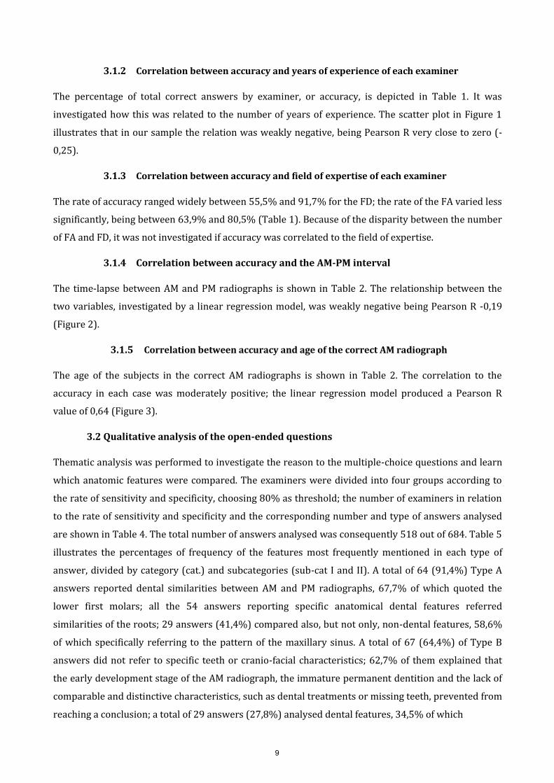
3.1.2 Correlation between accuracy and years of experience of each examiner
The percentage of total correct answers by examiner, or accuracy, is depicted in Table 1. It was
investigated how this was related to the number of years of experience. The scatter plot in Figure 1
illustrates that in our sample the relation was weakly negative, being Pearson R very close to zero (-
0,25).
3.1.3 Correlation between accuracy and field of expertise of each examiner
The rate of accuracy ranged widely between 55,5% and 91,7% for the FD; the rate of the FA varied less
significantly, being between 63,9% and 80,5% (Table 1). Because of the disparity between the number
of FA and FD, it was not investigated if accuracy was correlated to the field of expertise.
3.1.4 Correlation between accuracy and the AM-PM interval
The time-lapse between AM and PM radiographs is shown in Table 2. The relationship between the
two variables, investigated by a linear regression model, was weakly negative being Pearson R -0,19
(Figure 2).
3.1.5 Correlation between accuracy and age of the correct AM radiograph
The age of the subjects in the correct AM radiographs is shown in Table 2. The correlation to the
accuracy in each case was moderately positive; the linear regression model produced a Pearson R
value of 0,64 (Figure 3).
3.2 Qualitative analysis of the open-ended questions
Thematic analysis was performed to investigate the reason to the multiple-choice questions and learn
which anatomic features were compared. The examiners were divided into four groups according to
the rate of sensitivity and specificity, choosing 80% as threshold; the number of examiners in relation
to the rate of sensitivity and specificity and the corresponding number and type of answers analysed
are shown in Table 4. The total number of answers analysed was consequently 518 out of 684. Table 5
illustrates the percentages of frequency of the features most frequently mentioned in each type of
answer, divided by category (cat.) and subcategories (sub-cat I and II). A total of 64 (91,4%) Type A
answers reported dental similarities between AM and PM radiographs, 67,7% of which quoted the
lower first molars; all the 54 answers reporting specific anatomical dental features referred
similarities of the roots; 29 answers (41,4%) compared also, but not only, non-dental features, 58,6%
of which specifically referring to the pattern of the maxillary sinus. A total of 67 (64,4%) of Type B
answers did not refer to specific teeth or cranio-facial characteristics; 62,7% of them explained that
the early development stage of the AM radiograph, the immature permanent dentition and the lack of
comparable and distinctive characteristics, such as dental treatments or missing teeth, prevented from
reaching a conclusion; a total of 29 answers (27,8%) analysed dental features, 34,5% of which
9

asserting that the absence or the different location of the third molars in the AM radiograph were
unacceptable inconsistencies. For what concerns Type C answers, a total of 165 (55,5%) did not refer
to specific teeth or cranio-facial characteristics; 81,2% of them explained that the early development of
the AM radiograph, the immature permanent dentition and the lack of comparable and distinctive
characteristics such as dental treatments and missing teeth prevented from reaching a conclusion; a
total of 129 (43,4%) of Type C answers quoted dental features, 79,1% of which compared the molars;
out of 90 answers referring to dental anatomy, 43,3% reported dissimilarities in the root morphology.
A total of 34 Type D answers (72,3%) mentioned dental similarities, 76,5% of which referred to the
anatomy of the molars; out of 26 answers quoting a specific portion of the tooth, consistencies in the
root morphology accounted for 57,7%.
Table 1
Overall percentages of correct matches (TP), incorrect exclusions (FN), correct exclusions (TN), incorrect identifications (FP) and total correct answers (CA) obtained by each examiner; FD = forensic dentist, FA = forensic anthropologist; TP(%), TN(%) and CA(%) correspond to sensitivity, specificity and accuracy, respectively. Examiner Field Experience (y) TP (%) FN (%) TN (%) FP (%) CA (%)
1 FD 27 100 0 33,3 66,7 55,5
2 FD 28 50 50 87,5 12,5 75
3 FA 20 41,7 58,3 100 0 80,5
4 FD 25 0 100 100 0 66,7
5 FD 10 0 100 95,8 4,2 63,9
6 FD 26 58,3 41,7 100 0 86,1
7 FD 10 50 50 100 0 83,3
8 FD 15 100 0 79,2 20,8 86,1
9 FD 15 100 0 66,7 33,3 77,8
10 FD 30 25 75 87,5 12,5 66,7
11 FA 4 0 100 100 0 66,7
12 FD 5 91,7 8,3 79,2 20,8 83,3
13 FD 4 100 0 70,8 29,2 80,5
14 FD 6 66,7 33,3 87,5 12,5 80,5
15 FA 7 25 75 100 0 75
16 FD 12 50 50 91,7 8,3 77,8
17 FA 1 41,7 58,3 75 25 63,9
18 FD 1 91,7 8,3 91,7 8,3 91,7
10

19 FD 5 25 75 95,8 4,2 72,2
Table 2
Percentages of sensitivity, specificity and accuracy for each case in the questionnaire. Case Age of correct Correct AM- Sensitivity Specificity (%) Accuracy (%)
AM (y) PM interval (%)
(y)
1 9 18 47,4 89,5 75,4
2 12 3 63,2 81,6 75,4
3 13 3 73,7 84,2 80,7
4 9 18 52,6 86,8 75,4
5 12 3 47,4 94,7 78,9
6 9 5 47,4 92,1 77,2
7 9 5 26,3 94,7 71,9
8 13 3 78,9 84,2 82,5
9 13 5 68,4 89,5 82,5
10 9 9 31,6 89,5 70,2
11 12 5 47,4 78,9 68,4
12 13 6 57,9 71,1 66,7
Table 3
Sensitivity, specificity and accuracy of the methodology. Sensitivity (%) Specificity (%) Accuracy (%)
53,5 86,4 75,4
Table 4
Percentages of TP, TN, FP and FN in relation to the level of confidence of the examiners when answering to the multiple-choice question. (E=established; P=Possible; IND=Insufficient Data; X=Exclusion).
TP (%) TN (%) FN (%) FP (%)
E 34 ID 55 ID 81 E 3
P 66 X 45 X 19 P 97
11

Figure 1
Scatter plot obtained by the linear regression that show the relationship between the accuracy of each examiner and the number of years of experience (Pearson R = -0,25).
ACCURACY BY YEARS OF EXPERIENCE
100
80
60
40
20
0
0 5 10 15 20 25 30 35
Examiner's experience (y) y = -0.2406x + 78.61
R² = 0.0634
Figure 2
Scatter plot depicting the relationship between the accuracy for each case and the interval between PM and correct AM radiographs (Pearson R = -0,19).
12

ACCURACY BY AM-PM TIME-LAPSE
90
80
70
60
50
40
30
20
10
0 0 2 4 6 8 10 12 14 16 18 20
y = -0.1899x + 76.746 AM-PM interval (y) R² = 0.0382
Figure 3
Scatter plot depicting the correlation between the accuracy and the age of the correct AM radiograph in each case (R = 0,64).
13

ACCURACY BY AGE OF CORRECT AM RADIOGRAPH
90
80
70
case
(%
) 60
50
by
Accu
rac
y
40
30
20
10
0 0 2 4 6 8 10 12 14
y = 1.819x + 56.031 Age of correct AM radiograph (y) R² = 0.4149
Table 4
Type and number of answers to the open-ended question that were analysed according to the percentages of sensitivity and specificity of the examiners. Group of examiners No of Type of open-ended answer No of answers examiners
1. Sensitivity ≥ 80% 6 A. reason to the TP 70
2. Sensitivity < 80% 13 B. reason to the FN 104
3. Specificity ≥ 80% 13 C. reason to the TN 297
4. Specificity < 80% 6 D. reason to the FP 47
Table 5
Percentages of frequency of the dental and non-dental features most frequently mentioned in the open-ended answers A, B, C and D divided by categories (Cat.) and sub-categories I and II (Sub-cat.).
14

Answers Anatomical features
Type Cat. % Sub-cat. I % (out Sub-cat. II % (Out of % (out of Tot Answers) of Cat.) Sub-cat. I)
A Dental 91,4 Anatomy 84,4 Lower first 67,2 61,4 molars
Root anatomy 100 77,1
Non-dental 41,4 Anatomy 100 Maxillary sinus58,9 24,3 shape
B Dental 27,8 Number 24,1 Third molars 34,5 9,6
Anatomy 51,7
Other 64,4 No 68,6 - - 44,2 distinctive
features
C Dental 43,4 Anatomy 69,8 Molars (upper 79,1 34,3 and lower)
Root anatomy 43,3 13,1
Other 55,5 No 92,7 - - 51,5 distinctive
features
D Dental 72,3 Anatomy 76,5 Molars (upper 73,5 53,2 and lower)
Root anatomy 57,7 31,2
Non-dental 6,4 Anatomy 100 Maxillary sinus66,7 4,3 shape
Inferior 66,7 4,3 alveolar canal
4. Discussion
15

Dental identification relies on the similarities and inconsistencies between AM and PM data; for this
reason, clinical and radiographic visibility of dental restorations and anatomic abnormalities are very
useful to provide a conclusion of identity or exclusion [16–18]. When the number of comparable
distinctive characteristics decreases and the development stages differ significantly, identification is
expected to be more challenging [13,19]. The percentages of sensitivity, specificity and accuracy in our
research were inconsistent with previous studies testing dental identification by visual comparison of
radiographs; MacLean et al. [20] concluded that identification using dental bitewing was 93% accurate
even with few or no restorations and a time-lapse between 1 and 15 years; however, both AM and PM
radiographs were of permanent dentition; according to Kogon et al. [10], the methodology was 88%
accurate, but the study was conducted exclusively on children with restored dentition; Sholl et al. [21]
estimated that the average sensitivity was 93,3%, however the samples were 22 dry skulls from the
collection of the Royal College of Surgeons of Edinburgh, which could not show any anatomical
difference or development change between the simulated AM and PM radiographs; more recently,
Fridell et al. [12] concluded that the sensitivity within samples of children with unrestored dentitions
was 88,3%; Pretty et al. [22] found that the mean accuracy was 85,5%, but the study included restored
dentitions; Pinchi et al. [8] estimated that accuracy ranged between 76% and 97%, but identifications
were performed on samples with both restored and unrestored teeth. In our study, the increase of the
time interval between AM and PM radiographs was weakly correlated to the decrease of accuracy of
the examiners (Figure 2). On the other hand, as the age of the AM radiograph increased, the accuracy
also escalated (Figure 3); it can be assumed that the advancement of age and development stage of the
AM OPG resulted in a bigger number of similar traits with the corresponding PM radiograph,
facilitating the identification. According to the literature, training and experience in forensic
identification influence the performance [8,22,23]; for this reason, only practitioners with a forensic
background were recruited (Table 1); in our study, the relation between accuracy and years of
experience was very weak and negative (Figure 1); this means that, although not statistically
significant, there was a tendency of more experienced professionals to be less accurate. The web-
based questionnaire allowed to contact volunteers worldwide, facilitating the communication with the
Authors; this approach demonstrated to be a valid, accurate and reliable method to test error rate of
forensic identification by comparison of dental radiographs [22]. In our study, some radiographs did
not respond to high-quality resolution standards, particularly some traditional films which were
photographed and digitalized; additionally, the Authors did not have control over the clarity and
resolution of computer monitors of the participants, which might have also affected the quality of
some images; however, in real forensic cases, radiographs hardly come from machines of the same
make and AM high-quality are not always available, due to factors such as time since exposure,
development techniques or storage conditions. Although all cases were simulated, the examiners face
a true-to-life situation, in which the PM data of one deceased is often compared to the AM data of
several missing individuals. Full mouth-radiographs were preferred over intra-oral radiographs to
16

offer an overview of the whole dentition as well as of various cranio-facial structures, including the
nasal spine and turbinates, the maxillary sinus, the condyle and the TMJ, the ramus and body of the
mandible, the inferior alveolar canal and the mental foramen [5]. It was decided to upload half OPGs
instead of the whole radiographs for two reasons: 1) finding a bigger number of cases respecting all
inclusion criteria was a difficult task and the two halves of one radiograph were used for two separate
cases; 2) comparing 3 sets of radiographs per case was time consuming and the Authors did not want
to overwhelm the participants. The results of sensitivity and specificity of each examiner and of each
case showed that the identification of the correct match was more challenging than the exclusion of
the incorrect radiographs (Table 1 and 2). However, when reasoning why the radiographs were not
matched, most examiners stated that there was insufficient information to reach a conclusion rather
than listing specific anatomical inconsistencies. Overall, only a few examiners fully appreciated the
various anatomical traits visible in the radiographs and dental characteristics were mentioned more
frequently than non-dental features. Different assumptions can be elucidatory: firstly, 15 out of 19
examiners were dentists; supposedly, that they felt more comfortable in analysing mainly dental
characteristics; it is also possible that not all examiners were confident with the type of radiograph
used in this project: on an every-day clinical practice, dentists handle intra-oral radiographs
(periapical and bitewings) more frequently; this would suggest that dentists should master their
knowledge of all anatomical structures and their familiarity with any type of radiographic image;
secondly, there is not a standard procedure for comparing dental radiographs with identification
purposes [8] and the literature has not provided enough statistical evidence on the reliability of
maxillo-facial traits other than teeth for identification when comparing panoramic and intra-oral
radiographs [6,19,24]. As opposed to what expected by the Authors, FA did not contribute significantly
in providing information on which bone characteristics had identification validity. Examiners tended
to analyse the same category of dental characteristics when identifying or excluding a radiograph,
mainly the root morphology of the molars; this finding was consistent with previous studies [21,25];
this can be explained by the first molars being the first permanent teeth to be erupted or at an
advanced development stage by the time a child takes the first dental radiograph; additionally,
compared to the crown, the roots are less prone to natural modifications by erosion and abrasion and
show significant variability in terms of inclination, curvature, divergence and proximity to adjacent
teeth. The explanations to the FP showed that, even examining the same category of characteristics
within the same case, examiners reached different conclusions and consequently different levels of
sensitivity, specificity and accuracy. It must be also noted that the absence or the different location of
the third molars in the AM radiographs were considered by examiners with sensitivity <80% was
considered a sufficient factor to exclude the correct match; it can be assumed that examiners did not
acknowledge that the development stage of the AM radiograph was to early to see the germs. There
were some limitations in this study: firstly, the intra-rater agreement was not tested to verify the
consistency in the examiners’ answers after a significant amount of time; secondly, only four
17

anthropologists and no radiologists participated; the disparity between the number of FD and FA
prevented from comparing the performance and the features mentioned in the open-ended questions,
according to the field of expertise; the analysis of the correlation between accuracy and experience of
the examiners did not take into account differences in degrees, qualifications or number of positive
identifications performed during their forensic practice. Further research with a bigger number of
participants and a larger variability of professional background is recommended to confirm the
preliminary findings of this study and the intra-rater reliability. The contribution by radiologists
would be extremely beneficial because of their training in recognizing similarities and inconsistencies
in radiographic records.
5. Conclusions
The study tested the accuracy of dental identification of individuals with unrestored teeth by visual
comparison of radiographs with mixed dentition. Overall sensitivity, specificity and accuracy of the
methodology were 53,5%, 86,4% and 75,4%, respectively; in other words, excluding the incorrect
individuals was easier than matching the correct radiographs. However, more than half of the correct
exclusions were assessed due to insufficient data rather than distinctive inconsistencies. The accuracy
was weakly correlated to the experience of the examiners and the AM-PM interval; on the other hand,
it was moderately and positively correlated to the age of the AM radiographs. Qualitative analysis of
the explanations to the conclusions was also performed. The thematic analysis showed that, even
comparing the same characteristics, different examiners reached different conclusions. Similarities in
the anatomy of the molars and in the pattern of the maxillary sinus were mentioned to explain the
identification of both the correct and the incorrect AM radiographs. Most exclusions were explained as
the impossibility to detect comparable features because of the early development stage of the AM
radiograph and the lack of dental treatments; non-dental features were frequently overlooked. Finally,
the absence of the third molars in the correct AM radiograph was considered a sufficient factor for
some examiners to exclude a positive match; in other words, it was not acknowledged that the germs
can develop and become radiographically visible at a later age.
This research did not receive any specific grant from funding agencies in the public, commercial, or
not-for-profit sectors.
Bibliography
[1] R. Carabott, Dental human identification, in: Forensic Odontol. An Essent. Guid., John Wiley & Sons, Ltd, Chichester, UK, 2013: pp. 65–115. doi:10.1002/9781118526125.ch5.
[2] R.E. Wood, S.L. Kogon, Dental radiology considerations in DVI incidents: A review, Forensic Sci.
Int. 201 (2010) 27–32. doi:10.1016/j.forsciint.2010.04.018. [3] S. Martin-de-las-Heras, A. Valenzuela, J. de D. Luna, M. Bravo, The utility of dental patterns
in forensic dentistry, Forensic Sci. Int. 195 (2010) 166.e1-166.e5. doi:10.1016/j.forsciint.2009.11.004.
18

[4] B.J. Adams, Establishing personal identification based on specific patterns of missing, filled, and unrestored teeth., J. Forensic Sci. 48 (2003) 487–96. http://www.ncbi.nlm.nih.gov/pubmed/12762515 (accessed September 29, 2017).
[5] M.D. Viner, J. Robson, Post-Mortem Forensic Dental Radiography - a review of
current techniques and future developments, J. Forensic Radiol. Imaging. 8 (2017) 22–37. doi:10.1016/j.jofri.2017.03.007.
[6] R.E. Wood, Forensic aspects of maxillofacial radiology, Forensic Sci. Int. 159 (2006) S47–S55.
doi:10.1016/j.forsciint.2006.02.015. [7] A.B. Acharya, J.A. Taylor, Are a minimum number of concordant matches needed to
establish identity in forensic odontology?, J. Forensic Odontostomatol. 21 (2003) 6–13. [8] V. Pinchi, G.A. Norelli, F. Caputi, G. Fassina, F. Pradella, C. Vincenti, Dental identification by
comparison of antemortem and postmortem dental radiographs: Influence of operator qualifications and cognitive bias, Forensic Sci. Int. 222 (2012) 252–255. doi:10.1016/j.forsciint.2012.06.015.
[9] D.S. Bell, From Frye to Daubert, Aust. J. Forensic Sci. 32 (2000) 61–
64. doi:10.1080/00450610009410798. [10] S.L. Kogon, A.E. McKay, D.F. Maclean, the Validity of Bitewing Radiographs for the Dental
Identification of Children, J. Forensic Sci. 40 (1995) 1055–1057. doi:10.1520/JFS13875J. [11] R.E. Wood, N.J. Kirk, D.J. Sweet, Digital dental radiographic identification in the pediatric,
mixed and permanent dentitions., J. Forensic Sci. 44 (1999) 910–6. [12] S. Fridell, J. Ahlqvist, The use of dental radiographs for identification of children
with unrestored dentitions, J. Forensic Odontostomatol. 24 (2006) 42–46. [13] S.L. Kogon, D.F. MacLean, Long-term validation study of bitewing dental radiographs
for forensic identification., J. Forensic Sci. 41 (1996) 230–2. [14] ID & Bitemark guidelines | ABFO :: American Board of Forensic Odontology, (n.d.).
https://abfo.org/resources/id-bitemark-guidelines/ (accessed September 26, 2017). [15] V. Braun, V. Clarke, Using thematic analysis in psychology, Qual. Res. Psychol. 3 (2006) 77–
101. doi:10.1191/1478088706qp063oa. [16] G. Merlati, C. Savio, P. Danesino, G. Fassina, P. Menghini, Further study of restored and un-
restored teeth subjected to high temperatures, J. Forensic Odontostomatol. 22 (2004) 34–39. [17] D. De Angelis, C. Cattaneo, Implant Bone Integration Importance in Forensic Identification,
J. Forensic Sci. 60 (2015) 505–508. doi:10.1111/1556-4029.12640. [18] K. Khalid, S. Yousif, A. Satti, Discrimination potential of root canal treated tooth in
forensic dentistry, J. Forensic Odontostomatol. 34 (2016) 19–26. [19] H. Borrman, H.G. Grondahl, Accuracy in establishing identity in edentulous individuals
by means of intraoral radiographs, J. Forensic Odontostomatol. 10 (1992) 1–6. [20] D.F. MacLean, S.L. Kogon, L.W. Stitt, Validation of dental radiographs for human identification., J.
Forensic Sci. 39 (1994) 1195–200. [21] S.A. Sholl, G.H. Moody, Evaluation of dental radiographic identification: An experimental
study, Forensic Sci. Int. 115 (2001) 165–169. doi:10.1016/S0379-0738(00)00305-4. [22] I.A. Pretty, R.J. Pretty, B.R. Rothwell, D. Sweet, The reliability of digitized radiographs for
dental identification: a Web-based study., J. Forensic Sci. 48 (2003) 1–6. [23] H. Soomer, M.J. Lincoln, H. Ranta, A. Penttilä, E. Leibur, Dentists’ qualifications affect the
19

accuracy of radiographic identification., J. Forensic Sci. 48 (2003) 1121–6. doi:10.1520/JFS2003142.
[24] T.A. Xavier, A.S.S. Dias Terada, R.H.A. da Silva, Forensic application of the frontal and maxillary sinuses: A literature review, J. Forensic Radiol. Imaging. 3 (2015) 105–110. doi:10.1016/j.jofri.2015.05.001.
[25] D.T. Van Der Meer, P.C. Brumit, B.A. Schrader, S.B. Dove, D.R. Senn, Root Morphology and
Anatomical Patterns in Forensic Dental Identification: A Comparison of Computer-Aided Identification with Traditional Forensic Dental Identification*, J. Forensic Sci. 55 (2010) 1499– 1503. doi:10.1111/j.1556-4029.2010.01492.x.
20