university of groningen bone turnover and predictors of
TRANSCRIPT
University of Groningen
Bone turnover and predictors of response in ankylosing spondylitisArends, S.
IMPORTANT NOTE: You are advised to consult the publisher's version (publisher's PDF) if you wish to cite fromit. Please check the document version below.
Document VersionPublisher's PDF, also known as Version of record
Publication date:2012
Link to publication in University of Groningen/UMCG research database
Citation for published version (APA):Arends, S. (2012). Bone turnover and predictors of response in ankylosing spondylitis: results from theGLAS study Groningen: s.n.
CopyrightOther than for strictly personal use, it is not permitted to download or to forward/distribute the text or part of it without the consent of theauthor(s) and/or copyright holder(s), unless the work is under an open content license (like Creative Commons).
Take-down policyIf you believe that this document breaches copyright please contact us providing details, and we will remove access to the work immediatelyand investigate your claim.
Downloaded from the University of Groningen/UMCG research database (Pure): http://www.rug.nl/research/portal. For technical reasons thenumber of authors shown on this cover page is limited to 10 maximum.
Download date: 06-01-2019
The GLAS study was financially supported by unrestricted grants from Abbott BV, Pfizer BV,
and Wyeth Pharmaceuticals BV.
The printing of this thesis was financially supported by ABBOTT Immunology, MSD BV,
MT-Diagnostics Netherlands BV, Pfizer BV, Roche Nederland BV, University of Groningen,
University Medical Center Groningen, and Will Pharma Nederland BV.
© Suzanne Arends, Groningen 2012
All rights reserved. No part of this publication may be reproduced or transmitted in any form
or by any means, without the written permission of the author.
ISBN: 978-90-367-5594-8 (printed version)
ISBN: 978-90-367-5593-1 (digital version)
Printed by Ipskamp Drukkers, Enschede, The Netherlands

RIJKSUNIVERSITEIT GRONINGEN
BONE TURNOVER AND PREDICTORS OF RESPONSE
IN ANKYLOSING SPONDYLITIS
RESULTS FROM THE GLAS STUDY
Proefschrift
ter verkrijging van het doctoraat in de
Medische Wetenschappen
aan de Rijksuniversiteit Groningen
op gezag van de
Rector Magnificus, dr. E. Sterken,
in het openbaar te verdedigen op
maandag 25 juni 2012
om 16.15 uur
door
Suzanne Arends
geboren op 19 juni 1985
te Leeuwarden

Promotor: Prof. dr. C.G.M. Kallenberg
Copromotores: Dr. A. Spoorenberg
Dr. E. Brouwer
Dr. E. van der Veer
Beoordelingscommissie: Prof. dr. D.M.F.M. van der Heijde
Prof. dr. M. Boers
Prof. dr. B.H.R. Wolffenbuttel

CONTENTS
Chapter 1
Introduction 7
Chapter 2
The relation between bone mineral density, bone turnover markers,
and vitamin D status in ankylosing spondylitis patients with active
disease: a cross-sectional analysis.
17
Chapter 3
The effect of three years of tumor necrosis factor-alpha blocking
therapy on markers of bone turnover and their predictive value for
treatment discontinuation in ankylosing spondylitis: a prospective
longitudinal observational cohort study.
31
Chapter 4
Serum MMP-3 level as a biomarker for monitoring and predicting
response to etanercept treatment in ankylosing spondylitis.
47
Chapter 5
Baseline predictors of response and discontinuation of tumor
necrosis factor-alpha blocking therapy in ankylosing spondylitis:
a prospective longitudinal observational cohort study.
61
Chapter 6
Baseline predictors of response to TNF-α blocking therapy in
ankylosing spondylitis.
77
Chapter 7
The formation of autoantibodies and antibodies to TNF-α blocking
agents in relation to clinical response in patients with ankylosing
spondylitis.
93
Chapter 8
Daily physical activity in ankylosing spondylitis: validity and reliability
of two questionnaires and the relation with clinical assessments.
109
Chapter 9
Summary and general discussion 123
Chapter 10 Nederlandse samenvatting
Dankwoord
Curriculum Vitae
List of publications
List of abbreviations
133
143
147
150
153


CHAPTER 1
GENERAL INTRODUCTION

Chapter 1
8
Ankylosing spondylitis
Ankylosing spondylitis (AS; from Greek αγκύλος: stiff and σπόνδυλος: vertebrae) is a
rheumatic disease that predominantly affects the axial skeleton and sacroiliac joints. The
disease is characterized by the fascinating combination of inflammation, bone formation, and
bone loss causing severe pain, stiffness, and impaired functioning. Leonard Trask (1805-1861)
was the first documented patient with AS in the American literature. He suffered as a young
male from contortion of his neck and spine, which started after he fell from a horse.
Interestingly, some manifestations of AS were clearly described: “His neck and back have
continued to curve, more and more, every year, drawing his head downward on his breast.”
and “It was not until he had exercised for some time and got warmed up that he could perform
any labor, without suffering the most excruciating pains”. Mr. Trask’s condition remained a
medical mystery during his life, but he was diagnosed post mortem with AS.1,2
Epidemiology and etiology
In mid-Europe, AS has an estimated prevalence of 0.3-0.5%. The disease usually begins in the
second or third decade of life and onset after 45 years of age is rare.3 AS occurs more often in
males than in females (ratio 2-3:1), but this may partly be explained by the fact that most
women tend to experience milder symptoms and are therefore underdiagnosed.4 The cause of
AS is multifactorial and includes both genetic and environmental factors. There is a strong
association with the major histocompatibility complex (MHC) class I human leukocyte antigen
(HLA)-B27. Approximately 90% of patients with AS are positive for HLA-B27. Furthermore,
bacterial antigens have been suggested to be involved in the pathogenesis of AS.3
Clinical manifestations
The main clinical features of AS are chronic inflammatory back pain (IBP) and spinal stiffness.
The IBP is often of insidious onset and improves with exercise and worsens with rest. This pain
can be poorly localized by patients, but is most often felt in the sacroiliac region or deep in the
gluteal area with sometimes radiation of the pain to the upper leg. Other frequent symptoms
are peripheral arthritis, which mostly affects the shoulders and hips, and enthesitis, which can
occur at classic sites (i.e. Achilles tendon and plantar fascia) or at the spine. Furthermore, AS is
associated with extra-articular manifestations, such as acute anterior uveitis, psoriasis, and
inflammatory bowel disease (IBD).3
Bone formation as well as bone loss
AS is characterized by excessive bone formation, which can lead to the formation of
syndesmophytes, ankylosis of the spine and sacroiliac joints, and bony formations at enthesal
sites. Eventually, complete fusion or ankylosing of vertebrae can result in a so-called ‘bamboo
spine’.3 The relation between chronic inflammation and new bone formation is not fully
understood. The long-standing assumption is that inflammation and bone formation are
clearly linked and that inflammatory lesions are preferentially seen at sites that later on

Introduction
9
develop bony proliferations and ankylosis. The alternative hypothesis is that inflammation and
bone formation are simultaneously triggered and are uncoupled during disease progression.5
In addition to bone formation, bone loss is a well-recognized complication of AS and
osteoporosis can already be observed at early stages of the disease. Vertebral bone loss can be
associated with severe complications, particularly vertebral fractures and increased spinal
deformity.6,7
From AS to axial SpA: classification and diagnosis
For a long period of time, radiographic sacroiliitis, the hallmark of AS, was obligatory for the
diagnosis of AS. According to the modified New York criteria, patients are classified with AS if
radiographic sacroiliitis (grade ≥2 bilaterally or grade 3-4 unilaterally) is associated with at least
one clinical criterion.8 However, structural changes are often not visible on radiographs during
the first years of the disease, which results in a diagnostic delay. To shorten this delay, the
Amor and European Spondyloarthropathy Study Group (ESSG) criteria have been developed,
in which radiographic sacroiliitis is optional to classify patients with spondyloarthritis (SpA).9,10
The introduction of magnetic resonance imaging (MRI) made it possible to detect both
inflammation and early structural changes. The recently developed Assessment in
SpondyloArthritis international Society (ASAS) classification criteria for axial SpA included the
MRI as an important tool to identify patients with early disease. According to these criteria,
patients with back pain for >3 months and age at onset <45 years are classified as having axial
SpA if sacroiliitis on imaging (radiographs or MRI) is associated with at least one other SpA
feature, or if a patient is HLA-B27 positive and at least two other SpA features are present.11,12
An important remark is that these criteria sets have been developed as classification criteria
and not as diagnostic criteria. However, they are often used for early diagnosis in daily clinical
practice.
Treatment options
The standard treatment for axial symptoms of patients with AS consists of nonsteroidal
anti-inflammatory drugs (NSAIDs) and physical therapy. There is no evidence that
disease-modifying antirheumatic drugs (DMARDs), such as sulfasalazine or methotrexate, are
effective for the axial manifestations in AS, but the use of DMARDs can be considered in case
of peripheral arthritis.13
The introduction of tumor necrosis factor-alpha (TNF-α) blocking agents has been the most
important development in the treatment of AS in the past decades.14,15 These agents block the
effect of the proinflammatory cytokine TNF-α, which has been found at increased levels in
serum and synovium of affected patients.16 TNF-α blocking therapy is available for AS patients
with persistently active disease, who do not respond to conventional treatment.13
Currently, four TNF-α blocking agents are approved for AS: infliximab (2003), etanercept
(2004), adalimumab (2006), and golimumab (2009). Randomized controlled trials (RCTs) have
demonstrated that these agents are effective in controlling inflammation and improving
Chapter 1
10
clinical assessments such as fatigue, physical function, and disease-related quality of life in
patients with AS.17-20
ASAS group
The ASAS working group, consisting of international experts in the field of AS, was initiated in
1995. The mission of the ASAS group is to support and promote the research of AS to
improve the well-being and outcome of patients with AS. This goal is achieved by increasing
awareness and early diagnosis of the disease, by the development and validation of assessment
tools, and by the evaluation of treatment modalities.21 In 2009, the ASAS group published the
ASAS handbook, a guide to assess spondyloarthritis, which includes many important tools and
recommendations for AS.22 As far as available, we followed these guidelines while conducting
the cohort study presented in this thesis.
GLAS study
The Groningen Leeuwarden Ankylosing Spondylitis (GLAS) study is an ongoing prospective
longitudinal observational cohort study with follow-up visits according to a fixed protocol. The
general research goal of the GLAS study is to evaluate different aspects of TNF-α blocking
therapy in AS patients in daily clinical practice. A second goal is to obtain more knowledge on
AS-related bone formation and bone resorption, also in relation to TNF-α blocking therapy.
Data from observational studies such as the GLAS cohort are important, since they provide
information closer to clinical practice than RCTs.
Since November 2004, consecutive AS outpatients who start TNF-α blocking therapy at the
Medical Center Leeuwarden (MCL) and the University Medical Center Groningen (UMCG)
are included in the GLAS study. The inclusion was extended to all consecutive AS outpatients
who are treated by the rheumatologists at the MCL and UMCG in 2009. Inclusion criteria are
fulfilling the modified New York criteria for AS and being 18 years or older. In 2009, fulfilling
the ASAS axial SpA criteria including MRI was added in order to include also patients with
early disease. Exclusion criteria are no informed consent and legal incapacity.
According to the ASAS consensus statement,23 starting TNF-α blocking therapy is based on
active disease (Bath AS disease activity index (BASDAI) ≥4, range 0-10) despite treatment with
2 different NSAIDs for four weeks and/or positive expert opinion to start with TNF-α blocking
agents (physician’s global disease activity ≥4, range 0-10) based on clinical signs and/or rapid
radiological progression. The choice of the TNF-α blocking agent is based on the judgment of
the treating rheumatologist and/or the specific preference of the patient.
Patients treated with TNF-α blocking therapy are evaluated at baseline, after 3 and 6 months,
and then every 6 to 12 months. Patients without anti-TNF-α treatment are evaluated yearly. In
line with the ASAS core set of domains and measuring instruments that can be used for the
assessment of AS,22 standardized follow-up includes assessments of disease activity, physical
function, spinal mobility, quality of life, inflammation of entheses, and radiology. In addition,
Introduction
11
bone mineral density (BMD) is assessed and serum, plasma, and urine samples are collected
and stored to measure e.g. bone turnover markers (BTM) and vitamin D levels.
All manuscripts from this thesis are based on data derived from the GLAS cohort.
Outline of this thesis
This thesis covers several important topics of AS. The first part concerns different aspects of
AS-related bone loss (chapter 2) and investigates the effect TNF-α blocking therapy on BTM
and BMD (chapter 3). The second part focuses on response to TNF-α blocking therapy.
Potential biomarkers to monitor or predict response are studied (chapters 3-4), baseline
predictors of response to TNF-α blocking therapy are identified (chapters 5-6), and the effects
of antibodies against TNF-α blocking therapy are investigated (chapter 7). The last part
(chapter 8) describes a study comparing different methods to assess physical activity in AS.
Bone turnover
In AS, excessive bone formation can lead to the formation of syndesmophytes and ankylosing
of the spine, where on the other hand bone loss can result in low BMD and vertebral fractures.
This combination seems fascinating and the knowledge about the pathophysiology of
AS-related bone formation and bone loss is limited.
BTM reflect the metabolic activity of bone and can easily be measured in blood or urine. BTM
are traditionally categorized as markers of bone formation and bone resorption. Procollagen
type 1 N-terminal peptide (PINP; a product of collagen synthesis), bone-specific alkaline
phosphatase (BALP; an enzyme secreted by osteoblasts, which plays a key role in the
mineralization process), and osteocalcin (OC; a matrix protein with a regulating function in the
process of bone formation) are frequently used markers of bone formation. Furthermore,
serum type I collagen C-telopeptide (sCTX; a product of collagen degradation) is a well-known
marker of bone resorption.24,25 Measuring these BTM may provide a better insight in the bone
physiology of AS.26,27
A challenge of working with BTM is that they change with age and that there are differences
for gender (Figure 1). We have the unique availability of a healthy reference cohort on BTM,
which enables us to correct the BTM levels of individual AS patients for the normal influence
that age and gender have on bone turnover (using Z-scores; similar to the methodology of
interpreting BMD).

Chapter 1
12
Figure 1. Bone turnover markers in males and females of a healthy reference cohort. A: procollagen type 1
N-terminal peptide (PINP), B: bone-specific alkaline phosphatise (BALP), C: osteocalcin (OC), D: serum
type I collagen C-telopeptide (sCTX) (personal communication, Dr. E. van der Veer).
Chapter 2 investigates the relation between BMD, BTM, vitamin D, and clinical assessments
of disease activity and physical function in order to obtain more knowledge on AS-related
osteoporosis. Furthermore, the prevalence of low BMD and vertebral fractures is assessed to
show the importance of monitoring bone loss in AS. Finally, parameters that are related to low
BMD (osteopenia or osteoporosis) are identified.
TNF-α blocking agents have been shown to be effective in controlling inflammation and
improving clinical outcome in patients with AS. The next important question is whether TNF-α
blocking therapy also leads to a beneficial effect on bone turnover. To investigate this issue,
the effect of 3 years of TNF-α blocking therapy on BTM and BMD is described in chapter 3.
Predictors of response
In clinical practice, continuation of TNF-α blocking therapy is mainly based on subjective
measures such as the BASDAI and the global opinion of the patient and the physician. It would
be valuable to include also an objective measure in this evaluation process. In contrast to
patients with rheumatoid arthritis (RA), erythrocyte sedimentation rate (ESR) and C-reactive
protein (CRP) are elevated in only a minority of patients with AS.28,29 In search of a useful
Introduction
13
objective biomarker in AS, chapter 3 investigates the predictive value of early changes in BTM
for discontinuation of TNF-α blocking therapy. Subsequently, chapter 4 evaluates whether
matrix metalloproteinase-3 (MMP-3), an enzyme involved in degradation of extracellular
matrix components, can serve as a biomarker for monitoring and predicting response to
etanercept treatment.
Identifying characteristics of patients with AS before start of treatment which are able to
predict a beneficial response to TNF-α blocking therapy is relevant, especially in view of the
high costs and potential side effects of these agents. In chapter 5, baseline predictors of
response and discontinuation of TNF-α blocking therapy are identified in AS patients in daily
clinical practice. Chapter 6 provides an overview of clinical trials and observational studies
investigating baseline predictors of response after 3 to 6 months of TNF-α blocking therapy
and baseline predictors of long-term anti-TNF-α treatment continuation in AS.
Although the majority of AS patients respond very well to TNF-α blocking therapy,
approximately 30% fail to reach efficacy. This may in part be explained by the formation of
antibodies against TNF-α blocking agents. Chapter 7 investigates the influence of antibody
formation to TNF-α blocking agents on clinical response in AS patients treated with infliximab,
etanercept, or adalimumab. Furthermore, the association between the development of
autoantibodies and the formation of antibodies to TNF-α blocking agents is explored.
Daily physical activity
Physical activity seems to improve clinical assessments in patients with AS,30 but can also have
beneficial effects on BMD.31 Physical activity questionnaires are considered to be the most
applicable method to assess daily physical activity in population studies because of participant
convenience and minimal cost.32,33 However, the measurement properties of physical activity
questionnaires are not known in AS. Chapter 8 evaluates the construct validity and test-retest
reliability of the International Physical Activity Questionnaire (IPAQ) and Short QUestionnaire
to ASsess Health-enhancing physical activity (SQUASH) in patients with AS. Furthermore, the
relation between the amount of daily physical activity (i.e. household, work, transport, and
leisure time activities) and clinical assessments of disease activity, physical function, spinal
mobility, and quality of life is described in AS.
This thesis ends with a summary and general discussion provided in English and Dutch in
chapter 9 and 10, respectively.

Chapter 1
14
REFERENCES
1. Tucker D. Leonard Trask: the wonderful invalid. Portland 1858.
2. Jayson MI. Leonard Trask: the wonderful invalid: the first American description of ankylosing
spondylitis. Arthritis Rheum 2003;48:612-3.
3. Braun J, Sieper J. Ankylosing spondylitis. Lancet 2007;369:1379-90.
4. Lee W, Reveille JD, Weisman MH. Women with ankylosing spondylitis: a review. Arthritis Rheum
2008;59:449-54.
5. Elewaut D Protagonist D, Schett G Antagonist D. The development of ankylosis in ankylosing
spondylitis is largely dependent on inflammation. Arthritis Rheum 2012 Feb 21 [Epub ahead of
print].
6. Geusens P, Vosse D, van der Linden S. Osteoporosis and vertebral fractures in ankylosing
spondylitis. Curr Opin Rheumatol 2007;19:335-9.
7. Vosse D, Landewe R, van der Heijde D, et al. Ankylosing spondylitis and the risk of fracture: results
from a large primary care-based nested case-control study. Ann Rheum Dis 2009;68:1839-42.
8. van der Linden S, Valkenburg HA, Cats A. Evaluation of diagnostic criteria for ankylosing spondylitis.
A proposal for modification of the New York criteria. Arthritis Rheum 1984;27:361-8.
9. Amor B, Dougados M, Listrat V, et al. Are classification criteria for spondylarthropathy useful as
diagnostic criteria? Rev Rhum Engl Ed 1995;62:10-5.
10. Dougados M, van der Linden S, Juhlin R, et al. The European Spondylarthropathy Study Group
preliminary criteria for the classification of spondylarthropathy. Arthritis Rheum 1991;34:1218-27.
11. Rudwaleit M, Landewe R, van der Heijde D, et al. The development of Assessment of
SpondyloArthritis international Society classification criteria for axial spondyloarthritis (part I):
classification of paper patients by expert opinion including uncertainty appraisal. Ann Rheum Dis
2009;68:770-6.
12. Rudwaleit M, van der Heijde D, Landewe R, et al. The development of Assessment of
SpondyloArthritis international Society classification criteria for axial spondyloarthritis (part II):
validation and final selection. Ann Rheum Dis 2009;68:777-73.
13. Braun J, van den Berg R, Baraliakos X, et al. 2010 update of the ASAS/EULAR recommendations for
the management of ankylosing spondylitis. Ann Rheum Dis 2011;70:896-904.
14. Brandt J, Haibel H, Cornely D, et al. Successful treatment of active ankylosing spondylitis with the
anti-tumor necrosis factor alpha monoclonal antibody infliximab. Arthritis Rheum 2000;43:1346-52.
15. Van den Bosch F, Kruithof E, Baeten D, et al. Effects of a loading dose regimen of three infusions of
chimeric monoclonal antibody to tumour necrosis factor alpha (infliximab) in spondyloarthropathy:
an open pilot study. Ann Rheum Dis 2000;59:428-33.
16. Braun J, Bollow M, Neure L, et al. Use of immunohistologic and in situ hybridization techniques in
the examination of sacroiliac joint biopsy specimens from patients with ankylosing spondylitis.
Arthritis Rheum 1995;38:499-505.
17. Braun J, Deodhar A, Dijkmans B, et al. Efficacy and safety of infliximab in patients with ankylosing
spondylitis over a two-year period. Arthritis Rheum 2008;59:1270-8.
18. Dijkmans B, Emery P, Hakala M, et al. Etanercept in the longterm treatment of patients with
ankylosing spondylitis. J Rheumatol 2009;36:1256-64.
19. van der Heijde D, Schiff MH, Sieper J, et al. Adalimumab effectiveness for the treatment of
ankylosing spondylitis is maintained for up to 2 years: long-term results from the ATLAS trial.
Ann Rheum Dis 2009;68:922-9.
Introduction
15
20. Braun J, Deodhar A, Inman RD, et al. Golimumab administered subcutaneously every 4 weeks in
ankylosing spondylitis: 104-week results of the GO-RAISE study. Ann Rheum Dis 2012;71:661-7.
21. www.asas-group.org.
22. Sieper J, Rudwaleit M, Baraliakos X, et al. The Assessment of SpondyloArthritis international Society
(ASAS) handbook: a guide to assess spondyloarthritis. Ann Rheum Dis 2009;68:ii1-44.
23. van der Heijde D, Sieper J, Maksymowych WP, et al. 2010 Update of the international ASAS
recommendations for the use of anti-TNF agents in patients with axial spondyloarthritis.
Ann Rheum Dis 2011;70:905-8.
24. Seeman E, Delmas PD. Bone quality-the material and structural basis of bone strength and fragility.
N Engl J Med 2006;354:2250-61.
25. Vasikaran S, Eastell R, Bruyere O, et al. Markers of bone turnover for the prediction of fracture risk
and monitoring of osteoporosis treatment: a need for international reference standards.
Osteoporos Int 2011;22:391-420.
26. Borman P, Bodur H, Bingol N, et al. Bone mineral density and bone turnover markers in a group of
male ankylosing spondylitis patients: relationship to disease activity. J Clin Rheumatol 2001;7:315-
21.
27. Park MC, Chung SJ, Park YB, et al. Bone and cartilage turnover markers, bone mineral density, and
radiographic damage in men with ankylosing spondylitis. Yonsei Med J 2008;49:288-94.
28. Spoorenberg A, van der Heijde D, de Klerk E, et al. Relative value of erythrocyte sedimentation rate
and C-reactive protein in assessment of disease activity in ankylosing spondylitis. J Rheumatol
1999;26:980-4.
29. de Vries MK, van Eijk IC, van der Horst-Bruinsma IE, et al. Erythrocyte sedimentation rate, C-reactive
protein level, and serum amyloid a protein for patient selection and monitoring of anti-tumor
necrosis factor treatment in ankylosing spondylitis. Arthritis Rheum 2009;61:1484-90.
30. Dagfinrud H, Kvien TK, Hagen KB. Physiotherapy interventions for ankylosing spondylitis.
Cochrane Database Syst Rev 2008:CD002822.
31. Guadalupe-Grau A, Fuentes T, Guerra B, et al. Exercise and bone mass in adults. Sports Med
2009;39:439-68.
32. Vanhees L, Lefevre J, Philippaerts R, et al. How to assess physical activity? How to assess physical
fitness? Eur J Cardiovasc Prev Rehabil 2005;12:102-14.
33. Ainsworth BE. How do I measure physical activity in my patients? Questionnaires and objective
methods. Br J Sports Med 2009;43:6-9.

Chapter 1
16

CHAPTER 2
THE RELATION BETWEEN BONE MINERAL DENSITY, BONE TURNOVER
MARKERS, AND VITAMIN D STATUS IN ANKYLOSING SPONDYLITIS
PATIENTS WITH ACTIVE DISEASE: A CROSS-SECTIONAL ANALYSIS
Suzanne Arends1, Anneke Spoorenberg2, George A. W. Bruyn2,
Pieternella M. Houtman2, Martha K. Leijsma1, Cees G. M. Kallenberg1,
Elisabeth Brouwer1, Eveline van der Veer3
Departments of 1 Rheumatology and Clinical Immunology, 3 Laboratory Medicine,
University of Groningen, University Medical Center Groningen, 2 Rheumatology, Medical
Center Leeuwarden, The Netherlands
Osteoporosis Int. 2011;22:1431-9.

Chapter 2
18
ABSTRACT
Summary: Osteoporosis is a well recognized complication of ankylosing spondylitis (AS).
This study indicates that increased bone turnover, inflammation, and low vitamin D levels are
important in the pathophysiology of AS-related osteoporosis, and that bone turnover markers
(BTM) are valuable markers to detect bone loss in AS.
Introduction: The aim of this study was to elucidate the pathophysiology of AS-related
osteoporosis by investigating the relation between bone mineral density (BMD), BTM, vitamin
D, and clinical assessments of disease activity and physical function, as well as to identify
parameters that are related to low BMD (osteopenia or osteoporosis) in AS patients with
active disease.
Methods: One hundred twenty-eight consecutive Dutch AS outpatients were included in this
cross-sectional study. Bath AS Disease Activity Index (BASDAI), erythrocyte sedimentation
rate (ESR), C-reactive protein (CRP), AS Disease Activity Score (ASDAS), Bath AS Functional
Index (BASFI), bone formation markers procollagen type 1 N-terminal peptide (PINP) and
osteocalcin (OC), bone resorption marker serum C-telopeptides of type I collagen (sCTX),
25-hydroxyvitamin D (25OHvitD), lumbar spine and hip BMD, and vertebral fractures were
assessed. Z-scores of BTM were calculated using matched 10-year-cohorts of a Dutch
reference group to correct for the normal influence that age and gender have on bone
turnover.
Results: sCTX Z-score, OC Z-score, BASDAI, age, and gender were independently related to
low BMD. In addition, PINP Z-score, ESR, 25OHvitD, age, and gender were independently
related to sCTX and/or OC Z-score.
Conclusions: This study indicates that increased bone turnover, inflammation, and low
vitamin D levels are important in the pathophysiology of AS-related osteoporosis.
Furthermore, sCTX and OC Z-scores seem to be valuable markers to detect bone loss in AS
patients in daily clinical practice where BMD of the lumbar spine, measured by DXA, may be
overestimated due to osteoproliferation in patients with advanced AS.
.

Bone turnover in ankylosing spondylitis
19
INTRODUCTION
Ankylosing spondylitis (AS) is a chronic inflammatory disease that primarily affects the axial
skeleton. The disease is characterized by new bone formation, which leads to the formation of
syndesmophytes and ankylosis of the spine and sacroiliac joints. Osteoporosis is also a
well-recognized complication of AS and is already observed in early stages of the disease. Early
vertebral bone loss can be accompanied by severe complications. Previous studies have shown
that, in contrast to non-vertebral fractures, the risk of clinical vertebral fractures is increased in
AS patients1,2 and that vertebral fractures are frequently present in AS.3
Knowledge about the pathophysiology of AS-related osteoporosis is limited. Various studies
have shown involvement of inflammatory processes in the complex pathophysiological
mechanism of AS-related osteoporosis.4-9 Furthermore, various other factors such as drug
intake and decreased mobility in relation to pain and stiffness may contribute to the
development of osteoporosis in AS patients.10 In addition, recent studies in AS have suggested
that alterations in vitamin D metabolism are associated with inflammatory activity and bone
mineral density (BMD).7,11-13 Non-invasive assessment of biochemical bone turnover markers
(BTM) may provide more information about the pathophysiology of osteoporosis.14-16 So far,
conflicting data have been published about the relation between BTM, BMD, and disease
activity in AS.4,9,14,15,17-21
BMD is usually monitored with dual-energy x-ray absorptiometry (DXA).22 However, previous
studies have shown that the anterior-posterior lumbar spine BMD in AS can be overestimated
by the presence of syndesmophytes, ligament calcifications, and fusion of facet joints.23-25
Furthermore, measuring only hip BMD by DXA may not be sufficient to identify all patients
with AS-related osteoporosis since bone loss may primarily occur in the spine.23 Currently,
quantitative computed tomography (QCT) is considered to be the best technique to measure
spinal BMD in patients with advanced AS, since this technique can measure only trabecular
BMD.17,24,26 However, QCT is expensive and has a high radiation dose compared to DXA.27
Therefore, an alternative method to monitor bone loss in AS patients is desirable.
The aim of the present study was to elucidate the pathophysiology of AS-related osteoporosis
by investigating the relation between BMD, BTM, vitamin D, and clinical assessments of
disease activity and physical function in a cross-sectional cohort of AS patients with active
disease, and to identify parameters that are related to low BMD (osteopenia or osteoporosis)
in these patients.
Chapter 2
20
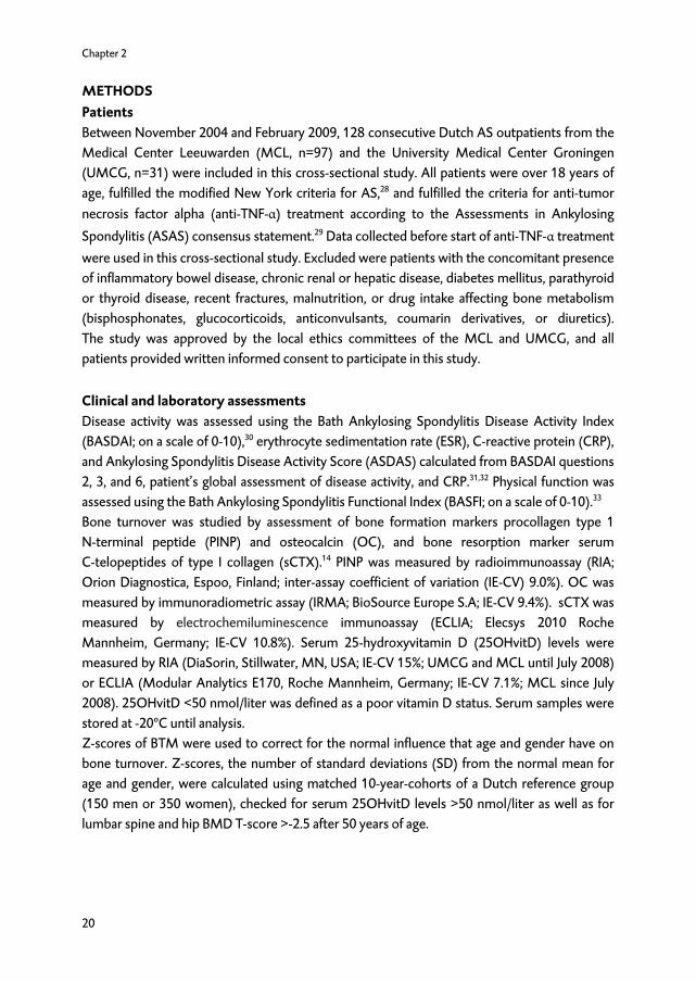
METHODS
Patients
Between November 2004 and February 2009, 128 consecutive Dutch AS outpatients from the
Medical Center Leeuwarden (MCL, n=97) and the University Medical Center Groningen
(UMCG, n=31) were included in this cross-sectional study. All patients were over 18 years of
age, fulfilled the modified New York criteria for AS,28 and fulfilled the criteria for anti-tumor
necrosis factor alpha (anti-TNF-α) treatment according to the Assessments in Ankylosing
Spondylitis (ASAS) consensus statement.29 Data collected before start of anti-TNF-α treatment
were used in this cross-sectional study. Excluded were patients with the concomitant presence
of inflammatory bowel disease, chronic renal or hepatic disease, diabetes mellitus, parathyroid
or thyroid disease, recent fractures, malnutrition, or drug intake affecting bone metabolism
(bisphosphonates, glucocorticoids, anticonvulsants, coumarin derivatives, or diuretics).
The study was approved by the local ethics committees of the MCL and UMCG, and all
patients provided written informed consent to participate in this study.
Clinical and laboratory assessments
Disease activity was assessed using the Bath Ankylosing Spondylitis Disease Activity Index
(BASDAI; on a scale of 0-10),30 erythrocyte sedimentation rate (ESR), C-reactive protein (CRP),
and Ankylosing Spondylitis Disease Activity Score (ASDAS) calculated from BASDAI questions
2, 3, and 6, patient’s global assessment of disease activity, and CRP.31,32 Physical function was
assessed using the Bath Ankylosing Spondylitis Functional Index (BASFI; on a scale of 0-10).33
Bone turnover was studied by assessment of bone formation markers procollagen type 1
N-terminal peptide (PINP) and osteocalcin (OC), and bone resorption marker serum
C-telopeptides of type I collagen (sCTX).14 PINP was measured by radioimmunoassay (RIA;
Orion Diagnostica, Espoo, Finland; inter-assay coefficient of variation (IE-CV) 9.0%). OC was
measured by immunoradiometric assay (IRMA; BioSource Europe S.A; IE-CV 9.4%). sCTX was
measured by electrochemiluminescence immunoassay (ECLIA; Elecsys 2010 Roche
Mannheim, Germany; IE-CV 10.8%). Serum 25-hydroxyvitamin D (25OHvitD) levels were
measured by RIA (DiaSorin, Stillwater, MN, USA; IE-CV 15%; UMCG and MCL until July 2008)
or ECLIA (Modular Analytics E170, Roche Mannheim, Germany; IE-CV 7.1%; MCL since July
2008). 25OHvitD <50 nmol/liter was defined as a poor vitamin D status. Serum samples were
stored at -20ºC until analysis.
Z-scores of BTM were used to correct for the normal influence that age and gender have on
bone turnover. Z-scores, the number of standard deviations (SD) from the normal mean for
age and gender, were calculated using matched 10-year-cohorts of a Dutch reference group
(150 men or 350 women), checked for serum 25OHvitD levels >50 nmol/liter as well as for
lumbar spine and hip BMD T-score >-2.5 after 50 years of age.

Bone turnover in ankylosing spondylitis
21
BMD measurement
BMD of lumbar spine (anterior-posterior projection at L1-L4) and hip (total proximal femur)
were measured using DXA (Hologic QDR Discovery (UMCG) or Hologic QDR Delphi (MCL),
Waltman, MA, USA). According to the World Health Organization (WHO) classification,
osteopenia was defined as a T-score between -1 and -2.5 and osteoporosis as a T-score ≤-2.5.34
Patients were categorized by the lowest T-score of the lumbar spine or hip. T-scores, the
number of SD from the normal mean obtained from young healthy adults, were calculated
using the NHANES reference database. DXA measurements of lumbar spine and hip were
available for 106 and 108 patients, respectively.
Vertebral assessment
Anterior, middle, and posterior heights of vertebrae T4 to L4 were measured on lateral
radiographs by two independent observers using a ruler. According to the Genant
classification, a vertebral fracture was defined based on reduction in anterior, middle, and/or
posterior height: grade 1, 20-25% reduction; grade 2, 25-40% reduction; and grade 3, >40%
reduction.35 In case of discrepancy between the two observers, a third independent observer
measured vertebral height in order to confirm the presence or absence of a vertebral fracture.
Radiographs were available for 106 patients.
Statistical analysis
Statistical analysis was performed with SPSS 16.0 software (SPSS, Chicago, IL, USA). Results
were expressed as mean ± SD or median (range) for parametric and nonparametric data,
respectively. Pearson’s and Spearman’s correlation coefficients were used as appropriate to
analyze the relationship between BMD, BTM, vitamin D, and clinical measures of disease
activity and physical function. Predictor analysis for low BMD, defined as lumbar spine or hip
BMD T-score ≤-1, was performed using univariate logistic regression and multivariate logistic
regression with conditional stepwise backward inclusion of variables that had a p-value ≤0.3 in
univariate analysis, together with variables that significantly correlated with lumbar spine or
hip BMD T-scores. The probability of P for stepwise removal was 0.10. Predictor analyses for
sCTX and OC Z-scores were performed using univariate linear regression and multivariate
linear regression with backward inclusion of variables that had a p-value ≤0.3 in univariate
analysis, together with variables that significantly correlated with sCTX or OC Z-scores. The
probability of F for removal was 0.10. P values <0.05 were considered statistically significant.

Chapter 2
22
RESULTS
Mean age of the 128 AS patients was 41.0 years (SD ± 11.1), median disease duration was 14
years (range 1-53), and 73% were male. Of the patients, 89% had a BASDAI score ≥4, 74% had
increased ESR levels, and 77% had increased CRP levels (Table 1).
Table 1. Characteristics of the AS study population (n = 128)
Age (yrs) 41.0 ± 11.1
Gender (male) (n, %) 93 (73)
Disease duration (yrs) 14 (1-53)
HLA-B27+ (n, %) 102 (84)
NSAID use (n, %) 100 (78)
DMARD use (n, %) 18 (14)
BASDAI (range 0-10) 6.0 ± 1.6 BASDAI ≥4 (n, %) 116 (89)
ESR (mm/h) 20 (2-90) Increased ESR (n, %) 95 (74)
CRP (mg/l) 14 (2-92) Increased CRP (n, %) 99 (77)
ASDAS 3.7 ± 0.8
BASFI (range 0-10) 5.6 ± 2.0
LS BMD T-score -0.68 ± 1.41 Osteopenia LS (n, %) 41 (39)
Osteoporosis LS (n, %) 9 (9)
Hip BMD T-score -0.52 ± 1.06 Osteopenia hip (n, %) 42 (39)
Osteoporosis hip (n, %) 2 (2)
VF (n, %) 41 (39) VF grade 1 (n, %) 27 (25)
VF grade 2 (n, %) 14 (13)
VF grade 3 (n, %) 0 (0)
PINP (μg/l) 42.7 (16.0-101.5)
PINP Z-score 0.14 (-1.74-3.55)
sCTX (pg/ml) 200.3 (13.4-780.9)
sCTX Z-score -0.36 (-2.58-5.90)
OC (μg/l) 12.7 (0.1-24.9)
OC Z-score -0.28 (-2.86-2.52)
25OHvitD (nmol/l) 61.4 (13.8-186) Poor vitD status (n, %) 30 (26)
Values are mean ± SD or median (range) unless otherwise indicated.
AS: ankylosing spondylitis; HLA-B27+: human leukocyte antigen B27 positive; NSAID: non-steroidal
anti-inflammatory drug; DMARD: disease-modifying antirheumatic drug; BASDAI: Bath ankylosing
spondylitis disease activity index; ESR: erythrocyte sedimentation rate; CRP: C-reactive protein; ASDAS:
ankylosing spondylitis disease activity score; BASFI: Bath ankylosing spondylitis functional index;
LS: lumbar spine; BMD: bone mineral density; VF: vertebral fracture; PINP: procollagen type 1
N-terminal peptide; sCTX: serum C-telopeptides of type I collagen; OC: osteocalcin; 25OHvitD:
25-hydroxyvitamin D.

Bone turnover in ankylosing spondylitis
23
Correlations between biochemical and clinical assessments
Correlations between BMD, BTM, vitamin D, and clinical assessments of disease activity and
physical function were calculated to obtain more knowledge about the pathophysiology of
AS-related osteoporosis (Table 2). There was a significant positive correlation between lumbar
spine and hip BMD T-scores. Lumbar spine BMD T-score correlated positively with BASDAI
(p<0.05) and hip BMD T-score correlated negatively with OC and sCTX Z-scores
(p<0.05).There were significant positive correlations between all BTM Z-scores. PINP Z-score
correlated positively with age (p<0.05) and PINP and sCTX Z-scores correlated positively with
disease duration (p<0.05). Finally, ESR, CRP, ASDAS, or BASFI were not significantly correlated
with any of the BMD T-scores or BTM Z-scores.
Table 2. Correlations between clinical and biochemical assessments in AS patients with active disease
(n=128)
Age Disease
duration
BASDAI ESR CRP ASDAS BASFI
Disease duration 0.600* -
BASDAI NS NS -
ESR NS NS NS -
CRP NS NS NS 0.693* -
ASDAS NS 0.187* 0.651* 0.374* 0.668* -
BASFI NS 0.203* 0.561* NS NS 0.472* -
PINP Z-score 0.362* 0.266* NS NS NS NS NS
sCTX Z-score NS 0.200* NS NS NS NS NS
OC Z-score NS NS NS NS NS NS NS
LS BMD T-score NS NS 0.205* NS NS NS NS
hip BMD T-score NS NS NS NS NS NS NS
25OHvitD NS NS NS NS NS NS NS
Table 2. (Continued)
PINP Z sCTX Z OC Z LS
BMD T
Hip
BMD T
sCTX Z-score 0.443* -
OC Z-score 0.578* 0.601* -
LS BMD T-score NS NS NS -
hip BMD T-score NS -0.380* -0.272* 0.626* -
25OHvitD NS NS NS NS NS
* Statistically significant correlation (p<0.05). See Table 1 for definitions.

Chapter 2
24
The difference between lumbar spine and hip BMD T-score correlated positively with disease
duration (ρ=0.340, p<0.05). As shown in Figure 1, patients with long disease duration never
had a lumbar spine BMD T-score that was much lower than their hip BMD T-score, which
indicates that osteoproliferation in the lumbar spine has resulted in an overestimation of the
lumbar spine BMD T-score in patients with advanced AS.
Figure 1. The difference between lumbar spine and hip BMD T-score correlated positively with disease
duration (ρ=0.340, p<0.05). Patients with long disease duration never had a lumbar spine BMD T-score
that was much lower than their hip BMD T-score, which indicates that osteoproliferation in the lumbar
spine has resulted in an overestimation of the lumbar spine BMD T-score in patients with advanced AS.
Vertebral fractures
Of the patients, 39% had at least 20% reduction in anterior, middle, and/or posterior vertebral
height, indicating vertebral fracture. In total, 74 vertebral fractures were detected; 59 wedge
fractures, 14 biconcave fractures, and one crush fracture. No significant differences between
patients with and without vertebral fractures were found in age (mean 43.1 years ± SD 11.1 vs.
39.9 years ± 11.0; p=0.149), disease duration (median 15 years (range 1-47) vs. 12 years (1-53);
p=0.925), BMD T-scores (lumbar spine -0.70 ± 1.33 vs. -0.71 ± 1.51; p=0.984, hip -0.47 ± 1.03
vs. -0.59 ± 1.10; p=0.591), and BTM Z-scores (PINP 0.15 (-1.74-3.08) vs. 0.22 (-1.65-3.55);
p=0.493), sCTX -0.21 (-2.28-5.90) vs. -0.23 (-2.58-3.92); p=0.778, OC -0.31 (-2.86-1.50) vs.
-0.18 (-2.66-2.52); p=0.460, respectively).

Bone turnover in ankylosing spondylitis
25
Predictors of low BMD
Predictor analysis was performed to identify parameters that are related to low BMD. In total,
57% of patients had a lumbar spine or hip BMD T-score of -1 or less, indicating low BMD. Male
gender, lower BASDAI score, higher PINP Z-score, higher OC Z-score, and higher sCTX
Z-score were significantly associated with low BMD in univariate regression analysis. Since
male gender was significantly associated with low BMD, variables that significantly differed
between men and women were included in multivariate analysis: age, ESR, OC Z-score, sCTX
Z-score, and 25OHvitD. Multivariate regression analysis showed that older age (odds ratio
(OR): 1.066, 95% confidence interval (CI): 1.008-1.129), lower BASDAI score (OR: 0.648,
0.445-0.923), higher ESR (OR: 1.025, 0.994-1.057), and higher sCTX Z-score (OR: 2.563, 1.370-
4.794) were independently related to low BMD (Table 3). OC Z-score was not included in
multivariate analysis, probably due to the strong correlation between sCTX Z-score and OC
Z-score (ρ=0.601, p=0.000). However, higher OC Z-score was also independently related to
low BMD in the presence of age, BASDAI, and ESR (OR: 2.255, 95%CI: 1.238-4.107), indicating
that both sCTX Z-score and OC Z-score are important. The Nagelkerke R2 of the multivariate
models including sCTX Z-score and OC Z-score were 0.381 and 0.338, respectively.
Table 3. Results of univariate and multivariate logistic regression analysis for low BMD
Univariate analysis Multivariate analysis
OR (95% CI) p-value OR (95% CI) p-value
Age (yrs)a 1.017 (0.981-1.055) 0.353 1.066 (1.008-1.129) 0.026
Genderb 4.368 (1.791-10.652) 0.001 *
Disease duration (yrs)a 1.012 (0.974-1.052) 0.539 *
BASDAI (range 0-10)c 0.728 (0.554-0.957) 0.023 0.648 (0.455-0.923) 0.016
ESR (mm/h)c 1.012 (0.980-1.034) 0.287 1.025 (0.994-1.057) 0.112
CRP (mg/l)c 1.018 (0.994-1.042) 0.143 *
ASDASc 0.769 (0.461-1.283) 0.315 **
BASFI (range 0-10)c 0.959 (0.790-1.165) 0.674 **
PINP Z-scorec 1.602 (1.043-2.461) 0.031 *
sCTX Z-scorec 1.878 (1.262-2.794) 0.002 2.563 (1.370-4.794) 0.003
OC Z-scorec 1.766 (1.135-2.749) 0.012 *
25OHvitD (nmol/l)c 0.998 (0.983-1.013) 0.787 *
VFd 0.902 (0.385-2.109) 0.811 **
OR refers to the risk of low BMD (lumbar spine or hip BMD T-score ≤ -1): a — per year; b — if gender is
male (versus female); c — per 1 grade or 1 point; d — if vertebral fracture is present (versus absent).
See Table 1 for definitions.
* The variable was not selected during multivariate regression analysis.
** The variable was not tested in multivariate regression analysis because of a p-value > 0.3 in univariate
regression analysis, no significant correlation with lumbar spine or hip BMD T-scores, and no significant
difference between men and women.

Chapter 2
26
Predictors of sCTX and OC Z-scores
Since sCTX and OC Z-scores seem to be valuable markers to detect bone loss, predictor
analyses for these markers were performed to get more information about the
pathophysiology of AS-related osteoporosis. Gender, PINP Z-score, OC Z-score, and
25OHvitD were significantly associated with sCTX Z-score in univariate regression analysis.
Since gender was significantly associated with sCTX Z-score, the previous mentioned variables
that significantly differed between men and women were included in multivariate analysis.
Multivariate regression analysis showed that ESR (B: 0.012, 0.000-0.025), PINP Z-score
(B: 0.292, 0.022-0.563), OC Z-score (B: 0.505, 0.243-0.768), and 25OHvitD (B: -0.009,-0.018-
0.000) were independently related to sCTX Z-score (Table 4). The R2 of this multivariate
model was 0.424.
Gender, PINP Z-score, and sCTX Z-score were significantly associated with OC Z-score in
univariate regression analysis. Since gender was significantly associated with OC Z-score, the
previous mentioned variables that significantly differed between men and women were
included in multivariate analysis. Multivariate regression analysis showed that age (B: -0.018,
-0.034--0.001), gender (B: -0.607, -0.999--0.214), PINP Z-score (B: 0.464, 0.282-0.646), and
sCTX Z-score (B: 0.243, 0.110-0.377) were independently related to OC Z-score (Table 5). The
R2 of this multivariate model was 0.509.
Table 4. Results of univariate and multivariate linear regression analysis for sCTX Z-score
Univariate analysis Multivariate analysis
B (95% CI) p-value B (95% CI) p-value
Age (yrs)a 0.018 (-0.004-0.041) 0.112 *
Genderb -0.680 (-1.211--0.148) 0.013 *
Disease duration (yrs)a 0.018 (-0.005-0.041) 0.114 *
BASDAI (range 0-10)c -0.060 (-0.213-0.092) 0.436 **
ESR (mm/h)c 0.011 (-0.002-0.025) 0.102 0.012 (0.000-0.025) 0.069
CRP (mg/l)c 0.007 (-0.007-0.021) 0.303 *
ASDASc 0.156 (-0.174-0.486) 0.351 **
BASFI (range 0-10)c 0.004 (-0.124-0.132) 0.953 **
PINP Z-scorec 0.581 (0.384-0.777) 0.000 0.292 (0.022-0.563) 0.035
OC Z-scorec 0.774 (0.577-0.971) 0.000 0.505 (0.243-0.768) 0.000
25OHvitD (nmol/l)c -0.011 (-0.020--0.002) 0.020 -0.009 (-0.018-0.000) 0.041
B refers to the influence on sCTX Z-score: a — per year; b — if gender is male (versus female); c — per 1
grade or 1 point.
See Table 1 for definitions.
* The variable was not selected during multivariate regression analysis.
** The variable was not tested in multivariate regression analysis because of a p-value > 0.3 in univariate
regression analysis, no significant correlation with sCTX Z-score, and no significant difference between
men and women.

Bone turnover in ankylosing spondylitis
27
Table 5. Results of univariate and multivariate linear regression analysis for OC Z-score
Univariate analysis Multivariate analysis
B (95% CI) p-value B (95% CI) p-value
Age (yrs)a 0.008 (-0.011-0.027) 0.409 -0.018 (-0.034--0.001) 0.036
Genderb -0.687 (-1.129--0.244) 0.003 -0.607 (-0.999--0.214) 0.003
Disease duration (yrs)a 0.007 (-0.012-0.026) 0.460 **
BASDAI (range 0-10)c -0.029 (-0.155-0.098) 0.655 **
ESR (mm/h)c 0.006 (-0.005-0.018) 0.284 *
CRP (mg/l)c 0.009 (-0.003-0.022) 0.130 *
ASDASc 0.052 (-0.222-0.326) 0.708 **
BASFI (range 0-10)c 0.035 (-0.071-0.141) 0.651 **
PINP Z-scorec 0.605 (0.453-0.756) 0.000 0.464 (0.282-0.646) 0.000
sCTX Z-scorec 0.464 (0.346-0.582) 0.000 0.243 (0.110-0.377) 0.000
25OHvitD (nmol/l)c -0.007 (-0.016-0.001) 0.076 *
B refers to the influence on OC Z-score: a — per year; b — if gender is male (versus female); c — per 1
grade or 1 point.
See Table 1 for definitions.
* The variable was not selected during multivariate regression analysis.
** The variable was not tested in multivariate regression analysis because of a p-value > 0.3 in univariate
regression analysis, no significant correlation with OC Z-score, and no significant difference between
men and women.
DISCUSSION
The present cross-sectional study in AS patients with active disease showed that sCTX and OC
Z-scores are independently related to low BMD, which indicates that sCTX and OC Z-scores
are valuable markers to detect bone loss in AS. An accurate and easily accessible marker of
bone loss is needed in patients with advanced AS, since the anterior-posterior lumbar spine
BMD measured by DXA can be overestimated by the presence of syndesmophytes, ligament
calcifications, and fusion of facet joints in these patients.23-25 Our finding that the difference
between lumbar spine and hip BMD correlated positively with disease duration indicates that
this overestimation also occurred in this study. Furthermore, our high prevalence of vertebral
fractures and of low BMD (osteopenia or osteoporosis) underlines the importance of
monitoring bone loss in AS.
In order to obtain more knowledge about the pathophysiology of AS-related osteoporosis, we
investigated the relation between BMD, BTM, vitamin D, and clinical assessments. Our results
demonstrate that increased bone turnover plays a significant role in the development of
osteoporosis in AS patients. First, significant positive correlations were found between age or
disease duration and PINP Z-score, a marker of bone formation, as well as between disease
duration and sCTX Z-score, a marker of bone resorption. Since the use of Z-scores corrects for
the normal influence that age and gender have on bone turnover, these correlations
demonstrate that AS is characterized by both increased bone formation and increased bone
resorption. Second, significant negative correlations were found between sCTX or OC

Chapter 2
28
Z-scores and hip BMD T-score, and a higher sCTX or OC Z-score was independently related to
low BMD, which indicates that high bone turnover is associated with bone loss in AS. This
finding is in agreement with the previous studies.4,14,15
The results of this study also demonstrate involvement of inflammatory processes in the
complex pathophysiological mechanism of AS-related osteoporosis. A higher ESR was
independently related to low BMD. Furthermore, ESR had independent influence on sCTX
Z-score. The importance of inflammatory processes was also shown in previous studies.4-9
Finally, our finding that 25OHvitD level had an independent significant inverse influence on
sCTX Z-score suggests that low vitamin D levels play a role in the development of AS-related
osteoporosis. The importance of vitamin D was also suggested in previous studies.7,11-13,36
Amento et al. reported that vitamin D is an endogenous modulator of the immune response,
which may slow down the inflammatory process by suppressing active T cells and cell
proliferation.36 Lange et al. found negative correlations between serum levels of vitamin D and
markers of disease activity or inflammation in AS patients. They also showed that AS patients
with osteoporosis had significantly lower vitamin D levels compared to AS patients with
normal BMD.7,11 Finally, Obermayer et al. suggested a close association of BMD, bone
metabolism, and inflammatory activity with Fok1 polymorphisms of the vitamin D receptor
gene in male AS patients.13
Unexpectedly, a lower BASDAI score was independently related to low BMD in this study.
A possible explanation for this finding may be that complaints related to new bone formation
influence the BASDAI, a subjective measure of disease activity, in AS patients with active
disease. The significant positive correlation between BASDAI and lumbar spine BMD T-score
found in this study seems to confirm this suggestion. Another explanation may be that BMD,
measured by DXA, reflects the influence of the disease on bone over time, while BASDAI
reflects the current status of disease activity.
There are some strengths and limitations to this study. The main limitation is that the study is
cross-sectional and that only AS patients with active disease were included. Further studies
with longer follow-up are needed to confirm the usefulness of sCTX and OC Z-scores in
monitoring bone loss in AS patients, as well as the importance of increased bone turnover,
inflammation, and low vitamin D levels in the development of AS-related osteoporosis.
Another limitation is that body mass index (BMI) was not assessed in this study. Therefore, it
was not possible to correct for low BMI in multivariate analysis. Finally, it was not clear if the
vertebral fractures occurred recently or if they were already present for many years. Therefore,
analyses investigating the relation between BTM and vertebral fractures were difficult. The
main strength is that Z-scores of BTM were calculated to correct for the influence that age and
gender have on bone turnover in healthy persons. In this way, male and female patients of
different age groups could be analyzed together.
In conclusion, this cross-sectional study in AS patients with active disease indicates that
increased bone turnover, inflammation, and low vitamin D levels are important in the
pathophysiology of AS-related osteoporosis. Furthermore, sCTX and OC Z-scores seem to be

Bone turnover in ankylosing spondylitis
29
valuable markers to detect bone loss in AS. Combining biochemical BTM and BMD
measurements may be useful to identify AS patients with osteoporosis in daily clinical practice
where lumbar spine BMD, measured by DXA, may be overestimated due to osteoproliferation
in patients with advanced AS.
REFERENCES
1. Cooper C, Carbone L, Michet CJ, et al. Fracture risk in patients with ankylosing spondylitis:
a population based study. J Rheumatol 1994;21:1877-82.
2. Vosse D, Landewe R, van der Heijde D, et al. Ankylosing spondylitis and the risk of fracture: results
from a large primary care-based nested case-control study. Ann Rheum Dis 2009;68:1839-42.
3. Geusens P, Vosse D, van der Linden S. Osteoporosis and vertebral fractures in ankylosing
spondylitis. Curr Opin Rheumatol 2007;19:335-9.
4. Franck H, Meurer T, Hofbauer LC. Evaluation of bone mineral density, hormones, biochemical
markers of bone metabolism, and osteoprotegerin serum levels in patients with ankylosing
spondylitis. J Rheumatol 2004;31:2236-41.
5. Ghozlani I, Ghazi M, Nouijai A, et al. Prevalence and risk factors of osteoporosis and vertebral
fractures in patients with ankylosing spondylitis. Bone 2009;44:772-6.
6. Gratacos J, Collado A, Pons F, et al. Significant loss of bone mass in patients with early, active
ankylosing spondylitis: a followup study. Arthritis Rheum 1999;42:2319-24.
7. Lange U, Teichmann J, Strunk J, et al. Association of 1.25 vitamin D3 deficiency, disease activity and
low bone mass in ankylosing spondylitis. Osteoporos Int 2005;16:1999-2004.
8. Maillefert JF, Aho LS, El Maghraoui A, et al. Changes in bone density in patients with ankylosing
spondylitis: a two-year follow-up study. Osteoporos Int 2001;12:605-9
9. Toussirot E, Ricard-Blum S, Dumoulin G, et al. Relationship between urinary pyridinium cross-links,
disease activity and disease subsets of ankylosing spondylitis. Rheumatology (Oxford) 1999;38:21—
27.
10. El Maghraoui A. Osteoporosis and ankylosing spondylitis. Joint Bone Spine 2004;71:291-5.
11. Lange U, Jung O, Teichmann J, et al. Relationship between disease activity and serum levels of
vitamin D metabolites and parathyroid hormone in ankylosing spondylitis. Osteoporos Int
2001;12:1031-5.
12. Mermerci Baskan B, Pekin Dogan Y, Sivas F, et al. The relation between osteoporosis and vitamin D
levels and disease activity in ankylosing spondylitis. Rheumatol Int 2010;30:375-81.
13. Obermayer-Pietsch BM, Lange U, Tauber G, et al. Vitamin D receptor initiation codon
polymorphism, bone density and inflammatory activity of patients with ankylosing spondylitis.
Osteoporos Int 2003;14:995-1000.
14. Borman P, Bodur H, Bingol N, et al. Bone mineral density and bone turnover markers in a group of
male ankylosing spondylitis patients: relationship to disease activity. J Clin Rheumatol 2001;7:315-
21.
15. Park MC, Chung SJ, Park YB, et al. Bone and cartilage turnover markers, bone mineral density, and
radiographic damage in men with ankylosing spondylitis. Yonsei Med J 2008;49:288-94.
16. Eastell R, Hannon RA. Biomarkers of bone health and osteoporosis risk. Proc Nutr Soc 2008;67:157-
62.

Chapter 2
30
17. El Maghraoui A, Tellal S, Chaouir S, et al. Bone turnover markers, anterior pituitary and gonadal
hormones, and bone mass evaluation using quantitative computed tomography in ankylosing
spondylitis. Clin Rheumatol 2005;24:346-51.
18. Karberg K, Zochling J, Sieper J, et al. Bone loss is detected more frequently in patients with
ankylosing spondylitis with syndesmophytes. J Rheumatol 2005;32:1290-8.
19. Sarikaya S, Basaran A, Tekin Y, et al. Is osteoporosis generalized or localized to central skeleton in
ankylosing spondylitis? J Clin Rheumatol 2007;13:20-4.
20. Yilmaz N, Ozaslan J. Biochemical bone turnover markers in patients with ankylosing spondylitis.
Clin Rheumatol 2000;19:92-8.
21. Vosse D, Landewe R, Garnero P, et al. Association of markers of bone- and cartilage-degradation
with radiological changes at baseline and after 2 years follow-up in patients with ankylosing
spondylitis. Rheumatology (Oxford) 2008;47:1219-22.
22. Kanis JA, McCloskey EV, Johansson H, et al. A reference standard for the description of
osteoporosis. Bone 2008;42:467-75.
23. Baek HJ, Kang SW, Lee YJ, et al. Osteopenia in men with mild and severe ankylosing spondylitis.
Rheumatol Int 2005;26:30-4.
24. Lee YS, Schlotzhauer T, Ott SM, et al. Skeletal status of men with early and late ankylosing
spondylitis. Am J Med 1997;103:233-41.
25. Meirelles ES, Borelli A, Camargo OP. Influence of disease activity and chronicity on ankylosing
spondylitis bone mass loss. Clin Rheumatol 1999;18:364-8.
26. Lange U, Kluge A, Strunk J, et al. Ankylosing spondylitis and bone mineral density--what is the ideal
tool for measurement? Rheumatol Int 2005;26:115-20.
27. Bessant R, Keat A. How should clinicians manage osteoporosis in ankylosing spondylitis?
J Rheumatol 2002;29:1511-9.
28. van der Linden S, Valkenburg HA, Cats A. Evaluation of diagnostic criteria for ankylosing spondylitis.
A proposal for modification of the New York criteria. Arthritis Rheum 1984;27:361-8.
29. Braun J, Davis J, Dougados M, et al. First update of the international ASAS consensus statement for
the use of anti-TNF agents in patients with ankylosing spondylitis. Ann Rheum Dis 2006;65:316-20.
30. Garrett S, Jenkinson T, Kennedy LG, et al. A new approach to defining disease status in ankylosing
spondylitis: the Bath Ankylosing Spondylitis Disease Activity Index. J Rheumatol 1994;21:2286-91.
31. Lukas C, Landewe R, Sieper J, et al. Development of an ASAS-endorsed disease activity score
(ASDAS) in patients with ankylosing spondylitis. Ann Rheum Dis 2009;68:18-24.
32. van der Heijde D, Lie E, Kvien TK, et al. ASDAS, a highly discriminatory ASAS-endorsed disease
activity score in patients with ankylosing spondylitis. Ann Rheum Dis 2009;68:1811-8.
33. Calin A, Garrett S, Whitelock H, et al. A new approach to defining functional ability in ankylosing
spondylitis: the development of the Bath Ankylosing Spondylitis Functional Index. J Rheumatol
1994;21:2281-5.
34. Kanis JA. Assessment of fracture risk and its application to screening for postmenopausal
osteoporosis: synopsis of a WHO report. WHO Study Group. Osteoporos Int 1994;4:368-81.
35. Genant HK, Wu CY, van Kuijk C, et al. Vertebral fracture assessment using a semiquantitative
technique. J Bone Miner Res 1993;8:1137-48.
36. Amento EP. Vitamin D and the immune system. Steroids 1987;49:55-72.

CHAPTER 3
THE EFFECT OF THREE YEARS OF TUMOR NECROSIS FACTOR-ALPHA
BLOCKING THERAPY ON MARKERS OF BONE TURNOVER AND THEIR
PREDICTIVE VALUE FOR TREATMENT DISCONTINUATION IN PATIENTS
WITH ANKYLOSING SPONDYLITIS: A PROSPECTIVE LONGITUDINAL
OBSERVATIONAL COHORT STUDY
Suzanne Arends1,2, Anneke Spoorenberg1,2, Pieternella M. Houtman2,
Martha K. Leijsma1, Reinhard Bos2, Cees G. M. Kallenberg1, Henk Groen3,
Elisabeth Brouwer1, Eveline van der Veer4
Departments of 1 Rheumatology and Clinical Immunology, 3 Epidemiology, 4 Laboratory Medicine, University of Groningen, University Medical Center Groningen, 2
Rheumatology, Medical Center Leeuwarden, The Netherlands
Arthritis Res Ther. 2012;14:R98.

Chapter 3
32
ABSTRACT
Introduction: The aim of this study was to investigate the effect of 3 years of tumor necrosis
factor-alpha (TNF-α) blocking therapy on bone turnover as well as to analyze the predictive
value of early changes in bone turnover markers (BTM) for treatment discontinuation in
patients with ankylosing spondylitis (AS).
Methods: Prospective cohort study of 111 consecutive AS outpatients who started TNF-α
blocking therapy. Clinical assessments and BTM were assessed at baseline, 3 and 6 months, as
well as 1, 2, and 3 years. Z-scores of BTM were calculated to correct for age and gender. Bone
mineral density (BMD) was assessed yearly.
Results: After 3 years, 72 patients (65%) were still using their first TNF-α blocking agent. In
these patients, TNF-α blocking therapy resulted in significantly increased bone-specific alkaline
phosphatase, a marker of bone formation; decreased serum collagen-telopeptide (sCTX),
a marker of bone resorption; and increased lumbar spine and hip BMD compared to baseline.
Baseline to 3 months decrease in sCTX Z-score (HR: 0.394, 95% CI: 0.263-0.591), AS disease
activity score (ASDAS; HR: 0.488, 95% CI: 0.317-0.752), and physician’s global disease activity
(HR: 0.739, 95% CI: 0.600-0.909) were independent inversely related predictors of time to
treatment discontinuation because of inefficacy or intolerance. Early decrease in sCTX Z-score
correlated significantly with good long-term response regarding disease activity, physical
function, and quality of life.
Conclusions: Three years of TNF-α blocking therapy results in a bone turnover balance that
favors bone formation (especially mineralization), in combination with continuous
improvement of lumbar spine BMD. Early change in sCTX can serve as an objective measure in
the evaluation of TNF-α blocking therapy in AS, in addition to the currently used more
subjective measures.
Influence of TNF-α blocking therapy on bone turnover
33
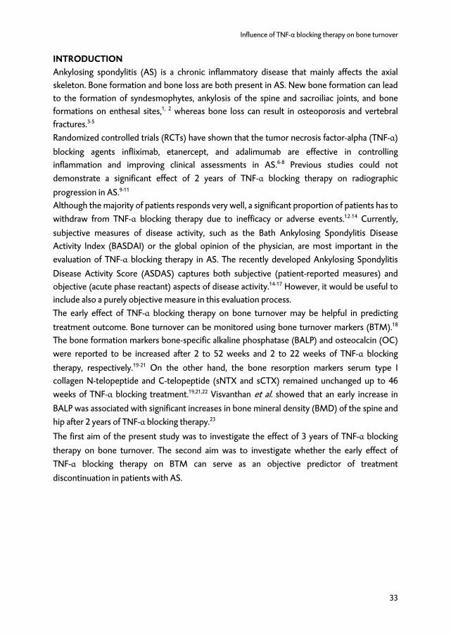
INTRODUCTION
Ankylosing spondylitis (AS) is a chronic inflammatory disease that mainly affects the axial
skeleton. Bone formation and bone loss are both present in AS. New bone formation can lead
to the formation of syndesmophytes, ankylosis of the spine and sacroiliac joints, and bone
formations on enthesal sites,1, 2 whereas bone loss can result in osteoporosis and vertebral
fractures.3-5
Randomized controlled trials (RCTs) have shown that the tumor necrosis factor-alpha (TNF-α)
blocking agents infliximab, etanercept, and adalimumab are effective in controlling
inflammation and improving clinical assessments in AS.6-8 Previous studies could not
demonstrate a significant effect of 2 years of TNF-α blocking therapy on radiographic
progression in AS.9-11
Although the majority of patients responds very well, a significant proportion of patients has to
withdraw from TNF-α blocking therapy due to inefficacy or adverse events.12-14 Currently,
subjective measures of disease activity, such as the Bath Ankylosing Spondylitis Disease
Activity Index (BASDAI) or the global opinion of the physician, are most important in the
evaluation of TNF-α blocking therapy in AS. The recently developed Ankylosing Spondylitis
Disease Activity Score (ASDAS) captures both subjective (patient-reported measures) and
objective (acute phase reactant) aspects of disease activity.14-17 However, it would be useful to
include also a purely objective measure in this evaluation process.
The early effect of TNF-α blocking therapy on bone turnover may be helpful in predicting
treatment outcome. Bone turnover can be monitored using bone turnover markers (BTM).18
The bone formation markers bone-specific alkaline phosphatase (BALP) and osteocalcin (OC)
were reported to be increased after 2 to 52 weeks and 2 to 22 weeks of TNF-α blocking
therapy, respectively.19-21 On the other hand, the bone resorption markers serum type I
collagen N-telopeptide and C-telopeptide (sNTX and sCTX) remained unchanged up to 46
weeks of TNF-α blocking treatment.19,21,22 Visvanthan et al. showed that an early increase in
BALP was associated with significant increases in bone mineral density (BMD) of the spine and
hip after 2 years of TNF-α blocking therapy.23
The first aim of the present study was to investigate the effect of 3 years of TNF-α blocking
therapy on bone turnover. The second aim was to investigate whether the early effect of
TNF-α blocking therapy on BTM can serve as an objective predictor of treatment
discontinuation in patients with AS.

Chapter 3
34
METHODS
Patients
Between November 2004 and December 2007, 111 consecutive Dutch outpatients with AS,
who started TNF-α blocking therapy at the University Medical Center Groningen (UMCG;
n=28) and the Medical Center Leeuwarden (MCL; n=83), were included in this longitudinal
analysis. All patients participated in the Groningen Leeuwarden Ankylosing Spondylitis (GLAS)
study, a prospective longitudinal observational cohort study with follow-up visits according to
a fixed protocol. For the present analysis, patients with recent fractures and/or use of
bisphosphonates were excluded because of major influence on bone metabolism. All patients
were over 18 years of age, fulfilled the modified New York criteria for AS (n=109)24 or the
Assessments in Ankylosing Spondylitis (ASAS) criteria for axial spondyloarthritis including
MRI (n=2).25 The patients started treatment with the TNF-α blocking agents infliximab (n=22),
etanercept (n=71), or adalimumab (n=18) because of active disease (BASDAI ≥4 and/or expert
opinion), according to the ASAS consensus statement.26 As described previously,14 infliximab
(5 mg/kg) was given intravenously at 0, 2, and 6 weeks and then every 8 weeks. In case of
inadequate response, the frequency of infliximab treatment was raised to every 6 weeks.
Etanercept was administered as a subcutaneous injection once (50 mg) or twice (25 mg) a
week. Adalimumab (40 mg) was administered as a subcutaneous injection on alternate weeks.
In 2004 and 2005, patients started treatment with either infliximab or etanercept as
adalimumab was only registered in the Netherlands from 2006. The choice of the TNF-α
blocking agent was based on the judgment of the treating rheumatologist and/or the specific
preference of the patient. Continuation of treatment was based on decrease in the BASDAI of
at least 50% or 2 units compared with baseline and/or expert opinion in favor of treatment
continuation. Reasons for discontinuation of TNF-α blocking therapy were classified into the
categories intolerance due to adverse events, inefficacy, or other reasons. Patients were
allowed to receive concomitant medication as usual in daily clinical practice. The GLAS study
was approved by the local ethics committees of the UMCG and the MCL. All patients
provided written informed consent according to the Declaration of Helsinki.
Clinical and laboratory assessments
Patients were evaluated at baseline and after 3 months (mean 3.3 mo, SD ± 0.5), 6 months
(mean 6.4 mo, SD ± 0.8), 1 year (mean 1.0 yr, SD ± 0.1), 2 years (mean 2.1 yr, SD ± 0.1), and 3
years (mean 3.1 yr, SD ± 0.1) of TNF-α blocking therapy. Disease activity was assessed using
the BASDAI (on a scale of 0-10), physician’s and patient’s global assessment of disease activity
(GDA; on a scale of 0-10), erythrocyte sedimentation rate (ESR), C-reactive protein (CRP), and
ASDASCRP (calculated from BASDAI questions 2, 3 and 6, patient’s GDA, and CRP).15,16
Increased ESR and CRP levels were based on local standardized values. Physical function was
assessed using the Bath Ankylosing Spondylitis Functional Index (BASFI; on a scale of 0-10).
Spinal mobility assessments included chest expansion, modified Schober test, occiput to wall
distance, and lateral lumbar flexion (left and right). Quality of life was assessed using the
Influence of TNF-α blocking therapy on bone turnover
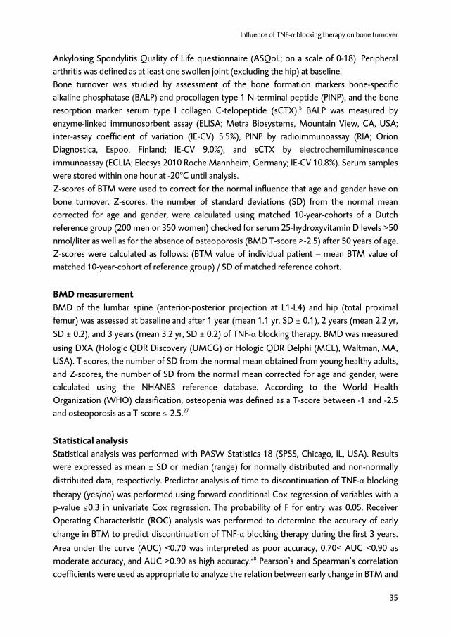
35
Ankylosing Spondylitis Quality of Life questionnaire (ASQoL; on a scale of 0-18). Peripheral
arthritis was defined as at least one swollen joint (excluding the hip) at baseline.
Bone turnover was studied by assessment of the bone formation markers bone-specific
alkaline phosphatase (BALP) and procollagen type 1 N-terminal peptide (PINP), and the bone
resorption marker serum type I collagen C-telopeptide (sCTX).5 BALP was measured by
enzyme-linked immunosorbent assay (ELISA; Metra Biosystems, Mountain View, CA, USA;
inter-assay coefficient of variation (IE-CV) 5.5%), PINP by radioimmunoassay (RIA; Orion
Diagnostica, Espoo, Finland; IE-CV 9.0%), and sCTX by electrochemiluminescence
immunoassay (ECLIA; Elecsys 2010 Roche Mannheim, Germany; IE-CV 10.8%). Serum samples
were stored within one hour at -20ºC until analysis.
Z-scores of BTM were used to correct for the normal influence that age and gender have on
bone turnover. Z-scores, the number of standard deviations (SD) from the normal mean
corrected for age and gender, were calculated using matched 10-year-cohorts of a Dutch
reference group (200 men or 350 women) checked for serum 25-hydroxyvitamin D levels >50
nmol/liter as well as for the absence of osteoporosis (BMD T-score >-2.5) after 50 years of age.
Z-scores were calculated as follows: (BTM value of individual patient — mean BTM value of
matched 10-year-cohort of reference group) / SD of matched reference cohort.
BMD measurement
BMD of the lumbar spine (anterior-posterior projection at L1-L4) and hip (total proximal
femur) was assessed at baseline and after 1 year (mean 1.1 yr, SD ± 0.1), 2 years (mean 2.2 yr,
SD ± 0.2), and 3 years (mean 3.2 yr, SD ± 0.2) of TNF-α blocking therapy. BMD was measured
using DXA (Hologic QDR Discovery (UMCG) or Hologic QDR Delphi (MCL), Waltman, MA,
USA). T-scores, the number of SD from the normal mean obtained from young healthy adults,
and Z-scores, the number of SD from the normal mean corrected for age and gender, were
calculated using the NHANES reference database. According to the World Health
Organization (WHO) classification, osteopenia was defined as a T-score between -1 and -2.5
and osteoporosis as a T-score ≤-2.5.27
Statistical analysis
Statistical analysis was performed with PASW Statistics 18 (SPSS, Chicago, IL, USA). Results
were expressed as mean ± SD or median (range) for normally distributed and non-normally
distributed data, respectively. Predictor analysis of time to discontinuation of TNF-α blocking
therapy (yes/no) was performed using forward conditional Cox regression of variables with a
p-value ≤0.3 in univariate Cox regression. The probability of F for entry was 0.05. Receiver
Operating Characteristic (ROC) analysis was performed to determine the accuracy of early
change in BTM to predict discontinuation of TNF-α blocking therapy during the first 3 years.
Area under the curve (AUC) <0.70 was interpreted as poor accuracy, 0.70< AUC <0.90 as
moderate accuracy, and AUC >0.90 as high accuracy.28 Pearson’s and Spearman’s correlation
coefficients were used as appropriate to analyze the relation between early change in BTM and

Chapter 3
36
clinical assessments. Generalized estimating equations (GEE) were used to analyze clinical
assessments, BTM, and BMD over time within subjects. Pairwise contrasts were used to
compare baseline and follow-up visits. P values <0.05 were considered statistically significant.
RESULTS
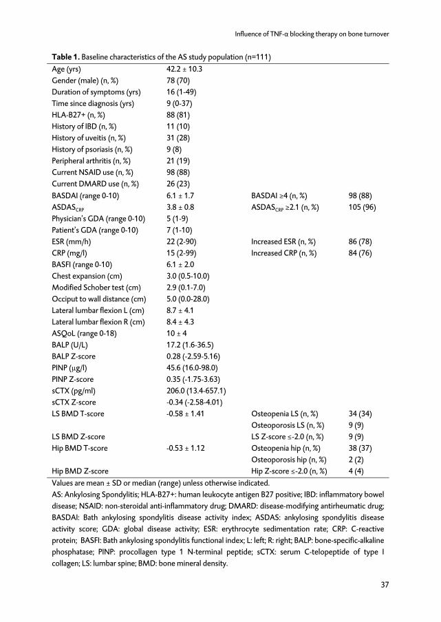
Mean age of the 111 AS patients was 42.2 years (SD ± 10.3), 70% were male, and median
disease duration was 16 years (range 1-49). All baseline characteristics are shown in Table 1.
After 3 years, 72 patients (65%) continued to use their first TNF-α blocking agent. In these
patients, all assessments of disease activity (Table 2), physical function, spinal mobility, and
quality of life (data not shown) improved after 3 months and remained significantly better
compared to baseline up to 3 years of TNF-α blocking therapy.
The remaining 39 patients (35%) discontinued treatment after a median follow-up of 7.0
months (range 1.1-36.2). Reasons for discontinuation of TNF-α blocking therapy were
inefficacy (n=17; 44%), adverse events (n=11; 28%: diarrhea or inflammatory bowel disease
(IBD; n=4); infection (n=3); allergic reaction (n=2); cardio-vascular disease (n=1);
demyelization problems (n=1)), both inefficacy and adverse events (n=5; 13%: diarrhea or IBD
(n=2); infection (n=1); allergic reaction (n=1); uveitis (n=1)), or other reasons (n=6; 15%: good
initial response, own choice (n=3); pregnancy wish (n=2); lost to follow up (n=1)).
Influence of TNF-α blocking therapy on bone turnover
37
Table 1. Baseline characteristics of the AS study population (n=111)
Age (yrs) 42.2 ± 10.3
Gender (male) (n, %) 78 (70)
Duration of symptoms (yrs) 16 (1-49)
Time since diagnosis (yrs) 9 (0-37)
HLA-B27+ (n, %) 88 (81)
History of IBD (n, %) 11 (10)
History of uveitis (n, %) 31 (28)
History of psoriasis (n, %) 9 (8)
Peripheral arthritis (n, %) 21 (19)
Current NSAID use (n, %) 98 (88)
Current DMARD use (n, %) 26 (23)
BASDAI (range 0-10) 6.1 ± 1.7 BASDAI ≥4 (n, %) 98 (88)
ASDASCRP 3.8 ± 0.8 ASDASCRP ≥2.1 (n, %) 105 (96)
Physician’s GDA (range 0-10) 5 (1-9)
Patient’s GDA (range 0-10) 7 (1-10)
ESR (mm/h) 22 (2-90) Increased ESR (n, %) 86 (78)
CRP (mg/l) 15 (2-99) Increased CRP (n, %) 84 (76)
BASFI (range 0-10) 6.1 ± 2.0
Chest expansion (cm) 3.0 (0.5-10.0)
Modified Schober test (cm) 2.9 (0.1-7.0)
Occiput to wall distance (cm) 5.0 (0.0-28.0)
Lateral lumbar flexion L (cm) 8.7 ± 4.1
Lateral lumbar flexion R (cm) 8.4 ± 4.3
ASQoL (range 0-18) 10 ± 4
BALP (U/L) 17.2 (1.6-36.5)
BALP Z-score 0.28 (-2.59-5.16)
PINP (μg/l) 45.6 (16.0-98.0)
PINP Z-score 0.35 (-1.75-3.63)
sCTX (pg/ml) 206.0 (13.4-657.1)
sCTX Z-score -0.34 (-2.58-4.01)
LS BMD T-score -0.58 ± 1.41 Osteopenia LS (n, %) 34 (34)
Osteoporosis LS (n, %) 9 (9)
LS BMD Z-score LS Z-score ≤-2.0 (n, %) 9 (9)
Hip BMD T-score -0.53 ± 1.12 Osteopenia hip (n, %) 38 (37)
Osteoporosis hip (n, %) 2 (2)
Hip BMD Z-score Hip Z-score ≤-2.0 (n, %) 4 (4)
Values are mean ± SD or median (range) unless otherwise indicated.
AS: Ankylosing Spondylitis; HLA-B27+: human leukocyte antigen B27 positive; IBD: inflammatory bowel
disease; NSAID: non-steroidal anti-inflammatory drug; DMARD: disease-modifying antirheumatic drug;
BASDAI: Bath ankylosing spondylitis disease activity index; ASDAS: ankylosing spondylitis disease
activity score; GDA: global disease activity; ESR: erythrocyte sedimentation rate; CRP: C-reactive
protein; BASFI: Bath ankylosing spondylitis functional index; L: left; R: right; BALP: bone-specific-alkaline
phosphatase; PINP: procollagen type 1 N-terminal peptide; sCTX: serum C-telopeptide of type I
collagen; LS: lumbar spine; BMD: bone mineral density.

Chapter 3
38
Table 2. The effect of 3 years of TNF-α blocking therapy on clinical assessments and bone turnover in patients
with AS (n=72)
Baseline 3 months p* 6 months p*
BASDAI 6.0 ± 1.7 2.5 ± 1.6 0.000 2.3 ± 1.7 0.000
ASDASCRP 3.9 ± 0.8 1.7 ± 0.7 0.000 1.6 ± 0.7 0.000
Physician’s GDA 5 (1-9) 2 (0-5) 0.000 1 (0-8) 0.000
Patient’s GDA 6 (1-10) 2 (0-8) 0.000 2 (0-8) 0.000
ESR (mm/h) 22 (2-90) 5 (2-35) 0.000 6 (2-36) 0.000
CRP (mg/l) 16 (2-99) 3 (2-20) 0.000 3 (2-21) 0.000
BALP Z-score 0.19 (-1.64-3.91) 0.94 (-1.49-5.12) 0.000 0.48 (-1.26-4.37) 0.000
PINP Z-score 0.36 (-1.75-3.63) 0.50 (-1.54-4.55) 0.047 0.54 (-1.47-5.33) 0.054
sCTX Z-score -0.02 (-2.58-4.01) -0.74 (-2.53-2.29) 0.000 -0.55 (-2.25-3.99) 0.002
LS BMD Z-score -0.36 ± 1.56
Hip BMD Z-score -0.40 ± 1.0
Table 2. (Continued)
12 months p* 24 months p* 36 months p*
BASDAI 2.7 ± 2.1 0.000 2.5 ± 1.8 0.000 2.6 ± 1.7 0.000
ASDASCRP 1.9 ± 0.9 0.000 1.9 ± 0.8 0.000 1.8 ± 0.8 0.000
Physician’s GDA 1 (0-7) 0.000 0 (0-6) 0.000 0 (0-3) 0.000
Patient’s GDA 2 (0-10) 0.000 2 (0-8) 0.000 2 (0-7) 0.000
ESR (mm/h) 7 (2-52) 0.000 9 (2-46) 0.000 8 (2-48) 0.000
CRP (mg/l) 3 (2-38) 0.000 3 (2-61) 0.000 3 (2-57) 0.000
BALP Z-score 0.87 (-1.34-6.69) 0.000 0.58 (-1.46-6.11) 0.000 1.09 (-1.33-9.27) 0.000
PINP Z-score 0.28 (-1.52-5.53) 0.102 0.57 (-1.51-7.17) 0.013 0.28 (-1.45-2.43) 0.247
sCTX Z-score -0.45 (-2.23-3.11) 0.011 -0.34 (-2.15-4.31) 0.119 -0.70 (-2.50-4.15) 0.019
LS BMD Z-score 0.04 ± 1.42 0.000 0.20 ± 1.39 0.000 0.48 ± 1.61 0.000
Hip BMD Z-score -0.32 ± 0.91 0.000 -0.24 ± 1.01 0.000 -0.16 ± 1.03 0.000
Values are mean ± SD or median (range).
See Table 1 for abbreviations.
* p-value compared to values recorded at baseline.

Influence of TNF-α blocking therapy on bone turnover
39
Effect of TNF-α blocking therapy on bone turnover
Data of the 72 AS patients who were still using their first TNF-α blocking agent after 3 years
were analyzed to investigate the effect of TNF-α blocking therapy on bone turnover (Table 2).
TNF-α blocking therapy resulted in a significant increase in the bone formation marker BALP
Z-score after 3 months (p<0.001) and BALP Z-score continued at a higher level up to 3 years.
The bone formation marker PINP Z-score was found to be significantly increased only after 3
and 24 months of TNF-α blocking therapy (p<0.05). The bone resorption marker sCTX Z-score
decreased significantly after 3 months (p<0.001) and remained decreased during 3 years of
treatment (Figure 1). The course of the absolute BTM values, analyzed separately for male and
female patients because of gender differences in BTM, were in line with the results for the
BTM Z-scores (data not shown).
Figure 1. The effect of 3 years of TNF-α blocking therapy on bone formation markers BALP (A) and PINP
(B), and bone resorption marker sCTX (C) in patients with AS (n=72). Z-scores were calculated to correct
for the normal influence that age and gender have on bone turnover. Data were available for 100%,
92.6%, 95.4%, 97.2%, 97.2%, and 90.3% of the patients at 0, 3, 6, 12, 24, and 36 months, respectively.
Box-and-whisker plots (Tukey): boxes indicate medians with interquartile ranges; + indicate means;
whiskers indicate 1.5 times the interquartile distances; • indicate outliers. a: p<0.001 compared to values
recorded at baseline. b: p<0.05 compared to values recorded at baseline. c: p<0.05 and p<0.01 compared
to values recorded at 6 and 24 months, respectively. d: p<0.05 and p<0.01 compared to values recorded
at 12 and 24 months, respectively.

Chapter 3
40
Lumbar spine and hip BMD Z-scores improved significantly after 1 year of TNF-α blocking
therapy (p<0.001). Subsequently, the lumbar spine BMD Z-score increased further after 2 and
3 years (p<0.05 and p<0.01, respectively). The hip BMD Z-score tended to increase further
after 2 years (p=0.050), but remained stable after 2 to 3 years of treatment (p=0.780) (Figure
2).
Figure 2. The effect of 3 years of TNF-α blocking therapy on lumbar spine (A) and hip (B) BMD in
patients with AS (n=72). Z-scores were calculated to correct for age and gender. Data were available for
92.4%, 93.1%, 89.6%, and 64.6% of the patients at 0, 12, 24, and 36 months, respectively.
Box-and-whisker plots (Tukey): boxes indicate medians with interquartile ranges; + indicate means;
whiskers indicate 1.5 times the interquartile distances; • indicate outliers. a: p<0.001 compared to values
recorded at baseline. b: p<0.05 compared to values recorded at 12 months. c: p<0.001 and p<0.01
compared to values recorded at 12 and 24 months, respectively.

Influence of TNF-α blocking therapy on bone turnover
41
Predictive value of early change in bone turnover
Data of 105 AS patients were analyzed to investigate the predictive value of early change
(0-3 months) in BTM for treatment discontinuation; 72 patients who continued versus 33
patients who discontinued their first TNF-α blocking agent because of inefficacy and/or
adverse events. Patients who discontinued treatment due to other reasons (n=6) were
excluded from this analysis. Baseline to 3 months decrease in BASDAI, ASDASCRP, patient’s
GDA, physician’s GDA, and sCTX Z-score were inversely associated with time to
discontinuation of TNF-α blocking therapy in univariate Cox regression. Multivariate analysis
showed that baseline to 3 months decrease in sCTX Z-score (HR: 0.394, 95% CI: 0.263-0.591),
ASDASCRP (HR: 0.488, 95% CI: 0.317-0.752), and physician’s GDA (HR: 0.739, 95% CI: 0.600-
0.909) were independent inversely related predictors of time to discontinuation of TNF-α
blocking therapy (Table 3).
When the ASDAS was excluded from the analysis, baseline to 3 months decrease in sCTX
Z-score (HR: 0.402, 95% CI: 0.273-0.593), BASDAI (HR: 0.702, 95% CI: 0.574-0.858), and
physician’s GDA (HR: 0.732, 95% CI: 0.602-0.891) were selected during multivariate analysis.
Since the number of female patients was relatively small, multivariate analysis using absolute
BTM values was performed only in male patients. Baseline to 3 months decrease in sCTX
(HR: 0.986, 95% CI: 0.979-0.993), BASDAI (HR: 0.707, 95% CI: 0.569-0.878) or alternatively,
ASDASCRP, and physician’s GDA (HR: 0.685, 95% CI: 0.522-0.898) were identified as
independent inversely related predictors of time to treatment discontinuation.
Table 3. Predictive value of baseline to 3 months change in clinical and laboratory parameters for time to
discontinuation of TNF-α blocking therapy (n=105)
Univariate analysis Multivariate analysis
Assessment HR (95% CI) p-value HR (95% CI) p-value
Δ0-3m BASDAI 0.672 (0.579-0.780) 0.000 *
Δ0-3m ASDASCRP 0.434 (0.327-0.577) 0.000 0.488 (0.317-0.752) 0.001
Δ0-3m physician’s GDA 0.643 (0.543-0.772) 0.000 0.739 (0.600-0.909) 0.004
Δ0-3m patient’s GDA 0.735 (0.641-0.844) 0.000 *
Δ0-3m CRP 0.977 (0.954-1.002) 0.068 *
Δ0-3m ESR 0.975 (0.950-1.001) 0.064 *
Δ0-3m BALP Z-score 1.291 (0.969-1.720) 0.081 *
Δ0-3m PINP Z-score 1.014 (0.688-1.493) 0.944 **
Δ0-3m sCTX Z-score 0.513 (0.367-0.717) 0.000 0.394 (0.263-0.591) 0.000
Δ0-3m: change from baseline to 3 months. See Table 1 for other abbreviations.
HR refers to the risk of anti-TNF-α treatment discontinuation per 1 grade or 1 point.
* The variable was not selected during multivariate Cox regression analysis (p≥0.05).
** The variable was not tested in multivariate Cox regression analysis because of a p-value >0.3 in
univariate analysis.

Chapter 3
42
The accuracy of baseline to 3 months change in sCTX Z-score to discriminate between
patients who continued and discontinued TNF-α blocking therapy during the first 3 years was
moderate, with an AUC of 0.741 (95% CI: 0.640-0.841), and comparable to the accuracy of
early change in ASDASCRP (AUC: 0.790, 95% CI: 0.699-0.880) or in physician’s GDA (AUC:
0.730, 95% CI: 0.624-0.837).
In addition, baseline to 3 months change in sCTX Z-score was significantly associated with
disease activity (BASDAI, ASDASCRP, physician’s and patient’s GDA, ESR, and CRP), physical
function (BASFI), spinal mobility (chest expansion), and quality of life (ASQoL) at last
follow-up (defined as at 3 years of TNF-α blocking therapy or at the moment of treatment
discontinuation) (Table 4).
Table 4. Correlations between baseline to 3 months change in bone resorption marker sCTX and clinical
assessments at last follow-up (n=105)
Δ0-3 sCTX Z-score
Assessment at last follow-upa correlation p-value
BASDAI (range 0-10) -0.388 0.000
ASDASCRP -0.463 0.000
Physician’s GDA (range 0-10) -0.321 0.001
Patient’s GDA (range 0-10) -0.260 0.010
ESR (mm/h) -0.273 0.006
CRP (mg/l) -0.288 0.004
BASFI (range 0-10) -0.268 0.008
Chest expansion (cm) 0.260 0.010
Modified Schober test (cm) -0.031 0.762
Occiput to wall distance (cm) 0.095 0.350
Lateral lumbar flexion L (cm) 0.096 0.346
Lateral lumbar flexion R (cm) 0.169 0.097
ASQoL (range 0-18) -0.296 0.003
See Tables 1 and 3 for abbreviations. a Defined as at 3 years of TNF-α blocking therapy or at the moment of treatment discontinuation.

Influence of TNF-α blocking therapy on bone turnover
43
DISCUSSION
This is the first study that investigates the predictive value of early changes in bone turnover
with regard to discontinuation of TNF-α blocking therapy in AS. Currently, in clinical practice,
continuation of TNF-α blocking therapy is mainly based on subjective measures such as the
BASDAI and the global opinion of the patient and the physician. Recent studies showed the
usefulness of the ASDAS as a more objective measure of disease activity.14-17,21 However, a
purely objective measure is still lacking in the evaluation process of TNF-α blocking therapy.
The present analysis shows that baseline to 3 months decrease in sCTX Z-score was inversely
related to time to discontinuation of TNF-α blocking therapy. Interestingly, sCTX Z-score
remained a significant predictor of treatment discontinuation in the presence of ASDAS and
physician’s GDA, which underlines the value of sCTX in addition to the currently used
measures. The accuracy of decrease in sCTX Z-score from baseline to 3 months in predicting
treatment continuation was comparable to the moderate accuracy of early decrease in ASDAS
or physician’s GDA. Furthermore, early decrease in sCTX Z-score was significantly associated
with good long-term response regarding disease activity, physical function, spinal mobility, and
quality of life. Based on these results, early change in sCTX can be useful as an objective
biomarker in the evaluation of TNF-α blocking therapy in patients with AS. A major advantage
of BTM is that they can easily be measured in the blood of patients at different time points
with relatively low costs. sCTX is widely available on automated immunoassay analysers or as
ELISA. However, it is important to standardize the serum sample collection to reduce
variability within and between patients.18
Until now, several studies investigated the influence of TNF-α blocking therapy on bone
formation and bone resorption up to 1 year of treatment.19-23 The present study shows that 3
years of TNF-α blocking therapy resulted in a significant increase in the bone formation marker
BALP, which plays a central role in the mineralization process of bone, at all time point
compared to baseline, while the effect on the bone formation marker PINP, a product of
collagen formation, was found to be less evident. Furthermore, a significant decrease in the
bone resorption marker sCTX, a product of collagen degradation, was found during 3 years of
TNF-α blocking therapy. The significant increase in bone formation is in line with previous
findings after 1 year of TNF-α blocking therapy.19-21 Until now, no clear effect of TNF-α blocking
therapy on bone resorption was reported.19,21-23 In the present study, we had the unique
availability of a healthy reference cohort on BTM, which allows us to correct the BTM levels of
an individual AS patient for age and gender (using Z-scores). In this way, the rate of bone
turnover can be studied without the confounding influence of age and gender, similar to the
methodology of interpreting BMD. Nevertheless, our findings using the absolute BTM values
(analysis split for gender) were in line with the results for the BTM Z-scores (data not shown).
The changes in BTM over time found in this study cannot be specifically attributed to TNF-α
blocking therapy because a placebo group is lacking. Visvanathan et al. showed no significant
changes in BALP or sCTX during 24 weeks of placebo treatment,23 which indicates that the
Chapter 3
44
present significant changes in BTM compared to baseline are the result of TNF-α blocking
therapy. How these results fit into the pathogenesis of AS remains to be studied.
Importantly, the present results regarding BTM should not be extrapolated to any possible
effect of TNF-α blocking therapy on radiographic progression in AS since no imaging method
was used to measure new bone formation resulting in the formation of syndesmophytes and
joint ankylosis. Furthermore, long-term observation is needed in order to see any effect of
TNF-α blocking therapy on new bone formation in patients with AS.
Interestingly, both lumbar spine and hip BMD improved significantly during 3 years of TNF-α
blocking therapy, which can be explained by the increase in mineralization and decrease in
bone resorption. Alternatively, the increase in lumbar spine BMD may in part be confounded
by the progression of formation of ligament calcifications and fusion of facet joints.5,29,30
However, we expect that excessive bone formation will only have minor influence on the
increase in lumbar spine BMD found in the present study, since previous studies reported a
radiological progression of approximately one point in modified stoke ankylosing spondylitis
spinal score (mSASSS; on a scale of 0-72) after 2 years of TNF-α blocking therapy.31-33
Moreover, the improvement in lumbar spine and hip BMD after TNF-α blocking therapy is in
line with previous findings.23, 34
CONCLUSIONS
This prospective longitudinal observational cohort study shows that 3 years of TNF-α blocking
therapy results in a bone turnover balance that favors bone formation (especially
mineralization), in combination with continuous improvement of lumbar spine BMD.
Furthermore, baseline to 3 months decrease in sCTX Z-score is identified as a significant
inversely related predictor of time to treatment discontinuation, independent from ASDAS
and physician’s GDA. Based on these results, early change in the bone resorption marker sCTX
seems useful as a purely objective biomarker in the evaluation of TNF-α blocking therapy in AS,
in addition to the currently used more subjective measures.

Influence of TNF-α blocking therapy on bone turnover
45
REFERENCES 1. Rudwaleit M, Khan MA, Sieper J. The challenge of diagnosis and classification in early ankylosing
spondylitis: do we need new criteria? Arthritis Rheum 2005;52:1000-8. 2. Baraliakos X, Listing J, Rudwaleit M, et al. Progression of radiographic damage in patients with
ankylosing spondylitis: defining the central role of syndesmophytes. Ann Rheum Dis 2007;66:910-5. 3. Geusens P, Vosse D, van der Linden S. Osteoporosis and vertebral fractures in ankylosing
spondylitis. Curr Opin Rheumatol 2007;19:335-9. 4. Vosse D, Landewe R, van der Heijde D, et al. Ankylosing spondylitis and the risk of fracture: results
from a large primary care-based nested case-control study. Ann Rheum Dis 2009;68:1839-42. 5. Arends S, Spoorenberg A, Bruyn GA, et al. The relation between bone mineral density, bone
turnover markers, and vitamin D status in ankylosing spondylitis patients with active disease: a cross-sectional analysis. Osteoporos Int 2011;22:1431-9.
6. Braun J, Deodhar A, Dijkmans B, et al. Efficacy and safety of infliximab in patients with ankylosing spondylitis over a two-year period. Arthritis Rheum 2008;59:1270-8.
7. Dijkmans B, Emery P, Hakala M, et al. Etanercept in the longterm treatment of patients with ankylosing spondylitis. J Rheumatol 2009;36:1256-64.
8. van der Heijde D, Schiff MH, Sieper J, et al. Adalimumab effectiveness for the treatment of ankylosing spondylitis is maintained for up to 2 years: long-term results from the ATLAS trial. Ann Rheum Dis 2009;68:922-9.
9. Baraliakos X, Listing J, Rudwaleit M, et al. Radiographic progression in patients with ankylosing spondylitis after 2 years of treatment with the tumour necrosis factor alpha antibody infliximab. Ann Rheum Dis 2005;64:1462-6.
10. van der Heijde D, Landewe R, Einstein S, et al. Radiographic progression of ankylosing spondylitis after up to two years of treatment with etanercept. Arthritis Rheum 2008;58:1324-31.
11. van der Heijde D, Salonen D, Weissman BN, et al. Assessment of radiographic progression in the spines of patients with ankylosing spondylitis treated with adalimumab for up to 2 years. Arthritis Res Ther 2009;11:R127.
12. Glintborg B, Ostergaard M, Krogh NS, et al. Predictors of treatment response and drug continuation in 842 patients with ankylosing spondylitis treated with anti-tumour necrosis factor: results from 8 years' surveillance in the Danish nationwide DANBIO registry. Ann Rheum Dis 2010;69:2002-8.
13. Kristensen LE, Karlsson JA, Englund M, et al. Presence of peripheral arthritis and male sex predicting continuation of anti-tumor necrosis factor therapy in ankylosing spondylitis: an observational prospective cohort study from the South Swedish Arthritis Treatment Group Register. Arthritis Care Res (Hoboken) 2010;62:1362-9.
14. Arends S, Brouwer E, van der Veer E, et al. Baseline predictors of response and discontinuation of TNF-alpha blocking therapy in ankylosing spondylitis: a prospective longitudinal observational cohort study. Arthritis Res Ther 2011;13:R94.
15. van der Heijde D, Lie E, Kvien TK, et al. ASDAS, a highly discriminatory ASAS-endorsed disease activity score in patients with ankylosing spondylitis. Ann Rheum Dis 2009;68:1811-8.
16. Lukas C, Landewe R, Sieper J, et al. Development of an ASAS-endorsed disease activity score (ASDAS) in patients with ankylosing spondylitis. Ann Rheum Dis 2009;68:18-24.
17. Xu M, Lin Z, Deng X, et al. The Ankylosing Spondylitis Disease Activity Score is a highly discriminatory measure of disease activity and efficacy following tumour necrosis factor-α inhibitor therapies in ankylosing spondylitis and undifferentiated spondyloarthropathies in China. Rheumatology (Oxford) 2011;50:1466-72.
18. Vasikaran S, Eastell R, Bruyere O, et al. Markers of bone turnover for the prediction of fracture risk and monitoring of osteoporosis treatment: a need for international reference standards. Osteoporos Int 2011;22:391-420.
19. Woo JH, Lee HJ, Sung IH, et al. Changes of clinical response and bone biochemical markers in patients with ankylosing spondylitis taking etanercept. J Rheumatol 2007;34:1753-9.

Chapter 3
46
20. Appel H, Janssen L, Listing J, et al. Serum levels of biomarkers of bone and cartilage destruction and new bone formation in different cohorts of patients with axial spondyloarthritis with and without tumor necrosis factor-alpha blocker treatment. Arthritis Res Ther 2008;10:R125.
21. Pedersen SJ, Sorensen IJ, Garnero P, et al. ASDAS, BASDAI and different treatment responses and their relation to biomarkers of inflammation, cartilage and bone turnover in patients with axial spondyloarthritis treated with TNFα inhibitors. Ann Rheum Dis 2011;70:1375-81.
22. Maksymowych WP, Rahman P, Shojania K, et al. Beneficial effects of adalimumab on biomarkers reflecting structural damage in patients with ankylosing spondylitis. J Rheumatol 2008;35:2030-7.
23. Visvanathan S, van der Heijde D, Deodhar A, et al. Effects of infliximab on markers of inflammation and bone turnover and associations with bone mineral density in patients with ankylosing spondylitis. Ann Rheum Dis 2009;68:175-82.
24. van der Linden S, Valkenburg HA, Cats A. Evaluation of diagnostic criteria for ankylosing spondylitis. A proposal for modification of the New York criteria. Arthritis Rheum 1984;27:361-8.
25. Sieper J, Rudwaleit M, Baraliakos X, et al. The Assessment of SpondyloArthritis international Society (ASAS) handbook: a guide to assess spondyloarthritis. Ann Rheum Dis 2009;68:ii1-44.
26. van der Heijde D, Sieper J, Maksymowych WP, et al. 2010 Update of the international ASAS recommendations for the use of anti-TNF agents in patients with axial spondyloarthritis. Ann Rheum Dis 2011;70:905-8.
27. Kanis JA. Assessment of fracture risk and its application to screening for postmenopausal osteoporosis: synopsis of a WHO report. WHO Study Group. Osteoporos Int 1994;4:368-81.
28. Swets JA. Measuring the accuracy of diagnostic systems. Science 1988;240:1285-93. 29. Meirelles ES, Borelli A, Camargo OP. Influence of disease activity and chronicity on ankylosing
spondylitis bone mass loss. Clin Rheumatol 1999;18:364-8. 30. Baek HJ, Kang SW, Lee YJ, et al. Osteopenia in men with mild and severe ankylosing spondylitis.
Rheumatol Int 2005;26:30-4. 31. van der Heijde D, Landewe R, Einstein S, et al. Radiographic progression of ankylosing spondylitis
after up to two years of treatment with etanercept. Arthritis Rheum 2008;58:1324-31. 32. van der Heijde D, Landewe R, Baraliakos X, et al. Radiographic findings following two years of
infliximab therapy in patients with ankylosing spondylitis. Arthritis Rheum 2008;58:3063-70. 33. van der Heijde D, Salonen D, Weissman BN, et al. Assessment of radiographic progression in the
spines of patients with ankylosing spondylitis treated with adalimumab for up to 2 years. Arthritis Res Ther 2009;11:R127.
34. Briot K, Gossec L, Kolta S, et al. Prospective assessment of body weight, body composition, and bone density changes in patients with spondyloarthropathy receiver anti-tumor necrosis factor-α treatment. J. Rheumatol 2008;35:855-61.

CHAPTER 4
SERUM MMP-3 LEVEL AS A BIOMARKER FOR MONITORING
AND PREDICTING RESPONSE TO ETANERCEPT TREATMENT
IN ANKYLOSING SPONDYLITIS
Suzanne Arends1, Eveline van der Veer2, Henk Groen3, Pieternella M. Houtman4,
Tim L. Th. A. Jansen4, Martha K. Leijsma1, Johan Bijzet1, Pieter C. Limburg1,2,
Cees G. M. Kallenberg1, Anneke Spoorenberg4, Elisabeth Brouwer1
Departments of 1 Rheumatology and Clinical Immunology, 2 Laboratory Medicine,
3 Epidemiology, University of Groningen, University Medical Center Groningen, 4 Rheumatology, Medical Center Leeuwarden, The Netherlands
J Rheumatol 2011;38:1644-50.

Chapter 4
48
ABSTRACT
Objective: To investigate whether level of serum matrix metalloproteinase-3 (MMP-3) can
serve as a biomarker for monitoring and predicting response to etanercept treatment in
patients with ankylosing spondylitis (AS) in daily clinical practice.
Methods: Ninety-two consecutive AS outpatients with active disease who started etanercept
treatment were included in this longitudinal observational study. Clinical data were collected
prospectively at baseline and after 3 and 12 months of treatment. At the same time points,
serum MMP-3 levels were measured retrospectively by ELISA.
Results: Since baseline serum MMP-3 levels were significantly higher in male compared to
female patients with AS, data analysis was split for gender. Changes in serum MMP-3 levels
after etanercept treatment correlated positively with changes in clinical assessments of disease
activity and physical function in both male and female patients. Receiver operating
characteristic analysis in male patients showed that baseline serum MMP-3 levels had poor
accuracy (AUC: <0.7) to discriminate between Assessments in Ankylosing Spondylitis 20
(ASAS20) or ASAS40 responders and nonresponders after 3 or 12 months of treatment. The
accuracy of change in serum MMP-3 levels from baseline to 3 months in predicting response
after 3 or 12 months of treatment was poor for ASAS40 (AUC: <0.7) or moderate for ASAS20
(AUC: 0.752 and 0.744, respectively), and was not superior to the accuracy of change in the
currently used objective biomarkers erythrocyte sedimentation rate and C-reactive protein.
Conclusion: Although significant changes in serum MMP-3 levels were found after
etanercept treatment, data analysis indicates that serum MMP-3 levels are not very useful for
monitoring and predicting response to etanercept treatment in patients with AS in daily
clinical practice.

Serum MMP-3 levels during etanercept treatment
49
INTRODUCTION
Ankylosing spondylitis (AS) is a chronic inflammatory disease that primarily affects the axial
skeleton. New bone formation can lead to the formation of syndesmophytes and ankylosing of
the spine and sacroiliac joints. Besides this ossification, osteoporosis is also a well recognized
complication of AS.1,2
The availability of tumor necrosis factor-alpha (TNF-α) blocking agents has significantly
improved clinical outcome in AS.3-5 In daily clinical practice, starting and continuation of TNF-α
blocking therapy in patients with AS is based mainly on subjective measures of disease activity
such as Bath Ankylosing Spondylitis Disease Activity Index (BASDAI) and the global opinion of
the physician, since more objective measures like erythrocyte sedimentation rate (ESR) and
C-reactive protein (CRP) are, in contrast to patients with rheumatoid arthritis (RA), elevated
only in a minority of patients with AS.6-8 Thus, better objective measures for evaluating
response to TNF-α blocking therapy in AS are needed.
Recently, Woo et al. suggested the potential usefulness of matrix metalloproteinase-3
(MMP-3; stromelysin-1) as a biomarker for monitoring response to TNF-α blocking therapy in
AS.9 MMP are produced in response to proinflammatory cytokines such as TNF-α and
interleukin 1 (IL-1)10,11 and play an important role in degradation of extracellular matrix
components.12 Studies in AS have shown that serum levels of MMP-3 are related to clinical
and laboratory measures of disease activity,9,13-16 and that baseline serum MMP-3 is an
independent predictor of 2-year radiographic progression of the spine.17 Various studies have
reported that TNF-α blocking therapy significantly reduces serum MMP-3 levels in patients
with AS.9,15,16,18-20 To date, knowledge of the predictive value of serum MMP-3 levels for
response to TNF-α blocking therapy is limited. Identification of objective predictors of
response to TNF-α blocking therapy seems important, especially in view of the costs and
potential side effects of these agents.
The aim of our study was to investigate whether serum MMP-3 levels can serve as a biomarker
for monitoring and predicting response to etanercept treatment in patients with AS in daily
clinical practice.

Chapter 4
50
MATERIALS AND METHODS
Patients
Ninety-two consecutive AS outpatients with active disease who started treatment with
etanercept at the Medical Center Leeuwarden (n=59) or the University Medical Center
Groningen (n=33) were included in this longitudinal observational study. All patients were age
≥18 years, fulfilled the modified New York criteria for AS,21 and started etanercept treatment
because of active disease according to the Assessments in Ankylosing Spondylitis (ASAS)
consensus statement.22 Patients were excluded if they had previously received TNF-α blocking
therapy. Etanercept was administered as subcutaneous injection once (50 mg) or twice
(25 mg) a week. Patients were allowed to receive concomitant medication as usual in daily
clinical practice. The study was approved by the local ethics committees of the Medical Center
Leeuwarden and University Medical Center Groningen and all patients provided written
informed consent according to the Declaration of Helsinki.
Clinical assessments
Clinical data were collected prospectively at baseline and after 3 months (mean 3.4 mo, SD ±
0.7) and 12 months (mean 12.5 mo, SD ± 1.8) of etanercept treatment. Disease activity was
assessed using BASDAI (scale of 0-10),23 physician and patient global assessment of disease
activity (GDA; scale of 0-10), ESR, CRP, and the Ankylosing Spondylitis Disease Activity Score
(ASDAS), a composite score calculated from BASDAI questions 2, 3 and 6, patient GDA, and
CRP.24,25 Physical function was assessed using the Bath Ankylosing Spondylitis Functional Index
(BASFI; on a scale of 0-10).26
Continuation of etanercept treatment was based on decrease in BASDAI of at least 50% or
2 units compared with baseline, and/or expert opinion in favor of continuation of treatment.
Response to etanercept treatment was defined using ASAS20 and ASAS40 response criteria.
ASAS20 response was defined as an improvement of at least 20% and absolute improvement
of at least 1 unit (scale of 0-10) compared with baseline in 3 or more of the 4 domains physical
function (BASFI), pain, patient GDA, and inflammation (mean from BASDAI questions 5 and
6), with no worsening by more than 20% in the remaining domain. ASAS40 response was
defined as an improvement of at least 40% and absolute improvement of at least 2 units
compared with baseline in 3 or more of the 4 domains, with no worsening at all in the
remaining domain.27
Laboratory assessments
Serum MMP-3 levels were measured retrospectively at baseline and after 3 months (mean 3.4
mo, SD ± 0.7) and 12 months (mean 12.5 mo, SD ± 1.8) of etanercept treatment. Samples
were stored at -20°C until analysis. Serum MMP-3 levels were measured by enzyme-linked
immunosorbent assay (ELISA; Invitrogen, Breda, The Netherlands) according to the
manufacturer’s instructions. The assay measures total human MMP-3 including pro-MMP-3,
active MMP-3, and MMP-3 in complex with tissue inhibitor of metalloproteinase (TIMP).

Serum MMP-3 levels during etanercept treatment
51
Statistical analysis
All data were analyzed on an intention-to-treat basis using SPSS 16.0 (SPSS Inc., Chicago, IL,
USA) and Analyse-It version 2.20 (Analyse-It Software, Ltd., Leeds, UK). Results were
expressed as mean ± SD or median (range) for normally distributed and non-normally
distributed data, respectively. Independent samples t test and Mann-Whitney U test were
used to compare differences between groups. Chi-Square test and Fisher Exact test were used
to compare differences in percentages between groups. Paired samples t test and Wilcoxon
signed-rank test were used to compare differences within groups. Spearman’s correlation
coefficients were used to analyze the relationship between serum MMP-3 levels and clinical
measures of disease activity and physical function. Receiver operating characteristic (ROC)
analysis was performed to determine the accuracy of baseline or change in serum MMP-3
levels to predict ASAS20 or ASAS40 response after 3 or 12 months of etanercept treatment.
Area under the curve (AUC) <0.70 was interpreted as poor accuracy, 0.70< AUC <0.90 as
moderate accuracy, and AUC >0.90 as high accuracy.28 A sample size of 29 responders and 29
nonresponders achieved 80% power to detect an AUC of 0.70 at a significance level of 0.05.
ROC analysis was performed to compare the accuracy (AUC) of change in serum MMP-3
levels to predict ASAS20 response after etanercept treatment with that of change in BASDAI,
ESR, CRP, or ASDAS scores. P values <0.05 were considered statistically significant.

Chapter 4
52
RESULTS
Mean age of the 92 patients with AS was 41.2 years (SD ± 9.9), median disease duration was
16 years (range 2-41), and 74% were male. At baseline, male patients showed significantly
higher serum MMP-3 levels (p<0.001) and lower BASDAI and patient GDA scores (p<0.05)
compared to female patients. Both groups were comparable for age, disease duration,
HLA-B27 status, concomitant presence of extraarticular manifestations or peripheral arthritis,
comedication, and baseline ESR, CRP, ASDAS, physician GDA, and BASFI scores (Table 1).
Since baseline serum MMP-3 levels were significantly different between male and female AS
patients, further data analysis was split for gender.
Table 1. Baseline characteristics of the ankylosing spondylitis (AS) study population. Values are mean ±
SD or median (range) unless otherwise indicated.
Characteristic Total Male Female p*
No. 92 68 24 -
Age (yrs) 41.2 ± 9.9 42.5 ± 10.4 37.9 ± 7.5 0.053
Duration of symptoms (yrs) 16 (2-41) 16 (2-41) 14 (3-36) 0.690
Time since diagnosis (yrs) 9 (0-37) 9 (0-37) 9 (0-26) 0.685
HLA-B27+ (%) 76 (85) 55 (83) 21 (91) 0.501
History of IBD (%) 4 (4) 3 (4) 1 (4) 1.000
History of uveitis (%) 29 (32) 22 (32) 7 (29) 0.773
History of psoriasis (%) 4 (4) 4 (6) 0 (0) 0.570
Peripheral arthritis (%) 21 (23) 15 (22) 6 (25) 0.782
Concomitant NSAID use (%) 81 (88) 59 (87) 22 (92) 0.722
Concomitant DMARD use (%) 20 (22) 14 (21) 6 (25) 0.652
BASDAI (range 0-10) 6.2 ± 1.8 6.0 ± 1.7 6.8 ± 1.8 0.048
ESR (mm/h) 21 (2-101) 19 (2-101) 27 (5-67) 0.294
CRP (mg/l) 15 (2-99) 15 (2-99) 12 (2-55) 0.800
ASDAS 3.8 ± 0.8 3.8 ± 0.9 4.1 ± 0.5 0.174
Physician GDA (range 0-10) 6 (1-9) 6 (1-9) 7 (1-8) 0.516
Patient GDA (range 0-10) 7 (1-10) 7 (1-10) 8 (2-10) 0.034
BASFI (range 0-10) 5.9 ± 2.0 6.0 ± 2.0 5.7 ± 2.2 0.567
MMP-3 (ng/ml) 10.8 (1.8-47.5) 13.6 (4.3-47.5) 5.9 (1.8-26.2) 0.000
* Male compared to female patients.
IBD: inflammatory bowel disease; NSAID: non-steroidal anti-inflammatory drug; DMARD: disease-
modifying antirheumatic drug; BASDAI: Bath Ankylosing Spondylitis Disease Activity Index; ESR:
erythrocyte sedimentation rate; CRP: C-reactive protein; ASDAS: Ankylosing Spondylitis Disease
Activity Score; GDA: global disease activity; BASFI: Bath Ankylosing Spondylitis Functional Index;
MMP-3: matrix metalloproteinase 3.

Serum MMP-3 levels during etanercept treatment
53
As shown in Table 2, all clinical assessments of disease activity and physical function
significantly improved after 3 and 12 months of etanercept treatment in both male and female
patients (p<0.01). Further, etanercept treatment resulted in a significant reduction in serum
MMP-3 levels in male patients after 3 months (p<0.05). The percentage of patients that
achieved ASAS20 response after 3 and 12 months of etanercept treatment was 79% and 66%
for male patients, respectively, and 50% and 46% for female patients. The percentage of
patients that reached ASAS40 response after 3 and 12 months of etanercept treatment was
50% and 47% for male patients, and 42% and 42% for female patients. The percentage of
patients that discontinued etanercept treatment after 3 and 12 months was 10% and 19% for
male patients, and 25% and 33% for female patients.
Table 2. Clinical and laboratory assessments at baseline and after 3 and 12 months of etanercept
treatment. Values are mean ± SD or median (range).
Assessment Baseline 3 Months p* 12 Months p**
Males (n=68)
BASDAI (range 0-10) 6.0 ± 1.7 3.0 ± 2.0 0.000 3.1 ± 2.3 0.000
ESR (mm/h) 19 (2-101) 4 (2-51) 0.000 5 (2-50) 0.000
CRP (mg/l) 15 (2-99) 3 (2-56) 0.000 3 (2-56) 0.000
ASDAS 3.8 ± 0.9 1.9 ± 0.8 0.000 2.1 ± 1.1 0.000
Physician GDA (range 0-10) 6 (1-9) 2 (0-5) 0.000 1 (0-8) 0.000
Patient GDA (range 0-10) 7 (1-10) 3 (0-9) 0.000 2 (0-10) 0.000
BASFI (range 0-10) 6.0 ± 2.0 3.8 ± 2.4 0.000 3.6 ± 2.6 0.000
MMP-3 (ng/ml) 13.6 (4.3-47.5) 13.1 (5.0-35.7) 0.016 12.7 (3.3-37.6) 0.053
Females (n=24)
BASDAI (range 0-10) 6.8 ± 1.8 3.8 ± 1.9 0.000 3.6 ± 2.0 0.000
ESR (mm/h) 27 (5-67) 15 (2-40) 0.000 17 (3-46) 0.012
CRP (mg/l) 12 (2-55) 4 (2-24) 0.001 6 (2-196) 0.019
ASDAS 4.1 ± 0.5 2.5 ± 1.0 0.000 2.6 ± 1.1 0.000
Physician GDA (range 0-10) 7 (1-8) 2 (0-7) 0.000 1 (0-9) 0.001
Patient GDA (range 0-10) 8 (2-10) 4 (0-10) 0.002 4 (1-9) 0.004
BASFI (range 0-10) 5.7 ± 2.2 3.2 ± 2.1 0.000 3.1 ± 2.3 0.001
MMP-3 (ng/ml) 5.9 (1.8-26.2) 5.6 (2.4-11.0) 0.983 7.2 (3.3-14.6) 0.191
See Table 1 for definitions.
* Values at baseline compared to 3 months. ** Values at baseline compared to 12 months.

Chapter 4
54
Concomitant peripheral arthritis
At baseline, peripheral arthritis (defined as at least one swollen joint) was observed in 15 (22%)
male and 6 (25%) female patients, respectively. After 3 and 12 months of etanercept
treatment, serum MMP-3 levels decreased significantly in male patients with concomitant
peripheral arthritis at baseline (p<0.05), but not in male patients with only axial disease
(Table 3).
Table 3. Clinical and laboratory assessments at baseline and after 3 and 12 months of etanercept
treatment in male AS patients with concomitant peripheral arthritis and with axial disease only. Values
are mean ± SD or median (range).
Assessment Baseline 3 Months p* 12 Months p**
Males with concomitant peripheral arthritis (n=15)
BASDAI (range 0-10) 6.7 ± 1.5† 3.0 ± 2.0 0.000 2.5 ± 1.8 0.000
ESR (mm/h) 26 (2-101) 4 (2-26) 0.001 5 (2-50) 0.001
CRP (mg/l) 24 (4-99)† 3 (2-14) 0.001 3 (2-15) 0.001
ASDAS 4.3 ± 0.8 1.8 ± 0.7 0.000 1.7 ± 0.7 0.000
MMP-3 (ng/ml) 17.0 (4.3-47.5) 14.7 (7.2-35.7) 0.016 10.9 (3.3-21.3) 0.004
Males with axial disease only (n=53)
BASDAI (range 0-10) 5.8 ± 1.8 3.0 ± 2.0 0.000 3.3 ± 2.4 0.000
ESR (mm/h) 15 (2-80) 5 (2-51) 0.000 6 (2-43) 0.000
CRP (mg/l) 12 (2-69) 2 (2-56) 0.000 3 (2-56) 0.000
ASDAS 3.6 ± 0.9 2.0 ± 0.8 0.000 2.2 ± 1.1 0.000
MMP-3 (ng/ml) 13.2 (4.9-37.2) 12.3 (5.0-29.6) 0.218 12.7 (4.6-37.6) 0.818
See Table 1 for definitions.
† Statistically significant difference (p<0.05) compared to values of males with axial disease only.
* Values at baseline compared to 3 months. ** Values at baseline compared to 12 months.
Correlations between serum MMP-3 levels and clinical assessments
At baseline, no statistically significant correlations were found between serum MMP-3 levels
and clinical assessments of disease activity or physical function in male patients. In female
patients, baseline serum MMP-3 levels correlated positively with baseline CRP and ASDAS
scores (p<0.05). After 3 and 12 months of etanercept treatment, changes in serum MMP-3
levels positively correlated with changes in BASDAI, ESR, CRP, ASDAS, physician GDA, and
patient GDA scores in male patients (p<0.05). In female patients, there were significant
positive correlations between changes in serum MMP-3 levels and changes in physician GDA
scores after 3 months as well as changes in BASDAI, CRP, ASDAS, and BASFI scores after 12
months (p<0.05) (Table 4).

Serum MMP-3 levels during etanercept treatment
55
Table 4. Spearman correlations between baseline or change in serum MMP-3 levels and clinical
assessments after 3 and 12 months of etanercept treatment
Baseline
Δ0-3/12
BASDAI
Baseline
Δ0-3/12
ESR
Baseline
Δ0-3/12
CRP
Baseline
Δ0-3/12
ASDAS
Baseline
Δ0-3/12
PhyGDA
Baseline
Δ0-3/12
PatGDA
Baseline
Δ0-3/12
BASFI
Males (n=68)
Baseline
MMP-3
ρ
p
-0.164
0.190
0.086
0.495
0.163
0.194
-0.016
0.898
0.173
0.172
-0.122
0.334
-0.050
0.695
Δ0-3
MMP-3
ρ
p
0.410
0.001
0.346
0.008
0.359
0.006
0.540
0.000
0.264
0.047
0.319
0.015
0.250
0.058
Δ0-12
MMP-3
ρ
p
0.319
0.018
0.343
0.013
0.394
0.003
0.330
0.015
0.328
0.015
0.277
0.041
0.251
0.065
Females (n=24)
Baseline
MMP-3
ρ
p
-0.027
0.905
0.370
0.090
0.639
0.001
0.492
0.023
-0.065
0.774
-0.217
0.346
0.076
0.738
Δ0-3
MMP-3
ρ
p
0.298
0.229
0.236
0.347
0.261
0.296
0.397
0.115
0.492
0.038
0.340
0.182
0.276
0.268
Δ0-12
MMP-3
ρ
p
0.561
0.037
0.098
0.727
0.602
0.018
0.700
0.004
0.444
0.112
0.402
0.138
0.549
0.034
Δ0-3: baseline to 3 months change; Δ0-12: baseline to 12 months change; PhyGDA: physician global
disease activity; PatGDA: patient global disease activity. See Table 1 for other definitions.
Accuracy of baseline serum MMP-3 levels in predicting response
Since the number of female patients was relatively small (n = 24), ROC analysis was performed
only in male patients. The accuracy of baseline serum MMP-3 levels to predict response after 3
and 12 months of etanercept treatment was poor, with an AUC of 0.685 (95% CI: 0.551-0.819)
and 0.655 (95% CI: 0.515-0.796), respectively, for ASAS20 response; and 0.568 (95% CI: 0.425-
0.710) and 0.607 (95% CI: 0.468-0.747), respectively, for ASAS40 response.
Accuracy of change in serum MMP-3 levels in predicting response
The accuracy of change in serum MMP-3 levels from baseline to 3 months to predict ASAS20
response after 3 and 12 months of etanercept treatment was moderate, with an AUC of 0.752
(95% CI: 0.618-0.886) and 0.744 (95% CI: 0.607-0.882), respectively. The best cutoff value of
change in serum MMP-3 levels from baseline to 3 months to discriminate between ASAS20
responders and nonresponders had a sensitivity of 72% and specificity of 75% after 3 months,
and of 73% and 72% after 12 months. When AUC values were compared, the AUC value of
baseline to 3 months change in serum MMP-3 levels to predict ASAS20 response after 3
months was significantly lower than that of baseline to 3 months change in BASDAI (p<0.001).
Further, no significant differences were found between AUC values of baseline to 3 months
change in serum MMP-3 levels to predict ASAS20 response after 3 or 12 months and those of
baseline to 3 months change in ESR or CRP levels.

Chapter 4
56
The accuracy of change in serum MMP-3 levels from baseline to 3 months to predict ASAS40
response after 3 and 12 months of etanercept treatment was poor, with an AUC of 0.610 (95%
CI: 0.458-0.762) and 0.670 (95% CI: 0.528-0.813), respectively (Table 5).
Table 5. Receiver operating characterisic analysis of baseline to 3 months change in clinical and
laboratory assessments predicting response after 3 and 12 months of etanercept treatment in male
patients with AS (n = 68)
AUC (95% CI) p* Optimal cutoff Sensitivity, % Specificity, %
ASAS20 response after 3 months
Δ0-3 MMP-3 0.752 (0.618-0.886) - 0.0 72 75
Δ0-3 ESR 0.708 (0.544-0.872) 0.556 6.5 77 64
Δ0-3 CRP 0.609 (0.436-0.781) 0.120 6.5 69 57
Δ0-3 BASDAI 0.953 (0.898-1.000) 0.000 1.9 90 86
Δ0-3 ASDAS 0.864 (0.744-0.984) 0.107 1.3 89 79
ASAS40 response after 3 months
Δ0-3 MMP-3 0.610 (0.458-0.762) - 0.1 76 55
Δ0-3 ESR 0.679 (0.546-0.811) 0.387 6.5 88 53
Δ0-3 CRP 0.584 (0.442-0.726) 0.761 10.5 64 67
Δ0-3 BASDAI 0.831 (0.731-0.932) 0.001 2.5 91 67
Δ0-3 ASDAS 0.777 (0.663-0.891) 0.007 1.3 97 52
ASAS20 response after 12 months
Δ0-3 MMP-3 0.744 (0.607-0.882) - 0.2 73 72
Δ0-3 ESR 0.779 (0.659-0.899) 0.662 6.5 88 70
Δ0-3 CRP 0.616 (0.472-0.761) 0.161 9.5 63 74
Δ0-3 BASDAI 0.816 (0.699-0.934) 0.395 2.6 81 83
Δ0-3 ASDAS 0.810 (0.694-0.926) 0.356 1.3 91 61
ASAS40 response after 12 months
Δ0-3 MMP-3 0.670 (0.528-0.813) - 0.5 74 61
Δ0-3 ESR 0.683 (0.552-0.814) 0.882 6.5 90 51
Δ0-3 CRP 0.578 (0.437-0.719) 0.268 9.5 63 61
Δ0-3 BASDAI 0.790 (0.680-0.900) 0.137 2.7 83 69
Δ0-3 ASDAS 0.739 (0.618-0.860) 0.325 1.9 70 72
* Compared to AUC of Δ0-3 MMP-3.
AUC: area under the curve. See Table 1 for other definitions.

Serum MMP-3 levels during etanercept treatment
57
DISCUSSION
This longitudinal observational study in daily clinical practice did not confirm the potential
usefulness of serum MMP-3 levels as a biomarker for monitoring response to TNF-α-blocking
therapy in AS, as suggested.9 At baseline, serum MMP-3 levels correlated positively with CRP
and ASDAS scores in female patients with AS. However, no statistically significant correlations
were found between baseline serum MMP-3 levels and clinical assessments of disease activity
or physical function in male AS patients. Changes in serum MMP-3 levels after 3 or 12 months
of etanercept treatment correlated positively with changes in clinical assessments of disease
activity and physical function in both male and female patients. However, ROC analysis in male
patients showed that baseline to 3 months change in serum MMP-3 levels had poor accuracy
to discriminate between ASAS40 responders and nonresponders after 3 months of etanercept
treatment. The accuracy of baseline to 3 months change in serum MMP-3 levels to predict
ASAS20 response after 3 months was moderate, but not superior to the accuracy of change in
the objective biomarkers ESR and CRP that are currently used.
The second aim of our study was to investigate whether serum MMP-3 levels can predict
response to etanercept treatment in AS. Our ROC analysis in male patients showed that
baseline serum MMP-3 level had poor accuracy to discriminate between ASAS20 or ASAS40
responders and nonresponders after 3 or 12 months of etanercept treatment in daily clinical
practice. This finding is in accord with results from Romero-Sanchez et al., who also reported
that serum MMP-3 levels did not predict ASAS20 or ASAS40 response at the same timepoint
or at later timepoints.19 Further, in our study, the accuracy of change in serum MMP-3 levels
from baseline to 3 months to predict ASAS20 or ASAS40 response after 12 months was
moderate and poor, respectively, and not superior to the accuracy of change in ESR or CRP
levels from baseline to 3 months.
A possible explanation for our results may be that serum MMP-3 levels primarily reflect
peripheral joint inflammation, which occurs in only a relatively small proportion of AS patients.
In our study, baseline serum MMP-3 levels seemed somewhat higher in male patients with
concomitant peripheral arthritis compared to male patients with only axial disease. In addition,
etanercept treatment significantly decreased serum MMP-3 levels only in male patients with
concomitant peripheral arthritis. Previous studies also reported differences in serum MMP-3
levels between patients with and those without concomitant peripheral arthritis.13,15,29
Moreover, serum MMP-3 levels were found to be markedly elevated in patients with RA, a
disease characterized by chronic inflammation of the joints.30-32 Unfortunately, the number of
AS patients with concomitant peripheral arthritis included in our study was too small to
investigate the usefulness of serum MMP-3 levels as a biomarker for monitoring and
predicting response to etanercept treatment in this particular group of AS patients.
Since baseline serum MMP-3 levels were significantly higher in male compared to female AS
patients, we decided to split further data analysis for gender. Our finding that male sex is
associated with higher serum MMP-3 levels is in accord with other findings in AS33 and in
healthy controls.30-32 In addition, Natoli et al. showed that the male sex steroid testosterone

Chapter 4
58
increased both gene and protein expression of MMP-3.34 A drawback of splitting our analysis
for gender was that analyses in female patients were limited by the relatively small number.
Although significant changes in serum MMP-3 levels after etanercept treatment were found,
especially in male AS patients with concomitant peripheral arthritis, further data analysis
indicated that measuring serum MMP-3 levels was not very useful for monitoring and
predicting response to etanercept treatment in AS patients in daily clinical practice.
REFERENCES
1. Braun J, Sieper J. Ankylosing spondylitis. Lancet 2007;369:1379-90.
2. Arends S, Spoorenberg A, Bruyn GAW, et al. The relation between bone mineral density, bone
turnover markers, and vitamin D status in ankylosing spondylitis patients with active disease:
a cross-sectional analysis. Osteoporos Int 2011;22:1431-9.
3. Calin A, Dijkmans BA, Emery P, et al. Outcomes of a multicentre randomised clinical trial of
etanercept to treat ankylosing spondylitis. Ann Rheum Dis 2004;63:1594-600.
4. van der Heijde D, Dijkmans B, Geusens P, et al. Efficacy and safety of infliximab in patients with
ankylosing spondylitis: results of a randomized, placebo-controlled trial (ASSERT). Arthritis Rheum
2005;52:582-91.
5. van der Heijde D, Schiff MH, Sieper J, et al. Adalimumab effectiveness for the treatment of
ankylosing spondylitis is maintained for up to 2 years: long-term results from the ATLAS trial.
Ann Rheum Dis 2009;68:922-9.
6. de Vries MK, van Eijk IC, van der Horst-Bruinsma IE, et al. Erythrocyte sedimentation rate, C-reactive
protein level, and serum amyloid a protein for patient selection and monitoring of anti-tumor
necrosis factor treatment in ankylosing spondylitis. Arthritis Rheum 2009;61:1484-90.
7. Ruof J, Stucki G. Validity aspects of erythrocyte sedimentation rate and C-reactive protein in
ankylosing spondylitis: a literature review. J Rheumatol 1999;26:966-70.
8. Spoorenberg A, van der Heijde D, de Klerk E, et al. Relative value of erythrocyte sedimentation rate
and C-reactive protein in assessment of disease activity in ankylosing spondylitis. J Rheumatol
1999;26:980-4.
9. Woo JH, Lee HJ, Sung IH, et al. Changes of clinical response and bone biochemical markers in
patients with ankylosing spondylitis taking etanercept. J Rheumatol 2007;34:1753-9.
10. MacNaul KL, Chartrain N, Lark M, et al. Discoordinate expression of stromelysin, collagenase, and
tissue inhibitor of metalloproteinases-1 in rheumatoid human synovial fibroblasts. Synergistic
effects of interleukin-1 and tumor necrosis factor-alpha on stromelysin expression. J Biol Chem
1990;265:17238-45.
11. Zhang Y, McCluskey K, Fujii K, et al. Differential regulation of monocyte matrix metalloproteinase
and TIMP-1 production by TNF-alpha, granulocyte-macrophage CSF, and IL-1 beta through
prostaglandin-dependent and -independent mechanisms. J Immunol 1998;161:3071-6.
12. Zhu J, Yu DT. Matrix metalloproteinase expression in the spondyloarthropathies. Curr Opin
Rheumatol 2006;18:364-8.
13. Chen CH, Lin KC, Yu DT, et al. Serum matrix metalloproteinases and tissue inhibitors of
metalloproteinases in ankylosing spondylitis: MMP-3 is a reproducibly sensitive and specific
biomarker of disease activity. Rheumatology (Oxford) 2006;45:414-20.

Serum MMP-3 levels during etanercept treatment
59
14. Maksymowych WP, Poole AR, Hiebert L, et al. Etanercept exerts beneficial effects on articular
cartilage biomarkers of degradation and turnover in patients with ankylosing spondylitis.
J Rheumatol 2005;32:1911-7.
15. Maksymowych WP, Rahman P, Shojania K, et al. Beneficial effects of adalimumab on biomarkers
reflecting structural damage in patients with ankylosing spondylitis. J Rheumatol 2008;35:2030-7.
16. Yang C, Gu J, Rihl M, et al. Serum levels of matrix metalloproteinase 3 and macrophage colony-
stimulating factor 1 correlate with disease activity in ankylosing spondylitis. Arthritis Rheum
2004;51:691-9.
17. Maksymowych WP, Landewe R, Conner-Spady B, et al. Serum matrix metalloproteinase 3 is an
independent predictor of structural damage progression in patients with ankylosing spondylitis.
Arthritis Rheum 2007;56:1846-53.
18. Appel H, Janssen L, Listing J, et al. Serum levels of biomarkers of bone and cartilage destruction and
new bone formation in different cohorts of patients with axial spondyloarthritis with and without
tumor necrosis factor-alpha blocker treatment. Arthritis Res Ther 2008;10:R125.
19. Romero-Sanchez C, Robinson WH, Tomooka BH, et al. Identification of acute phase reactants and
cytokines useful for monitoring infliximab therapy in ankylosing spondylitis. Clin Rheumatol
2008;27:1429-35.
20. Wendling D, Cedoz JP, Racadot E. Serum levels of MMP-3 and cathepsin K in patients with
ankylosing spondylitis: effect of TNFalpha antagonist therapy. Joint Bone Spine 2008;75:559-62.
21. van der Linden S, Valkenburg HA, Cats A. Evaluation of diagnostic criteria for ankylosing spondylitis.
A proposal for modification of the New York criteria. Arthritis Rheum 1984;27:361-8.
22. Braun J, Davis J, Dougados M, et al. First update of the international ASAS consensus statement for
the use of anti-TNF agents in patients with ankylosing spondylitis. Ann Rheum Dis 2006;65:316-20.
23. Garrett S, Jenkinson T, Kennedy LG, et al. A new approach to defining disease status in ankylosing
spondylitis: the Bath Ankylosing Spondylitis Disease Activity Index. J Rheumatol 1994;21:2286-91.
24. Lukas C, Landewe R, Sieper J, et al. Development of an ASAS-endorsed disease activity score
(ASDAS) in patients with ankylosing spondylitis. Ann Rheum Dis 2009;68:18-24.
25. van der Heijde D, Lie E, Kvien TK, et al. ASDAS, a highly discriminatory ASAS-endorsed disease
activity score in patients with ankylosing spondylitis. Ann Rheum Dis 2009;68:1811-8.
26. Calin A, Garrett S, Whitelock H, et al. A new approach to defining functional ability in ankylosing
spondylitis: the development of the Bath Ankylosing Spondylitis Functional Index. J Rheumatol
1994;21:2281-5.
27. Sieper J, Rudwaleit M, Baraliakos X, et al. The Assessment of SpondyloArthritis international Society
(ASAS) handbook: a guide to assess spondyloarthritis. Ann Rheum Dis 2009;68:ii1-44.
28. Swets JA. Measuring the accuracy of diagnostic systems. Science 1988;240:1285-93.
29. Vandooren B, Kruithof E, Yu DT, et al. Involvement of matrix metalloproteinases and their inhibitors
in peripheral synovitis and down-regulation by tumor necrosis factor alpha blockade in
spondylarthropathy. Arthritis Rheum 2004;50:2942-53.
30. Posthumus MD, Limburg PC, Westra J, et al. Serum levels of matrix metalloproteinase-3 in relation
to the development of radiological damage in patients with early rheumatoid arthritis.
Rheumatology (Oxford) 1999;38:1081-7.
31. Ribbens C, Porras M, Franchimont N, et al. Increased matrix metalloproteinase-3 serum levels in
rheumatic diseases: relationship with synovitis and steroid treatment. Ann Rheum Dis 2002;61:161-
6.

Chapter 4
60
32. Yamanaka H, Matsuda Y, Tanaka M, et al. Serum matrix metalloproteinase 3 as a predictor of the
degree of joint destruction during the six months after measurement, in patients with early
rheumatoid arthritis. Arthritis Rheum 2000;43:852-8.
33. Appel H, Janssen L, Listing J, et al. Influence of HLA-B27 and sex on serum biomarkers of cartilage
and bone turnover parameters in patients with ankylosing spondylitis. Ann Rheum Dis 2009;68
Suppl 3:641.
34. Natoli AK, Medley TL, Ahimastos AA, et al. Sex steroids modulate human aortic smooth muscle cell
matrix protein deposition and matrix metalloproteinase expression. Hypertension 2005;46:1129-
34.

CHAPTER 5
BASELINE PREDICTORS OF RESPONSE AND DISCONTINUATION
OF TUMOR NECROSIS FACTOR-ALPHA BLOCKING THERAPY IN
ANKYLOSING SPONDYLITIS: A PROSPECTIVE LONGITUDINAL
OBSERVATIONAL COHORT STUDY
Suzanne Arends1, Elisabeth Brouwer1, Eveline van der Veer2, Henk Groen3,
Martha K. Leijsma1, Pieternella M. Houtman4, Tim L. Th. A. Jansen4,
Cees G. M. Kallenberg1, Anneke Spoorenberg4,
Departments of 1 Rheumatology and Clinical Immunology, 2 Laboratory Medicine,
3 Epidemiology, University of Groningen, University Medical Center Groningen, 4 Rheumatology, Medical Center Leeuwarden, The Netherlands
Arthritis Res Ther. 2011;13:R94.

Chapter 5
62
ABSTRACT
Introduction: Identifying ankylosing spondylitis (AS) patients who are likely to benefit from
tumor necrosis factor-alpha (TNF-α) blocking therapy is important, especially in view of the
costs and potential side effects of these agents. Recently, the AS Disease Activity Score
(ASDAS) has been developed to assess both subjective and objective aspects of AS disease
activity. However, data about the predictive value of the ASDAS with respect to clinical
response to TNF-α blocking therapy are lacking. The aim of the present study was to identify
baseline predictors of response and discontinuation of TNF-α blocking therapy in AS patients
in daily clinical practice.
Methods: AS outpatients who started TNF-α blocking therapy were included in the
Groningen Leeuwarden Ankylosing Spondylitis (GLAS) study, an ongoing prospective
longitudinal observational cohort study with follow-up visits according to a fixed protocol. For
the present analysis, patients were excluded if they had previously received anti-TNF-α
treatment. Predictor analyses of response and treatment discontinuation were performed
using logistic and Cox regression models, respectively.
Results: Between November 2004 and April 2010, 220 patients started treatment with
infliximab (n=32), etanercept (n=137), or adalimumab (n=51). At 3 and 6 months, 68% and
63% of patients were Assessments in Ankylosing Spondylitis (ASAS)20 responders, 49% and
46% ASAS40 responders, and 49% and 50% Bath Ankylosing Spondylitis Disease Activity Index
(BASDAI)50 responders, respectively. Baseline predictors of response were younger age, male
gender, higher ASDAS score, higher erythrocyte sedimentation rate (ESR) level, higher
C-reactive protein (CRP) level, presence of peripheral arthritis, higher patient’s global
assessment of disease activity, and lower modified Schober test. In August 2010, 64% of
patients were still using their TNF-α blocking agent with a median follow-up of 33.1 months
(range 2.4 to 68.2). Baseline predictors of discontinuation of TNF-α blocking therapy were
female gender, absence of peripheral arthritis, higher BASDAI, lower ESR level, and lower CRP
level.
Conclusions: Besides younger age and male gender, objective variables such as higher
inflammatory markers or ASDAS score were identified as independent baseline predictors of
response and/or continuation of TNF-α blocking therapy. In contrast, higher baseline BASDAI
score was independently associated with treatment discontinuation. Based on these results, it
seems clinically relevant to include more objective variables in the evaluation of anti-TNF-α
treatment.

Predicting response and treatment continuation
63
INTRODUCTION
Randomized controlled trials (RCTs) have demonstrated that the tumor necrosis factor alpha
(TNF-α) blocking agents infliximab, etanercept, and adalimumab are effective in the treatment
of Ankylosing Spondylitis (AS). However, a significant proportion of patients has to withdraw
from TNF-α blocking therapy due to inefficacy or adverse events.1-3 Identifying patients who
are likely to benefit from TNF-α blocking therapy is important, especially in view of the costs
and potential side effects of these agents.
Several studies using clinical data from RCTs have focused on the identification of predictors of
response to anti-TNF-α treatment in AS.4-6 However, many patients who are treated with
TNF-α blocking therapy in daily clinical practice would have been excluded in RCTs. Until now,
three population based registries have investigated predictors of response and/or continuation
of TNF-α blocking therapy. These registries showed that raised inflammatory markers, lower
Bath Ankylosing Spondylitis Functional Index (BASFI), and younger age at baseline were
associated with clinical response,7,8 whereas male gender, raised inflammatory markers, low
visual analogue scale (VAS) fatigue, and presence of peripheral arthritis were baseline
predictors of longer drug survival.7,9
Disease activity in AS encompasses a wide range of concepts and is therefore difficult to
measure. Recently, the Ankylosing Spondylitis Disease Activity Score (ASDAS) has been
developed.10,11 This new index is a composite score of patient-reported measures and acute
phase reactants developed in order to capture both subjective and objective aspects of AS
disease activity. Currently, information about the predictive value of the ASDAS with respect
to response to TNF-α blocking therapy or drug survival is lacking due to absence of ASDAS
data in previous studies. The aim of the present study was to identify baseline predictors of
response and discontinuation of TNF-α blocking therapy in AS patients in daily clinical practice.
MATERIALS AND METHODS
Patients
Since 2004 AS outpatients with active disease, who started treatment with the TNF-α blocking
agents infliximab, etanercept, or adalimumab at the Medical Center Leeuwarden (MCL) and
the University Medical Center Groningen (UMCG), were included in the Groningen
Leeuwarden Ankylosing Spondylitis (GLAS) study, an ongoing prospective longitudinal
observational cohort study with follow-up visits according to a fixed protocol. All patients
were over 18 years of age, fulfilled the modified New York criteria for AS or the Assessments
in Ankylosing Spondylitis (ASAS) criteria for axial spondyloarthritis including MRI,12 and
started anti-TNF-α treatment because of active disease according to the ASAS consensus
statement.13 For the present analysis, patients were excluded if they had previously received
anti-TNF-α treatment. Infliximab (5mg/kg) was given intravenously at 0, 2, and 6 weeks and
then every 8 weeks. In case of inadequate response, the frequency of infliximab treatment was
raised to every 6 weeks. Etanercept was administered as a subcutaneous injection once

Chapter 5
64
(50 mg) or twice (25 mg) a week. Adalimumab (40 mg) was administered as a subcutaneous
injection on alternate weeks. In the first years of this study, patients were treated with either
infliximab or etanercept since adalimumab was only registered in the Netherlands since 2006.
The choice of the TNF-α blocking agent was based on the judgment of the treating
rheumatologist (chiefly) and/or the specific preference of the patient. Patients were allowed to
receive concomitant medication as usual in daily clinical practice. The study was approved by
the local ethics committees of the UMCG and MCL and all patients provided written informed
consent according to the Declaration of Helsinki to participate in this study.
Clinical assessments
Patients were evaluated at baseline, after 3 and 6 months of anti-TNF-α treatment, and then
every 6 months. Disease activity was assessed using the Bath Ankylosing Spondylitis Disease
Activity Index (BASDAI; on a scale of 0-10),14 physician’s and patient’s global assessment of
disease activity (GDA; on a scale of 0-10), erythrocyte sedimentation rate (ESR), C-reactive
protein (CRP), and ASDAS calculated from BASDAI questions 2, 3, and 6, patient’s GDA, and
CRP.10,11 Physical function was assessed using BASFI (on a scale of 0-10).15 Spinal mobility
assessments included chest expansion, modified Schober test, occiput to wall distance, and
lateral lumbar flexion (left and right). Peripheral arthritis was defined as at least one swollen
joint (excluding the hip) at baseline.
Response
At every visit, continuation of treatment was based on decrease in BASDAI, amounting at least
50% (BASDAI50 response) or 2 units compared with baseline, and/or expert opinion in favor
of treatment continuation. The ASAS20 and ASAS40 response criteria have been developed
for defining treatment response in clinical trials. ASAS20 response was defined as an
improvement of at least 20% and absolute improvement of at least 1 unit (on a scale of 0-10)
compared with baseline in 3 or more of the 4 domains: physical function (BASFI), pain (VAS),
patient’s GDA (VAS), and inflammation (mean from BASDAI questions 5 and 6), with no
worsening by more than 20% in the remaining domain. ASAS40 response was defined as an
improvement of at least 40% and an absolute improvement of at least 2 units compared with
baseline in 3 or more of the 4 domains, with no worsening at all in the remaining domain.12,16
In the present analysis, the ASAS20, ASAS40, and BASDAI50 response criteria were used to
define treatment response. Patients who did not respond to TNF-α blocking therapy in the first
3 months were classified as primary non-responders and patients who lost their initial clinical
response as secondary non-responders.
Antibody assessment
Antibodies to TNF-α blocking agents were measured in patients who discontinued infliximab
or adalimumab treatment due to inefficacy. Antibodies were detected by radioimmunoassay
(RIA) as described in detail previously.17,18 The assay measures specific high-avidity IgG

Predicting response and treatment continuation
65
antibodies to infliximab or adalimumab by an antigen-binding test. In short, serum (1 μl/test)
was pre-incubated with Sepharose-immobilized protein A (1 mg/test; Pharmacia, Uppsala,
Sweden) in Freeze buffer (Sanquin, Amsterdam, the Netherlands). Non-bound serum
components were removed by washing before 50 μl 125I-radiolabeled F(ab)’2 fragment of
infliximab or adalimumab was added. After overnight incubation, non-bound radiolabel was
washed away and Sepharose-bound radioactivity was measured. Test results were converted
into arbitrary units per milliliter (AU/ml) by comparison with dilutions of a reference serum.
The reference value was set at 12 AU/ml, as derived from 100 healthy donors.
Statistical analysis
Statistical analysis was performed with SPSS 16.0 software (SPSS, Chicago, IL, USA). Results
were expressed as mean ± SD or median (range) for normally and non-normally distributed
data, respectively. The Independent Samples T test and Mann-Whitney U test were used to
compare differences between groups. The Chi-Square test and Fisher Exact test were used to
compare percentages between groups. Predictor analyses of ASAS20, ASAS40, and BASDAI50
response (yes/no) were performed using binary logistic regression. Predictor analysis of time
to discontinuation of TNF-α blocking therapy (yes/no) was performed using Cox regression.
Multivariate analysis was performed with conditional stepwise forward inclusion of predictors
that had a p-value ≤0.3 in the univariate analysis. P-values <0.05 were considered statistically
significant.
RESULTS
Between November 2004 and April 2010, a total of 220 patients (MCL: n=163; UMCG: n=57)
started treatment with a first TNF-α blocking agent; 32 receiving infliximab, 137 etanercept,
and 51 adalimumab. Mean age of all patients was 42.9 years (SD ± 11.9), median disease
duration was 15 years (range 1-53), and 69% were male. The three treatment groups were
comparable for age, gender, HLA-B27 status, BASDAI, ASDAS, patient’s GDA, CRP, ESR,
concomitant medication, and presence of peripheral arthritis at baseline. In the infliximab
group, time since diagnosis was significantly longer, the percentage of patients with a history
of inflammatory bowel disease (IBD) was significantly higher, and occiput to wall distance was
significantly larger compared to the etanercept and adalimumab groups. In the adalimumab
group, the percentage of patients with a history of uveitis and physician’s GDA were
significantly lower and chest expansion was significantly higher compared to the infliximab
and/or etanercept group (Table 1).

Chapter 5
66
Table 1. Baseline characteristics of the AS study population
Total IFX ETA ADA
Number of patients 220 32 137 51
Age (yrs) 42.9 ± 11.9 45.8 ± 10.1 41.9 ± 11.6 43.7 ± 13.3
Gender (male) (n, %) 152 (69) 20 (63) 96 (70) 36 (71)
Duration of symptoms (yrs) 15 (1-53) 21 (2-49) 15 (1-47) 11 (1-53)
Time since diagnosis (yrs) 7 (0-45) 16 (0-35)* 7 (0-44) 6 (0-45)
HLA-B27+ (n, %) 174 (81) 24 (75) 108 (82) 42 (82)
History of IBD (n, %) 20 (9) 8 (26)† 7 (5) 4 (8)
History of uveitis (n, %) 64 (29) 13 (40) 44 (32) 7 (14)‡
History of psoriasis (n, %) 13 (6) 3 (9) 8 (6) 13 (6)
Peripheral arthritis (n, %) 37 (17) 5 (16) 27 (20) 5 (10)
Current NSAID use (n, %) 158 (72) 24 (75) 102 (75) 32 (63)
Current DMARD use (n, %) 45 (21) 10 (31) 28 (20) 7 (14)
BASDAI (range 0-10) 6.1 ± 1.7 6.1 ± 1.4 6.2 ± 1.7 5.9 ± 1.7
ASDAS 3.8 ± 0.8 3.8 ± 0.6 3.8 ± 0.8 3.7 ± 0.9
Physician’s GDA (range 0-10) 5 (0-9) 5 (0-8) 5 (0-9) 3 (0-9)‡
Patient’s GDA (range 0-10) 7 (1-10) 6 (1-9) 7 (1-10) 6 (1-10)
ESR (mm/h) 21 (2-101) 24 (2-90) 20 (2-101) 23 (2-74)
CRP (mg/l) 13 (2-99) 15 (2-74) 12 (2-99) 14 (2-92)
BASFI (range 0-10) 6.1 (0.3-9.7) 6.3 (1.9-9.6) 5.9 (0.3-9.7) 6.3 (0.4-9.5)
Chest expansion (cm) 3.0 (0.5-43.0) 2.5 (0.5-7.0) 3.0 (0.5-22.0) 3.5 (0.0-43.0)§
Modified Schober test (cm) 2.9 (0.0-7.0) 2.4 (0.5-6.0) 2.8 (0.1-7.0) 3.2 (0.0-5.5)
Occiput to wall distance (cm) 4.9 (0.0-34.5) 9.0 (0.0-26.0)^ 4.5 (0.0-34.5) 3.5 (0.0-30.0)
Lateral lumbar flexion L (cm) 8.0 (0.0-30.0) 7.0 (2.0-15.0) 9.0 (0.0-30.0) 8.0 (1.0-20.5)
Lateral lumbar flexion R (cm) 8.0 (0.0-29.0) 8.0 (0.5-17.0) 8.0 (0.0-29.0) 7.5 (1.0-20.0)
Values are mean ± SD or median (range) unless otherwise indicated.
AS: Ankylosing Spondylitis; IFX: infliximab; ETA: etanercept; ADA: adalimumab; HLA-B27+: human
leukocyte antigen B27 positive; IBD: inflammatory bowel disease; NSAID: non-steroidal anti-
inflammatory drug; DMARD: disease-modifying antirheumatic drug; BASDAI: Bath Ankylosing
Spondylitis Disease Activity Index; ASDAS: Ankylosing Spondylitis Disease Activity Score; GDA: global
disease activity; ESR: erythrocyte sedimentation rate; CRP: C-reactive protein; BASFI: Bath Ankylosing
Spondylitis Functional Index; L: left; R: right.
* p<0.05 compared to ETA group and p=0.052 compared to ADA group.
† p<0.05 compared to ETA group and p=0.050 compared to ADA group.
‡ p<0.05 compared to IFX and ETA groups.
§ p<0.05 compared to ETA group.
^ p<0.05 compared to ETA and ADA groups.

Predicting response and treatment continuation
67
Table 2. Response and drug survival rate in AS patients treated with TNF-α blocking therapy
Total IFX ETA ADA
Number of patients 220 32 137 51
ASAS20 responders at 3 months
(number of patients)
68%
(145 of 214)
80%
(24 of 30)
66%
(88 of 133)
65%
(33 of 51)
ASAS20 responders at 6 months
(number of patients)
63%
(132 of 209)
71%
(22 of 31)
66%
(86 of 131)
51%
(24 of 47)
ASAS40 responders at 3 months
(number of patients)
49%
(104 of 214)
63%
(19 of 30)
47%
(62 of 133)
45%
(23 of 51)
ASAS40 responders at 6 months
(number of patients)
46%
(97 of 209)
52%
(16 of 31)
48%
(63 of 131)
38%
(18 of 47)
BASDAI50 responders at 3 months
(number of patients)
49%
(105 of 214)
60%
(18 of 30)
46%
(61 of 133)
51%
(26 of 51)
BASDAI50 responders at 6 months
(number of patients)
50%
(104 of 209)
48%
(15 of 31)
51%
(67 of 131)
47%
(22 of 47)
1-year drug survival
(number of patients)
71%
(136 of 192)
76%
(22 of 29)
72%
(88 of 123)
65%
(26 of 40)
2-year drug survival
(number of patients)
66%
(97 of 148)
70%
(19 of 27)
69%
(66 of 96)
48%
(12 of 25)
See Table 1 for definitions.
No statistical differences were found between treatment groups (p≥0.05).
ASAS20 response
The percentage of ASAS20 responders to TNF-α blocking therapy was 68% and 63% at 3 and 6
months, respectively. No significant differences were found in the percentage of ASAS20
responders between the three TNF-α blocking agents at 3 or 6 months (p=0.297 and p=0.128,
respectively) (Table 2).
Results of univariate and multivariate logistic regression analysis for ASAS20 response at 3 and
6 months of anti-TNF-α treatment are presented in Tables 3 and 4, respectively. Male gender
(OR: 2.166) was identified as a significant baseline predictor of ASAS20 response in univariate
logistic regression analysis. Therefore, variables that significantly differed between men and
women at baseline were included in multivariate analysis: age, patient’s GDA, ESR, chest
expansion, and occiput to wall distance. Multivariate logistic regression analysis showed that
younger age (OR: 0.972), male gender (OR: 3.151), and higher ESR level (OR: 1.023) or
alternatively, higher CRP level (OR: 1.024) or higher ASDAS score (OR: 1.728) were
independent baseline predictors of ASAS20 response at 3 months of anti-TNF-α treatment
(Table 3).
At 6 months of anti-TNF-α treatment, younger age (OR: 0.960), male gender (OR: 2.991), and
higher ASDAS score (OR: 1.573) or alternatively, presence of peripheral arthritis (OR: 2.518)
and higher patient’s GDA (OR: 1.173), were independent baseline predictors of ASAS20
response (Table 4).

Chapter 5
68
Table 3. Baseline predictors of ASAS20 response at 3 months of anti-TNF-α treatment
Univariate analysis Multivariate analysis
OR (95% CI) p-value OR (95% CI) p-value
Age (yr)†a 0.982 (0.959-1.006) 0.150 0.972 (0.947-0.998) 0.035
Gender Female 1 - -
Male 2.166 (1.185-3.958) 0.012 3.151 (1.580-6.285) 0.001
Duration of symptoms (yr)† 1.001 (0.976-1.028) 0.914 ***
HLA-B27 Neg. 1 - -
Pos. 0.779 (0.363-1.675) 0.523 ***
Peripheral arthritis Absent 1 - -
Present 2.120 (0.876-5.129) 0.096 **
BASDAI (range 0-10)‡ 0.946 (0.793-1.129) 0.538 ***
ASDAS‡ 1.458 (0.992-2.144) 0.055 *
Physician’s GDA (range 0-10)‡ 1.122 (0.983-1.282) 0.089 **
Patient’s GDA (range 0-10)‡a 1.029 (0.882-1.201) 0.714 **
ESR (mm/h)‡a 1.016 (1.000-1.032) 0.049 1.023 (1.005-1.041) 0.014
CRP (mg/l)‡ 1.021 (1.003-1.040) 0.025 *
BASFI (range 0-10)‡ 0.939 (0.816-1.081) 0.382 ***
Chest expansion (cm)‡a 1.081 (0.948-1.233) 0.243 **
Modified Schober test (cm)‡ 1.026 (0.861-1.224) 0.773 ***
Occiput to wall distance (cm)‡a 0.981 (0.942-1.022) 0.364 **
Lateral lumbar flexion L (cm)‡ 1.027 (0.965-1.092) 0.402 ***
Lateral lumbar flexion R (cm)‡ 1.029 (0.968-1.094) 0.352 ***
TNF-α blocking agent ETA 1 - -
IFX 2.045 (0.780-5.364) 0.146 **
ADA 0.938 (0.476-1.846) 0.852 **
See Table 1 for definitions.
OR refers to the risk of achieving ASAS20 response: † per year; ‡ per 1 grade or 1 point. a Significant difference (p<0.05) between men and women at baseline.
* CRP and ASDAS were not selected during forward conditional logistic regression due to the strong correlation
with ESR (ESR and CRP: ρ=0.669, p=0.000; ESR and ASDAS: ρ=0.412, p=0.000). Although, higher CRP level
(OR: 1.024, 95% CI: 1.004-1.044) and higher ASDAS level (OR: 1.728, 95% CI: 1.126-2.652) were also significant
predictors of ASAS20 response at 3 months in the presence of age and gender.
** The variable was not selected during multivariate regression analysis (p≥0.05).
*** The variable was not tested in multivariate regression analysis because of a p-value >0.3 in univariate
regression analysis and no significant difference between men and women at baseline.

Predicting response and treatment continuation
69
Table 4. Baseline predictors of ASAS20 response at 6 months of anti-TNF-α treatment
Univariate analysis Multivariate analysis
OR (95% CI) p-value OR (95% CI) p-value
Age (yr)†a 0.977 (0.954-1.002) 0.069 0.960 (0.934-0.987) 0.004
Gender Female 1 - -
Male 1.995 (1.087-3.659) 0.026 2.991 (1.519-5.890) 0.002
Duration of symptoms (yr)† 0.997 (0.972-1.023) 0.821 ***
HLA-B27 Neg. 1 - -
Pos. 1.086 (0.520-2.266) 0.827 ***
Peripheral arthritis Absent 1 - -
Present 2.218 (0.952-5.165) 0.065 *
BASDAI (range 0-10)‡ 1.031 (0.873-1.219) 0.717 ***
ASDAS‡ 1.356 (0.945-1.946) 0.099 1.573 (1.051-2.354) 0.028
Physician’s GDA (range 0-10)‡ 1.087 (0.955-1.239) 0.207 **
Patient’s GDA (range 0-10)‡a 1.124 (0.973-1.300) 0.113 *
ESR (mm/h)‡a 1.005 (0.991-1.019) 0.499 **
CRP (mg/l)‡ 1.009 (0.993-1.024) 0.281 **
BASFI (range 0-10)‡ 0.989 (0.861-1.135) 0.872 ***
Chest expansion (cm)‡a 1.108 (0.953-1.289) 0.183 **
Modified Schober test (cm)‡ 0.900 (0.755-1.074) 0.243 **
Occiput to wall distance (cm)‡a 0.989 (0.950-1.030) 0.591 **
Lateral lumbar flexion L (cm)‡ 0.985 (0.928-1.044) 0.606 ***
Lateral lumbar flexion R (cm)‡ 1.018 (0.960-1.079) 0.557 ***
TNF-α blocking agent ETA 1 - -
IFX 1.279 (0.544-3.008) 0.573 **
ADA 0.546 (0.278-1.076) 0.079 **
See Table 1 for definitions.
OR refers to the risk of achieving ASAS20 response: † per year; ‡ per 1 grade or 1 point. a Significant difference (p<0.05) between men and women at baseline.
* Presence of peripheral arthritis and patient’s GDA were not selected during forward conditional logistic
regression due to the significant difference in ASDAS score between patients with and without peripheral arthritis
(mean 4.2 vs. 3.7, p=0.001) and the strong correlation between ASDAS and patient’s GDA (ρ=0.508, p=0.000).
Although, presence of peripheral arthritis (OR: 2.518, 95% CI: 1.053-6.025) and higher patient’s GDA (OR: 1.173,
95% CI: 1.003-1.372) were also significant predictors of ASAS20 response at 6 months in the presence of age and
gender.
** The variable was not selected during multivariate regression analysis (p≥0.05).
*** The variable was not tested in multivariate regression analysis because of a p-value >0.3 in univariate
regression analysis and no significant difference between men and women at baseline.

Chapter 5
70
ASAS40 response
The percentage of ASAS40 responders to TNF-α blocking therapy was 49% and 46% at 3 and 6
months, respectively. No significant differences were found in the percentage of responders
between the three TNF-α blocking agents at 3 or 6 months (p=0.216 and p=0.421,
respectively) (Table 2).
Multivariate logistic regression analysis showed that younger age (OR: 0.970, 95% CI: 0.946-
0.994) was the only independent baseline predictors of ASAS40 response at 3 months of
anti-TNF-α treatment.
At 6 months of anti-TNF-α treatment, younger age (OR: 0.961, 95% CI: 0.935-0.987), male
gender (OR: 2.488, 95% CI: 1.235-5.014), and higher patient’s GDA (OR: 1.258, 95% CI: 1.067-
1.483) or alternatively, higher ASDAS score (OR: 1.721, 95% CI: 1.159-2.555) were
independent baseline predictors of ASAS40 response.
BASDAI50 response
The percentage of BASDAI50 responders to TNF-α blocking therapy was 49% and 50% at 3 and
6 months, respectively. No significant differences were found in the percentage of responders
between the three TNF-α blocking agents at 3 or 6 months (p=0.358 and p=0.866,
respectively) (Table 2).
Multivariate logistic regression analysis showed that younger age (OR: 0.975, 95% CI: 0.951-
0.999), male gender (OR: 2.572, 95% CI: 1.346-4.913), and higher CRP level (OR: 1.025, 95% CI:
1.008-1.042) or alternatively, higher ESR level (OR: 1.026, 95% CI: 1.009-1.042) were
independent baseline predictors of BASDAI50 response at 3 months of anti-TNF-α treatment.
At 6 months of anti-TNF-α treatment, younger age (OR: 0.957, 95% CI: 0.929-0.985), male
gender (OR: 2.598, 95% CI: 1.302-5.186), presence of peripheral arthritis (OR: 4.991, 95% CI:
2.054-12.124), and lower modified Schober test (OR: 0.751, 95% CI: 0.610-0.924) were
independent baseline predictors of BASDAI50 response.

Predicting response and treatment continuation
71
Treatment discontinuation
In August 2010, 141 (64%) patients were still using their TNF-α blocking agent with a median
follow-up of 33.1 months (range 2.4-68.2). The remaining 79 (36%) patients discontinued
TNF-α blocking therapy after median treatment duration of 7.0 months (range 0.2-55.6).
Reasons for discontinuation of TNF-α blocking therapy were inefficacy (n=40; 51%), adverse
events (n=21, 27%: infection (n=8); allergic reaction (n=4); diarrhea or IBD (n=5);
cardio-vascular disease (n=2); demyelization problems (n=1); bladder cancer (n=1)), both
inefficacy and adverse events (n=8, 10%: recurrent infections (n=3); allergic reaction (n=1);
diarrhea or IBD (n=2); uveitis (n=1); malaise (n=1)), or other reasons (n=10, 13%: good initial
response, own choice (n=3); pregnancy wish (n=5); lost to follow up (n=2)).
Antibodies to TNF-α blocking agents were measured in patients who discontinued infliximab
(n=7) or adalimumab (n=14) treatment due to inefficacy. Antibody data were missing for one
adalimumab patient. Antibodies against infliximab and adalimumab were detected in 5 of 7
(71%) and in 8 of 13 (62%) patients who discontinued treatment due to inefficacy, respectively.
In total, 5 of 13 (38%) patients with antibodies to TNF-α blocking agents were primary
non-responders and 8 of 13 (62%) patients were secondary non-responders.
The 1-year and 2-year TNF-α blocking therapy survival rates were 71% and 66%, respectively.
No significant differences were found in 1-year or 2-year survival rates between the three
TNF-α blocking agents (p=0.593 and p=0.127, respectively) (Table 2).
Results of univariate and multivariate Cox regression analysis for discontinuation of anti-TNF-α
treatment are presented in Table 5. Since female gender (HR: 0.503) and absence of peripheral
arthritis (HR: 0.382) were significantly associated with treatment discontinuation in univariate
Cox regression analysis, baseline variables that significantly differed between men and women
(age, patient’s GDA, ESR, chest expansion, and occiput to wall distance) or between patients
with and without peripheral arthritis (BASDAI, ASDAS, physician’s GDA, and CRP) were
included in multivariate analysis. Multivariate Cox regression analysis showed that female
gender (HR: 0.406), absence of peripheral arthritis (HR: 0.320), higher BASDAI score
(HR: 1.225), and lower ESR level (HR: 0.983) or alternatively, lower CRP level (HR: 0.984) were
independent baseline predictors of discontinuation of anti-TNF-α treatment (Table 5).

Chapter 5
72
Table 5. Baseline predictors of anti-TNF-α treatment discontinuation
Univariate analysis Multivariate analysis
HR (95% CI) p-value HR (95% CI) p-value
Age (yr)†a 0.994 (0.975-1.014) 0.561 **
Gender Female 1 - -
Male 0.503 (0.321-0.787) 0.003 0.406 (0.251-0.657) 0.000
Duration of symptoms (yr)† 0.981 (0.959-1.002) 0.082 **
HLA-B27 Neg. 1 - -
Pos. 0.823 (0.468-1.448) 0.500 ***
Peripheral arthritis Absent 1 - -
Present 0.382 (0.176-0.830) 0.015 0.320 (0.144-0.712) 0.005
BASDAI (range 0-10)‡b 1.162 (1.016-1.329) 0.028 1.225 (1.053-1.424) 0.008
ASDAS‡b 1.005 (0.759-1.330) 0.974 **
Physician’s GDA (range 0-10)‡b 0.907 (0.816-1.008) 0.070 **
Patient’s GDA (range 0-10)‡a 1.075 (0.958-1.208) 0.219 **
ESR (mm/h)‡a 0.987 (0.974-0.999) 0.039 0.983 (0.969-0.997) 0.018
CRP (mg/l)‡b 0.986 (0.972-1.000) 0.049 *
BASFI (range 0-10)‡ 1.045 (0.935-1.168) 0.438 ***
Chest expansion (cm)‡a 0.986 (0.903-1.076) 0.753 **
Modified Schober test (cm)‡ 1.189 (1.036-1.365) 0.014 **
Occiput to wall distance (cm)‡a 0.971 (0.938-1.006) 0.971 **
Lateral lumbar flexion L (cm)‡ 1.018 (0.973-1.066) 0.434 ***
Lateral lumbar flexion R (cm)‡ 1.016 (0.971-1.062) 0.498 ***
TNF-α blocking agent ETA 1 - -
IFX 0.847 (0.441-1.627) 0.618 ***
ADA 1.334 (0.769-2.314) 0.305 ***
See Table 1 for definitions.
HR refers to the risk of anti-TNF-α treatment discontinuation: † per year; ‡ per 1 grade or 1 point. a Significant difference (p<0.05) between men and women at baseline. b Significant difference (p<0.05) between patients with peripheral arthritis (defined as at least one swollen joint)
and only axial disease at baseline.
* CRP was not selected during forward conditional logistic regression due to the strong correlation with ESR
(ρ=0.669, p=0.000) and the significant difference in CRP level between patients with and without peripheral
arthritis (median 17 vs. 12, p=0.014). Although, lower CRP level (HR: 0.984, 95% CI: 0.969-0.999) was also a
significant predictor of treatment discontinuation in the presence of gender and BASDAI.
** The variable was not selected during multivariate regression analysis (p≥0.05).
*** The variable was not tested in multivariate regression analysis because of a p-value >0.3 in univariate regression
analysis and no significant difference between men and women at baseline.

Predicting response and treatment continuation
73
DISCUSSION
In this prospective longitudinal observational cohort study, ASAS20 and ASAS40 response was
reached by 51% to 80% and 38% to 63% of AS patients at 3 to 6 months of anti-TNF-α
treatment, respectively. These results from daily practice are in line with the findings in RCTs.1-3
Although TNF-α blocking therapy is effective in the majority of AS patients, identifying patients
who are likely to benefit from TNF-α blocking therapy is important, especially in view of the
potential side effects and financial burden of these agents. Data from observational studies are
necessary, since inclusion criteria of RCTs are very strict and therefore not completely
comparable to the criteria for starting TNF-α blocking therapy in daily clinical practice. Our
finding that younger AS patients respond significantly better to anti-TNF-α treatment is in line
with previous studies using data from RCTs and population based registries.5-7 Previous studies
in rheumatoid arthritis (RA) also found that females were less likely to achieve remission on
anti-TNF-α treatment.19,20 Furthermore, female gender was significantly associated with
discontinuation of TNF-α blocking therapy in registries of arthritic rheumatic diseases.21,22 and
AS.7,9 Unfortunately, it is still unclear why male patients respond better to TNF-α blocking
therapy.
Multiple studies have shown the importance of raised inflammatory markers with regard to
achieving clinical response 4-7 or treatment continuation.7 This study also confirms the
predictive value of high ESR or CRP levels. Our finding that absence of peripheral arthritis is
associated with treatment discontinuation is in accordance with Kristensen et al., who
reported that patients with peripheral arthritis are more likely to continue TNF-α blocking
therapy.9 In the present study, presence of peripheral arthritis was also independently related
to ASAS20 and BASDAI50 response at 6 months in the presence of age and gender, indicating
that concomitant peripheral arthritis is a predictor of both response and continuation of
anti-TNF-α treatment.
Recently, the ASDAS has been developed to assess a broader spectrum of disease activity.10,11
A new and interesting finding is that higher ASDAS score was identified as a significant
baseline predictor of ASAS20 and ASAS40 response to TNF-α blocking therapy in this study.
Until now, in clinical practice, starting and continuation TNF-α blocking therapy is mainly based
on BASDAI response, which is solely based on the opinion of the patient. In this study, more
objective variables such as higher inflammatory markers and higher ASDAS score were
identified as independent baseline predictors of response and/or continuation of anti-TNF-α
treatment. In contrast, a higher baseline BASDAI score was independently associated with
treatment discontinuation. Based on these results, it seems clinically relevant to include more
objective variables in the evaluation of anti-TNF-α treatment.
Our finding that the majority of AS patients discontinued TNF-α blocking therapy because of
inefficacy is in accordance with Glintborg et al.,7 but other registries found an almost equal
distribution between treatment withdrawal due to adverse events and inefficacy9,23 or even a

Chapter 5
74
higher discontinuation rate because of adverse events.21,22 These differences may be explained
by variation in the classification of reasons for stopping TNF-α blocking therapy.
Since previous studies in AS patients treated with etanercept have reported that no antibodies
against etanercept could be detected,24,25 antibodies were only measured in patients who
discontinued infliximab and adalimumab due to inefficacy in this study. Antibody formation
seems to be related to inefficacy of infliximab and adalimumab since these antibodies were
detected in almost two third of patients (13 out of 20) who discontinued infliximab or
adalimumab treatment due to inefficacy. This is in line with our previous findings in a smaller
group of AS patients. In this study, patients with antibodies had significantly lower serum
TNF-α blocker levels compared to patients without antibodies and significant negative
correlations between serum levels of TNF-α blocking agents and assessments of disease
activity were found.24 Based on these results, it seems useful to determine antibody formation
to TNF-α blocking agents in non-responsive AS patients.
In the present study, we did not find significant differences in the percentage of ASAS20,
ASAS40, or BASDAI50 responders at 3 and 6 months or in 1-year and 2-year drug survival
rates between the three TNF-α blocking agents. Furthermore, the type of anti-TNF-α treatment
(infliximab, etanercept, or adalimumab) was not significantly associated with achieving
response or discontinuation of treatment. However, these findings should be interpreted with
caution since there were differences in disease duration, the percentage of patients with
extra-articular manifestations, physician’s GDA, and spinal mobility measures at baseline and
there was an uneven distribution of patients among the different treatment groups.
CONCLUSIONS
This prospective longitudinal observational cohort study identified higher ASDAS score,
higher ESR or CRP level, presence of peripheral arthritis, younger age, male gender, lower
modified Schober test, higher patient’s GDA, and lower BASDAI as independent baseline
predictors of response and/or continuation of TNF-α blocking therapy in AS patients. These
findings may help clinicians to identify AS patients who are more likely to benefit from TNF-α
blocking therapy in daily clinical practice.

Predicting response and treatment continuation
75
REFERENCES
1. Braun J, Deodhar A, Dijkmans B, et al. Efficacy and safety of infliximab in patients with ankylosing
spondylitis over a two-year period. Arthritis Rheum 2008;59:1270-8.
2. Dijkmans B, Emery P, Hakala M, et al. Etanercept in the longterm treatment of patients with
ankylosing spondylitis. J Rheumatol 2009;36:1256-64.
3. van der Heijde D, Schiff MH, Sieper J, et al. Adalimumab effectiveness for the treatment of
ankylosing spondylitis is maintained for up to 2 years: long-term results from the ATLAS trial.
Ann Rheum Dis 2009;68:922-9.
4. Davis JC, Jr., Van der Heijde DM, Dougados M, et al. Baseline factors that influence ASAS 20
response in patients with ankylosing spondylitis treated with etanercept. J Rheumatol
2005;32:1751-4.
5. Rudwaleit M, Listing J, Brandt J, et al. Prediction of a major clinical response (BASDAI 50) to tumour
necrosis factor alpha blockers in ankylosing spondylitis. Ann Rheum Dis 2004;63:665-70.
6. Rudwaleit M, Claudepierre P, Wordsworth P, et al. Effectiveness, safety, and predictors of good
clinical response in 1250 patients treated with adalimumab for active ankylosing spondylitis.
J Rheumatol 2009;36:801-8.
7. Glintborg B, Ostergaard M, Krogh NS, et al. Predictors of treatment response and drug continuation
in 842 patients with ankylosing spondylitis treated with anti-tumour necrosis factor: results from 8
years' surveillance in the Danish nationwide DANBIO registry. Ann Rheum Dis 2010;69:2002-8.
8. Lord PA, Farragher TM, Lunt M, et al. Predictors of response to anti-TNF therapy in ankylosing
spondylitis: results from the British Society for Rheumatology Biologics Register. Rheumatology
(Oxford) 2010;49:563-70.
9. Kristensen LE, Karlsson JA, Englund M, et al. Presence of peripheral arthritis and male sex predicting
continuation of anti-tumor necrosis factor therapy in ankylosing spondylitis: an observational
prospective cohort study from the South Swedish Arthritis Treatment Group Register.
Arthritis Care Res 2010;62:1362-9.
10. Lukas C, Landewe R, Sieper J, et al. Development of an ASAS-endorsed disease activity score
(ASDAS) in patients with ankylosing spondylitis. Ann Rheum Dis 2009;68:18-24.
11. van der Heijde D, Lie E, Kvien TK, et al. The ASDAS is a highly discriminatory ASAS-endorsed disease
activity score in patients with ankylosing spondylitis. Ann Rheum Dis 2008;68:1811-8.
12. Sieper J, Rudwaleit M, Baraliakos X, et al. The Assessment of SpondyloArthritis international Society
(ASAS) handbook: a guide to assess spondyloarthritis. Ann Rheum Dis 2009;68:ii1-44.
13. Braun J, Davis J, Dougados M, et al. First update of the international ASAS consensus statement for
the use of anti-TNF agents in patients with ankylosing spondylitis. Ann Rheum Dis 2006;65:316-20.
14. Garrett S, Jenkinson T, Kennedy LG, et al. A new approach to defining disease status in ankylosing
spondylitis: the Bath Ankylosing Spondylitis Disease Activity Index. J Rheumatol 1994;21:2286-91.
15. Calin A, Garrett S, Whitelock H, et al. A new approach to defining functional ability in ankylosing
spondylitis: the development of the Bath Ankylosing Spondylitis Functional Index. J Rheumatol
1994;21:2281-5.
16. Anderson JJ, Baron G, van der Heijde D, et al. Ankylosing spondylitis assessment group preliminary
definition of short-term improvement in ankylosing spondylitis. Arthritis Rheum 2001;44:1876-86.
17. Bartelds GM, Wijbrandts CA, Nurmohamed MT, et al. Clinical response to adalimumab: relationship
to anti-adalimumab antibodies and serum adalimumab concentrations in rheumatoid arthritis.
Ann Rheum Dis 2007;66:921-6.
Chapter 5
76
18. Wolbink GJ, Vis M, Lems W, et al. Development of antiinfliximab antibodies and relationship to
clinical response in patients with rheumatoid arthritis. Arthritis Rheum 2006;54:711-5.
19. Atzeni F, Antivalle M, Pallavicini FB, et al. Predicting response to anti-TNF treatment in rheumatoid
arthritis patients. Autoimmun Rev 2009;8:431-7.
20. Hyrich KL, Watson KD, Silman AJ, et al. Predictors of response to anti-TNF-alpha therapy among
patients with rheumatoid arthritis: results from the British Society for Rheumatology Biologics
Register. Rheumatology (Oxford) 2006;45:1558-65.
21. Carmona L, Gomez-Reino JJ. Survival of TNF antagonists in spondylarthritis is better than in
rheumatoid arthritis. Data from the Spanish registry BIOBADASER. Arthritis Res Ther 2006;8:R72.
22. Heiberg MS, Koldingsnes W, Mikkelsen K, et al. The comparative one-year performance of
anti-tumor necrosis factor alpha drugs in patients with rheumatoid arthritis, psoriatic arthritis, and
ankylosing spondylitis: results from a longitudinal, observational, multicenter study. Arthritis Rheum
2008;59:234-40.
23. Konttinen L, Tuompo R, Uusitalo T, et al. Anti-TNF therapy in the treatment of ankylosing
spondylitis: the Finnish experience. Clin Rheumatol 2007;26:1693-700.
24. Arends S, Lebbink HR, Spoorenberg A, et al. The formation of autoantibodies and antibodies to
TNF-alpha blocking agents in relation to clinical response in patients with ankylosing spondylitis.
Clin Exp Rheumatol 2010;28:661-8.
25. de Vries MK, van der Horst-Bruinsma IE, Nurmohamed MT, et al. Immunogenicity does not
influence treatment with etanercept in patients with ankylosing spondylitis. Ann Rheum Dis
2009;68:531-5.
CHAPTER 6
BASELINE PREDICTORS OF RESPONSE TO TNF-α BLOCKING THERAPY
IN ANKYLOSING SPONDYLITIS
Suzanne Arends1,2, Eveline van der Veer3, Cees G. M. Kallenberg1,
Elisabeth Brouwer1, Anneke Spoorenberg1,2,
Departments of 1 Rheumatology and Clinical Immunology, 3 Laboratory Medicine,
University of Groningen, University Medical Center Groningen, 2 Rheumatology, Medical
Center Leeuwarden, The Netherlands
Curr Opin Rheumatol. 2012;24:290-8.

Chapter 6
78
ABSTRACT
Purpose of review: Identifying the characteristics of patients with ankylosing spondylitis (AS)
before start of treatment which are able to predict a beneficial response to tumor necrosis
factor-alpha (TNF-α) blocking therapy is relevant, especially in view of the high costs and
potential side effects of these agents. This review provides an overview of clinical trials and
observational studies investigating baseline predictors of response after 3-6 months of TNF-α
blocking therapy and baseline predictors of long-term anti-TNF-α treatment continuation in
AS.
Recent findings: In multiple studies, increased acute phase reactants, higher disease activity,
higher functional status, younger age, and HLA-B27 positivity were identified as independent
baseline predictors of achieving clinical response to TNF-α blocking therapy. Increased acute
phase reactants, presence of peripheral arthritis, and male sex were repeatedly identified as
independent baseline predictors of anti-TNF-α treatment continuation.
Summary: Several studies using multivariate analyses identified comparable baseline
predictors of response and/or continuation of TNF-α blocking therapy. The single predictors
identified have, at best, moderate capacity to predict treatment response in the individual
patient. The development of a prediction model may lead to a more robust instrument to
support physicians in decision-making on TNF-α blocking therapy in AS in daily clinical
practice.

Baseline predictors of response: an overview
79
INTRODUCTION
Randomized controlled trials (RCTs) have demonstrated that tumor necrosis factor-alpha
(TNF-α) blocking agents are effective in controlling inflammation and improving clinical
assessments in ankylosing spondylitis (AS).1-4 According to the recently published 2010 update
of the Assessment in SpondyloArthritis international Society (ASAS) recommendations,
patients who start TNF-α blocking therapy should fulfill the modified New York criteria for
definitive AS or the ASAS criteria for axial spondyloarthritis (SpA), have active disease [Bath
AS Disease Activity Index (BASDAI) ≥4 and a positive expert opinion] for at least 4 weeks, and
have tried at least two nonsteroidal anti-inflammatory drugs (NSAIDs) for a minimum of 4
weeks in total.5 It is relevant to identify patient characteristics before start of treatment which
are able to predict a beneficial response to TNF-α blocking therapy, especially considering the
economic burden and potential side effects of these agents. The purpose of this review is to
provide an overview of the RCTs and observational studies that investigated baseline
predictors of achieving response after 3-6 months of TNF-α blocking therapy and baseline
predictors of long-term anti-TNF-α treatment continuation in AS.
Ankylosing spondylitis
AS is a chronic inflammatory rheumatic disease that primarily affects the axial skeleton. The
disease is characterized by new bone formation, which can lead to the formation of
syndesmophytes, ankylosis of the spine and sacroiliac joints, and bony formations on enthesal
sites.6,7 In addition to bone formation, AS is also characterized by bone loss. Osteoporosis of
the spine can already be observed at early stages of the disease. Vertebral bone loss can be
associated with severe complications, particularly vertebral fractures and increased spinal
deformity.8-10 Other frequently occurring symptoms in patients with AS are peripheral arthritis,
enthesitis, and extra-articular manifestations such as acute anterior uveitis, psoriasis, and
inflammatory bowel disease.11
AS has an estimated prevalence of 0.3-0.5% (mid-Europe), starts usually in the third decade of
life, and manifests more often in males than in females (ratio 2:1). The cause of AS is suggested
to be multifactorial, including both endogenous factors, such as genetic factors, in particular
expression of the major histocompatibility complex (MHC) class I antigen HLA-B27, and
exogenous factors, such as bacterial antigens.11
Treatment of ankylosing spondylitis
The standard treatment for axial symptoms of patients with AS consists of NSAIDs and
physical therapy. The use of classic disease-modifying antirheumatic drugs (DMARDs), such as
sulfasalazine or methotrexate, can be considered in case of peripheral arthritis. There is no
evidence that DMARDs are effective for the axial manifestations in AS. Treatment with TNF-α
blocking agents is available for AS patients with persistently active disease (BASDAI ≥4 and
expert opinion), who do not respond to conventional treatment.12 The ASAS working group

Chapter 6
80
made recommendations for decision-making on TNF-α blocking therapy in AS in daily clinical
practice.5
Currently, there are four TNF-α blocking agents approved for AS: infliximab, a monoclonal
chimeric antibody which is given intravenously at a dose of 5 mg/kg every 6-8 weeks;1
etanercept, a human TNF receptor fusion protein which is administered as subcutaneous
injection at a dose of 50 mg once a week or 25 mg twice a week;2 adalimumab, a humanized
monoclonal antibody which is administered as subcutaneous injection at a dose of 40 mg on
alternate weeks;3 and golimumab, a fully human monoclonal antibody which is administered as
subcutaneous injection at a dose of 50 mg once a month.4
Efficacy of TNF-α blocking therapy
TNF-α blocking therapy has been shown to significantly improve clinical outcome and disease-
related quality of life in AS. Continuation of treatment is based on a decrease in BASDAI,
amounting to at least 50% (BASDAI50 response) or two units compared with baseline, and
expert opinion in favor of treatment continuation.5 RCTs have demonstrated that BASDAI50
response is achieved by 45-60% of AS patients after 3-6 months of TNF-α blocking therapy.13-15
The ASAS20 and ASAS40 response criteria have been developed for defining treatment
response in RCTs. ASAS20 response is defined as an improvement of at least 20% and absolute
improvement of at least 1 unit (on a scale of 0 to 10) compared with baseline in three or more
of the following four domains, physical function, back pain, patient’s global assessment of
disease activity (GDA), and inflammation, with no worsening of more than 20% and more than
1 unit in the remaining domain. ASAS40 response is defined as an improvement of at least
40% and at least 2 units compared with baseline in three or more of the four domains, with no
worsening at all in the remaining domain.16,17 RCTs have demonstrated that ASAS20 and
ASAS40 response are achieved by 55-65% and 40-50% of AS patients after 3-6 months of
TNF-α blocking therapy, respectively.13,14,18,19
Data from observational studies are of additional value, as inclusion criteria of RCTs are very
strict and, therefore, not completely comparable to criteria for starting anti-TNF-α treatment in
daily clinical practice. Cohort studies and national registries have reported BASDAI50, ASAS20,
and ASAS40 response rates of 50-70%, 60-70%, and 45-50% after 3-6 months of TNF-α
blocking therapy, respectively.20,21*-23*,24**
Although the majority of AS patients respond very well to TNF-a blocking therapy, a significant
proportion of patients have to withdraw from treatment due to inefficacy or adverse events. In
clinical practice, the reported one-year and two-year treatment survival rates are 70-85% and
60-75%, respectively.22*,24**,25,26* The high costs involved27 and the potential side effects28
indicate that there is a clear need to identify characteristics of AS patients which are able to
predict treatment response and/or continuation of TNF-a blocking therapy.

Baseline predictors of response: an overview
81
BASELINE PREDICTORS OF RESPONSE TO TNF-α BLOCKING THERAPY
Especially in the last 2 years, several studies using data from RCTs, cohort studies, or
population-based registries have investigated whether patient characteristics before start of
treatment can predict a beneficial response to TNF-α blocking therapy in AS.
Independent predictors
Large sample sizes and multivariate analyses are required to determine to what degree a
parameter contributes independently to the prediction of clinical response after 3-6 months
and to the prediction of long-term treatment continuation. An overview of the results from all
trials, cohort studies, and registries using multivariate analyses is given in Table 1. The clinical
assessments and response criteria used in these studies are described in Table 2.
The presence of inflammation at baseline was found to be an important independent predictor
of achieving response to TNF-α blocking therapy in almost all studies. Increased levels of
C-reactive protein (CRP) or erythrocyte sedimentation rate (ESR) were predictive for
BASDAI50, ASAS20, ASAS40, ASAS partial remission, and AS Disease Activity Score (ASDAS)
major improvement,15,20,22*,23*,24**,29,31**,33,35,36 as well as for continuation of TNF-α blocking
therapy.22*,24**,25,34 The presence of peripheral arthritis was also identified as a predictor of both
response (BASDAI50 and ASAS20) and treatment continuation.22*,26* Higher Berlin MRI spine
score (reflecting the extent of bone marrow edema) was shown to be predictive for BASDAI50
response.35 These results clearly indicate that AS patients with active inflammatory disease are
more likely to benefit from TNF-α blocking therapy than patients with chronic, less
inflammatory disease. Kristensen et al. suggested that the predictive value of peripheral
arthritis for achieving response and treatment continuation may be related to the fact that it is
clinically easier to distinguish active inflammation from irreversible damage in the peripheral
joints than in the spine.26*
Recently, the ASDAS, a composite score of patient-reported measures and acute phase
reactants, has been developed to capture both subjective and objective aspects of AS disease
activity.37,38 Interestingly, the ASDAS was found to be an independent predictor of ASAS20
and ASAS40 response.22* Furthermore, clinical responders had significantly higher ASDAS
scores at baseline than non-responders.21* The results for the completely subjective measures
of disease activity were somewhat less clear. Identified predictors of response were higher
patient’s GDA (ASAS20 and ASAS40),22*,29,33 higher BASDAI (BASDAI50 and ASAS20),15,23*,29
higher score for back pain (ASAS20),29 and higher score for morning stiffness (ASAS40).33 On
the other hand, lower score for back pain and lower physician’s GDA were found to be
predictive for BASDAI50 and ASAS40 response, respectively.33 Lower BASDAI22* and low
visual analogue scale (VAS) fatigue24** were related to continuation of TNF-α blocking therapy.
These discrepancies may partly be explained by the fact that some predictors are components
of the BASDAI or ASAS response criteria and are therefore not independent variables.29
Furthermore, a subjectively generated score, such as BASDAI, is not able to differentiate

Chapter 6
82
between back pain caused by inflammation or structural damage (ankylosis or joint
destruction), or myalgia.26*,30
Multiple studies have identified higher functional status as an independent baseline predictor
of clinical response to anti-TNF-α treatment. Lower Bath AS Functional Index (BASFI) score
(indicating better physical function) was found to be related to BASDAI50, ASAS20, ASAS40,
and ASAS partial remission.15,23*,24**,29,31**,33 In addition, lower Bath AS Metrology Index
(BASMI)33 and lower modified Schober test22* (reflecting mobility of the spine) were
predictive for ASAS partial remission and BASDAI50 response, respectively. Younger age was
identified as a predictor of BASDAI50, ASAS20, ASAS40, and ASAS partial
remission,22*,24**,31**,33 and shorter disease duration was related to BASDAI50 and ASAS40
response.15 The fact that younger AS patients with shorter disease duration and higher
functional status respond better to TNF-α blocking therapy indicates that less structural
damage has occurred and more acute inflammation is present in these patients.15
Male sex was found to be predictive for both response (BASDAI50 ASAS20, and ASAS40)22*
and treatment continuation.22*,24**,25,26* It is still unclear why male AS patients respond better to
TNF-α blocking therapy. Female patients were found to have lower CRP or ESR levels and
higher subjective scores of disease activity compared to male patients.22*,26* It may be
hypothesized that female patients score higher on subjective measures of disease activity
because of different musculoskeletal performance or a general tendency towards reporting
poorer scores in questionnaires.24** Furthermore, the lower objective and higher subjective
disease activity scores may indicate a higher level of comorbidity with chronic pain syndromes
such as fibromyalgia in female patients, which may affect subjectively perceived treatment
efficacy.26*
Two studies that included a very large number of patients in their analyses identified HLA-B27
positivity as a predictor of BASDAI50, ASAS40, and ASAS partial remission.31**,33 It is unclear
whether this predictive value can be explained by the fact that HLA-B27 positivity results in
earlier diagnosis or that the disease biology differs between HLA-B27 positive and negative
patients.31**

Baseline predictors of response: an overview
83
Table 1. Overview of studies investigating baseline predictors of response and treatment continuation in AS
using multivariate analyses
Authors Sample size
predictor
analysis
Study design Identified predictors before start of treatment
that are independently associated with achieving
clinical response or continuation of TNF-α
blocking therapy
Davis et al.
[29]
138
etanercept
Randomized
controlled trial
ASAS20 (over time, 6m): higher CRP, lower
BASFI, higher score for back pain / patient’s GDA /
BASDAI / inflammation (BASDAI Q5-Q6)
Rudwaleit et al.
[15]
99
69 infliximab
30 etanercept
Randomized
controlled trial
BASDAI50 (3m): shorter disease duration, lower
BASFI, higher CRP, higher BASDAI
ASAS40 (3m): shorter disease duration, lower
BASFI, higher CRP
Rudwaleit et al.
[35]
62
infliximab
(majority)
etanercept
Randomized
controlled trial
BASDAI50 (3m): higher Berlin MRI spine score
Vastesaeger et al.
[32**]
635
201 infliximab
268 golimumab
156 placebo
Randomized
controlled trial
BASDAI50 and ASAS partial remission (6m):
young age, low BASFI, absence of enthesitis, high
CRP, HLA-B27 positivity, anti-TNF-α treatment
Wagner et al.
[41*]
100
76 golimumab
24 placebo
Randomized
controlled trial
BASDAI50 (3m): lower leptin levels, higher
immunoglobulin M, higher VEGF
ASAS20 (3m): lower PINP, lower insulin levels
Rudwaleit et al.
[31]
1250
adalimumab
Open-label trial BASDAI50 (3m): younger age, higher CRP, HLA-
B27 positivity, TNF antagonist naivety, lower
BASFI, lower score for total back pain
ASAS40 (3m): younger age, higher CRP, HLA-B27
positivity, TNF antagonist naivety, higher score for
morning stiffness, lower physician’s GDA, higher
patient’s GDA, use of glucocorticoids
ASAS partial remission (3m): younger age, higher
CRP, HLA-B27 positivity, TNF antagonist naivety,
lower BASFI, lower BASMI
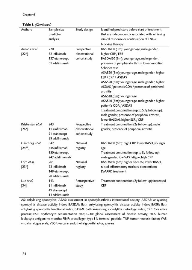
Chapter 6
84
Table 1. (Continued)
Authors Sample size
predictor
analysis
Study design Identified predictors before start of treatment
that are independently associated with achieving
clinical response or continuation of TNF-α
blocking therapy
Arends et al.
[22*]
220
32 infliximab
137 etanercept
51 adalimumab
Prospective
observational
cohort study
BASDAI50 (3m): younger age, male gender,
higher CRP / ESR
BASDAI50 (6m): younger age, male gender,
presence of peripheral arthritis, lower modified
Schober test
ASAS20 (3m): younger age, male gender, higher
ESR / CRP / ASDAS
ASAS20 (6m): younger age, male gender, higher
ASDAS / patient’s GDA / presence of peripheral
arthritis
ASAS40 (3m): younger age
ASAS40 (6m): younger age, male gender, higher
patient’s GDA / ASDAS
Treatment continuation (up to 5.7y follow-up):
male gender, presence of peripheral arthritis,
lower BASDAI, higher ESR / CRP
Kristensen et al.
[26*]
243
113 infliximab
91 etanercept
39 adalimumab
Prospective
observational
cohort study
Treatment continuation (2y follow-up): male
gender, presence of peripheral arthritis
Glintborg et al.
[24**]
842
445 infliximab
150 etanercept
247 adalimumab
National
registry
BASDAI50 (6m): high CRP, lower BASFI, younger
age
Treatment continuation (up to 8y follow-up):
male gender, low VAS fatigue, high CRP
Lord et al.
[23*]
261
93 infliximab
148 etanercept
20 adalimumab
National
registry
BASDAI50 (6m): higher BASDAI, lower BASFI,
raised inflammatory markers, concomitant
DMARD treatment
Luc et al.
[34]
143
81 infliximab
49 etanercept
13 adalimumab
Retrospective
study
Treatment continuation (2y follow-up): increased
CRP
AS: ankylosing spondylitis; ASAS: assessment in spondyloarthritis international society; ASDAS: ankylosing
spondylitis disease activity index; BASDAI: Bath ankylosing spondylitis disease activity index; BASFI: Bath
ankylosing spondylitis functional index; BASMI: Bath ankylosing spondylitis metrology index; CRP: C-reactive
protein; ESR: erythrocyte sedimentation rate; GDA: global assessment of disease activity; HLA: human
leukocyte antigen; m: months; PINP: procollagen type I N-terminal peptide; TNF: tumor necrosis factor; VAS:
visual analogue scale; VEGF: vascular endothelial growth factor; y: years
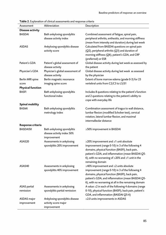
Baseline predictors of response: an overview
85
Table 2. Explanation of clinical assessments and response criteria
Assessment Abbreviation Description
Disease activity
BASDAI Bath ankylosing spondylitis
disease activity index
Combined assessment of fatigue, spinal pain,
peripheral arthritis, enthesitis, and morning stiffness
(mean from intensity and duration) during last week
ASDAS Ankylosing spondylitis disease
activity score
Calculated from BASDAI questions on spinal pain
(Q2), peripheral arthritis (Q3) and duration of
morning stiffness (Q6), patient’s GDA, and CRP
(preferred) or ESR
Patient’s GDA Patient’s global assessment of
disease activity
Global disease activity during last week as assessed by
the patient
Physician’s GDA Physician’s global assessment of
disease activity
Global disease activity during last week as assessed
by the physician
Berlin MRI spine
score
Berlin magnetic resonance
imaging spine score
Extent of bone marrow edema (grade 0-3) for 23
vertebral units from C2/C3 to L5/S1
Physical function
BASFI Bath ankylosing spondylitis
functional index
Includes 8 questions relating to the patient’s function
and 2 questions relating to the patient’s ability to
cope with everyday life
Spinal mobility
BASMI Bath ankylosing spondylitis
metrology index
Combination assessment of tragus to wall distance,
lumbar flexion (modified Schober test), cervical
rotation, lateral lumbar flexion, and maximal
intermalleolar distance
Response criteria
BASDAI50 Bath ankylosing spondylitis
disease activity index 50%
improvement
≥50% improvement in BASDAI
ASAS20 Assessments in ankylosing
spondylitis 20% improvement
≥20% improvement and ≥1 unit absolute
improvement (range 0-10) in 3 of the following 4
domains, physical function (BASFI), back pain,
patient’s GDA, and inflammation (mean BASDAI Q5-
6), with no worsening of ≥20% and ≥1 unit in the
remaining domain
ASAS40 Assessments in ankylosing
spondylitis 40% improvement
≥40% improvement and ≥2 units absolute
improvement (range 0-10) in 3 of the following 4
domains, physical function (BASFI), back pain,
patient’s GDA, and inflammation (mean BASDAI Q5-
6), with no worsening at all in the remaining domain
ASAS partial
remission
Assessments in ankylosing
spondylitis partial remission
A value ≤2 in each of the following 4 domains (range
0-10), physical function (BASFI), back pain, patient’s
GDA, and inflammation (BASDAI Q5-6)
ASDAS major
improvement
Ankylosing spondylitis disease
activity score major
improvement
≥2.0 units improvements in ASDAS

Chapter 6
86
Biomarkers as potential predictors
Several studies reported promising data regarding the potential value of biomarkers, for
example markers of inflammation or bone and cartilage metabolism, as baseline predictors of
response to TNF-α blocking therapy. Romero-Sanchez et al.39 tested 22 cytokines and found
that only serum levels of IL-1α before start of treatment were able to distinguish ASAS40
responders from non-responders. However, in receiver operating characteristics analysis, the
accuracy to predict treatment response was moderate (area under the curve: 0.71) and the
best cutoff value had a sensitivity of 84.9% and a specificity of 53.8%.
De Vries et al.20 reported that elevated baseline levels of serum amyloid A (SAA), an acute
phase reactant, were related to BASDAI50 response in univariate analysis. In addition, they
showed that the combination of elevated CRP and SAA levels was the strongest predictor of
BASDAI50 response.
Woo et al.40 suggested the potential usefulness of matrix metalloproteinase-3 (MMP-3), an
enzyme involved in degradation of extracellular matrix components, as a biomarker to
monitor response to TNF-α blocking therapy. A recent study found that axial SpA patients with
major improvement in ASDAS after anti-TNF-α treatment had significantly higher baseline
MMP-3 levels compared to patients without major improvement.35 However, two other
studies showed that serum MMP-3 levels at baseline could not predict ASAS20 or ASAS40
response after TNF-α blocking therapy.39,41
Pedersen et al.35 found that baseline levels of vascular endothelial growth factor (VEGF), a
biomarker of angiogenesis, were higher and that baseline levels of the cartilage glycoprotein
YKL-40, a biomarker of chronic inflammation, were lower in patients achieving BASDAI
response and ASDAS major improvement. Furthermore, ASDAS responders had higher
baseline levels of interleukin 6 (IL-6) and C-terminal crosslinking telopeptide of type II collagen
(CTX-II), a biomarker of cartilage degradation. The results regarding IL-6 are in line with
Visvanathan et al.,36 who reported that a greater percentage of AS patients with elevated IL-6
before start of treatment achieved BASDAI50 and ASAS20 responses compared to those with
low IL-6.
Finally, Wagner et al.32* tested 92 serum proteins and identified lower baseline levels of leptin,
a hormone that plays a central role in fat metabolism, and higher baseline levels of
immunoglobulin M and VEGF as the strongest predictors of BASDAI50 response after TNF-α
blocking therapy. Furthermore, they showed that lower baseline levels of insulin, a hormone
regulating carbohydrate and fat metabolism, and procollagen type 1 N-terminal peptide
(PINP), a marker of bone formation, were most predictive for ASAS20 response.
Until now, the results of these studies are either not confirmed by other study groups or
confirmed in studies that used less robust techniques of data analysis. Further studies using
multivariate analyses are needed to confirm the predictive value of these biomarkers,
in addition to the currently known predictors.
Baseline predictors of response: an overview
87
FUTURE PERSPECTIVES
Several studies tested a large group of serum proteins instead of following a hypothesis-driven
approach.32*,39 These analyses can contribute to the identification of potentially valuable
predictors. The pathogenetic relevance of such predictive proteins still has to be proven.
For evaluating TNF-α blocking therapy, not only the response regarding disease activity is
important, but also the effect on disease-related quality of life and radiographic outcome.
Therefore, it may also be interesting to use these parameters as endpoints in studies
investigating whether patient characteristics before start of treatment are able to predict a
beneficial effect of TNF-α blocking therapy. A drawback of using radiographic progression as
an endpoint is the need for long-term observation in order to be able to see any effect of
TNF-α blocking therapy.
The currently identified single predictors have, at best, moderate capacity to predict treatment
response in the individual patient and, therefore, they should not be mandatory for allowing
AS patients to be treated with TNF-α blocking agents. Recently, Vastesaeger et al.31** took the
first step to create a model that provides a potential basis for patient selection for TNF-α
blocking therapy. The development of such a prediction model may lead to a more robust
instrument to support physicians to make evidence-based decisions to start anti-TNF-α
treatment in daily clinical practice.
CONCLUSION
Although TNF-α blocking therapy is effective in the majority of patients with AS, identifying
patients who are most likely to benefit from TNF-α blocking therapy is important, especially
considering the high costs and potential side effects of these agents. Currently,
recommendations for starting TNF-α blocking therapy in AS are primarily based on inadequate
response to conventional treatment and less on the expectation that anti-TNF-α treatment will
be effective in a particular patient. Multiple studies using data from clinical trials and
observational studies in AS have identified increased acute phase reactants, presence of
peripheral arthritis, higher disease activity, higher functional status, younger age, male sex, and
HLA-B27 positivity as independent baseline predictors for achieving clinical response after 3-6
months and/or for long-term continuation of TNF-α blocking therapy in multivariate analyses
(Figure 1). Currently, the predictive value of single parameters is not strong enough to predict
treatment response in the individual AS patient. The development of a prediction model may
lead to a better predictive instrument to support physicians when deciding on TNF-α blocker
use in daily clinical practice.

Chapter 6
88
Figure 1. Repeatedly identified as independent baseline predictors for achieving clinical response after 3
to 6 months and/or for long-term continuation of TNF-α blocking therapy in patients with AS
KEY POINTS
Identifying AS patients who are most likely to benefit from TNF-α blocking therapy is
relevant, especially in view of the high costs and potential side effects of these agents.
Increased acute phase reactants, presence of peripheral arthritis, higher disease activity,
higher functional status, younger age, male sex, and HLA-B27 positivity are identified as
independent predictors for achieving clinical response and/or for continuation of TNF-α
blocking therapy in multivariate analyses.
The predictive value of single parameters is not strong enough to predict treatment
response in the individual patient, but the development of a prediction model may
support physicians in decision-making on TNF-α blocking therapy in AS in daily clinical
practice.
Baseline predictors of response: an overview
89
REFERENCES
1. Braun J, Deodhar A, Dijkmans B, et al. Efficacy and safety of infliximab in patients with ankylosing
spondylitis over a two-year period. Arthritis Rheum 2008;59:1270-8.
2. Dijkmans B, Emery P, Hakala M, et al. Etanercept in the longterm treatment of patients with
ankylosing spondylitis. J Rheumatol 2009;36:1256-64.
3. van der Heijde D, Schiff MH, Sieper J, et al. Adalimumab effectiveness for the treatment of
ankylosing spondylitis is maintained for up to 2 years: long-term results from the ATLAS trial.
Ann Rheum Dis 2009;68:922-9.
4. Braun J, Deodhar A, Inman RD, et al. Golimumab administered subcutaneously every 4 weeks in
ankylosing spondylitis: 104-week results of the GO-RAISE study. Ann Rheum Dis 2012;71:661-7.
5. van der Heijde D, Sieper J, Maksymowych WP, et al. 2010 Update of the international ASAS
recommendations for the use of anti-TNF agents in patients with axial spondyloarthritis.
Ann Rheum Dis 2011;70:905-8.
6. Rudwaleit M, Khan MA, Sieper J. The challenge of diagnosis and classification in early ankylosing
spondylitis: do we need new criteria? Arthritis Rheum 2005;52:1000-8.
7. Baraliakos X, Listing J, Rudwaleit M, et al. Progression of radiographic damage in patients with
ankylosing spondylitis: defining the central role of syndesmophytes. Ann Rheum Dis 2007;66:910-5.
8. Geusens P, Vosse D, van der Linden S. Osteoporosis and vertebral fractures in ankylosing
spondylitis. Curr Opin Rheumatol 2007;19:335-9.
9. Vosse D, Landewe R, van der Heijde D, et al. Ankylosing spondylitis and the risk of fracture: results
from a large primary care-based nested case-control study. Ann Rheum Dis 2009;68:1839-42.
10. Arends S, Spoorenberg A, Bruyn GA, et al. The relation between bone mineral density, bone
turnover markers, and vitamin D status in ankylosing spondylitis patients with active disease:
a cross-sectional analysis. Osteoporos Int 2011;22:1431-9.
11. Braun J, Sieper J. Ankylosing spondylitis. Lancet 2007;369:1379-90.
12. Braun J, van den Berg R, Baraliakos X, et al. 2010 update of the ASAS/EULAR recommendations for
the management of ankylosing spondylitis. Ann Rheum Dis 2011;70:896-904.
13. van der Heijde D, Kivitz A, Schiff MH, et al. Efficacy and safety of adalimumab in patients with
ankylosing spondylitis: results of a multicenter, randomized, double-blind, placebo-controlled trial.
Arthritis Rheum 2006;54:2136-46.
14. Inman RD, Davis JC,Jr, van der Heijde D, et al. Efficacy and safety of golimumab in patients with
ankylosing spondylitis: results of a randomized, double-blind, placebo-controlled, phase III trial.
Arthritis Rheum 2008;58:3402-12.
15. Rudwaleit M, Listing J, Brandt J, et al. Prediction of a major clinical response (BASDAI 50) to tumour
necrosis factor alpha blockers in ankylosing spondylitis. Ann Rheum Dis 2004;63:665-70.
16. Sieper J, Rudwaleit M, Baraliakos X, et al. The Assessment of SpondyloArthritis international Society
(ASAS) handbook: a guide to assess spondyloarthritis. Ann Rheum Dis 2009;68:ii1-44.
17. Anderson JJ, Baron G, van der Heijde D, et al. Ankylosing spondylitis assessment group preliminary
definition of short-term improvement in ankylosing spondylitis. Arthritis Rheum 2001;44:1876-86.
18. van der Heijde D, Dijkmans B, Geusens P, et al. Efficacy and safety of infliximab in patients with
ankylosing spondylitis: results of a randomized, placebo-controlled trial (ASSERT). Arthritis Rheum
2005;52:582-91.
19. Davis JC,Jr, van der Heijde D, Braun J, et al. Recombinant human tumor necrosis factor receptor
(etanercept) for treating ankylosing spondylitis: a randomized, controlled trial. Arthritis Rheum
2003;48:3230-6.

Chapter 6
90
20. de Vries MK, van Eijk IC, van der Horst-Bruinsma IE, et al. Erythrocyte sedimentation rate, C-reactive
protein level, and serum amyloid a protein for patient selection and monitoring of anti-tumor
necrosis factor treatment in ankylosing spondylitis. Arthritis Rheum 2009;61:1484-90.
21. Pedersen SJ, Sorensen IJ, Hermann KG, et al. Responsiveness of the Ankylosing Spondylitis Disease
Activity Score (ASDAS) and clinical and MRI measures of disease activity in a 1-year follow-up study
of patients with axial spondyloarthritis treated with tumour necrosis factor alpha inhibitors.
Ann Rheum Dis 2010;69:1065-71.
* The construct validity and high responsiveness of ASDAS in comparison with conventional clinical
measures of disease activity is demonstrated in patients with axial SpA treated with TNF-α blocking
agents.
22. Arends S, Brouwer E, van der Veer E, et al. Baseline predictors of response and discontinuation of
TNF-alpha blocking therapy in ankylosing spondylitis: a prospective longitudinal observational
cohort study. Arthritis Res Ther 2011;13:R94.
* This study shows the predictive value of characteristics of patients with AS before start of
treatment, including ASDAS, for clinical response and discontinuation of TNF-α blocking therapy
using multivariate analyses.
23. Lord PA, Farragher TM, Lunt M, et al. Predictors of response to anti-TNF therapy in ankylosing
spondylitis: results from the British Society for Rheumatology Biologics Register. Rheumatology
(Oxford) 2010;49:563-70.
* This study evaluates the effectiveness of TNF-α blocking therapy in AS patients during routine
clinical practice and identifies variables before start of treatment which are independently
associated with improvements in disease activity and function.
24. Glintborg B, Ostergaard M, Krogh NS, et al. Predictors of treatment response and drug continuation
in 842 patients with ankylosing spondylitis treated with anti-tumour necrosis factor: results from 8
years' surveillance in the Danish nationwide DANBIO registry. Ann Rheum Dis 2010;69:2002-8.
** In this large study using long-term data from the Danish nationwide rheumatological database,
independent baseline predictors of drug survival and clinical response are identified in patients with
AS receiving their first TNF-α blocking agent in routine care.
25. Pavelka K, Forejtova S, Stolfa J, et al. Anti-TNF therapy of ankylosing spondylitis in clinical practice.
Results from the Czech national registry ATTRA. Clin Exp Rheumatol 2009;27:958-63.
26. Kristensen LE, Karlsson JA, Englund M, et al. Presence of peripheral arthritis and male sex predicting
continuation of anti-tumor necrosis factor therapy in ankylosing spondylitis: an observational
prospective cohort study from the South Swedish Arthritis Treatment Group Register. Arthritis
Care Res (Hoboken ) 2010;62:1362-9.
* The potential value of clinical characteristics to predict 2-year drug survival is examined in AS
patients treated with TNF-α blocking therapy in clinical practice.
27. Schabert VF, Goodman S, Watson C, et al. Annual costs of tumor necrosis factor inhibitors using
real-world data in a commercially insured population in the United States. J Med Econ 2012;15:264-
75.
28. Gerard-Benoist S, Leux C, Le Goff B, et al. Frequency of hospitalization and severe events in 497
spondylarthritis patients treated with anti-TNF drugs: Comparisons with 423 RA patients. Joint Bone
Spine 2012;79:204-5.
29. Davis JC,Jr., van der Heijde DM, Dougados M, et al. Baseline factors that influence ASAS 20 response
in patients with ankylosing spondylitis treated with etanercept. J Rheumatol 2005;32:1751-4.

Baseline predictors of response: an overview
91
30. Rudwaleit M, Schwarzlose S, Hilgert ES, et al. MRI in predicting a major clinical response to
anti-tumour necrosis factor treatment in ankylosing spondylitis. Ann Rheum Dis 2008, 67:1276-
1281.
31. Vastesaeger N, van der Heijde D, Inman RD, et al. Predicting the outcome of ankylosing spondylitis
therapy. Ann Rheum Dis 2011,70:973-81.
** This is a large study using data from two clinical trials that identifies baseline predictors of various
disease-state and disease-outcome instruments in AS. Subsequently, the authors create a model
that identifies AS subpopulations likely to respond optimally to TNF-α blocking therapy.
32. Wagner C, Visvanathan S, Braun J, et al. Serum markers associated with clinical improvement in
patients with ankylosing spondylitis treated with golimumab. Ann Rheum Dis 2012;71:674-80.
* Approximately 100 different serum proteins are evaluated to identify markers associated with
clinical response on golimumab treatment in AS.
33. Rudwaleit M, Claudepierre P, Wordsworth P, et al. Effectiveness, safety, and predictors of good
clinical response in 1250 patients treated with adalimumab for active ankylosing spondylitis.
J Rheumatol 2009;36:801-8.
34. Luc M, Gossec L, Ruyssen-Witrand A, et al. C-reactive protein predicts tumor necrosis factor-alpha
blocker retention rate in axial ankylosing spondylitis. J Rheumatol 2007;34:2078-81.
35. Pedersen SJ, Sorensen IJ, Garnero P, et al. ASDAS, BASDAI and different treatment responses and
their relation to biomarkers of inflammation, cartilage and bone turnover in patients with axial
spondyloarthritis treated with TNFα inhibitors. Ann Rheum Dis 2011;70:1375-81.
36. Visvanathan S, Wagner C, Marini JC, et al. Inflammatory biomarkers, disease activity and spinal
disease measures in patients with ankylosing spondylitis after treatment with infliximab.
Ann Rheum Dis 2008;67:511-7.
37. Lukas C, Landewe R, Sieper J, et al. Development of an ASAS-endorsed disease activity score
(ASDAS) in patients with ankylosing spondylitis. Ann Rheum Dis 2009;68:18-24.
38. van der Heijde D, Lie E, Kvien TK, et al. ASDAS, a highly discriminatory ASAS-endorsed disease
activity score in patients with ankylosing spondylitis. Ann Rheum Dis 2009;68:1811-8.
39. Romero-Sanchez C, Robinson WH, Tomooka BH, et al. Identification of acute phase reactants and
cytokines useful for monitoring infliximab therapy in ankylosing spondylitis. Clin Rheumatol
2008;27:1429-35.
40. Woo JH, Lee HJ, Sung IH, et al. Changes of clinical response and bone biochemical markers in
patients with ankylosing spondylitis taking etanercept. J Rheumatol 2007;34:1753-9.
41. Arends S, van der Veer E, Groen H, et al. Serum MMP-3 level as a biomarker for monitoring and
predicting response to etanercept treatment in ankylosing spondylitis. J Rheumatol 2011;38:1644-
50.

Chapter 6
92

CHAPTER 7
THE FORMATION OF AUTOANTIBODIES AND ANTIBODIES TO TNF-α
BLOCKING AGENTS IN RELATION TO CLINICAL RESPONSE IN PATIENTS
WITH ANKYLOSING SPONDYLITIS
Suzanne Arends1*, Helma R. Lebbink1*, Anneke Spoorenberg2, Laura Bungener3,
Caroline Roozendaal3, Eveline van der Veer3, Pieternella M. Houtman2, Eduard N. Griep2,
Pieter C. Limburg1,3, Cees G. M. Kallenberg1, Gert Jan Wolbink4, Elisabeth Brouwer1
Departments of 1 Rheumatology and Clinical Immunology, 3 Laboratory Medicine,
University of Groningen, University Medical Center Groningen, 2 Rheumatology,
Medical Center Leeuwarden, 4 Sanquin Research / Jan van Breemen Research Institute,
Amsterdam, The Netherlands, * Both authors contributed equally to this work.
Clin Exp Rheumatol. 2010;28;661-8.

Chapter 7
94
ABSTRACT
Objective: To investigate the influence of antibody formation to TNF-α blocking agents on
the clinical response in AS patients treated with infliximab (IFX), etanercept (ETA), or
adalimumab (ADA), and to investigate the development of ANA, ANCA, and anti-dsDNA
antibodies in association with the formation of antibodies to TNF-α blocking agents.
Methods: Consecutive AS outpatients with active disease who started treatment with IFX
(n=20), ETA (n=20), or ADA (n=20) were included in this longitudinal observational study.
Clinical data were collected prospectively at baseline and after 3, 6, and 12 months of
anti-TNF-α treatment. At the same time points, serum samples were collected. In these
samples, antibodies to TNF-α blocking agents, serum TNF-α blocker levels, and ANA, ANCA,
and anti-dsDNA antibodies were measured retrospectively.
Results: Anti-IFX, anti-ETA, and anti-ADA antibodies were induced in 20%, 0%, and 30% of
patients, respectively. Although ANA, ANCA, and anti-dsDNA antibodies were detected
during anti-TNF-α treatment, no significant association was found between the presence of
these autoantibodies and the formation of antibodies to TNF-α blocking agents. Patients with
anti-IFX or anti-ADA antibodies had significantly lower serum TNF-α blocker levels compared
to patients without these antibodies. Furthermore, significant negative correlations were
found between serum TNF-α blocker levels and assessments of disease activity.
Conclusion: This study indicates that antibody formation to IFX or ADA is related to a
decrease in efficacy and early discontinuation of anti-TNF-α treatment in AS patients.
Furthermore, autoantibody formation seems not to be associated with antibody formation to
TNF-α blocking agents.

Formation of antibodies to TNF-α blocking agents
95
INTRODUCTION
Ankylosing Spondylitis (AS) is a chronic inflammatory rheumatic disease that primarily affects
the axial skeleton. Peripheral joints and extra-articular structures may also be involved. The
availability of the tumor necrosis factor alpha (TNF-α) blocking agents infliximab (IFX),
etanercept (ETA), and adalimumab (ADA) has significantly improved outcome in AS.1
Although the majority of patients respond very well to these TNF-α blocking agents,
approximately 30% fail to reach efficacy.2-4 One possible explanation for this failure could be
that the formation of antibodies to TNF-α blocking agents may decrease serum TNF-α blocker
levels. Previous studies in rheumatoid arthritis (RA) have shown that the development of
neutralizing antibodies is associated with allergic reactions and reduced response to
treatment.5-7 Recently, de Vries et al. published 3 studies on antibody formation to TNF-α
blocking agents in different AS cohorts. They detected antibodies in 29% and 31% of the
patients treated with IFX and ADA, respectively, and no antibodies in patients treated with
ETA.8-10
Unlike several other inflammatory auto-immune diseases, there are no disease specific
autoantibodies found in AS. However, the presence of well-known autoantibodies such as
antinuclear antibodies (ANA), anti-neutrophil cytoplasmatic antibodies (ANCA), and
anti-double-stranded DNA (anti-dsDNA) antibodies has been described in AS patients.11,12
Furthermore, previous studies have shown that ANA and anti-dsDNA antibodies can be
induced during anti-TNF-α treatment.12-15 Since patients who produce autoantibodies have a
more activated immune system, it may be suggested that these patients are more prone to the
production of antibodies to TNF-α blocking agents.
The objective of the present study was to investigate the influence of antibody formation to
TNF-α blocking agents on the clinical response in AS patients treated with IFX, ETA, or ADA in
daily clinical practice. The second goal was to investigate the development of ANA, ANCA, and
anti-dsDNA antibodies in association with the formation of antibodies to TNF-α blocking
agents.
PATIENTS AND METHODS
Patients
Consecutive AS outpatients with active disease who started treatment with infliximab (IFX,
n=20), etanercept (ETA, n=20), or adalimumab (ADA, n=20) at the Medical Center
Leeuwarden (MCL) or the University Medical Center Groningen (UMCG) were included in
this longitudinal observational study. All patients were aged ≥18 years, fulfilled the modified
New York criteria for AS,16 and started anti-TNF-α treatment because of active disease
according to the Assessments in Ankylosing Spondylitis (ASAS) consensus statement.17
Patients were excluded if they had previously received anti-TNF-α treatment. IFX (5mg/kg) was
given intravenously at 0, 2 and 6 weeks and then every 8 weeks. ETA was administered as a
subcutaneous injection once (50 mg) or twice (25 mg) a week. ADA (40 mg) was administered
as a subcutaneous injection every 2 weeks. The choice of the TNF-α blocking agent was based

Chapter 7
96
on the judgment of the treating rheumatologist and/or the specific preference of the patient.
Patients were allowed to receive concomitant medication as usual in daily clinical practice. The
study was approved by the local ethics committees of the MCL and UMCG and all patients
provided written informed consent to participate in this study.
Clinical assessments
Clinical data were collected prospectively at baseline and after 3, 6, and 12 months of
anti-TNF-α treatment. Continuation of anti-TNF-α treatment was based on decrease in Bath
Ankylosing Spondylitis Disease Activity Index (BASDAI), amounting 50% or 2 units compared
with baseline, and/or expert opinion in favor of treatment continuation. Response to
anti-TNF-α treatment was defined using BASDAI and ASAS20 response criteria. ASAS20
response was defined as an improvement of at least 20% and absolute improvement of at least
1 unit (on a scale of 0-10) compared with baseline in 3 or more of the 4 domains: physical
function (Bath Ankylosing Spondylitis Functional Index (BASFI)), pain, patient’s global
assessment of disease activity, and inflammation (mean from BASDAI questions 5 and 6), with
no worsening by more than 20% in the remaining domain.18 Disease activity was assessed
using BASDAI, erythrocyte sedimentation rate (ESR), C-reactive protein (CRP), and the
Ankylosing Spondylitis disease activity score (ASDAS) calculated from BASDAI questions 2, 3
and 6, patient’s global assessment of disease activity, and CRP.19,20
Laboratory assessments
Antibodies to TNF-α blocking agents, serum TNF-α blocker levels, and ANA, ANCA, and
anti-dsDNA antibodies were measured retrospectively at baseline and after 3, 6, and 12
months of anti-TNF-α treatment. Analyses in samples from one patient were always performed
in one assay to avoid inter-assay variability. Samples were stored at -20°C until analysis.
Anti-IFX and anti-ADA antibodies were detected by radioimmunoassay (RIA) as described in
detail previously.5,6 The assay measures specific high-avidity IgG antibodies to IFX or ADA by an
antigen-binding test. In short, serum (1 μl/test) was pre-incubated with Sepharose-
immobilized protein A (1 mg/test; Pharmacia, Uppsala, Sweden) in Freeze buffer (Sanquin,
Amsterdam, The Netherlands). Non-bound serum components were removed by washing
before 50 μl 125I-radiolabeled F(ab)’2 fragment of IFX or ADA was added. After overnight
incubation, non-bound radiolabel was washed away and Sepharose-bound radioactivity was
measured. Test results were converted into arbitrary units per milliliter (AU/ml) by
comparison with dilutions of a reference serum. The reference value was set at 12 AU/ml, as
derived from 100 healthy donors. Anti-ETA antibodies were detected using a two-site assay
RIA using Sepharose-immobilized ETA as solid phase for capturing ETA-specific antibody and 125I-radiolabeled ETA for detection.9 It should be noted that antibodies to the TNF-α blocking
agent may be underestimated or undetectable in patients with high serum concentrations of
TNF-α blocker since the presence of TNF-α blocking agent interferes with the assay.

Formation of antibodies to TNF-α blocking agents
97
Serum IFX, ETA, and ADA levels were measured by enzyme-linked immunosorbent assay
(ELISA; Sanquin, Amsterdam, The Netherlands) as described previously.8,9 The ELISA is based
on the principle that the TNF-α blocking agent is captured through its ability to bind TNF-α.
The sensitivity of detection for IFX, ETA and ADA was 0.0003 μg/ml, 0.001 μg/ml, and 0.001
μg/ml, respectively.
Serum samples were tested for ANA by indirect immunofluorescence using fixed Hep-2000
cells (ImmunoConcepts, Biomedical Diagnostics, Antwerp, Belgium) as recommended by the
manufacturer. ANA titer and pattern were reported. An ANA titer ≥1:40 was considered
positive. Detection of ANCA by indirect immunofluorescence was performed on ethanol-fixed
granulocytes as described before21,22 with minor modifications.23 An ANCA titer ≥1:40 was
considered positive. Anti-dsDNA antibodies were measured by Farr RIA (Siemens Healthcare
Diagnostics, Breda, The Netherlands) and by anti-dsDNA IgG and IgM ELISA on the Alegria
(ORGENTEC, supplied by Siemens Healthcare Diagnositics). The Farr assay detects both IgG
and IgM antibodies against dsDNA using 125I-labeled recombinant dsDNA as a substrate.
A result ≥10 IU/ml was considered positive. To be able to distinguish between IgG and IgM
responses, all samples positive for anti-dsDNA in the Farr assay were also measured in the
separate IgG and IgM assays on the Alegria. A result ≥20 IU/ml was considered positive.
Statistical analysis
Statistical analysis was performed with SPSS 16.0 software (SPSS, Chicago, IL, USA). Results
were expressed as mean ± SD or median (range) for parametric and nonparametric data,
respectively. The Independent Samples T test and Mann-Whitney U test were used to
compare differences between groups. The Chi-Square test and Fisher Exact test were used to
compare percentages between groups. The Paired Samples T test and Wilcoxon Signed Rank
test were used to compare differences within groups. The Fisher Exact test was used to
analyze the relationship between the presence or absence of antibodies to TNF-α blocking
agent and the presence or absence of ANA, ANCA, and anti-dsDNA antibodies. The
Spearman’s correlation coefficient was used to analyze the relationship between serum levels
of the TNF-α blocking agent and BASDAI, CRP, ESR, and ASDAS. Patients who had temporarily
stopped their anti-TNF-α treatment in the period just before their 3, 6, or 12 month visit were
excluded from the corresponding analysis. P values <0.05 were considered statistically
significant.
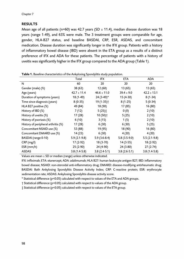
Chapter 7
98
RESULTS
Mean age of all patients (n=60) was 42.7 years (SD ± 11.4), median disease duration was 18
years (range 1-49), and 63% were male. The 3 treatment groups were comparable for age,
gender, HLA-B27 status, and baseline BASDAI, CRP, ESR, ASDAS, and concomitant
medication. Disease duration was significantly longer in the IFX group. Patients with a history
of inflammatory bowel disease (IBD) were absent in the ETA group as a results of a distinct
preference of IFX and ADA for these patients. The percentage of patients with a history of
uveitis was significantly higher in the IFX group compared to the ADA group (Table 1).
Table 1. Baseline characteristics of the Ankylosing Spondylitis study population.
Total IFX ETA ADA
N 60 20 20 20
Gender (male) (%) 38 (63) 12 (60) 13 (65) 13 (65)
Age (years) 42.7 ± 11.4 46.6 ± 11.0 39.4 ± 9.0 42.2 ± 13.1
Duration of symptoms (years) 18 (1-49) 24 (3-49)* 15 (4-30) 8 (1-34)
Time since diagnosis (years) 8 (0-35) 19 (1-35)† 8 (1-25) 5 (0-34)
HLA B27 positive (%) 49 (84) 18 (90) 17 (85) 16 (80)
History of IBD (%) 7 (12) 5 (25)‡ 0 (0) 2 (10)
History of uveitis (%) 17 (28) 10 (50)† 5 (25) 2 (10)
History of psoriasis (%) 6 (10) 3 (15) 1 (5) 2 (10)
History of peripheral arthritis (%) 17 (28) 6 (30) 6 (30) 5 (25)
Concomitant NSAID use (%) 53 (88) 19 (95) 18 (90) 16 (80)
Concomitant DMARD use (%) 14 (23) 6 (30) 4 (20) 4 (20)
BASDAI (range 0-10) 5.9 (2.1-9.8) 5.9 (3.6-8.4) 5.8 (3.5-9.0) 5.5 (2.1-9.8)
CRP (mg/l) 17 (2-92) 18 (3-70) 14 (3-55) 18 (2-92)
ESR (mm/h) 25 (2-90) 24 (4-90) 24 (3-80) 27 (2-74)
ASDAS 3.8 (1.4-5.8) 3.8 (2.4-5.1) 3.8 (2.6-5.1) 3.8 (1.4-5.8)
Values are mean ± SD or median (range) unless otherwise indicated.
IFX: infliximab; ETA: etanercept; ADA: adalimumab; HLA B27: human leukocyte antigen B27; IBD: inflammatory
bowel disease; NSAID: non-steroidal anti-inflammatory drug; DMARD: disease-modifying antirheumatic drug;
BASDAI: Bath Ankylosing Spondylitis Disease Activity Index; CRP: C-reactive protein; ESR: erythrocyte
sedimentation rate; ASDAS: Ankylosing Spondylitis disease activity score.
* Statistical difference (p<0.05) calculated with respect to values of the ETA and ADA groups.
† Statistical difference (p<0.05) calculated with respect to values of the ADA group.
‡ Statistical difference (p<0.05) calculated with respect to values of the ETA group.

Formation of antibodies to TNF-α blocking agents
99
Antibody formation to TNF-α blocking agents
At baseline, 1 patient tested positive for anti-ADA antibodies (24 AU/ml) and these antibodies
were still found after 3 months of treatment (18 AU/ml). In the treatment period of one year,
antibodies to TNF-α blocking agents were induced in 4 (20%) patients treated with IFX and in 6
(30%) patients treated with ADA. No antibodies to the TNF-α blocking agent were detected in
patients treated with ETA (Table 2). In 6 patients (IFX, n=1; ADA, n=5), the antibodies were
detected for the first time at 3 months. In the remaining 4 patients (IFX, n=3; ADA, n=1), the
antibodies were detected for the first time at 6 months. Serum concentrations of antibodies
were between 12 and 5154 AU/ml. The formation of antibodies to TNF-α blocking agents was
not related to the use of concomitant non-steroidal anti-inflammatory drugs (NSAIDs) or
disease-modifying antirheumatic drugs (DMARDs) (data not shown).
Autoantibody formation
No statistically significant differences were found in ANA, ANCA, and anti-dsDNA formation
between the 3 treatment groups. ANA and ANCA were present at baseline and new ANA and
ANCA were induced during anti-TNF-α treatment in all 3 groups (Table 2). ANA patterns
varied between homogeneous, coarse speckled, and nucleolar. The majority of ANCA showed
a p-ANCA fluorescence pattern. No anti-dsDNA antibodies were detected in the Farr assay at
baseline. In all treatment groups, anti-dsDNA antibodies were induced during anti-TNF-α
treatment in 20% to 30% of patients (Table 2). In 33% to 50% of these patients, the anti-dsDNA
antibodies disappeared at 12 months. Serum concentrations of anti-dsDNA autoantibodies
were low, ranging from 10 to 23 IU/ml. The anti-dsDNA autoantibodies detected in the Farr
assay were of IgM isotype in 5 patients, of IgG isotype in 1 patient, and of both isotypes in
5 patients, as determined by separate IgG and IgM assays on the Alegria. In the remaining
5 patients, no positive IgG or IgM anti-dsDNA response was seen in the Alegria test, mostly
due to responses just below the reference value of the assays. The seroconversion to IgG
anti-dsDNA antibodies was not associated with the appearance of clinically relevant lupus-like
symptoms.
No statistically significant association was found between the presence of ANA, ANCA, or
anti-dsDNA antibodies and the formation of antibodies to TNF-α blocking agents in both the
IFX and ADA group.

Chapter 7
100
Table 2. Antibodies to TNF-α blocking agents and autoantibodies in the IFX (n=20), ETA (n=20), and
ADA (n=20) groups.
Present at baseline Induction during treatment
IFX
Anti-IFX (%) 0 (0) 4 (20)
ANA (%) 10 (50) 6 (30)
ANA ≥ 1:80 (%) 4 (20) 3 (15)
ANCA (%)† 3 (15) 3 (15)
Anti-dsDNA Farr (%)
Only IgM
Only IgG
Both IgM and IgG
IgM and IgG negative‡
0 (0)
-
-
-
-
6 (30)
4
-
1
1
ETA
Anti-ETA (%) 0 (0) 0 (0)
ANA (%) 12 (60) 3 (15)
ANA ≥ 1:80 (%) 4 (20) 1 (5)
ANCA (%)† 1 (5) 3 (15)
Anti-dsDNA Farr (%)
Only IgM
Only IgG
Both IgM and IgG
IgM and IgG negative‡
0 (0)
-
-
-
-
4 (20)
1
-
1
2
ADA
Anti-ADA (%) 0 (0) 6 (30)
ANA (%) 6 (30) 4 (20)
ANA ≥ 1:80 (%) 4 (20) 3 (15)
ANCA (%)† 1 (5) 3 (15)
Anti-dsDNA Farr (%)
Only IgM
Only IgG
Both IgM and IgG
IgM and IgG negative‡
0 (0)
-
-
-
-
6 (30)
-
1
3
2
ANA: antinuclear antibodies; ANCA: anti-neutrophil cytoplasmatic antibodies; anti-dsDNA Farr:
anti-double-stranded DNA measured by Farr. See Table 1 for other definitions.
† The majority of positive ANCA showed a p-ANCA pattern of fluorescence.
‡ No positive IgG or IgM anti-dsDNA response was seen in the Alegria test, mostly due to responses
just below the reference value of the assays.

Formation of antibodies to TNF-α blocking agents
101
Antibody formation to TNF-α blocking agents and ASAS20 response
In all 3 groups, BASDAI, CRP, ESR, and ASDAS significantly improved after 3, 6, and 12 months
of anti-TNF-α treatment compared with baseline, except for ESR in the ADA group after 12
months of treatment (data not shown). The percentage of patients that reached BASDAI
response ranged from 60% to 85% for IFX, from 60% to 75% for ETA, and from 45% to 55% for
ADA over time. At 3 months, the percentage of BASDAI responders was significantly lower for
ADA compared to IFX. Furthermore, there was a trend suggesting a lower percentage of
BASDAI responders to ADA compared to IFX at 12 months (p=0.053). The percentage of
patients that reached ASAS20 response ranged from 65% to 85% for IFX, from 70% to 75% for
ETA, and from 45% to 65% for ADA over time. At 6 months, there was a trend suggesting a
lower percentage of ASAS20 responders to ADA compared to both IFX and ETA (p=0.053)
(Table 3).
During the first year, 15 patients discontinued anti-TNF-α treatment because inefficacy (IFX,
n=3; ETA, n=1; ADA, n=7), adverse events (ETA, n=1; ADA, n=1), inefficacy and adverse events
(IFX, n=1), or other reasons (IFX, n=1). There was a trend suggesting a higher percentage of
patients that discontinued ADA treatment compared to ETA treatment (p=0.065) (Table 3).
Of patients who discontinued anti-TNF-α treatment because of inefficacy, antibodies to TNF-α
blocking agents were detected in 3 of 4 patients treated with IFX and in 4 of 7 patients treated
with ADA. No antibodies were detected in the patient treated with ETA.
Antibodies to TNF-α blocking agents were also detected in 3 patients who continued
anti-TNF-α treatment. One patient was BASDAI and ASAS20 responder at all time points,
although anti-IFX antibodies were detected at 6 and 12 months (24 and 20 AU/ml,
respectively), resulting in very low serum IFX levels (1.05 and 1.22 μg/ml, respectively). Also an
increased ESR level was found in this patient at 12 months. In the second patient, anti-ADA
antibodies were detected at 3 and 6 months (60 and 18 AU/ml, respectively), resulting in low
serum ADA levels (1.81 and 1.64 μg/ml, respectively) and non-response. At 12 months, this
patient reached BASDAI and ASAS20 response and antibodies were not found anymore. In the
final patient, anti-ADA antibodies were detected at 3, 6 and 12 months (12, 5154 and 1120
AU/ml, respectively), resulting in low and undetectable serum ADA levels (2.77, 0.02 and 0.02
μg/ml, respectively) and increased ESR levels. However, the patient reached BASDAI and
ASAS20 response at 12 months.

Chapter 7
102
Table 3. Serum TNF-α blocker levels, treatment response and reasons for treatment discontinuation of
the IFX (n=20), ETA (n=20), and ADA (n=20) groups at 3, 6 and 12 months of anti-TNF-α treatment.
At 3 months At 6 months At 12 months Total
IFX
Serum IFX levels (μg/ml) 30.6 ± 30.0 36.3 ± 43.2 23.4 ± 26.9
BASDAI response (%) 17 (85) 14 (70) 12 (60)
ASAS20 response (%) 17 (85) 15 (75) 13 (65)
Treatment discontinuation (%)
Allergic reaction, inefficacy
Inefficacy
Pregnancy wish
1 (5)
1
0 (0) 4 (20)
3
1
5 (25)
1
3
1
ETA
Serum ETA levels (μg/ml) 3.1 ± 1.2 2.8 ± 1.6* 2.9 ± 1.0
BASDAI response (%) 15 (75) 12 (60) 15 (75)
ASAS20 response (%) 15 (75) 15 (75) 14 (70)
Treatment discontinuation (%)
Inefficacy
Cardiac complaints
2 (10)
1
1
0 (0) 0 (0) 2 (10)
1
1
ADA
Serum ADA levels (μg/ml) 5.7 ± 4.0 5.5 ± 3.8 8.0 ± 5.1
BASDAI response (%) 11 (55) † 11 (55) 9 (45) †††
ASAS20 response (%) 13 (65) 9 (45) †† 10 (50)
Treatment discontinuation (%)
Inefficacy
Diarrhea
3 (15)
3
4 (20)
3
1
1 (5)
1
8 (40)
7
1
Values are mean ± SD unless otherwise indicated. See Table 1 for definitions.
* Statistical difference (p<0.05) calculated with respect to values at 3 months.
† Statistical difference (p<0.05) calculated with respect to values of the IFX group.
†† Trend to statistical difference (p=0.053) calculated with respect to values of the IFX and ETA groups.
††† Trend to statistical difference (p=0.053) calculated with respect to values of the ETA group.
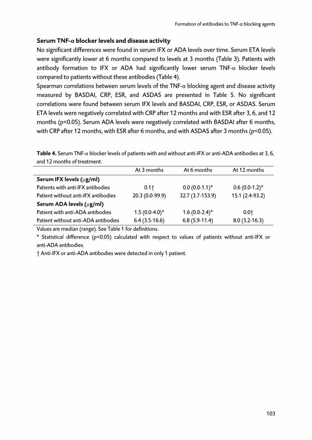
Formation of antibodies to TNF-α blocking agents
103
Serum TNF-α blocker levels and disease activity
No significant differences were found in serum IFX or ADA levels over time. Serum ETA levels
were significantly lower at 6 months compared to levels at 3 months (Table 3). Patients with
antibody formation to IFX or ADA had significantly lower serum TNF-α blocker levels
compared to patients without these antibodies (Table 4).
Spearman correlations between serum levels of the TNF-α blocking agent and disease activity
measured by BASDAI, CRP, ESR, and ASDAS are presented in Table 5. No significant
correlations were found between serum IFX levels and BASDAI, CRP, ESR, or ASDAS. Serum
ETA levels were negatively correlated with CRP after 12 months and with ESR after 3, 6, and 12
months (p<0.05). Serum ADA levels were negatively correlated with BASDAI after 6 months,
with CRP after 12 months, with ESR after 6 months, and with ASDAS after 3 months (p<0.05).
Table 4. Serum TNF-α blocker levels of patients with and without anti-IFX or anti-ADA antibodies at 3, 6,
and 12 months of treatment.
At 3 months At 6 months At 12 months
Serum IFX levels (μg/ml)
Patients with anti-IFX antibodies 0.1† 0.0 (0.0-1.1)* 0.6 (0.0-1.2)*
Patient without anti-IFX antibodies 20.3 (0.0-99.9) 32.7 (3.7-153.9) 15.1 (2.4-93.2)
Serum ADA levels (μg/ml)
Patient with anti-ADA antibodies 1.5 (0.0-4.0)* 1.6 (0.0-2.4)* 0.0†
Patient without anti-ADA antibodies 6.4 (3.5-16.6) 6.8 (5.9-11.4) 8.0 (3.2-16.3)
Values are median (range). See Table 1 for definitions.
* Statistical difference (p<0.05) calculated with respect to values of patients without anti-IFX or
anti-ADA antibodies.
† Anti-IFX or anti-ADA antibodies were detected in only 1 patient.

Chapter 7
104
Table 5. Spearman correlations between serum TNF-α blocker levels and BASDAI, CRP, ESR, and ASDAS
of the IFX (n=20), ETA (n=20), and ADA (n=20) groups at 3, 6, and 12 months of anti-TNF-α treatment.
BASDAI CRP ESR ASDAS
IFX
Serum IFX levels at 3 months NS NS NS NS
Serum IFX levels at 6 months NS NS NS NS
Serum IFX levels at 12 months NS NS NS NS
ETA
Serum ETA levels at 3 months NS NS -.561* NS
Serum ETA levels at 6 months NS NS -.519* NS
Serum ETA levels at 12 months NS -.599* -.808* NS
ADA
Serum ADA levels at 3 months NS NS NS -0.552*
Serum ADA levels at 6 months -.570* NS -.604* NS
Serum ADA levels at 12 months NS -.848* NS NS
NS : not significant. See Table 1 for other definitions.
* Statistical significant correlation (p<0.05).
DISCUSSION
The present study indicates that antibody formation to IFX or ADA is related to a decrease in
efficacy and early discontinuation of anti-TNF-α treatment in daily clinical practice, since
antibodies were detected in the majority of patients who discontinued IFX or ADA treatment
due to inefficacy. Furthermore, patients with anti-IFX or anti-ADA antibodies had significantly
lower serum TNF-α blocker levels compared to patients without these antibodies. Significant
negative correlations were observed between serum ADA levels and BASDAI, CRP, ESR, or
ASDAS, which suggests the importance of sufficiently high serum TNF-α blocker levels to
obtain clinical response.
The percentage of patients that developed anti-IFX and anti-ADA antibodies was comparable.
No anti-ETA antibodies were detected in this study, which is consistent with the report of de
Vries et al..9 The absence of antibodies to ETA may be explained by the fact that the dimeric
fusion protein ETA has a less immunogenic structure in comparison with the 2 monoclonal
antibodies IFX and ADA. Significant negative correlations between serum ETA levels and CRP
or ESR were found, which again seems to reflect the importance of sufficiently high serum
anti-TNF-α levels to obtain response in terms of decreased inflammation. However, no
significant correlation between serum ETA levels and BASDAI was found. BASDAI is a disease
activity score solely based on the opinion of the patient. A possible explanation for these
findings may thus be that patients with fewer subjective complaints are less compliant to
therapy, which results in low serum ETA levels, high CRP and ESR levels, and normal BASDAI
scores.
Interestingly, anti-ADA antibodies first appeared and then disappeared in one patient. This
finding is in accordance with Wolbink et al., who found that treatment continuation with IFX

Formation of antibodies to TNF-α blocking agents
105
resulted in a decrease of anti-IFX antibodies in RA patients.6 However, they continued IFX
treatment with increased dosages after the detection of anti-IFX antibodies and in the present
study we did not change the dose of ADA. Our patient had relatively low anti-ADA antibody
titers and low, but detectable serum ADA levels. Therefore, continuation of treatment may be
effective in patients with low antibody titers, whereas switching to another TNF-α blocking
agent may be a better therapeutic option in patients with high antibody titers.
When we compare clinical data of the IFX, ETA, and ADA groups, the response to ADA
treatment seems to be somewhat lower in comparison with the other two agents, and the
percentage of patients that discontinued ADA treatment seems to be somewhat higher in
comparison with ETA. However, the response rates to IFX and ETA found in this observational
study were comparable to those reported in randomized clinical trials,24,25 while the response
rate to ADA was somewhat inferior to that reported in the literature.4 The small-sized
treatment groups may have contributed to the differences in response rates in this study.
There were two unexpected findings in our study. First, one baseline sample behaved in an
atypical manner, showing a slightly positive test result for antibodies against ADA. This is
remarkable, since this patient had not yet received the drug at the time when the blood sample
was drawn. Inhibition experiments did not lead to conclusive results, which means that we
have to consider this sample as false-positive. This is a rare phenomenon which we have not
encountered previously, neither during test validation using 100 supposedly negative samples,
nor during extensive ADA testing which we perform routinely. Second, we found that
2 patients were responders to treatment according to the BASDAI and ASAS20 criteria,
despite the presence of antibodies to the TNF-α blocking agent and low or undetectable
serum TNF-α blocker levels. Moreover, ESR levels were increased in these patients. This
indicates that it may be useful to consider more objective measures such as acute phase
reactants as well as patient-related outcome measures when assessing clinical response to
anti-TNF-α treatment in AS.
ANA and ANCA antibodies were present at baseline and additionally developed during
anti-TNF-α treatment in all 3 groups. Anti-dsDNA antibodies were not present at baseline, but
were induced during anti-TNF-α treatment in all 3 groups. Interestingly, the anti-dsDNA
antibodies disappeared in approximately half of the patients after 12 months of treatment. The
presence of anti-dsDNA antibodies did not seem to be clinically relevant in this study since
serum concentrations were low and seroconversion to the more pathogenic IgG class
anti-dsDNA antibodies was not associated with the appearance of lupus-like symptoms. These
findings are in agreement with previous studies.12,13,26 To our knowledge, this is the first study
that investigates the relation between autoantibodies and antibodies to TNF-α blocking
agents. Although ANA, ANCA, and anti-dsDNA antibodies were detected in AS patients
treated with TNF-α blocking agents, no significant association was found between the
presence of these autoantibodies and the induction of antibodies to TNF-α blocking agents.
There are some limitations to this study. First, the 3 treatment groups were small and differed
significantly in disease duration as well as in the percentage of patients with extra-articular

Chapter 7
106
manifestations. Second, the collection of blood samples was not linked to the timing of
administration of the TNF-α blocking agent. The administration of anti-TNF-α treatment to
patients with antibodies to TNF-α blocking agents results in the formation of immune
complexes, which may accelerate the clearance of the applied agent and the antibodies.7
Therefore, the frequency of antibody formation to TNF-α blocking agents may have been
underestimated in our study. In addition, the lack of standardization of the collection of blood
samples increases the variation in measured serum TNF-α blocker levels. Despite this variation,
we still found significant negative correlations between serum anti-TNF-α levels and
assessments of disease activity. Only in the IFX group, no significant correlation was found.
This may be explained by the fact that the lack of standardization of the collection of blood
samples influences the results of the IFX group most since IFX is administered every 8 weeks.
In conclusion, this longitudinal observational study in daily clinical practice indicates that the
presence of ANA, ANCA, or anti-dsDNA antibodies is not associated with the formation of
antibodies to TNF-α blocking agents. The percentage of patients that developed anti-IFX and
anti-ADA antibodies was comparable in this study, while no anti-ETA antibodies were
detected. The formation of anti-IFX and anti-ADA antibodies seems to be related to a decrease
in efficacy and early discontinuation of anti-TNF-α treatment in AS patients, since anti-IFX or
anti-ADA antibodies were detected in the majority patients who discontinued treatment due
to inefficacy. Furthermore, patients with antibodies had significantly lower serum TNF-α
blocker levels compared to patients without antibodies. Finally, significant negative
correlations were found between serum levels of TNF-α blocking agents and assessments of
disease activity. Further studies in larger groups of AS patients are needed to confirm the
relation between antibody formation to TNF-α blocking agents, serum TNF-α blocker levels,
and assessments of disease activity. In these studies, blood samples should be taken
immediately before administration of the TNF-α blocking agent. However, based on the results
of this study it seems useful to determine antibody formation to TNF-α blocking agents in
non-responsive AS patients.

Formation of antibodies to TNF-α blocking agents
107
REFERENCES
1. Braun J, Kalden JR. Biologics in the treatment of rheumatoid arthritis and ankylosing spondylitis.
Clin Exp Rheumatol 2009;27:S164-7.
2. Calin A, Dijkmans BA, Emery P, et al. Outcomes of a multicentre randomised clinical trial of
etanercept to treat ankylosing spondylitis. Ann Rheum Dis 2004;63:1594-600.
3. van der Heijde D, Dijkmans B, Geusens P, et al. Efficacy and safety of infliximab in patients with
ankylosing spondylitis: results of a randomized, placebo-controlled trial (ASSERT). Arthritis Rheum
2005;52:582-91.
4. van der Heijde D, Schiff MH, Sieper J, et al. Adalimumab effectiveness for the treatment of
ankylosing spondylitis is maintained for up to 2 years: long-term results from the ATLAS trial.
Ann Rheum Dis 2009;68:922-9.
5. Bartelds GM, Wijbrandts CA, Nurmohamed MT, et al.: Clinical response to adalimumab:
relationship to anti-adalimumab antibodies and serum adalimumab concentrations in rheumatoid
arthritis. Ann Rheum Dis 2007; 66(7): 921-6.
6. Wolbink GJ, Vis M, Lems W, et al. Development of antiinfliximab antibodies and relationship to
clinical response in patients with rheumatoid arthritis. Arthritis Rheum 2006;54:711-5.
7. Wolbink GJ, Aarden LA, Dijkmans BA. Dealing with immunogenicity of biologicals: assessment and
clinical relevance. Curr Opin Rheumatol 2009;21:211-5.
8. de Vries MK, Wolbink GJ, Stapel SO, et al. Decreased clinical response to infliximab in ankylosing
spondylitis is correlated with anti-infliximab formation. Ann Rheum Dis 2007;66:1252-4.
9. de Vries MK, van der Horst-Bruinsma IE, Nurmohamed MT, et al. Immunogenicity does not
influence treatment with etanercept in patients with ankylosing spondylitis. Ann Rheum Dis
2009;68:531-5.
10. de Vries MK, Brouwer E, van der Horst-Bruinsma IE, et al. Decreased clinical response to
adalimumab in ankylosing spondylitis is associated with antibody formation. Ann Rheum Dis
2009;68:1787-8.
11. Calvo-Romero JM, Romero-Requena J, Arevalo-Lorido JC. Antineutrophil cytoplasmic antibodies
(ANCA) in ankylosing spondylitis. Clin Exp Rheumatol 2003;21:528.
12. De Rycke L, Baeten D, Kruithof E, et al. Infliximab, but not etanercept, induces IgM anti-double-
stranded DNA autoantibodies as main antinuclear reactivity: biologic and clinical implications in
autoimmune arthritis. Arthritis Rheum 2005;52:2192-201.
13. Bacquet-Deschryver H, Jouen F, Quillard M, et al.. Impact of three anti-TNFalpha biologics on
existing and emergent autoimmunity in rheumatoid arthritis and spondylarthropathy patients.
J Clin Immunol 2008;28:445-55.
14. Ferraro-Peyret C, Coury F, Tebib JG, et al. Infliximab therapy in rheumatoid arthritis and ankylosing
spondylitis-induced specific antinuclear and antiphospholipid autoantibodies without autoimmune
clinical manifestations: a two-year prospective study. Arthritis Res Ther 2004;6:R535-43.
15. Gonnet-Gracia C, Barnetche T, Richez C, et al. Anti-nuclear antibodies, anti-DNA and C4
complement evolution in rheumatoid arthritis and ankylosing spondylitis treated with TNF-alpha
blockers. Clin Exp Rheumatol 2008;26:401-7.
16. van der Linden S, Valkenburg HA, Cats A. Evaluation of diagnostic criteria for ankylosing spondylitis.
A proposal for modification of the New York criteria. Arthritis Rheum 1984;27:361-8.
17. Braun J, Davis J, Dougados M, et al. First update of the international ASAS consensus statement for
the use of anti-TNF agents in patients with ankylosing spondylitis. Ann Rheum Dis 2006;65:316-20.

Chapter 7
108
18. Anderson JJ, Baron G, van der Heijde D, et al. Ankylosing spondylitis assessment group preliminary
definition of short-term improvement in ankylosing spondylitis. Arthritis Rheum 2001;44:1876-86.
19. Lukas C, Landewe R, Sieper J, et al. Development of an ASAS-endorsed disease activity score
(ASDAS) in patients with ankylosing spondylitis. Ann Rheum Dis 2009;68:18-24.
20. van der Heijde D, Lie E, Kvien TK, et al. ASDAS, a highly discriminatory ASAS-endorsed disease
activity score in patients with ankylosing spondylitis. Ann Rheum Dis 2009;68:1811-8.
21. van der Woude FJ, Rasmussen N, Lobatto S, et al. Autoantibodies against neutrophils and
monocytes: tool for diagnosis and marker of disease activity in Wegener's granulomatosis. Lancet
1985;1:425-9.
22. Wiik A. Delineation of a standard procedure for indirect immunofluorescence detection of ANCA.
APMIS 1989;97:12-3.
23. van der Giessen M, Huitema MG, Cohen Tervaert JW, et al. A technical note on the routinely
performed ANCA detection. APMIS 1989;97:37.
24. Braun J, Deodhar A, Dijkmans B, et al. Efficacy and safety of infliximab in patients with ankylosing
spondylitis over a two-year period. Arthritis Rheum 2008;9:1270-8.
25. Dijkmans B, Emery P, Hakala M, et al. Etanercept in the longterm treatment of patients with
ankylosing spondylitis. J Rheumatol 2009;36:1256-64.
26. De Rycke L, Baeten D, Kruithof E, et al. The effect of TNFalpha blockade on the antinuclear antibody
profile in patients with chronic arthritis: biological and clinical implications. Lupus 2005;14:931-7.

CHAPTER 8
DAILY PHYSICAL ACTIVITY IN ANKYLOSING SPONDYLITIS:
VALIDITY AND RELIABILITY OF TWO QUESTIONNAIRES
AND THE RELATION WITH CLINICAL ASSESSMENTS
Suzanne Arends1,2, Marianne Hofman1,3, Yvo P.T. Kamsma3,
Eveline van der Veer4, Pieternella M. Houtman2, Cees G. M. Kallenberg1,
Anneke Spoorenberg1,2, Elisabeth Brouwer1
Departments of 1 Rheumatology and Clinical Immunology, 3 Center for Human Movement
Sciences, 4 Laboratory Medicine, University of Groningen, University Medical Center
Groningen, 2 Rheumatology, Medical Center Leeuwarden, The Netherlands
Submitted for publication

Chapter 8
110
ABSTRACT
Objectives: To investigate the construct validity and test-retest reliability of the International
Physical Activity Questionnaire (IPAQ; long form) and the Short QUestionnaire to ASsess
Health-enhancing physical activity (SQUASH) and to investigate the relation between daily
physical activity and clinical assessments in patients with ankylosing spondylitis (AS).
Methods: The self-report questionnaires IPAQ and SQUASH were compared with daily
physical activity assessed with the ActiGraph accelerometer during 7 consecutive days in 63
AS outpatients. The IPAQ and SQUASH were administered on two different occasions
approximately one week apart in 52 AS outpatients. In all 115 patients, clinical assessments
were performed at the outpatient clinic.
Results: IPAQ and SQUASH total scores correlated significantly with accelerometer outcome:
ρ=0.38 and r=0.35, respectively. Intraclass correlation coefficients between first and second
assessments of the IPAQ and SQUASH were 0.83 and 0.89, respectively. Bland-Altman
analyses showed no systemic bias, but in particular for the IPAQ the 95% limits of agreement
were wide. Daily physical activity assessed by accelerometer, IPAQ, and SQUASH correlated
significantly with disease activity, physical activity, and quality of life. A relation with spinal
mobility was found only for the accelerometer and SQUASH.
Conclusion: Both physical activity questionnaires showed moderate construct validity. The
SQUASH showed good test-retest reliability, superior to the IPAQ. These results indicate that
the SQUASH can be used in AS population studies, but the development of a disease-specific
physical activity questionnaire may be desirable. Further studies are needed to investigate the
causality of the relation between daily physical activity and clinical assessments.
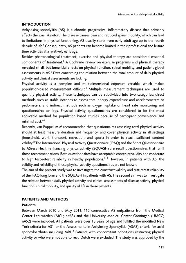
Measurement of daily physical activity
111
INTRODUCTION
Ankylosing spondylitis (AS) is a chronic, progressive, inflammatory disease that primarily
affects the axial skeleton. The disease causes pain and reduced spinal mobility, which can lead
to limitations in physical functioning. AS usually starts from early adult age up to the fourth
decade of life.1 Consequently, AS patients can become limited in their professional and leisure
time activities at a relatively early age.
Besides pharmacological treatment, exercise and physical therapy are considered essential
components of treatment.2 A Cochrane review on exercise programs and physical therapy
revealed small, but beneficial effects on physical function, spinal mobility, and patient global
assessments in AS.3 Data concerning the relation between the total amount of daily physical
activity and clinical assessments are lacking.
Physical activity is a complex and multidimensional exposure variable, which makes
population-based measurement difficult.4 Multiple measurement techniques are used to
quantify physical activity. These techniques can be subdivided into two categories: direct
methods such as stable isotopes to assess total energy expenditure and accelerometers or
pedometers, and indirect methods such as oxygen uptake or heart rate monitoring and
questionnaires or logs. Physical activity questionnaires are considered to be the most
applicable method for population based studies because of participant convenience and
minimal cost.4-7
Recently, van Poppel et al. recommended that questionnaires assessing total physical activity
should at least measure duration and frequency, and cover physical activity in all settings
(household, work, transport, recreation, and sport) in order to reach sufficient content
validity.8 The International Physical Activity Questionnaire (IPAQ) and the Short QUestionnaire
to ASsess Health-enhancing physical activity (SQUASH) are recall questionnaires that fulfill
these recommendations. Both questionnaires have acceptable construct validity and moderate
to high test-retest reliability in healthy populations.9,10 However, in patients with AS, the
validity and reliability of these physical activity questionnaires are not known.
The aim of the present study was to investigate the construct validity and test-retest reliability
of the IPAQ long form and the SQUASH in patients with AS. The second aim was to investigate
the relation between daily physical activity and clinical assessments of disease activity, physical
function, spinal mobility, and quality of life in these patients.
PATIENTS AND METHODS
Patients
Between March 2010 and May 2011, 115 consecutive AS outpatients from the Medical
Center Leeuwarden (MCL; n=63) and the University Medical Center Groningen (UMCG;
n=52) were included. All patients were over 18 years of age and fulfilled the modified New
York criteria for AS11 or the Assessments in Ankylosing Spondylitis (ASAS) criteria for axial
spondyloarthritis including MRI.12 Patients with concomitant conditions restricting physical
activity or who were not able to read Dutch were excluded. The study was approved by the

Chapter 8
112
local ethics committees of the MCL and UMCG and written informed consent according to
the Declaration of Helsinki was obtained from all patients.
Construct validity
Construct validity of the physical activity questionnaires was examined by correlating the IPAQ
and SQUASH total scores to the accelerometer outcome. The accelerometer provides an
objective and valid estimate of overall physical activity.5 On the first day, the long version of
the IPAQ and the SQUASH were administered at the outpatient clinic in 63 AS patients. In
succession, daily physical activity was assessed using the ActiGraph accelerometer during 7
consecutive days.
IPAQ. The IPAQ long form refers to an average week in the past month and comprises
questions in the following domains: occupational, household and gardening, transport, and
leisure time.9 Each of these domains provides a specific activity score calculated by multiplying
the number of minutes per week of the performed activities with the accompanying mean
metabolic equivalent (MET)-value of these activities.13,14 The total activity score was calculated
by the sum of these domain scores and was reported in MET-minutes/week. According to the
IPAQ guidelines, data were excluded if the total minutes of activity per day exceeded 960 or
when missing values were present.9
SQUASH. The SQUASH refers to an average week in the past month and contains questions in
the following domains: commuting activities, household activities, leisure time and sports
activities, and activities at work and school. Activity scores per domain were calculated by
multiplying the number of minutes per week with an intensity score (range 1-9) of the
activities performed.10 The intensity score was based on the reported intensity of an activity
combined with the activity intensity classification according to Ainsworth’s Compendium of
Physical Activities.13,14 The total activity score was calculated as the sum of the scores per
domain. According to the SQUASH protocol, data were excluded if the total minutes of
activity per day exceeded 960.10 Since the present study investigates the validity and reliability
of the SQUASH, data were also excluded if missing values were present.
Accelerometer. The ActiGraph accelerometer (GT1M, MTI, Fort Walton Beach, FI) is a small,
lightweight, uniaxial accelerometer, which senses vertical accelerations between 0.05 and 2.0
gravitational acceleration (ĝ) with a sample frequency of once per minute. The accelerometer
was worn on the right hip during waking hours, except for periods of showering or other water
activities. The duration of accelerometer wearing periods and the performed physical activities
lasting over 10 minutes were noted in a diary. The outcome of the accelerometer was
expressed in average kilo counts per day (kcounts/day), calculated by dividing the total activity
kcounts by the total number of days the monitor was worn. Data were excluded if the
accelerometer was worn for less than 10 hours per day, less than five days, or when it was not
worn during both weekend days.15

Measurement of daily physical activity
113
Test-retest reliability
The test-retest reliability of an instrument is based on the assumption that the construct has
not changed over time and thus the outcome of the measure can be reproduced. To test the
reproducibility of the IPAQ and SQUASH over time, the questionnaires were filled out by 52
AS patients on two different occasions approximately one week apart. This time period was
chosen because it is unlikely that the disease status will change in a week and this period is
long enough to avoid re-call bias. The first assessment was performed at the outpatient clinic
and the second assessment was conducted at home.
Clinical assessments
In all 115 patients, clinical assessments were performed at the outpatient clinic on the first day
of the questionnaire assessments. Disease activity was assessed using the Bath Ankylosing
Spondylitis Disease Activity Index (BASDAI; on a scale of 0-10), erythrocyte sedimentation rate
(ESR), C-reactive protein (CRP), and the Ankylosing Spondylitis Disease Activity Score
(ASDASCRP; calculated from BASDAI questions 2, 3, and 6, patient’s global assessment of
disease activity, and CRP).16,17 Physical function was assessed using the Bath Ankylosing
Spondylitis Functional Index (BASFI; on a scale of 0-10). Spinal mobility assessments included
occiput-to-wall distance, chest expansion, modified Schober test, lateral spinal flexion (left and
right) and cervical rotation (left and right). Quality of life was assessed using the Ankylosing
Spondylitis Quality of Life questionnaire (ASQoL; on a scale of 0-18).
Statistical analysis
Data were analyzed using PASW Statistics 18 (SPSS, Chicago, IL, USA). Construct validity was
examined by calculating Spearman’s and Pearson’s correlation coefficients between
accelerometer activity counts and IPAQ and SQUASH total scores, respectively. Correlations
below 0.3 were interpreted as poor association, between 0.3 and 0.6 as moderate association,
between 0.6 and 0.8 as good association and above 0.8 as excellent association. The ActiGraph
accelerometer is limited in measuring cycling and fitness activities and is not capable of
measuring swimming or other water activities. Therefore, the accelerometer outcome was also
correlated with an adjusted total score of the SQUASH without these activities. A z-test with
Fisher’s transformation was used to compare this correlation with the correlation of the
unadjusted SQUASH total score.
Test-retest reliability of the IPAQ and SQUASH was investigated by calculating intraclass
correlation coefficients (ICCagreement; two way random effects model) between the first and
the second assessments of the questionnaires. Reliability was assessed for both total scores
and activity scores per domain. ICC values of at least 0.70 indicate good reliability.18
Additionally, Bland-Altman analysis was performed on the total scores.19 Differences between
first and second IPAQ and SQUASH assessments were examined using Wilcoxon signed rank
and paired t-tests, respectively.

Chapter 8
114
Pearson’s and Spearman’s correlation coefficients were used as appropriate to analyze the
relation between daily physical activity and clinical assessments. P values <0.05 were
considered statistically significant.
RESULTS
Characteristics of the 115 AS patients included in the validity (n=63) and reliability (n=52)
studies are presented in Table 1.
Table 1. Characteristics of the AS study population
Total group Validity study Reliability study
Number of patients 115 63 52
Age (years) 44.6 ± 12.1 43.2 ± 12.3 46.3 ± 11.8
Gender (male) (n, %) 71 (62) 40 (64) 31 (60)
BMI 26.4 ± 4.4 26.2 ± 4.8 26.6 ± 3.9
Duration of symptoms (years) 16 (0-54) 18 (2-54) 16 (0-53)
Time since diagnosis 10 (0-42) 11 (1-42) 9 (0-37)
HLA-B27+ (n, %) 82 (76) 49 (82) 33 (69)
Anti-TNF use (n, %) 78 (70) 39 (62) 39 (81)*
NSAID use (n, %) 44 (40) 24 (39) 20 (42)
DMARD use (n, %) 17 (15) 13 (21) 4 (8)
BASDAI (range 0-10) 3.7 (0.0-9.0) 3.8 (0.4-8.6) 3.5 (0.0-9.0)
ESR (mm/h) 11 (2-60) 13 (2-60) 10 (2-39)
CRP (mg/l) 3 (2-46) 3 (2-44) 4 (2-46)
ASDASCRP 2.3 (0.7-4.4) 2.3 (0.9-4.2) 2.3 (0.7-4.4)
BASFI (range 0-10) 3.8 ± 2.4 3.6 ± 2.4 4.2 ± 2.4
Occiput-to-wall distance (cm) 0.0 (0.0-29.0) 0.0 (0.0-29.0) 0.0 (0.0-20.0)
Chest expansion (cm) 5.0 (1.0-14.0) 6.0 (1.0-10.5) 4.0 (1.0-14.0)**
Modified Schober test (cm) 4.0 (0.4-7.0) 4.0 (0.4-6.2) 3.9 (0.0-7.0)
Lateral spinal flexion left (cm) 10.5 (0.0-28.0) 11.0 (0.0-28.0) 10.0 (0.5-27.5)
Lateral spinal flexion right (cm) 10.0 (0.0-27.0) 10.5 (0.0-22.0) 10.0 (1.0-27.0)
Cervical rotation left (degrees) 60 (0-100) 74 (5-100) 55 (0-95)*
Cervical rotation right (degrees) 62 (5-100) 65 (5-100) 61 (10-100)
ASQoL (range 0-18) 6 (0-18) 6 (0-17) 6 (0-18)
* Statistically significant difference compared to the validity study (p<0.05).
** Statistically significant difference compared to the validity study (p<0.01).
Values are mean ± SD or median (range) unless otherwise indicated.
AS: ankylosing spondylitis; BMI: body mass index; HLA-B27+: human leukocyte antigen B27 positive;
TNF: tumor necrosis factor; NSAID: non-steroidal anti-inflammatory drug; DMARD: disease modifying
antirheumatic drug; BASDAI: Bath ankylosing spondylitis disease activity index; ESR: erythrocyte
sedimentation rate; CRP: C-reactive protein; ASDAS: ankylosing spondylitis disease activity score;
BASFI: Bath ankylosing spondylitis functional index; ASQoL: ankylosing spondylitis quality of life.
Measurement of daily physical activity
115
Construct validity
Complete data to examine the validity of the IPAQ were available for 45 of the 63 (71%)
patients. In these patients, the median IPAQ total score was 5937 (MET-minutes/week)
(interquartile range (IQR) 2126-11601) and mean accelerometer outcome was 234
kcounts/day (SD±107). The remaining patients had missing IPAQ values (n=12) and/or did not
meet the accelerometer criteria (n=7). The correlation between IPAQ total score and
accelerometer activity counts was 0.38, (p<0.05), which indicates moderate validity of the
IPAQ.
Complete data to examine the validity of the SQUASH were available for 53 of the 63 (84%)
patients. In these patients, the mean SQUASH total score was 7267 (SD±3453) and mean
accelerometer outcome was 236 kcounts/day (SD±107). The remaining patients had missing
(n=2) or invalid (n=1) SQUASH values or did not meet the accelerometer criteria (n=7). The
correlation between SQUASH total score and accelerometer activity counts was 0.35 (p<0.05),
which indicates moderate validity of the SQUASH.
According to the SQUASH, 68%, 34%, and 19% of AS patients reported cycling, fitness, and
swimming, respectively, which are activities that cannot be (properly) registered by the
accelerometer. The correlation between SQUASH total score adjusted for these activities and
accelerometer outcome was 0.39 (p<0.01). This correlation did not significantly differ from the
correlation with the unadjusted SQUASH total score (z=-0.23, p=0.82).

Chapter 8
116
Test-retest reliability
Complete data for the first and second assessment of the IPAQ were available for 32 of the 52
(62%) patients. The remaining patients had missing values (n=18) or the total minutes of
activity per day exceeded 960 (n=2). The ICC for the IPAQ total score was 0.83, indicating
good reliability. ICCs for the sub scores ranged from 0.60 (household and garden activities) to
0.88 (transport activities) (Table 2).
Complete data for the first and second assessment of the SQUASH were available for 33 of the
52 (63%) patients. The remaining patients had missing values (n=16), the total minutes of
activity per day exceeded 960 (n=2), or the second questionnaire was missing (n=1). The ICC
for the SQUASH total score was 0.89, indicating good reliability. ICCs for sub scores of the
SQUASH ranged from 0.48 (commuting activities) to 0.88 (work and school activities) (Table
2).
Bland-Altman analyses for the IPAQ and SQUASH total scores showed that the mean
difference between the first and second assessment was small and not significantly different
from zero, which implies that no systematic bias was present. However, in particular for the
IPAQ, the 95% limits of agreement (LOA) were wide, indicating that only large changes can be
considered as true changes (Figure 1).
Table 2. Physical activity of the AS patients (with complete data) included in the reliability study
1st assessment 2nd assessment ICC 95% CI
IPAQ (MET-mins/week) (n=32)
Total activity score 3849 (1470-8132) 4349 (2120-7508) 0.83* 0.68-0.91
Work activity score 0 (0-3710) 50 (0-3197) 0.80* 0.63-0.90
Transport activity score 653 (284-1607) 737 (209-1589) 0.88* 0.77-0.94
Household & garden activity score 580 (53-1481) 245 (90-1406) 0.60* 0.33-0.79
Leisure time activity score 657 (195-1718) 678 (111-1832) 0.73* 0.52-0.86
SQUASH (n=33)
Total activity score 5760 (3360-6890) 5600 (3715-7540) 0.89* 0.79-0.95
Work and school activity score 1080 (0-4440) 600 (0-4680) 0.88* 0.77-0.94
Commuting activity score 0 (0-341) 0 (0-438) 0.48* 0.17-0.70
Household activity score 900 (420-1815) 840 (240-1635) 0.77* 0.59-0.88
Leisure time & sports activity score 1350 (775-1795) 1410 (480-2635) 0.87* 0.76-0.93
* Statistically significant intraclass correlation (p<0.001).
No significant differences were found between the first and second assessments of the physical activity
questionnaires.
Values are median (interquartile range) unless otherwise indicated.
AS: ankylosing spondylitis; IPAQ: International Physical Activity Questionnaire; SQUASH: Short
QUestionnaire to ASses Health-enhancing physical activity; ICC: intraclass correlation; CI: confidence
interval

Measurement of daily physical activity
117
Figure 1. Bland-Altman plots. Difference between the total scores on the first and second IPAQ (A) and
SQUASH (B) plotted against the mean of both assessments, together with 95% limits of agreement
(LOA).

Chapter 8
118
Relation between daily physical activity and clinical assessments
Accelerometer, IPAQ and SQUASH total scores correlated significantly with disease activity
assessed using ASDASCRP. In addition, the IPAQ and SQUASH correlated significantly with
BASDAI and the accelerometer correlated significantly with ESR and CRP. All three daily
physical activity measures correlated significantly with physical function and quality of life,
while a significant correlation with spinal mobility was found only for the accelerometer and
SQUASH (Table 3).
Table 3. Correlations between daily physical activity and clinical assessments in AS patients
IPAQ
(MET-mins/week)
SQUASH Accelerometer
(kcounts/day)
Number of patients 86a 94a 55b
Disease activity
BASDAI (range 0-10) -0.220*c -0.326**c NS
ESR (mm/h) NS NS -0.460***c
CRP (mg/l) NS NS -0.289*c
ASDASCRP -0.243*c -0.311**c -0.283*c
Physical function
BASFI (range 0-10) -0.387***c -0.476***d -0.274*d
Spinal mobility
Occiput-to-wall distance (cm) NS -0.297**c NS
Chest expansion (cm) NS NS NS
Modified Schober test (cm) NS 0.260*c 0.338*c
Lateral spinal flexion left (cm) NS NS 0.344*d
Lateral spinal flexion right (cm) NS NS 0.385**d
Cervical rotation left (degrees) NS 0.286**c 0.358**c
Cervical rotation right (degrees) NS 0.296**c 0.285*c
Quality of life
ASQoL -0.282**c -0.500***c -0.356**c
* Statistically significant correlation (p<0.05)
** Statistically significant correlation (p<0.01)
*** Statistically significant correlation (p<0.001) a Only patients with complete data on the first IPAQ or SQUASH assessment and available clinical
data were included b Only patients with complete accelerometers data and available clinical data were included. c Spearman’s correlation coefficient d Pearson’s correlation coefficient
See Tables 1 and 2 for abbreviations.

Measurement of daily physical activity
119
DISCUSSION
This is the first study that investigates the measurement properties of physical activity
questionnaires in AS. The construct validity of the IPAQ (long form) and SQUASH compared
with accelerometer activity counts was found to be moderate, with correlations of 0.38 and
0.35, respectively. A large study in healthy populations from different countries showed
comparable agreement between the IPAQ total score and accelerometer outcome (pooled
correlation of 0.33).9 The results for the SQUASH total score were reported to be somewhat
better, with correlations of 0.45 in healthy adults and 0.67 in patients after total hip
arthroplasty.10,20 Recently, Terwee et al. have developed the Quality Assessment of Physical
Activity Questionnaire (QAPAQ) checklist to appraise the qualitative attributes and
measurement properties of physical activity questionnaires. They stated that the correlation
between total physical activity assessed by a questionnaire and accelerometer total counts
should be at least 0.50.21 Based on these guidelines, the standard to prove the construct
validity of the IPAQ and SQUASH was not completely reached in patients with AS.
Both questionnaires showed good test-retest reliability based on ICC values (0.83 and 0.89 for
the IPAQ and SQUASH total scores, respectively). Previous studies reported Spearman’s
correlation coefficients of 0.81 (pooled correlation) for the IPAQ long form9 and 0.58 and 0.57
for the SQUASH.10,20 In the present study, ICC’s were used instead of correlation coefficients,
because correlation coefficients do not take systematic measurement errors into account.21
For the comparison with previous studies, correlation coefficients were also calculated,
resulting in values of 0.74 for the IPAQ and 0.90 for the SQUASH (data not shown). Bland
Altman analysis was performed in only one of the previous studies. In accordance with our
results, the authors reported no systematic bias between SQUASH assessments, but their
95% LOA were approximately 3 times larger than those found in the present study.20 We
found wide 95% LOA for the IPAQ in comparison to the SQUASH, which indicates that only
large changes can be considered as true changes.
Interestingly, objective accelerometer daily activity was significantly correlated with the
objective measures ESR and CRP and the subjective IPAQ and SQUASH were significantly
correlated with the subjective measure BASDAI. Accelerometer, IPAQ, and SQUASH scores
were all significantly correlated to the ASDASCRP, a composite score of patient-reported
measures and CRP developed in order to capture both subjective and objective aspects of AS
disease activity. The inverse relation between accelerometer outcome and CRP is in line with
recent findings of Plasqui et al.,22 but the relation between daily physical activity and clinical
assessments has not been studied before. Some support for the presence of these relations
can be obtained from intervention studies concerning exercise programs and physical therapy
in AS. Although exercise is only a part of a person’s total physical activity,23 an increase in
exercise means in general an increase in total physical activity. Until now, conflicting data have
been published about the relation between physical activity and disease activity in intervention
studies.24,25 Randomized controlled trials showed an improvement in physical function and
spinal mobility after an 8-week to 4-month exercise program,26-28 which supports a relation
Chapter 8
120
between these clinical assessments and daily physical activity in AS. Further studies are needed
to investigate the causality of the relation between higher daily physical activity and lower
disease activity and better physical function, spinal mobility, and quality of life found in the
present study.
A limitation of this study was the multitude of missing values in both questionnaires.
Therefore, a relatively large group of patients was excluded from the analyses. The frequent
occurrence of missing values may hamper the use of these physical activity questionnaires in
daily practice. In contrast to the IPAQ, the SQUASH guidelines clearly prescribe how to deal
with missing data.10 However, the reliability of the SQUASH total score was insufficient
(ICC=0.60) in our analysis including patients with imputation for missing values according to
the SQUASH guidelines (data not shown). A second limitation is that the ActiGraph
accelerometer is an uniaxial accelerometer and therefore is limited in the measurement of
physical activities that require little vertical movement, such as cycling or fitness. In addition,
the accelerometer is not waterproof and therefore swimming and other water-related
activities could not be measured. Even though, the correlation between the accelerometer
outcome and the SQUASH score without these activities was comparable to the correlation of
the total SQUASH score. The IPAQ only discriminates between moderate and vigorous leisure
time physical activities. For that reason, a correction for cycling, fitness, and swimming on the
IPAQ total score could not be performed. Finally, the timeframes for the questionnaires and
accelerometer were not completely comparable. Both questionnaires referred to a usual week
in the past month (retrospective assessment), whereas the accelerometer daily activity was
assessed prospectively for 5 to 7 days. Strength of our study is that wearing the accelerometer
could not influence the reliability assessments of the physical activity questionnaires, since
validity and reliability were examined in two different groups of AS patients.
In conclusion, the construct validity of the IPAQ (long form) and SQUASH was found to be
moderate in patients with AS. Both questionnaires showed good test-retest reliability based
on ICC values and Bland-Altman analyses showed no systemic bias between assessments. In
particular for the IPAQ the 95% LOA were wide, which indicates that the degree of
repeatability is not sufficient for this questionnaire. Daily physical activity assessed by
accelerometer outcome (objective) and IPAQ and SQUASH total scores (subjective) was
found to be significantly related to clinical assessments of disease activity, physical function,
and quality of life. A relation with spinal mobility was found only for the accelerometer and
SQUASH. Based on these results, the SQUASH seems superior to the IPAQ to assess daily
physical activity in AS population studies. However, the development of a disease-specific
physical activity questionnaire may be desirable. Further studies are needed to investigate the
causality of the relation between higher daily physical activity and better clinical assessments in
AS.
Measurement of daily physical activity
121
REFERENCES
1. Braun J, Sieper J. Ankylosing spondylitis. Lancet 2007;369:1379-90. 2. Braun J, van den Berg R, Baraliakos X, et al. 2010 update of the ASAS/EULAR recommendations for
the management of ankylosing spondylitis. Ann Rheum Dis 2011;70:896-904. 3. Dagfinrud H, Kvien TK, Hagen KB. Physiotherapy interventions for ankylosing spondylitis.
Cochrane Database Syst Rev 2008:CD002822. 4. Lamonte MJ, Ainsworth BE. Quantifying energy expenditure and physical activity in the context of
dose response. Med Sci Sports Exerc 2001;33:S370-8. 5. Vanhees L, Lefevre J, Philippaerts R, et al. How to assess physical activity? How to assess physical
fitness? Eur J Cardiovasc Prev Rehabil 2005;12:102-14. 6. Pereira MA, FitzerGerald SJ, Gregg EW, et al. A collection of Physical Activity Questionnaires for
health-related research. Med Sci Sports Exerc 1997;29:S1-205. 7. Ainsworth BE. How do I measure physical activity in my patients? Questionnaires and objective
methods. Br J Sports Med 2009;43:6-9. 8. van Poppel MN, Chinapaw MJ, Mokkink LB, et al. Physical activity questionnaires for adults:
a systematic review of measurement properties. Sports Med 2010;40:565-600. 9. Craig CL, Marshall AL, Sjostrom M, et al. International physical activity questionnaire: 12-country
reliability and validity. Med Sci Sports Exerc 2003;35:1381-95. 10. Wendel-Vos GC, Schuit AJ, Saris WH, et al. Reproducibility and relative validity of the short
questionnaire to assess health-enhancing physical activity. J Clin Epidemiol 2003;56:1163-9. 11. van der Linden S, Valkenburg HA, Cats A. Evaluation of diagnostic criteria for ankylosing spondylitis.
A proposal for modification of the New York criteria. Arthritis Rheum 1984;27:361-8. 12. Sieper J, Rudwaleit M, Baraliakos X, et al. The Assessment of SpondyloArthritis international Society
(ASAS) handbook: a guide to assess spondyloarthritis. Ann Rheum Dis 2009;68:ii1-44. 13. Ainsworth BE, Haskell WL, Leon AS, et al. Compendium of physical activities: classification of energy
costs of human physical activities. Med Sci Sports Exerc 1993;25:71-80. 14. Ainsworth BE, Haskell WL, Whitt MC, et al. Compendium of physical activities: an update of activity
codes and MET intensities. Med Sci Sports Exerc 2000;32:S498-504. 15. Trost SG, McIver KL, Pate RR. Conducting accelerometer-based activity assessments in field-based
research. Med Sci Sports Exerc 2005;37:S531-43. 16. Lukas C, Landewe R, Sieper J, et al. Development of an ASAS-endorsed disease activity score
(ASDAS) in patients with ankylosing spondylitis. Ann Rheum Dis 2009;68:18-24. 17. van der Heijde D, Lie E, Kvien TK, et al. ASDAS, a highly discriminatory ASAS-endorsed disease
activity score in patients with ankylosing spondylitis. Ann Rheum Dis 2009;68:1811-8. 18. Terwee CB, Bot SD, de Boer MR, et al. Quality criteria were proposed for measurement properties
of health status questionnaires. J Clin Epidemiol 2007;60:34-42. 19. Bland JM, Altman DG. Statistical methods for assessing agreement between two methods of clinical
measurement. Lancet 1986;1:307-10. 20. Wagenmakers R, van den Akker-Scheek I, Groothoff JW, et al. Reliability and validity of the short
questionnaire to assess health-enhancing physical activity (SQUASH) in patients after total hip arthroplasty. BMC Musculoskelet Disord 2008;9:141.
21. Terwee CB, Mokkink LB, van Poppel MN, et al. Qualitative attributes and measurement properties of physical activity questionnaires: a checklist. Sports Med 2010;40:525-37.
22. Plasqui G, Boonen A, Geusens P, et al. Physical activity and body composition in patients with ankylosing spondylitis. Arthritis Care Res (Hoboken) 2012;64:101-7.
23. Shephard RJ. Limits to the measurement of habitual physical activity by questionnaires. Br J Sports Med 2003;37:197-206.
24. Karapolat H, Akkoc Y, Sari I, et al. Comparison of group-based exercise versus home-based exercise in patients with ankylosing spondylitis: effects on Bath Ankylosing Spondylitis Indices, quality of life and depression. Clin Rheumatol 2008;27:695-700.

Chapter 8
122
25. Sweeney S, Taylor G, Calin A. The effect of a home based exercise intervention package on outcome in ankylosing spondylitis: a randomized controlled trial. J Rheumatol 2002;29:763-6.
26. Kraag G, Stokes B, Groh J, et al. The effects of comprehensive home physiotherapy and supervision on patients with ankylosing spondylitis--a randomized controlled trial. J Rheumatol 1990;17:228-33.
27. Lim HJ, Moon YI, Lee MS. Effects of home-based daily exercise therapy on joint mobility, daily activity, pain, and depression in patients with ankylosing spondylitis. Rheumatol Int 2005;25:225-9.
28. Ince G, Sarpel T, Durgun B, et al. Effects of a multimodal exercise program for people with ankylosing spondylitis. Phys Ther 2006;86:924-35.

CHAPTER 9
SUMMARY AND GENERAL DISCUSSION

Chapter 9
124
This thesis covers several important topics of AS. The first part (chapters 2-3) concerns
different aspects of bone turnover in AS. The second part (chapters 3-7) focuses on the
identification of predictors of response and investigates antibody formation to TNF-α blocking
agents in AS. The final study of this thesis (chapter 8) concerns the assessment of daily physical
activity in patients with AS.
GLAS study
All manuscripts from this thesis are based on data derived from the Groningen Leeuwarden
Ankylosing Spondylitis (GLAS) study. This is an ongoing prospective longitudinal observational
cohort study of AS patients from the outpatient clinics of the Medical Center Leeuwarden
(MCL) and the University Medical Center Groningen (UMCG), which started in November
2004. The general research goals of the GLAS study are to obtain more knowledge on
AS-related bone formation and bone resorption, and to evaluate the effect of tumor necrosis
factor-alpha (TNF-α) blocking therapy in AS patients in daily clinical practice.
The GLAS study is conducted according to the operative guidelines established by the
Assessment in SpondyloArthritis international Society (ASAS) working group.1 Patients
treated with TNF-α blocking therapy (n=260 until 2012) are evaluated at baseline, after 3 and 6
months, and then every 6 to 12 months. Patients without anti-TNF-α treatment (inclusion
started in 2009; n=140 until 2012) are evaluated yearly. Standardized follow-up visits include
assessments of disease activity, physical function, spinal mobility, quality of life, inflammation
of entheses, bone mineral density (BMD), and radiology. Furthermore, serum, plasma, and
urine samples are collected and stored to measure e.g. bone turnover markers (BTM) and
vitamin D levels.
All patients from the GLAS cohort fulfill the modified New York criteria for AS2 (>95% of
patients) and/or the ASAS criteria for axial spondyloarthritis.3,4 Approximately two-third of the
patients is male. At study entry, the mean age was 43 years (standard deviation: 12 years), the
median duration of symptoms was 15 years (range: 1-59 years), and the median time since
diagnosis was 6 years (range: 0-54 years).
Bone turnover
In AS, excessive bone formation can lead to the formation of syndesmophytes and ankylosis of
the spine and sacroiliac joints.5 On the other hand, excessive bone loss can result in low BMD
(osteopenia or osteoporosis) and vertebral fractures.6,7
BTM reflect the metabolic activity of bone and can easily be measured in blood or urine.
Previous studies have demonstrated that measuring BTM can provide a better insight in the
bone physiology of AS.8-10 A challenge of working with BTM (as well as BMD) is that they
change with age and that there are differences for gender. To correct for these influences, we
calculated BTM Z-scores using a Dutch healthy reference cohort. This corresponds to the
methodology of interpreting BMD.

Summary
125
In order to obtain more knowledge on AS-related osteoporosis, we studied the relation
between BMD, BTM, vitamin D levels, and clinical assessments of disease activity and physical
function in a cross-sectional analysis of baseline data from AS patients with active disease. The
results presented in chapter 2 show that higher levels of bone resorption marker serum type I
collagen C-telopeptide (sCTX), bone formation marker osteocalcin (OC), and erythrocyte
sedimentation rate (ESR) were independently related to low BMD. Furthermore, higher ESR
and lower 25-hydroxyvitamin D levels were independently related to sCTX Z-score. Based on
these results, high bone turnover, inflammatory processes, and low vitamin D levels seem all
to play a role in AS-related bone loss. In addition, previous studies have suggested the
importance of adverse effects of medication (for example glucocorticoids) and decreased
mobility in relation to pain and stiffness in the multifactorial etiology of osteoporosis in AS.11
Another important finding in chapter 2 was that the difference between lumbar spine and hip
BMD T-score correlated positively with disease duration, which indicates that
osteoproliferation in the lumbar spine has resulted in an overestimation of the lumbar spine
BMD, measured by dual-energy x-ray absorptiometry (DXA), in patients with advanced AS.
Therefore, measuring both BTM and BMD seems useful to identify osteoporosis in these
patients. The high prevalence of vertebral fractures (39%) and low BMD (57%) in our study
underlines the importance of monitoring bone loss in AS in daily clinical practice.
Many randomized controlled trials (RCTs) have shown that TNF-α blocking agents are
effective in controlling inflammation and improving clinical assessments in patients with
AS.12,15 These improvements were also found in our cohort. Subsequently, we investigated the
effect of TNF-α blocking therapy on BTM and BMD. The results of chapter 3 show that
3 years of TNF-α blocking therapy resulted in a significant increase in bone-specific alkaline
phosphatase (BALP), which plays a central role in the mineralization process of bone, while the
effect on procollagen type 1 N-terminal peptide (PINP), a product of collagen formation, was
less evident. Furthermore, a significant decrease in sCTX, a product of collagen degradation,
was found. Interestingly, both lumbar spine and hip BMD improved significantly during 3 years
of TNF-α blocking therapy, which may be explained by the increase in mineralization and
decrease in bone resorption.
Predictors of response to TNF-α blocking therapy
In clinical practice, continuation of TNF-α blocking therapy is mainly based on subjective
measures such as the Bath AS Disease Activity Index (BASDAI) and the global opinion of the
patient and the physician. Recent studies showed the usefulness of the AS disease activity
score (ASDAS) as a more objective measure of disease activity.16-19 However, if available, it
would be useful to include also a purely objective measure in the evaluation of TNF-α blocking
therapy in patients with AS. In search of such an objective biomarker, we investigated the
predictive value of BTM and matrix metalloproteinase-3 (MMP-3), an enzyme involved in

Chapter 9
126
degradation of extracellular matrix components, using longitudinal data from AS patients
treated with TNF-α blocking agents.
In chapter 3, we found that decrease in bone resorption marker sCTX and improvements in
ASDAS and physician’s global disease activity (GDA) during the first 3 months of treatment
were independently related to longer continuation of TNF-α blocking therapy. The accuracy of
early change in sCTX to discriminate between patients who continued and discontinued TNF-α
blocking therapy during the first 3 years was moderate and comparable to the accuracy of early
change in ASDAS or physician’s GDA. In addition, baseline to 3 months decrease in sCTX was
significantly associated with good long-term response regarding disease activity, physical
function, spinal mobility, and quality of life. Based on these results, early change in sCTX seems
useful as a purely objective biomarker in the evaluation of TNF-α blocking therapy in AS, in
addition to the currently used more subjective measures.
In chapter 4, we found that etanercept treatment resulted in a significant reduction in serum
MMP-3 levels in male patients with AS. This decrease appeared to be significant in male
patients with concomitant peripheral arthritis at baseline, but not in male patients with only
axial disease. Data analysis was split for gender, since baseline serum MMP-3 levels were
significantly different between male and female patients with AS. Subsequently, we showed
that baseline serum MMP-3 levels had poor accuracy to discriminate between responders and
nonresponders after 3 or 12 months of etanercept treatment. The accuracy of decrease in
serum MMP-3 levels from baseline to 3 months to predict response after 3 or 12 months of
treatment was poor to moderate. Furthermore, this accuracy was comparable to those of
change in ESR and CRP, which are elevated only in a minority of patients with AS.20,21
Therefore, measuring serum MMP-3 levels seems not very useful for monitoring and
predicting response to etanercept treatment in AS patients in daily clinical practice.
Besides biomarkers to evaluate treatment in AS, it is relevant to identify patient characteristics
before start of treatment which are able to predict a beneficial response to TNF-α blocking
therapy, especially in view of the high costs and potential side effects of these agents.
In chapter 5, we identified male gender, higher ESR or CRP level, and the presence of
peripheral arthritis as independent baseline predictors of both treatment response and
continuation of TNF-α blocking therapy in daily clinical practice. Furthermore, a higher ASDAS
score was found to be a significant baseline predictor of response to TNF-α blocking therapy.
In contrast, a higher BASDAI score at baseline was independently associated with treatment
discontinuation. Currently, starting and continuation TNF-α blocking therapy is mainly based
on the BASDAI, but these results suggest that more objective measures should be included in
this process.
Chapter 6 provided an overview of clinical trials and observational studies investigating
baseline predictors of response after 3-6 months of TNF-α blocking therapy and baseline
predictors of long-term anti-TNF-α treatment continuation in AS. Multiple studies have
identified increased acute phase reactants, presence of peripheral arthritis, higher disease

Summary
127
activity, higher functional status, younger age, male sex, and HLA-B27 positivity as
independent baseline predictors for achieving clinical response after 3-6 months and/or for
long-term continuation of TNF-α blocking therapy in multivariate analyses. Furthermore,
several studies reported promising data regarding the potential value of biomarkers, for
example markers of inflammation or bone and cartilage metabolism, as baseline predictors of
response to TNF-α blocking therapy. Until now, the results of these studies are either not
confirmed by other study groups or confirmed in studies that used less robust techniques of
data analysis. Further studies using multivariate analyses are needed to confirm the predictive
value of these biomarkers, in addition to the currently known predictors.
Although the majority of AS patients respond very well to TNF-α blocking therapy,
approximately 30% fail to reach efficacy. This may in part be explained by the formation of
antibodies against these agents.22,23 Chapter 7 showed that during one year of treatment,
antibodies against infliximab, etanercept, and adalimumab were induced in 20%, 0%, and 30%
of patients, respectively. This antibody formation was related to a decrease in efficacy and early
discontinuation of anti-TNF-α treatment, since antibodies were detected in the majority of
patients who discontinued treatment due to inefficacy. Furthermore, patients with these
antibodies had significantly lower serum TNF-α blocker levels compared to patients without
these antibodies. Significant negative correlations were found between serum TNF-α blocker
levels and assessments of disease activity, which suggests the importance of sufficiently high
serum TNF-α blocker levels to obtain clinical response.
Daily physical activity
Besides pharmacological treatment, exercise and physical therapy are essential components of
treatment. Physical activity questionnaires are considered to be the most applicable method to
assess daily physical activity in population studies because of participant convenience and
minimal cost.24,25 The International Physical Activity Questionnaire (IPAQ) and the Short
QUestionnaire to ASsess Health-enhancing physical activity (SQUASH) are recall
questionnaires with acceptable construct validity and moderate to high test-retest reliability in
healthy populations. In chapter 8, we investigated the measurement properties of these
physical activity questionnaires in patients with AS. The results showed that the construct
validity of the IPAQ and SQUASH compared with accelerometer activity counts was moderate.
Furthermore, both questionnaires had good test-retest reliability based on intraclass
correlation coefficients. Bland-Altman analyses showed no systemic bias, but in particular for
the IPAQ the 95% limits of agreement were wide, which indicates that only large changes can
be considered as true changes. A limitation of the study was the multitude of missing values in
both questionnaires, which may hamper the use of these physical activity questionnaires in
daily practice. Based on our results, the SQUASH seems superior to the IPAQ to assess daily
physical activity in AS population studies. However, the development of a disease-specific
physical activity questionnaire may be desirable. Finally, we showed that higher daily physical

Chapter 9
128
activity was significantly related to lower disease activity and better physical function, spinal
mobility, and quality of life.
Resume and perspective
The present thesis describes different aspects of bone turnover in AS as well as several topics
in relation to the clinical management of this disease in daily clinical practice.
First, we demonstrated that BTM can be useful to identify bone loss in AS, especially in
patients with advanced disease. Further longitudinal studies are needed to investigate the
usefulness of BTM in predicting outcome such as osteoporosis and/or radiographic damage in
AS. A major advantage of BTM is that they can easily be measured in the blood of patients at
different time points with relatively low costs. However, it is important to standardize serum
sample collection and the assays used for BTM measurements to reduce variability within and
between patients.26,27 The use of T-scores and Z-scores helps to interpret BTM and BMD.
The introduction of TNF-α blocking agents has been the most important development in the
treatment of AS in the past decades. RCTs have demonstrated that these agents are effective
in controlling inflammation and improving clinical assessments.12-15 Data from observational
studies such as the GLAS cohort are also important, since they provide information closer to
clinical practice than RCTs. Furthermore, follow-up is often longer in cohort studies and it
gives, in case of a sufficient amount of included patients, the possibility of head to head
comparison of several different expensive therapies such as TNF-α blocking agents.
A limitation of observational studies in clinical practice is the introduction of bias, but this
problem can partly be solved by using sophisticated analysis techniques.
Besides improvements in clinical assessments, it was shown that TNF-α blocking therapy also
has a beneficial effect on lumbar spine and hip BMD. Based on our BTM analysis, this can
largely be explained by an increase in mineralization and decrease in bone resorption. Still, the
important question remains whether TNF-α blocking agents can diminish or stop radiographic
progression and can prevent vertebral fractures in AS. There are concerns that, although
inflammation will resolve after TNF-α blocking therapy, the focal fatty lesions seen on
magnetic resonance imaging (MRI) will persist. This may lead to ongoing repair processes,
resulting in formation of syndesmophytes and progression of radiographic damage.28 Recent
studies did not show a significant difference in radiographic progression after 2 years of TNF-α
blocking therapy, when compared to radiographic data of TNF-α blocker naive patients from
historical cohorts.29-31 It may be hypothesized that long-term treatment with TNF-α blocking
agents will decelerate radiographic progression over time, since by preventing new
inflammatory lesions, repair processes will extinguish over time. In line with this hypothesis,
less radiographic progression was reported between 4 and 8 years of infliximab treatment
compared to a historical cohort.32 Further studies with long-term follow-up are needed to
confirm these findings. The influence of long-term TNF-α blocking therapy on radiographic
progression will also be an important research question in our longitudinal GLAS study.

Summary
129
In clinical practice, continuation of TNF-α blocking therapy is mainly based on subjective
measures such as the BASDAI and the global opinion of the patient and the physician. In our
opinion, it would be useful to also include an objective measure in this evaluation process.
This thesis confirmed the usefulness of the ASDAS, a composite score of patient-reported
measures and an acute phase reactant developed in order to capture both subjective and
objective aspects of AS disease activity. Besides, we identified early decrease in bone
resorption marker sCTX as a significant predictor of continuation of TNF-α blocking therapy,
independent from ASDAS and physician’s GDA. Of course, confirmation of these results in an
independent cohort will strengthen sCTX as an objective measure.
At this moment, recommendations for starting TNF-α blocking therapy in AS are primarily
based on inadequate response to conventional treatment and less on the expectation that
anti-TNF-α treatment will be effective in a particular patient. In the last few years, several
studies including our own study focused on the identification of characteristics of AS patients
before start of treatment which are able to predict a beneficial response to TNF-α blocking
therapy. However, the predictive value of single parameters identified is not strong enough to
predict treatment response in the individual patient. The development of a prediction model33
may lead to an instrument that can support physicians to make evidence-based decisions to
start TNF-α blocking therapy in patients with AS in daily clinical practice.
The fact that some patients with AS fail to reach efficacy to TNF-α blocking therapy can in part
be explained by the immunogenicity of these agents. In our study, antibody formation to
infliximab or adalimumab was associated with treatment discontinuation and lower serum
TNF-α blocker levels. Sufficiently high serum TNF-α blocker levels seem to be important to
obtain clinical response, since significant negative correlations were found between serum
TNF-α blocker levels and assessments of disease activity. In line with our findings, a recent
study in a large group of patients with rheumatoid arthritis (RA) showed that the development
of antibodies against adalimumab was associated with lower serum levels of adalimumab,
earlier treatment discontinuation, higher disease activity, and absence of clinical remission.
The authors also reported that antibodies against adalimumab were already detectable long
before the patient discontinued anti-TNF-α treatment.34 It can therefore be useful to measure
serum TNF-α blocker levels and antibodies to TNF-α blocking agents in all patients, so that
treatment regimens can be adapted if necessary.
Finally, our results demonstrated that the amount of daily physical activity (i.e. household,
work, transport, and leisure time activities) is related to disease activity, physical function,
spinal mobility, and quality of life in patients with AS. Further studies are needed to investigate
the causality of these relations. Furthermore, we showed reasonable measurement properties
for the SQUASH, a general physical activity questionnaire, in these patients. However, the
exact measurement of daily physical activity remains difficult and the development of an
AS-specific physical activity questionnaire may therefore be desirable.

Chapter 9
130
REFERENCES 1. Sieper J, Rudwaleit M, Baraliakos X, et al. The Assessment of SpondyloArthritis international Society
(ASAS) handbook: a guide to assess spondyloarthritis. Ann Rheum Dis 2009;68:ii1-44. 2. van der Linden S, Valkenburg HA, Cats A. Evaluation of diagnostic criteria for ankylosing spondylitis.
A proposal for modification of the New York criteria. Arthritis Rheum 1984;27:361-8. 3. Rudwaleit M, Landewe R, van der Heijde D, et al. The development of Assessment of
SpondyloArthritis international Society classification criteria for axial spondyloarthritis (part I): classification of paper patients by expert opinion including uncertainty appraisal. Ann Rheum Dis 2009;68:770-6.
4. Rudwaleit M, van der Heijde D, Landewe R, et al. The development of Assessment of SpondyloArthritis international Society classification criteria for axial spondyloarthritis (part II): validation and final selection. Ann Rheum Dis 2009;68:777-83.
5. Baraliakos X, Listing J, Rudwaleit M, et al. Progression of radiographic damage in patients with ankylosing spondylitis: defining the central role of syndesmophytes. Ann Rheum Dis 2007;66:910-5.
6. Geusens P, Vosse D, van der Linden S. Osteoporosis and vertebral fractures in ankylosing spondylitis. Curr Opin Rheumatol 2007;19:335-9.
7. Vosse D, Landewe R, van der Heijde D, et al. Ankylosing spondylitis and the risk of fracture: results from a large primary care-based nested case-control study. Ann Rheum Dis 2009;68:1839-42.
8. Borman P, Bodur H, Bingol N, et al. Bone mineral density and bone turnover markers in a group of male ankylosing spondylitis patients: relationship to disease activity. J Clin Rheumatol 2001;7:315-21.
9. Park MC, Chung SJ, Park YB, et al. Bone and cartilage turnover markers, bone mineral density, and radiographic damage in men with ankylosing spondylitis. Yonsei Med J 2008;49:288-94.
10. Vosse D, Landewe R, Garnero P, et al. Association of markers of bone- and cartilage-degradation with radiological changes at baseline and after 2 years follow-up in patients with ankylosing spondylitis. Rheumatology (Oxford) 2008;47:1219-22.
11. El Maghraoui A. Osteoporosis and ankylosing spondylitis. Joint Bone Spine 2004;71:291-5. 12. Braun J, Deodhar A, Dijkmans B, et al. Efficacy and safety of infliximab in patients with ankylosing
spondylitis over a two-year period. Arthritis Rheum 2008;59:1270-8. 13. Dijkmans B, Emery P, Hakala M, et al. Etanercept in the longterm treatment of patients with
ankylosing spondylitis. J Rheumatol 2009;36:1256-64. 14. van der Heijde D, Schiff MH, Sieper J, et al. Adalimumab effectiveness for the treatment of
ankylosing spondylitis is maintained for up to 2 years: long-term results from the ATLAS trial. Ann Rheum Dis 2009;68:922-9.
15. Braun J, Deodhar A, Inman RD, et al. Golimumab administered subcutaneously every 4 weeks in ankylosing spondylitis: 104-week results of the GO-RAISE study. Ann Rheum Dis 2012;71:661-7.
16. van der Heijde D, Lie E, Kvien TK, et al. ASDAS, a highly discriminatory ASAS-endorsed disease activity score in patients with ankylosing spondylitis. Ann Rheum Dis 2009;68:1811-18.
17. Lukas C, Landewe R, Sieper J, et al. Development of an ASAS-endorsed disease activity score (ASDAS) in patients with ankylosing spondylitis. Ann Rheum Dis 2009;68:18-24.
18. Xu M, Lin Z, Deng X, et al. The Ankylosing Spondylitis Disease Activity Score is a highly discriminatory measure of disease activity and efficacy following tumour necrosis factor-α inhibitor therapies in ankylosing spondylitis and undifferentiated spondyloarthropathies in China. Rheumatology (Oxford) 2011;50:1466-72.
19. Pedersen SJ, Sorensen IJ, Garnero P, et al. ASDAS, BASDAI and different treatment responses and their relation to biomarkers of inflammation, cartilage and bone turnover in patients with axial spondyloarthritis treated with TNFα inhibitors. Ann Rheum Dis 2011;70:1375-81.
20. Spoorenberg A, van der Heijde D, de Klerk E, et al. Relative value of erythrocyte sedimentation rate and C-reactive protein in assessment of disease activity in ankylosing spondylitis. J Rheumatol 1999;26:980-4.

Summary
131
21. de Vries MK, van Eijk IC, van der Horst-Bruinsma IE, et al. Erythrocyte sedimentation rate, C-reactive protein level, and serum amyloid a protein for patient selection and monitoring of anti-tumor necrosis factor treatment in ankylosing spondylitis. Arthritis Rheum 2009;61:1484-90.
22. de Vries MK, Wolbink GJ, Stapel SO, et al. Decreased clinical response to infliximab in ankylosing spondylitis is correlated with anti-infliximab formation. Ann Rheum Dis 2007;66:1252-4.
23. de Vries MK, Brouwer E, van der Horst-Bruinsma IE, et al. Decreased clinical response to adalimumab in ankylosing spondylitis is associated with antibody formation. Ann Rheum Dis 2009;68:1787-8.
24. Vanhees L, Lefevre J, Philippaerts R, et al. How to assess physical activity? How to assess physical fitness? Eur J Cardiovasc Prev Rehabil 2005;12:102-14.
25. Ainsworth BE. How do I measure physical activity in my patients? Questionnaires and objective methods. Br J Sports Med 2009;43:6-9.
26. Vasikaran S, Eastell R, Bruyere O, et al. Markers of bone turnover for the prediction of fracture risk and monitoring of osteoporosis treatment: a need for international reference standards. Osteoporos Int 2011;22:391-420.
27. Eastell R, Garnero P, Audebert C, et al. Reference intervals of bone turnover markers in healthy premenopausal women: Results from a cross-sectional European study. Bone 2012;50:1141-7.
28. Chiowchanwisawakit P, Lambert RG, Conner-Spady B, et al. Focal fat lesions at vertebral corners on magnetic resonance imaging predict the development of new syndesmophytes in ankylosing spondylitis. Arthritis Rheum 2011;63:221525.
29. van der Heijde D, Landewe R, Einstein S, et al. Radiographic progression of ankylosing spondylitis after up to two years of treatment with etanercept. Arthritis Rheum 2008;58:1324-31.
30. van der Heijde D, Salonen D, Weissman BN, et al. Assessment of radiographic progression in the spines of patients with ankylosing spondylitis treated with adalimumab for up to 2 years. Arthritis Res Ther 2009;11:R127.
31. Baraliakos X, Listing J, Rudwaleit M, et al. Radiographic progression in patients with ankylosing spondylitis after 2 years of treatment with the tumour necrosis factor alpha antibody infliximab. Ann Rheum Dis 2005;64:1462-1466.
32. Baraliakos X, Listing J, Fritz C, et al. Persistent clinical efficacy and safety of infliximab in ankylosing spondylitis after 8 years--early clinical response predicts long-term outcome. Rheumatology (Oxford) 2011;50:1690-9.
33. Vastesaeger N, van der Heijde D, Inman RD, et al. Predicting the outcome of ankylosing spondylitis therapy. Ann Rheum Dis 2011;70:973-81.
34. Bartelds GM, Krieckaert CL, Nurmohamed MT, et al. Development of antidrug antibodies against adalimumab and association with disease activity and treatment failure during long-term follow-up. JAMA 2011;305:1460-8.

Chapter 9
132

CHAPTER 10
NEDERLANDSE SAMENVATTING

Chapter 10
134
Ankyloserende spondylitis
In dit proefschrift worden de resultaten beschreven van een onderzoek betreffende patiënten
met ankyloserende spondylitis (AS), ook wel de ziekte van Bechterew genoemd. AS is een
chronische reumatische aandoening, waarbij vooral de wervelkolom is aangedaan. De ziekte
wordt gekenmerkt door ontsteking in combinatie met botvorming en botverlies. De vorming
van nieuw bot kan leiden tot ankylose (verstijving) van de wervelkolom en de sacroiliacale (SI;
heiligbeen) gewrichten. Tegelijkertijd kan botverlies resulteren in osteoporose (botontkalking).
De meest voorkomende klachten bij AS zijn stijfheid en pijn onder in de rug, vaak met
uitstraling in de bil. Kenmerkend is dat deze klachten verbeteren door bewegen en verergeren
tijdens rust. Naast de betrokkenheid van de wervelkolom en het bekken kunnen ook
ontstekingen optreden in andere gewrichten zoals de schouders, heupen of knieën (perifere
artritis), de peesaanhechtingen (enthesitis), het regenboogvlies van het oog (uveitis), de huid
(psoriasis) en de darmen (ziekte van Crohn of colitis ulcerosa).
AS kan niet worden genezen, maar door behandeling kunnen de klachten wel aanzienlijk
worden verlicht. Oefentherapie gericht op het behouden van de beweeglijkheid en het
voorkomen van vergroeiingen is een belangrijk onderdeel van de behandeling. Om de
ontsteking en daarmee ook de pijn te verminderen worden vaak ontstekingsremmers
(NSAID’s) gebruikt. Daarnaast zijn ongeveer 10 jaar geleden geneesmiddelen beschikbaar
gekomen die het ontstekingseiwit tumor necrosis factor-alfa (TNF-α) remmen. Op dit moment
zijn er vier TNF-α blokkerende geneesmiddelen geregistreerd voor AS: infliximab, etanercept,
adalimumab en golimumab. Een patiënt komt pas in aanmerking voor behandeling met deze
kostbare middelen als de ziekte zeer actief is en als er onvoldoende verbetering is op de
reguliere behandeling.
GLAS studie
Alle hoofdstukken in dit proefschrift zijn gebaseerd op gegevens van de Groningen
Leeuwarden AS (GLAS) studie. De GLAS studie is een prospectief observationeel cohort
onderzoek (d.w.z. het vervolgen van een groep patiënten in de tijd zonder de mogelijke
uitkomsten te beïnvloeden) van patiënten met AS die worden behandeld op de poliklinieken
van het Medisch Centrum Leeuwarden (MCL) en het Universitair Medisch Centrum
Groningen (UMCG). Het algemene doel van de GLAS studie is om meer informatie te
verkrijgen over de processen van botvorming en botafbraak bij AS en om het effect van de
verschillende TNF-α blokkerende geneesmiddelen te evalueren bij een grote groep AS
patiënten in de dagelijkse klinische praktijk.
De GLAS studie wordt uitgevoerd volgens de tot nu toe beschikbare richtlijnen van de ASAS
werkgroep (groep van internationale experts op het gebied van AS). Vanaf 2004 zijn 260 AS
patiënten, behandeld met TNF-α blokkerende geneesmiddelen, opgenomen in het onderzoek.
Ze worden voor start van de behandeling, na 3 en 6 maanden en vervolgens elke 6-12
maanden gezien op geprotocolleerde spreekuren. Vanaf 2009 doen ook 140 AS patiënten
zonder anti-TNF behandeling mee aan de studie, die jaarlijks op het speciale spreekuur komen.

Samenvatting
135
Tijdens alle bezoeken vult de patiënt een aantal vragenlijsten in met betrekking tot
ziekteactiviteit, fysiek functioneren en kwaliteit van leven. Daarnaast vindt er een uitgebreid
lichamelijk onderzoek plaats en wordt bloed afgenomen en opgeslagen om o.a. botmarkers en
vitamine D spiegels te meten. Tot slot worden iedere 2 tot 4 jaar botdichtheidmetingen
gedaan en röntgenfoto’s gemaakt.
Alle patiënten van de GLAS studie voldoen aan de gemodificeerde New York criteria voor AS
(>95% van de patiënten) en/of aan de ASAS criteria voor axiale spondylarthropathie (SpA).
Ongeveer tweederde van de patiënten is man en bij de aanvang van het onderzoek was de
gemiddelde leeftijd 43 jaar (variërend van 18-79 jaar), de duur van symptomen 15 jaar
(variërend van 1-59 jaar) en de tijd sinds diagnose 6 jaar (variërend van 0-54 jaar).
Botmetabolisme
Bij patiënten met AS kan overmatige botvorming leiden tot de vorming van syndesmofyten
(botvorming in de ligamenten van de tussenwervelgewrichten van de wervelkolom) en
ankylose (verstijving) van de wervelkolom en de SI gewrichten. Daarnaast kan overmatig
botverlies resulteren in een lage botmineraaldichtheid (BMD) en wervelfracturen.
De BMD kan worden gemeten met behulp van een DEXA scan (soort röntgenonderzoek). De
uitslag wordt vergeleken met de gemiddelde piekbotmassa van jonge volwassenen (T-score)
en de gemiddelde botmassa van mensen met dezelfde leeftijd en geslacht (Z-score). Een
T-score tussen de -1 en -2.5 wordt geïnterpreteerd als osteopenie (verminderde botmassa) en
bij een T-score lager dan -2.5 is er sprake van osteoporose.
Botmarkers weerspiegelen de aanmaak en afbraak van het bot en kunnen gemakkelijk worden
gemeten in bloed of urine. Procollageen type N-terminale peptide (PINP; een product van
collageenaanmaak, het hoofdbestanddeel van de botstructuur), botspecifieke alkalische
fosfatase (BALP; een enzym dat een belangrijke rol speelt in het mineralisatieproces van bot)
en osteocalcine (OC; een eiwit met een regulerende functie in het proces van botaanmaak)
worden vaak gebruikt als markers van botaanmaak. Daarnaast is serum type I collageen
C-telopeptide (sCTX; komt in het bloed bij collageenafbraak) een bekende marker van
botafbraak. Het lastige van het werken met botmarkers is dat ze net als de BMD veranderen
met de leeftijd en dat er verschillen zijn tussen mannen en vrouwen. Om hiervoor te
corrigeren, berekenen we Z-scores met behulp van een referentiegroep. Deze Z-score wordt
als volgt berekend: (waarde botmarker van de patiënt — gemiddelde waarde van een
referentiegroep met vergelijkbare leeftijd en geslacht) / standaard deviatie (SD) van deze
referentiegroep.
In hoofdstuk 2 hebben we de relatie tussen BMD, botmarkers, vitamine D spiegels en
klinische maten van ziekteactiviteit en fysiek functioneren onderzocht in een cross-sectionele
analyse (d.w.z. één meetmoment per patiënt) van AS patiënten met actieve ziekte (voor start
van de behandeling met TNF-α blokkerende geneesmiddelen). De resultaten laten zien dat
hogere waardes van botafbraak marker sCTX, botaanmaak marker OC en bezinking in het

Chapter 10
136
bloed onafhankelijk van elkaar gerelateerd zijn aan een lage BMD. Daarnaast zijn lagere
vitamine D spiegels en een hogere bezinking onafhankelijk gerelateerd aan botafbraak (sCTX
Z-score). Zowel een hoog botmetabolisme als ontstekingsprocessen en lage vitamine D
spiegels lijken dus een rol te spelen tijdens botverlies bij AS. Eerdere studies hebben
gesuggereerd dat ook de nadelige effecten van medicijnen (bijvoorbeeld glucocorticoïden
zoals prednison) en een verminderde mobiliteit in relatie tot pijn en stijfheid van belang zijn
tijdens het ontstaan van osteoporose bij patiënten met AS.
Belangrijk is dat het verschil tussen de BMD van de lumbale wervelkolom (onderste deel van
de wervelkolom) en de heup positief gerelateerd is aan de ziekteduur. Dit kan betekenen dat
de vorming van nieuw bot heeft geleid tot een overschatting van de BMD in de lumbale
wervelkolom, gemeten met behulp van een DEXA-scan, bij AS patiënten met gevorderde
ziekte. Daarom concluderen we in hoofdstuk 2 dat het bij deze patiënten zinvol is zowel
botmarkers als BMD te meten om osteoporose op te sporen. Het grote aantal patiënten met
wervelfracturen (39%) en een lage BMD (57%) in ons onderzoek onderstreept het belang van
het monitoren van botverlies bij AS in de dagelijkse klinische praktijk.
Meerdere gerandomiseerde klinische studies (studies waarbij het toeval beslist welke patiënt
in welke behandelgroep komt) hebben aangetoond dat TNF-α blokkerende geneesmiddelen
verbetering geven in klinische uitkomstmaten bij patiënten met AS. Deze klinische respons op
anti-TNF behandeling zien we ook bij de patiënten die deelnemen aan de GLAS studie.
Daarnaast hebben we het effect van TNF-α blokkerende geneesmiddelen op het
botmetabolisme en de BMD onderzocht. De resultaten in hoofdstuk 3 laten zien dat 3 jaar
anti-TNF behandeling een significante verhoging geeft in BALP (belangrijk voor
botmineralisatie), terwijl het effect op PINP (product van botaanmaak) minder duidelijk is.
Tevens is er een significante afname gevonden in sCTX (product van botafbraak). De BMD van
de lumbale wervelkolom en de heup zijn aanzienlijk verbeterd gedurende 3 jaar behandeling
met TNF-α blokkerende geneesmiddelen, waarschijnlijk als gevolg van de toename in
botmineralisatie en de afname in botafbraak.
Het voorspellen van respons op anti-TNF behandeling
Op dit moment zijn de BASDAI (vragenlijst in te vullen door de patiënt met vragen over
vermoeidheid, pijn en stijfheid) en het algemene oordeel van de patiënt en de arts over de
mate van ziekteactiviteit bepalend voor het continueren van de anti-TNF behandeling in de
klinische praktijk. Dit zijn echter allemaal subjectieve maten. Recente studies hebben
aangetoond dat de ASDAS, een combinatie van een vragenlijst en de ontstekingswaarde in het
bloed, kan worden gebruikt als een meer objectieve maat voor ziekteactiviteit. Het zou echter
nuttig zijn om, indien beschikbaar, ook een volledig objectieve maat te gebruiken bij de
beoordeling van het effect van anti-TNF behandeling bij patiënten met AS. Op zoek naar een
dergelijke objectieve maat hebben we gekeken naar de waarde van de eerder genoemde
botmarkers en van matrix metalloproteinase-3 (MMP-3), een enzym dat betrokken is bij de

Samenvatting
137
afbraak van extracellulaire matrix componenten (structuren die deel uitmaken van biologische
weefsels maar die zich buiten de cellen bevinden). Hiervoor hebben we de longitudinale
gegevens (d.w.z. meerdere achtereenvolgende meetmomenten per patiënt) gebruikt van AS
patiënten die zijn behandeld met TNF-α blokkerende geneesmiddelen.
In hoofdstuk 3 hebben we aangetoond dat de afname in botafbraak marker sCTX en de
verbetering in ASDAS en het algemene oordeel van de arts tijdens de eerste 3 maanden van de
anti-TNF behandeling onafhankelijk van elkaar het voorzetten van deze behandeling kunnen
voorspellen. Het vermogen van de vroege verandering in sCTX om onderscheid te maken
tussen patiënten die stoppen en doorgaan met de behandeling is redelijk en vergelijkbaar met
het onderscheidend vermogen van de vroege verandering in ASDAS of in het algemene
oordeel van de arts. Daarnaast is deze vroege afname in sCTX geassocieerd met een goede
lange termijn respons met betrekking tot ziekteactiviteit, fysieke functie, beweeglijkheid van
de wervelkolom en kwaliteit van leven. Op basis van deze resultaten lijkt een vroege
verandering in sCTX bruikbaar als objectieve maat tijdens de klinische evaluatie van anti-TNF
behandeling, in aanvulling op de momenteel gebruikte meer subjectieve maten.
In hoofdstuk 4 wordt beschreven dat behandeling met etanercept een afname geeft van
MMP-3 spiegels in het bloed bij mannen met AS. Deze daling bleek statistisch significant te
zijn bij mannelijke AS patiënten met perifere artritis, maar niet bij mannelijke AS patiënten met
alleen betrokkenheid van de wervelkolom. De data-analyse werd opgesplitst voor geslacht,
omdat de MMP-3 spiegels voor start van de behandeling verschillend zijn tussen mannen en
vrouwen. Vervolgens vonden we dat zowel de MMP-3 spiegels voor start van de behandeling
als de afname in MMP-3 spiegels tijdens de eerste 3 maanden van de behandeling geen
duidelijk onderscheid kunnen maken tussen patiënten die wel of geen goede respons bereiken
na 3 en/of 12 maanden behandeling met etanercept. Het meten van MMP-3 spiegels in het
bloed lijkt dan ook niet erg zinvol voor het monitoren en voorspellen van respons op anti-TNF
behandeling bij AS patiënten in de dagelijkse klinische praktijk.
Naast biologische markers om de respons op anti-TNF behandeling objectief te kunnen
beoordelen, is het ook van belang om patiëntkarakteristieken en labwaarden te vinden die in
staat zijn om vooraf een goede respons op deze behandeling te voorspellen. Dit geldt zeker
met het oog op de hoge kosten en mogelijke bijwerkingen van deze geneesmiddelen. In de
longitudinale studie beschreven in hoofdstuk 5 vonden we dat mannelijk geslacht,
aanwezigheid van perifere artritis en hogere bezinking of CRP voor start van de behandeling
onafhankelijk van elkaar het optreden van respons en het voorzetten van de behandeling
kunnen voorspellen. Tevens bleek een hogere ASDAS score voor start van de behandeling een
belangrijke voorspeller te zijn voor een goede respons op anti-TNF behandeling, terwijl een
hogere BASDAI score voor start van de behandeling geassocieerd was met het staken van de
behandeling in de dagelijkse klinische praktijk. Op dit moment wordt de BASDAI in combinatie
met de mening van de arts gebruikt voor het starten en continueren van de behandeling met

Chapter 10
138
TNF-α blokkerende geneesmiddelen. De bovengenoemde resultaten suggereren echter dat er
meer objectieve maten moeten worden meegenomen in dit proces.
Hoofdstuk 6 geeft een overzicht van de gerandomiseerde klinische studies en observationele
onderzoeken naar voorspellende variabelen voor respons na 3-6 maanden behandeling met
TNF-α blokkerende geneesmiddelen en voor het langdurig voorzetten van deze behandeling
bij patiënten met AS. Meerdere studies hebben met behulp van multivariate analyses (analyses
om het verband tussen meerdere variabelen te onderzoeken) hogere bezinking of CRP in het
bloed, aanwezigheid van perifere artritis, hogere ziekteactiviteit, betere functionele status,
jongere leeftijd, mannelijk geslacht en aanwezigheid van HLA-B27 (een genetische factor)
geïdentificeerd als variabelen voor de start van de behandeling die onafhankelijk het optreden
van klinische respons na 3-6 maanden en/of het langdurig voortzetten van anti-TNF
behandeling kunnen voorspellen. Tevens laten verschillende studies veelbelovende gegevens
zien met betrekking tot potentiële biologische markers, bijvoorbeeld ontstekingsmarkers of
markers van het bot- en kraakbeenmetabolisme, als voorspellers van respons op anti-TNF
behandeling. Tot nu toe zijn de resultaten van deze onderzoeken echter nog niet door andere
onderzoeksgroepen bevestigd of zijn ze bevestigd met behulp van minder robuuste
data-analyse technieken. Verder onderzoek met multivariate analyses is daarom nodig om de
voorspellende waarde van deze biologische markers te bevestigen, in aanvulling op de reeds
bekende voorspellende variabelen.
Hoewel de meerderheid van de patiënten met AS goed reageren op TNF-α blokkerende
geneesmiddelen is het effect van deze medicatie onvoldoende bij ongeveer 30% van de
patiënten. Dit kan deels worden verklaard door het ontstaan van antistoffen tegen deze
geneesmiddelen. In hoofdstuk 7 bleek dat gedurende één jaar behandeling 20%, 0% en 30%
van de patiënten antistoffen vormen tegen respectievelijk infliximab, etanercept en
adalimumab. De vorming van antistoffen is gerelateerd aan een afname van de effectiviteit en
het vroegtijdig staken van de anti-TNF behandeling. Er zijn antistoffen gevonden bij de meeste
patiënten die moesten stoppen met infliximab en adalimumab behandeling wegens
ineffectiviteit. Bovendien hebben patiënten met deze antistoffen lagere medicatiespiegels in
het bloed dan patiënten zonder antistoffen. Een interessante bevinding is dat lagere
medicatiespiegels in het bloed gerelateerd zijn aan hogere ziekteactiviteit, wat het belang van
voldoende hoge medicatiespiegels impliceert voor het bereiken van klinische respons.
Dagelijkse fysieke activiteit
Naast geneesmiddelen zijn lichaamsoefeningen en fysiotherapie essentiële onderdelen van de
behandeling van AS. Vragenlijsten worden beschouwd als de meest geschikte methode om
dagelijkse fysieke activiteit te meten in populatieonderzoek, vanwege de
gebruiksvriendelijkheid en de relatief lage kosten. De IPAQ en SQUASH zijn vragenlijsten voor
het meten van fysieke activiteit met aanvaardbare validiteit (d.w.z. dat er wordt gemeten wat
de bedoeling is) en gemiddelde tot hoge betrouwbaarheid (d.w.z. dat als de meting wordt

Samenvatting
139
herhaald, dit dezelfde resultaten oplevert; de resultaten zijn reproduceerbaar) bij gezonde
proefpersonen. In hoofdstuk 8 hebben we deze eigenschappen van de IPAQ en SQUASH
onderzocht bij patiënten met AS. Onze resultaten tonen aan dat de validiteit van de IPAQ en
SQUASH vergeleken met een beweegmeter redelijk is. Daarnaast hebben beide vragenlijsten
een goede test-hertest betrouwbaarheid op basis van de intraclass correlatie coëfficiënt (ICC;
een statistische maat voor reproduceerbaarheid). De Bland-Altman grafiek laat zien dat er geen
sprake is van een systematische meetfout. De 95% grenzen van overeenstemming (d.w.z. de
waarden waarbinnen 95% van de gevonden verschillen zich bevinden) zijn echter erg breed,
vooral voor de IPAQ. Dit betekent dat alleen grote veranderingen kunnen worden beschouwd
als werkelijke veranderingen. Een beperking van onze studie is dat er relatief veel ontbrekende
waarden waren in beide vragenlijsten. Dit kan het gebruik van deze vragenlijsten in de
dagelijkse klinische praktijk belemmeren. De huidige resultaten geven aan dat de SQUASH
geschikter is dan de IPAQ voor het meten van dagelijkse fysieke activiteit in AS
populatiestudies. Het lijkt echter wenselijk om een ziektespecifieke vragenlijst betreffende
fysieke activiteit te ontwikkelen voor patiënten met AS. Tot slot hebben we gevonden dat
hogere dagelijkse fysieke activiteit gerelateerd is aan lagere ziekteactiviteit en betere fysieke
functie, beweeglijkheid van de wervelkolom en kwaliteit van leven.
Samenvatting en verder onderzoek
Dit proefschrift beschrijft verschillende aspecten van het botmetabolisme in AS en een aantal
onderwerpen met betrekking tot de behandeling van deze ziekte in de dagelijkse klinische
praktijk.
Allereerst hebben we aangetoond dat het meten van botmarkers nuttig kan zijn om botverlies
op te sporen in AS, vooral bij patiënten met gevorderde ziekte. Verdere longitudinale studies
zijn nodig om de waarde van botmarkers te onderzoeken voor het voorspellen van ziekte-
uitkomst, zoals osteoporose en/of röntgenschade. Een groot voordeel van botmarkers is dat
ze gemakkelijk op meerdere tijdstippen kunnen worden gemeten in het bloed van patiënten
tegen relatief lage kosten. Het is echter belangrijk de afname van bloed en de
laboratoriumtesten te standaardiseren om de variatie in de uitslagen binnen en tussen
patiënten te verminderen. Het gebruik van T-scores en/of Z-scores om te corrigeren voor de
invloed van leeftijd en geslacht helpt bij de interpretatie van botmarkers en BMD.
De introductie van TNF-α blokkerende geneesmiddelen is de belangrijkste ontwikkeling
geweest voor de behandeling van AS in de afgelopen decennia. Gerandomiseerde klinische
studies hebben aangetoond dat deze geneesmiddelen effectief zijn tegen ontstekingen en dat
ze verbetering geven in klinische uitkomstmaten. Observationele studies zoals de GLAS studie
zijn echter ook van belang, aangezien deze studies informatie geven vanuit de klinische praktijk
en de follow-up vaak lang is. Mits er voldoende patiënten meedoen, geven ze tevens de
mogelijkheid om verschillende kostbare geneesmiddelen zoals TNF-α blokkerende
geneesmiddelen te vergelijken. Een beperking van observationele studies in de klinische

Chapter 10
140
praktijk is de introductie van bias (vertekening van resultaten), maar dit probleem kan deels
worden opgelost door het gebruik van geavanceerde analysetechnieken.
We hebben aangetoond dat TNF-α blokkerende geneesmiddelen een gunstig effect hebben
op de BMD van de lumbale wervelkolom en de heup bij patiënten met AS. Op basis van onze
botmarker analyse lijkt dit grotendeels verklaard te kunnen worden door een toename in de
botmineralisatie en een afname in de botafbraak. Een belangrijke vraag blijft echter of anti-TNF
behandeling de radiologische progressie (toename van de afwijkingen die zichtbaar zijn op
röntgenfoto’s) kan verminderen of stoppen en wervelfracturen kan voorkomen. Meerdere
studies hebben geen significant verschil gevonden in radiologische progressie tussen AS
patiënten die twee jaar behandeld werden met TNF-α blokkerende geneesmiddelen en
AS patiënten zonder anti-TNF behandeling (historisch cohort). Een recente studie
rapporteerde wel minder radiologische progressie tijdens de periode van 4 tot 8 jaar
behandeling met infliximab in vergelijking met een historisch cohort. Deze bevinding
suggereert dat langdurige anti-TNF behandeling radiologische progressie kan remmen.
Verdere onderzoeken waarin patiënten zeer lang worden vervolgd zijn nodig om deze
bevinding te bevestigen. Het effect van langdurige behandeling met TNF-α blokkerende
geneesmiddelen op het ontstaan van radiologische schade zal de komende jaren een
belangrijke onderzoeksvraag zijn binnen onze GLAS studie.
De voortzetting van anti-TNF behandeling wordt in de klinische praktijk vooral gebaseerd op
subjectieve maten, zoals de BASDAI en het algemene oordeel van de patiënt en de arts.
Volgens ons zou het zinvol zijn om tevens een objectieve maat mee te nemen in dit
evaluatieproces. Onze studie bevestigt de bruikbaarheid van de ASDAS, een samengestelde
score van vragen ingevuld door de patiënt en de ontstekingswaarde in het bloed om zowel
subjectieve als objectieve aspecten van ziekteactiviteit te meten. Daarnaast vonden we dat de
afname in botafbraak marker sCTX tijdens de eerste 3 maanden van anti-TNF behandeling het
voortzetten van deze behandeling kan voorspellen, onafhankelijk van de ASDAS en het
algemene oordeel van de arts. Natuurlijk zal bevestiging van deze resultaten in een
onafhankelijk cohort de betekenis van sCTX als objectieve maat versterken.
Op dit moment zijn de richtlijnen voor het starten van anti-TNF behandeling bij AS
voornamelijk gebaseerd op onvoldoende respons op de standaard behandeling en minder op
de verwachting dat anti-TNF behandeling effectief zal zijn in een bepaalde patiënt. In de
afgelopen jaren hebben verschillende studies, waaronder ons eigen onderzoek, zich gericht op
de identificatie van patiëntkarakteristieken die in staat zijn om vooraf een goede respons op
behandeling met TNF-α blokkerende geneesmiddelen te voorspellen. Helaas is de
voorspellende waarde van de huidige parameters niet sterk genoeg om de respons op deze
behandeling te kunnen voorspellen in de individuele AS patiënt. De ontwikkeling van een
predictiemodel kan echter leiden tot een instrument dat artsen ondersteuning kan bieden bij
de besluitvorming rond het starten van anti-TNF behandeling in de dagelijkse klinische praktijk.
Het feit dat sommige patiënten met AS geen baat (meer) hebben bij anti-TNF behandeling kan
voor een deel worden verklaard door het ontstaan van antistoffen. In onze studie hebben we

Samenvatting
141
een duidelijke relatie gevonden tussen het ontstaan van antistoffen tegen infliximab of
adalimumab en het staken van de behandeling en lagere medicatiespiegels in het bloed.
Daarnaast waren de medicatiespiegels gerelateerd aan ziekteactiviteit. Het hebben van
voldoende hoge medicatiespiegels lijkt dus van belang te zijn voor het bereiken van een
klinische respons. In lijn met onze bevindingen heeft een recente studie in een grote groep
patiënten met reumatoïde artritis (RA) laten zien dat de vorming van antistoffen tegen
adalimumab geassocieerd is met lagere medicatiespiegels, eerder stoppen van de behandeling,
hogere ziekteactiviteit en afwezigheid van klinische remissie (vermindering van
ziekteverschijnselen). De onderzoekers melden ook dat de antistoffen tegen adalimumab al
waarneembaar waren lang voordat de patiënt stopte met de behandeling. Het lijkt daarom
zinvol om medicatiespiegels en antistoffen te meten bij alle patiënten met AS, zodat indien
nodig de behandeling vroegtijdig kan worden aangepast.
Tot slot laten onze resultaten zien dat de totale hoeveelheid dagelijkse fysieke activiteit
(huishouden, werk, vervoer en vrije tijd) geassocieerd is met klinische maten van
ziekteactiviteit, fysieke functie, beweeglijkheid van de wervelkolom en kwaliteit van leven bij
patiënten met AS. Verdere studies zijn nodig om de oorzakelijkheid van deze relaties te
onderzoeken. Daarnaast vonden we een redelijke validiteit en goede betrouwbaarheid van de
SQUASH vragenlijst betreffende fysieke activiteit bij AS patiënten. Echter, het meten van
dagelijkse fysieke activiteit blijft lastig met een algemene vragenlijst en de ontwikkeling van
een AS-specifieke fysieke activiteit vragenlijst is daarom wenselijk.
De rode draad in onze GLAS studie is de evaluatie van behandeling met TNF-α blokkerende
geneesmiddelen bij patiënten met AS in de dagelijkse klinische praktijk. Belangrijke
onderwerpen hierbij zijn het objectief kunnen beoordelen van de effectiviteit van anti-TNF
behandeling en het vooraf kunnen voorspellen welke patiënten wel en niet goed zullen
reageren op deze behandeling. De resultaten beschreven in dit proefschrift dragen bij aan de
ontwikkelingen op dit gebied.

Chapter 10
142

DANKWOORD

Dankwoord
144
DANKWOORD Dit proefschrift is tot stand gekomen dankzij de hulp van vele personen. Ik wil graag van deze gelegenheid gebruik maken om mijn waardering hiervoor uit te spreken. Allereerst wil ik mijn copromotoren Anneke Spoorenberg, Liesbeth Brouwer en Eveline van der Veer bedanken voor hun uitstekende begeleiding. Beste Anneke, je passie voor de ziekte van Bechterew werkt aanstekelijk. Je weet alles van het ziektebeeld en houdt het belang van de patiënt altijd in je achterhoofd. Je stimulerende manier van feedback geven, heb ik als zeer prettig ervaren. Beste Liesbeth, jouw expertise ligt voornamelijk in het basale onderzoek, al begin je het klinische (database) onderzoek steeds meer te waarderen. Je hebt mij de grondbeginselen van het labonderzoek bijgebracht. Verder kon ik altijd bij je terecht met allerlei (vaak praktische) vragen. Beste Eveline, je bent een expert op het gebied van de botstofwisseling. Als ik iets wilde overleggen, maakte je altijd tijd vrij. Regelmatig kwam ik naar je kamer of belden we elkaar (want je houdt niet van mailen) om even te brainstormen over de aanpak van analyses, de interpretatie van resultaten of de lijn van het opschrijven ervan. Naast dat ik de afgelopen jaren veel van jullie drieën heb geleerd, zijn jullie ook hele fijne personen om mee samen te werken. Ik denk met plezier terug aan de avondvergaderingen afwisselend bij een ieder thuis en aan de congressen en cursussen die we gezamenlijk hebben bezocht. Ik ben blij dat we onze samenwerking de komende jaren kunnen voortzetten. Ook wil ik mijn promotor Cees Kallenberg bedanken. Beste Cees, qua onderwerp ben ik misschien een beetje het buitenbeentje van jouw promovendi. Toch hield je mijn voortgang altijd goed in de gaten en voorzag je mijn manuscripten vliegensvlug van waardevol commentaar, waarvoor dank. De leden van de leescommissie, prof. dr. D.M.F.M. van der Heijde, prof. dr. M. Boers en prof. dr. B.H.R. Wolffenbuttel wil ik graag bedanken voor de snelle beoordeling van mijn proefschrift. Nella Houtman, Martha Leijsma, Helma Lebbink, Laura Bungener, Caroline Roozendaal, Piet Limburg, Johan Bijzet, Gertjan Wolbink, Tim Jansen, George Bruyn, Ed Griep, Reinhard Bos en Yvo Kamsma, bedankt voor jullie aandeel in de verschillende manuscripten. Henk Groen, ik vond het erg fijn dat ik bij jou terecht kon met mijn statistische en methodologische vragen, dank hiervoor. Judith Vierdag, Wietie Lolkema, Attje Krol, Anneke Hamstra en Karin Rasing, veel dank voor jullie inzet bij het verzamelen van alle klinische data. Lampkje Bulstra, Janita Bulthuis, Janny Havinga en Kiki Bugter, bedankt voor jullie logistieke ondersteuning.

Dankwoord
145
Johan Bijzet, Berber Doornbos, Jetske Anema, Janna Hoving, Lineke Inia, Hanetta Kamminga, Karin Koerts, Lucie Wagenmarkers en Anneke Weiland, bedankt voor jullie bijdrage aan de verschillende labbepalingen en het labarchief. Marianne Hofman, Joost Nieuwstad, Kristian Pool en Fleur Kamps, jullie studentprojecten hebben zeker bijgedragen aan de GLAS studie, dank hiervoor. Paul Koenes en Frank de Vries, bedankt voor jullie ondersteuning op ICT gebied. Jorrit Waslander, Rolf Tonckens, Emma Tonckens, Marsha de Vries, Margot van der Haar en Annelie Musters, bedankt voor jullie hulp bij de data-invoer. Mijn kamergenoten van de afgelopen jaren Niels van der Geest, Paulina Chalan, Deena Abdulahad, Alexandre Silva de Souza, Hans Nienhuis, Nynke Jager, Sebastian Dolff, Henko Tadema, Wayel Abdulahad, Nan Hu, Birgit Buhl, Bert Holvast, Min Chen, Fleur Schaper, Judith Land en Koen Janssen wil ik bedanken voor de goede werksfeer en de leuke gesprekken. De afgelopen jaren heb ik op diverse andere onderwerpen met mensen samengewerkt. In het bijzonder wil ik Freke Wink (artritis psoriatica), Petra Meiners en Hendrika Bootsma (syndroom van Sjögren) en Jaap van Doormaal (mastocytose) bedanken voor de prettige samenwerking. Marc Bijl en Jo Berden wil ik graag bedanken voor de goede begeleiding tijdens het uitwerken van de data van de lupus nefritis studies. Ik heb onze discussies over de interpretatie van de resultaten altijd als erg positief ervaren. Daarnaast waardeer ik het dat jullie mij het laatste half jaar de ruimte hebben gegeven om mijn proefschrift af te ronden. Nu is het tijd om ook de data van de 2e studie op een goede manier op papier te zetten. Niels van der Geest en Ria Wolkorte, bedankt voor jullie hulp bij de voorbereidingen en jullie bijstand op mijn promotiedag als paranimfen. Beste Niels, ondanks dat onze projecten niets met elkaar te maken hebben, is het erg fijn om regelmatig even te bespreken wat ons bezighoudt. Lieve Ria, de stap naar Groningen was voor jou behoorlijk groot, maar wat is het toch heerlijk om zo’n goede vriendin zo dichtbij te hebben. Er volgen ongetwijfeld nog vele rondjes om het ziekenhuis tijdens de lunchpauze. Lieve vrienden, bedankt voor de gezellige etentjes, de concerten en de (korte) vakantietripjes. Door de drukke schema’s en soms verre reisafstanden is het wel eens lastig plannen, maar ik heb de afgelopen jaren vele leuke momenten met jullie beleefd. En niet te vergeten mijn tennismaatjes, bedankt voor de vele uren die we samen op de tennisbaan hebben doorgebracht. Zonder twijfel de beste manier om in je vrije tijd lekker te ontspannen! Tot slot mag mijn familie natuurlijk niet ontbreken. Lieve papa en mama, jullie hebben mij altijd voor de volle 100% gesteund in de dingen die ik graag wilde doen. Het is erg fijn om te weten dat je ouders er altijd voor je zullen zijn. Niet voor niets heb ik mijn laatste stelling aan jullie gewijd.

Dankwoord
146
Lieve Geertje, van jongs af aan ben je als een 2e moeder voor ons geweest. Bedankt dat je deur altijd voor ons open staat. Lieve Sander, als mixpartner ben je mijn niveau inmiddels ontgroeid (gelukkig maar!). Ondanks dat we elkaars tegenpolen zijn, is onze band als broer en zus sterk, iets wat ik enorm waardeer. Lieve Richard, wat blijft het toch bijzonder om alles met iemand te kunnen delen. Bedankt voor je onvoorwaardelijke steun, je interesse, je begrip als er weer eens iets nog ‘even’ af moest en het af en toe eens op de rem trappen (voor mij zeker niet onbelangrijk). Ik kijk met veel plezier uit naar wat de toekomst ons samen zal brengen.
Suzanne

CURRICULUM VITAE
&
LIST OF PUBLICATIONS

Curriculum Vitae
148
Curriculum Vitae
149
CURRICULUM VITAE
Suzanne Arends was born on 19 June 1985 in Leeuwarden, The Netherlands. After graduating from the grammar school (Stedelijk Gymnasium, Leeuwarden) in 2003, she studied Biomedical Sciences at the Radboud University Nijmegen. She performed her bachelor internship at the Department of Physiology of the Radboud University Nijmegen (supervisors: J.T. Groothuis and prof. dr. M.T.E. Hopman), participating in a project about autonomic dysreflexia in patients with spinal cord injury. During the study, she was tutor and worked as student assistant at the Department of Epidemiology. The training for her master in Human Movement Sciences (research profile) started in 2006 and included a minor in Neurology and a master graduation project in Sport Science. Her minor internship was about balance confidence in Parkinson’s disease and was performed at the Department of Neurology of the Radboud University Nijmegen Medical Center (supervisors: L.B. Oude Nijhuis and prof. dr. B.R. Bloem). During her major internship, she investigated the relation between stress and recovery and the occurrence of injuries in elite Dutch youth soccer players. This project was a collaboration between professional football club sc Heerenveen and the University Center for Sport, Movement, and Health of the University of Groningen (supervisors: M.S. Brink and dr. K.A.M.P. Lemmink). After graduating (bene meritum) in 2008, she started as a PhD student at the Department of Rheumatology and Clinical Immunology of the University Medical Center Groningen (supervisors: dr. A. Spoorenberg, dr. E. Brouwer, dr. E. van der Veer, and prof. dr. C.G.M. Kallenberg). In cooperation with the Department of Rheumatology of the Medical Center Leeuwarden and the Department of Laboratory Medicine of the University Medical Center Groningen, she worked on the Groningen Leeuwarden Ankylosing Spondylitis (GLAS) study. This resulted in the present thesis entitled ‘bone turnover and predictors of response in ankylosing spondylitis’. She followed several courses on research methodology and statistics during her PhD project. The next years, she will continue her career at the Department of Rheumatology and Clinical Immunology as a post doc researcher, working on the GLAS study and other cohort studies. Suzanne lives together with Richard Boezerooij in Wolvega and they married in 2010.
Publications
150
LIST OF PUBLICATIONS
Arends S, Hofman M, Kamsma YPT, van der Veer E, Houtman PM, Kallenberg CGM,
Spoorenberg A, Brouwer E. Daily physical activity in ankylosing spondylitis: validity and
reliability of two questionnaires and the relation with clinical assessments. Submitted for
publication.
Wink F*, Arends S*, McKenna SP, Houtman PM, Brouwer E, Spoorenberg A. Validity and
reliability of the Dutch adaptation of the Psoriatic Arthritis Quality of Life (PsAQoL)
questionnaire. Submitted for publication.
Van Doormaal JJ, Arends S, Brunekreeft KL, van der Wal B, Sietsma J, van Voorst Vader PC,
Oude Elberink JNG, Kluin-Nelemans JC, van der Veer E, de Monchy JGR. Prevalence of indolent
systemic mastocytosis in a Dutch region. Submitted for publication.
Meiners PM, Arends S, Brouwer E, Spijkervet FKL, Vissink A, Bootsma. H Responsiveness of
disease activity indices ESSPRI and ESSDAI in patients with primary Sjögren’s syndrome
treated with rituximab. Ann Rheum Dis. 2012 Jan 17. [Epub ahead of print]
Arends S, Grootscholten C, Derksen RHWM, Berger SP, de Sévaux RG, Voskuyl AE, Bijl M,
Berden JHM. Long-term follow-up of a randomized controlled trial of azathioprine/
methylprednisolone versus cyclophosphamide in patients with proliferative lupus nephritis.
Ann Rheum Dis. 2012;71:966-73.
Van Doormaal JJ, van der Veer E, van Voorst Vader PC, Kluin PhM, Mulder AB, van der Heide S,
Arends S, Kluin-Nelemans JC, Oude Elberink JNG, de Monchy JGR. Tryptase and histamine
metabolites as diagnostic indicators of indolent systemic mastocytosis without skin lesions.
Allergy 2012;67:683-90.
Arends S, Spoorenberg A, Houtman PM, Leijsma MK, Bos R, Kallenberg CGM, Groen H,
Brouwer E, van der Veer E. The effect of three years of tumor necrosis factor-alpha blocking
therapy on markers of bone turnover and their predictive value for treatment discontinuation
in ankylosing spondylitis: a prospective longitudinal observational cohort study. Arthritis Res
Ther. 2012;14:R98.
Arends S, van der Veer E, Kallenberg CGM, Brouwer E, Spoorenberg A. Baseline predictors of
response to TNF-α blocking therapy in ankylosing spondylitis. Curr Opin Rheumatol.
2012;24:290-8.

Publications
151
Arends S, Brouwer E, van der Veer E, Groen H, Leijsma MK, Houtman PM, Jansen TLThA,
Kallenberg CGM, Spoorenberg A. Baseline predictors of response and discontinuation of
tumor necrosis factor-alpha blocking therapy in ankylosing spondylitis: a prospective
longitudinal observational cohort study. Arthritis Res Ther. 2011;13:R94.
Arends S, van der Veer E, Groen H, Houtman PM, Jansen TLThA, Leijsma MK, Bijzet J, Limburg
PC, Kallenberg CGM, Spoorenberg A, Brouwer E. Serum MMP-3 level as a biomarker for
monitoring and predicting response to etanercept treatment in ankylosing spondylitis.
J Rheumatol. 2011;38:8;1644-50.
Arends S, Spoorenberg A, Bruyn GAW, Houtman PM, Leijsma MK, Kallenberg CGM, Brouwer
E, van der Veer E. The relation between bone mineral density, bone turnover markers, and
vitamin D status in ankylosing spondylitis patients with active disease: a cross-sectional
analysis. Osteoporosis Int. 2011;22:1431-9.
Arends S*, Lebbink HR*, Spoorenberg A, Bungener LB, Roozendaal C, van der Veer E, Houtman
PM, Griep EN, Limburg PC, Kallenberg CGM, Wolbink GJ, Brouwer E. The formation of
autoantibodies and antibodies to TNF-α blocking agents in relation to clinical response in
patients with ankylosing spondylitis. Clin Exp Rheumatol. 2010;28:661-8.
Brink MS, Visscher C, Arends S, Zwerver J, Post WJ, Lemmink KAMP. Monitoring stress and
recovery; new insights for the prevention of injuries and illnesses in elite youth soccer players.
Br J Sport Med. 2010;44:809-15.
Oude Nijhuis LB, Arends S, Borm GF, Visser JE, Bloem BR. Balance confidence in Parkinson’s
disease. Mov Disord. 2007;22:2450-1.
Arends S, Oude Nijhuis LB, Visser JE, Stolwijk L, Frenken D, Bloem BR. Het meten van valangst
bij patiënten met de ziekte van Parkinson (Article in Dutch). Tijdschr Neurol Neurochir.
2007;8:375-80.
* these authors contributed equally

Publications
152

153
LIST OF ABBREVIATIONS

Abbreviations
154
LIST OF ABBREVIATIONS
25OHvitD 25-hydroxyvitamin D
ADA adalimumab
ANA antinuclear antibodies
ANCA anti-neutrophil cytoplasmatic antibodies
Anti-dsDNA anti-double-stranded DNA
AS ankylosing spondylitis
ASAS Assessment in SpondyloArthritis international Society
ASDAS Ankylosing Spondylitis Disease Activity Score
ASQoL Ankylosing Spondylitis Quality of Life
AUC area under the curve
B regression coefficient
BALP bone-specific alkaline phosphatase
BASDAI Bath Ankylosing Spondylitis Disease Activity Index
BASFI Bath Ankylosing Spondylitis Functional Index
BASMI Bath Ankylosing Spondylitis Metrology Index
BMD bone mineral density
BMI body mass index
BTM bone turnover markers
CI confidence interval
CRP C-reactive protein
CTX-II C-terminal crosslinking telopeptide of type II collagen
DMARD disease-modifying antirheumatic drug
DXA dual-energy x-ray absorptiometry
ECLIA electrochemiluminescence immunoassay
ELISA enzyme-linked immunosorbent assay
ESR erythrocyte sedimentation rate
ESSG European Spondyloarthropathy Study Group
ETA etanercept
GDA global disease activity
GEE generalized estimating equations
GLAS Groningen Leeuwarden Ankylosing Spondylitis
HLA human leukocyte antigen
HR hazard ratio
IBD inflammatory bowel disease
IBP inflammatory back pain
ICC intraclass correlation coefficient
IE-CV inter-assay coefficient of variation
IFX infliximab
IL-1 interleukin 1

Abbreviations
155
IL-6 interleukin 6
IPAQ International Physical Activity Questionnaire
IRMA immunoradiometric assay
LOA limits of agreements
LS lumbar spine
MCL Medical Center Leeuwarden
MET metabolic equivalent
MHC major histocompatibility complex
MMP-3 matrix metalloproteinase-3
MRI magnetic resonance imaging
mSASSS modified Stoke Ankylosing Spondylitis Spinal Score
NSAID nonsteroidal anti-inflammatory drug
OC osteocalcin
OR odds ratio
PINP procollagen type 1 N-terminal peptide
QAPAQ Quality Assessment of Physical Activity Questionnaire
QCT quantitative computed tomography
RA rheumatoid arthritis
RCT randomized controlled trial
RIA radioimmunoassay
ROC receiver operating characteristic
SAA serum amyloid A
sCTX serum type I collagen C-telopeptide
SD standard deviation
SQUASH Short QUestionnaire to ASsess Health-enhancing physical activity
sNTX serum type I collagen N-telopeptide
SpA spondyloarthritis
TIMP tissue inhibitor of metalloproteinase
TNF-α tumor necrosis factor-alpha
UMCG University Medical Center Groningen
VAS visual analogue scale
VEGF vascular endothelial growth factor
VF vertebral facture
WHO World Health Organization