university of southampton research repository eprints soton · 2.3 f)-d-galactosidase approach 47...
TRANSCRIPT

University of Southampton Research Repository
ePrints Soton
Copyright © and Moral Rights for this thesis are retained by the author and/or other copyright owners. A copy can be downloaded for personal non-commercial research or study, without prior permission or charge. This thesis cannot be reproduced or quoted extensively from without first obtaining permission in writing from the copyright holder/s. The content must not be changed in any way or sold commercially in any format or medium without the formal permission of the copyright holders.
When referring to this work, full bibliographic details including the author, title, awarding institution and date of the thesis must be given e.g.
AUTHOR (year of submission) "Full thesis title", University of Southampton, name of the University School or Department, PhD Thesis, pagination
http://eprints.soton.ac.uk

UNIVERSITY OF SOUTHAMPTON
Development of Novel Fluorescent Oligonucleotide Probes
for Use in Nucleic Acid Sequence Analysis
Lynda Jane Brown
A Thesis submitted for the Degree of Doctor of Philosophy
Department of Chemistry
January 2000

UNIVERSITY OF SOUTHAMPTON
ABSTRACT
FACULTY OF SCIENCE
CHEMISTRY
Doctor of Philosophy
DEVELOPMENT OF NOVEL FLUORESCENT OLIGONUCLEOTIDE PROBES FOR
USE IN NUCLEIC ACID SEQUENCE ANALYSIS
by Lynda Jane Brovm
DNA and RNA labelled probes are used for target recognition in complex systems of
nucleic acids. The development of two novel probe systems (heterogeneous and
homogeneous assay) for use in nucleic acid sequence analysis is described.
The homogeneous assay proposed required two probes, one terminating in an enzyme (3' ,
~-D-galactosidase) and the other tenninating in a fluorophore modified with ~-D
galactosyl groups (enzyme substrates, 5') to remove its fluorescent properties. Synthesis of
each probe is outlined. Fluorescence properties of the two probes hybridised adjacent to
one another on a target nucleic acid was investigated.
The heterogeneous assay described involved a resin-bound nucleic acid probe synthesised
with a stem and loop structure. The stem consisted of two short annealed ann sequences,
one terminating in a quencher and the other in a fluorophore. In buffer only, the resin
bound probe was' closed', quenching occurred and the clear glass resin was non
fluorescent. Introduction of a target nucleic acid complementary to the loop region causes
the probe to 'open' and the glass beads became brightly fluorescent.
11

For Richard
III

Contents
Declaration Abstract Contents Abbreviations Acknowledgements
Chapter 1 Introduction to DNA and its Detection
1.1 Nucleic acid background
1.1.1 Primary Structure
1.1.2 Secondary Structure
1.1.3 The Genetic Code
1.2
1.3
1.4
Detecting DNA
1.2.1 DNA Diagnostics
1.2.2 Classical Methods
1.2.3 Non-Isotopic Labelling
1.2.3.1 The Signal Emitting Moiety
1.2.3.2 The Spacer
1.2.3.3 The Reactive Group
Labelling Strategies
1.3.1 Indirect Labelling
1.3.2 Direct Labelling
1.3.2.1 Radioactive Labels
1.3.2.2 Enzyme Labels
1.3.2.3 Chemiluminescent Labels
1.3.2.4 Fluorescent Labels
1.3.2.5 Lanthanide Labels
Fundamentals of Fluorescence
1.4.1 The Nature of Fluorescence
11
IV
Vl11
XlI
1
1
1
2
3
4
5
6
7
8
9
9
11
11
12
13
13
13
14
16
17
17
1.4.2 Fluorescence Quenching 18
1.4.3 Fluorescence Resonance Energy Transfer (FRET) 19
1.4.4 Time Resolved Fluorescence 20
1.4.5 Fluorescence Spectrometry 22
1.5 Molecular Biological Methods Utilised in Fluorescence Assay 23
IV

1.5.1 Polymerase Chain Reaction 23
1.5.1.1 Hot Start PCR 24
1.5.2 Fluorescence In Situ Hybridisation 25
1.53 UV Thermal Melting 26
1.6 Practical Applications of Fluorescent Probe Systems 27
1.6.1 Taqman Assay 27
1.6.2 Molecular Beacons 28
1.6.3 Scorpion Primers 30
1.6.4 Invader Assay 32
1.6.5 PNA Probes 33
1.6.6 SUNRlSE Primers 33
1.6.7 DNA Chip Technology 34
Chapter 2 Development of Novel Homogeneous Assay Systems 35
2.1 Introduction 35
2.2 Alkaline Phosphatase Approach 37
2.2.1 Introduction 37
2.2.2 Solid Phase Strategy 38
2.2.2.1 4,4 '-Dimethoxytrityl Ether 38
2.2.2.2 p-Methoxybenzyl Ether 40
2.2.2.3 t-Butyldimethyl silyl Ether 41
2.2.2.4 Levulinyl Esters 41
2.2.2.5 Disiloxyl Linker 41
2.2.3 Solution Phase Strategy 45
2.2.4 Conclusions 46
2.3 f)-D-Galactosidase Approach 47
2.3.1 Introduction 47
2.3.2 Synthesis of Signal Probe 47
2.3.3 Evaluation of the Signal Probe 52
2.3.4 f)-Galactosidase-Oligonucleotide Enzyme Probe 1 53
2.3.4.1 Synthesis of MBS Oligonucleotide 55
2.3.4.2 Conjugation to f3-Galactosidase 56
2.3.5 Enzyme Probe 1 Activity 58
v

2.3.6 SDS Page Electrophoresis 59
2.3.7 Assay Evaluation 60
2.3.8 Enzyme Probe 1 Hybridisation 63
2.3.8.1 UV Thermal Melting 64
2.3.8.2 Dot Blot Assay 65
2.3.9 Factors Influencing Formation and Stability of Hybrids 66
2.3.10 p-Galactosidase-Oligonucleotide Enzyme Probe 2 68
2.3.11 Enzyme Probe 2 Hybridisation 68
2.3.11.1 UV Thermal Melting 68
2.3.11.2 Dot Blot Assay 69
2.3.11.3 Molecular Beacon Approach 69
2.3.12 Assay Evaluation 71
2.4 Conclusions 74
76
76
76
76
78
Chapter 3 Heterogeneous Assay
3.1 Introduction
3.2 Molecular Beacons on Beads
3.2.1 Introduction
3.3
Chapter 4
4.1
4.2
3.2.2 Resin Derivatisation
3.2.3 Heterogeneous Hybridisation 79
3.2.4 Molecular Beacons on Controlled Pore Glass 81
3.2.5 Molecular Beacons Immobilised on Alternative Supports 84
3.2.5.1 Polystyrene Beads 85
3.2.5.2 TentagefTM Beads 86
3.2.5.3 Macroporous Beads 86
3.2.6 Conclusions and Future Work 87
Heterogeneous Capture Assay
3.3.1 Principle
3.3.2 Results
3.3.3 Conclusions
Enzyme Labile Linkers
Introduction
p-Galactose Linker
VI
89
89
89
91
92
92
94

4.3
Chapter 5
5.1
5.2
5.3
4.2.1 Principle and Preparation
4.2.2 Oligonucleotide Synthesis and Linker Cleavage
4.2.3 Conclusions
Peptoid Linkers
4.3.1 Phenylalanine Linker 1
94
97
97
98
98
4.3.1.1 Principle and Preparation 98
4.3.1.2 Oligonucleotide Assembly and Linker Testing 99
4.3.1.3 Conclusions 102
4.3.2 Phenylalanine Linker 2 102
4.3.2.1 Principle and Preparation 102
4.3.2.2 Linker Cleavage 104
4.3.2.3 Conclusions 106
4.3.3 Lysine Linker 107
4.3.3.1 Principle and Preparation 107
4.3.3.2 Linker Cleavage 109
4.3.3.3 Conclusions 111
Experimental
Preparation of Compounds
5. 1. 1 General Methods
5.1.2 List of Compounds
5.1.3 Experimental
Preparation of Resins
5.2.1 General Methods
5.2.2 Resin Derivatisation
Preparation of Synthetic Oligonucleotides
5.3.1 General Methods
5.3.2 Oligonucleotide Sequences
5.4 Molecular Biology
112
112
112
114
117
166
166
166
169
169
170
174
174
175
5.4. 1 General Methods
5.4.2 Experimental
Chapter 6 Publication
Chapter 7 References 185
Vll

Abbreviations
A Angstrom unit (=10- 10 m)
A
a
ABI
Abs
Ac
AMPS
Aq
BCIP
BSA
tBu
C
BC
CDCb
CPG
CPG-LCAA
Cy
o DAB
DABCYL
DBU
DCC
Dde
DDQ
DIPEA
DIC
DIG
DMAP
DMF
DMSO
adenine
attomole
Applied Biosystems
absorbance
acetyl
ammonium persulphate
aqueous
5-bromo-4-chloro-3-indolyl phosphate
bovine serum albumin
tertiary buty I
cytosine
carbon [NMR]
deuteriated chloroform
controlled pore glass
controlled pore glass-long chain alkyl amino
cyanine ( dye)
chemical shift (parts per million)
diaminobenzimide tetrahydrochloride
4-(4' -dimethylaminophenylazo )benzoic acid
1 ,8-diazabicyclo( 5.4.0 )undec-7 -ene
dicyclohexylcarbodiimide
4,4-dimethyl-2,6-dioxocyclohex-l-y(idene )ethane
2,3-dichloro-5 ,6-dicyano-l ,4-benzoquinone
diisopropy lethy lamine
disopropy lcarbodimide
digoxigenin
4-dimethy laminopyridine
N,N-dimethylfonnamide
dimethylsulphoxide
Vlll
DMT
DNA
DNP
DVB
dNTP
EDANS
EDC
EDTA
ES
EtOH
EtOAc
F
FAM
FAB
FISH
Fmoc
~gal
G
lH
Hepes
HEX
hr
HRP
HPLC
Hz
mer
MBS
MeCN
MeOH
mp
NBT
4,4' -dimethoxytrityl
deoxyribonucleic acid
2, 4-dinitropheny 1
diviny lbenzene
2' -deoxynucleoside triphosphate
molar extinction coefficient
5-(2' -aminoethyl)aminonapthalene-l-sulfonic acid
dimethylaminopropyl-3-ethylcarbodiimide
ethylenediamine tetracetic acid
electrospray (mass spectrometry)
ethanol
ethy 1 acetate
fluorophore
5( 6)carboxyfluorescein
fast atom bombardment
fluorescence in situ hybridisation
fluorenylmethyloxycarbonyl
~-galactosidase
guanme
proton [NMR]
4-(2-hydroxyethy I)-I-piperazine ethanesulfonic acid
hexachlorinated fluorescein
hour
horseradish peroxidase
high perfonnance liquid chromatography
Hertz
nucleotides in length
maleimidobenzoy 1-N-hy droxysuccinimide ester
acetonitrile
methanol
melting point
nitroblue tetrazolium chloride
IX

NBS N-hydroxysuccinimide ester
NMP N-methyl pyrrolidone
NMR nuclear magnetic resonance
OD optical density
PAGE polyacrylamide gel electrophoresis
PBS phosphate buffered saline
PC personal computer
PCR polymerase chain reaction
PEG polyethylene glycol
Ph phenyl
Phe phenylalanine
PNA peptide nucleic acid
ppm parts per million
PS polystyrene
Piv pivaloyl
Q quencher
RNA ribonucleic acid
RP reverse phase
SDS sodium dodecyl sulphate
SSC trisodium citrate
T thymidine
TBAF tetrabutylammonium fluoride
TBS t-butyldimethylsilyl
TCA trichloroacetic acid
Temed N,N,N',N',-tetramethylethylenediamine
TET tetrachlorinated fluorescein
THF tetrahydrofuran
TG tentagel
tIc thin layer chromatography
Tm melting temperature
Tris tris(hydroxymethyl)aminomethane
U uri dine
x

UV
Vis
Xgal
ultraviolet
visible
5-bromo-4-chloro-3-indolyl-f3-D-galactopyranoside
Xl

Acknowledgements
To my supervisor, Prof. Torn Brown, thank you for the ideas, the support and the
encouragement over the years, without which the research would not have been possible.
My gratitude also to Nycomed Amersham, not only for their generous financial support of
my research, but for the continued advice and inspiration of my two industrial supervisors
Dr. Alan Hamilton and Dr. Jon Cummins. The remainder of the project funding was
provided by the BBSRC.
A special thanks to Dr. Sarah Allinson and Dr. Richard Brown for their time spent proof
reading this entire document, and to all the past and present members of the Brown group I
have had the pleasure of working alongside over the years.
Thank you to Dr. John Langley and Miss Julie Herniman for all their mass spectrometry
expertise and assistance and to Mrs Joan Street for providing excellent NMR resources.
Thank you to Mr. Jamie Nicol for preparation of the Methyl Red quencher, to Miss Cathy
Richards for synthetic assistance, to Dr. Andrew Stuart (Oswel Research Products) for
guidance in protein purification and to Dr. Catherine McKeen (Oswel Research Products)
for the early experimental investigations.
To my family for their encouragement through the good times and the bad, an especial
thanks to you for always being there.
And finally, to Richard for his continued love and support.
XlI

Chapter 1
Introduction to DNA and its Detection
~H,aLJL'''''' 1 - Introduction
1.0 Introduction to DNA and its Detection
Nucleic Acid Backgroun.d
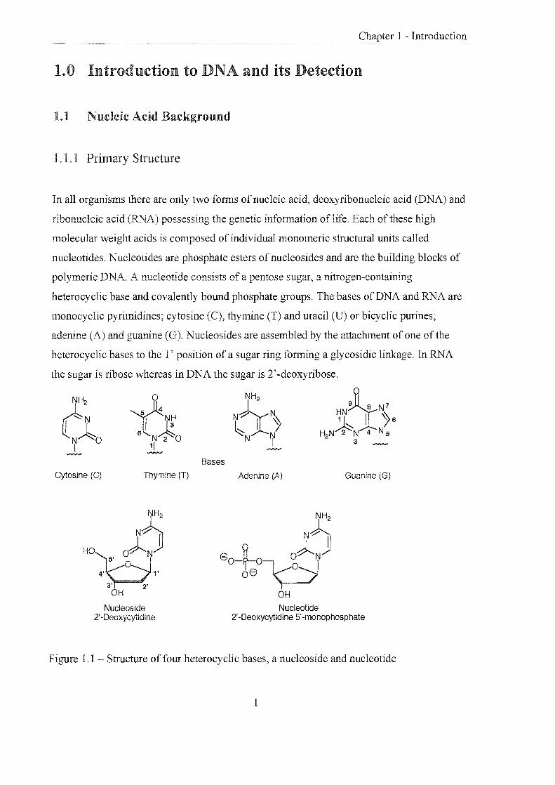
1.1.1 Primary Structure
In all organisms there are only two forms of nucleic acid, deoxyribonucleic acid (DNA) and
ribonucleic acid (RNA) possessing the genetic infonnation of life. Each of these high
molecular weight acids is composed of individual monomeric structural units called
nucleotides. Nucleotides are phosphate esters ofnucleosides and are the building blocks of
polymeric DNA. A nucleotide consists of a pentose sugar, a nitrogen-containing
heterocyclic base and covalently bound phosphate groups. The bases of DNA and RNA are
monocyclic pyrimidines; cytosine (C), thymine (T) and uracil (U) or bicyclic purines;
adenine (A) and guanine (G). Nucleosides are assembled by the att.achment of one of the
heterocyclic bases to the l' position of a sugar ring fonning a glycosidic linkage. In RNA
the sugar is ribose whereas in DNA the sugar is 2'-deoxyribose.
c: 0 r I ~H
IN~O l~ 6 N 2 0
I 1/ ~
Cytosine (C) Thymine (T)
HO
Nucleoside 2'-Deoxycytidine
NH2 ~N7 eY~ ~N I ~6 ~ Y-/' H2N~N '4 75
N I 3 ....,."..,. """"'"
Bases
Adenine (A) Guanine (G)
NH2
N~ 8 ~ O~NjJ o-rr-°-Vo-J
08
)---/ OH
Nucleotide 2'-Deoxycytidine 5'-nlonophospllate
Figure 1.1 Structure of four heterocyclic bases, a nucleoside and nucleotide
1

1 - Introduction
The addition of one or more phosphate groups to the 5', 3' or 2' position of the pentose ring
gives rise to nucleoside mono-, di- or triphosphates. In DNA and RNA,
deoxyribonucleotides or ribonucleotides respectively are joined into a polymer by the
covalent linkage of phosphate groups of the 5' hydroxyl of one ribose and the 3' hydroxyl
of the next. This linkage is kl10v.m as the phosphodiester bond and it is this that gives the
nucleic acid direction. At physiological pH each phosphate group is deprotonated (hence
the tenn 'acid') causing nucleic acids to be highly charged molecules.
1.1.2 Secondary Structure
The characteristic secondary structure of DNA is known as the "double helix". James
Watson and Francis Crick first deduced the structure of this highly orga.TJ.ised duplex in
1953 1,2 making use of the X-ray fibre diffraction data of Rosemary Franklin3,4 and
Maurice Wilkins5,6 and the infonnation contained in Chargaffs rules (see later). The
structure is described as 1:\vo separate, antiparallel chains of DNA. wound around each other
following a helical pathway. The coiling produces a double helix that is right-handed; the
negatively charged sugar-phosphate chain forms the external backbone.
5' end 9H N 0
eO--!Jv-v{:Ct G
W NH2 cJ
eo--k~ /0, ... ~NH2 C
6' --V NyN
" 0 (J 80~_ ~~NH2 A
II q I o ~OVN r \\
\.J Nd C{ -L
8 0-f-o r-Yo o "(yN/rNH
HO":-< 0
T
3'end
Figure 1.2 - Phosphodiester bonds and covalent structure of a DNA strand
2

1 - Introduction
The planar heterocyclic bases form the core of the helix by stacking one above the other
providing considerable stability to the helix. This conformation leads to the formation of
major and minor grooves that follow the coiled path of the molecule. The two strands are
held together by "base-pairs" formed by hydrogen bonds between individual bases on
facing strands and around 10 base pairs make up one tum of the helix.
Under the influence of early work by Avery, MacLeod and McCarty7, Erwin Chargaffin
the early 1950's published rules indicating the amount of adenine always equalled the
amount of thymine and the same was true for guanine and cytosine in DNA 8. This feature
is explained, as the two strands are complementary in terms of sequence. The base pairs are
always purine-pyrimidine pairs, where A only pairs with T and G only pairs with C,
therefore the sequence of one strand directly defines the sequence of the other.
A:T G:C
Figure 1.3 - Hydrogen bonding between the DNA base pairs
1.1.3 The Genetic Code 9
The human genome contains approximately 105 genes, encoded by about 3% to 5% of the
total 3 x 10⁹ base pairs in human DNA. The DNA sequence is found on 23 chromosomes;
22 pairs of autochromosomes and 1 pair of sex chromosomes. A gene is simply a segment
of a DNA molecule that codes for the synthesis of a protein, and in eukaryotes is divided
into coding regions (exons) and non-coding regions (introns). Each gene can consist of up
to 2 million base pairs.
3

1 - Introduction
Replication
n DNA
TranscriPt/,
RNA .. PROTEIN Translation
Figure 1.4 Central dogma of molecular biology
The central dogma of molecular biology describes the flow of the genetic information from
DNA through RNA to a final protein product. The sequence in which amino acids in a
protein are joined together during protein synthesis is ultimately determined by the
sequence of nucleotide bases in a gene encoding that protein. Each amino acid is coded by
a codon (groups of three nucleotides). There are 64 non-overlapping codons which make up
the" genetic code", 61 coding for amino acids and 3 stop codons. As there are twenty amino
acids, the code is degenerate, some amino acids being coded by more than one triplet. The
code is universal and the basis of the heredity information of nucleic acids.
In order for the sequence of bases to be deciphered during protein synthesis the double
stranded DNA is transcribed into single-stranded mRNA. The information carried by the
mRNA is then used as a template for the synthesis of protein molecules during translation
in which other RNA molecules, principally tRNA and rRNA, act as key intermediates 1 0, 1 1.
1.2 Detecting DNA
The fundamentals of using DNA diagnostically involves: (i) defining the nucleic acid
sequences that are essential in the expression of proteins associated with disease; (ii)
determination of the presence of these sequences in an individual. Nucleic acid
hybridisation is the most powerful method for revealing and quantifYing specific DNA or
RNA. The association of complementary strands can be exploited in the detection of
nucleic acids. A probe is a short piece of DNA or RNA that is labelled to allow its
detection, subsequent to hybridisation. A suitable probe system should be highly specific,
have low detection limits and be simple to prepare and purifj 12-15.
4

1 - Introduction
1.2.1 DNA Diagnostics
Applications of routine nucleic acid screening depend upon the development of sensitive,
rapid, accurate and economical procedures, \vhere ideally PCR amplification is used in
conjunction with appropriate probe technology.
Single nucleotide substitutions and insertions and deletion mutations can lead to mutant
alleles or gene variants that are associated with genetic disease e.g. sickle cell anaemia16,
cystic fibrosis, phenylketonuria 17 and Huntington'S disease. These defects can be screened
for directly or by probing for linked genetic markers coinherited with the mutant gene18.
Analysis is important in pre~natal diagnosis and in genetic counselling in affected families
for subsequent pregnancies. Genetic information can be also be used to determine
susceptibility in individuals to exogenous risks such as diet or environmental factors.
Infectious disease e.g. measles, rubella, HIV19 Hepatitis A and B, salmonella and candida
can be clinically diagnosed by the detection of pathogenic organisms by means of DNA
probes. Selection of DNA sequence allows tailoring of a test to a particular disease, for
example HIV screening of donated blood, or for differentiation between closely related
organisms e.g. Herpes Simplex Virus types 1 and 2. Genes related to resistance to
antibiotics for treatment of disease can also be screened.
Applications in forensic science based upon the identification of individuals at a genetic
level are more accurate than traditional identification by blood types, fingerprints or
physical characteristics. DNA can be extracted from body fluids and be used to deduce if
individuals are related in paternity testing, or for compatibility in bone marrow transplants.
Criminal investigation employs the technique ofDN.A fingerprinting which exploits the
highly variable micro satellite sequences of the human genome to yield a "bar code" of an
individuapo .
5
,-,U<U.IJ' .. d 1 - Introduction
1.2.2 Classical Methods
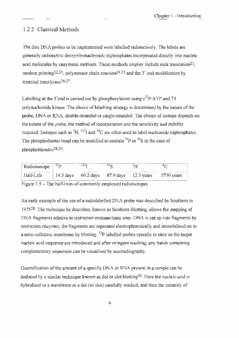
The first DNA probes to be implemented were labelled radioactively. The labels are
generally radioactive deoxyribonucleoside triphosphates incorporated directly into nucleic
acid molecules by enzymatic methods. These methods employ include nick translation21,
random priming22,23, polymerase chain reaction24,25 and the 3' end modification by
terminal transferase26,27.
Labelling at the 5'end is carried out by phosphorylation using l2P-ATP and T4
polynucleotide kinase. The choice of labelling strategy is detennined by the nature of the
probe, DNA or RNA, double-stranded or single-stranded. The choice of isotope depends on
the nature of the probe, the method of incorporation and the sensitivity and stability
required. Isotopes such as 3H, 1251 and 14C are often used to label nucleoside triphosphates.
The phosphodiester bond can be modified to contain 32p or 35S in the case of
phosphothioates28,29.
60.2 days 87.9 days 12.3 years 5730 years
Figure 1.5 - The half-lives of commonly employed radioisotopes
An early example of the use of a radiolabelled DNA probe was described by Southern in
197528. The technique he describes, known as Southern Blotting, allows the mapping of
DNA fragments relative to restriction endonuclease sites. DNA is cut up into fragments by
restriction enzymes; the fragments are separated electrophoretically and immobilised on to
a nitro-cellulose membrane by blotting. 32p labelled probes specific to sites on the target
nucleic acid sequence are introduced and after stringent washing, any bands containing
complementary sequences can be visualised by autoradiography.
Quantification of the amount of a specific DNA or RNA present in a sample can be
deduced by a similar technique known as dot or slot blotting30. Here the nucleic acid is
hybridised to a membrane as a dot (or slot) carefully washed, and then the intensity of
6

,-,nULlL,",' 1 - Introduction
probe concentration remaining on the membrane is measured. The reading directly
represents the amount of target sequence present in the sample.
Radiolabelling has the advantages of very high sensitivity. For example 1251 has a lower
detection limit of lOamol, and radiolabels are easily incorporated. However, there are
numerous conflicting disadvantages associated with this technique:
.. hazardous handing; regulated by the Home Office
short half-life; short shelf-life
expensIve
limited signal emissions
time consuming methodology
waste disposal
1.2.3 Non-isotopic Labelling
In order to overcome the disadvantages associated with radioactive labels the search began
for safe and convenient alternatives. The most common methods to emerge employed
colourimetric31 ,32, chemiluminescent33 , bioluminescent34 or fluorescent35,36 reporter
groups. The labels allow the incorporation of more than one label molecule per
oligonucleotide37 and also permit the simultaneous detection of several DNA targets within
one experiment. This has also enabled the development of automated DNA sequence
analysis as a different coloured fluorophore can be used for each reaction specific for each
of the four bases38. Combinatorial labelling of probes has been used to increase the number
of target sequences that can be detected at the same time39.
A label is a molecule that can be attached to a protein or nucleic acid and is capable of
releasing a signal. Ideally a label for a DNi\. probe would possess the following properties:
.. attached to DNA or RNA under mild conditions with a simple, cheap and reproducible
protocol
.. attachment should not significantly alter the signal generating properties of the label or
effect hybridisation properties of oligonucleotide
7

,,",U,CLIJ"'-" 1 - Introduction
detectable at low concentrations using simple instrumentation
produce a hybridisation signal which is distinguishable from that of an unreacted signal
stable to hybridisation conditions e.g. elevated temperatures, detergents and solvents40
the corresponding reagent is highly specific for its target
chemically cheap and variable to permit synthetic modification of functionality
(changes to physical properties such as solubility, emission characteristics and charge)
co allow simultaneous detection of several labels in one experiment
co multiple addition of the label to a single system
co stable to long term storage
co easily disposed of
The label essentially consists of three parts, the signal emitting moiety, a spacer and a
reactive group.
,,~__ __~) \. ____ -., ~_--"J \. .... __ ...... ,.... __ ~J - Y . v v
Reactive group Spacer Signal emitting moiety
Figure 1.6 Components of a label
1.2.3.1 The Signal Emitting Moiety
In direct labelling strategies this is generally a luminescent 1abel, but can be an enzyme for
example alkaline phosphatase directly conjugated to an oligonucleotide probe41 . The label
molecule has to either exhibit a satisfactory quantum yield in aqueous solution or turnover
a signal-generating substrate. Indirect methods employ either enzymes or haptens (small
foreign molecules that can be attached to a macromolecule to which antibodies can be
raised) as the label, and the signal is generated sequentially. Enzymes include horseradish
peroxidase conjugated to an antibody42 and a common hapten system is based on the
highly specific binding between biotin and strepavidin43 .
8

'---'UCl.LJL'-l 1 - Introduction
1.2.3.2 The Spacer
Many simple aliphatic and aromatic molecules have been used to separate the luminescent
moiety from the labelled substance44. An important prerequisite of the spacer is that it does
not interfere with the luminescent properties of the label. This bridging group can be used
to change the hydrophobicity or hydrophilicty of the molecule, and alter the flexibility of
the label relative to the labelled substance thereby assisting hybridisation.
1.2.3.3 The Reactive Group
general terms, this can be any group capable of reacting with a nucleic acid, under mild
conditions, to fonn a covalent linkage. There are a few examples of non-covalent labelling
of nucleic acids such as the intercalation of ethidium bromide45 in double-stranded DNA,
however covalent coupling has proved to be more popular and versatile. Examples of some
significant labelling reactions are described:
(i) Free amino groups on DNA or nucleosides can couple with N-hydroxysuccinimide
esters, isothiocyanates or activated carboxyl groups of luminescent dyes yielding stable
thiourea or amide bonds. The amino groups are either those present on the heterocyclic
bases or generated by the derivatisation of 5' or 3' end of DNA. These reactions are usually
mild and can be carried out in aqueous solution (a and b, figure 1.7)46-50.
(ii) The nucleic acid is synthesised with a free thiol at one terminus. This undergoes
Michael addition with a,j3-unsaturated ketones attached to the luminescent label (c, figure
1.7)51.
(iii) Labels are functionalised with azido groups which upon activation form highly reactive
nitrene intermediates that react with many different types of chemical bonds in the nucleic
acids. However this results in non-specific labelling of the nucleic acid52,53.
(iv) Labelling of nucleoside triphosphates by the reaction of activated sites on the label and
free amino groups attached to the purine or pyrimidine bases at convenient sites. The
triphosphates are enzymatically incorporated into the DNA probe by methods such as nick
translation or peR Cd, figure 1.7)54.
9

1 - Introduction
(v) Labels are synthesised as phosphoramidites that can be incorporated into DNA during
solid phase oligonucleotide synthesis. One or more of these monomers can be placed
internally or at the 3' or 5' terminus of the oligonucleotide (e, figure 1.7)32,35,55
(a)
8-spacer-{ ) + O-N
" o
P H2N~ --,... 8-spacer---{
HN~
(b)
8-spacer-N=C=S 8-spacer-NH NH-0 [( S
(c)
~ (0-s pace r-y
o + HS-@J - 8-spacer-~s/~
o
o (d) NH2 CL)l
I U Spacer NH N:::;.-'
OH 0 \ I \ ~:::;.-'
o 18 0 8 0 ~- 0 I o 3 - --,... 08
3 OH OH
(e)
fl'\--spacer----0-1--< 01.,,"000>'" ~ V 0 + HO----1'NAl s~ 0-spacer-0-p-0~
( ~ ~ 68
CN
8 = luminescent molecule ~ = nucleic acid
Figure 1.7- Popular methods of label incorporation into nucleic acids
10

1 - Introduction
1.3 Labelling Strategies
1.3.1 Indirect Labelling
Indirect labelling requires a DNA probe labelled with a small molecule that is detectable by
a highly specific binding protein. A classic case is the incorporation of a hapten into a
probe that is recognised by an antibody. The antibody is either covalently linked to a
luminescent molecule or conjugated to an enzyme (alkaline phosphatase, horseradish
peroxidase). The antibody will bind tightly to the hapten and its presence is detected
directly, or in the case of an enzyme case by the turnover of a substrate that results in a
colourimetric, chemiluminescent or fluorescent product. A more complicated, yet more
sensitive method involves the binding of a primary antibody to the hapten of the probe,
followed by binding of a secondary antibody that recognises the first antibody. In this case
it is the secondary antibody which is labelled for detection. Antibodies that recognise
specific haptens are produced in mammals by exposure to the hapten coupled to a protein
carrier for immunisation.
Substrate \
.,ace \ D'
Enzyme (E') '---- Q .. Y 0 Colour or light
Antibody ~ Hap!e,
Hybridised DNA probe '''''''' ""'" ""T A """" ""III "'"'' ""'" '""" "'"" "'"'" Target ON Membrane
Figure 1.8 - Indirect labelling strategy
The most commonly studied hapten is biotin56. Linking to DNA occurs by the reaction of
reagents such as N-biotinyl-6-aminocaproic acid N-hydroxysuccinimide ester or biotin
hydrazide57 (figure 1.9) with an amino group on DNA, or by enzymatic incorporation of
biotinylated nucleotides. Reporter groups are attached to strepavidin and avidin, proteins
(not antibodies) that bind very tightly to biotin, and hybridisation can be detected. Biotin
1 1 ~1

~u.aIJL',",J. 1 Introduction
has the disadvantage that it is present at high levels in certain tissues, leading to high
background signals. However, amplification of signal occurs in the presence of extra biotin
as there are four possible binding sites on avidin only one of which is occupied by the
biotin attached to the probe. Other widely applied indirect detection systems include
digoxigenin (DIG) in combination with anti-DIG antibodies, and dinitrophenyl group
(DNP) with anti-DNP antibodies58,59.
(i) HO (ii) H
Figure 1.9 - i, N-biotinyl-6-aminocaproic acidN-hydroxysuccinimide ester, ii, digoxigenin
1.3.2 Direct Labelling
Direct labelling occurs when a reporter molecule is bound immediately to a nucleic acid
probe. The specific molecular recognition of a nucleic acid probe annealing to its
complementary target sequence enables rapid direct detection60. The label must not
interfere with the hybridisation process and must be stable to hybridisation conditions. The
nucleic acid probe can be a long polynucleotide or a short synthetic strand. Synthetic
probes are advantageous as they can be easily prepared in large quantities, and the probe
length can be used to control the melting temperature of the probe-target duplex. Alteration
of the hybridisation temperature can be used to favour formation of the desired duplex over
mismatch hybrids, and can allow the assay to proceed at lower temperatures and with
shorter annealing times.
\ I Colour or light
/"" Label 0=J -'"
Hybridised DNA probell'""' "11'" "liT IlIHII! niH U Jill III In Iill lillill IInll: H1Hili Target DNA
Membrane
Figure 1.10 - Direct labelling strategy
12

,--,wc<uv'-" 1 - Introduction
1.3.2.1 Radioactive Labels
These are commonly employed and exhibit high signal sensitivities especially when the
probe is multiply labelled. However radio labels are associated with numerous
disadvantages (section 1.2.2).
1.3.2.2 Enzyme Labels
E!lzymes are covalently attached to nucleic acids. The enzymes can be detected by the
turnover of a substrate to produce colour or light. Typical systems use alkaline
phosphatase41 ,61 with 5-bromo-4-chloro-3-indolyl phosphate/nitroblue tetrazolium
(BCIP/~TBT) or horseradish peroxidase62 with diaminobenzimide tetrahydrochloride
(DAB). The conditions of the assay must not cause the enzyme to be denatured; therefore
the activity of the enzyme must be monitored. These labels offer the highest sensitivities
with low background signals and visualisation is rapid.
1.3.2.3 Chemiluminescent Labels
Chemilumescence is the emission of light as a result of a chemical reaction. The ent"IJalpy
of the reaction causes an atom to be promoted to a vibronically excited state, when it
decays a photon is emitted.
C + Reactant I>' Pr* ---_)I> Pr+ tru Catalyst
The emitter is chemically different from the original molecule therefore one molecule can
only produce one photon. However in some cases the product is capable of fluorescing but
may only be transiently stable. Chemiluminescent molecules have been used to detect
nucleic acids, for example luminol is a chemilwninescent substrate for horseradish
peroxidase, and has been used in conjugation with a biotin-strepavidin system63.
Fluorescamine is intrinsically non-fluorescent but reacts in milliseconds with primary
aliphatic amines to yield a fluorescent derivative (figure 1.11).
13

1 - Introduction
CY.~ I O~. H r.OOH ---... I
RNH2 N I ~
~ i h
Luminal Fluorescamine
Figure 1. 11 - Chemiluminescent molecules
1.3.2.4 Fluorescent Labels
Fluorescent molecules absorb light of a specific wavelength and emit light of Imver energy
and longer wavelength. Attachment ofthese molecules to nucleic acids pennits direct
detection if monitored at the correct emission wavelength. In principle, fluorescence
measurements offer extremely high sensitivities, however in practice the sensitivity can be
limited by light scattering, background fluorescence and quenching effects. Many
fluorescent dyes are commercially available and their excitation and emission wavelengths
are represented in figure 1. 12
F----JJ>. F* ____ JJ>. F + hv Light
The most commonly employed fluorescent moieties include 6-carboxyfluorescein
(F AM)35,60,64,65, its tetra and hexachlorinated analogues (TET and HEX) and carboxy-X
rhodamine (ROX)66. These have proved popular due to their high absorption and emission
wavelengths and the range of dyes will allow the detection of several targets within one
experiment67. On the negative side, the broad nature of their emission peaks complicates
the simultaneous detection of multiple sequences. They are also sensitive to pH68 and
susceptible to photobleaching. The inadequacies of these dyes has led to the development
of alternatives such as the CyDyes ™ and the BODIPY™ spectral range dyes.
14

BODIPYTR
BODiPY FL
Texas Red
Rhodamine
Casade Blue
o.
0 • 0
DIll
u •
0
• 0
'-..-uuvv,,< 1 - Introduction
o. CIIl!!I
o • • O •
• O.
0 • DIIIII
Lucifer Yellow r---r---,.---,r---r--r--r-0--r----r---,-,.--...,.-iIII-,--r---r---r-.--.,..--.--r--,
I I I I I I I I I I I I I I I I I I I I 300 400 500 600 700
Wavelength (nm)
o Excitation • Emission
Figure 1.12 - Excitation and emission maxima of a range of fluorescent dyes
Cyanine Dyes (CyDye™)
These covalent labelling agents, introduced by Amersham Life Science69, emit in the far-
red region of the visible spectrum and have the general structure shown in figure 1.13.
These dyes have many of the properties of ideal fluorescent probes including high
extinction coefficients, high quantum yields, and excellent photostability. The spectral
properties (excitation and emission maxima 500-750nm) are selected by the appropriate
choice of heterocyclic nuclei eX and Y) and the length of the polymethine chain (n). The
groups Rl -~ are variable, providing the desired functionality, charge, reactivity and
solubility. Cyanine dyes are popular, as they are commercially available and pH insensitive.
They can be used to detect proteins 70,71, antibodies, peptides and nucleic acids 72, 73.
15

x + Y = S,O,NR,C(CH3l2
n = 1,2,3, .....
Rl - R4 = Substituent
Figure 1.13 - Generic structure of a cyanine dye
BODIPY™ dyes
,->U,CUJV'-'i 1 - Introduction
These versatile dyes which have been patented by Molecular Probes Inc. span the visible
spectrum by alteration of the substituents RJ and R2 which can either increase or decrease
the level of conjugation74. The substituents can be used to alter the solubility and the dyes
are pH insensitive (figure 1.14). The improved spectral characteristics of these dyes have
been exploited in automated DNA sequencing. It is reported that the improved sensitivity
allows a reduction in reagent consumption by 33%, compared to conventional dye
primers75 .
Rr R2 = Substituent
R3 = Linker
Figure 1.14 Generic structure of a BODIPY dye
1.3.2.5 Lanthanide Labels
Lanthanides form chelates that are highly fluorescent with large Stokes shifts and
extremely long lifetimes76-78 . DNA has been directly labelled with lanthanide chelates by
incorporation of labelled deoxynuc1eoside triphosphates by nick translation, random
priming, peR and ruthenium phosphoramidites. Hurskainen et ai. describe a method using
the amino groups on cytidine, which undergo a transamination reaction in the presence of
sodium bisulfite diamine. The free primary aliphatic amine groups react with an
16

'"-'uuvv'"', 1 - Introduction
isothiocyanate derivative of an europium chelate. This process causes multiple labelling of
the DNA, which can affect the efficiency of hybridisation by significantly altering the
melting temperature of the probe-target duplex. It has been reported that the optimum
system contains four to eight europium chelates per hundred bases. The sensitivity of these
chelates is comparable to the detection limits achieved with radioisotopes.
NH2
~Jp . ~N~ NH I H2N~ 2
NaHS03, pH 6
Figure 1.15 Labelling of cytosine with a europium chelate
1.4 Fundamentals of Fluorescence
1.4.1 The Nature of Fluorescence79,80
Fluorescence is the property of some atoms and molecules to absorb photons of incident
light of a particular wavelength (excitation wavelength) and after a short period
(fluorescence lifetime) re-emit a lower energy photon (emission wavelength). The process
begins when a fluorophore absorbs a photon from an external light source, which causes the
promotion of an electron from its ground state (So) to the first excited singlet state (Sl). The
excited molecule then undergoes a conformational change and is subject to collisions with
surrounding molecules. These interactions cause discharge of thennal energy from the
molecule to its environment (internal conversion). After a short fluorescence lifetime the
electron falls back to the ground state and a photon of lower energy, longer wavelength is
emitted (fluorescence). Each fluorophore can repeat the cyclic process many times before
17

1 - Introduction
photobleaching prevails. The 'shift' in energy or wavelength from excitation to emission is
called the Stokes shift and this is fundamental to the sensitivity of fluorescence
measurements, a large Stokes shift leads to low background signals.
The fluorescence quantum yield is the ratio of the number of fluorescence photons emitted
to the number of photons absorbed. It is a measure of the emission efficiency of a
fluorophore. Physical measurements can be complicated by background signals that may
originate from endogenous sample constituents.
2----------------S2 1
o
Absorbtion (10'1 5S)
2 So 1
o
; ,
i Internal conversion(10·12s) , ;
J
Fluoresence (1 O,9S)
Figure 1.16 - Jablonski energy level diagram
1.4.2 Fluorescence Quenching81 ,82
The fluorescence energy of an excited fluorophore can be transferred non-radiative1y to a
molecule in close proximity resulting in the loss of fluorescence emission. No photons are
emitted and the phenomenon is known as quenching. There are two main quenching
processes both requiring molecular contact between fluorophore and quencher:
(i) Collisional (or dynamic); quencher molecules collide with the excited fluorophore and
energy is dissipated as heat, significantly reducing the fluorescence lifetime of the
fluorophore. Collisional quenchers include molecular oxygen, acrylamide, nitromethane,
purines, pyrimidines and some olefins.
(ii) Static (or complex); a complex is formed between the fluorophore and quencher which
is non-fluorescent. Alternatively resonance energy transfer (section 1.4.3) quenches the
fluorophore.
18

'--'~'lUIJ.'-'~ 1 - Introduction
Collisional quenching only affects the excited states of the fluorophore and therefore no
changes in the absorption spectra are observed. In contrast, ground state complex formation
will result in perturbation of the absorption spectrum of the fluorophore.
Ex
F*
I"',~ '#
Fluorescence emission
Collisional quenching
F*
;v·'-I 'V
F + a ---"'ks
Fluorescence emission
Figure 1.17 Principles of collisional and static quenching
(F. 0)"
I no I emission
'V
F.O
Static quenching
1.4.3 Fluorescence Resonance Energy Transfer (FRET)
An excited fluorophore (donor, D) may transfer its energy to a neighbouring chromophore
or fluorophore (acceptor A) non-radiatively through induced dipole-induced dipole
interactions. This occurs when the dipoles are in an approximately parallel orientation. The
criterion for occurrence is the overlap of the emission spectra of the donor and the
excitation spectmm of the acceptor. Forster in the late 1940' s first proposed a theof'j 83 ,
which was later con finned by Stryer and co-workers84, describing long-range molecular
interactions by resonance energy transfer. His theory related the interchromophore distance
(r) and the spectroscopic properties of the chromophore. The rate of energy transfer
between the donor and the acceptor is inversely proportional to the sixth power of the
distance (1/r6). The optimum distance (r) for non-radiative transfer of energy is between 10-
IooA and it is this distance that governs the efficiency of the process. The extent of energy
transfer can be measured because the fluorescence of the donor (both intensity and lifetime)
is significantly reduced, and the acceptor, if it is fluorescent, increases in emission
(sensitised emission).
FRET has been used in many biochemical and structural biological applications as a
qualitative or quantitative tool. The subject has been extensively reviewed with references
to applications for peptide and protein interactions and the use of FRET as a spectroscopic
19

Chapter 1 - Introduction
ruler84-87. FRET is an extremely powerful tool for probing nucleic acid structure88,
sequence89 and hybridisation90. Examples include examining triple helical DNA91 and the
structure of a four-way DNA junction at varying salt concentrations92.
", . , : -. . : .. · . · . · . · . · . · . · ~ .
Donor Absorbance
Normal Donor Emission
Quenched Donor Emission
Acceptor Absorbance
Acceptor Emission
Sensitised Emission
Figure 1.18 - Illustration of quenched donor emission and sensitised acceptor emission
1.4.4 Time Resolved Fluorescence93,94
Fluorescent measurements are plagued by background signals arising from Raman and
Rayleigh scattering or the emission of organic fluorophores within biological samples (350-
600nm). This can limit the sensitivities of the commonly employed short-lived fluorescent
dyes (10-9 to lO-1OS) . However, the use offluorophores with long fluorescent lifetimes
permits selective detection of signals, by delaying measurements until all other species
have decayed. For an interference-free measurement of a signal, the label lifetime must be
at least ten times longer (> lOOns) than the decay of background signals (::::; IOns).
It was the advent of rare-earth metal ion complexes used as labels that transformed the
technology of time-resolved fluorescence. The chelates ofEu (III)77, Tb (III)78, Sm (III)
and Ru (III) with aromatic chromophores, all have long fluorescent lifetimes (IllS to 1 ms),
large Stokes shifts (>200nm), narrow emission bandwidths (10nm) and long emission
wavelengths.
Luminescence of these chelates is not strictly fluorescence or phosphorescence. The
organic ligand absorbs energy and is promoted from So to the excited SI state, where it
rapidly loses energy (intersystem crossing) and falls to the excited Tl state. From this level
20

1 - Introduction
the molecule can either return to So ground state (phosphorescence) or transfer energy
intramolecularly to the 4 f energy level of the central metal ion. If this phenomenon occurs
the metal ion moves into its own Sl state and can decay to So emitting radiation (ion
fluorescence). When a sample is excited by short light pulses short-lived fluorophores will
quickly dissipate to zero. Time-resolved fluorescent measurements are taken after this
period eliminating unwanted background signals.
DELFIA® (dissociation enhanced lanthanide fluorescence immunoassay, Wallac Oy,
Turku, Finland) is a commercially available assay format that uses a non-luminescent
lanthanide chelate (e.g. diazaphenyl-ethylenediamine tetraacetic acid-Eu (III)) as a label for
an antibody or nucleic acid. The final step of the assay involves lowering the pH to induce
dissociation of the lanthanide complex and capture of the lanthanide in a solubilising
micelle. The micelle contains reagents capable of forming a luminescent chelate with the
ion (e.g. fluorinated aromatic ~-diketones) enabling detection95.
Flash excitation
I~
I Fluorescence I intensity
o
''-'/ ' , . "
. "
200
Short-lived fluorescence
Long-lived fluorescence
/ '" ....
600 I
1000
Time (ns)
Figure 1.19 - Principle of time-resolved fluorescence
Enzymes as either labels or analytes can be detected by the turnover of a substrate to
release a product that can chelate witl) lanthanide ion and fonn a luminescent complex. An
example of this is the glucose oxidase substrate 1,10 phenanthroline-2,9-dicarboxylic acid
dihydrazide that gives the detectable chelate Eu (III)-phenanthroline-dicarboxylic acid96.
21

Chapter 1 - Introduction
1.4.5 The Fluorescence Spectrometer
The basic task performed by a fluorimeter is delivery of excitation energy to the
fluorescencing species, and separation of the weaker emitted light from the brighter
excitation light97. This ensures only emitted light is detected and a sensitive and defined
image is generated. Most modem spectrometers consist of four main elements; an
excitation source, wavelength selection devices, the sample cell, and a detector. The basic
construction can be seen in figure 1.20. Light emerges from the source (e.g. xenon or
mercury lamps) and enters the excitation monochromator. In modem spectrometers,
monochromators are based on diffraction gratings, not prisms, allowing the selection of a
narrow band of light by rotation of the concave grating. Typical monochromators have
entrance and exit slits, which are variable. Large slit widths produce increased signal levels
but with higher signal: noise ratios, smaller slit widths give higher resolution but at the
expense of light intensity. The chosen wavelength of light travels to the sample, the emitted
light generated then passes through the emission monochromator to a photomultiplier.
Here, incident photons hitting the photocathode are accelerated through a series of dynodes
towards an anode, this causes the signal to be amplified and the final output is proportional
to the emitted light intensity (figure 1.20).
L = lens S = variable slit C = sample cell Mex = excitation monochromator Mem = emission monochromator P = photomultiplier
--s
-s
P
PC
Figure 1.20 - Typical set-up of a fluorescence spectrometer
22

'-'HUIJL' .... ' 1 - Introduction
To measure the excitation spectrum of a fluorophore, the emission monochromator is set at
a desired wavelength (usually the emission maximum) and the excitation monochromator is
scanned through the absorption bands. To measure the emission spectrum the appropriate
excitation wavelength is chosen and wavelength scanned with the emission monochromator
to detennine the distribution of light emitted by the sample.
Fluorescence spectroscopy is a highly sensitive technique; signals can originate from
sources other than those of the fluorophore of interest. Interference can arise due to
Rayleigh and Raman scattering, background fluorescence of solvents, light leaks in the
instrumentation or stray scatter due to particulates. Acquisition of spectra is complicated by
the wavelength-dependent nature of the source, monochromators and photomultipler tubes
all playing an important part in instrument design98.
1.5 Molecular Biological Methods Utilised in Fluorescence Assay
1.5.1 Polymerase Chain Reaction
The polymerase chain reaction (peR) is a technique used widely in molecular biology99 to
amplify a section of target DNA that is flanked by two kno'vvn genetic sequences. Two
short oligonucleotides are prepared (usually synthetically) and are designed such that each
is complementary to one end of the target strand. These oligonucleotides are known as
primers, and are typically 18-30 bases in length, with similar (%G+C) content to ensure
similar annealing temperatures. The region of the template bound by the primers is
amplified by a series of cycles. In the first cycle the duplex target is dissociated into two
single stands by heating to 95°C, subsequent cooling to approximately 55°C allows the
primers to anneal with their 3' ends pointing towards each other. The temperature is now
increased to 72°C, the optimum temperature for activity of the thermostable Taq
polymerase. This polymerase is isolated from the thermophilic bacterium Thermus
aquaticus and has a half-life over two hours at 95°C so is suited well to the experiment 100.
The polymerase, in the presence ofMg2+, uses nucleotide triphosphates to extend the
primers along the length of the target producing two new double-stranded sections of DNA.
23

Chapter 1 - Introduction
The second cycle begins again by heating to 95°C effecting the denaturation of the newly
synthesised molecules. Each single strand now acts as a template for primer annealing and
extension. During the second cycle primers annealing to newly synthesised molecules can
only be extended as far as the first primer, affording molecules of the correct length. All of
the following cycles amplify the correct length products, which soon outnumber the
original target molecules. In practice n cycles will amplify a particular sequence 2n
times24,101 .
3' ~: Target DNA -5' .,--- .. - - -.............. ... ,../
~
(i) .. 3h== 5' •
/ '--- ---- -y 3'===- 5' ............. -3' !:$ 5' 3' 5'
(iii) 5' 3' 5' .- 3'
3' .,---..
(ii)
"" 5'
,../ c ............. - c 5' 3' - '--- -I> .,--- C ..
Cycle 1 : (i) denaturation c -- .-(ii) primer annealing ... (iii) polymerisation
Cycle 2: (i) denaturation (ii) primer annealing (iii) polymerisation
Cycle 3: (i) denaturation (ii) primer annealing (iii) polymerisation
-- DNA bracketed by the primers
- DNA outside primer region
-~ Primer 1
- Primer2
Figure 1.21 - The first three cycles of a polymerase chain reaction
1.5.1.1 Hot Start peR
Hot start PCR is designed to alleviate some of the problems associated with background
signals in PCR such as primer-dimer formation and non-specific priming. This technique is
carried out manually. Initially the reaction mixture lacks a crucial component such as
magnesium ions or Taq polymerase. Heating during the first cycle ensures there is no
binding, at this point the tube can be opened and the missing component added. PCR now
24

1 - Introduction
continues as nonnal. The major disadvantage of this technique is the possibility of
contamination during the addition ofthe missing component to the open reaction tube.
Alternatives include adding a reagent that can bind to the polymerase and inhibit its action
(e.g. an antibody) which is denatured in the first heating cycle102, or enclosing an essential
component in wax, again released upon heating.
1.5.2 Fluorescence In Situ Hybridisation (FISH)103
Fluorescence in situ hybridisation is an essential tool in genetic analysis as it allows the
identification of the presence and location of cellular DN.A or RNA within morphological
preserved chromosome preparations with sensitivity and ease. The principle lies in the
annealing of a labelled probe to its complementary strand in fixed cells or tissues followed
by detection of the label (radioactive or lwninescent). FISH is different to other probe
systems as the target is embedded in a complex matrix that can hinder probe access and
destabilise probe: target hybrids formed. The probe (DNA or RNA) is usually prepared by
one of four methods: nick-translation; random priming; peR or chemical synthesis and is
labelled during each process. The length of the probe is dependent on the application, but
the typical length is between 200 bp and lkb. Longer probes increase non-specific
background but short probes can be difficult to detect due to insufficient hybridisation, high
probe concentrations decrease the signal: noise ratio. It is important that the target is
accessible to the probe and must be retained in situ, not degraded by nucleases.
Visualisation limits span from an entire chromosome to a 40kb chromosomal section. FISH
employs both indirect and direct labelling strategies, however methods generating signal
amplification have proved most popular.
FISH has been used in toxicological studies to monitor the effect of exposure to radiation
on chromosomal aberrations (structtl.ral and numerical) 1 04. Chromosome painting105,106 by
combinatorial or ratio labelling of specific probes has led to the painting of all twenty four
different human chromosomes being labelled with distinct colours which can provide a
general screening test for chromosome abnormalities. Other applications include
monitoring changes in aneuploidy in sperm (a major cause of birth defects) and genetic
mapping by the use of probes labelled with multiple colours allows gene order along a
25
Chapter 1 - Introduction
chromosome to be determined l07. Complex and unusual structural rearrangements in genes
that lead to tumours can be assessed by comparative genomic hybridisation (CGH). Two
probes are generated one specific for normal DNA and the other for tumour DNA, each are
labelled differently. Equimolar amounts of the probes are introduced into the test cells; the
nature of the signal is a measure of the extent of hybridisation of one probe over
another 1 08.
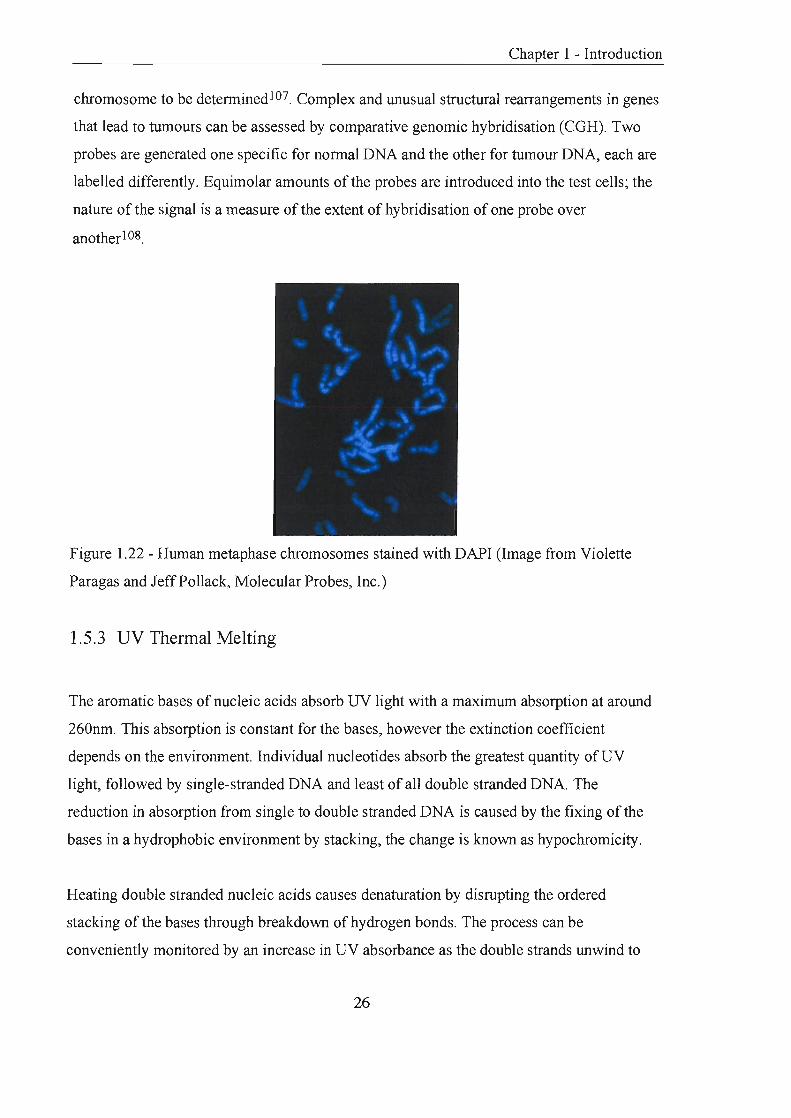
Figure 1.22 - Human metaphase chromosomes stained with DAPI (Image from Violette
Paragas and Jeff Pollack, Molecular Probes, Inc.)
1.5.3 UV Thennal Melting
The aromatic bases of nucleic acids absorb UV light with a maximum absorption at around
260nm. This absorption is constant for the bases, however the extinction coefficient
depends on the environment. Individual nucleotides absorb the greatest quantity ofUV
light, followed by single-stranded DNA and least of all double stranded DNA. The
reduction in absorption from single to double stranded DNA is caused by the fixing of the
bases in a hydrophobic environment by stacking, the change is known as hypochromicity.
Heating double stranded nucleic acids causes denaturation by disrupting the ordered
stacking of the bases through breakdown of hydrogen bonds. The process can be
conveniently monitored by an increase in UV absorbance as the double strands unwind to
26

1 - Introduction
single strands. The thermal denaturation of dsDNA is co-operative indicating that the ends
and the AT-rich internal regions de stabilise adjacent regions of the helix. This leads to a
progressive and concerted melting of the whole structure at a well-defined temperature,
corresponding to the mid-point of a smooth transition. This temperature is known as the
melting temperature (Tm). UV melting experiments can provide both quantitative and
qualitative data about the nature, purity and degree of hybrid is at ion of a sample.
1.6 Practical Examples of Fluorescent Probe Systems
1.6.1 Taqrnan™ Assay
The Taqman™ assay is a widely used format for the detection of accumulation ofPCR
specific products 1 09, 110. A linear probe is assembled consisting of an oligonucleotide
labelled with a fluorogenic donor (5' FAM) and acceptor (internal or 3' TAMRA).
Irradiation of the intact probe causes excited emission by the donor, which is quenched by
the process ofFRPT (section 1.4.3). The probe can hybridise to the complementary
template and during polymerisation the probe will be cleaved due to the inherent 5' ~ 3'
nucleolytic activity ofTaq DNA polymerase. Cleavage causes separation of the donor and
acceptor that resulting in an increase in fluorescent intensity, due to loss of quenching.
Measurement of the rise in fluorescent intensity directly indicates the generation of probe
specific amplicons.
The design and performance of the probe requires three main considerations:
(i) Degree of quenching; dependent on choice and separation of the fluorophore and
quencher pair, the conformational flexibility and purity of the probe.
(ii) Hybridisation of the probe; internal quenchers can disrupt base pairing and reduce Tm,
a 2~3°C reduction in Tm has been reported for tvvo internal TAMRA molecules109.
(iii) Efficiency of probe cleavage by Taq polymerase; dependant on the accessibility of the
probe to the enzyme and the complementarity between the probe and template.
27

1. Polymersation
Forward 5' primer Q Probe ,. 3"==~"~:::::~--- 5'
2. Probe Displacement
5,~~===f~~~~3~' __ _ 3~ 5'
3. Cleavage of Probe
, , 5' '\.."..... 3,======~"::----I'l==~3r.' ___ 5'
4. Completion of Polymerisation
- -" , .. 3' '-'" .....
5' 3"~~============~·~5'
Figure 1.23 - The Taqman™ assay
• Quencher
o Fluorophore (quenched)
Fluorophore
Chapter 1 - Introduction
The Taqman fonnat has applied to be rapid and accurate detection of hepatitis C virus in 48
patient samples 111 and detection of salmonella in food samples in less than 20 hours
producing no false negatives or false positive results 112. The assay has also been used to
develop a test for parental DNA for a deletion sequence linked to the genetic syndrome
cutaneous malignant melanoma 113.
1.6.2 Molecular Beacons
Tyagi and Kramer have described the recent discovery of a novel probe technology for the
detection of specific nucleic acids in homogeneous solutionl14. These probes, named
'Molecular Beacons', fluoresce only upon hybridisation to their target. They produce
sensitive, real-time signals that indicate the degree of hybrid is at ion of the probe to a target
nucleic acid. The essential feature of the probe is their stem and loop structure. The loop
portion is an oligodeoxynucleotide sequence that probes for its complementary target
nucleic acid in solution. The stem is constructed of two short oligodeoxynucleotide arms
28

Chapter 1 - Introduction
either side of the loop, one ann is tenninally labelled with a fluorophore and the other with
a quencher. Ideally the quencher should be non-fluorescent. Annealing of the anns causes
intennolecular energy transfer from the fluorophore to the quencher and this energy is
dissipated as heat. In this confonnation the probe is non-fluorescent. Hybridisation of the
loop to its target causes a confonnational change, the stem anns dissociate, and the
fluorophore and quencher are no longer in close proximity; fluorescence is restored.
+
Molecular Beacon Probe Non-fluorescent
Target Nucleic Acid
Probe-Target Hybrid Fluorescent
Figure 1.24 - Principles of a molecular beacon assay
The original molecular beacons synthesised by Kramer's group utilised 5-(2'
aminoethyl)aminonapthalene-1-sulfonic acid (EDANS) as a fluorophore and 4-(4 '
dimethylaminophenylazo) benzoic acid (DABCYL) as the quencher. DABCYL was later
shown to be an efficient quencher (98%) ofa series of spectral dyes (emission maxima 475-
615nm) due to Van der Waal contact of the fluorophore and quencher pennitting direct
energy transfer 115.
H
~~NH2
~ 0=1=0 0 8 Na@ EDANS
Figure 1.25 - Structure of a commonly employed fluorophore and quencher pair
29

'-../H'U.IJV ..... 1 - Introduction
Molecular beacons must be designed so that the arm sequences are unrelated to the target
and are long enough to form a stem (>4 bp) but not so long that dissociation is difficult
«12 bp). In addition, hybridisation of the arm sequences in the hairpin loop must produce a
weaker interaction than target-to-probe annealing.
Molecular beacons have been used to detect and quantify PCR ampIicon concentration by
the intensity of fluorescence at the mmealing stage in each cycle. They are pa.rticularly
suited to this application, exhibiting fast hybridisation kinetics and allowing sealed tube
experiments. The probes exhibit allelic discrimination, only binding to their perfectly
complementa.ry targets and have the ability to recognise single base mutations. These
principles have allowed the use of multicoloured probes in a single experiment 115,116.
Practical examples of their application to real systems include detection of the single point
mutation of cytosine to thymidine in the methylenetetrahydrofolate reductase (MTHFR)
gene. This mutation has been related to an increased risk of cardiovascular disease and
neural tube defects. Molecular beacons specific for the wild type or mutant sequence
demonstrated high levels of specificity for their target 117. it mutant form arising from a
conservative substitution (V641) in the coding region of the CCR2 genotype has a
protective effect against HIV-l disease progression. A molecular beacon assay that
accurately discriminated between the wild type labelled with fluorescein, and mutant form
labelled with hexachlorinated fluorescein has been described118.
In addition the probes have been used for the detection of mRNA119, 120, in surface
immobilised hybridisation studies 121, as PNA-DNA hybrid probes122, sequence analysis of
amplified segments of Mtuberculosis DNA123 and in the spectral genotyping of the alleles
of the ~-chemokine receptor S (CCRS)19.
1.6.3 Scorpion Primers
A recently described technology combines a highly specific probing region with a primer
for a peR reaction65 . The assay is unimolecular and therefore kinetically and
30

Chapter 1 - Introduction
thermodynamically favoured. The probe region is chemically attached to the 5' end of one
of the PCR primers and is blocked against copying by a hexaethylene glycol spacer. The
primer binds to the target and is extended by polymerisation. The newly synthesised strand
contains a sequence complementary to the probe sequence, annealing occurs and
fluorescence is detected. The principal superiority of the assay is its unimolecular nature,
inferring zero-order kinetics increasing the efficiency of the probing process (the probe is
held in close proximity to its target) and reducing the time-scale of hybrid is at ion.
Intermolecular systems (TaqmanTM109 and molecular beacons 124) are limited by the
formation of alternative intra-strand secondary structures at the assay temperature and the
competition of re-annealing complementary PCR amplicons.
Scorpions have shown high specificity in single tube genotyping experiments of the two
main variants of the Hereditary Haemochromatosis gene: C282Y, the major disease causing
mutation and H63D, a prevalent polymorphism of uncertain clinical relevance.
Scorpions have also been shown to accurately distinguish between matched and mis
matched allelic polymorphs of ex on 10 of the BRCA2 gene.
Scorpion in Dark State
Primer Extension
Probe-Target Hybridisation
• Quencher
o Fluorophore (quenched) Fluorophore
I Blocker ~PCRprimer
Figure 1.26 - Stages of a Scorpion detection assay
31
Chapter 1 - Introduction
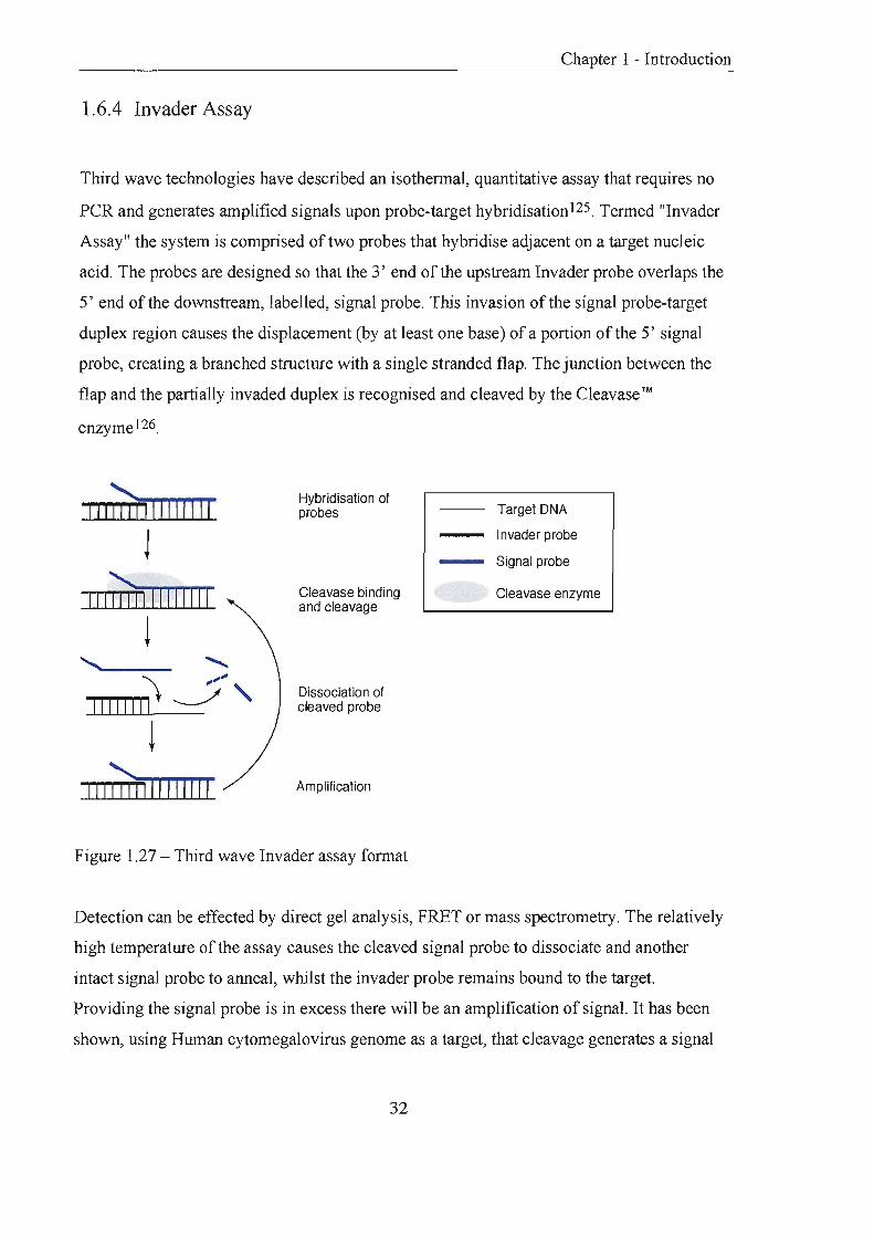
1.6.4 Invader Assay
Third wave technologies have described an isothermal, quantitative assay that requires no
PCR and generates amplified signals upon probe-target hybridisation125. Termed "Invader
Assay" the system is comprised of two probes that hybridise adjacent on a target nucleic
acid. The probes are designed so that the 3' end of the upstream Invader probe overlaps the
5' end of the dO\VIlstream, labelled, signal probe. This invasion of the signal probe-target
duplex region causes the displacement (by at least one base) of a portion of the 5' signal
probe, creating a branched structure with a single stranded flap. The junction between the
flap and the partially invaded duplex is recognised and cleaved by the Cleavase ™
enzyme 1 26.
111;m 1111111 11
J
",;W IIIII IIII !
......... .......... :t ~~
1111110 J " !
111 7irrn 11111 11
Hybridisation of probes
Cleavase binding and cleavage
Dissociation of cleaved probe
Amplification
Figure 1.27 - Third wave Invader assay format
Target DNA
I nvader probe
- Signal probe
Cleavase enzyme
Detection can be effected by direct gel analysis, FRET or mass spectrometry. The relatively
high temperature of the assay causes the cleaved signal probe to dissociate and another
intact signal probe to anneal, whilst the invader probe remains bound to the target.
Providing the signal probe is in excess there will be an amplification of signal. It has been
shown, using Human cytomegalovirus genome as a target, that cleavage generates a signal
32

1 - Introduction
that is proportional to the number of copies of target DNA present. Carry-over
contamination, false-positives and background signals are all alleviated as signal is only
procured in the presence of a target127.
l.6.5 PNA Probes
PNA oligomers are not substrates for DNA, RNA or protein modifying enzymes, and will
hybridise to DNA and RNA by normal \Vatson and Crick base pairing128. They bind
strongly under conditions in which normal nucleic acids hybrids are disfavoured, for
example at low salt concentrations. PNA probes are assembled by standard FMOC or BOC
chemistry and C~'1 be labelled directly with fluorescent dyes122, enzymes, or visualised
indirectly with haptens such as biotin and dinitrophenol. PNA probes have high specificity,
affinity and stability at low salt concentrations in comparison with DNA probes. DNA
probes often fail to distinguish certain regions of highly structured rR..NA. However PNA
probes detect these highly structured rRNA regions with significantly higher fluorescent
signals, exhibiting improved performance at high temperatures and low salt concentrations.
l.6.6 SUNRISE Primers
An amplification system has been developed called SUNRlSETNI The basis is the
incorporation of fluorescence energy-transfer labelled primers into PCR products. These
primers have specific target sequences at the 3' end and hairpin structures at the 5' end that
are non-fluorescent prior to PCR. Incorporation of the primer during PCR causes the
hairpin to be disrupted a,'ld fluorescence is detectable. The fluorescent signal intensifies
with increasing number ofPCR cycles as more primers are incorporated I 29.
Following this a Universal SUNRISE Primer was reported to allow the incorporation of an
~d"'ntl·cal hal·rp~n ~~l·me" ;nto an'\! tArget nnAl",;,., "c~d130 An ;nl·tl·al PI'R st"'p l·ncorpo~"'+"'s " i \..- ~ 1 "1-1-1 JJI 1.. 1 .1 .1.1 ".lJ "a U.\o..IL'-'J.\.I u .1 . • l.l .1 1. ~1- \,.;.L 1u(,.\.,.o U
primer that carries an oligonucleotide tail (15 bases) that is complementary to the sequence
of the universal hairpin primer. Following incorporation of the tail, the hairpin primer takes
over and is unwound in the same manner as before.
33

,,--,u,~ .. n~i 1 - Introduction
The primers have been successfully applied to a closed tube amplification detection fonnat
to distinguish between the normal (\VG4) and mutant (RG4) alleles ofthe ~(3)-adrenergic
receptor gene 131.
1.6.7 DNA Chip Technology
DNA chip technology utilises high-density microscopic arrays of nucleic acids immobilised
on solid surfaces for biochemical analysis. DNA or RNA samples from biological sources
are labelled enzymatically by the incorporation of labelled primers or nucleoside
triphosphates during peR amplification. Hybridisation of the targets to the microarrays and
subsequent detection can be used for polymorphism detection, DNA sequencing and
genotyping. For example analysis of the CFTR gene was carried out by the preparation ofa
microplate loaded with 428 features to identifY mutations in exon 11, and a microarray
containing 96,600 20mers was used to identifY mutations over the entire 3.45 kb of exon
11132.
The oligonucleotide arrays are often prepared on glass due to its inert chemical properties,
low intrinsic fluorescence a.lld the ability to derivitise its surface. The strategies for the
preparation of these libraries using photolithography and micro-spotting, has been
excellently reviewed by B.Lemieux et al. 133 and E.M. Southern 134
34

Chapter 2
Development of Novel Homogeneous
Assay Systems

Chapter 2 - Homogeneous Assay
2.0 Development of Novel Homogeneous Assay Systems
The preceding chapter introduced the reagents, techniques and strategies employed in the
design of systems to probe genetic sequences. The following chapter aims to record the
work carried out in the design, synthesis and testing of a novel two-probe assay.
2.1 Introduction
The aim of this project was to further develop the use of fluorescence as a detection method
for DNA sequences. When designing a novel assay the incorporation of the following
features was considered to be beneficial:
(i) Homogeneous assay (all components in solution) is advantageous over its heterogeneous
counterpart135 (solid and solution phase components) as it does not rely on the
hybridisation of a probe to an insoluble support and therefore displays faster hybridisation
kinetics. In homogenous assay formats there is no requirement for washing steps as the
signal is generated only when the labelled probe is hybridised to the target.
(ii) Enzyme-linked probes are highly specific for their substrates and provide high detection
sensitivities, similar to those attained using radioisotopes61 .
(iii) Isothermal signal generation simplifies an assay by reducing the number of steps
perfonned.
(iv) In certain cases, the use of more than one probe in an assay increases the specificity for
a particular target as two or more adjacent nucleic acid sequences can be probed.
An assay was proposed (figure 2. 1) that required a probe labelled at the 5' end that is
initially non-fluorescent and complementary to a known sequence on a target nucleic acid
(signal probe). This probe is 'dark', as the fluorescent label it bears has been chemically
modified with masking groups to remove its fluorescent characteristics. The masking
35

Chapter 2 - Homogeneous Assay
groups are substrates that can be cleaved from the label by an enzyme-catalysed reaction. A
second probe consists of the conjugate formed when an enzyme is linked to the 3' end of an
oligonucleotide (enzyme-probe). The enzyme-probe is complementary to a sequence
downstream (further along the target DNA) of the upstream signal probe sequence on the
target DNA. There will be little or no fluorescence from either the signal probe or enzyme
probe individually in solution. When the two probes are mixed there may be a small
amount of background fluorescence detected due to the enzymatic hydrolysis occurring
when the two probes meet in solution. However, in the presence of a target nucleic acid the
two probes can hybridise adjacent to each other, bringing the 3' enzyme and the 5' substrate
molecules into close contact. Cleavage of the masking groups by the enzyme will release a
strong fluorescent signal, which can be detected.
(i)
(ii)
(iii)
(iv)
1 ~
J
~
J
o Nonfluorescent label
Fluorescent label • Enzyme
Q Substrate protecting groups
- Probe DNA
- Target DNA
Figure 2. 1- (i) An oligonucleotide is labelled with a fluorophore (5') which has been
chemically modified to remove its fluorescent properties (signal probe), the probe is
introduced to the target nucleic acid. (ii) The probe hybridises to its complementary
sequence on the target nucleic acid. (iii) An oligonucleotide labelled with a enzyme (3 ' ) is
introduced (enzyme-probe). (iv) The oligonucleotide-enzyme conjugate hybridises adjacent
to the signal probe, the enzyme cleaves the substrate groups attached to the fluorophore and
fluorescence is visualised.
36

Chapter 2 - Homogeneous Assay
The assay is desirable as is homogeneous, isothermal, requires two probes and exploits
enzyme detection. The close proximity of the substrate and enzyme is caused only by
hybridisation of the two probes adjacent to one another on a target; consequently
fluorescence is only generated in the presence of a target nucleic acid. The assay format is
sensitive, specific and the fluorescent signal generated \vill be directly proportional to the
number of enzyme-probes hybridised.
Fluorescein is a well-known dye that will only fluoresce when its phenolic hydroxyl groups
are unsubstituted. This allows the lone pairs on the oxygen to participate in the highly
conjugated delocalised system and stabilise the lowest excited state. When the phenolic
hydroxyl groups are unprotected the wavelength of emission is in the visible region and
fluorescence is observed. However, protection of the hydroxyl groups masks this
fluorescence by shifting the wavelength of emission into the ultraviolet region. A non
fluorescent probe can be created by attachment of a phenol-protected fluorescein monomer
to the 5'end of an oligonucleotide.
2.2 Alkaline Phosphatase Approach
2.2.1 Introduction
Alkaline phosphatase catalyses the hydrolysis of phosphomonoesters to produce inorganic
phosphate and the corresponding alcohol. It is a relatively small enzyme with a molecular
weight of about 89 kDa and is dimeric in structure. Alkaline phosphatase has been applied
to indirect136 and direct labelling strategies61 ,62 in nucleic acid hybridisation probe
technology with detection reported at the level of one attomole137. Indirect procedures
described in the literature involve attachment to a hapten or to an antibody specific for a
hapten, in both cases detection is achieved by enzymatic turnover of a substrate releasing
light or colour. Probe assembly in direct methodologies involves either covalently attaching
the enzyme or the substrate to an oligonucleotide. Hybridisation is measured by the
increase in colour or light caused by turnover of the substrate such as 5-bromo-4-chloro-3-
indolyl phosphate I nitroblue tetrazoliuIn (BCIP/NBT). The chemistry involved in
37

Chapter 2 - Homogeneous Assay
conjugation of alkaline phosphatase to oligonucleotides is routine138 presenting a
convenient starting point for assay development.
Preparation of a signal probe required the attachment of a label modified with substrates for
alkaline phosphatase (phosphate esters). Fluorescein diphosphate was chosen as a suitable
label modified with a reactive spacer for attachment to an oligonucleotide (figure 2.2).
Several synthetic approaches to prepare the signal probe are described in the following
sections.
e Q '/
eif'\\ o
o
e o 0, I p, e d 0
H R N o-p.-o~ ~ 6 e oligonucleotide
Figure 2.2 - Fluorescein diphosphate signal probe
2.2.2 Solid Phase Strategy
This approach involved synthesising a fully protected phosphoramidite monomer of
fluorescein that was compatible with standard solid phase DNA synthesis 139. The monomer
could be coupled to the 5' end of a resin bound oligonucleotide on a DNA synthesiser, and
following selective removal of the fluorescein protecting groups, on-resin phosphorylation
of the phenolic hydroxyls could be attempted. The choice of protecting groups for tl}e
phenolic hydroxyls of fluorescein was imperative, as their selective removal required
orthogonality between these protecting groups and those of the heterocyclic bases. In
addition any deprotection reagents applied had to leave the oligonucleotide-resin linkage
intact allowing subsequent on-resin phosphorylation.
2.2.2.14,4'-Dimethoxytrityl Ether
4,4' -Dimethoxytrityl ethers are acid labile and repeatedly removed from the 5' hydroxy! of
nucleosides during solid phase DNA synthesis, inferring compatibility for protection of the
38
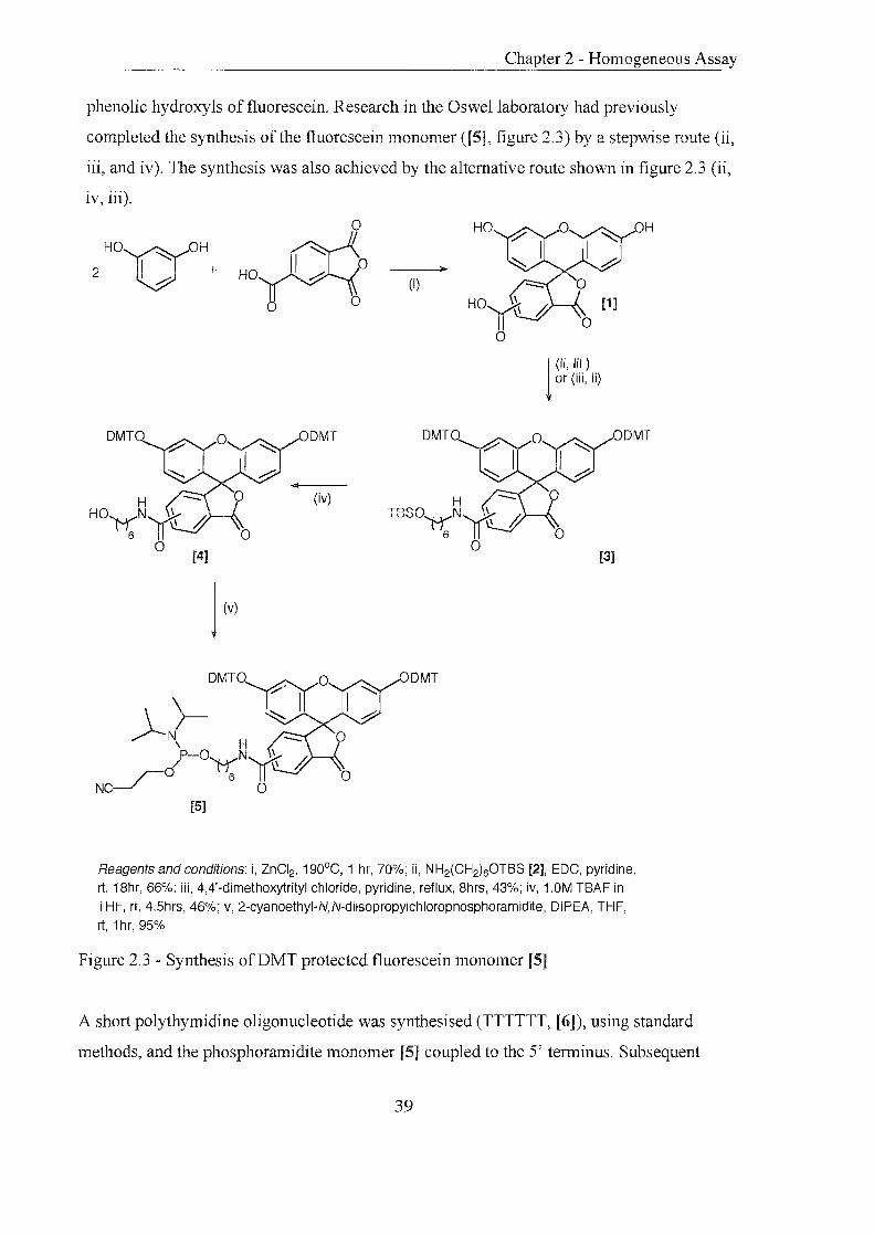
Chapter 2 - Homogeneous Assay
phenolic hydroxyls of fluorescein. Research in the Oswellaboratory had previously
completed the synthesis of the fluorescein monomer ([5], figure 2.3) by a stepwise route (ii,
iii, and iv). The synthesis was also achieved by the alternative route shown in figure 2.3 (ii,
iv, iii).
o H01)'0H
2 I + h HOP(
o 0
(iv)
DMTO~O \ >- I~ II J-N, H-
p-o N~\ / ~ \
r-O 6 NC~ 0
[5]
(i)
DMTO
H TBS0'M;;N~
o
DMT
j (ii, iii ) or (iii, ii)
o
ODMT
[3]
Reagents and conditions: i, ZnCI2, 190°C, 1 hr, 70%; ii, NH2(CH2)60TBS [2], EDC, pyridine,
rt, 18hr, 66%; iii, 4,4'-dimethoxytrityl chloride, pyridine, reflux, 8hrs, 43%; iv, 1.0M TBAF in
THF, rt, 4.5hrs, 46%; v, 2-cyanoethyl-N,N-diisopropyichlorophosphoramidite, DIPEA, THF,
rt, 1 hr, 95%
Figure 2.3 - Synthesis ofDMT protected fluorescein monomer [5]
A short poly thymidine oligonucleotide was synthesised (TTTTTT, [6]), using standard
metlJods, and the phosphoramidite monomer [5] coupled to the 5' terminus. Subsequent
39

Chapter 2 - Homogeneous Assay
treatment with trichloroacetic acid in CH2Ch (3%) was expected to release the 4,4-
dimethoxytrityl cation. Normally, the highly visible bright orange cation is carried away by
solvent flow. Acid treatment of the monomer turned the resin orange indicating release of
the cation. However, all attempts to separate the cation from the resin by repeated washing
or addition of known efficient trityl cation scavengers, (pyrrole and triethylsilane140) failed.
It was concluded that the cation was strongly, ionically associated with the phenolic anion
(figure 2.4), a phenomenon that can occur when nucleophilic species remain bound to the
resin141 . The tight association of the DMT cation led to the search for other suitable
hydroxyl protecting groups.
Q~~-~~-~~oj-O'Y..Y~rr -; I Q'Y~Y~Y '8 II. ,
a 6 ''"
Expecty
~ H nnn-O-p-(l . . ~ N[(\r'
'8'-W \ 0 a 6 o [6]
DMT
o
Fluorescent Resin, orange deprotection solution
8® DMT
o
Orange Resin, colourless deprotection solution
Figure 2.4 - Expected and observed results: acidic deprotection of resin-bound labelled
oligonucleotide
2.2.2.2 p-Methoxybenzyl Ether (PMB)
p-Methoxybenzyl ethers are cleaved oxidatively using 2,3-dichloro-5,6-dicyano-l,4-
benzoquinone (DDQ) in water142, or by ceric ammonium nitrate in aqueous acetonitrile to
release alcohols. A range of reagents and solvents were investigated to protect 5(6)
40

Chapter 2 - Homogeneous Assay
carboxyfluorescein as a PMB ether. These included using sodium hydride, triethylamine
and diisopropylethylamine in THF, DMF and pyridine. Reactions in THF proved
problematic due to poor solubility of carboxyfluorescein. Reactions in DMF and pyridine
displayed good solubility but consistently gave low yields of the desired product and higher
yields of the triply protected product (PMB ethers and PMB ester).
2.2.2.3 t-Butyldimethysilyl Ether (TBS)
TBS ethers are relatively acid and base stable but labile to fluoride ions. 5(6)
carboxyfluorescein was reacted with t-butyldimethylsilyl chloride and
dimethylaminopyridine in pyridine to give the desired compound. However, subsequent
purification and handling revealed a high degree of instability to acid, indicated by the
constant regeneration of fluorescence.
2.2.2.4 Levulinyl Esters
Levulinyl groups have been used to protect of hydroxyl functions of 0ligonucleotides143 .
The esters are cleaved by O.5M hydrazine hydrate in pyridine/acetic acidlwater144 and
under these conditions the oligonucleotide-support remains intact. Reactions oflevulinyl
anhydride with 5(6) carboxyfluorescein produced a product that exhibited instability in the
presence of amines. Introduction of the linker amine [2] or the use oftetrabutylammonium
fluoride caused the appearance of fluorescence, indicating deprotection had occurred.
2.2.2.5 Disiloxyllinker
Kwiatkowski et al. reported the synthesis of a disiloxyl resin-oligonucleotide linker to
replace the conventional succinyllinker to overcome the production of defective molecules,
in particular depurinated molecules, during standard oligonucleotide synthesis145.
DMT°L--o~ ~ase
O--~~ o
(i) (ii)
Figure 2.5 - i, Conventional base labile succinyllinker, ii, fluoride labile disiloxyllinker
41

Chapter 2 - Homogeneous Assay
1,3-dichloro-l,1,3,3-tetraisopropyl disiloxane is coupled to the 3'hydroxyl ofthe first
nucleoside to be attached to the resin, this is then coupled to an alcohol derivatised resin.
The linker is stable to mild base but cleaved by fluoride ions. Fluorescein dipivalate is
commercially available as its phosphoramidite monomer, utilising the monomer in
conjunction with the disiloxyllinker ,,'ould permit selective deprotection of fluorescein.
---l>-
(i)
[7]
(iv)
[10J
Reagents and conditions: i, 2'-deoxythymidine,4-dimethylaminopyridine, 4,4'-dimethoxytrityl chloride, pyridine, rt, 24hrs, 82% ii, imidazole,
[9]
1 ,3-dichloro-1 ,1 ,3,3-tetraisopropyl disiloxane, pyridine, rt, 24hrs, iii, 1 ,6-hexanediol, pyridine imidazole, rt, 12hrs, 76% (last two steps), iv, diisopropylethylamine, 2-cyanoethyl-N,N-diisopropylchlorophosphoramidite, C~Cl2' rt, 1.5hrs, argon, 98%
Figure 2.6 - Preparation of disiloxyl phosphoramidite monomer
Synthesis of the disiloxyl nucleoside phosphoramidite monomer [10] was achieved. The 5'
hydroxyl of 2'-deoxythyrnidine was protected as a 4,4' -dimeLh.oxytrityl ether. 1,3-dichloro-
1,1,3,3-tetraisopropyl disiloxane was coupled to the 3' hydroxyl and following conversion
42

Chapter 2 - Homogeneous Assay
of the starting material, hexan-l,6-diol added to the reaction mixture. Finally the primary
alcohol of the hexyl spacer was transformed to the phosphoramidite (figure 2.6, [10]).
LCAA-CPG was derivatised with a poly alkyl linker (figure 2.7). The linker was assembled
by protecting the primary hydroxyl group of 12-hydroxydodecanoic acid as 4,4' -
dimethoxytrityl ether [11]. The following reaction coupled the carboxylic acid of this
molecule with free amino moieties on the resin. Resin loading was calculated by acid
catalysed detritylation, DMT cation release was quantified at 495nm. The resin was capped
with acetic anhydride prior to use in automated oligonucleotide synthesis. The first
nucleoside coupled to the CPG (3') was the monomer [10] thus placing the fluoride scissile
linkage between the oligonucleotide and the resin. Sequential addition of twelve standard
thymidine monomers, followed by fluorescein dipivalate completed the synthesis of the
resin-bound oligonucleotide [13].
R./OH R./OH HO "11n - DMTO ·11n -->-
o (i) 0 (ii, iii)
[11]
~-O DMTO~ -->-
o [12]
Automated oligonucleotide synthesis
Reagents and conditions: i, DMTCI, pyridine, rt, 2hrs; ii, LCAA-CPG, DIC, HOST, DIPEA, CI12CI2, rt, 1 hr; iii, AC20, pyridine, THF: 1-methylimaidazole, THF (1:1), rt, 1 hr
Figure 2.7 - Derivatisation of CPG for oligonucleotide synthesis with monomer [10]
The stability of the disiloxyllinker to the conditions used for the deprotection of the
fluorescein trimethylacetyl esters was established. Methanolic ammonia is the typical
reagent used for such deprotections. However, the disiloxyllinker was labile when
subjected to ammonia with significant loss of the oligonucleotide from the resin after 30
minutes, contrary to the published results 145. Figure 2.7 shows the stability of the disiloxyl
linker to methanol saturated with aImnonia compared to the stability of the conventional
succinyllinker, known to completely cleave in 30 minutes.
43

4.5
4
3.5
~ 3 c 25 2.5
~ 2 ~ 1.5
1
0.5
- Succinyl linker
- Disiloxyl linker
o 10 20 30 40 50 60 70 80 90 100 110 120 Time (seconds)
Chapter 2 - Homogeneous Assay
Figure 2.8 - Increase in absorbance at 260nm as oligonucleotide is released into solution
from CPG derivatised with succinyl or disiloxyllinkers
High losses of oligonucleotide from the resin were impractical for on-resin fluorescein
phosphorylation. Alternative deprotection systems (figure 2.8) were investigated in a series
of model reactions on fluorescein dipivalate [18]. The results revealed DBU: MeOH (1: 20)
as a suitable choice of deprotection reagent and subsequent experiments demonstrated the
stability ofthe disiloxyllinker to this reagent over the 45 minutes reaction period.
Deprotection conditions
Ammonia: MeOH (1 : 20)
Triethylamine: MeOH (1 :20)
Triethylamine: MeOH (1: 4)
Triethylamine: MeOH (1: 1)
KHC03 (0. 1M): MeOH (1: 20)
LiBH4 (2.0M): THF: MeOH (1: 2: 2)
DBU: MeOH (1: 20)
Deprotection time (Hrs)
18
18
18
12
18
6
0.75
Figure 2.9 - Conditions and time periods from acetyl removal from fluorescein dipivalate
An oligonucleotide sequence FTTTTTTTTTTTT ([14]), F = fluorescein monomer, figure
2.10) was synthesised using the disiloxyllinker and resin. The fluorescein monomer was
deprotected with DBU: MeOH (1: 20) producing the expected fluorescent resin. Initial
phosphorylation of the fluorescein hydroxyl groups using the phosphate monomer shown in
44

Chapter 2 - Homogeneous Assay
figure 2.10 appeared successful (monitored by the disappearance of resin fluorescence).
The oligonucleotide was removed from the resin using 3HF .. Et3N: NMP: Et3N to give a
colourless solution. (NE. using tetrabutylammonium fluoride gave a fluorescent solution
indicating loss of phosphate groups) The cyanoethyl and ethylsulfonyl protecting groups on
the phosphate monomer were removed with triethylamine: CH2Ch (1: 4). RPHPLC
analysis of the oligonucleotide gave a maj or fluorescent peak corresponding to F AMT 12,
and a minor peak that was fluorescent and therefore could not be the desired product.
Repeated attempts at on-resin phosphorylation failed to yield the desired phosphorylated
fluorescein labelled oligonucleotide.
(~ChOCOUO ~ I
"-
OCO(CH3h
o
o
(i) (ii)
Figure 2.10 - i, phosphate phosphoramidite, ii, fluorescein phosphoramidite
2.2.3 Solution Phase Strategy
Synthetic oligonucleotides derivatised at the 3' or 5' terminus with an amino function
(aminolin..l() react with N-hydroxysuccinimde esters of dyes in a simple labelling reaction.
5(6) Carboxyfluorescein N-hydroxysuccinimide ester [15] was treated with phosphorus
oxychloride to give non-fluorescent crude solid. Reaction of the crude product with an
aminolink: oligonucleotide [17] in bicarbonate buffer, pH 9.75, gave one main peak by
RPHPLC shown to be unreacted amino link oligonucleotide. Two small peaks were
observed; one fluorescent and one colourless. It was proposed that these corresponded to
fluorescein and fluorescein diphosphate labelled products, however the amounts of product
collected was insufficient for mass spectral analysis. The yield of the desired
oligonucleotide was very low, and attempts to improve this were unsuccessful.
45

Chapter 2 - Homogeneous Assay
(i,ii)
[1 ]
Reagents and conditions: i, N-hydroxysuccinimide, EDC, EtOAc, rt, 3hrs, 61%; ii, POCI3 , pyridine, OoC, 2hrs
Figure 2.11 - Synthesis of phosphorylated fluorescein N-hydroxysuccinimide ester
2.2.4 Conclusions
Literature describes the use of fluorescein diphosphate and fluorescein monophosphate as a
substrate to study alkaline phosphatase activity146,147. Rotman et al148 encountered
difficulties associated with diphosphate preparation, but successful synthesis has been
described using fluorescein 1 46. However, the literature methods do not subject the
molecule to acidic or basic conditions. Phosphorylation of fluorescein derivatives has not
been reported.
5(6) Carboxyfluorescein diphosphate derivatives appear to be relatively unstable to acid
and base. Fluorescein is a good leaving group and its phosphates can be readily hydrolysed
allowing fluorescein to tautomerise and regain fluorescence. Unfortunately, the inherent
instability of this molecule deemed it unsuitable for the assay application described.
R
Figure 2.12 - Hydrolysis of fluorescein phosphate
46

2.3 ~-D-Galactosidase Approach
2.3.1 Introduction
~-D-Galactosidase (Ee 3.2.1.23) is oblate ellipsoid in shape and has a molecular weight of
around 500,000. The native enzyme is tetrameric, comprising of four identical subunits,
each with a molecular weight in the range of 130,000. The hydrolysis of ~-D-galactosides
by ~-D-galactosidase may be followed by monitoring the liberation of either the glycone or
aglycone products. The substrate fluorescein di-~-D-galactopyranoside (FDG) as been used
to study the activity of the enzyme 149, 150 which releases fluorescein in a two step
hydrolysis process151 ,152. The mechanism involves the hydrolysis of fluorescein di-~-D
galactoside to the fluorescein mono-~-D-galactoside (FMG) intennediate which is in tutTI
hydrolysed by one of two pathways: (i) binding - the FMG binds to enzyme in competition
with FDG and is hydrolysed (ii) channelling - the FMG is retained in the binding site until
completely hydrolysed into fluorescein and galactose before diffusing away. The synthesis
of this substrate by the silver (1) catalysed glycosylation of fluorescein with 2,3,4,6-tetra-O
acetyl-a-D-galactopyransoyl bromide has been described148 but reported yields are poor.
Synthesis of fluorescein-~-D-galactopyranoside phosphoramidite monomer is not found in
the literature. This molecule covalently linked to the 5' end of an oligonucleotide creates the
non-fluorescent signal probe. Linkage of the ~-D-galactosidase to the 3'end ofa second
oligonucleotide creates the enzyme-probe, for signal probe detection.
2.3.2 Synthesis of the Signal Probe
Figure 2.15 outlines the final stepwise synthesis ofthe fluorescein-bis-~-D
galactopyranoside, non-fluorescent, phosphoramidite monomer. The reaction of two
molecules of resorcinol with benzenetricarboxylic anhydride results in results in roughly
equal mixture of the 5 and 6 regiosiomers of carboxy fluorescein. For the purposes of early
synthetic exploration this starting material was used as an isomeric mixture. To reduce the
complexity ofNMR data and simplify each step of the synthesis, the isolation ofthe
47

Chapter 2 - Homogeneous Assay
isomers was investigated. The two isomers are inseparable by simple chromatographic and
crystallisation purification methods, but acylation of the phenolic hydroxyls causes greater
resolution between the isomers by TLC. Recrystallisation of the 5-isomer of the acetyl
diester in isopropanol has been reported 153 but all endeavours to crystallise or isolate either
isomer by column or radial crJomatography failed. Derivatisation as a dipivalate ester
allowed isolation of both isomers by column chromatography, however the quantity and
purity of the individual compounds was insufficient to continue the stepwise monomer
synthesis.
RO
HO
[1 ]
PivO
[18]
R = (CH3lsCCO (Piv)
(iv) C R' = i'Pr2NH 2<±> [19]
R=H
R'=H [20]
OH
OPiv
OH
RO
R= (CH3hCCO
Reagents and conditions: i, pivaloyl chloride, pyridine, rt, 3hrs, 64%; ii, diisopropylamine, ethanol, boiling, 19%; iii, 1 M HCI wash then cold nitromethane; iv, NaOH in MeOH,then 4M HCI,92%
Figure 2.13 - Procedure for isolation of isomers of 5(6) carboxyfluorescein
48

Rossi et at. reported the separation of the isomers by derivatisation and precipitation (figure
2.13)154. Repetition of the method described by the authors failed to produce either isomer
pure. In contrast, a slight modification of the method achieved acceptable yields of the pure
6-isomer. 5(6) carboxyfluorescein dipivalate was prepared by reaction with pivaloyl
chloride instead of the a,riliydride described by Rossi, to give a cleaner product with greater
conversion of starting material. The crude product was dissolved in a minimum volume of
boiling ethanol and diisopropyiamine was added using fewer equivalents than reported.
Slow cooling to -20oe produced the diisopropylamine salt of the 6-isomer (90% by NMR)
as a fine white solid, which was easily converted to 6-carboxyfluorescein by base catalysed
removal of the pivaloyl groups. This provided sufficient material to complete the stepwise
synthesis of the monomer [25]. The authors described the cf'jstallisation of the 5-isomer
from the acidified filtrate in nitromethane, yet all attempts to duplicate were unsuccessful
(figure 2.13).
6-Aminohexanol was protected as a TBS ether, which was in turn coupled to 6-
carboxyfluorescein to give the amide ([21], figure 2.15). Glycosylation of fluorescein in
low yields (14%) was originally performed by Rotman et a1148. The reaction follows
classic Koenigs-Knorr chemistry with inversion of the a-galactosyl halide to give
exclusively the ~-D-galactoside. In the presence of heavy-metal salts, a neighbouring group
participation mechanism is favoured, dictating exclusive attack of the glycosyl acceptor
from above the plane of the ring (figure 2.] 4). The correct configuration of the glycosidic
linkage was essential for enzyme recognition and was confirmed by N1v1R and enzyme
hydrolysis.
Rotman's reaction was reproduced using the amide [21], 2,3,4,6-tetra-O-acetyl-a-D
galactopyranosyl bromide (prepared by the reaction of hydrogen bromide in acetic acid
with penta-O-acetyl-~-D-galactopyranoside) and silver oxide. The reaction was slow,
proceeding through a fluorescein mono-p-D-galactopyranoside intermediate, and gave a
low yield. The reaction was hindered by the insolubility of the fluorescein amide in
benzene and failed to produce enough material for stepwise synthesis.
49
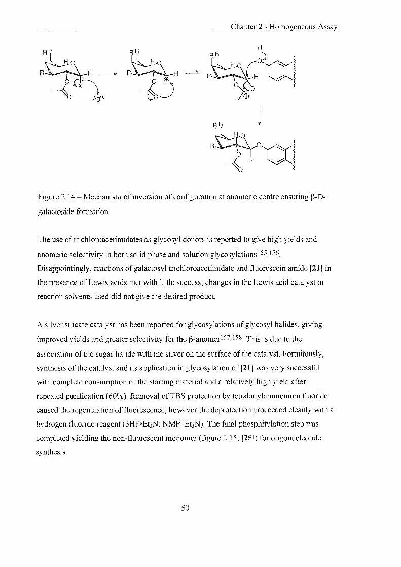
Chapter 2 - Homogeneous Assay
Figure 2.14 - Mechanism of inversion of configuration at anomeric centre ensuring (3-D
galactoside fonnation
The use oftrichloroacetimidates as glycosyl donors is reported to give high yields and
anomeric selectivity in both solid phase and solution glycosylations155,156.
Disappointingly, reactions of galactosyl trichloroacetimidate and fluorescein amide [21] in
the presence of Lewis acids met with little success; changes in the Lewis acid catalyst or
reaction solvents used did not give the desired product.
A silver silicate catalyst has been reported for glycosylations of glycosyl halides, giving
improved yields and greater selectivity for the p-anomer157,158. This is due to the
association of the sugar halide with the silver on the surface of the catalyst. Fortuitously,
synthesis of the catalyst and its application in glycosylation of [21] was very successful
with complete consumption of the starting material and a relatively high yield after
repeated purification (60%). Removal ofTBS protection by tetrabutylammonium fluoride
caused the regeneration of fluorescence, however the deprotection proceeded cleanly with a
hydrogen fluoride reagent (3HFoEt3N: NMP: Et3N). The final phosphitylation step was
completed yielding the non-fluorescent monomer (figure 2.15, [25]) for oligonucleotide
synthesis.
50

[20]
Ac OAc
HO
---~
(i)
TBSO~HN
Chapter 2 - Homogeneous Assay
OH
We
ACO'\"'YO
o
o
Aco.. fAC ~ -'( I" ~OAc
ACO\""YO
o
OAc
Acrl ( ~ ~OAC
ACO\\"'YO
-...;::: 0 [241
[25]
Reagents and conditions: i, EDC, pyridine, rt, 24hrs, 66%; ii, a-D-tetraacetate galactosyl
bromide [22], silver silicate catalyst, CH2CI2, dark, rt, 2 days, 55% or a-D-tetraacetate
galactosyl bromide [22], silver oxide, quinoline, benzene, dark, rt, 3 days, 19%; iii,
3HF'Et3 N:NMP:Et3 N (2: 3: 1.5), dark, 6O oC, 1 hr, 81 %;
iv, 2-cyanoethyl-N',N-diisopropyl-chiorophosphorarnidite, DIPEA, THF, rt, 1 hr, argon, 82%
Figure 2.15 - Synthesis of non-fluorescent fluorescein-bis-p-D-galactoside phosphoramidite
monomer
51

Chapter 2 - Homogeneous Assay
Two oligonucleotides were prepared using standard solid phase DNA synthesis; TIS [26]
control and BT 15 ([27], where indicates phosphoramidite [25] used). Two peaks eluted on
RPHPLC of [27] (figure 2.16); the earlier peak (a) had the same retention time as [26]
control indicating this peak was a TIS oligonucleotide, the later peak (b) was the desired
non-fluorescent labelled oligonucleotide. The identity of both peaks was confirmed by
electrospray mass spectrometry.
b ./\
-------- \ \
\ \
5212,0
(i) (ii)
Figure 2.16 - i, RPHPLC of [27], ii, ES- [27], calculated MW 5360.97
2.3.3 Evaluation of the Signal Probe [27]
A cleavage assay, in which the oligonucleotide labelled with the J3-D-galactosyl protected
fluorescein monomer [25] (the signal probe, [27]) was exposed to J3-D-galactosidase,
confirmed the galactosyl fluorescein ethers as effective substrates for the enzyme 1 59 (figure
2.17). A tube only containing signal probe emitted a very low level of background
fluorescence (tube 1). A tube containing [26] and J3-D-galactosidase emitted no
fluorescence (tube 2). However, the tube comprising of signal probe [27] and J3-D
galactosidase became intensely fluorescent over a 30minute period (tube 3). The contents
of tube 1 and 3 were examined by UV spectroscopy, both samples absorbed at 260nm, but
tube 3 displayed a characteristic fluorescein absorbance at 495nm.
52

Chapter 2 - Homogeneous Assay
tube 1 tube 2 tube 3
Figure 2.17 - Assay results (as viewed under transilluminator)
An identical assay performed in a UV spectrometer cell, monitoring at 495nm,
demonstrated the increase in absorbance with time due to the enzymatic turnover of
substrate (figure 2.18, i). A control experiment omitting the f3-D-galactosidase showed
insignificant change over the thirty minute period (figure 2.18, ii) ..
-0.05
fl -0.06 c ~ -0.07 5 ~ -0.08 ~
-0.09 t - ----- - ---- - ii
o 200 400 600 600 1000 1200 1400 1600 1600
Time (seconds)
Figure 2.18 - UV monitored assay, i, assay of [27], ii, control (no f3-D-galactosidase)
2.3.4 ~-D-Galactosidase-Oligonuc1eotide Enzyme-probe 1
Following the successful synthesis and enzymatic recognition of the signal probe [27] , the
next step was to examine the conjugation of f3-D-galactosidase to an oligonucleotide.
Coupling of enzymes to proteins is a well-established technique 160, and the same principles
have been applied in the preparation of oligonucleotide-enzyme conjugates 137. Literature
53

Chapter 2 - Homogeneous Assay
searches produced no evidence of p-D-galactosidase coupled to oligonucleotides, however
methods for attachment to various proteins have been described161,162. Attachment is
achieved using a range of crosslinking reagents specific for any of the twenty free thiols on
p-D-galactosidase and reactive groups on the protein. One example of conjugation between
p-D-galactosidase and an antibody is shown in figure 2.19. Crosslinking reagents are homo
or heterobifunctional molecules containing two reactive functionality's that target reactive
groups (amines, thiois, and carboxylic acids) on proteins or oligonucleotides 163. The
crosslinking reagent covalently links two biological molecules, providing a spacer that can
reduce steric interactions. It is imperative that the cross linking reagent or crosslinking
reaction does not interfere with the performance of any biological functions.
---~
HS-G y
~o o~~ N~/'..N
S 0 0
Figure 2.19 - Conjugation of p-D-galactosidase (P-GAL) to an antibody (AB) using
dimaleimide, N,N'-oxydimethylenedimaleimide crosslinking reagent
By modifying reported methods a convenient strategy for conjugation of p-D-galactosidase
to an oligonucleotide was proposed. Experiments were primarily performed on the 5'
terminus of an oligonucleotide in order to establish the correct conditions for attachment;
subsequent experiments employed 3' aminolink oligonucleotides ([31], figure 2.20).
54

Chapter 2 - Homogeneous Assay
?! GATCTGACTTGGTAGH-O-l-~NH2 [31]
I OH 6
(i)
y?! \} GATCTGACTTGGTAGH-O-f-O'l... yNH ~ ;1
OH \~J6 I h N
J (ii) 0 II
GATCTGACTTGGTAGH-O-~D...t VNH\}J!; OH \'16 I
h N S
[32] 0
Reagents and conditions: i, maleimidobenzoyl-N--hydroxysuccinimde ester, Na2COa buffer, pH 6.0, rt, 3hrs; ii, f3-D-galactosidase, Na2HP04 buffer, pH 5.6, rt, 18hrs
Figure 2.20 - Conjugation of f3-D-galactosidase to a 3' aminolink oligonucleotide
2.3.4.1 Synthesis ofMBS Oligonucleotide
The preparation of aminolink oligonucleotides is carried out using commercial aminohexyl
phosphoramidite monomers, the synthetic details have been well documented by
Agrawal50. Initial attempts to couple maleimidobenzoyl-N-hydroxsuccinimde ester (MBS)
at pH 7.0 to an amino-derivatised oligonucleotide [28] gave poor yields and demanded
optimisation. Reactions of N-hydroxysuccimide esters of dye molecules with aminolink
oligonucleotide are generally carried out at pH 8.75; therefore the pH of the reaction buffer
was raised to this value. This change appeared to dramatically improve the yield (70%),
however, the isolated product (RPHPLC) of the reaction failed to couple with f3-D
galactosidase, as analysed by gel filtration and ion exchange chromatography of the
products. Examination ofthe product revealed a molecular ion corresponding to a hydrated
product formed by attack of hydroxide at the double bond (figure 2.21).
~NdH~A H~O Figure 2.21 - Hydrated MBS product
55

Chapter 2 - Homogeneous Assay
This indicated a conjugate Michael addition of water had occurred at pH 8.75 rendering the
derivative umeactive towards crosslinking with the enzyme. It was concluded that at pH 7.0
the major peak by RPHPLC (retention time 1O.8min) was the desired product. At pH 8.75 a
small peak of the same retention time is present (8%) but the major peak (52%, retention
time of 10. 1 min) corresponds to the hydrated product (figure 2.22, i). When the reaction
was repeated at pH 6.0 the undesirable hydrated peak contributed 3% of the total product
and the desired alkene 34% of the total (54% unreacted starting material, figure 2.22, ii). It
was concluded that the reaction ofMBS with primary aminolink oligonucleotides should be
carried out at pH 6.0 to prevent hydration of the desired unsaturated product, this
observation has been made by others 164.
lOO
80
B
'0
.0
20
0 .0 0 0.00 5 . 00 10. 00 15 . 00 20 . 00
(i) (ii)
Figure 2.22 - RPHPLC i, pH 8.75; ii, pH 6.00; A: umeacted starting material; B: hydrated
product; C: desired product
2.3.4.2 Conjugation olMES derivatised oligonucleotide to f3-D-Galactosidase
The purified MBS oligonucleotide was reacted with B-D-galactosidase in phosphate buffer
at pH 5.6. Purification of the conjugate was achieved by gel filtration followed by ion
exchange chromatography on a Pharmacia FPLC system. Peak elution was monitored by
the ratio of absorption at 2601280nm; proteins giving large negative peaks, oligonucleotides
strong positive peaks and conjugates weak negative peaks.
Gel filtration separates according to size, larger molecules are eluted first, as they can not
enter the pores of the gel as easily as smaller molecules, and elution is achieved
isocratically. The trace obtained from the native enzyme is a single peak (49min) with a
56

Chapter 2 - Homogeneous Assay
strong absorbance at 280nm (a, figure 2.23). The trace obtained from the conjugate
produced two peaks, the first (45min) gave a shallower absorbance peak corresponding to
the conjugate, the second (85min) was unreacted oligonucleotide absorbing strongly at
260nm (b, figure 2.23).
~-:-==-:::-.,:"":," -. __ . -_-_.~:~ . ....: .~--. - - ....: '..:.5----:
1) .. ... . . _- - ._ -._- -- _. - -.. - - 8 -
., . -~ ... ----.-~--. ---.-.---.---- - . ·0 ··· ·--·-
I
___ c __ ; ____ ~-_---~--L2--.-), . 1: ~. :
(a) (b)
Figure 2.23 - Gel filtration chromatography: a, B-D-galactosidase, b, conjugation reaction
Ion Exchange separates according to charge and gives greater resolution, separating the
conjugate into several peaks of varying degrees of substitution of the enzyme by the
oligonucleotide. The trace obtained from the native enzyme was a single peak (a, figure
2.24), whereas the conjugate gave approximately nine peaks (b, figure 2.24).
~ ... . ... .. . . .
(a) (b)
Figure 2.24 - Ion exchange chromatography: a, B-D-galactosidase b, conjugation
57

Chapter 2 - Homogeneous Assay
With these results in hand a large-scale synthesis of the conjugate with a 3' amino
derivatised oligonucleotide [31] was performed, coupling was again highly successful
producing the enzyme probe 1 [32].
2.3.5 Enzyme-probe 1 Activity
Enzyme activity of each fraction was verified by the turnover of the substrate 0-
nitrophenol-f3-D-galactopyranoside. Hydrolysis of this substrate releases the yellow product
o-nitrophenol which absorbs 405nm (figure 2.25).
~N02.o-f°H Vl--~OH
.i"' .. HO OH
~-galactosidase
Figure 2.25 - Hydrolysis of o-nitrophenol-f3-D-galactopyranoside by f3-D-galactosidase
The conjugate fractions from ion exchange chromatography were also analysed for the
necessary absorptions at 260mn for DNA, and 280mn for protein (figure 2.26, i). The peaks
that displayed high rates of substrate turnover (figure 2.26, ii), and the correct absorption
maxima for the protein (280nm) and the oligonucleotide (260nm), were combined [32].
In order to establish the relative activity ofthe conjugate compared to that of the native
enzyme, the turnover of one micromole of substrate by f3-D-galactosidase was monitored at
405nm at 37°C over a five minute period. The experiment was repeated using the same
quantity of substrate and lO!-!L of conjugate. From the gradient of the linear section of the
two curves, and taking into account the relative amounts of native enzyme to conjugate
used, calculations showed the conjugate exhibited a sixth of the activity of the free enzyme.
The approximate amount of DNA present in the conjugate was calculated to be 9.4!-!gmL-1
from the optical density reading at 260nm, the molecular weight of the oligonucleotide, and
its molar extinction coefficient.
58

Chapter 2 - Homogeneous Assay
0'05~ i 0.04
" 0.03
'" ~ 0.02
0.01
o r 230 255 280 305 330
~1~~ ~ 0.5
o I I o 100 200 300
Time (seconds)
400
Wavelength (nm)
(i) (ii)
Figure 2.26 - UV traces i, typical spectrum of conjugate, ii, hydrolysis of o-nitrophenol-~
D-galactopyranoside by the ~-D-galactosidase-oligonucleotide conjugate [32]
2.3.6 SDS Page Electrophoresis
To investigate further the nature of the ~-D-galactosidase conjugate SDS-polyacrylamide
gel electrophoresis was performed, and the bands were visualised by highly sensitive silver
staining.
Lane
212
170
116
76
53
1 2
- +n t
3 4
••
.. Bands of varying degree of
. oligonucleotide substitution of ~-D
galactosidase
Lane:
1 + 4 = molecular markers
2 = conjugate [32]
3 = ~-D-galactosidase
Figure 2.27 - Gel electrophoresis demonstrating the substitution of ~-D-galactosidase with
oligonucleotides
59

Chapter 2 - Homogeneous Assay
The gel obtained clearly displayed the conjugate with four bands of varying degrees of
oligonucleotide substitution of the enzyme. The first two bands, corresponding to one and
two oligonucleotides substituting the enzyme, are the most intense, therefore the most
abundant. The molecular marker at 116 and lane 3 are native f3-D-galactosidase proving
there was little or no free enzyme present in the conjugate mixture [32].
2.3.7 Assay Evaluation (as described section 2.1)
The f3-D-galactosidase conjugate (enzyme probe 1, [32]) and signal probe had been
assembled enabling the assay to be designed and evaluated. Oligonucleotides to act as
target DNA and the signal probe were designed and synthesised. Figure 2.28 depicts the
three-strand system (1) which brought the substrate and enzyme in head to head contact and
system (2) which incorporated a short stem to keep the substrate and enzyme away from the
target third strand. Enzyme and substrate were separated from the oligonucleotide strands
by hexaethylene glycol spacers, this was to increase the degree of freedom to try and
promote interactions between the pair.
3' "HH 3' 5' [33}-- GCT AGC CGC GTA GCA GAT GGT TCA GTC TAG-[32] system 1 [34}-- CGA TCG GCG CAT CGT CTA CCA AGT CAG ATC
S ~
• -g
5' HH 3' CG TA AT
3' C( ~ 5' [35}-- GCT AGC CGC GTG TIC AGT CTA G-[32] system 2 [36]- CGA TCG GCG CAC AAG TCA GAT C
S ~
Figure 2.28 - Systems (1) and (2) designed to test the assay
To assess the performance of individual components of the assay experiments 1-5 were
conducted in a fluorimeter cuvette and the increase in fluorescence absorption (495nm) was
monitored in each case.
60

o = FAM (fluorescent)
o = FAM (non-fluorescent)
o = galactose IIIIII
= f3-galactosidase
= oligonucleotide
i) The substrate [24] was placed in buffer but was relatively insoluble even with the addition
ofDMSO, ~-D-galactosidase was subsequently added. Fluorescence intensity increased from
125 to 632 over a 24 hour period indicating the slow hydrolysis of the substrate (figure 2.29).
700
600
-f 500 <D
E 400 <D g Il 300 " ~ ~200
100
O ~----~------~----~------~----~ o 5 10 15 20 25
Time (hours)
Figure 2.29 - Hydrolysis of ~-galactose F AM molecule [17] by ~-galactosidase over 24 hour,
monitored by increase in fluorescence emission at 520nm.
[33]
ii) The oligonucleotide substrate (signal probe, [33]) was placed in buffer and ~-D
galactosidase was added. Fluorescence intensity increased from 670 to over 1000 over 24
hours (figure 2.30). The background fluorescence of the substrate was higher than in the first
experiment and the reaction proceeded at a much faster rate. These observations were
explained by the increased solubility of the substrate in aqueous buffer due to the hydrophilic
oligonucleotide.
61

1100
1050
1000 ~ ~ 950 ~ B 900
~ 850
~ 800 ::J
IT: 750
700
650 -I----_-_--~--~--~ o 5 10 15 20 25
Time (hours)
Figure 2.30 -Hydrolysis of ~-galactose signal probe [33] by ~-galactosidase over 24 hours,
monitored by increase in fluorescence emission at 520nm.
~Q [33] II II II I
[32]
,.,D°o ""tTt TTIIT"t trlr
iii) The signal probe [33] was placed in buffer and the ~-D-galactosidase oligonucleotide
conjugate (enzyme-probe, [32]) was added, no target strand was present therefore the two
oligonucleotides remained free in solution. The fluorescence intensity reading increased from
360 to 462 over 24 hours (figure 2.31). The rate of hydrolysis of the signal probe by the
enzyme-probe free in solution was slow and the change in fluorescence intensity was small.
The slow hydrolysis was desirable if a significantly increased rate caused by hybridisation of
the two probes adjacent to one another was to be observed.
470
450
£ i? 430
~ g 410
" ~ ~ 390 ::J
IT:
370
350 0 5 10 15 20 25
Time (hours)
Figure 2.31 - Hydrolysis of ~-galactose signal probe [33] by ~-galactosidase-oligonucleotide
conjugate (enzyme probe, [32]) in the absence of a target nucleic acid, over 24 hours.
Monitored by increase in fluorescence emission at 520nm.
62
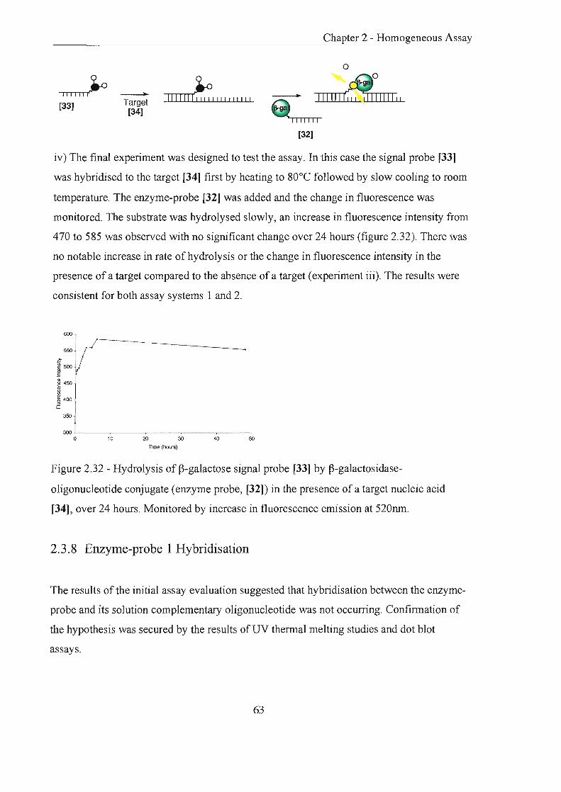
[33]
----lIo
Target [34]
III IIII~' I " II I " I ~
Chapter 2 - Homogeneous Assay
o
IIIIII~~IIIIII 9 ..,., "11'" TOil ,-,
[32]
iv) The final experiment was designed to test the assay. In this case the signal probe [33]
was hybridised to the target [34] first by heating to 80°C followed by slow cooling to room
temperature. The enzyme-probe [32] was added and the change in fluorescence was
monitored. The substrate was hydrolysed slowly, an increase in fluorescence intensity from
470 to 585 was observed with no significant change over 24 hours (figure 2.32). There was
no notable increase in rate of hydrolysis or the change in fluorescence intensity in the
presence of a target compared to the absence of a target (experiment iii). The results were
consistent for both assay systems 1 and 2.
600
550
350
300 ~--~----~----~----~--~ o 10 20 30 40 50
Time (hours)
Figure 2.32 - Hydrolysis of f)-galactose signal probe [33] by f)-galactosidase
oligonucleotide conjugate (enzyme probe, [32]) in the presence of a target nucleic acid
[34], over 24 hours. Monitored by increase in fluorescence emission at 520nm.
2.3.8 Enzyme-probe 1 Hybridisation
The results of the initial assay evaluation suggested that hybridisation between the enzyme
probe and its solution complementary oligonucleotide was not occurring. Confirmation of
the hypothesis was secured by the results of UV thermal melting studies and dot blot
assays.
63

Chapter 2 - Homogeneous Assay
2. 3. 8.1 UV Thermal Melting
Slow heating of dsDNA causes the unwinding of the ordered helical structure into the ~NO
single-stranded constituents. This can be measured by a smooth transition of increasing UV
absorption (260nm), the mid-point corresponding to the precise melting temperature of the
duplex. UV melting experiments can be perfonned to determine the extent of annealing of
two complementary strands by observing the shape of the curve produced on heating
(section 1.5.3).
Equal samples of the target oligonucleotide [34] and the enzyme-probe [32] were mixed in
hybridisation buffer. The sample was heated to 90°C and cooled slowly to room
temperature, allowing efficient annealing of the two strands. The sample was then heated
from 5°C to 85°C over 80 minutes following the change in absorbance at 260nm, this was
repeated three times. Each melt showed the same gradual increase in absorbance from
0.231 to 0.291 (figure 2.33, i) but the transition expected due to the dissociation of the
strands was not observed (figure 2.33, ii). A sample of the native f3-D-galactosidase was
melted under the same condition this produced an extremely similar curve to that of the
hybridisation experiment.
Proteins also absorb UV light (Amax=280nm) due to the bulky, aromatic, hydrophobic side
chains of phenylalanine, tyrosine and tryptophan amino acids. At the wavelength of the
experiment (260nm) there will be some absorbance by the protein. Elevated temperatures
will denature proteins, causing the unwinding of the tertiary structure; this increase in
disorder exposes a greater number of aromatic amino acid residues to UV irradiation and
the absorbance will increase.
It was concluded that the observed increase in absorbance was due to conformational
change of the enzyme with rising temperature, and not to the unwinding of the annealed
oligonucleotides. The question of hybrid is at ion of the target and enzyme-probe had not
been resolved by this experiment, the effects of the protein masked the occurrence of a
hybrid melting transition.
64

I Abs I
) I Abs t-----------jTm
. '-----'"""--- -- - - -----
___ * ___ L~ .. ____ ...
Temperature Temperature
(i) ( ii)
Figure 2.33 - UV thennal melting i, observed curve, ii, expected curve
2.3.8.2 Dot Blot Assay
The principle lies in the UV crosslinking of an oligonucleotide to a nitro-cellulose or nylon
membranes (neutral or positively charged) via its heterocyclic bases3o. Multiple samples of
DNA are spotted next to each other on a single filter in dots ofunifonn diameter. For
quantitative analysis knO\xm amounts of DNA are applied, usually by serial dilution of a
standard to evaluate the extent ofhybridisation32. As little as 1-3pg of DNA can be
detected. The membrane is exposed to a solution of a complementary labelled
oligonucleotide, which becomes immobilised on the membrane by annealing to the
membrane-crosslinked oligonucleotide. Following stringent washing procedures, a label
bound to the second oligonucleotide can be detected directly or, in the case of an enzyme
label, by means of hydrolysis of a substrate releasing a coloured product.
In the case of the ~-D-galactosidase conjugate ([32], enzyme-probe), firstly the target
oligonucleotide [34] was UV cross-lirlked to a nylon membrane. Secondly this membrane-
bound oligonucleotide was exposed to an excess of ~-D-galactosidase conjugate [32] for
annealing. Unbound material was removed by membrane washing; the presence of bound
conjugate was detected by the turnover ofXgal that produces an insoluble blue/green
product on contact with ~-D-galactosidase. After 3 days there was little evidence of the
colourimetric product fonning insinuating either the target had not bound to the membrane
or more probably there had been no ap.nealing between target and conjugate.
65

The conjugate [32] was directly spotted onto the nylon membrane and the oligonucleotide
was covalently attached by UV crosslinking, but again colour failed to develop with Xgal
substrate. The oligonucleotide of the conjugate was not able to crosslink: to the membrane.
From the UV melting and dot blot experiments it was concluded that the oligonucleotide
linked to the j3-D-galactosidase was not fonning a duplex with its complementary target.
This conjecture explained the slow hydrolysis observed in initial attempts to test the assay
in the presence of a target oligonucleotide (experiment iv, section 2.3.7). Lack of, or poor
annealing of the ep..zyme-probe to the target implied all ep..zymatic cleavage that occurred
was due to random collision of the substrate and enzyme in solution, thus explaining the
similarity in result between experiments iii and iv, section 2.3.7.
2.3.9 Factors Influencing Fonnation and Stability of Hybrids
The disappointing result from initial assay evaluation called for a re-examination of the
conditions required for efficient enzyme-probe-target hybrid formation. Numerous factors
effect the rate and stability of hybrid formation40,165. One factor is the relative
concentrations of the probe or target, however all hybridisation experiments were
performed with either a large excess of target, a large excess of enzyme-probe or equimolar
quantities of each strand and in all cases hybridisation failed to occur. Monovalent cation
concentration influences hybridisation and can alter the Tm of a duplex, yet results showed
hybridisation was not occurring either at O.lM or at l.OM sodium chloride concentrations.
Generally, as temperature increases the rate of hybridisation increases, provided the
temperature is below the Tm of the duplex in question. The experiments performed gave no
indication of a dsDNA to ssDNA transition; therefore further investigation of the effect of
temperature was not necessary.
The three most important factors controlling the annealing of the enzyme-probe with a
complementary target are length of the oligonucleotide, base composition of the
oligonucleotide, and steric hindrance caused by the enzyme. The length of an
oligonucleotide probe has a considerable effect on the rate and stability of the hybrid
formed. Hybrids of a hundred or more base pairs are essentially stable at room temperature,
66

Chapter 2 - Homogeneous Assay
however, for shorter probes a decrease in length causes a decrease in Tm of the hybrid,
some hybrids requiring temperatures belmv room temperature to fonn efficiently.
Altering the base composition of a duplex can raise or lower its Tm, as oligonucleotides
with a higher (%G+C) form more stable hybrids. The effect has been determined
empirically by measuring the dissociation temperature of hybrids relative to the length and
(%G+C) content of oligonucleotides.
Bolton and McCarthy proposed an equation that is said to hold for oligonucleotides from
14 to 70 base pairs in length166
Tm = 81.5°C - 16.6 (lOglO M) + 0.41 (%G + C) -600 / L
Where M is the concentration of monovalent cation, L is the chain length of the hybrid in
base pairs, Tm is the temperature at which 50% of the hybrids are dissociated and G and C
are the number of corresponding nucleotides in the sequence.
Applying this equation to the 15mer f3-D-galactosidase enzyme-probe it can be seen that the
(%G+C) content is 46.6% and the predicted Tm is 77.2°C. In an attempt to improve
hybridisation of the enzyme-probe to its target, an oligonucleotide for a new enzyme-probe
was designed GAT CTG ACT TGG TAG ACT A TG CGC CGA TCG HHH (H
hexaethylene glycol. The (%G +C) content ofthe oligonucleotide was raised slightly to
53.3% but the length was doubled giving a new predicted Tm of99.9°C (based on a
constant salt concentration ofO.1M throughout). Therefore, the change in oligonucleotide
length and content produces a more stable probe-target hybrid as the Tm of the duplex is
elevated.
The final problem of steric interference by the f3-D-galactosidase was a more difficult issue.
Linking a longer oligonucleotide to the enzyme may increase the stability of a hybrid
formed but if the oligonucleotide attached to the enzyme is inaccessible to its target then
the hybrid will not form regardless of oligonucleotide length. Building a bridge of
hexaethylene glycol spacers between the oligonucleotide and the enzyme allows both
67

Chapter 2 - Homogeneous Assay
partners enhanced flexibility. The original 15-mer enzyme-probe had one hexaethylene
glycol spacer benveen ~-D-galactosidase and the 3' of the oligonucleotide; the new probe
was designed to incorporate three.
2.3.10 ~-D-Ga1actosidase-Oligonuc1eotide Enzyme-Probe 2
The second longer enzyme-probe 2 [37] was prepared and analysed by the same methods as
the first enzyme-probe 1 [32] (sections 2.3.4,2.3.5, and 2.3.6). The 30-mer oligonucleotide
derivatised at the 3' end with an amino moiety was reacted with MBS in a 31.1 % yield and
subsequently coupled to ~-D-galactosidase and analysed by gel filtration, ion exchange
chromatography and SDS page electrophoresis. The enzymatic activity of the conjugate 2
[37] was confinned as before (section 2.3.5) and the amount of DNA present in the
conjugate was calculated to be 19.6!J,gmL-1 from the optical density reading at 260nm
(O.54mL-1), the molecular weight of the oligonucleotide (10,480), and its molar extinction
coefficient (287.28).
2.3.11 Enzyme-probe 2 Hybridisation
2.3. J J. J UV Thermal Melting
In a control experiment an oligonucleotide identical in sequence to the oligonucleotide of
the enzyme-probe [38] was annealed to its perfectly complementary target [39]. UV
thermal melting of the duplex gave a Tm in excess of 85°C in agreement with a computer
generated prediction of 92°C. l\ second control experiment heated the enzyme probe 2 [37]
in buffer alone from 5.3°C to 83.3°C. This displayed a gradual rise in absorbance, without
any significant transition, as the protein unfolds. UV melting the conjugate annealed to its
complementary target from 6°C to 84°C displayed a weak transition at 51.3°C.
Unfortunately a control UV melt of the target [39] on its own produced a similar transition
at 50°C, the nature of this transition was investigated by computer simulation but still
remains unkno'v'vTI.
68
Chapter 2 - Homogeneous Assay
2.3.11.2 Dot Blot Assay
The target [34] was UV cross-lin.1(ed to a nylon membrane, and then incubated with excess
of enzyme-probe 2 [37] for annealing. Unbound material was removed by membrane
washing; the presence of bound conj ugate was investigated by the turnover of the substrate
Xaa1 no bhlelarpen nrodnf't anppared b .L, -'"-..l.-"-J ... \,- f O.&-- 1-' \,.i...... Y ",,"'" .
The enzyme-probe 2 [37] was directly spotted onto the nylon membrane and the
oligonucleotide was bound by UV crosslinking, but again after washing, the blue colour
failed to develop with Xgal substrate. However, repetition ofthe experiment with very
concentrated samples of the probe produced very faint spots.
Finally native (3-D-galactosidase control and the enzyme-probe 2 [37] were spotted onto
nitro-cellulose membrane (binds proteins directly). (3-D-galactosidase developed the
expected strong blue! green spot, the probe gave a weak coloured spot.
Hybridisation of two oligonucleotides in solution is sterically favoured over membrane
bound hybridisation. It can be concluded from the dot blots that the enzyme-probe can not
hybridise to a membrane-bound target or bind via the oligonucleotide to a membrane.
However, target-probe hybrids may be forming in solution. UV thermal melting results
\A/Ould nonnally confirm hybridisation but in this case the results are complicated and
inconclusive
2.3.11.3 Molecular Beacon Approach
I\101ecular beacons are single-stranded oligonucleotides probes designed to detect specific
nucleic acids in homogeneous solution by the generation of fluorescence upon
hybridisationl14 (see section 1.6.2). A molecular beacon was synthesised with a loop
sequence complementary to the oligonucleotide portion of the enzyme-probe 2
FCGCACGCGATCGGCGCATAGTCTACCAAGTCAGATCCGTGCGM [40]
(i)The molecular beacon ([40], O.040D) was heated to 90°C and allowed to cool slowly to
room temperature, this achieves efficient annealing of the stems. The sample was heated
and cooled between 20°C to 80°C in the absence of a target strand (figure 2.34). The curve
displays the expected result, increasing temperature causes increasing fluorescence due to
69

Chapter 2 - Homogeneous Assay
stem dissociation and the molecular beacon adopting random conformations. Decreasing
temperature allows re-annealing of the stem and a drop in fluorescence to original
background signal. The melting temperature of the hairpin was 60°C.
418.7 400
350
300
250
D1 200
150
1()()
50
28.7 +-=C====~--~,---r-, --~,------==,=, 43.0 45 50
Figure 2.34 -
ffi 00 ffi mo Terrp.(q
Molecular beacon [40] heated and cooled between 20°C to 80°C in
the absence of a target strand, monitored at 520 nm.
Derivative of fluorescent melting curve displaying a hairpin melting
temperature of 60°C
(ii) Addition of an excess of target ([38], 0.1980D, 4.4eq) to the molecular beacon sample,
followed by heating to 80°C produced the same stem-dissociation curve as observed in (i).
However, no drop in fluorescence intensity was observed when cooled to 20°C or reheated
to 80°C, the entire molecular beacon was annealed to the target (figure 2.35).
6OJO
550
500
450
400
350 rt
300
250 Heating
200 Cooling 150
1000 I I I
200 30 40 50 fl) 70 00.0 Terrn(Q
Figure 2.35 - Molecular beacon [40] heating and cooling between 20°C and 80°C after
annealing to a target in excess ([38] , 4.4eq), monitored at 520nm
70

Chapter 2 - Homogeneous Assay
(iii) The molecular beacon was heated with an excess of enzyme-probe 2 ([37], 0.1080D,
2.4eq) to 80°C giving stem dissociation curve initially observed. Cooling to 20°C caused a
fall in fluorescence intensity yet the final intensity reached was a third of its original value
(figure 2.36).
477.7
450
400
350
1nt300
250
200 /~ //
/-'
158.5 +-~~=--'---'------r---r----'-----'------' 20.0 30 40 50
Terrp.(C) 60 70 80.0
Heating
Cooling
Figure 2.36 - Molecular beacon [40] heating from 20°C to 80°C with the enzyme-probe 2
(2.4eq), and subsequent cooling from 80°C and 20°C, monitored at 520 run
It was concluded a proportion of the oligonucleotides attached to p-D-galactosidase were
sterically accessible to the molecular beacon and hybridisation of these oligonucleotides
maintained a level of fluorescence. The remaining molecular beacon re-adopted the hairpin
conformation at lower temperatures and fluorescence intensity was reduced. The result
demonstrated that hybridisation of the molecular beacon to the enzyme-probe was taking
place, but the extent of hybrid is at ion was not known.
2.3.12 Assay Evaluation (as described in section 1.2)
Evidence of enzyme-probe hybridisation prompted the re-evaluation of the assay. Figure
2.37 depicts assay systems (1) and (2) that required four new oligonucleotides, two targets
([42], [44]) and two signal probes ([41], [43]). System (1) brought the substrate and enzyme
in head to head contact and system (2) incorporated a short stem to keep the substrate and
enzyme away from the target third strand. Enzyme and substrate were separated from the
oligonucleotide strands by hexaethylene glycol spacers to increase their flexibility
encouraging enzymatic scission.
71

Chapter 2 - Homogeneous Assay
. ~ system 1
3' 5' ~~3 3' 5' [41]-GAA CGT CCG AATTAT GCT AGC CGC GTA TCA GAT GGTTCA GTCTAG--[37] [42~ cn GCA GGC n A AT A CGA TCG GCG CAT AGT CT A CCA AGT CAG ATC
5' • -~ H8
5' C2G3 3'
GC
3'
A T system 2 T A CG
3' q 9 5' [43] -GAA CGT CCG CGC GTA TCA GAT GGT TCA GTC TAG--[37] [44]-CTT GCA GGC GCG CAT AGT CTA CCA AGT CAG ATC
5' 3'
Figure 2.37 - Systems (1) and (2) designed to re-test assay
Each assay was conducted in stages in a fluorescence spectrometer and the change in
fluorescence was monitored over time.
o = FAM (fluorescent)
--~ @ = FAM (non-fluorescent) = ~-galactosidase
[41] or [43] o = galactose I I I II I = oligonucleotide
i) The signal probe ([41], [43], O.19nmol) was placed in buffer, excess p-D-galactosidase
was added and the change in fluorescence intensity was monitored over 1 hour. The signal
probes from system 1 and 2 gave similar curves of increasing intensity with the final
reading a three fold increase of the background reading (figure 2.38, i).
~-Q [41] or [43] Ii IIIi I
[37]
00 ..r'0
-'I"TI T'TI 1"'-1 T"'i, r
ii) The signal probe ([41], [43], O.19nmol) was placed in buffer and an equimolar amount
of enzyme-probe ([37], O.19nmol) was added, no target strand was present and the two
oligonucleotides remained free in solution. Over the course of one hour both systems 1 and
2 showed no significant change in fluorescent intensity (figure 2.38, ii).
72

Chapter 2 - Homogeneous Assay
o system 1
[41] ----Target [42]
IIIIIII~I I " I II I I I --. Q
""1"-1 1"1 rrl 1"-1
IIIIII~~IIIIII o
[37] system 2
[43] ----Target [44]
IIII"I~IIIIIIIIII -e Lrl ""1 1"'1 TTl I-r-I
[37]
iii) The assay was tested with both systems, in each case the signal probe ([41], (43],
O.19nmol) was hybridised to the target ((42], [44]) by heating followed by slow cooling.
The enzyme-probe ([37], 0.19nmol) was added and the change in fluorescence was
monitored over I hour. Again, both systems expressed no notable change in fluorescent
intensity (figure 2.38, iii).
153.5 ! i (iii)
140 l:,.:..::---........;.~~----------1!---:--:'--::"~" ~, -:-::.:-.. -:. --, tl- -:, ~--,=--.:-, -, nl "." 120
100
Int
80
60
36.6 -
0.0
Figure 2.38 -
i -- -- +- - -~ ..
_.L •
500 1000 1500 s
(ii)
(i) ---.--- -~- - ---- --i-j
• I , I
, , --
___ 1 ___ . __ . _______ . __
2000 2500 3000
i I ,
-; i I I
I
_ ____ .l. ~
3600.0
Signal probe [41] with excess f)-galactosidase, fluorescence intensity at
520nm over 1 hour.
Signal probe [41] with an equimolar amount of enzyme-probe 2 ([37]), no
target strand, fluorescence intensity at 520nm over one hour.
Signal probe [41] with an equimolar amount of enzyme-probe 2 ([37]), with
target strand [42], fluorescence intensity at 520nm over one hour.
73

Chapter 2 - Homogeneous Assay
2.3.13 Conclusions
The synthesis of a non-fluorescent phosphoramidite monomer [25] was accomplished, and
the monomer was utilised in the assembly of a range of oligonucleotide signal probes. The
signal probe was an efficient substrate for native j3-D-galactosidase, demonstrated by the
visualisation of fluorescence and the curve generated upon enzymatic hydrolysis of the
substrate. Conjugation between j3-D-galactosidase and a 15mer oligonucleotide was
achieved creating an enzyme probe 1 for the intended assay. However, results established
that the enzyme probe would not hybridise to a complementary synthetic DNA target. In
the absence of annealing, the rate of signal probe hydrolysis and the intensity of
fluorescence released were shown to be similar with or without a target nucleic acid.
On the basis of these results a second enzyme probe 2 [37] was designed which was longer
(30mer) and slightly more GC rich. Initial investigation suggested although tlJ.e
oligonucleotide of the enzyme probe would not anneal to a membrane, hybridisation may
occur in homogeneous solution. Disappointingly, attempts to validate the proposed assay in
two different systems did not produce significantly different changes in fluorescent
intensity in the presence or absence of the target.
Previous successful enzyme probes have used much smaller enzymes such as alkaline
phosphatase (AP, 89kDa) and horseradish peroxidase (40kDa). In these cases the
conjugation to oligonucleotides has little affect on the hybridisation kinetics, selectivity or
efficiency of the oligonucleotide itself (HRP conjugation; no change in Tm, AP
conjugation; approximately lOoC fall in Tm167). j3-D-Galactosidase is a relatively large
enzyme with a molecular weight of approximately 500kDa 168 and has twenty free thiols for
conjugation. However, the location of the thiols may mean that not all are involved in the
conjugation reaction and the number of oligonucleotides per enzyme molecule may vary.
Both enzyme probes prepared possessed oligonucleotides relatively small (l5mer: 5kDa,
30mer: lOkDa) in comparison with j3-D-galactosidase and it is conceivable that
hybridisation is prevented for steric reasons.
74

Chapter 2 - Homogeneous Assay
The assay was carried out at the optimum pH for I3-D-galactosidase activity (pH 7.5), at this
pH the overall charge of the tetrameric enzyme is -158.78. This large negative charge
excludes the possibility of electrostatic attraction between the protein and the conjugated
oligonucleotides which bear an overall negative charge due to their phosphate backbones.
However, poor ap,-,'1ealing bet\veen the oligonucleotides linked to the protein and target
nucleic acids may be explained by electrostatic repulsion of the incoming target
oligonucleotides by the protein. This is an inherent problem as the pI of the enzyme is
5.28169 it requires a pH of 5.28 or lower to possess a positive charge, however at this pH
the enzyme is no longer active.
To study the assay further requires an enzyme probe capable of hybrid is at ion to its
complementary target. This might be achieved using a longer oligonucleotide for
conjugation and a longer spacer between the oligonucleotide and enzyme. However, an
intrinsic problem lies in the size of I3-D-galactosidase which may always sterically prevent
annealing. Another consideration for future development is signal amplification. Thermal
cycling the annealing and enzymatic cleavage steps would enhance the fluorescent signal,
yet this would require a thermostable I3-D-galactosidase.
75

Chapter 3
Heterogeneous Assay

Chapter 3 - Heterogeneous Assay
300 Heterogeneous Assay
This chapter explains the basis of heterogeneous assay and the development of new novel
heterogeneous assays, which use support-bound probes to detect nucleic acid sequences in
solution.
3.1 Introduction
Heterogeneous or separation assay is probably the most commonly used format for binding
assays, and involves immobilisation of single-stranded DNA or RNA onto an inert solid
phase135 . The immobilised nucleic acid is capable of forming a hybrid with a single
stranded probe added in solution, when incubated under conditions that favour duplex
formation. The remainder of unbound probe can simply be washed away. The probing
species can be either labelled directly or visualised after additional binding steps with
secondary labelled species. The sensitivity of this assay format can be greater that its
homogeneous rival as backgrotmd signals and interference are reduced by repeated washing
procedures removing excess unbound label.
3.2 Molecular beacons on beads
3.2.1 Introduction
Tyagi and Kramer recently reported a novel probe technology for the detection of specific
nucleic acids in homogeneous solution1l4. The probes named 'Molecular Beacons' produce
sensitive, real-time fluorescent signals that indicate hybridisation of a probe to a target
nucleic acidl15,118. They have a stem and loop structure, the loop portion probing for
complementary target nucleic acids in solution and the stem consisting of t\VO short arms
one terminally labelled with a fluorophore and the other with a quencher. Annealing of the
arms causes intramolecular energy transfer from the fluorophore to the quencher and the
probe is non-fluorescent. Hybridisation ofthe loop to its target causes the stem arms to
76

Chapter 3 - Heterogeneous Assay
dissociate, the fluorophore and quencher are no longer in close proximity and fluorescence
is emitted (see section 1.6.2). Molecular beacons are designed so that the arm sequences are
unrelated to the target and hybridisation of the arm sequences in the hairpin loop must
produce a weaker interaction than target-probe annealing. There is only one recent example
of molecular beacons supported on a solid phase. This involves labelling the beacon with
biotin, the biotin then binds to surface-immobilised avidin linking the molecular beacon to
the silica support 121.
The aim of the project was to adapt this technology to heterogeneous assay by supporting
molecular beacons on solid phase. The intention was to show that resin-bound beacons
exhibit similar properties to their homogeneous predecessors, non-fluorescent in the
absence of a target nucleic acid and fluorescent in the presence of a complementary target.
This tedulo1ogy could be harnessed in nucleic acid screening as hybridisation of resin
bound beacons to target DNA or RNA would generate fluorescent beads that could be
isolated and analysed either directly or by cleavage of the oligonucleotide from the resin.
The oligonucleotides could be examined by mass spectrometry 170, 171.
The principle behind the molecular beacon beads is shown in figure 3.1. A molecular
beacon is synthesised by standard automated DNA synthesis and deprotected. Initially the
resin is dry, therefore the conditions are unsuitable for hairpin formation and the beads are
fluorescent. Placing the resin in hybridisation buffer allows the stems to anneal and the
fluorescence is quenched. A probe-target hybrid is formed upon exposure to a loop
complementary nucleic acid, the arms are separated and the resin regains its fluorescence.
Development of molecular beacon heterogeneous assay required a suitable solid support
that was (i) suitable for automated oligonucleotide synthesis giving satisfactory coupling
efficiencies (>98%) at each step, (ii) allow annealing between the immobilised DNA and
the solution target nucleic acid, (iii) easily manipulated for bead isolation and
characterisation.
77

-------4--8 1
8~""O J Target DNA
~~~I ~I~I~I_. ~
Chapter 3 - Heterogeneous Assay
Probe in 'open' state = fluorescent
Probe in 'closed' state = non-fluorescent
Probe-target hybrid = fluorescent
Figure 3.1 - Mechanism of action of molecular beacon on beads
3.2.2 Resin Derivatisation
The purpose of the initial experiments was to find a suitable resin for supporting the
synthesis of an oligonucleotide, and to establish suitable conditions for the hybridisation of
the support bound oligonucleotide to complementary oligonucleotides in solution. All
resins investigated displayed different swelling, shrinking and sinking properties, which
would influence the efficiency of annealing. Four resins were examined: long chain
aminoalkyl-controlled pore glass (LCAA-CPG, Link Technologies), aminoalkylpolystyrene
(Link Technologies), Novasyn TentaGeFM amino resin (NovaBiochem) and TentaGeITM N
amino resin (Rapp Polymere).
Each resin was derivatised with a poly-alkyl linker as shown in figure 3.2. Firstly the
primary hydroxyl of 12-hydroxydodecanoic acid was protected as a 4,4' -dimethoxytrityl
ether. Secondly, the carboxy lie acid moiety was coupled to free amino groups on the resin
using DICIHOBT in 1 % DIPEAICH2CH2 creating a stable amide bond between the linker
and resin. The loading of the linker was determined by acid-catalysed detritylation followed
by quantitation of the DMT cation release from the resin at 495nm.The appropriate amount
of each resin for 0.2 micromole scale oligonucleotide synthesis was utilised on an ABI 394
DNA synthesiser.
78

R.",.oH HO . i1IT -(i)
n. ...... OH DMTO . i1Tf
o [11]
o H II ,.A. ...... N
OligonUCleotide-O-'l-u . hIT 08 0
o (ii,iii)
-(v)
Chapter 3 - Heterogeneous Assay
H
DMT0ki¥N
o [12,45-47]
j (iv)
Reagents and conditions: i, DMTCI, pyridine, rt, 2hrs, 87%; ii, resin, DIC, HOST, DIPEA, CH2CI2, rt, 1-2hrs, loading = 10 to 67jlmolg-1; iii, A~O, pyridine, THF:
1-methylimidazole, THF (1: 1), rt, 1 hr; iv, 5% TCA, CH2CI2, rt, 5mins; v, standard
automated DNA synthesis
Figure 3.2 - Generic scheme for resin derivatisation
In order to establish suitable conditions for the hybridisation of resin-bound
oligonucleotides to solution nucleic acids, oligonucleotides [49-52] were assembled on each
of the resins, each oligonucleotide preceded by a hexaethylene glycol spacer. The resins
were heated (60°C) in conc. aq. ammonia: ethanol (4:1) for four hours and washed
thoroughly with anhydrous CH2Cb and Et20, to remove the heterocyclic base and
phosphate protecting groups.
3.2.3 Heterogeneous Hybridisation
Oligonucleotides of complementary sequence to the resin-bound oligonucleotide were
synthesised and purified using standard methods [53-54]. The solution oligonucleotide
(0.25 OD) was mixed with the resin-bound oligonucleotide in buffer such that the resin
bound oligonucleotide was in approximately four times excess. The heterogeneous mixture
was allowed to stand at room temperature for 3 hours, after which the absorbance (260nm)
of the resin solution was measured. The resins were also assessed for uniform shape and
good sinking and swelling properties
The Novasyn TentaGel™ (TG) amino resin gave substandard synthesis giving low
coupling efficiencies (coupling efficiency ofTG = 91.5%, compared to CPG >98.5%)
79

Chapter 3 - Heterogeneous Assay
leading to a low overall yield. The resin was designed for peptide synthesis 172; the beads
swell considerably, the large size rendered the resin unsuitable for oligonucleotide
synthesis.
The Rapp TentaGeFM amino resin coupled with efficiencies >96.4% which was just
acceptable for short sequences. The beads were uniform and medium in size but were
sticky and difficult to dry, making manipulation difficult. Heterogeneous annealing was
partially successful with a drop in UV absorbance ofO.060D corresponding to annealing
24% ofthe solution oligonucleotide.
The aminopolystyrene beads were relatively small and uniform and gave high coupling
efficiencies of99.1 %, but were slow sinking in UV experiments. Hybridisation ofthe
immobilised oligonucleotide to the solution oligonucleotide was very effective, with a fall
in UV absorbance of 0.21 corresponding to a reduction of 84% of oligonucleotide in
solution. UV melting of the resin solution gave a smooth transition as the oligonucleotide
was released from the resin (Tm = 83.6°C, figure 3.3).
oms ,..----------, 0.03
0.025
dA/dT 0.02
0.015
0.01
0.005
0~~1---_r--_r--~
o 500 1000 1500 2000
Time (seconds)
0.3 ...,---------------------,
0.25 0.2
Absorbance 0.15
0.1
0.05 ;----~
o o 25 50 75 100125 150175200
Time (minutes)
Figure 3.3 - UV thermal melting ofimmobilised hybrid
Controlled-Pore Glass, a rigid, porous borosilicate glass support has the advantage of being
a well-known support for DNA synthesis. CPG is hydrophilic and can be dried or frozen
allowing long-term storage of immobilised biomolecules, it is also heat stable and therefore
amenable to temperature cycling. Oligonucleotide assembly on LCAA-CPG proceeded
with >98.5% coupling efficiencies, hybridisation was successful and the particles were easy
80

to handle, sinking quickly in aqueous buffer, which is an important factor for immediate
UV readings. The only drawback was the random structure of the glass particles, but
despite this controlled pore glass was chosen for initial resin-bound molecular beacon
studies.
3.2.4 Molecular Beacons on Controlled Pore Glass
Two molecular beacons were assembled by solid phase DNA synthesis on the
polyalkylamide CPG previously described [12J. Both oligonucleotides were labelled at the
5' position with a fluorescein fluorophore and at 3' end with a methyl red quencher (figure
3.4); CGCACGCTTAAAGTCACTTCATTTTCGTGCG [49] and
CGCACGl\TGTAGCACATCAGAAGCGTGCG [55J. Three hexaethylene glycol spacers
were coupled to the resin preceeding the quencher to minimise steric interactions between
the resin and the oligonucleotide during annealing (figure 3.4,3.5).
(ii)
~ 0Y'c o o
o
(iii)
Figure 3.4 - i Methyl red phosphoramidite, ii, hexaethylene glycol phosphoramidite, iii,
fl uorescein phosphoramidite
81
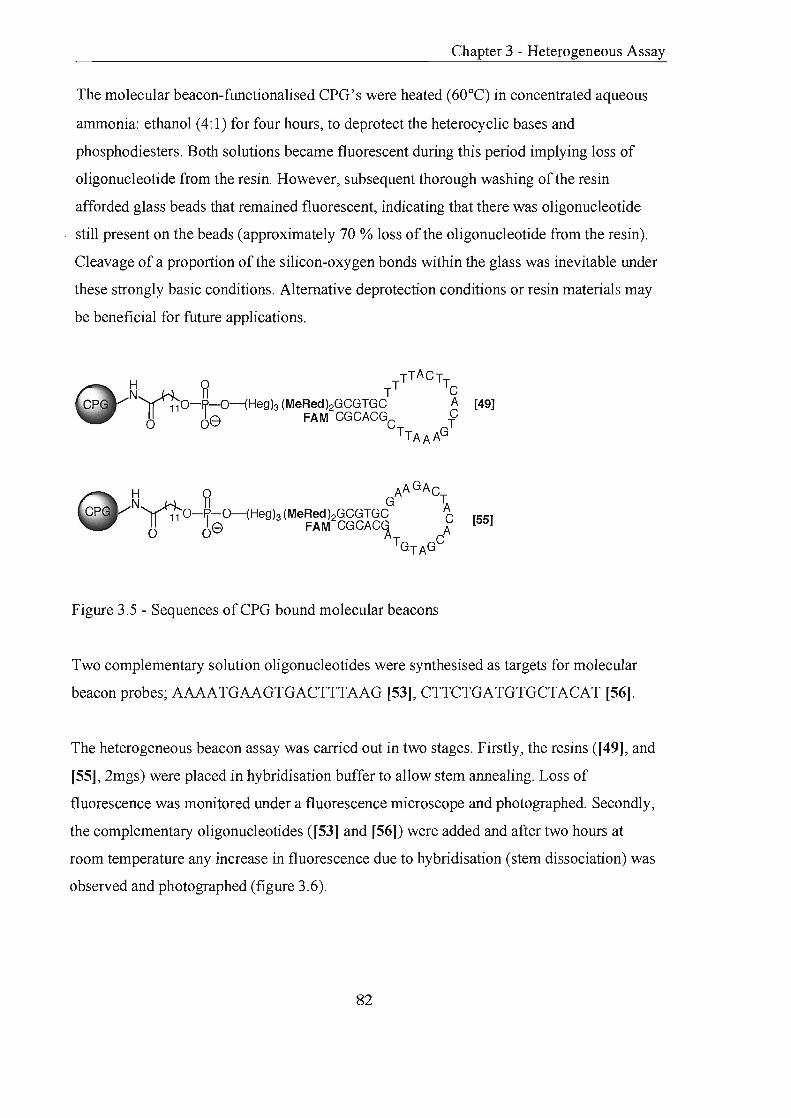
Chapter 3 - Heterogeneous Assay
The molecular beacon-functionalised CPG's were heated (60°C) in concentrated aqueous
ammonia: ethanol (4: 1) for four hours, to deprotect the heterocyclic bases and
phosphodiesters. Both solutions became fluorescent during this period implying loss of
oligonucleotide from the resin. However, subsequent thorough washing of the resin
afforded glass beads that remained fluorescent, indicating that there was oligonucleotide
still present on the beads (approximately 70 % loss of the oligonucleotide from the resin).
Cleavage of a proportion of the silicon-oxygen bonds within the glass was inevitable under
these strongly basic conditions. Alternative deprotection conditions or resin materials may
be beneficial for future applications.
[49]
[55]
Figure 3.5 - Sequences of CPG bound molecular beacons
Two complementary solution oligonucleotides were synthesised as targets for molecular
beacon probes; AAAATGAAGTGACTTTAAG [53], CTTCTGATGTGCTACAT [56].
The heterogeneous beacon assay was carried out in two stages. Firstly, the resins ([49], and
[55], 2mgs) were placed in hybridisation buffer to allow stem annealing. Loss of
fluorescence was monitored under a fluorescence microscope and photographed. Secondly,
the complementary oligonucleotides ([53] and [56]) were added and after two hours at
room temperature any increase in fluorescence due to hybridisation (stem dissociation) was
observed and photographed (figure 3.6).
82

Chapter 3 - Heterogeneous Assay
(v)
Figure 3.6 - i, ii dry fluorescent CPG; iii, iv, buffered non-fluorescent CPG; v, vi, buffered
fluorescent CPG and target
83

Chapter 3 - Heterogeneous Assay
Figure 3.7 shows the approximate percentages of the total number of non-fluorescent or
fluorescent beads / total number of beads viewed. Lower percentages than expected were
observed due to loss of oligonucleotide during ammonia deprotection.
Dry Resin Resin in buffer Resin in buffer with target
[49] fluorescent beads non-fluorescent beads fluorescent beads
(~ 70%) (~90 %) 50%)
[55] fluorescent beads non-fluorescent beads fluorescent beads
(~ 70%) (~90 %) (~ 50%)
Figure 3.7 - Approximate percentages of bead types under different experimental
conditions
The results showed that when the resin was dry the glass beads possessing probes were
fluorescent, Ll-terefore the fluorophore a.l1d quencher must be separated (stems are
dissociated). Addition of a suitable annealing buffer caused the beads to become non
fluorescent, indicating closure of the probes by stem annealing. Finally, introduction of a
complementary solution oligonucleotide caused a significant increase in fluorescence on
the beads; the loop portion of the probe has hybridised to its target causing the solid
supported molecular beacons to exist in the fluorescent 'open' state. Exposure of the CPG
beacons to a non-complemental)' oligonucleotide in buffer did not increase the level of
fluorescence on the beads.
3.2.5 Molecular Beacons Immobilised on Alternative Supports
Initial experiments proved molecular beacons bound to glass particles would anneal to their
targets, and display similar behaviour to homogenous molecular beacons. However a
number of weaknesses were uncovered during the experiments using glass particles that
may be overcome by other solid phase materials: i, deprotection in aqueous base caused
significant loss of the probe from the resin reducing the intensity of fluorescence observed,
this might be avoided by milder cleavage conditions or using resins chemically inert to
base, ii, isolation of individual particles for analysis would be tricky, as the glass chips
84

Chapter 3 - Heterogeneous Assay
were irregular in shape and size. These disadvantages led to a search for alternative resin
t<ipes.
3.2.5.1 Polystyrene Beads
Polystyrene beads have the advantage of being spherical and uniform in shape allowing
easy isolation. Previous results with aminopolystrene resin (Link Technologies) recorded
good coupling efficiencies and excellent heterogeneous hybridisation properties. However
molecular beacons synthesised on polystyrene beads [57] persistently displayed low levels
of fluorescence caused by a number of factors.
(i) The beads were not as suited to oligonucleotide synthesis as ePG, displaying average
coupling efficiencies of around 96%. This causes high loading of the quencher at the 3'
terminus at the beginning of the synthesis but low loading of the fluorescent dye at the 5'
terminus at the end of the synthesis. The consequence of an excess of quencher to
fluorophore is that the beads are always quenched either intra or intermolecularly.
However, couplings were improved by using a RNA coupling cycle which prolongs the
coupling time of each monomer to 20 minutes and this increased the coupling efficiency to
98.5% [58].
Oi) There was a small amount of quenching of the fluorescent dyes by the polystyrene itself
raLl}er tha.'1 the quencher molecules. This leads to reduced fluorescence of the beads even
when the probes are in the open state (i.e. when hybridised to a target). In an attempt to
overcome this problem hexaethylene spacers were coupled to the resin prior to the
molecular beacon [59], [60].
(iii) Polyaromatic polystyrene resin is highly hydrophobic being made up of mainly 1 %
divinylbenzene (DVB) cross-lipL<1ced polystyrene and does not swell well in aqueous
environments. If the beads are not swollen there may be a large degree of inter- and
intramolecular quenching and access of target oligonucleotides may be prevented. In
polystyrene resin the spacer separating the bound species and the hydrophobic resin matrix
is generally short, this can disfavour reactions involving incoming polar or charged species
such as target oligonucleotides.
85

Chapter 3 - Heterogeneous Assay
Due to the problems described, it was concluded that polystyrene was an unsuitable solid
phase for the heterogeneous assay. The resin cannot afford to be hydrophobic nature, as
annealing of the immobilised molecular beacon to target oligonucleotides requires aqueous
environments. Hexaethyleneglycol spacers attached primarily to the resin can increase the
hydrophilicPj and swelling of polystyrene but not to a satisfactory level for the assay. It was
concluded that the physico-chemical properties of a resin required careful consideration for
an aqueous assay.
3.2.5.2 TentaGefTM Beads
The architecture of TentaGeFM resin is based on a very small proportion of cross-linked
1 % DVB-polystyrene backbone that is extensively grafted with long PEG spacers making
up 70% of the total resin weight 173. The reactive sites are therefore situated at the end of
long, flexible spacers and are well-separated fonn the polystyrene backbone; this reduces
problems caused by the hydrophobic nature of the polystyrene matrix and steric hindrance
of incoming molecules. TentaGeFM appeared an appropriate resin for hybridisation
experiments swelling in polar aqueous solvents due to its increased hydrophilicity. Previous
experiments with Rapp amino TentaGeFM gave disappointing stepwise coupling
efficiencies, however improved yields (>96.9%) were achieved using an RNA cycle
extending the coupling time to 10 minutes [61]. All attempts to repeat the molecular beacon
assay gave results similar to those using polystyrene in that the beads were consistently
orange and non-fluorescent.
3.2.5.3 JvJacroporous Beads
This iype of resin has a pennanent well-developed porous structure even when completely
dry, and because of the unchanging structure the beads do not need to swell to allow access
to the interior. Provided the surfaces of the pores are wetted with solvent the pores can be
entered, essentially all solvents can be used, even water174. Amino Argopore™ is a
macroporous resin with a high internal surface area that provides low and predictable
swelling 1 75. The resin was derivatised and used in automated synthesis, but unfortunately
the average stepwise yield was >85.5 giving an overall yield of7% for a 35mer molecular
beacon oligonucleotide [62]. Attempts to increase the coupling by extending the reaction
times did not significantly improve the yield.
86

Chapter 3 - Heterogeneous Assay
3.2.6 Conclusions and Future Work
Molecular beacons attached to glass beads have been synthesised. The glass-bound beacons
in the absence of a complementary DNA sequence are 'closed', quenching occurs and the
glass particles are non-fluorescent. The clear glass beads become brightly fluorescent when
exposed to a complementary target nucleic acid, indicating that the probes are in their
'open' state. Ranges of structurally dissimilar resins have been investigated for the same
purpose, however each resin proved unsuitable for the different reasons described.
This technology could be harnessed in nucleic acid screening by hybridisation of molecular
beacons to target DNA or RNA, followed by isolation and analysis of the fluorescent
beads. lfthe assay were to be carried out in a multiplex format it would require a
characteristic dye for each molecular beacon sequence. However, this is currently limited
by the availabilii'j of fluorophores \vitl:l suitable emission characteristics.
A few tentative experiments exploring the possibility of using only one fluorophore and
one quencher, to screen any number of targets were performed. A lin.ker that was stable to
DNA synthesis and deprotection but cleaved by UV irradiation176 was placed between the
resin and the molecular beacon (figure 3.8). Synthesis of a poly thymidine oligonucleotide
showed coupling to be successful [63], however, mixed sequence oligonucleotide [64]
required ammonia deprotection that caused high losses of oligonucleotide from the resin
(",,60%). Continuation of this work employing fast-deprotecting phosphoramidite
monomers or alternative deprotection conditions may lead to interesting applications.
A range of different molecular beacons could be synthesised and combined into a single
vessel 3..11d exposed to target nucleic acids. Hybridisation of target to the probes would be
indicated by the generation of fluorescence on the complementary beads that could be
isolated. UV irradiation of the beads would release the oligonucleotides into solution,
subsequent mass spectral analysis would elucidate the molecular beacon and therefore its
complementary target nucleic acid (figure 3.9).
87

Chapter 3 - Heterogeneous Assay
DMT~No, I~ --" 4
3 [65] 0 3 OH
98 ~o-p-o
oligonucleotide ~ q N02
I~ HsCO ~ 0
O~~ Reagents and conditions: i, 4,4'-dimethoxytrityl chloride, DMAP, pyridine, 6QoC, 2hrs, 86%; ii,
LCAA-CPG, HOBT, DIC, DIPEA, CH2CI 2, rt, 2hrs, loading = 22pmolg-1; iii, acetic anhydride,
pyridine, THF : 1-methylimidazole, THF (1 :1), rt, O.5hr; iv, 3% TCA in CH2CI2; v, automated
DNA synthesis
Figure 3.8 - Derivatisation of controlled pore glass with UV linker
Other linkers that could be applied to the same or similar uses with optimisation are
described in chapter 4. The linkers are cleaved by enzymatic hydrolysis and contain amide
bonds. Therefore they are completely orthogonal to automated DNA synthesis and
deprotection. Advances in MALDI-TOF mass spectrometry allow molecular ions of
samples in the types of aqueous buffers required for enzyme scission to be detected 177,178.
---~
resin sets combined
mass spectrometry ... ---
<) non-fluorescent bead 0 fluorescent bead
target nucleic acids
linker cleaved
I isolation of fluorescent
y beads
aMB4 ()'MBl
MB molecular beacon oligonucleotide
Figure 3.9 - Representation of multiplex molecular beacon assay
88

Chapter 3 - Heterogeneous Assay
3.3 Heterogeneous Capture Assay
A new strategy for the use of solid supported probes was investigated. The idea involved
the capture a.l1d release of oligonucleotide probes on and off a solid support
3.3.1 Principle
Single-stranded oligonucleotides, assembled by solid phase synthesis, that are attached to
the solid support by inert linkers will remain bound to the side-chain after deprotection.
Synthesis of resin-bound oligonucleotides labelled at the 3' end with a quencher can be
used to capture complementary oligonucleotides (denoted as capture oligonucleotides). The
complementaf'j solution oligonucleotides can be labelled (5') with fluorescent molecules
(denoted as probe oligonucleotides) and are longer than the resin-bound capture
oligonucleotides.
The principle lies in the hybridisation of the probe oligonucleotide to the capture
oligonucleotide on the resin (figure 3.10, i). In this state the fluorophore and quencher are
adjacent and fluorescence will be quenched. As the probe oligonucleotide is longer than the
capture oligonucleotide it will have a 3' tail. This is important, as heating the resin in the
presence of a probe complementaf'j target will cause dissociation of the fluorescent probe
from the resin (figure 3.10, iv). Upon cooling the probe will preferentially hybridise to the
longer target. Target-probe annealing is signalled by a gain in fluorescence of the resin
solution, in the presence of non-complementaf'j targets the probe oligonucleotide will
simply re-anneal to the resin and fluorescence will be lost by quenching (figure 3.10, v).
3.3.2 Results
Polystyrene beads were derivatised with inert linkers [45] and used to assemble single
stranded oligonucleotides, labelled at the 3' end \'lith a quencher, that remained bound to
the resin after synthesis and deprotection (capture oligonucleotide, [67]). A second 5'
fluorescently labelled solution oligonucleotide was synthesised, deprotected and purified
89

Chapter 3 - Heterogeneous Assay
[68]. This longer oligonucleotide was complementary to the capture oligonucleotide on the
reSIn.
C). IIII 1111
--:~ O~IIIIIIIIIIIIII
i, annealing of fluorescent probe to resin
01111111111111"11" Q.IIIIII~ ~.
v, annealing of fluorescent probe with target
iv, strand dissociation with heat
(;) ." "'I' I resin-bound 3' quencher capture probe o I i II i II i' I II 5' fluorescently labelled probe
II II 1111 II 111111 target DNA
Figure 3.10 - Principle of capture assay
ii , resin washed
j
iii, target introduced
The resin was heated to 90°C in a solution ofthe fluorescent oligonucleotide [68] and
cooled to room temperature to allow annealing between the fluorescent oligonucleotide and
the capture oligonucleotide [67] (i, figure 3.10). The resin was then washed to remove any
unbound fluorescent oligonucleotide (ii, figure 3.10). To ensure that the fluorescent
oligonucleotide was being held on the resin, the resin was heated from 15 to 90°C over a
period of75 minutes and the increase in absorbance at 495nm and 260nm followed. Both
melts showed characteristic S shaped curves for the release of the fluorescent
oligonucleotide into solution as the two strands dissociated, Tm = 72°C (figure 3.11). The
beads were allowed to cool for re-annealing, this process was slower than the dissociation
due to the dilution of the resin required for UV measurements.
90

2.5°1
Abs
0.82
15
Temperature
(i) A = 260nm
Abs
0.21
90 15
Temperature
(ii) A = 495nm
3.11 - Results of resin heating at i, 260nm, ii, 495nm
Chapter 3 - Heterogeneous Assay
90
A sample of the re-annealed resin was placed in a cuvette with hybridisation buffer and an
excess of target solution oligonucleotide [69] was added (iii, figure 3.10). The sample was
heated to 90°C (iv, figure 3.10) and the increase in absorbance at 495nm and 260nm was
recorded. At 495nm the absorbance rose from 0.048 to 0.15, and at 260nm from 3.49 to
3.65 indicating release of the fluorescent oligonucleotide from the resin. The sample was
cooled causing an initial drop in absorbance to 0.125 but remained constant at this value for
48 hours as the fluorescent oligonucleotide was bound to its target (v, figure 3.10).
A control experiment (buffer only, no target) showed the an increase in absorption at
495nm of 0.057 to 0.455 at 90°C, cooling caused a drop to 0.252, but after 48 hours the
reading was back down to 0.08, as the fluorescent oligonucleotide was re-annealed to the
resIn.
3.3.3 Conclusions
The initial experiment performed confirmed the principles of the assay. The re-annealing of
the probe oligonucleotide to the resin-bound capture oligonucleotide was slow and would
require optimisation to allow the assay to be thermal cycled in a PCR fonnat, which
requires numerous heating and cooling cycles. Saturation of the solution with beads may
enhance arulealing, however this may make fluorescent reading difficult. Another approach
could involve coating the walls of a cuvette or capillary with the capture oligonucleotide
this would simplify fluorescent readings by removing any obstructions.
91

Chapter 4
Enzyme Labile Linkers

Labile Linkers
4.0 Enzyme Labile Linkers
This chapter explains the need for chemically stable linkers between an oligonucleotide and
a resin, or to separate different sections of an oligonucleotide. The synthesis of four linkers
is described and successful enzymatic cleavage of two tripeptide linkers.
4.1 Introduction
Solid phase chemistry of oligonucleotides employs linkers that are cleaved chemically, for
example the previously described succinyl and disiloxyllinkers145 (section 2.2.2.5), or by
UV irradiation 179. A linker is described as a bifunctional protecting group that can be
easily attached to the molecule to be synthesised by a bond labile to the cleavage
conditions, but is held on the solid support via a more stable bond or a bond with
completely orthogonal reactivity. Ideally linkers allow the attachment of the starting
molecule in high yields and are stable to the chemistry required for synthesis of the
supported molecule. It is also important that the conditions for linker cleavage are
sufficiently mild, so not to destroy the product. A recent comprehensive review of linker
strategy in solid-phase synthesis has been published 141.
The most commonly used linker in oligonucleotide assembly, the succinyllinker, is base
labile. This means manipulation ofthe oligonucleotide on-resin under basic conditions (e.g.
selective removal of protecting groups) is impractical. Therefore manipulations of this kind
are generally performed after the oligonucleotide has been released from the resin into
solution. However, performing chemical manipulations on the oligonucleotide in solution is
non-selective due to the numerous reactive sites and can require laborious purifications.
The need for linkers exhibiting a higher degree of orthogonality to the conditions of
automated DNA synthesis has therefore increased180.
Developments in probe technology have increased the demand for modified
oligonucleotides. Assembly of modified molecules 'whilst still attached to a solid support
simplifies both synthesis and purification. Linkers are generally employed to retain a
92
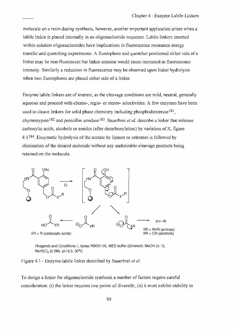
Chapter 4 - Enzyme Labile Linkers
molecule on a resin during synthesis, however, another important application arises when a
labile linker is placed internally in an oligonucleotide sequence. Labile linkers inserted
within solution oligonucleotides have implications in fluorescence resonance energy
transfer and quenching experiments. A fluorophore and quencher positioned either side of a
linker may be non-fluorescent but linker scission would cause increased in fluorescence
intensity. Similarly a reduction in fluorescence may be observed upon linker hydrolysis
when two fluorophores are placed either side of a linker.
Enzyme labile linkers are of interest, as the cleavage conditions are mild, neutral, generally
aqueous and proceed with chemo-, regio- or stereo- selectivities. A few enzymes have been
used to cleave linkers for solid phase chemistry including phosphodiesterase181 ,
chymotrypsin 182 and penicillin amidase 183 . Sauerbrei et aI. describe a linker that releases
carboxylic acids, alcohols or ami des (after decarboxylation) by variation of X, figure
4.1184. Enzymatic hydrolysis of the acetate by lipases or esterases is followed by
elimination of the desired molecule without any undesirable cleavage products being
retained on the molecule.
c o
6
~
o HO)lXR ...
(i)
o 8~R--" XH-R
XR = R (carboxylic acids) XR = NHR (amines) XR = OR (alcohols)
Reagents and Conditions: i, lipase RB001-05, MES buffer (SOmmo!): MeOH (4: 1),
NaHS03 (O .2M), pH 6.5, 30°C
Figure 4.1 - Enzyme-labile linker described by Sauerbrei et al.
To design a linker for oligonucleotide synthesis a number of factors require careful
consideration: (i) the linker requires two points of diversity; Oi) it must exhibit stability to
93

Labile Linkers
the conditions of automated oligonucleotide synthesis; (iii) the linker should be easily
incorporated during DNA synthesis. For enzyme labile linkers the choice of enzyme is
important, the linker must be a recognisable substrate, and the enzyme must not harm the
oligonucleotide.
The linkers designed were tenninated at one end with a protected hydroxyl and at the other
with a phosphoramidite. This was to allow the linker to be coupled using standard DNA
chemistry. Automated oligonucleotide synthesis would then be continued following
detritylation of the hydroxyl group. This phosphoramidite method permitted incorporation
of the lin..l(er not only directly to the resin, but also internally within an oligonucleotide for
solution hydrolysis. Synthesis of a phosphoramidite also ensured linker incorporation
maintained high coupling yields, which are essential for efficient DNA synthesis. Enzyme
labile linkers of the types shovm in figure 4.2 were initially proposed.
o R II ~ ~ tfN~'1
.,' H II ,,' 0
enzyme labile
I function 1 J--o~ Han . enzyme labile o ,,' ~
HO : 0---: ::. ':
iSH ~OH
Ir-fu-n-ct-io-n --'2t-O~"'jIOH
HO
Figure 4.2 - Proposed enzyme labile linkers based on monosaccharides, disaccaharides or
peptoids.
4.2 f)-Galactose Linker
4.2.1 Principle and Preparation
It was envisaged that this linker would be cleaved by exposure to a glycosy lase enzyme
severing the glycosidic linkage a.lld releasing an alcohol.
94

Labile Linkers
H0x5~~-R2 4 ~ o + HO 6 0-P-0-R1
'e HO : OH 0-
OH
Figure 4.3 - ~-galactosyllinker cleaves to release alcohols from galactose-containing
residue
The commercially available ~-galactose pentaacetate was selectively deprotected at the
anomeric centre to give a mixture of ~ and a anomers (a: ~, 1:3)185. Subsequent
conversion to the crystalline trichloroacetimidate [71) was first attempted using a simple
biphasic reaction between trichloroacetonitrile and 50% aqueous potassium hydroxide,
containing a catalytic amount oftetrabutylammonium hydrogen sulfate186. The method
was successful in producing the trichloroacetimidate, yet resulted in cleavage of the acetyl
protecting groups. An alternative method using sodium hydride, gave the desired a-anomer
selectively in a satisfactory yield, however, the yield was improved using 1,8-
diazabicyclo[5.4.0]undec-7-ene (DBU) as the base for the reaction187,188.
Monosilylation of the symmetric hexan-l,6-diol was achieved in reasonable yield by
generating the monosodium alkoxide salt and reacting it with tert-butyldimethylsilyl
chloride 1 89. The gIycosylation step between the trichloroacetimidate glycosyl donor and
the alcohol acceptor was catalysed by Lewis acid 190,191. Results showed trimethy lsily 1
trifluoromethanesulphonate proved to be a superior catalyst than boron trifluoride etherate
giving only the ~-anomer in high yield. It was important to ensure that the ~-anomer was
retained, as the molecule needed to be a recognisable substrate for ~-galactosidase. NMR
analysis confirmed the configuration as the ~-anomer, an axial-axial configuration of the
hydrogens at positions C 1 and C2 gave rise to a large coupling constant.
Removal of the acetyl groups by base-catalysed transesterification proceeded as expected.
The primary hydroxyl was protected as the 4,4'-dimethoxytrityl ether with only a small
amount ofbis- and tris- tritylation observed. The three remaining hydroxyl functions were
95
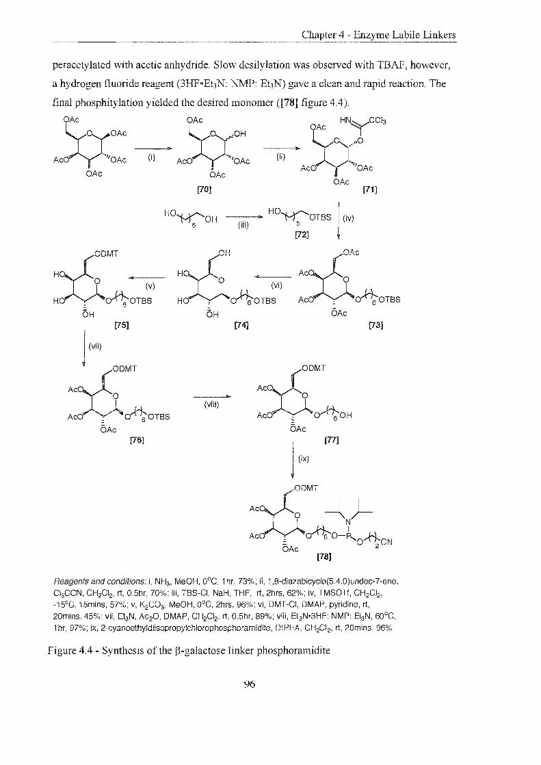
Labile Linkers
peracetylated with acetic anhydride. Slow desilylation was observed with TBAF, however,
a hydrogen fluoride reagent (3HF .. Ehl'J: NMP: Ehl'J) gave a clean and rapid reaction. The
final phosphitylation yielded the desired monomer ([78] figure 4.4).
OAc OAc
AXx::: --(i)-:~ AXx::C HNy CCl3
O)?AC 0 .,,0 (ii) ".. .-
AcO ""'OAc OAc OAc
[70]
HO~OH 5
OAc [71]
--(ii-O __ J> HO~OTBS I (iv)
[72] ~
Ac £"""OMT
HO ~. ____ _ o (v)
HO ~ ~OTBS OH
(OH Han <{ ° (vi)
HO ~ ~OTBS CH
:bOAc
AcO ,0 ~OTBS CAc
[75] [74] [73]
I (vii)
A~ fODMT
ACOV~OTBS (viii)
AcO .... fO
OMT
Ac}0c0i;-OH ---"..
CAc CAc [76]
Reagents and conditions: i, NHs, MeOH, OOC, 1 hr, 73%; ii, 1,8-diazabicyclo(5.4.0)undec-7-ene,
CI3CCN, CH2CI2, rt, 0.5hr, 70%; iii, TBS-CI, NaH, THF, rt, 2hrs, 62%; iv, TMSOTf, CH2CI2,
-i5°C, i5mins, 57%; v, K2C03, MeOH, DOC, 2hrs, 96%; vi, OMT-CI, OMAP, pyridine, rt,
20mins, 45%; vii, Et3N, AC20, OMAP, CH2CI2, rt, O.5hr, 89%; viii, Et3N·3HF: NMP: EtsN, 60°C,
1 hr, 97%; ix, 2-cyanoethyldiisopropylchlorophosphoramidlte, DIPEA, CH2CI2, rt, 20mins, 96%
Figure 4.4 - Synthesis of the ~-galactose linker phosphoramidite
96

Chapter 4 - Enzyme Labile Linkers
4.2.2 Oligonucleotide synthesis and linker cleavage
Two oligonucleotides were assembled placing the linker between a fluorescent dye and a
polyt~ymidine oligonucleotide. The coupling efficiency of the monomer was> 99%.
FST 15 [79], FSST 15 [80] where S indicates incorporation of monomer [78] and F =
fluorescein.
The oligonucleotides were designed such that cleavage of the linker by p-galactosidase
\vould separate t~e fluorescent dye and the colourless polyT oligonucleotides. The cleavage
reaction was perfonned and the products analysed by gel filtration. Successful scission
would be characterised by elution of a colourless polyT oligonucleotide, with the
fluorescent dye fragment retained on the gel. However, when the reaction was passed
through the gel the labelled fluorescent oligonucleotide was eluted as a single entity
indicating cleavage was unsuccessful. Mass spectrometry confirmed the observation; no
cleavage products were detected, the only molecular ion being that of the starting
oligonucleotide.
4.2.3 Conclusions
Results showed the linker failed to hydrolyse in solution, this implied cleavage from a resin
\vas extremely unlikely. The disappointing result could be caused by poor or no recognition
of the linker as a substrate for the enzyme or that the enzyme could not access the linker. In
favour of the fonner hypothesis, there is some evidence to suggest p-D-galactosidase may
not tolerate substitution in the C-6 position 168. Future work could utilise the precursor to
the phosphoramidite [77] to synthesise a disaccharide linker. For example linking galactose
to glucose would form a lactose-based linker that could be a substrate for p-galactosidase or
other endoglycosylases. However, due to the large size of the ~-galactosidase causing
potential steric problems in solid supported applications, attention switched to alternative
linkers and enzymes.
97

Chapter 4 - Enzyme Labile Linkers
4.3 Peptoid Linkers
4.3.1 Phenylalanine Linker 1
4.3.1.1 Principle and preparation
Chymotrypsin is a monomeric, serine protease that hydrolyses peptide bonds in the middle
of polypeptide chains (endopeptidase). It has a large, hydrophobic binding pocket that
accommodates the side chains of amino acids such as phenylalanine, tyrosine and
tryptophan 1 92. Cleavage occurs at the C-terminus of the L-isomers of any of these residues.
Coloured and luminescent substrates based of these three amino acids have traditionally
been used to assay the activity of chymotrypsin. A linker was designed that incorporated
alkyl spacers either side of a central phenylalanine residue.
rY R'--O-~OJN~'H"O-~O-R2 08 11 H II 6 0 8
o Figure 4.5 - Phenylalanine Linker 1
Synthesis of the linker began with the commercially available Fmoc-phenylalanine, which
was coupled to the bifunctional 6-aminohexan-l-01 to give the desired amide with no
significant ester formation. Tritylation proceeded in high yield to give the white crystalline
product [82], subsequent Fmoc deprotection afforded the free amine [83]. In order to
generate an alcohol at the ].[-tenninus, the amine was refluxed with butyrolactone in an
attempt to achieve ring-opening amide formation 193 . However, this resulted in slow
detritylation and gave none of the desired amide. An alternative reaction coupled the amine
with 12-hydroxydodecanoic acid in good yield, producing a more flexible, long chain,
primary alcohol [84]. Subsequent conversion to the phosphoramidite gave the desired
monomer [85] (figure 4.6) that was incorporated into an oligonucleotide by solid phase
synthesis with 99.5% coupling efficiency.
98

Labile Linkers
en r:; FmocNH~
II o
(i) ~I (ii) 10 ~ OOMT FmocNH 'H":' FmocNH . II 6
o [82J
J (iii)
(v)
[84} [831
j (iv)
.y 00 >-N~p_O II ( "~'-l y.oOMT
9 't-('N~ r-'!6
YJ 0 NC 2
[85]
Reagents and conditions: i, 6-aminohexan-1-ol, EOC, OMF, rt, 18hrs, 62%; ii, 4,4'-dimethoxytrityl chloride, OMAP, pyridine, rt, 18hrs, 92%; iii, 20% piperidine in OMF, rt, 2hrs, 96%; iv, 12-hydroxydodecanoic acid, EOC, CH2CI2, rt, 18hrs, 76%; v, 2-cyanoethyldiisopropylchlorophosphoramidite, OIPEA, CH2CI2, rt, 20 mins, 92%
Figure 4.6 - Synthesis of phenylalanine linker 1 phosphoramidite
4.3.1.2 Oligonucleotide assembly and linker testing
Two oligonucleotides were prepared XplTJ5 [86], XHplHT15 [87] (where X =
hexachlorinated fluorescein, H = hexaethylene glycol and pI indicates incorporation of the
phenylalanine linker 1 monomer [85]). The oligonucleotides were designed such that after
hydrolysis of the amide bond at the C-terminus of the phenylalanine residue by
chymotrypsin, the dye would be released as a small molecule fragment from the
oligonucleotide chain.
The oligonucleotide was placed in buffer with an excess of chymotrypsin for 24 hours and
the reaction products were examined by gel filtration (size-exclusion chromatography). If
99

Chapter 4 - Enzyme Labile Linkers
cleavage had occurred the small fluorescent dye fragment would be retained in the pores of
the gel and the large colourless oligonucleotide would be eluted. After prolonged exposure
to chymotrypsin both oligonucleotides were eluted though the gel as fluorescent solutions,
suggesting no cleavage had occurred.
Further analysis of the assay products was carried out by gel electrophoresis, lanes
containing unreacted oligonucleotides should appear as single bands, whereas lanes
containing cleavage products should display a band for the released oligonucleotide and a
fluorescent band of the dye. Staining was achieved with SYBR Green rfM which stains
single-stranded DNA.
Figure 4.7 shows the result of the agarose gel electrophoresis, lane 1 contained PCR
markers to ensure staining was accomplished, lanes 2 and 4 contained [86] and [87]
respectively and displayed single fluorescent bands only, as expected. Lanes 3 and 5
contained the products of the chymotrypsin cleavage assay of [86] and [87] respectively.
There was an obvious change in migration through the gel in both cases, however, the
expected result in which there would be two bands, one from the oligonucleotide and
another from the fluorescent dye was not observed. Nucleic acid staining ensured that any
oligonucleotide fragments would be detected, removing the possibility that the band
visualised was a dye fragment and not the labelled oligonucleotide. The shift in migration
suggested the dye-labelled oligonucleotides were forming a complex with the enzyme, the
increase in size causing the oligonucleotides to move more slmvly through the gel. This
complex formation is feasible as the fluorescein based dye possesses aromatic
functionalities that could interact with the hydrophobic pocket at the active site of
chymotrypsin.
100
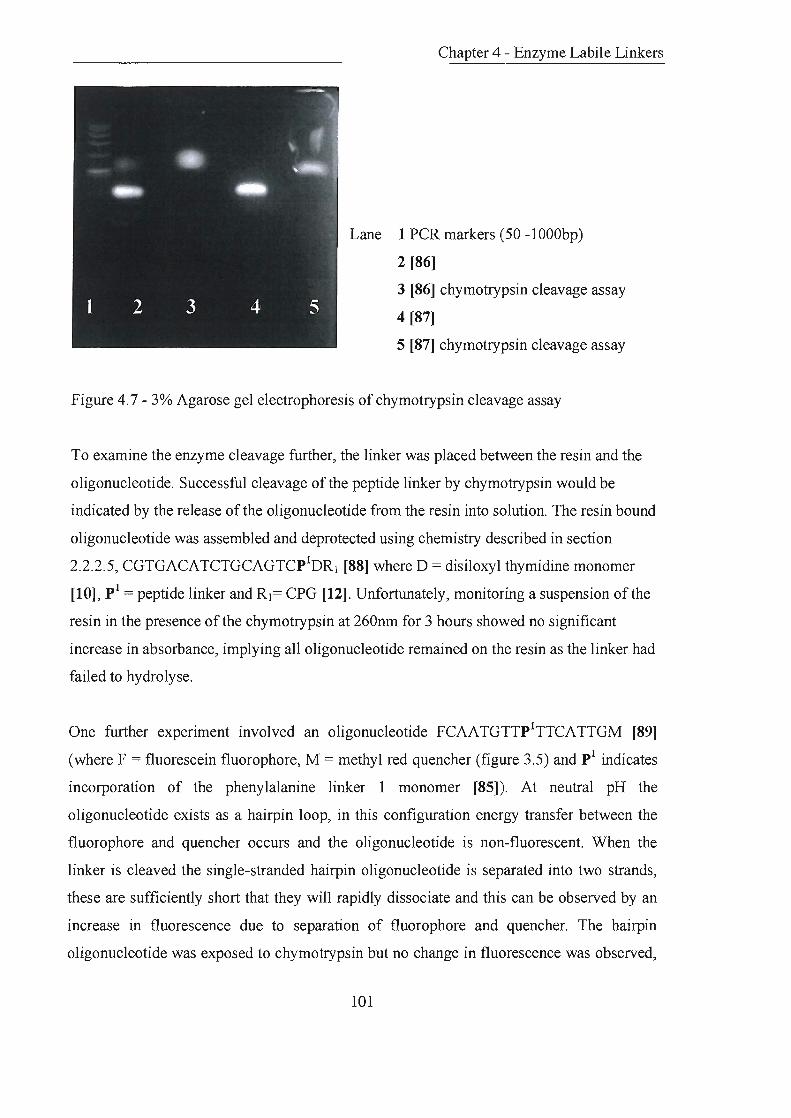
Chapter 4 - Enzyme Labile Linkers
Lane 1 PCR markers (50 -lOOObp)
2 [86]
3 [86] chymotrypsin cleavage assay
4 [87]
5 [87] chymotrypsin cleavage assay
Figure 4.7- 3% Agarose gel electrophoresis of chymotrypsin cleavage assay
To examine the enzyme cleavage further, the linker was placed between the resin and the
oligonucleotide. Successful cleavage of the peptide linker by chymotrypsin would be
indicated by the release of the oligonucleotide from the resin into solution. The resin bound
oligonucleotide was assembled and deprotected using chemistry described in section
2.2.2.5, CGTGACATCTGCAGTCpIDR] [88] where D = disiloxyl thymidine monomer
[10], pI = peptide linker and R]= CPG [12]. Unfortunately, monitoring a suspension of the
resin in the presence of the chymotrypsin at 260nm for 3 hours showed no significant
increase in absorbance, implying all oligonucleotide remained on the resin as the linker had
failed to hydrolyse.
One further experiment involved an oligonucleotide FCAATGTTpITTCATTGM [89]
(where F = fluorescein fluorophore, M = methyl red quencher (figure 3.5) and pI indicates
incorporation of the phenylalanine linker 1 monomer [85]). At neutral pH the
oligonucleotide exists as a hairpin loop, in this configuration energy transfer between the
fluorophore and quencher occurs and the oligonucleotide is non-fluorescent. When the
linker is cleaved the single-stranded hairpin oligonucleotide is separated into two strands,
these are sufficiently short that they will rapidly dissociate and this can be observed by an
increase in fluorescence due to separation of fluorophore and quencher. The hairpin
oligonucleotide was exposed to chymotrypsin but no change in fluorescence was observed,
101

Labile Linkers
indicating no cleavage. Confonnation that the single stranded hairpin loop oligonucleotide
remained intact was achieved by UV thermal melting which gave the same transition (Tm =
61 DC), corresponding to stem dissociation, before and after exposure to chymotrypsin.
4.3.1.3 Conclusions
The results shmved the peptide linker was not able to act as a substrate for chymotrypsin,
there was either no recognition of the linker by the enzyme or in the case of a dye-labelled
oligonucleotide, a complex between the dye and the enzyme may be forming. To overcome
these difficulties design of a new linker required greater rigidity of the recognition site by a
more peptidic linker structure in attempt to gain substrate recognition.
4.3.2 Phenylalanine Linker 2
4.3.2.1 Principle and Preparation
Design focused around a tripeptide substrate for chymotrypsin, phenylalanine was
sandwiched between two glycine residues and alkyl spacers incorporated each side.
Successful cleavage of the linker would occur at the C-tenninal amide of the phenylalanine
residue (figure 4.8).
Figure 4.8 - Phenylalanine Linker 2
The tripeptide section of the linker was assembled by solution phase peptide synthesis
beginning with the protected hydroxyamine spacer [92]. A series of carbodiimide coupling
and piperidine deprotection reactions generated a tripeptide amine onto which a twelve
carbon spacer was added to provide the final alcohol for phosphitylation (figure 4.9).
102

HO",-vNH2 --l>-
\~16 (i)
DMTOUNH~ 6 II NH
o [96}
I (vii, v)
'If
DMTO"-.,,/NH~ II NH
[98]
[99J
o
I (viii)
'f
J (ix)
DMT0"t--rNH~ 6 II NH
o
[1001
Chapter 4 - Enzyme Labile Linkers
o
HO~NHJlCF3 6
[90]
(ii)
o
DMTO~NHJlCF3 6
[91J
I (iii) 'f
NH2 DMTO ...... l/NH~, "" \"'76 II NH2
(vi, v) ° .. DMTO~NH2
(iv, v) 6
I ~ [94J h
[92]
o NH~ II OH
/I NH~1 a
'I ~
~ ~ k NH~ ~U-P\
II NH 11 a o ~CN ~
Reagents and conditions: i, EtOCOCF3, Et3N, CH2CI2, rt,12hrs, 71%; ii, DMTCI, DMAP, pyridine, rt, 15hrs, 81%; iii, NH3 (g), CH30H, rt, 18hrs, 88%; iv, Fmoc-Gly-OH, EDC, CH2CI2,
DMF, 67%; v, 20% piperidine, DMF, rt, 1-24hrs, [94J = 53%, [96] = 83%, [98] = 92%; vi, Fmoc-Phe-OH, EDC, CH2CI 2, DMF, rt, 14hrs, 91%; vii, Fmoc-Gly-OH, EDC, CH2CI2, DMF, rt, 1 hr, 83%; viii, 12-hydroxydodecanoic acid, EDC, CH2CI2, rt, 12hrs, 83%; xi, 2-cyanoethyldiisopropylchlorophosphoramidite, DIPEA, CH2CI2, rt, 1 hr, 92%
Figure 4,9 - Synthesis of phenylalanine linker 2 phosphoramidite
103

Chapter 4 - Enzyme Labile Linkers
4.3.2.2 Linker cleavage
Initial experiments to examine the hydrolysis of the linker were performed in solution. Two
oligonucleotides were synthesised and purified FTTTTTTP2TTTTTT [101], FP2TTTTTT
[102] (where F = fluorescein and p2 indicates incorporation of the phenylalanine liner 2
monomer [100], figure 4.10)
100
100
so 80
60
40
20
10 lS 20 5. 00 10.00 15.00
(i) (ii)
Figure 4.10 - Phenylalanine linker 2 oligonucleotide prior to hydrolysis (t = 0 min) (i)
[101], (ii) [102]
Initial experiments incubated the oligonucleotides in buffer with a large excess of
chymotrypsin at 37°C for 3 days. The enzyme was removed by centrifugal ultrafiltration
and the reaction products analysed by RP.HPLC, eluting under identical conditions to
initial oligonucleotide purification.
The experiment was performed on both oligonucleotides and showed in each case a
complete disappearance of starting material with the formation of new product peaks
corresponding to complete hydrolysis of the linker. To assess the rate of cleavage, a time
controlled study was performed with both oligonucleotides [101] and [102] using an excess
of enzyme.
Six samples of the modified oligonucleotides in separate tubes were incubated at 3rC, in
buffer, five tubes containing chymotrypsin. After time periods of lOmins, 30mins, 1hr,
3hrs, and 24hrs a tube was removed, denaturation was achieved by rapid heating to 95°C
and the enzyme removed by centrifugal filtration. RP.HPLC analysis of the reaction
104

Chapter 4 - Enzyme Labile Linkers
products showed almost complete conversion of starting oligonucleotide to hydrolysed
product after lOmins with very little change after longer time periods (figure 4.11).
B
c
B
0.00 5 . 00 10.00 15 .00 20 . 00 0 . 00
(i) (ii)
Figure 4.11 - (i) [101], (ii) [102] almost complete hydrolysis of starting oligonucleotide (A)
by chymotrypsin, generating cleavage fragments (fluorescent product B and non
fluorescent product C). All other peaks are enzyme fragments (t = 10 min).
The results were consistent for both oligonucleotides and collection of the individual
cleavage peaks revealed one fluorescent product and one colourless product as expected.
Mixed injection of starting oligonucleotide with the reaction products confirmed that the
oligonucleotide had been completely hydrolysed (figure 4.12). Repetition of the experiment
using one equivalent of chymotrypsin showed the formation of the products on a much
slower timescale, the reaction was almost complete after 24 hrs.
To examine the hydrolysis of the linker placed between a resin and an oligonucleotide, the
following sequences were assembled and left attached to the resin FP2TTTTTTR2 [103],
FP2HH-CPG [104] (where H= hexaethleneglycol, p2 indicates incorporation of the
phenylalanine linker 2 monomer [100] and R2 = polystyrene). The compound [103] was
designed to release dye from the resin into solution. A sample of the resin in buffer was
incubated with excess chymotrypsin and monitored for increase in absorbance at 495nm
over time. The reading increased from 0.059 to 0.275 over 21 hours, however, the
experiment showed only qualitative cleavage of the linker by detachment of the dye.
105
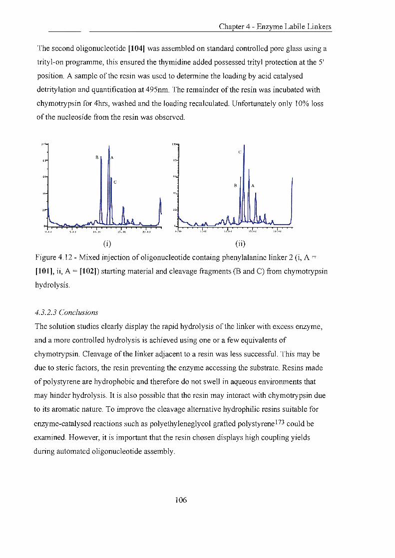
Chapter 4 - Enzyme Labile Linkers
The second oligonucleotide [104] was assembled on standard controlled pore glass using a
trityl-on programme, this ensured the thymidine added possessed trityl protection at the 5'
position. A sample of the resin was used to determine the loading by acid catalysed
detritylation and quantification at 495nm. The remainder of the resin was incubated with
chymotrypsin for 4hrs, washed and the loading recalculated. Unfortunately only 10% loss
of the nucleoside from the resin was observed.
0.00 5 .00 10.00 IS.00 20.00 0.00 10.00 1S.00 20.00
(i) (ii)
Figure 4.12 - Mixed injection of oligonucleotide containg phenylalanine linker 2 (i, A =
[101], ii, A = [102]) starting material and cleavage fragments (B and C) from chymotrypsin
hydrolysis.
4.3.2.3 Conclusions
The solution studies clearly display the rapid hydrolysis of the linker with excess enzyme,
and a more controlled hydrolysis is achieved using one or a few equivalents of
chymotrypsin. Cleavage of the linker adjacent to a resin was less successful. This may be
due to steric factors, the resin preventing the enzyme accessing the substrate. Resins made
of polystyrene are hydrophobic and therefore do not swell in aqueous environments that
may hinder hydrolysis. It is also possible that the resin may interact with chymotrypsin due
to its aromatic nature. To improve the cleavage alternative hydrophilic resins suitable for
enzyme-catalysed reactions such as polyethyleneglycol grafted polystyrene173 could be
examined. However, it is important that the resin chosen displays high coupling yields
during automated oligonucleotide assembly.
106

Chapter 4 - Enzyme Labile Linkers
4.3.3 Lysine Linker
4.3.3.1 Principle and preparation
A previous problem encountered had been the fonnation of complexes because of the
similarities between the aromatic rings of dye labels or the aromatic heterocyclic bases of
nucleotides and chymotrypsin substrates. A second tripeptide linker was designed that
incorporated lysine as the central amino acid. This linker was proposed as a substrate for
trypsin, which requires an amino acid with a basic side chain, such as lysine or arginine, for
catalysis.
Figure 4.13 - Lysine Linker
The E-amino moiety oflysine was protected with 4,4'-dimethyl-2,6-dioxocyclohex-l-y
(idene)ethane (Dde), as the conditions for removal of this group (hydrazine in DMF) are
orthogonal to the conditions of DNA synthesis. This choice of protecting group allowed the
molecule to be assembled by solution phase peptide chemistry and deprotected after
oligonucleotide assembly. The tripeptide section of the link:er was assembled from the with
the protected hydroxy amine spacer [92]. A series of carbodiimide coupling and piperidine
deprotection reactions generated a tripeptide amine, onto which a twelve-carbon spacer was
added to provide the final alcohol for phosphitylation (figure 4.14).
107

Labile Linkers
(ii)
j (iii) o
DMTOX.vNHy-' ~II NH2 DMTO ...... yNH~ DMTO ...... ,.....NH2 1-'6 NH .. \-'6 II NH2 .. \-'6
o ) (vi, v) 0 (iv, v)
[106] H2N 4 [94] [92]
J (vii, v)
Reagents and conditions: i, EtOCOCF3, Et3N, CH2CI2, rt,12hrs, 71%; ii, DMTCI, DMAP, pyridine, rt, 15hrs, 81 %; iii, NH3 (g), CH30H, rt, 18hrs, 88%; iv, Fmoc-Gly-OH, EDC, CH2CI 2, DMF, 53%; v, 20% piperidine, DMF, rt, 1-24hrs, [94] = 67%, [106] = 69%, [108] = 82%; vi, Fmoc-Lys(Dde)-OH, EDC, CH2CI2, rt, 1 hr, 88%; vii, Fmoc-Gly-OH, EDC, CH2CI 2, DMF, rt, 2.5hrs, 67%; viii, 12-hydroxydodecanoic acid, EDC, CH~12' rt, 2.5hrs, 82%; xi, 2-cyanoethyldiisopropylchlorophosphoramidite, DIPEA, CH~12' rt, 2.5hrs, 91%
Figure 4.14 - Synthesis oflysine linker phosphorarnidite
108

Chapter 4 - Enzyme Labile Linkers
4.3.3.2 Linker cleavage
Initial experiments to examine the hydrolysis of the linker were performed in solution. Two
oligonucleotides were synthesised and purified by RPHPLC
TTTTTTLTTTTTTTTTTTTTTTT [111], TTCTTCTTLTCCTCTTCTTCTCCTTT [112]
(where L indicates the incorporation of the lysine linker monomer [110], figure 4.15).
) 00 )00
" "
60 60
40 40
20 20
0 . 00 0.00
(i) (ii)
Figure 4.15 - Lysine linker oligonucleotide prior to hydrolysis (t = 0 min) i, [111] ii, [112]
Deprotection of the oligonucleotides with concentrated aqueous ammonia was shown to
remove Dde protection from the lysine side chain by RPHPLC and mass spectral analysis.
The fully deprotected oligonucleotides were incubated, in buffer, with a large excess of
trypsin at 37°C for 24 hours. Trypsin was removed by centrifugal ultrafiltration and the
reaction products were analysed by RPHPLC eluting under identical conditions to initial
oligonucleotide purification. Hydrolysis of the lysine linker was shown by almost total
conversion of starting material to two product peaks (figure 4.16, i). Mixed injection of
starting oligonucleotide with the reaction products confirmed that the oligonucleotide had
been completely hydrolysed (figure 4.16, ii).
109
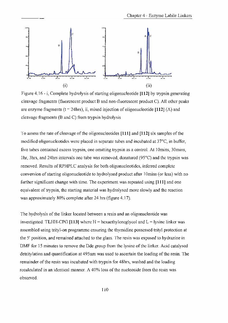
Chapter 4 - Enzyme Labile Linkers
100
c A C
B
5.00 10.00 15.00
(i) (ii)
Figure 4.16 - i, Complete hydrolysis of starting oligonucleotide [112] by trypsin generating
cleavage fragments (fluorescent product B and non-fluorescent product C). All other peaks
are enzyme fragments (t = 24hrs), ii, mixed injection of oligonucleotide [112] (A) and
cleavage fragments (B and C) from trypsin hydrolysis
To assess the rate of cleavage of the oligonucleotides [111] and [112] six samples of the
modified oligonucleotides were placed in separate tubes and incubated at 37°C, in buffer,
five tubes contained excess trypsin, one omitting trypsin as a control. At 10mins, 30mins,
Ihr, 3hrs, and 24hrs intervals one tube was removed, denatured (95°C) and the trypsin was
removed. Results ofRPHPLC analysis for both oligonucleotides, inferred complete
conversion of starting oligonucleotide to hydrolysed product after 10mins (or less) with no
further significant change with time. The experiment was repeated using [111] and one
equivalent of trypsin, the starting material was hydrolysed more slowly and the reaction
was approximately 80% complete after 24 hrs (figure 4.17).
The hydrolysis of the linker located between a resin and an oligonucleotide was
investigated. TLHH-CPG [113] where H = hexaethyleneglycol and L = lysine linker was
assembled using trityl-on programme ensuring the thymidine possessed trityl protection at
the 5' position, and remained attached to the glass. The resin was exposed to hydrazine in
DMF for 15 minutes to remove the Dde group from the lysine of the linker. Acid catalysed
detritylation and quantification at 495nm was used to ascertain the loading of the resin. The
remainder of the resin was incubated with trypsin for 48hrs, washed and the loading
recalculated in an identical manner. A 40% loss of the nucleoside from the resin was
observed.
110

Chapter 4 - Enzyme Labile Linkers
A
(i) (ii)
Figure 4.17 - Lysine linker oligonucleotide [111] (A) hydrolysis using one equivalent of
trypsin generating cleavage fragments (B and C). All other peaks are enzyme fragments i, t
= 10 mins, ii, t = 1 hour
4.3.3.3 Conclusions
Results of solution studies demonstrate that hydrolysis of the lysine linker is very fast with
excess enzyme. Retardation of the rate is achieved when only one equivalent of trypsin is
available. Cleavage of the linker adjacent to a resin was more successful than cleavage of
the phenylalanine tripeptide linker. However, the exposure time was extended which may
explain the result. Incomplete cleavage may be due to steric factors between resin and
enzyme; improved yield might be achieved with more hydrophilic resins, or inserting
additional spacer molecules between resin and linker.
111

Chapter 5
Experimental

Chapter 5 - Experimental
5.0 Experimental
5.1 Preparation of Compounds
5.1.1 General Methods
All reactions requiring anhydrous conditions were performed in dry glassware under an
atmosphere of nitrogen or argon. Methylene chloride, pyridine and triethylamine were
distilled from calcium hydride. Tetrahydrofuran was freshly distilled from sodium wire and
benzophenone. Methanol was distilled from magnesium and iodine. Quinoline was distilled
from 3A molecular sieves. Anhydrous benzene, dimethylformamide and
diisopropylethylamine were purchased from Aldrich. Other reagents were purchased from
Aldrich/Sigma or Lancaster.
Column chromatography was carried out under pressure on Merck 60-mesh silica.
TLC was carried out using Merck Kieselgel60 F24 (0.22mm thickness, aluminium backed).
Compounds were visualised by irradiation at 254nm or staining with p-anisaldehyde:
glacial acetic acid: concentrated sulphuric acid: ethanol (5: 1: 1: 100) or ninhydrin: ethanol
(1: 100) or concentrated H2S04: ethanol (1: 10) or polymolybidic acid: ethanol (l: 10).
Solvent systems A- Q are described:
A: CH2Clz: MeOH 4: 1 + 1 % AcOH
B: CH2Ch: MeOH: Et3N 8.75:0.25: 1.00
C: CH2Ch: MeOH: Et3N 8.5:0.5: 1.0
D: CH2Ch: MeOH 9: 1
E: EtOAc
F: CHCh: MeOH 19:1
G: EtOAc: Et3N 9: 1
H: CH2Ch: MeOH: Et3N 8.75:1.00:0.25
I: CH2Ch: MeOH: Et3N 9: 1: 1
J: EtOAc: hexane 9: 1
112
K: EtOAc: hexane 1:9
L: EtOAc: hexane 3:7
M: EtOAc: hexane 1: 1
0: CH2Ch: MeOH 4: 1
P: CH2Ch: MeOH 19: 1
Q: EtOAc: MeOH 19: 1
Chapter 5 - Experimental
Infrared spectra were measured on a Perkin-Elmer 1600 series FTIR machine using
samples supported on sodium chloride cells, or on a BIORAD FT-IR instrument using a
Golden Gate adapter and BIORAD WIN-IR software. Absorbtions are described as strong
(s), medium (m), weak, or broad (br).
Melting points were measured on a Gallenkamp electrothermal melting point apparatus.
Ultraviolet spectra were measured on a Perkin Elmer UV/Vis Lambda 2 spectrometer.
Proton NMR was recorded at 300MHz and carbon NMR at 75.5MHz on a Brucker AM 300
spectrometer in either deuterated chloroform or D6-DMSO. The multiplicites of carbon-13
signals were determined using distortionless enhancement by phase transfer (DEPT)
spectral editing technique.
Mass Spectra were recorded on a Fisons VG platform instrument or MALDI TOF
Microanalytical data was obtained from University College, London.
113

5.1.2 List of Compounds
[1] 5(6) Carboxyfluorescein 117
[2] 6-(t-Butyldimethylsilyloxy)-1-hexylamine 118
[7] 5' -0-(4,4' -Dimethoxytrityl)-2' -deoxythymidine 119
[9] 5' -0-(4,4' -Dimethoxy )trity 1-2' -deoxythymidy 1-3' -0-( 1,1,3,3-tetra-
isopropyl-disiloxyl-3-) hexanol 120
[10] 5' -0-(4,4' -Dimethoxytrityl)-2' -deoxythymidyl-3' -O-(1,l,3,3-tetra-iso
propy l-disiloxy 1 -3' O-cyanoethoxy)-N,N-diisopropy lamino ) phosphine 121
[11) 12-0' -(4,4' -Dimethoxytrity loxy )dodecanoic acid 122
[15] 5(6)-Carboxyfluorescein-N-hydroxysuccinimide ester 123
[18] 5( 6)-Carboxyfluorescein Dipiva1ate 124
[19] 6-Carboxyfluorescein dipivalate diisopropylamine salt 125
[20] 6-Carboxyfluorescein 126
[21] 6-(t -B uty 1dimethy lsily loxy )-1-hexy 1-6-carboxyfluoresceinamide 127
[22J a-D-Tetraacetate galactosyl bromide 128
[23] 6-(t-Butyldimethylsilyloxy)-I-hexyl-6-carboxyfluoresceinamide-
(3,6)-bis-~-D-galactose tetraacetate 129
[24] 1-0-Hexyl-6-carboxyfluoresceinamide -(3 ,6)-bis-~-galactose tetraacetate 131
[25] 1-0-( cyanoethoxy -N,N-diisopropy lamino phosphine) hexy 1-6-
carboxyfl uoresceinamide(3, 6)-b is-~-Ga1actose tetraacetate 132
[65] 4-( 4-(0-4, 4'-dimethoxytrityl)-hydroxmethyl-2-methoxy-5-nitro-
phenoxy)-butyric acid 133
[70] 2,3,4 ,6-Tetra-O-acety 1-D-ga1actose 134
[71] 0-(2,3,4,6-Tetra-0-acetyl)-a-D-galactosyl trichloroacetimidate 135
[72] 6-(t-Butyldimethylsi1yl)oxy-l-hexanol 136
[73] 0-~-D-[(2,3,4,6-Tetra-0-acetyl)-galactosyl-[6-(t-butyldimethylsilyl)
oxy hexane] 136
[74] 0-~-D-[6-(t-Buty1dimethy1silyl)oxy hexane]-galactose 137
[75] 0-~-D-(6-0-4,4'-Dimethyloxytrityl)-galactosyl-[6-(t-butyldimethylsilyl)
oxy hexane] 138
114

Chapter 5 - Experimental
[76] 0-~-D-[(2,3,4-Tri-0-acetyl)-6-0-( 4,4'-dimethyloxytrityl)]-galactosyl
-[6-(t-butyldimethylsilyl)oxy] hexane 139
[77] 0-~-D-[(2,3,4-Tri-0-acetyl)-6-0-( 4,4'-dimethyloxytrityl)]-galactosyl
hexan-6-o1 141
[78] 0-~-D-[(2,3,4-Tri-0-acetyl)-6-0-( 4,4'-dimethyloxytrityl)]-galactosyl-
[6-(2-cyanoethyl diisopropylphosphoramidyl)oxy hexane] 142
[81] Fmoc-pheny lalanine-6-hy droxy hexy lam ide 143
[82] Fmoc-pheny lalanine-6-hydroxy( -0-4,4' dimethoxytrity 1-)hexy lamide 144
[83] Pheny lalanine-6-hydroxy(-0-4,4' dimethoxytrityl-)hexy lamide 145
[84] 12-Hydroxy-dodecanamido-phenylalanine-6-hydroxy
( -0-4,4' dimethoxytrity 1-) hexy 1amide 146
[85] 12-(2-Cyanoethy I-diisopropy Iphosphoramidy 1 )oxy -dodecanamido-
pheny lalanine-6-hy droxy( -0-4,4' dimethoxytrity 1-)hexy lamide 147
[90] I-Hexanol-6-trifluoroacetamide 147
[91] 1- 0' -(4,4' -Dimethoxytrityloxy) hexyl-6-trifluoroacetamide 148
[92] 1-0-(4,4' -Dimethoxytrityloxy)-6-hexylamine 149
[93] Fmoc-glycine-6-hy droxy( -0-4,4' -dimethoxytrity 1 )hexy lamide 150
[94] Glycine-6-hydroxy(-0-4,4' -dimethoxytrityl)hexylamide 151
[95] Fmoc-phenylalanine-glycine-6-hydroxy( -0-4,4' -dimethoxytrity1)
hexylamide 152
[96] Pheny 1alanine-glycine-6-hydroxy( -0-4,4' -dimethoxytrity l)hexy 1amide 153
[97] F moc-glycine-pheny lalanine-glycine-6-hy droxy( -0-4,4' -
dimethoxytrity l)hexy lam ide 154
[98] Glycine-pheny lalanine-glycine-6-hydroxy( -0-4, 4' -dimethoxytrity 1)
hexylamide 155
[99] 12-H Y droxydodecanamido-glycine-pheny lalanine-glycine-6-hy droxy
(-0-4,4' -dimethoxytrityl)hexy lamide 156
[100] 12-(2-Cyanoethy 1 diisopropy Iphosphoramidy 1 )oxy -dodecanamido-glycine-
pheny lalanine-glycine-6-hy droxy (-0-4,4' -dimethoxytrity l)hexylamide 157
[105] Fmoc-1ysine(Dde )-glycine-6-hydroxy( -0-4,4' -dimethoxytrityl)
hexylamide 158
[106] L ysine(Dde )-glycine-6-hydroxy( -0-4,4' -dimethoxytrity l)hexy lamide 159
115

Chapter 5 - Experimental
[107] Fmoc-glycine-lysine(Dde )-glycine-6-hydroxy( -0-4,4' -dimethoxytrity 1)
hexy lamide 161
[108] Glycine-Iysine-(Dde) -glycine-6-hydroxy (-0-4,4' -dimethoxytrityl)
hexylamide 162
[109] 12-Hydroxydodecanamido-glycine-lysine-(Dde )-glycine-6-hydroxy
(-0-4,4' -dimethoxytrityl)hexylamide 163
[110] 12-(2-Cyanoethyl-diisopropy Iphosphoramidyl )oxy-dodecanamido-glycine-
lysine(Dde )-glycine-6-hydroxy (-0-4,4' -dimethoxytrity l)hexy lamide 164
Silver oxide catalyst
Silver silicate catalyst
116
165
165

5.1.3 Experimental
+
5(6) Carboxyfluorescein [1]194
Method 1
o
Chapter 5 - Experimental
C21H120y Mol. Wt.: 376.32
C, 67.02; H, 3.21; 0, 29.76
4-Carboxyphthalic anhydride (4.80g, 2S.0mmol) was ground to a fine powder and placed in
an open metal pot. Resorcinol (S.4Sg, 49.Smmol) was added, and the mixture heated to
190°C for 1 hr with manual stirring. During this period anhydrous zinc chloride (1.7Sg,
12.Smmol) was added portionwise, and the reaction stirred until too viscous to continue.
The red solid was cooled to room temperature and then allowed to stand for 1hr, water
(50mL) and aqueous hydrochloric acid (concentrated, 2.SmL) were added. The mixture was
heated to boiling, filtered and the residue dissolved in aqueous sodium hydroxide solution
(2M, 40mL). This was dropped into rapidly stirring aqueous hydrochloric acid (4M, SOmL)
affording an orange precipitate, which was removed by filtration. The orange suspension
was taken up in MeOH (SOmL) to aid handling and then the solvent removed in vacuo. The
residue was triturated with Et20 (30mL), filtered and evaporated to dryness. The product
was dried over P205, in vacuo, for 24 hr to give orange flakes that were ground to a fine
powder (S.32g, 14.1mmol, S7%, improved to 70%).
Method 2 4-Carboxyphthalic anhydride (38.40g, 0.2mol) was ground to a fine powder and place in an
open metal pot. Resorcinol (43.60g, O.4mol) was added, and the mixture heated to 190°C
for 1 hr with manual stirring. The resulting red solid was cooled to room temperature and
allowed to stand for Ihr, dissolved in aqueous sodium hydroxide solution (2M, 300mL) and
dropped into rapidly stirring aqueous hydrochloric acid (4M, 300mL). This gave an orange
precipitate that was filtered, dissolved in MeOH (200mL) and the solvent removed in
117

vacuo, The product was dried over P205, in vacuo for 24 hrs to give orange flakes that were
ground to a fine powder (64,00g, 170,2mmol, 85%).
Rf(A): 0.78; LRMS (ES- mode): mlz 375.2 (M-), 489.2 (MTFA-); HRMS: C2IH130 7
requires 377.06613, found 377.06613; Amax (MeOH): 483nm; mp: 350-355°C (lit.mp: 356-
358°C)
IR (CH2Ch): v = 2922.9 (s), 2842.7 (s), 1629.4 (m), 1459.2 (m), 1374.0 (m), 722.5 (w) cm-
IH (d6-DMSO): () 8.40 (lH, s, H4,), 8.32 (lH, d, J 9.5Hz, H4), 8.20 (lH, d, J 8. 1Hz,
Hl4'), 8.12 (lH, d,J 8. 1Hz, H5), 7.65 (lH, s, H6), 7.40 (lH, d,J = 8. 1Hz, H6,), 6.75 (4H,
S, HI), 6,58-6.61 (8H, m, H2,3), 3.50 (2H, br, OH)
l3C (~-DMSO):{) = 167.9, 166.2 (C7, 15), 159.7 (C lO), 156.1 (ClI ), 151.9 (C l3), 136.2 (Cl2),
133.2 (CI4), 129.3, 126.9, 125.6, 124.6 (C3-6, 9), 112.8 (C2), 109.0 (Cs), 102.4 (C I)
6-(t-Butyldimethylsilyloxy)-1-hexylamine [2] 195
To a solution of6-amino-1-hexanol (6.21g, 53.1mmol) in anhydrous pyridine (50rnL) was
added t-butyldimethylsilyl chloride (9.52g, 57.2mmol). The yellow solution was stirred at
room temperature, under argon, for 20hrs. The solvent was removed in vacuo, and the
resulting viscous brown oil was dissolved in CH2Ch (1 OOrnL), washed with saturated
sodium bicarbonate solution (2xlOOmL) and dried (Na2S04). Removal of the solvents in
vacuo afforded an opaque yellow oil, which was purified by silica gel column
chromatography eluting with CH2Ch: MeOH (9: 1). The title compound was obtained as a
pale yellow oil, which was dried over P205 in vacuo for 24 hrs (9.63g, 41.6mmol, 79%).
Rf (H): 0.40; LRMS (ES+ mode): mlz 232 (MH+); HRMS: CI2H30NOSi requires 232.20967,
found 232.20967; Amax (MeOH): 254nm
IR (CH2Ch): v = 3350.0 (w), 2829.7 (s), 2374.6 (m), 1473.0 (m), 1254.9 (s), 1095.3 (s),
834.6 (s) cm-1
118

IH (CDCh): {j = 3.55 (2H, t, J = 6.6 Hz, CH20), 2.67 (2H, t, J = 6.6 Hz, CH)NH2), l.70
(2H, br s, NH2), 1.52-1A2 (4H, m, CfuCH20, CfuCH2NH2), 1.38-1.25 (4H, m, CH2), 0.90
(9H, s, (CH3)3CSi), 0.00 (6H, s, (CH3)2Si)
I3C (CDCh): (j = 63.3 (CH20Si), 42.2 (CH2NH2), 33.7, 32.9(CH2), 26.8 (CH2), 26.]
((CH3)3C), 25.9 (CH2), 18.5 ((CH3)3C), -3.3 (CH3Si)
0--
o
- 5H~~3 0p20ANjJj2
11 7
9 8 OH
10
~1H32N207 Mol. Wt.: 544.60
C, 68.37; H, 5.92; N, 5.14; 0, 20.56
5'-O-(4,4'-Dimethoxytrityl)-2'-deoxythymidine [7]
Anhydrous pyridine (2 x 20mL) was coevaporated from thymidine (6.06g, 25.0mmol), 4-
dimethylaminopyridine (0.36g, 2.9mmol) was added, and pyridine (20mL) was
coevaporated from the mixture a second time. The resulting mixture was dissolved in
pyridine (30mL) and a solution of 4,4' -dimethoxytrityl chloride (9.32g, 27.5mmol) in
pyridine (lOmL) was added portionwise over 1.5 hrs. After stirring under argon for 24hrs,
the reaction was quenched with MeOH (20mL), and the solvents removed in vacuo. The
resulting solid was dissolved in EtOAc (150mL), washed with saturated sodium
bicarbonate solution (3 x 1 OOmL), dried (MgS04), and the solvents removed in vacuo to
give a yellow foam. Recrystallisation from benzene afforded the title compound as a white
solid, which was dried over P20 S in vacuo for 24 hrs (l1.10g, 20Ammol, 82%).
Rf CD): 0.63; LRMS (ES+ mode): mlz 303.2 (DMT+), 567.2 (MNa+); HRMS: C3IH32N20 7
requires 544.22095, found 544.22095; Amax (MeOH): 269, 234nm; mp: 124-127°C
IRv = 3036.8 (w), 2834.16 (w), 1677.6 (s), 1606.3 (m), 1252.6 (s), 1175.8 (s), cm-I
IH (CDCh): {j = 11.40 (lH, s, H s), 7.50 (lH, s, HI), 7AO-7.15 (9H, m, PhH, HIS), 6.90 (4H,
d, J = 8.8 Hz, HI6), 6.23 (lH, t, J = 6.6 Hz, H7), 5.35 (lH, d, J 4A Hz, H9), 4.33 (lH, s,
119

H IO), 3.92-3.88 (lH, m, Hll ), 3.75 (6H, s, OCH3), 3.35 (3H, s, H3), 3.24-3.16 (2H, m, HIZ),
2.35-2.10 (2H, m, H8)
l3C (CDCh): (; = 163.8 (C4), 158.3 (C17), 150.5 (C6), 144.8 (C I8), 135.5 (C J4), 129.8 (C l ),
128.4, 128.0, 127.8, 126.9 (CPh, 15), 113.3 (C J6), 109.7 (C2), 86.0 (C13), 85.6 (C7), 83.9 (C9),
70.7 (Cll ), 63.9 (C l2), 55.1 (OCH3), 39.6 (C8), 1l.8 (C3)
0--
C49H72N201OSi2 Mol. Wt.· 905.27
C, 65.01; H, 8.02; N, 3.09; 0, 17.67; Si, 6.20
5' -0-( 4,4' -Dimethoxy)trityl 2' -deoxythymidyl-3' -0-(1,1,3,3-tetra-isopropyl-disHoxyl-
3-) hexanol [9] 145
Freshly distilled pyridine (2 x lOmL) was coevaporated from the alcohol [7] (2.60g,
4.8mmol), imidazole was added (0.64g, 9.6mmol) and pyridine (lOmL) was co evaporated
from the mixture. To a stirring suspension ofthe mixture in pyridine (40mL), under argon,
was added 1,3-dichloro-l,1,3,3-tetraisopropyl disiloxane (l.50g, 4.8mmol). The reaction
was stirred at room temperature for 24 hrs. This afforded 5' -0-(4,4' -Dimethoxytrityl)-2'
deoxythymidy13' -0-(1,1,3,3-tetra-isopropyl-disiloxyl) chloride [8], which was used
directly reacted in the following reaction. Hexane-l,6-diol (5.00g, 42.4mmol) was added
and the reaction stirred for a further 12 hrs. The solvent was removed in vacuo and the
resulting cloudy oil was dissolved in CH2Ch (150mL), washed with saturated sodium
bicarbonate solution (3 x 100mL), then dried (MgS04). Removal of the solvent in vacuo
yielded a cloudy oil that was purified by silica gel column chromatography eluting with
MeOH: CH2Ch (0: 100 then 2:98 then 1: 19). Drying over P20s for 12 hrs in vacuo afforded
the title compound as a clear oil (3. 15g, 3.5mmol, 76%).
120

Rf(F): 0.65; LRMS (ES+ mode): m/z 303.1 (DMT+), 926.9 (MNa+); "'-max (MeOH): 268,
233nm
IR: V = 2936.06 (m), 2864.85 (m), 1687.4 (s), 1248.6 (m), 1607.27 (m), 1032.7 (s), cm-l
IH (DMSO): i5 = 11.40 (lH, s, H5), 7.50 (IH, S, HI), 7.40-7.20 (9H, m, PhH, H I5), 6.90
(4H, d, J 8.6 Hz, H I6), 6.23 (lH, t, J 6.0 Hz, H7), 4.60-4.58 (lH, m, H9), 4.35 (lH, q, J
5.1 Hz, H ll ), 3.95 (lH, s, OH), 3.75 (6H, s, OCH3), 3.60 (2H, t, J 6.9 Hz, CH20H),
3.35 (3H, s, H3), 3.31-3.28 (2H, m, CH20Si), 3.25-3.20 (2H, m, H12), 2.35-2.32 (2H, m,
Hs), 1.50-1.20 (l2H, m, CH2, CHSi), 0.80-0.11 (24H, m, CH3CHSi)
13C (CDCh): i5 = 164.2 (C4), 158.8 (C l7), 150.6 (C6), 144.4 (C I8), 136.2 (C14), 135.5 (CI),
130.3, 128.3, 127.3, 123.9 (CPh• 15), 113.4 (CI6), 111.3 (C2), 87.1 (C7), 85.3 (C13), 84.9 (C9),
72.7 (C ll ), 63.7 (CH20H), 62.8 (C12), 55.4 (OCH3), 46.1 (CH20Si), 41.8 (Cs), 32.8 (CH2),
25.7 (CH2), 17.6, 17.4, 17.3 (CH3CHSi), 13.5, 13.2, 13.0 (CH3CHSi), 11.9 (C3)
0-
C5sHs9N4011 PSi2 Mol. Wt.: 1105.49
C, 63.01; H, 8.11; N, 5.07; 0,15.92; P, 2.80; Si, 5.08
5'-O-(4,4'-Dimethoxytrityl)-2'-deoxythymidyl-3'-O-(1,1,3,3-tetra-isopropyl-disiloxyl-
3'O-cyanoethoxy)-N,N-diisopropylamino) phosphine [10] 145
To a stirring solution of [9] (1. 109, 1.2mmol) in CH2Ch (10mL) was added anhydrous N,N
diisopropylethylamine (0.52g, 0.77mL, 4.5mmol) under argon. 2-Cyanoethyl-N,N
diisopropylchlorophosphoramidite (0.32g, 0.3mL, l.4mmol) was added to the mixture
dropwise, and the reaction allowed to stir at room temperature, under argon, for 1.5 hrs.
EtOAc (50mL) was added, the organic layer washed with saturated potassium chloride
solution (2 x 50mL), dried (MgS04) and the solvents removed in vacuo. The resulting oil
121
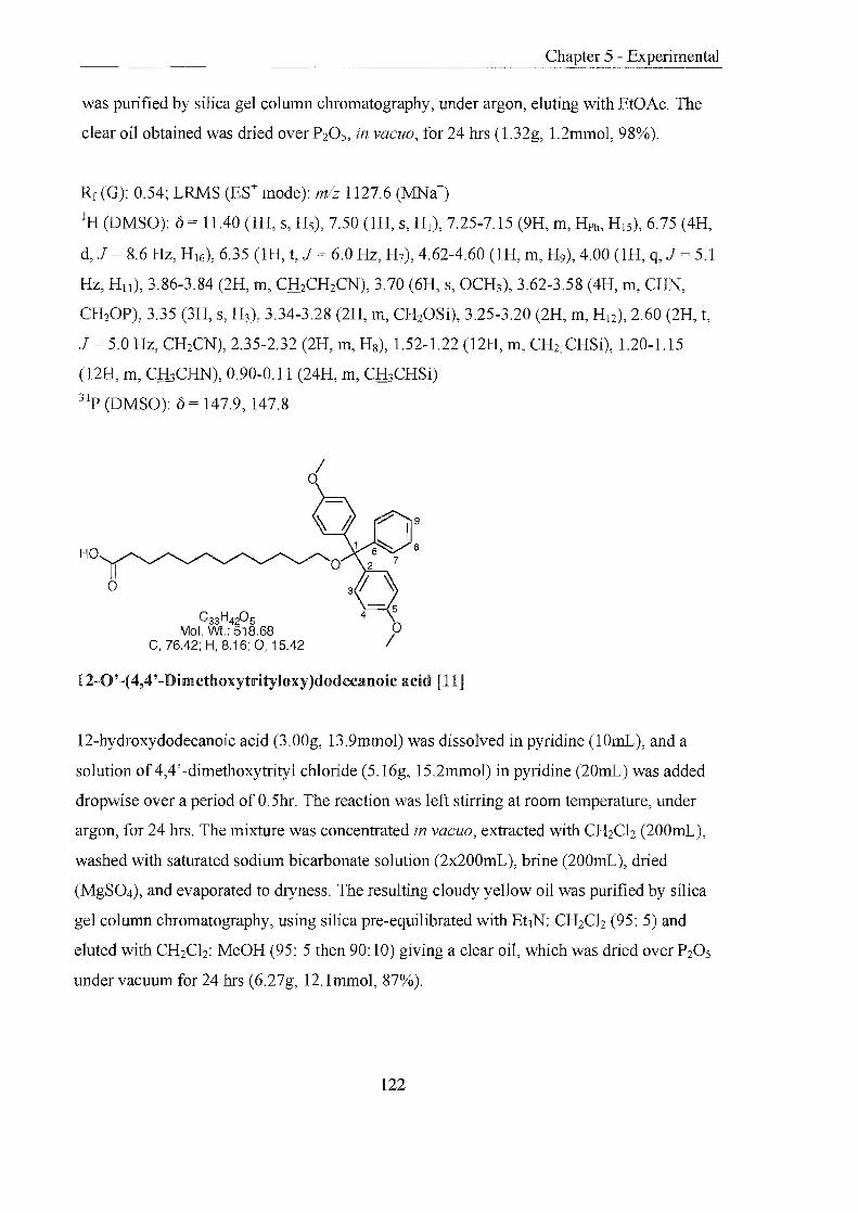
Chapter 5 - Experimental
was purified by silica gel column chromatography, under argon, eluting with EtOAc. The
clear oil obtained was dried over P20 S, in vacuo, for 24 hrs (1.32g, 1.2mmol, 98%).
Rf (G): 0.54; LRMS (ES+ mode): m/z 1127.6 (MNa+)
IH (DMSO): {) = 11.40 (lH, s, Hs), 7.50 (lH, S, HI), 7.25-7.15 (9H, m, Hph, HIS), 6.75 (4H,
d, J = 8.6 Hz, H16), 6.35 (lH, t, J 6.0 Hz, H7), 4.62-4.60 (lH, m, H9), 4.00 (lH, q, J = 5.1
Hz, Hl1 ), 3.86-3.84 (2H, m, CfuCH2CN), 3.70 (6H, s, OCH3), 3.62-3.58 (4H, m, CHN,
CH20P), 3.35 (3H, s, H3), 3.34-3.28 (2H, m, CH20Si), 3.25-3.20 (2H, m, H12), 2.60 (2H, t,
J = 5.0 Hz, CH2CN), 2.35-2.32 (2H, m, H8), 1.52-1.22 (l2H, m, CH2, CHSi), 1.20-1.15
(l2H, m, CH3CHN), 0.90-0.11 (24H, m, CH3CHSi)
31p (DMSO): {) = 147.9, 147.8
HO
C33H4205 Mol. Wt.: 518.68
C, 76.42; H, 8.16; 0, 15.42
12-0'-(4,4'-Dimethoxytrityloxy)dodecanoic acid [11]
12-hydroxydodecanoic acid (3.00g, 13.9mmol) was dissolved in pyridine (lOrnL), and a
solution of 4,4'-dimethoxytrityl chloride (5.16g, 15.2mmol) in pyridine (20rnL) was added
dropwise over a period ofO.5hr. The reaction was left stirring at room temperature, under
argon, for 24 hrs. The mixture was concentrated in vacuo, extracted with CH2Cb (200mL),
washed with saturated sodium bicarbonate solution (2x200mL), brine (200rnL), dried
(MgS04), and evaporated to dryness. The resulting cloudy yellow oil was purified by silica
gel column chromatography, using silica pre-equilibrated with Et3N: CH2Cb (95: 5) and
eluted with CH2Cb: MeOH (95: 5 then 90: 10) giving a clear oil, which was dried over P20 S
under vacuum for 24 hrs (6.27g, 12.1mmol, 87%).
122

Rf (1): 0.61; LRMS (APCr mode): mlz 517.4 (M-), (ES- mode): m1z 631.5 (MTFA-);
HRMS: C33H420S requires 518.30322, found 518.30322; Amax (MeOH): 232nm
IR: v = 2925.2 (m), 2853.23 (m), 1712.7 (w), 1507.94 (m) cm-1
lH (CDCh):o = 8.50 (lH, br s, OH), 7.45-7.10 (9H, m, J 8.6 Hz, H3, ?-9), 6.65 (4H, d, J =
8.6 Hz, H4), 3.70 (6H, s, CH30), 3.00 (2H, t, J = 7.1 Hz, CH20), 2.12 (2H, t, J 6.4 Hz,
CH2CO), 1.60-1.45 (4H, m, CH2CH20, ClliCH2CO), 1.24-1.21 (14H, m, CH2)
13C (CDCh): 0 = 179.1 (CO), 158.4 (C5), 145.6 (C6), 136.9 (C2), 130.2 (C3), 128.4 (Cs),
127.8 (C7), 126.7 (C9), 113.1 (C4), 85.7 (Cl), 63.7 (CH20), 55.3 (CH30), 35.9 (CH2CO),
30.3 (CH2CH20), 29.8, 29.7, 29.6, 26.5, (CH2) 25.8 (CH2CH2CO)
r1: HO
'(N_O
o 0
OH
+
C25H15NOg Mol. Wt.· 473.39
c}-o o 0
C, 63.43; H, 3.19; N, 2.96; 0, 30.42
5(6)-Carboxyfluorescein-N-hydroxysuccinimide ester [15] 196, 197
OH
To a suspension of 5(6)-carboxyfluorescein (1.00g, 2.7mmol) in EtOAc (30mL) was added
EDC (0.51g, 2.7mmol) followed by N-hydroxysuccinimide (0.34g, 2.9mmol). The orange
solution was stirred at room temperature, under argon, for 3 hrs. The solvent was removed
in vacuo and the resulting viscous red oil was dissolved in CH2Ch (lOOmL), washed with
saturated potassium chloride solution and dried (MgS04). The crude mixture was purified
by silica gel column chromatography eluting with MeOH: CH2Ch (1: 19). The product was
dried over P20 S in vacuo for 12 hrs to give an crude orange solid that was used directly in a
labelling reaction (0.76g, 1.6mmol, 61%).
Rf(D): 0.55; LRMS (ES- mode): mlz 472.2 (M-), Amax (MeOH): 276, 466nm
123

4 C31H2S09 Mol. wt.: 544.55
C, 68.37; H, 5.18; 0, 26.44
5(6)-Carboxyfluorescein Dipivalate [18] 154
Chapter 5 - Experimental
~7 JO
1{~
o
Anhydrous pyridine (80mL) was co-evaporated three times from 5(6)-carboxyfluorescein
(l9.01g, 50.5mmol) and the residue suspended in pyridine (40mL). Trimethylacetyl
chloride (24.50g, 25mL, 0.2mol) was added dropwise to the stirring solution at O°C, under
argon. After 3 hrs the solvents were removed in vacuo. The residue was dissolved in
CH2Ch (50mL), washed with saturated potassium chloride solution (3 x 50mL) then dried
(Na2S04). The solvents were removed in vacuo affording a yellow oil, which was purified
by silica gel column chromatography eluting with CH2Ch: Et3N: MeOH (1:0:0 then
80: 10: 10). The title compounds were obtained as a yellow foam that was dried over P20S,
in vacuo, for 24 hrs (16.78g, 30.8mmol, 64%).
Rf(B): 0.52,0.38; LRMS (ES- mode): mlz 657.2 (M-), 1201.3 (2MTFA-); HRMS: C3lH2909
requires 545.18116, found 545.18116; Amax (MeOH): 276, 229nm; mp: 105-110°C
IR (CH2Ch): v = 3053.1 (w), 2983.0 (w), 1754.8 (s), 1694.7 (w), 1419.1 (m), 1258.7 (m)
IH (CDCh): 0 = 8.60 (lH, s, H4,), 8.26 (lH, d, J = 8.1 Hz, H6,), 8.23 (lH, d, J 8.1 Hz,
H4), 8.10 (lH, d, J 8.1 Hz, Hs), 7.86 (lH, s, J 8.1 Hz, H6), 7.28 (lH, s, Hs'), 7.08 (4H,
s, HI), 6.80-6.78 (8H, m, H2, 3), 1.38 (36H, s, H19)
l3C (CDCh): 0= 176.7 (CI6), 168.0 (CIS), 165.5 (C7), 156.9, 153.4, 152.9, 151.7, 151.6
(ClO-14), 136.8, 136.4, 132.7, 13l.5, 129.5, 128.9, 127.0, 126.7 (C3-6), 125.5, 125.4 (C9),
118.0 (C2), 115.7, 115.6 (C8), 110.6 (CI), 39.4 (C l7), 27.2 (CI8)
124

C37H42N09 Mol. wt.: 644.73
C, 68.93; H, 6.57; N, 2.17; 0, 22.33
6-Carboxyfluorescein dipivalate diisopropylamine salt [19] 154
Chapter 5 - Experimental
5(6) Carboxyfluorescein (20.00g, 53.2mmol) was dissolved in anhydrous pyridine at O°e.
Trimethylacetyl chloride (28mL, 0.2mol) was added to the dark orange solution dropwise,
and the reaction allowed to stir for 3 hrs, whilst warming to room temperature. The pale
yellow mixture was diluted with CH2Ch (200mL), washed with saturated potassium
chloride solution (2 x 250mL) and dried (Na2S04). Removal of the solvents in vacuo gave a
dark brown oil, which was converted to a stable foam, by coevaporation of CH2Ch
(5x30mL). The foam (25.00g) was dissolved in boiling EtOH (55mL) to give a clear brown
solution to which diisopropylamine (2.5mL) was added. The solution was cooled to room
temperature then at -20°C for 18 hrs. A fine white precipitate was formed, which was
removed by filtration and dried over P20s in vacuo for 24 hrs to give the desired compound
as white solid (5.22g, 9.6mmol, 19%).
Rr(C): 0.73; LRMS (ES- mode): mJz 543.0 (M-), 657.0 (MTFA-); HRMS: C3IH2909
requires 545.18116, found 545.18116; Amax (MeOH): 282, 233nm; mp: 222-227°C
IR (CH2Ch): v = 3052.1 (w), 2980.0 (w), 1754.8 (m), 1694.7 (w), 1609.5 (m), 1419.1 (m),
1258.7 (m) cm-I
Found C: 68.78, H: 6.47, N: 1.82%, C37H42N09 requires C: 68.93, H: 6.57, N: 2.17%
IH (CDCh): 0 = 8.25 (IH, d, J 8.1 Hz, H4), 7.94 (lH, d, J 8.1 Hz, H5), 7.62 (IH, s, H6),
6.97 (2H, d, J = 2.2 Hz, HI), 6.80 (2H, d, J = 8.8 Hz, H3), 6.68 (2H, dd, J 8.8,2.2 Hz,
H2), 3.15 (2H, sept, J 6.0 Hz, H I9), 1.30 (I8H, s, H18), 1.16-1.14 (I2H, m, H20)
l3C (CDCh): 0 176.7 (C16), 170.0 (C7,lS), 169.4 (C12,14), 153.1, 152.6, 151.6 (C13,Il,lO),
131.3,129.2,124.7,124.6 (C3-6), 127.1 (C9), 117.8 (C2), 116.4 (C8), 110.3 (C I), 46.6 (C19),
39.3 (C17), 27.2 (CI8), 19.2 (C20)
125
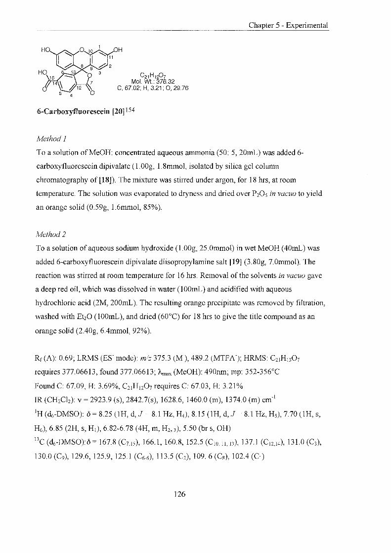
H
C21 H1P7 Mol. wt.: 376.32
C, 67.02; H, 3.21; 0, 29.76
6-Carboxyfluorescein [20] 154
Method 1
Chapter 5 - Experimental
To a solution of MeOH: concentrated aqueous aImnonia (50: 5, 20rnL) was added 6-
carboxyfluorescein dipivalate (1.00g, 1.8mmol, isolated by silica gel column
chromatography of [18]). The mixture was stirred under argon, for 18 hrs, at room
temperature. The solution was evaporated to dryness and dried over P20S in vacuo to yield
an orange solid (0.59g, 1.6rnrnol, 85%).
Method 2
To a solution of aqueous sodium hydroxide (1.00g, 25.0mmol) in wet MeOH (40mL) was
added 6-carboxyfluorescein dipivalate diisopropylaInine salt [19] (3.80g, 7.0mmol). The
reaction was stirred at room temperature for 16 hrs. Removal of the solvents in vacuo gave
a deep red oil, which was dissolved in water (100rnL) and acidified with aqueous
hydrochloric acid (2M, 200rnL). The resulting orange precipitate was removed by filtration,
washed with Et20 (100mL), and dried (60°C) for 18 hrs to give the title compound as an
orange solid (2.40g, 6.4mmol, 92%).
Rf(A): 0.69; LRMS (ES- mode): mlz 375.3 (M-), 489.2 (MTFA-); HRMS: C21H I307
requires 377.06613, found 377.06613; ~nax (MeOH): 490mn; mp: 352-356°C
Found C: 67.09, H: 3.69%, C21H 120 7 requires C: 67.03, H: 3.21%
IR (CH2Ch): v = 2923.9 (s), 2842.7(s), 1628.6, 1460.0 (m), 1374.0 (m) cm-I
IH (dt;-DMSO): () = 8.25 (1H, d, J = 8.1 Hz, H4), 8.15 (lH, d, J 8.1 Hz, H5), 7.70 (lH, s,
H6), 6.85 (2H, S, HI), 6.82-6.78 (4H, m, H2, 3),5.50 (br s, OH)
I3C (d6-DMSO):{) = 167.8 (C7,IS), 166.1, 160.8, 152.5 (CIO II, 13), 137.1 (C12,I4), 131.0 (C3),
130.0 (C9), 129.6, 125.9, 125.1 (C6-6), 113.5 eC2), 109. 6 eCs), 102.4 (CI)
126
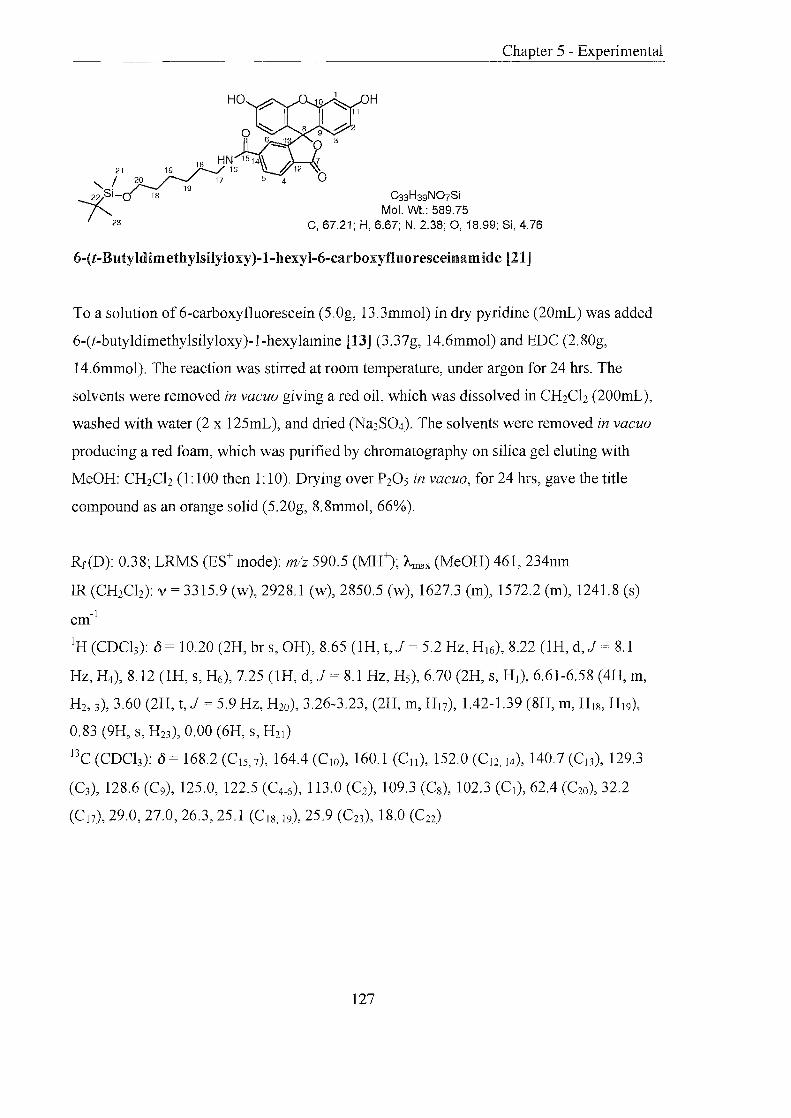
H
HN 15 14 \ 7 21~9 18 16 \\ #12
"I 20 17 5 4 0 22 Si- 18 19 C33H39N07Si o Mol. Wt.: 589.75
23 C, 67.21; H, 6.67; N, 2.38; 0,18.99; Si, 4.76
6-(t-Butyldimethylsilyloxy)-1-hexyl-6-carboxyfluoresceinamide [21]
To a solution of6-carboxyfluorescein (5.0g, 13.3mmol) in dry pyridine (20mL) was added
6-(t-butyldimethylsilyloxy)-1-hexylamine [13] (3.37g, 14.6mmol) and EDC (2.80g,
14.6mmol). The reaction was stirred at room temperature, under argon for 24 hrs. The
solvents were removed in vacuo giving a red oil, which was dissolved in CH2Ch (200mL),
washed with water (2 x 125mL), and dried (Na2S04). The solvents were removed in vacuo
producing a red foam, which was purified by chromatography on silica gel eluting with
MeOH: CH2Ch (1: 100 then 1'10). Drying over P205 in vacuo, for 24 hrs, gave the title
compound as an orange solid (5.20g, 8.8mmol, 66%).
Rf(D): 0.38; LRMS (ES+ mode): m/z 590.5 (MH+); Amax (MeOH) 461, 234nm
lR (CH2Ch): v = 3315.9 (w), 2928.1 (w), 2850.5 (w), 1627.3 (m), 1572.2 (m), 1241.8 (s)
-1 cm
IH (CDCb): () 10.20 (2H, br s, OH), 8.65 (lH, t, J = 5.2 Hz, H I6), 8.22 (lH, d, J 8.1
Hz, H4), 8.12 (lH, s, H6), 7.25 (lH, d, J 8.1 Hz, H5), 6.70 (2H, S, HI), 6.61-6.58 (4H, m,
H2, 3), 3.60 (2H, t, J = 5.9 Hz, H20), 3.26-3.23, (2H, m, H17), 1.42-1.39 (8H, m, H I8, H I9),
0.83 (9H, s, H23), 0.00 (6H, s, H21 )
l3C (CDCb): () = 168.2 (C I5, 7), 164.4 (ClO), 160.1 (Cll ), 152.0 (C12, 14), 140.7 (C13), 129.3
(C3), 128.6 (C9), 125.0, 122.5 (C4-6), 113.0 (C2), 109.3 (C8), 102.3 (Cl), 62.4 (C20), 32.2
(C l7), 29.0, 27.0, 26.3, 25.1 (C I8, 19), 25.9 (C23), 18.0 (C22)
127

~ ° '1 ° 6)00 ,\IBr
)lo 3 2~",j( 0y
°
C14H1gBr09 Mol. wt: 411.20
C, 40.89; H, 4.66; Br, 19.43; 0, 35.02
a-D-Tetraacetate galactosyl bromide [22] 198,199
To p-D-galactose pentaacetate (13.80g, 35.4mmol) was added a solution of30% (w: w)
hydrogen bromide in acetic acid (27.60g, 340.0mmol, 20.4mL). The reaction mixture was
stirred, under argon, at O°C for 2.5 hrs. CH2Cb (100mL) was added and the reaction poured
onto ice water (100mL), washed further with ice water (2 x100mL), dried (Na2S04), and
the solvents removed in vacuo. The resulting white foam was dissolved in a minimal
amount ofEt20 (lOmL) and petroleum ether (40-60) was added dropwise until a persistent
cloudiness was observed. Standing at O°C for 1 hr afforded a white solid which was
removed by filtration, washed with cold hexane and dried over P20s for 18 hrs (12. 86g,
31.3mmol, 88%).
Rf(L): 0.27; LRMS (EI+ mode): mlz 332.56 (M-Br+); Amax (MeOH): 202nm; mp: 83-85°C
Found C: 40.87, H: 4.53%, Cl~19Br09 requires C: 40.89, H: 4.66%
IR (CH2Cb): v = 2987.9 (w), 2966.9 (w), 2948.2 (w), 1739.5 (s), 1210.4 (s), 749.0 (m) cm-
IH (CDCh): (j = 6.70 (lH, d, J = 3.7 Hz, HI), 5.52 (lH, dd, J = 4.4, 1.4 Hz, H4), 5.41 (lH,
dd, J = 10.3,2.9 Hz, H3), 5.05 (lH, dd, J 10.3,3.7 Hz, H2), 4.49 (lH, t, J = 6.5 Hz, Hs),
4.19 (2H, dd, J = 11.0,5.9 Hz, H6), 4.11 (lH, dd, J 11.0, 7.3 Hz, H6) 2.01 (3H, s, CH3),
2.06 (3H, s, CH3), 2.12 (3H, s, CH3), 2.16 (3H, s, CH3)
BC (CDCb): (j = 170.4, 170.1, 169.9, 169.8 (CO), 88.1 (CHBr), 71.1 (CH), 68.0 (CH), 67.8
(CH), 66.9 (CH), 60.9 (CH), 20.8 (CH3), 20.7 (CH3), 20.6 (CH3), 20.5 (CH3)
128

Y21 o \,.,
(j 20 19 AO o
Chapter 5 - Experimental
C61 H75N025Si Mol. Wt.: 1250.33
C, 58.60; H, 6.05; N, 1.12; 0, 31.99; Si, 2.25
6-(t-ButyldimethylsHyloxy)-1-hexyl-6-carboxyfluoresceinamide-(3,6)-bis-f3-D-galactose
tetraacetate [23]
lv'fethod 1148
To a suspension of the fluorescein derivative [21] (1.00g, 1.7mmol), freshly prepared silver
oxide (0.39g, 1.7mmol), freshly distilled quinoline (O.5mL) in anhydrous benzene (15mL)
was added a-D-tetraacetyl galactosyl bromide [22] (2.79g, 6.8mmol). The reaction was
stirred in the dark, under argon, at room temperature for 72 hrs. The mixture was filtered
through celite, CH2Ch (IOOmL) added, and the organic layer washed with water (150mL).
Removal of the solvents in vacuo gave an orange foam, which was purified by silica gel
column chromatography eluting with cyclohexane: EtOAc (1:0 then 4: 1 then 2:1 then 1: 1
then 0: 1). The crude product was repurified by silica gel column chromatography eluting
with a gradient of hexane: EtOAc (1:0 then 2: 1 then 5:3 then 0: 1), yielding the desired
compound as a light yellow foam (0.40g, 0.3mmol, 19%).
Method 2. 157,158,200 , A mixture of the fluorescein derivative [21] (6.00g, 1O.lmmol) and silver silicate catalyst
(20.00g) was taken up in anhydrous CH2Ch (250mL) and then the solvent removed in
vacuo. To a suspension of the orange powder in CH2Ch (lOOmL) was added crushed
molecular sieves (20.00g) and the mixture stirred, in the dark, at room temperature for 1 hr.
The mixture was cooled to -20°C, and a solution of a-D-tetraacetyl galactosyl bromide [22]
(16.70g, 40.8mmol) in CH2Cb (50mL) was added dropwise. The reaction mixture was
129

Chapter 5 - Experimental
stirred in the dark, under argon, at room temperature for 24 hrs. A second portion of a-D
tetraacetyl-galactosyl bromide [22] (3.00g, 7.3mmol) and silver silicate catalyst (6.00g)
were added and the reaction stirred for a further 24 hrs. The mixture was diluted with
CH2Ch (200mL), filtered through celite; washed with CH2Ch (200mL) and the solvents
removed in vacuo affording a pale brown foam (21.00g). This was purified by silica gel
column chromatography eluting with EtOAc: Et20 (0: 1 then 1: 1). The resulting pale
yellow foam (1O.80g) was purified by silica gel column chromatography eluting with
MeOH: CH2Ch (0: 1 then 1: 9) to give an off white foam. This was recrystallised from a
minimum amount of CH2Ch and Et20 to give the title compound as white crystalline solid
(6.70g, 5.4mmol, 55%).
Rf(D): 0.88; LRMS (ES+ mode): mlz 1250.5 (MH+); Amax (MeOH): 264, 233nm; mp: 60-
65°C
IR (CH2Ch): v = 2943.1 (m), 1752.5 (s), 1577.1 (w), 1227.6 (s), 1029.7 (s), 738.5 (s) cm-I
IH (CDCh): () = 8.07 (lH, d, J 8.1 Hz, H4), 7.96 (IH, d, J 8.1Hz, Hs), 7.50 (lH, s, H6),
6.90 (2H, d, J = 4.4 Hz, HI), 6.72 (4H, S, H2, 3), 6.23 (lH, t, J = 5.9 Hz, H I6), 5.48-5.46
(4H, m, H I9, H23), 5.16-5.07 (4H, m, H24), 4.27-4.06 (6H, m, H20-22), 3.59 (2H, t,J 5.9
Hz, H I8), 3.48 (2H, q, J = 7.4 Hz, H l7), 2.20-2.00 (24H, m, CH3), 1.60-1.20 (8H, m, CH2),
0.88 (6H, s, CHSi), 0.05 (9H, s, (CH3)3Si)
l3C (CDCh): () = 170.5, 170.2, 169.5 (CH3CO) 168.6 (CIS, 7) 165.7 (C lO) 158.6 (C ll ), 152.3
(CI2,I4), 137.8 (C l3), 129.4 (C3), 128.8 (C9), 125.7, 123.4, 122.8 (C4-6), 113.5 (C2), 104.4
(C8), 99.2 (CI), 71.4, 70.8, 68.5, 66.9 (C I9-23) 62.2 (C I8), 61.4 (C24), 40.6 (Cl7), 32.9, 27.0,
26.1. 25.7 (CH2), 20.8 (CH3CO) 18.5 (CH3)3C, -3.5 (CH3Si)
130
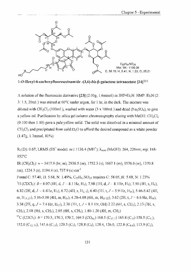
Y21 o Cf"20
A 0
19
o
Cs5H61 N025 Mol. Wt.: 1136.06
C, 58.15; H, 5.41; N, 1.23; 0, 35.21
1-0-Hexyl-6-carboxyfluoresceinamide -(3,6)-bis-(3-galactose tetraacetate [24]201
A solution of the fluorescein derivative [23] (2.00g, 1.6mmol) in 3HP .. EhN: NMP: Et3N (2:
3: 1.5, 20mL) was stirred at 60°C under argon, for 1 hr, in the dark. The mixture was
diluted with CH2Ch (lOOmL), washed with water (3 x 100mL) and dried (Na2S04), to give
a yellow oil. Purification by silica gel column chromatography eluting with MeOH: CH2Ch
(0: 100 then 1: 10) gave a pale yellow solid. The solid was dissolved in a minimal amount of
CH2Ch and precipitated from cold Et20 to afford the desired compound as a white powder
(1.47g, 1.3mmol, 81 %).
Rf (D): 0.67; LRMS (ES+ mode): mlz 1136.4 (MH+); Amax (MeOH): 264, 226nm; mp: 148-
152°C
IR (CH2Cb): v 3417.9 (br, m), 2936.5 (m), 1752.3 (s), 1667.1 (m), 1576.6 (w), 1370.8
em), 1224.3 (s), 1194.4 (s), 737.9 (s) cm-1
Pound C: 57.40, H: 5.68, N: 1.49%, CS5H61N025 requires C: 58.05, H: 5.68, N: 1.23%
IH (CDCb): 0 = 8.07 (lH, d, J 8.1 Hz, H4), 7.98 (lH, d, J = 8. 1Hz, Hs), 7.50 (lH, s, H6),
6.82 (2H, d, J 4.4Hz, HI), 6.72 (4H, S, H2, 3),6.40 (lH, t, J = 5.9 Hz, H I6), 5.46-5.42 (4H,
m, H19, 23), 5.16-5.08 (4H, m, H24), 4.28-4.08 (6H, m, H20-22), 3.62 (2H, t, J 6.6 Hz, HI8),
3.38 (2H, q, J = 7.4 Hz, H17), 2.30 (lH, t, J 8.1 Hz, OH) 2.22 (6H, s, CH3), 2.13 (3H, s,
CH3), 2.08 (9H, s, CH3), 2.05 (6H, s, CH3), 1.80-1.20 (8H, m, CH2)
l3C (CDCh): () = 170.5, 170.3, 170.2, 169.5 (COAc) 168.5 (CIS, 7) 165.8 (C lO) 158.5 (Cll ),
152.0 (C12, 14), 141.6 (C13), 129.5 (C3), 128.8 (C9), 128.4, 126.0, 122.8 (C4-6), 113.9 (C2),
131
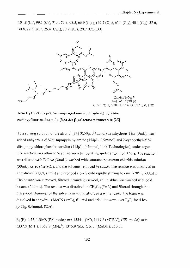
Chapter 5 - Experimental
104.8 (C8), 99.1 (CI), 71.4, 70.8, 68.5, 66.9 (C I9-23 ) 62.7 (CI8), 61.4 (C24), 40.4 (Cl7), 32.6,
30.8,29.5,26.7,25.4 (CH2), 20.9, 20.8, 20.7 (CH3CO)
o
1 ~9°Y ° 10 ~ 0yZ' 0 I 11 0
8 9 ~2 0 ~ 3
o
oA C64H78N3026P
Mol. Wt.: 1336.28 C, 57.52; H, 5.88; N, 3.14; 0, 31.13; P, 2.32
I-D-(Cyanoethoxy-N,N-diisopropylamino phosphine) hexyl-6-
carboxyfluo resceinam id e-(3,6)-bis-f3-galactose tetraacetate [25]
To a stirring solution of the alcohol [24] (0.50g, O.4mmol) in anhydrous THF (3mL), was
added anhydrous N,N-diisopropylethylamine (154JlL, 0.9mmo1) and 2-cyanoethyl-N,N
diisopropylchlorophosphoramidite (115JlL, 0.5mmol, Link Technologies), under argon.
The reaction was allowed to stir at room temperature, under argon, for 0.5hrs. The reaction
was diluted with EtOAc (30mL), washed with saturated potassimn chloride solution
(30mL), dried (Na2S04), and the solvents removed in vacuo. The residue was dissolved in
anhydrous CH2Clz (3mL) and dropped slowly onto rapidly stirring hexane (-20°C, 300mL).
The hexane was removed, filtered through glasswool, and residue was washed with cold
hexane (200mL). The residue was dissolved in CH2Clz (5mL) and filtered through the
glasswool. Removal of the solvents in vacuo afforded a white foam. The foam was
dissolved in anhydrous MeCN (4mL), filtered and dried in vacuo over P20s for 4 hrs
(0.52g, O.4mmol, 82%).
Rr(E): 0.77; LRMS (ES- mode): mlz 1334.1 (M-), 1449.2 (MTFA-); (ES+ mode): mlz
1337.0 (MH+), 1359.9 (MNa+), 1375.9 (MK+); Amax (MeOH): 250nm
132
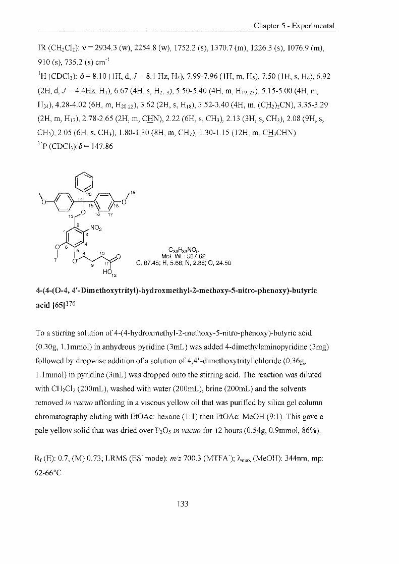
IR (CH2Ch): v = 2934.3 (w), 2254.8 (w), 1752.2 (s), 1370.7 (m), 1226.3 (s), 1076.9 (m),
910 (s), 735.2 (s) cm-1
IH (CDCh): {j = 8.10 (lH, d, J 8.1 Hz, H4), 7.99-7.96 (lH, m, H5), 7.50 (lH, s, H6), 6.92
(2H, d,J 4.4Hz, HI), 6.67 (4H, S, H2, 3), 5.50-5.40 (4H, m, HI9,23), 5.15-5.00 (4H, m,
H24), 4.28-4.02 (6H, m, H20-22), 3.62 (2H, S, HIS), 3.52-3.40 (4H, m, (CH)2CN), 3.35-3.29
(2H, m, H17), 2.78-2.65 (2H, m, CRN), 2.22 (6H, s, CH3), 2.13 (3H, s, CH3), 2.08 (9H, s,
CH3), 2.05 (6H, s, CH3), 1.80-1.30 (8H, m, CH2), 1.30-1.15 (l2H, m, CH3CRN)
3lp (CDCh):{j = 147.86
C33H33NOg Mol. Wt.: 587.62
C, 67.45; H, 5.66; N, 2.38; 0, 24.50
4-(4-(0-4, 4'-Dimethoxytrityl)-hydroxmethyl-2-methoxy-5-nitro-phenoxy)-butyric
acid [65] 176
To a stirring solution of 4-( 4-hydroxmethyl-2-methoxy-5-nitro-phenoxy )-butyric acid
(0.30g, 1.1mmol) in anhydrous pyridine (3rnL) was added 4-dimethylaminopyridine (3mg)
followed by dropwise addition of a solution of 4,4' -dimethoxytrityl chloride (0.36g,
1.1mmol) in pyridine (3rnL) was dropped onto the stirring acid. The reaction was diluted
with CH2Ch (200mL), washed with water (200rnL), brine (200mL) and the solvents
removed in vacuo affording in a viscous yellow oil that was purified by silica gel column
chromatography eluting with EtOAc: hexane (1: 1) then EtOAc: MeOH (9: 1). This gave a
pale yellow solid that was dried over P205 in vacuo for 12 hours (0.54g, 0.9mmol, 86%).
Rf(E): 0.7, (M) 0.73; LRMS (ES- mode): m/z 700.3 (MTFA-); Amax (MeOH): 344nm, mp:
62-66°C
133
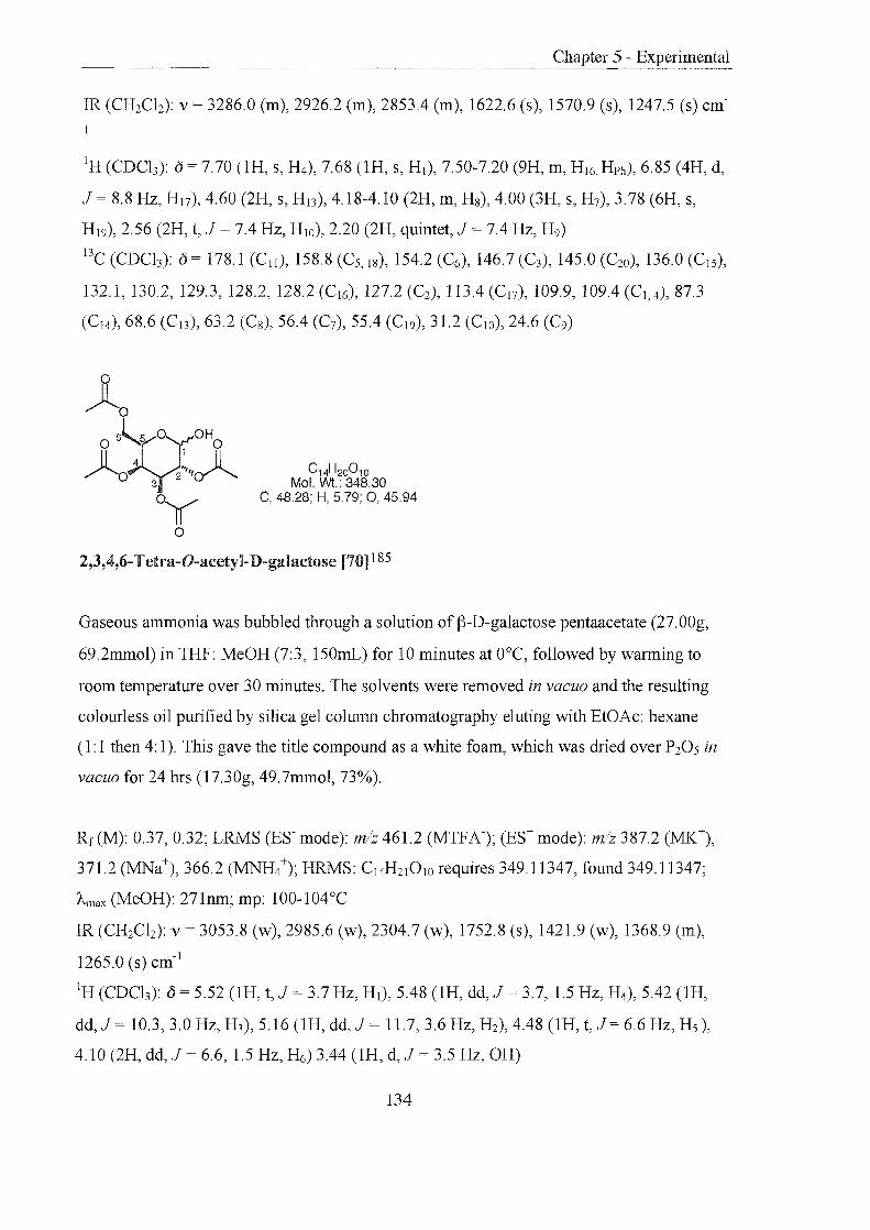
IR (CH2Ch): v = 3286.0 (m), 2926.2 (m), 2853.4 (m), 1622.6 (s), 1570.9 (s), 1247.5 (s) cm-
IH (CDCb): ()= 7.70 (lH, s, H4), 7.68 (lH, s, HI), 7.50-7.20 (9H, m, H16, Hph), 6.85 (4H, d,
J 8.8 Hz, H17), 4.60 (2H, s, H J3), 4.18-4.10 (2H, m, H8), 4.00 (3H, s, H7), 3.78 (6H, s,
H I9), 2.56 (2H, t, J = 7.4 Hz, H lO), 2.20 (2H, quintet, J = 7.4 Hz, H9)
l3C (CDCh): () 178.1 (C ll ), 158.8 (C5, 18), 154.2 (C6), 146.7 (C3), 145.0 (C20), l36.0 (CIS),
132.1, l30.2, 129.3, 128.2, 128.2 (C16), 127.2 (C2), 1l3.4 (C 17), 109.9, 109.4 (CI , 4), 87.3
(CI4), 68.6 (C l3), 63.2 (C8), 56.4 (C7), 55.4 (C I9), 31.2 (C lO), 24.6 (C9)
0y °
C14H2001O Mol. Wt.: 348.30
C, 48.28; H, 5.79; 0, 45.94
2,3,4,6· Tetra-O-acetyl-D-galactose [70] 185
Gaseous ammonia was bubbled through a solution of ~-D-galactose pentaacetate (27.00g,
69.2mmol) in THF: MeOH (7:3, 150mL) for 10 minutes at O°C, followed by warming to
room temperature over 30 minutes. The solvents were removed in vacuo and the resulting
colourless oil purified by silica gel column chromatography eluting with EtOAc: hexane
(l: 1 then 4: 1). This gave the title compound as a white foam, which was dried over P20S in
vacuo for 24 hrs (l7.30g, 49.7mmol, 73%).
Rf(M): 0.37,0.32; LRMS (ES- mode): mlz 461.2 (MTFA-); (ES+ mode): mlz 387.2 (MK+),
371.2 (MNa+), 366.2 (MNH/); HRMS: C14H21 0 lO requires 349.11347, found 349. 1 l347;
Amax (MeOH): 271nm; mp: 100-104°C
IR (CH2Ch): v = 3053.8 (w), 2985.6 (w), 2304.7 (w), 1752.8 (s), 1421.9 (w), l368.9 (m),
1265.0 (s) cm-I
IH (CDCh): () = 5.52 (lH, t, J = 3.7 Hz, HI), 5.48 (lH, dd, J = 3.7, 1.5 Hz, H4), 5.42 (lH,
dd, J 10.3,3.0 Hz, H3), 5.16 (lH, dd, J = 11.7,3.6 Hz, H2), 4.48 (lH, t, J = 6.6 Hz, H5 ),
4.10 (2H, dd, J = 6.6, 1.5 Hz, H6) 3.44 (lH, d, J = 3.5 Hz, OH)
134

l3C (CDCh): 0 = 170.8, 170.6, 170.5, 170.3 (CO), 96.8 (CHanorneric), 68.4, 68.3, 67.4, 66.4
(CH), 62.0 (CH2), 21.0, 21.0, 20.8 (CH3CO)
C16H20CI3NOlO Mol. Wt.: 492.69
C, 39.00; H, 4.09; CI, 21.59; N, 2.84; 0, 32.47
0-(2,3,4,6-Tetra-O-acetyl)-a-D-galactosyl trichloroacetimidate [71] 187
To a solution of2,3,4,6-tetra-O-acetyl-D-galactose [70] (16.40g, 47.09mol) in CH2Ch
(30mL) under argon, was added trichloroacetonitrile (l4.20mL, 0.14mol). Following
gradual addition of 1,8-diazabicyclo(5.4.0)undec-7-ene (0.3mL, 1.9mmol) the reaction was
allowed to stir for 30 minutes. A colour change from clear, colourless to yellow was
observed. The reaction was diluted with CH2Ch (50mL), washed with aqueous
hydrochloric acid (2M, 50mL), brine (2 x 50mL) and dried (Na2S04). The solvents were
removed in vacuo, to leave a white solid that was recrystallised from Et20 and hexane
affording a white crystalline solid that was dried over P20s in vacuo for 3 days (l5.90g,
32.3mmol, 70%).
Rf(N): 0.735; LRMS (ES- mode): m1z492.1 (MTFA-), (ES+ mode): mlz 331.2 (M
(NH2COccht); HRMS: C16H24ChN201O requires 509.04965, found 509.04965; Arnax
(MeOH): 271nm; mp: 52-55°C
Found C: 39.08, H: 3.99, N: 2.75%, C16H20ChNOlO requires C: 39.01, H: 4.09, N: 2.84%
IR (CH2Ch): v = 3341.9 (w), 3341.0 (w), 1750.0 (s), 1674.9 (m), 1617.0 (m) cm-I
IH (CDCh): 0 = 8.60 (lH, s, NH), 6.61 (lH, d, J 2.9 Hz, HI), 5.57 (lH, d, J =" 2.9 Hz,
H4), 5.43 (lH, dd, J = 11.0,3.0 Hz, H2 or 3),5.37 (lH, dd, J 11.0,3.0 Hz, H2 or 3),4.44
(lH, t, J = 7.0 Hz, Hs), 3.45 (lH, dd, J = 10.3, 7.0 Hz, H6) 3.43 (lH, dd, J = 11.0, 7.0 Hz,
H6), 2.45 (3H, s, CH3), 2.18 (3H, s, CH3), 2.03 (6H, S, 2(CH3» l3C (CDCh): 0= 170.5,170.3,170.2 (CO), 161.1 (C=NH), 93.6 (CHanorneric), 69.1, 67.7,
67.5,67.0 (CH), 61.4 (CH2), 20.8, 20.7 (CH3)
135

Chapter 5 - Experimental
XS·/O~OH / " C12H2802Si
Mol. Wt.: 232.44 C, 62.01; H, 12.14; 0,13.77; Si, 12.08
6-(t-ButyldimethylsiIyl)oxy-l-hexanol [72] 188
Sodium hydride (washed with anhydrous hexane, 2.03g, 84.6mmol) was suspended in
anhydrous THY Following addition of 1,6-hexandiol (1O.00g, 84.7mmol) the solution was
stirred under argon, at room temperature, for 45 minutes. t-Butyldimethylsilyl chloride
(l2.75g, 84.7mmol) was added and the mixture stirred for a further 1 hI. The reaction was
diluted with Et20 (50mL), washed with 10% potassium carbonate solution (200mL), brine
(200mL) and dried (Na2S04). Removal of the solvents in vacuo gave a viscous oil, which
was purified by silica gel column chromatography eluting with EtOAc: hexane (1: 10) to
yield a yellow oil which was dried over P20s in vacuo for 8 hrs (12.20g, 52.5mmol, 62%).
Rr (D): 0.44; LRMS (ES+ mode): mlz 202.2 (M-2CH3); HRMS: C12H290 2Si requires
233.19368, found 233.19368; Amax (MeOH): 270nm
IR (CH2Cb): v = 3386.4 (s), 2965.6 (s), 2926.6 (s), 1435.9 (m), 1373.5 (m), 1259.6 (s),
1102.1 (s), 876.8 (m) cm- l
IH (CDCh):£5 = 3.60-3.50 (4H, m, CH20), 1.55-1.45 (4H, m, CfuCH20), 1.40-1.25 (4H, m,
CH2), 0.85 (9H, s, (CH3)3Si), 0.05 (6H, s, (CH3)2Si)
l3C (CDCh): £5 = 62.3,62.9 (CH20), 32.9, 32.8 (CH2CH20), 26.1 (CH3)3C, 25.7, 25.7
(CH2), 18.5 (CH3)3C, -3.5 (CH3Si)
O-f}-D-[(2,3,4,6-Tetra-O-acetyI)-galactosyl-[6-(t-butyldimethylsilyl)oxy hexane]
[73] 189,190,202
136
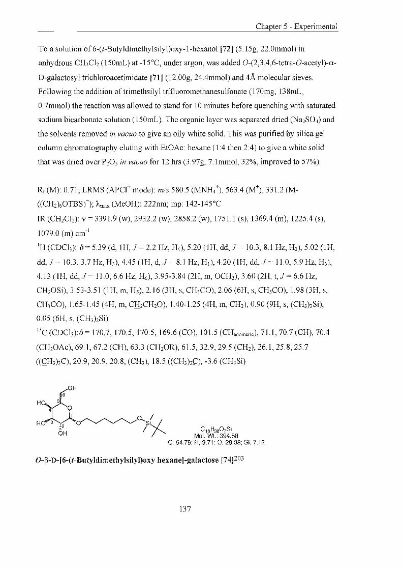
To a solution of6-(t-Butyldimethylsilyl)oxy-1-hexanol [72J (5.15g, 22.0mmol) in
anhydrous CH2Cb (150mL) at -15°C, under argon, was added O-(2,3,4,6-tetra-O-acetyl)-a
D-galactosyl trichloroacetimidate [71] (12.00g, 24.4mmol) and 4A molecular sieves.
Following the addition oftrimethsilyl trifluoromethanesulfonate (170mg, 138mL,
0.7mmol) the reaction was allowed to stand for 10 minutes before quenching with saturated
sodium bicarbonate solution (150mL). The organic layer was separated dried (Na2S04) and
the solvents removed in vacuo to give an oily white solid. This was purified by silica gel
column chromatography eluting with EtOAc: hexane (1:4 then 2:4) to give a white solid
that was dried over P20 S in vacuo for 12 hrs (3.97g, 7.1mmol, 32%, improved to 57%).
Rf(M): 0.71; LRMS (APCt mode): mlz 580.5 (MNH/), 563.4 (M+), 331.2 (M
((CH2)60TBSt); Amax (MeOH): 222nm; mp: 142-145°C
IR (CH2Cb): v = 339l.9 (w), 2932.2 (w), 2858.2 (w), 1751.1 (s), 1369.4 (m), 1225.4 (s),
1079.0 (m) cm- l
IH (CDCh): 0 5.39 (d, 1H, J = 2.2 Hz, H4), 5.20 (lH, dd, J 10.3,8.1 Hz, H2), 5.02 (lH,
dd, J 10.3,3.7 Hz, H3), 4.45 (lH, d, J = 8.1 Hz, HI), 4.20 (lH, dd, J 11.0,5.9 Hz, H6),
4.13 (lH, dd, J 11.0,6.6 Hz, ~), 3.95-3.84 (2H, m, OCH2), 3.60 (2H, t, J 6.6 Hz,
CH20Si), 3.53-3.51 (lH, m, H5), 2.16 (3H, s, CH3CO), 2.06 (6H, s, CH3CO), 1.98 (3H, s,
CH3CO), 1.65-1.45 (4H, m, CH2CH20), 1.40-1.25 (4H, m, CH2), 0.90 (9H, s, (CH3)3Si),
0.05 (6H, s, (CH3)2Si)
BC (CDCh):o = 170.7, 170.5, 170.5, 169.6 (CO), 101.5 (CHanomeric), 71.1, 70.7 (CH), 70.4
(CH20Ac), 69.1, 67.2 (CH), 63.3 (CH20R), 61.5, 32.9, 29.5 (CH2), 26.1, 25.8, 25.7
((CH3)3C), 20.9, 20.9, 20.8, (CH3), 18.5 ((CH3)3C), -3.6 (CH3Si)
O-~-D-[ 6-(t-Butyldimethylsilyl)oxy hexane]-galactose [74]203
137

A solution the (3-D-galactose derivative [73] (3.20g, 5.7mmol) in anhydrous MeOH (50mL)
was cooled to O°C and potassium carbonate (0.24g, 1.7mmol) added. The reaction was left
standing for 2 rus, the solvents removed in vacuo and the resulting foam dissolved in
CH2Ch (50mL). The organic layer was washed with water (50mL), brine (3 x 50mL), dried
(Na2S04) and the solvents removed in vacuo. This gave the desired compound as a pale
yellow foam, which was dried over P205 in vacuo for 12 hrs (2.15g, 5.5mmol, 96%).
Rf(O): 0.60; LRMS (ES- mode): mlz 507.0 (MTFA-); Amax (MeOH): 219nm
IR (CH2Ch): v = 3054.3 (m), 2987.2 (w), 2305.4 (w), 1422.0 (m), 1265.0 (s) cm- I
IH (CDCh): 0 = 4.20 (lH, d, J 7.4 Hz, HI), 3.95-3.85 (lH, m, H4), 3.84 (lH, d, J = 2.9
Hz, H3), 3.75-3.70 (2H, m, H2, 5), 3.63 (2H, t, J 6.6 Hz, OCH2), 3.55-3.45 (4H, m, H6,
CH20Si), 1.70-1.60 (2H, m, CH2CH20), 1.55-1.45 (2H, m, ClliCH20), 1.40-1.30 (4H, m,
CH2), 0.90 (9H, s, (CH3)3Si), 0.05 (6H, s, (CH3)2Si)
BC (CDCh): 104.9 (CHanomeric), 76.4, 74.9, 72.5, (CH), 70.7 (CH20Ac), 70.4, 70.2, 64.2
(CH2), 62.3, 33.8, 30.8 (CH2), 26.8, 26.8, 26.4 ((CH3)3C), 19.1 ((CH3)3C), -3.5 (CH3Si)
15
I ~ 14
h 13
\ 'I ~ 12
o 11 8 7 / o
6 Mol. Wt.: 696.94 )5° 90
H C, 67.21; H, 8.10; 0, 20.66; Si, 4.03
4 0
_~ ~ ~ ,,0 I HO 3 : 2 v ....., "V' ........ 'SiJ
OH I /"
0-(3-D-( 6-0-4,4! -Dim eth yloxytrityl)-galactosyl-[6-(t-butyldim ethyIsilyl)oxy hexane]
[75]
A solution of 4,4'-dimethyloxytrityl chloride (1.22g, 3.6mmol) in pyridine (4mL) was
added dropwise to a solution of the (3-D-galactose derivative [74] (l.41g, 3.6mmol) and 4-
dimethylaminopyridine (43mg, 360.0!-lmol) in anhydrous pyridine (8mL), under argon.
After stirring at room temperature for 15mins CH2Ch (300mL) was added, the organic
layer washed with sodium bicarbonate solution (200mL), brine (200mL) and dried
138

Chapter 5 - Experimental
(NaZS04). Removal of the solvents in vacuo gave an orange oil, which was purified by
silica gel column chromatography eluting with Et3N: CHzCh (3: 100). This gave a yellow
foam which was dried over PZ0 5 in vacuo for 18hrs (1.13g, 1.6mmol, 45%).
Rf (G): 0.46; LRMS (ES+ mode): mlz 720.0 (MNa+), 715.0 (MNH/) 303.4 (DMT+);
HRMS: C39H5609SiNa requires 719.35913, found 719.35913; Amax (MeOH): 263, 241nm;
mp: 62-65°C
Found C: 66.95, H: 8.08%, C39H5609Si requires C: 67.21, H: 8.10%
IR (CH2Ch): v = 3054.3 (w), 2360.2 (w), 1653.5 (w), 1265.0 (s), 738.9 (s) cm-!
!H (CDCb): () = 7.50-7.20 (9H, m, H9, 13-15),6.83 (4H, d, J = 8.8 Hz, H IO), 4.23 (lH, d, J =
6.6 Hz, HI), 4.05 (lH, d, J =2.2 Hz, H4), 3.95-3.85 (lH, m, H5), 3.80 (6H, s, CH30), 3.65-
3.50 (6H, m, Hz, 3, OCHz, CH20Si), 3.43 (lH, dd, J =9.6,5.9 Hz, H6), 3.38 (lH, dd, J =
9.6,6.6 Hz, H6), 1.70-1.60 (2H, m, CfuCH20), 1.57-1.48 (2H, m, CfuCHzO), 1.40-1.32
(4H, m, CH2), 0.90 (9H, s, (CH3)3Si), 0.05 (6H, s, (CH3)zSi)
13C (CDCh):{) = 158.7 (Cll ), 144.9 (C l2), 136.1, 136.0 (Cs), 130.2, 128.2, 128.0, 127.0 (C9,
13-15), 113.3 (C IO), 103.1 eCI), 86.6 (C7), 73.7, 72.4 eC2-5), 70.1 eC6), 69.3 (Cz-s), 63.3, 62.5
(CHzO), 55.4 (CH30), 32.9, 29.8, 26.0, 25.8 eCH2), 26.2 ((CH3)3CSi), 18.6 ((CH3)3C), -3.4
(CH3Si)
'1'\/ 11 _ 8 7 ~ /; a C45H62012Si
)50 9 a Mol. Wt.:823.05 y, ' ° C, 65,67; H, 759; 0,23.33; Si, 3.41
a 1~o.../
AY lY-a
O-f3-D-[(2,3,4-Tri-O-acetyl)-6-0-( 4,4' -dim ethyloxytrityl) ]-galactosyl- [6-(t-
butyldimethylsilyl)oxy] hexane [76]204
139

To a solution of the ~-D-galactose derivative [75] (0.95g, 1.4mmol) in anhydrous CH2Ch
(20mL) was added Et3N (0.57mL, 4.1mmol), 4-dimethylaminopyridine (l6.60mg,
140.0mol) and acetic anhydride (0.39mL, 4.1mmol). The reaction was stirred, at room
temperature, under argon for 1.5 hrs. The reaction was diluted with CH2Ch (IOOmL),
washed with saturated sodium bicarbonate solution (2 x 100mL), water (100mL), brine (2 x
100mL) and dried (Na2S04). Removal of the solvents in vacuo gave a yellow foam, which
was purified by silica gel column chromatography eluting with EtOAc: hexane. (3: 1). The
pale yellow foam obtained was dried over P20S in vacuo for 12 hrs (0.99g, 1.2mmol, 89%).
Rf(M): 0.77; LRMS (ES+ mode): mlz 845.8 (MNa+), 840.8 (MNH/) 303.4 (DMT+);
HRMS: C4sH63012Si requires 823.40888, found 823.40888; "'-max (MeOH): 273, 234nm;
mp: 60-64°C
Found C: 65.69, H: 7.68%, C45H62012Si requires C: 65.67, H: 7.59%
IR (CH2Ch):v = 3054.2 (m), 2986.4 (w), 2359.9 (w), 1750.2 (m), 1608.1 (w), 1264.9 (s),
746.2 (s) cm- l
IH (CDCh): 7.40-7.19 (9H, m, H9, 13-15),6.85-6.78 (4H, m,J = 8.8 Hz, H IO), 5.60 (lH, d,J
= 3.7 Hz, H4), 5.10 (lH, dd, J = 10.3, 7.4Hz, H2or3), 5.12 (lH, dd, J = 10.3,3.7 Hz, H20r
3),4.43 (lH, d, J = 7.4 Hz, HI), 3.90-3.81 (2H, m, OCH2), 3.80 (6H, s, CH30), 3.58 (2H, t,
J = 6.6 Hz, CH20Si), 3.50-3.35 (2H, m, H6), 3.10 (lH, t, J = 8.8 Hz, H5), 2.05 (3H, s,
CH3CO), 2.00 (3H, s, CH3CO), 1.90 (3H, s, CH3CO), 1.60-1.43 (4H, m, CfuCH20), 1.38-
1.25 (4H, m, CH2), 0.90 (9H, s, (CH3)3Si), 0.05 (6H, s, (CH3)2Si)
l3C (CDCb): 0 = 170.5, 170.2, 169.6 (CO), 158.7 (Cll ), 144.3 (C12), 135.8, 135.5 (Cs),
130.2, 130.1, 128.3, 128.0, 127.0 (C9, 13-15), 113.3 (CIO), 101.4 (Cl ), 86.4 (C7), 72.2, 71.4
(C2-5), 70.2 (C6), 69.4 (C2-5), 67.5, 63.3, 60.6 (CH2), 55.4 (CH30), 32.9, 29.5, 25.8, 25.7
(CH2), 26.1 ((CH3)3CSi), 20.9, 20.9, 20.8 (CH3CO) 18.5 ((CH3)3C), -3.2 (CH3Si)
140

r; ~ cI 11 _ 8 7 ~ /; C39H48012
y 4 ' C, 66.09; H, 6.83; 0, 27.09 )50 9 Mol. Wt.:708.79
o 1~OH 3;2 0
o~y o
O-~-D-[(2,3,4-Tri-O-acetyl)-6-0-(4,4'-dimethyloxytrityl)]-galactosyl hexan-6-o1 [77]201
The silyl ether [76] (0.92g, l.lmmol) was suspended in 3HF-Et3N: NMP: Et3N (2: 3: 1.5,
3.5mL) and left standing at 65°C for 20minutes. The reaction was diluted with Et20
(70mL), washed with water (50mL), saturated sodium bicarbonate solution (3 x 50mL),
water (50mL), brine (70mL) and dried (Na2S04). The solvents were removed in vacuo
giving a white foam which was purified by silica gel column chromatography eluting with
EtOAc: hexane (3:2 then 3: 1 then 1:0). The resulting white foam was dried over P205 in
vacuo for 14 hrs (0.77g, 1.1mmol, 97%).
Rf(N): 0.40; LRMS (APCI+ mode): mlz 731.7 (MNa+), 708.8 (~) 303.4 (DMT+), 138.9
((CH3)3C(CH2)2SiF+); HRMS: C39H4S012 requires 708.31458, found 708.31458; "'-max
(MeOH): 272, 234nm; mp: 63-66°C
Found C: 66.38, H: 6.65%, C39H4S012 requires C: 66.09, H: 6.83%
IR (CH2Ch): v = 3054.2 (w), 2986.4 (w), 1750.2 (s), 1607.9 (w), 1265.5 (s), 739.8 (s) cm-1
IH (CDCh): () = 7.40-7.20 (9H, m, H9, 13-15),6.85-6.78 (4H, m, H IO), 5.58 (lH, d, J 2.9
Hz, H4), 5.10 (lH, dd, J 10.3,8.1 Hz, H2), 5.12 (lH, dd, J 10.3,2.9 Hz, H3), 4.43 (lH,
d, J 7.4 Hz, HI), 3.90-3.81 (2H, m, OCH2), 3.80 (6H, S, CH30), 3.63 (2H, t, J 6.6 Hz,
CH20H), 3.50-3.35 (2H, m, H6), 3.10 (lH, t, J 8.8 Hz, H5), 2.05 (3H, s, CH3CO), 2.00
(3H, S, CH3CO), 1.90 (3H,s, CH3CO), 1.60-1.50 (4H, m, ClliCH20), 1.40-1.35 (4H, m,
CH2)
13C (CDCh): () = 170.5, 170.2, 169.7 (CO), 158.7 (Cll ), 144.3 (C12), 135.8, 135.6 (Cs),
130.2, 130.1, 128.3, 128.0, 127.0 (C9, 13-15), 113.3 (C IO), 101.4 (Cl ), 86.4 (C7), 72.3, 71.4,
141

Chapter 5 - Experimental
(C2-S), 70.2 (C6), 69A (C2-S), 67.5, 62.9, 60.7 (CH2), 55A (CH30), 32.8, 29.5, 25.8, 25.6
(CH2), 21.0, 20.9, 20.8 (CH3CO)
o
C48H65N2013P Mol. Wt.: 909.01
C, 63.42; H, 7.21; N, 3.08; 0, 22.88; P, 3.41
0-13-D-[ (2,3,4-Tri-0-acetyl)-6-0-( 4,4' -dim ethyloxytrityl) ]-galactosyl-[6-(2-cyanoethyl
diisopropylpbosphoramidyl)oxy hexane] [78]
To the alcohol [77] (300mg, OAmmol) in anhydrous CH2Clz (5mL) was added N,N
diisopropylethylamine (295/-!L, 1.7mmol) and 2-cyanoethyl-diisopropyl
chlorophosphoramidite (l14/-!L, 510.0/-!mol) and the reaction stirred under argon. After
stirring at room temperature, for 20 minutes the clear solution was diluted with EtzO
(25mL), washed with saturated potassium chloride solution (25mL), dried (Na2S04) under
argon, and then the solvents removed in vacuo. The oil obtained was purified by silica gel
column chromatography, under argon, eluting with Et20 to give the title compound as
white foam, which was dried over P20 S for 24 hrs (370mg, OAmmol, 96%).
Rf (N): 0.81; LRMS (ES+ mode): mlz 909.7 (M+), 931.6 (MNa+)
IH (CDCh): () = 7AO-7.15 (9H, m, H9, 13-1S), 6.90-6.80 (4H, m, J = 8.8 Hz, H lO), 5.60 (lH,
d, J 2.2 Hz, ~), 5.20-5.05 (2H, m, H2, 3), 4A3 (lH, d, J 7A Hz, HI), 3.90-3.81 (2H, m,
OCH2), 3.80 (6H, s, CH30), 3.62-3.58 (4H, m, CHN, CfuCH2CN), 3.50-3.35 (4H, m, H6
CH20P), 3.10 (lH, t, J 8.8 Hz, Hs), 2.65 (2H, t, J = 6.6 Hz, CH2CfuCN), 2.05 (3H, s,
CH3CO), 2.00 (3H, s, CH3CO), 1.90 (3H, s, CH3CO), 1.40-1.21 (8H, m, CfuCH20, CH2),
1.20 (l2H, dd, J 6.6,5.9 Hz, CH3CHN)
31p (CDCh):{) = 147.90
142

C30H34N204 Mol. Wt.: 486.60
C, 74.05; H, 7.04; N, 5.76; 0,13.15
Fmoc-phenylalanine-6-hydroxyhexylamide [81]
Chapter 5 - Experimental
To a solution ofFmoc-phenylalanine (9.68g, 25.0mmol) and 6-amino-1-hexanol (2.93g,
25.0mmol) in anhydrous THF: DMF (5:1, 50mL) was addedEDC (5.00g, 26.0mmol) and
the mixture stirred, under argon, at room temperature for 24 hrs. The reaction was diluted
with EtOAc (200mL), washed with sodium bicarbonate (200mL), water (200mL), brine
(200mL) and then dried (Na2S04). Removal of the solvents in vacuo gave a white, waxy
solid that was purified by silica gel column chromatography eluting with MeOH: CH2Ch
(0: 1 then 1: 10). The white powder obtained was dried over P20s in vacuo for 24 hrs (7. 50g,
15.4mmol, 62%).
Rr(D): 0.58; LRMS (ES+ mode): m/z 509.5 (MNa+), 487.6 (MH+); HRMS: C30H3SN204
requires 487.25968, found 487.25968; Arnax (MeOH): 305,295, 265nm; mp: 142-146°C
IR (Nujol mull): v = 3295.9 (s), 2725.8 (w), 1689.3 (s), 1646.6 (m), 1460.6 (m), 1136.6
(w), 739.0 (m) cm- l
IH (CDCh):b = 7.95 (lH, t, J = 5.2 Hz, H6), 7.87 (2H, d, J = 8.1 Hz, H12), 7.67-7.60 (2H,
m, H13), 7.45-7.18 (9H, m, H14, 16-19),4.36 (lH, t, J 5.2 Hz, H lO), 4.24-4.10 (4H, m, H2,
4),3.40-3.35 (2H, m, H9), 3.15-2.90 (3H, m, H7, s), 2.18 (lH, dd, J 10.3,9.5 Hz, HI),
1.43-1.33 (4H, m, Hs), 1.29-1.17 (4H, m, Hs)
13C (CDCh):b = 171.2 (C22), 155.8 (C21 ), 143.8 (Cll ), 140.7 (C20), 138.3 (CIS), 129.3,
129.0,128.1,127.7,127.4,127.1,126.3,125.5,125.4,121.5, 120.2 (C 12-14, 16-19),65.7 (C4),
60.8 (C9), 56.3 (Cs), 38.6 (C7), 37.9 (C2), 32.6, 29.3, 26.8, 26.1 (Cs)
143

Chapter 5 - Experimental
-0
8 8
C51H52N206 /0 Mol. Wt.: 788.97
C, 77.64; H, 6.64; N, 3.55; 0, 12.17
Fmoc-pbenylalanine-6-hydroxy(-O-4,4'dimethoxytrityl-)bexylamide [82]
To a solution of the alcohol [81] (4.00g, 8.2mmol) in anhydrous pyridine (20mL) was
added a solution of 4,4'-dimethoxytrityl chloride (3.07g, 9.1mmol) in pyridine (20mL)
dropwise over a period of 10 minutes. The mixture was stirred under argon at room
temperature for 18 hrs. The solvents were removed in vacuo and the residue dissolved in
CH2Clz (200mL), washed with saturated sodium bicarbonate solution (200mL), water
(200mL), brine (200mL) and then dried (Na2S04)' Removal of the solvents in vacuo gave a
pale yellow solid, which was recrystallised from CH2Clz and petroleum ether. The resulting
white crystals were dried over P205 for 24 hrs (5.95g, 7.5mmol, 92%).
Rr(D): 0.86; LRMS (ES+ mode): mlz 812.2 (MNa+), 487.8 (M-DMT+), 303.4 (DMT+); ~nax
(MeOH): 305, 290, 265nm; mp: 94-98°C
IR (Nujol mull): v 3297.5 (m), 1689.3 (m), 1646.6 (m), 1607.0 (w), 1460.7 (m), 1175.2
(w), 737.2 (m), 706.7 (m) cm-1
IH (CDCh): 0 = 7.78 (2H, d, J =7.4 Hz, H 12), 7.56 (2H, m, H 13), 7.48-7.20 (I8H, m, H14, 16-
19,25,29-31),6.85 (4H, d, J = 8.8 Hz, H26), 5.65-5.40 (2H, m, H3, 6) 4.45-4.30 (3H, m, H4, 5),
4.20 (IH, t, J 6.6 Hz, HI), 3.80 (6H, s, OCH3), 3.20-3.05 (4H, m, H2, 7),3.00 (2H, t, J
6.6 Hz, H9), 1.55 (2H, q, J = 6.6 Hz, Hg), 1.40-1.25 (4H, m, Hg), 1.20-1.05 (2H, m, Hg)
13C (CDCb): 0= 170.6 (C22), 158.5 (C21 ,27), 145.6 (C1l ), 143.9 (C20,2g), 141.5 (C I5), 136.8
(C24), 130.2, 129.5, 128.9, 128.3, 127.9, 127.9, 127.3, 126.8, 125.2, 120.2 (C12-14, 16-19,25,29-
31), 113.1 (C26), 85.8 eC23), 67.2 eC4), 63.4 eC9), 56.7 (OCH3), 55.4 eCI), 47.3 eC5), 39.7
(C7), 39.1 eC2), 30.1, 29.4,26.3,25.4 (Cg)
144

° C36H42N204
Mol. Wt.: 566.73 /0 C, 76.29; H, 7.47; N, 4.94; 0, 11.29
Phenylalanine-6-hydroxy(-O-4,4'dimethoxytrityl-)hexylamide [83]
To a solution of piperidine in DMF (20%, 40mL) was added to the peptide [82] (5.00g,
6.3mmol) and the mixture stirred, under argon, for 2 hrs. This gave a cloudy yellow
suspension. The solvents were removed in vacuo to give a viscous oil, was diluted with
CHzCh (200mL), washed with saturated sodium bicarbonate solution (200mL), water
(200mL), brine (200mL) and then dried (NaZS04). Removal of the solvents in vacuo
afforded a yellow oil which was purified by silica gel column chromatography eluting with
MeOH: CHzCh (0: 1 then 1 :20) to give a yellow oil that was dried over PzOs for 24 hrs
(3.44g, 6.1mmol, 96%).
Rf(D): 0.54; LRMS (ES+ mode): mlz 589.0 (MNa+), 303.4 (DMT+); Amax (MeOH): 270,
240mn
IR (Nujol mull):v = 3361.1 (m), 3059.9 (m), 3028.7 (m), 1652.2 (s), 1607.0 (m), 1459.5
(m), 1176.8 (m), 702.3 (m) cm- l
IH (CDCh): 0 = 7.46 (2H, d, J =7.4 Hz, H 1Z), 7.37-7.20 (l2H, m, H 13,l4, Z5, Z9-31), 6.85 (4H,
d, J = 8.8 Hz, H26), 3.80 (6H, s, OCH3), 3.60 (lH, dd, J= 4.4,3.7 Hz, H6), 3.32-3.20 (3H,
m, H3,7), 3.05 (2H, t, J 6.6 Hz, H9), 2.70 (lH, dd, J 9.5,3.5 Hz, HI), 1.55 (2H, q, J
7.4 Hz, H2), 1.54-1.25 (8H, m, H8)
l3C (CDCh): 0= 174.2 (Czz), 158.5 (CZl ,27), 145.6 (Cll ), 138.2 (CZ8), 136.9 (CZ4), 130.2,
129.5, 128.9, 128.3, 127.9, 126.9, 126.7 (C12-14, CZ5,Z9-31), 113.1 (CZ6), 85.8 (CZ3), 63.5 (C9),
56.6 (OCH3), 55.4 (Cl ), 41.3 eC7), 39.2 eCz), 30.2, 29.7 27.0, 26.2 (Cg)
145

HO 35
34
C48H64N206 Mol. VVt.: 765.03
o
C, 75.36; H, 8.43; N, 3.66; 0,12.55
Chapter 5 - Experimental
12-Hydroxy dodecanam ido-phen ylalsnine-6-byd roxy( -0-4,4' dim ethoxytrityl-) hexylamide [84]
To a solution of the amine [83] (3.32g, 5.9mmol) and 12-hydroxydodecanoic acid (l.27g,
5.9mmol) in CH2Clz (30mL) was added EDC (l.13g, 5.9mmol). The mixture was stirred,
under argon, for 2 hrs. The reaction was diluted with CH2Clz (200mL), washed with
saturated sodium bicarbonate solution (200mL), water (200mL), brine (200mL) and then
dried (Na2S04). Removal ofthe solvents in vacuo afforded a pale yellow solid, which was
recrystallised from CH2Clz and petroleum ether. The title compound was obtained as a
white solid, which was dried, in vacuo, over P205 for 24 hrs (3.42g, 4.5mmol, 76%).
Rr(D): 0.72; LRMS (ES+ mode): mlz 303.2 (DMT+), 787.5 (MNa+), 1551.9 (2MNa+); Amax
(MeOH): 275, 240nm; mp: 70-74°C
IR (CH2Clz): v = 3280.5 (m), 2361.6 (w), 1633.9 (m), 1461.1 (s) cm-1
IH (CDCh): () = 7.44 (2H, d, J c=7.4 Hz, H 12), 7.36-7.20 (l2H, m, H13,14, 25, 29-31),6.83 (4H,
d, J = 8.8 Hz, H26), 6.46 (lH, d, J 8.1 Hz, H3), 6.00 (lH, t, J = 5.9 Hz, H6), 4.68-4.62
(lH, m, HI), 3.80 (6H, s, OCH3), 3.65 (2H, t, J = 6.6 Hz, H34), 3.00-2.70 (6H, m, H2, 7, 9),
2.17 (2H, t,J 5.4 Hz, H33), 1.60 (lH, s, H35), 1.45-1.30 (8H, m, CH2), 1.20-1.00 (l8H, m,
CH2)
BC (CDCb): () = 173.3 (C22), 170.9 (C32), 158.4 (C27), 145.5 (Cll ), 137.0 (C2S), 136.8 (C24),
130.2,129.4,128.7,128.3,127.9,127.1,126.7 (C12-14, 25,29-31),113.1 (C26), 85.8 (C23), 63.4
(C9), 63.1 (C34), 55.4 (OCH3), 54.8 (C I), 39.6 (C7), 38.9 (C2), 36.7, 32.9, 30.1, 29.6, 29.5,
29.5,29.4,29.2,26.9,26.1,25.9,25.7 (C33, CH2)
146
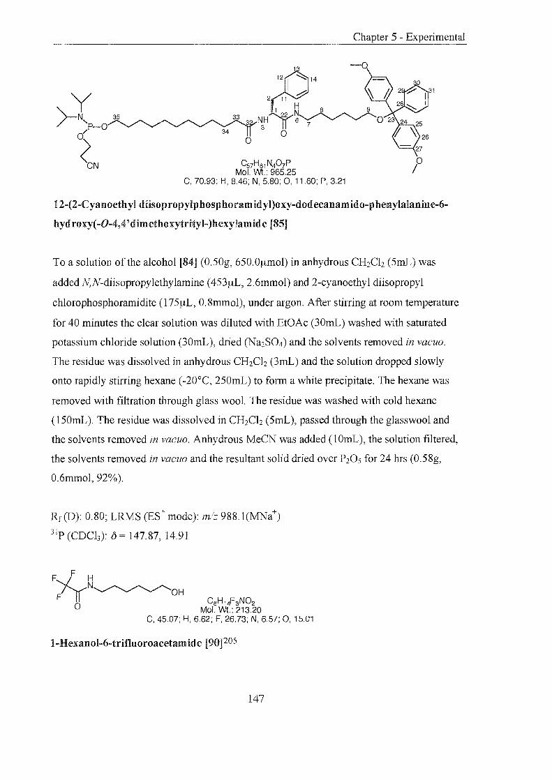
33
° ° C5yHa1N40yP
Mol. Wt.: 965.25 C, 70.93; H, 8.46; N, 5.80; 0, 11.60; P, 3.21
Chapter 5 - Experimental
12-(2-Cyanoethyl diisopropylphosphoramidyl)oxy-dodecanamido-phenylalanine-6-
hydroxy( -0-4,4' dimethoxytrityl-)bexyJamide [85]
To a solution of the alcohol [84] (0.50g, 650.0~mol) in anhydrous CH2Ch (5mL) was
added N,N-diisopropylethylamine (453~L, 2.6mmol) and 2-cyanoethyl diisopropyl
chlorophosphoramidite (175~L, 0.8mmol), under argon. After stirring at room temperature
for 40 minutes the clear solution was diluted with EtOAc (30mL) washed with saturated
potassium chloride solution (30mL), dried (Na2S04) and the solvents removed in vacuo.
The residue was dissolved in anhydrous CH2Ch (3mL) and the solution dropped slowly
onto rapidly stirring hexane (-20°C, 250mL) to form a white precipitate. The hexane was
removed with filtration through glass wool. The residue was washed with cold hexane
(150mL). The residue was dissolved in CH2Ch (5mL), passed through the glasswool and
the solvents removed in vacuo. Anhydrous MeCN was added (1 OmL), the solution filtered,
the solvents removed in vacuo and the resultant solid dried over P20s for 24 hrs (0.58g,
0.6mmol, 92%).
Rr (D): 0.80; LRMS (ES+ mode): mlz 988. 1 (MNa+)
31p (CDCb): 0 147.87, 14.91
F F H
~N~OH o CaH14F3N02
Mol. Wt.: 213.20 C, 45.07; H, 6.62; F, 26.73; N, 6.57; 0, 15.01
I-Hexanol-6-trifluoroacetamide [90]205
147

To a solution of6-amino-I-hexanol (l3.30g, O.lImol) in CH2Ch (lOOmL) was added ethyl
trifluoroacetate (l4.16mL, I6.9Ig, O.lmmol) and the mixture stirred at room temperature,
under argon, for 12 hrs. Et3N (2mL) was added to raise the pH to 8.0, followed by a second
amount of ethyl trifluoroacetate (3mL, 3.58g, 25.2mmol). After stirring for 4 hrs, the
solvents were removed in vacuo. The yellow oil obtained was dissolved in CH2Ch
(200mL), washed with 10% citric acid solution (2 x 200mL), dried (Na2S04), and the
solvents removed in vacuo. The clear yellow oil was recrystallised from a minimal amount
of toluene: chloroform (4:1). This afforded a white crystalline solid that was dried over
P20S in vacuo for 12 hrs (l6.95g, 79.5mmol, 71 %).
Rf(J): 0.61; LRMS (ES- mode): m/z 326.2(MTFA-); HRMS: CgHlgF3N202 requires
231.13204, found 231.13204; Amax (MeOH): 268nm; mp: 41-43°C
IR: v = 3386.9 (m), 3305.7 (m), 2931.8 (m), 2858.1 (m), 1696.8 (s), 1549.6 (s), 1176.2 (s),
1151.9 (s) cm-1
lH (CDCb):o 6.80 (lH, s, NH), 3.63 (2H, t, J 7.6 Hz, CH20), 3.35 (2H, q, J = 7.5 Hz,
CH2NH), 1.95 (lH, s, OH), 1.65-1.52 (4H, m, CfuCH20H, CH2CH2NH), 1.43-1.35 (4H,
m, CH2)
l3C (CDCb):o = 157.8 (CF3), 114.1 (CO), 62.7 (CH20H), 39.9 (CH2NH), 32.5
(CH2CH2NH), 26.4 (CH2CH20H), 25.3, 26.2 (CH2)
C2g1-iS2FSN04 Mol. WI.: 515.56
C, 67.56; H, 6.26; F, 11.05; N, 2.72; 0, 12.41
0' -(4,4' -Dimethoxytrityloxy) hexyl-6-trifluoroacetamide [91]
A solution ofrecrystallised 4,4' -dimethoxytrityl chloride (19.00g, 56.1mmol) in pyridine
(30mL) and added portionwise to a solution of I-hexanol-6-trifluoroacetamide [90]
148

(l1.95g, 56.1mmol) and 4-dimethylaminopyridine (137mg, 1. 12mol) in pyridine (18mL).
The mixture was stirred at room temperature, under argon, for 15 hrs and quenched with
MeOH (50mL). The solvents were removed in vacuo and the oil obtained dissolved in
CH2Cb (200mL), washed with saturated sodium bicarbonate solution (2 x 200mL), dried
(Na2S04), and the solvents removed in vacuo. The yellow oil was purified by silica gel
column chromatography eluting with hexane: EtOAc (10:0 then 9:1 then 8:2 then 5:5 then
0: 10) Drying over P20S in vacuo, for 24hrs, afforded the title compound as a yellow oil
(23.54g, 45.7mmol, 81%).
Rf(K): 0.1; LRMS (ES- mode): m/z 628.2 (MTFA-), (ES+ mode): mlz 537.5 (MNa+);
HRMS: C29H32F3N04 requires 515.22834, found 515.22834; Amax (MeOH): 274, 233nm
IR: v = 3301.2 (w), 2934.8 (m), 2860.78 (m), 1704.8 (s), 1507.7 (s), 1247.3 (s), 1172.0 (s),
1153.8 (s) cm-]
IH (CDCh): 0= 7.47-7.42 (4H, m, H4) 7.36-7.17 (9H, m, H3, 7-9), 6.50 (lH, s, NH), 3.79
(6H, s, OCH3), 3.34 (2H, q, J = 5.1 Hz, CH2NH), 3.05 (2H, t, J 6.0 Hz, CH20), 1.66-1.51
(4H, m, CH2CH20, CH2CH2NH), 1.43-l.35 (4H, m, CH2)
l3C (CDCh): 0= 207.3 (CF3), 171.4 (CO), 158.5 (Cs), 145.5 (C2), 136.8 (C6), 130.1 (Cg),
128.3 (C3), 127.9 (C7), 126.7 (C9), 113.1 (C4), 85.8 (Cl ), 63.2 (CH20), 55.3 (OCH3), 40.1
(CH2NH), 30.0 (CH2CH20), 29.0 (CH2CH2NH), 26.7, 26.0 (CH2)
C27H33N03 Mol. Wt.: 419.56
/ o
C, 77.29; H, 7.93; N, 3.34; 0, 11.44
1-0-(4,4'-Dimethoxytrityloxy)-6-hexylamine [92]
Ammonia gas was bubbled through MeOH (200mL) for 20 minutes. To the saturated
solution was added 1-0-(4,4'-dimethoxytrityloxy) hexyl-6-trifluoroacetamide [91] (9.56g,
18.6mmol) and ammonia gas continuously bubbled through the stirred solution for 18 hrs.
149
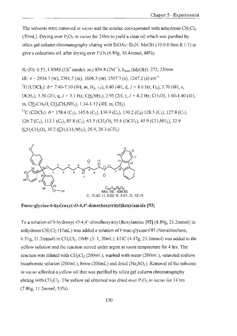
Chapter 5 - Experimental
The solvents were removed in vacuo and the residue co evaporated with anhydrous CH2Ch
(50mL). Drying over P20S in vacuo for 24hrs to yield a clear oil which was purified by
silica gel column chromatography eluting with EtOAc: Et3N: MeOH (l 0:0:0 then 8: 1: 1) to
give a colourless oil, after drying over P20 S (6.89g, 16.4mmol, 88%).
Rf(D): 0.51~ LRMS (ES+ mode): mlz 839.8 (2M+); Amax (MeOH): 272, 230nm
IR: v 2934.3 (w), 2361.5 (m), 1606.5 (m), 1507.3 (s), 1247.2 (s) cm-l
lH (CDCb): ()= 7.40-7.10 (9H, m, H3, 7-9),6.80 (4H, d, J 8.6 Hz, H4), 3.70 (6H, s,
OCH3), 3.30 (2H, q, J 5.1 Hz, CH2NH2), 2.95 (2H, t, J 4.2 Hz, CH20), 1.60-1.40 (4H,
m, CfuCH20, CfuCH2NH2), 1.34-1.12 (4H, m, CH2)
13C (CDCh): () = 158.4 (Cs), 145.6 (C2), 136.9 (C6), 130.2 (Cg) 128.3 (C3), 127.8 (C7),
126.7 (C9), 113.1 (C4), 85.8 (Cl), 63.5 (CH20), 55.4 (OCH3), 41.9 (CH2NH2), 32.9
(CH2CH20), 30.2 (CH2CH2NH2), 26.9, 26.3 (CH2)
C44H46 N20 6 Mol. Wt.: 698.85
C, 75.62; H, 6.63; N, 4.01; 0,13.74
Fm oc-glycine-6-hydroxy( -0-4,4' -dim ethoxytrityl)hexylamid e [93]
To a solution of 6-hydroxy(-O-4,4' -dimethoxytrityl)hexylamine [92] (8.89g, 21.2mmol) in
anhydrous CH2Ch (l5mL) was added a solution ofFmoc-glycine-OH (NovaBiochem,
6.31g, 21.2mmol) in CH2Clz: DMF (3: 1, 20mL). EDC (4.47g, 23.3mmol) was added to the
yellow solution and the reaction stirred under argon at room temperature for 4 hrs. The
reaction was diluted with CH2Ch (200mL), washed with water (200mL), saturated sodium
bicarbonate solution (200mL), brine (200mL) and dried (Na2S04)' Removal of the solvents
in vacuo afforded a yellow oil that was purified by silica gel column chromatography
eluting with CH2Ch. The yellow oil obtained was dried over P20 S in vacuo for 14 hrs
(7.80g, l1.2mmol, 53%).
150

Rf(P): 0.71; LRMS (ES+ mode): mlz 800.1 (MEt3N+), 721.0 (MNa+); "-max (MeOH): 271,
232nm; decomposed: 155-160°C
IR: v 2932.6 (w), 2896.7 (w), 2855.7 (w), 2365.3 (m), 2341.3 (m), 1652.4 (m), 1606.4
(m), 1506.8 (m), 1247.6 (s), 749.6 (s) cm-l
lH (CDCb): 0 = 7.40-7.08 (l7H, m, H 14-l7, 21, 26-28),6.70 (4H, d, J = 9.0 Hz, Hn ), 4.35-4.30
(2H, m, H4), 4.20 (lH, t, J = 7.0 Hz, H12), 3.73 (6H, s, H24), 3.27 (2H, s, H9), 3.23 (2H, q, J
= 6.6 Hz, H6), 2.98 (2H, t, J = 6.6 Hz, HI), 1.80-1.20 (8H, m, H2, 3, 5)
13C (CDCh): 0 = 158.4 (C 23 ), 145.6 (C20), 143.9 (C ll ), 141.5 (C8), 136.8 (C25), 130.2 (C27),
128.9 (C 13), 128.3 (C2l), 127.9 (C26), 127.3 (C 17, 14), 126.7 (C 28), 125.2 (C 16), 121.2 (C 18),
120.2 (CIS), 113.1 (C22), 85.8 (C19), 67.3 (C4), 63.4 (C l ), 55.4 (C24), 47.3 (C6), 43.3 (C 12),
39.8 (C9), 30.1, 29.9 (CS, 2), 26.9, 26.2 (C 3)
C:?gH36N204 Mol. Wt.: 476.61
C, 73.08; H, 7.61; N, 5.88; 0, 13.43
Glycine-6-hyd roxy( -0-4,4' -dimethoxytrityl)hexylamide [94]
To a solution of piperidine in DMF (20%, 50mL) was added the Fmoc-protected compound
[93] (6.00g, 8.6mmol) and the mixture stirred under argon, at room temperature for 24 hrs.
The solvents were removed in vacuo and the resulting oil dissolved in CH2Ch (200mL),
washed with water (200mL), saturated sodium bicarbonate solution (200mL), brine
(200mL) and dried (Na2S04). Removal of the solvents in vacuo gave a pale yellow solid
which was purified by silica gel column chromatography eluting with MeOH: CH2Ch (0: 1
then 1: 10) to give the desired compound as a colourless oil that was dried over P20 S in
vacuo (2.75g, 5.8mmol, 67%).
Rf(D): 0.44; LRMS (ES+ mode): mlz 499.4 (MNa+), 303.3 (DMT+); HRMS: C29H36N204
requires 476.26751, found 476.26751; "-max (MeOH): 269, 234nm
151

Chapter 5 - Experimental
IR: v = 3329.1 (w), 2931.7 (w), 2364.2 (m), 2341.3 (m), 1654.2 (m), 1606.6 (m), 1507.4
(m), 1246.1 (s), 1032.1 (s), 730.7 (s) cm-1
IH (CDCh):o= 7.40-7.13 (9H, m, HZL26-Z8), 6.77 (4H, d,J = 9.0 Hz, Hzz), 3.71 (6H, s,
Hz4), 3.27 (2H, s, H9), 3.25 (2H, q, J = 6.6 Hz, H6), 3.05 (2H, t, J = 6.6 Hz, HI), 1.60-1.16
(lOH, m, HZ-5, 10)
13C (CDCb): 0 = 172.8 (Cs), 158.5 (C23), 145.6 (Czo), 136.8 (Czs), 130.2 (C27), 128.3 (CZI),
127.9 (CZ6), 126.7 (Czs), 113.1 (Czz), 85.8 (C19), 63.5 (C l ), 55.4 (CZ4), 44.9 (C9), 39.1 (C6),
30.1,29.8 (CZ, 5), 27.0, 26.2 (C3,4)
0-24
36
C53H55N307 Mol. Wt.: 846.02
C, 75.24; H, 6.55; N, 4.97; 0,13.24
Fmoc-pbenylalanine-glycine-6-hydroxy(-O-4,4'-dimethoxytrityl)hexylamide [95]
To a solution of the amine [94] (l.lOg, 2.3mmol) in anhydrous CHzClz (lOmL) was added
Fmoc-phenylalanine-OH (NovaBiochem, 0.90g, 2.3mmol) and EDC (0,49g, 2.5mmol). The
solution was stirred at room temperature, under argon, for 14 hrs. The reaction was diluted
with CHzClz (50mL), washed with water (50mL), saturated sodium bicarbonate solution
(50mL), brine (50mL) and then dried (NaZS04). Removal of the solvents in vacuo gave a
pale yellow foam (1.92g, 98%), that was taken through to the next step crude. A small
amount (1 OOmg) was purified by silica gel column chromatography, for analysis, eluting
with MeOH: CHzClz (0: 1 then 1 :50) to give an off white solid which was dried over PzOs
in vacuo (83mg, 83%).
RdD): 0.67, (E) 0.65; LRMS (ES+ mode): m1z 868.9 (MNa+), 543.4 (M-DMT+), 303.3
(DMT+); HRMS: CS3H55N307 requires 845.4040017, found 845.4040017; Amax (MeOH):
265nm; mp: 78-82°C
152

IR: v 3388.2 (w), 2930.2 (w), 2858.7 (w), 2363.5 (m), 2341.6 (m), 1653.9 (m), 1607.0
(m), 1506.6 (m), 1246.7 (s), 1031.5 (s), 748.5 (s) cm-1
IH (CDCh): {) = 7.69-7.07 (22H, m, H I4-17, 21,26-28, 33-35), 6.87 (lH, t, J = 8.8 Hz, HIO), 6.75
(4H, d, J = 7.9 Hz, Hn), 6.36-6.32 (lH, m, H7), 5.12-5.08 (lH, m, H36), 4.40-4.30 (2H, m,
H4), 4.23 (lH, t, J = 7.0 Hz, Hu), 4.08 (lH, t, J 6.6 Hz, H30), 3.89-3.72 (2H, m, H9), 3.71
(6H, s, H24), 3.14-3.03 (2H, m, H6), 2.95 (2H, t, J = 6.6 Hz, HI), 1.57-1.53 (2H, m, H31 ),
1.42-1.12 (8H, m, H2, 3, 5)
l3C (CDCh): {) = 171.8 (C8), 168.5 (C29), 158.4 (C23), 156.4 (Cll), 145.6 (C20), 143.7 (Cl3),
141.4 (C32), 136.9 (C25), 136.4 (C I8), 130.2, 129.4, 128.9, 128.4, 128.0, 127.9, 127.3, 127.3,
126.8, 125.1, 120.2 (C I4-17, 21, 26-28, 33-35), 113.1 (C22), 85.8 (C19), 67.2 (C4), 63.4 (Cl ), 56.7
(C30), 55.4 (C24), 47.2 (C6), 43.4 (C l2), 39.8 (C9), 38.3 (C31 ), 30.2, 29.5 (C5,2), 27.0, 26.2
(C3)
7
0-24
Cs8H4SNs°5 Mol. Wt.: 623.78
C, 73.17; H, 7.27; N, 6.74; 0,12.82
Phenylalanin e-glycin e-6-hydroxy( -0-4,4' -dimethoxytrityl)hexylamide [96]
To a solution of piperidine in DMF (20%, 25mL) was added the Fmoc protected amide [95J
(1.90g, crude) and the mixture stirred under argon, at room temperature for 12 hrs. The
solvents were removed in vacuo and the resulting oil dissolved in CH2Ch (150mL), washed
with water (l50mL), saturated sodium bicarbonate solution (l50mL), brine (150mL) and
then dried (Na2S04). Removal of the solvents in vacuo gave a pale yellow solid (2. 14g),
which was taken through to the next step crude. A small amount (1 OOmg) was purified by
silica gel column chromatography eluting with MeOH: CH2Ch (0: 1 then 1: 10) to give the
desired compound as a white waxy solid, which was dried over P205 in vacuo (50mg, 91 %
based on yield of last step).
153

Rf (D): 0.70, (E): 0.13; LRMS (ES+ mode): mlz 646.5 (MNa+), 303.3 (DMT+); Amax
(MeOH): 262nm
IR: v = 3313.7 (w), 2932.1 (w), 2854.6 (w), 2359.9 (m), 1649.9 (m), 1606.6 (m), 1507.7
(s), 1248.0 (s), 103l.4 (s), 753.2 (s) cm-!
IH (CDCb):(5 = 8.82-8.79 (lH, m, H lO), 7.40-7.10 (l4H, m, H2!, 26-28, 33-35),6.75 (4H, d, J =
9.0 Hz, H22), 6.34-6.31 (lH, m, H7), 3.84 (2H, d, J = 6.5 Hz, H9), 3.71 (6H, s, H24), 3.62-
3.56 (lH, m, H30), 3.21-3.10 (2H, m, H6), 2.97 (2H, t, J 6.6 Hz, HI), 1.83-1.72 (2H, m,
H31 ), 1.56-1.12 (8H, m, H2• 3,S)
13C (CDCb): (5 = 175.2 (C8), 166.8 (C29), 158.4 (C23), 145.4 eC20), 137.5 (C32), 136.8 (C25),
130.0, 129.3, 128.8, 128.2, 127.7, 126.9, 126.6 (C21 . 26-28, 33-35), 113.0 (C22), 85.7 (C I9), 63.3
(C I), 56.4 (C30), 55.2 (C24), 43.4 (C9), 41.0 (C31 ), 39.6 (C6), 30.0, 29.5 eC2, 5),26.8,26.0
(C3)
7 ° C55H58N408
Mol. WI.: 903.07 C, 73.15; H, 6.47; N, 6.20; 0,14.17
15
Fm oc-glycine-p henylalanin e-glycine-6-hyd roxy( -0-4,4' -dim ethoxytrityl)hexylamid e
[97]
To a solution of the amine [96] (2.14g, crude) in anhydrous CH2Ch (lOmL) was added
Fmoc-phenylalanine-OH (NovaBiochem, 0.90g, 2.3mmol) and EDC (0.49g, 2.5mmol). The
solution was stirred at room temperature, under argon, for 1 hr. The reaction was diluted
with CH2Ch (50mL), washed with water (50mL), saturated sodium bicarbonate solution
(50mL), brine (50mL) and then dried (Na2S04). Removal of the solvents in vacuo gave a
pale yellow foam (2.27g), which was taken through to the next step crude. A small amount
(110mg) was purified by silica gel column chromatography eluting with MeOH: CH2Ch
154
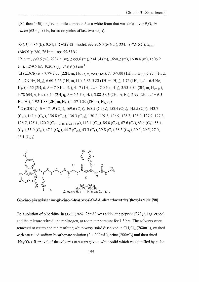
(0: 1 then 1 :50) to give the title compound as a white foam that was dried over P205 in
vacuo (63mg, 83%, based on yields oflast two steps).
Rf(D): 0.86 (E): 0.54; LRMS (ES+ mode): mlz 926.0 (MNa+), 224.1 (FMOC+); Amax
(MeOH): 280, 261nm; mp: 55-57°C
IR: v = 3299.6 (w), 2934.5 (w), 2359.6 (m), 2341.4 (m), 1650.2 (m), 1608.4 (m), 1506.9
(m), 1259.5 (s), 1030.8 (s), 749.9 (s) cm- l
IH (CDCb):o = 7.77-7.00 (22H, m, HI4-17.21, 26-28, 33-35), 7.10-7.00 (lH, m, HIO), 6.80 (4H, d,
J = 7.9 Hz, H22), 6.60-6.56 (lH, m, H7), 5.86-5.83 (lH, m, H39), 4.72 (lH, d, J 6.5 Hz,
H36), 4.35 (2H, d, J = 7.0 Hz, H4), 4.17 (1H, t, J = 7.0 Hz, H l2), 3.93-3.84 (3H, m, H30, 38),
3.78 (6H, s, H24), 3.14 (2H, q, J = 6.5 Hz, H6), 3.08-3.05 (2H, m, H9), 2.99 (2H, t, J = 6.5
Hz, HI), 1.92-1.88 (2H, m, H31 ), 1.57-1.20 (8H, m, H2, 3, 5)
l3c (CDCh): 0 = 171.9 (Cll ), 169.6 (C37), 168.5 (C8,29), 158.4 (C23 ), 145.5 (C20), 143.7
(C 13), 141.4 (C32), 136.8 (C25 ), 136.3 (CI8), 130.2, 129.3, 128.9, 128.3, 128.0, 127.9, 127.3,
126.7, 125.1, 120.2 (C 14-l7,2l,26-28, 33-35), 113.1 (C22), 85.8 (C I9), 67.6 (C4), 63.4 (C l), 55.4
(C24), 55.0 (C30), 47.1 (C l2), 44.7 (C38), 43.3 (C6), 39.8 (C9), 38.5 (C3l ), 30.1,29.5,27.0,
26.1 (C2-5)
31 0 II 39
30 NH~NH2 36 38
C40H48N406 Mol. Wt.: 680.83
C, 70.56; H, 7.11; N, 8.23; 0,14.10
Glycine-phenylalanine-glycine-6-hydroxy(-O-4,4'-dimethoxytrityl)hexylamide [98]
To a solution of piperidine in DMF (20%, 25mL) was added the peptide [97] (2.17g, crude)
and the mixture stirred under nitrogen, at room temperature for 1.5 hrs. The solvents were
removed in vacuo and the resulting white waxy solid dissolved in CH2Cb (200mL), washed
with saturated sodium bicarbonate solution (2 x 200mL), brine (200mL) and then dried
(Na2S04). Removal of the solvents in vacuo gave a white solid which was purified by silica
155

Chapter 5 - Experimental
gel column chromatography eluting with MeOH: CH2Ch (0: 1 then 1: 10) to give the desired
compound as a white crystalline solid, which was dried over P20s in vacuo (0.90g,
1.3mmol, 92%, 57% overall yield oflast 4 steps,).
Rf(D): 0.31; LRMS (ES+ mode): mlz 703.6 (MNa+), 303.3 (DMT+); HRMS:
C4oH48N406Na requires 703.34716, found 703.34716; Amax (MeOH): 273, 234nm; mp: 63-
68°C
IR: v = 3298.9 (w), 2928.6 (w), 2855.7 (w), 2363.1 (m), 2341.6 (m), 1648.8 (m), 1606.7
(m), 1506.8 (m), 1245.8 (s), 1030.6 (s), 750.2 (s) cm-1
IH (CDCh): 0 = 7.87 (lH, t, J 7.4 Hz, H IO), 7.40-7.05 (14H, ill, H2I ,26-28, 33-3S), 6.75 (4H,
d, J = 8.8 Hz, H22), 6.60-6.56 (lH, m, H7), 4.50 (lH, d, J = 6.5 Hz, H36), 3.88-3.72 (lH, m,
H30), 3.73 (6H, s, H24), 3.20 (2H, S, H39), 3.12-3.06 (2H, m, H6), 2.95 (2H, t, J 6.0 Hz,
HI), 2.08-1.98 (2H, m, H38), 1.60-1.10 (l2H, m, H2-5, 9, 31)
l3c (CDCb): 0 = 173.7 (C37), 171.8 (C8), 168.9 (C29), 158.4 (C23), 145.5 (C20), 136.8 (C32),
136.6 eC25), 130.2, 129.3, 128.9, 128.3, 127.8, 127.3, 126.7, (C21 , 26-28, 33-35), 113.1 (C22),
85.8 (CI9), 63.5 (CI), 55.4 (C24), 55.1 (C30), 46.5 (C38), 3.3 (C6), 39.8 (C9), 37.8 (C31 ), 30.2,
29.5,27.0, 26.2 (C2-S)
31 0 39 II H 43
9 ~N~041 5 30 NH 37 38 42 7 44 OH
36 46 o 0
C52H70N408 0-24 Mol. Wt.: 879.13
C, 71.04; H, 8.03; N, 6.37; 0, 14.56
12-Hydroxydodecanamido-glycine-pbenylalanine-glycine-6-bydroxy(-O-4,4'
dimethoxytrityl)hexylamide [99]
To a solution of the amine [98] (0.50g, 740.0f,lmol) in anhydrous CH2Ch (5mL) was added
12-hydroxydodecanoic acid (0.90g, 740.0f,lmol) and EDC (155mg, 0.8mmol). The solution
was stirred at room temperature, under argon, for 12 hrs. The reaction was diluted with
156
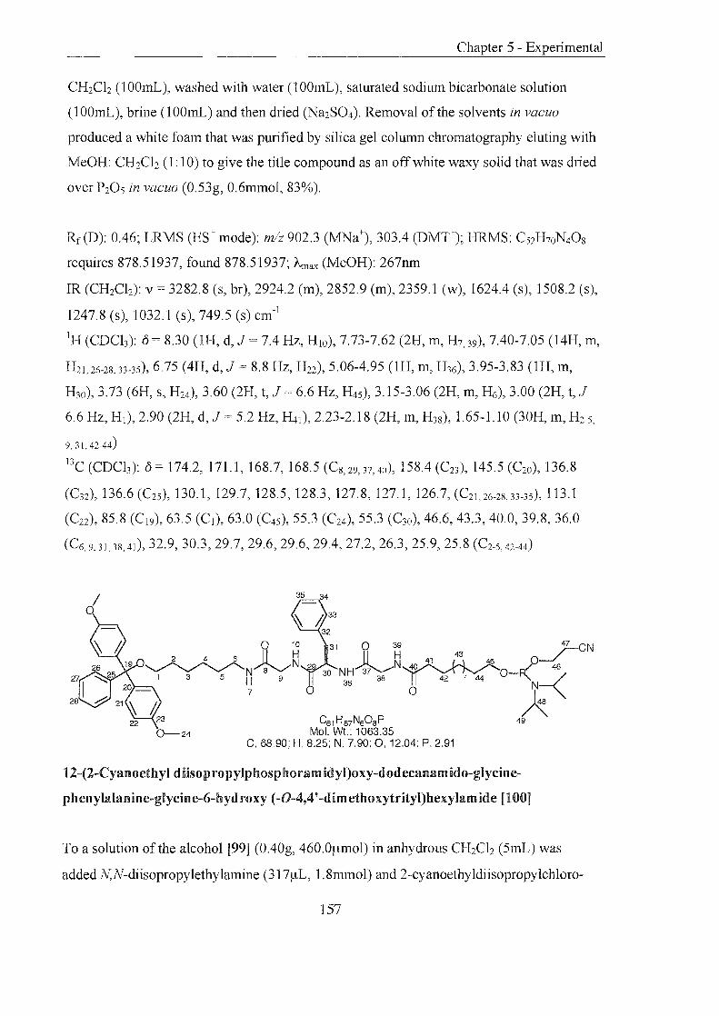
CH2Ch (100mL), washed with water (1 OOmL), saturated sodium bicarbonate solution
(lOOmL), brine (1001nL) and then dried (Na2S04). Removal of the solvents in vacuo
produced a white foam that was purified by silica gel column chromatography eluting with
MeOH: CH2Ch (1: 10) to give the title compound as an off white waxy solid that was dried
over P20S in vacuo (0.53g, 0.6mmol, 83%).
Rf(D): 0.46; LRMS (ES+ mode): mlz 902.3 (MNa+), 303.4 (DMT+); HRMS: C52H70N40S
requires 878.51937, found 878.51937; Arnax (MeOH): 267nm
IR (CH2Ch): v = 3282.8 (s, br), 2924.2 (m), 2852.9 (m), 2359.1 (w), 1624.4 (s), 1508.2 (s),
1247.8 (s), 1032.1 (s), 749.5 (s) cm- l
IH (CDCh): 0 8.30 (1H, d, J = 7.4 Hz, H IO), 7.73-7.62 (2H, m, H7,39), 7.40-7.05 (14H, m,
H21 , 26-2S, 33-35),6.75 (4H, d, J 8.8 Hz, H22), 5.06-4.95 (1H, m, H36), 3.95-3.83 (lH, m,
H30), 3.73 (6H, s, H24), 3.60 (2H, t, J 6.6 Hz, H4S ), 3.15-3.06 (2H, m, H6), 3.00 (2H, t, J =
6.6 Hz, HI), 2.90 (2H, d, J = 5.2 Hz, H41 ), 2.23-2.18 (2H, m, H3S), 1.65-1.10 (30H, m, H2-5,
9,31,42-44)
l3C (CDCh): 0= 174.2,171.1,168.7,168.5 (CS,29, 37, 40), 158.4 (C23 ), 145.5 (C20), 136.8
(C 32 ), 136.6 (C2S ), 130.1, 129.7, 128.5, 128.3, 127.8, 127.1, 126.7, (C21 ,26-28,33-35), 113.1
(C22 ), 85.8 (C I9 ), 63.5 (C l ), 63.0 (C45 ), 55.3 (C24), 55.3 (C30), 46.6, 43.3, 40.0, 39.8, 36.0
(C6,9,31,38,41), 32.9, 30.3, 29.7, 29.6, 29.6, 29.4, 27.2, 26.3, 25.9, 25.8 (C2-5,42-44)
.4 33
32
31 )1 39 47 eN ~~~1 43 5 p~
30 NH 37 38 40 42 7 44 0-F(-<46 36 N
o ~ C61H87N608P 49
7
Mol. Wt.: 1063.35 C, 68.90; H, 8.25; N, 7.90; 0, 12.04; P, 2.91
12-(2-Cyanoethyl diisopropylphosphoramidyI)oxy-dodecanamido-gIycine
phenylalanine-glycine-6-hyd roxy (-0-4,4' -dimethoxytrityl)hexylamide [100]
To a solution of the alcohol [99] (0.40g, 460.0pmol) in anhydrous CH2Ch (5mL) was
added N,N-diisopropylethylamine (317~L, 1.8mmol) and 2-cyanoethyldiisopropylchloro-
157

phosphoramidite (111/-AL, 1 18mg, 0.5mmol), under argon. After stirring at room
temperature for 1 hr N,N-diisopropylethylamine (lOO/-AL, 0.6mmol) and 2-
cyanoethyldiisopropylchloro- phosphoramidite (lOO/-AL, 106mg, 450.0/-Amol) were added.
After another 1 hr the pale yellow solution was diluted with EtOAc (25rnL), washed with
saturated potassium chloride solution (30mL), dried (Na2S04), and the solvents removed in
vacuo. Anhydrous CH2Clz (3rnL) was added and this solution dropped slowly onto rapidly
stirring hexane (-20°C, 250mL) fonning a white precipitate. The hexane was removed with
filtration, through glasswool. The residue was washed with cold hexane (150mL) and then
dissolved in CH2Clz (5mL), passed through the glasswool and the solvents removed in
vacuo. Anhydrous MeCN was added (lOmL), the solution filtered, the solvents removed in
vacuo and dried over P20S for 24 hrs (0.45g, 420.0/-Amol, 92%).
Rf(D): 0.64,0.51 (Q): 0.81,0.7; LRMS (ES+ mode): mlz 1086.6 (MNa+); Amax (MeOH):
276nm
31p (CDCh): 0 = 147.2
o 51
36
C60H70N40s 15 16 Mol. Wt.: 991.22
C, 72.70; H, 7.12; N, 5.65; 0,14.53
Fm oc-lysine(Dde )-glycine-6-hydroxy( -0-4,4' -dimethoxytrityl)hexylamide [105]
To a solution of the amine [94] (0.89g, 1.9mmol) in anhydrous CH2Clz (lOmL) was added
Fmoc-lysine(Dde)-OH (NovaBiochem, 1.00g, 1.9mmol) and EDC (0.39g, 2.0mmol). The
solution was stirred at room temperature, under argon, for 1 hr. The reaction was diluted
with CH2Clz (lOOmL), washed with water (l50mL), saturated sodium bicarbonate solution
158

(150mL), brine (l50mL) and then dried (Na2S04). Removal of the solvents in vacuo gave a
white foam (1.81g, crude). A small amount (76mg) was purified by silica gel column
chromatography, for analysis, eluting with MeOH: CH2Cb (1: 20) to give an off white solid
which was dried over P205 in vacuo (67mg, 88%).
Rf(D): 0.43; LRMS (ES+ mode): mlz 1030.2 (MK+), 991.2 (M+), 303.3 (DMT+); Amax
(MeOH): 294, 257nm; mp: 69-73°C
IR (CH2Cb): v = 3276.8 (w), 2933.2 (m), 2360.2 (m), 1650.0 (m), 1564.5 (m), 1506.9 (m),
1257.4 (s), 1028.8 (s), 749.8 (s) cm-1
IH (CDCh): {) = 7.80-7.20 (17H, m, H I4-17. 21.26-28),6.90 (1H, t, J = 8.3 Hz, H IO), 6.75 (4H,
d, J = 7.9 Hz, H22), 6.36-6.32 (IH, m, H7), 5.65 (lH, d, J = 6.6 Hz, H36), 4.40-4.30 (2H, m,
H4), 4.18 (IH, t, J = 7.0 Hz, H l2), 4.10-4.05 (lH, m, H30), 3.85-3.77 (2H, m, H9), 3.71 (6H,
S, H 24), 3.35-3.25 (2H, Ill, H34), 3.17-3.08 (2H, m, H6), 2.95 (2H, t, J 6.6 Hz, HI), 2.41
(3H, s, H S1 ), 2.28 (4H, s, H55), 1.95-1.10 (14H, m, H2, 3, 5, 31-33),0.93 (6H, S, H57)
BC (CDCh): {) = 173.81 (CS8 , 54), 172.10 (C8), 170.03 (C29), 158.43 (C23), 156.66 (C ll ),
145.53 (C20), 143.8 (C l3 , 53), 141.5 (C I8), 136.8 (C25 ), 130.2, 128.3, 128.0, 127.8, 127.3,
126.7,125.1,120.2 (CI4-17, 21, 26-28), 113.1 (C22), 108.1 (C52), 85.8 (C19), 67.2 (C4), 63.4
(C l), 55.4 (C24), 47.3 (C6, 34),43.3 (C l2), 43.1 (C33, 55) 39.8 (C9), 30.3 (C S6 ), 30.1, 29.5 (C2.
s), 28.4 (CSI ), 26.9, 26.1 (C3), 23.0 (C3I , 32), 18.1 (CS7)
° 51
34
32
C45H60N407 Mol. Wt.: 768.98
C, 70.29; H, 7.86; N, 7.29; 0,14.56
Lysine(Dde)-glycine-6-hydroxy(-O-4,4'-dimethoxytrityl)hexylamide [106]
159
To a solution of piperidine in DMF (20%, 25mL) was added the peptide [105] (1.74g,
1.8mmol) and the mixture stirred under argon, at room temperature for 45 minutes. The
solvents were removed in vacuo and the resulting oil dissolved in CH2Ch (200mL), washed
with water (200mL), saturated sodium bicarbonate solution (200mL), brine (150mL) and
then dried (Na2S04). After removal of the solvents in vacuo an off white foam (1.35g
crude) was obtained which was taken through to the next step crude. A small amount
(96mg) was purified by silica gel column chromatography eluting with MeOH: CH2Ch
(1:20 then 1: 10) to give the title compound as a white foam which was dried over P20s in
vacuo (59mg, 69% based on yield of last step).
Rf(D): 0.20; LRMS (ES+ mode): mlz 767.8 (M+), 790.9 (MNa+); Amax (MeOH): 297,
235nm
IR (CH2Ch): v 3308.4 (w), 2930.3 (m), 2861.4 (m), 2359.9 (m), 1638.9 (m), 1567.1 (m),
1507.3 (m), 1257.1 (s), 1030.9 (s), 750.6 (s) cm-l
lH (CDCh): 0= 7.92-7.88 (1H, m, H lO), 7.45-7.15 (9H, m, H21 ,26-2S), 6.80 (4H, d,J 8.3
Hz, H22), 6.65 (1H, t, J 5.9 Hz, H7), 3.80 (2H, d, J 5.2 Hz, H9), 3.76 (6H, s, H24), 3.52-
3.49 (lH, m, H30), 3.40-3.30 (2H, m, H34), 3.23-3.10 (4H, m, H6, 36),2.98 (2H, t, J 6.6
Hz, HI), 2.41 (3H, s, H S1 ), 2.28 (4H, S, HS5), 1.90-1.10 (14H, m, H2-5,31-33), 0.93 (6H, s, HS7 )
l3C (CDCh): 0= 198.0 (C54), 175.2 (CS3 ), 173.7 (C5S), 169.0 (C29) 162.7 (Cs), 158.4 (C23),
145.5 (C20), 136.8 (C25), 130.1,129.3,128.8,128.3,127.8,126.7, (C21,26-28), 113.1 (C22),
108.0 (C52), 85.8 (C 19), 63.4 (C1), 55.3 (C24), 44.8, 43.3, 39.8 (C6, 9, 34),36.7 (C30), 34.26,
32.9 (C33,55), 30.3 (C56), 30.1, 29.8 (C2,5), 28.4 (C51 ), 27.0, 26.2 (C3,4), 22.5 (C3l ,32), 18.1
(C57)
160
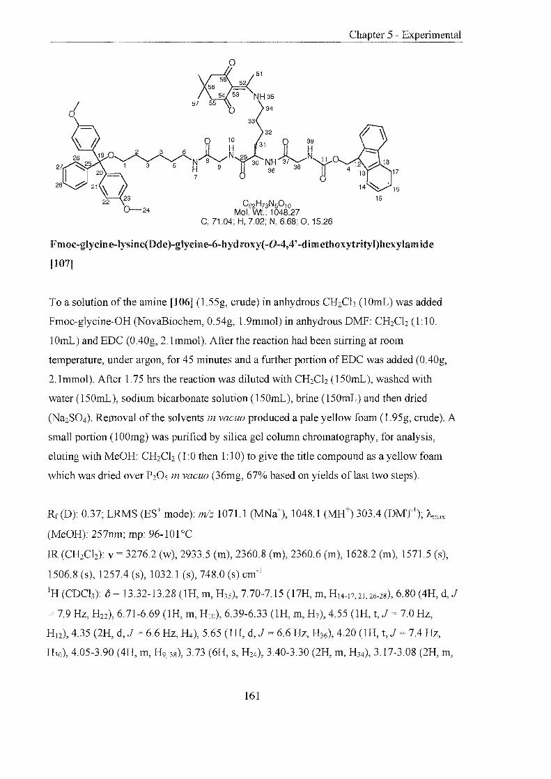
0--24
° 51
C62H73N5010 Mol. Wt.: 1048.27
C, 71.04; H, 7.02; N, 6.68; 0, 15.26
15
Fmoc-glycine-lysine(Dde )-glycine-6-hyd roxy( -0-4,4' -dim ethoxytrityl)hexylam ide
[107]
To a solution of the amine [106] (1.55g, crude) in anhydrous CH2Ch (1OmL) was added
Fmoc-glycine-OH (NovaBiochem, 0.54g, 1.9mmol) in anhydrous DMF: CH2Ch (1:10.
1OmL) and EDC (OAOg, 2.1mmol). After the reaction had been stirring at room
temperature, under argon, for 45 minutes and a further portion ofEDC was added (OAOg,
2.1mmol). After 1.75 hrs the reaction was diluted with CH2Ch (l50mL), washed with
water (l50mL), sodium bicarbonate solution (l50mL), brine (l50mL) and then dried
(Na2S04). Removal of the solvents in vacuo produced a pale yellow foam (1.95g, crude). A
small portion (1 OOmg) was purified by silica gel column chromatography, for analysis,
eluting with MeOH: CH2Clz (1:0 then 1: 10) to give the title compound as a yellow foam
which was dried over P20S in vacuo (36mg, 67% based on yields of last two steps).
Rr(D): 0.37; LRMS (ES+ mode): m/z 1071.1 (MNa+), 1048.1 (MH+) 303A (DMT+); Amax
(MeOH): 257nm; mp: 96-101 °C
IR (CH2Ch): v = 3276.2 (w), 2933.5 (m), 2360.8 (m), 2360.6 (m), 1628.2 (m), 1571.5 (s),
1506.8 (s), 1257.4 (s), 1032.1 (s), 748.0 (s) cm-1
IH (CDCh): (5 = 13.32-13.28 (lH, m, H35), 7.70-7.15 (l7H, m, H 14-17, 21, 26-28),6.80 (4H, d, J
= 7.9 Hz, H22), 6.71-6.69 (lH, m, HIO), 6.39-6.33 (lH, m, H7), 4.55 (lH, t, J 7.0 Hz,
H 12), 4.35 (2H, d, J 6.6 Hz, H4), 5.65 (IH, d, J = 6.6 Hz, H36), 4.20 (lH, t, J 7A Hz,
H30), 4.05-3.90 (4H, m, H9,38), 3.73 (6H, s, H24), 3.40-3.30 (2H, m, H34), 3.17-3.08 (2H, m,
161

Chapter 5 - Experimental
H6), 3.00 (2H, t, J 6.6 Hz, HI), 2.50 (3H, s, HSI ), 2.30 (4H, s, H55), 1.90-1.10 (14H, m,
H2,3,5,31-33), 0.95 (6H, s, H57)
l3C (CDCh): 0= 173.9 (C5S,54), 171.8 (Cs), 169.9 (C29), 168.5 (C37), 158.4 (C23), 156.1
(C ll ), 145.5 (C20), 143.8 (C]3,53), 141.4 (CIS), 136.8 (C25), 130.2, 128.3, 128.0, 127.8,
127.3,126.7,125.1,120.2 (C I4-17,21,26-2S), 113.1 (C22), 108.1 (CS2 ), 85.8 (C I9), 67.5 (C4),
63.4 (CI), 55.4 (C24), 55.2, 52.9 (C5S ), 47.2, 44.8, 43.1, 39.8, 31.8 (C6,9,33,34,3S), 43.3 (C l2),
30.3 (C56), 30.1, 29.5 (C2,5), 28.4 (C51 ), 28.1 (C30), 27.0, 26.1 (C3), 22.8 (C31 , 32), 18.2 (C57)
51
34
C47H63N50S Mol. Wt.: 826.03
C, 68.34; H, 7.69; N, 8.48; 0,15.50
Glycine-Iysine-(Dde) -glycine-6-hydroxy (-O-4,4'-dimethoxytrityl)hexylamide [108]
To a solution of piperidine in DMF (20%, 25mL) was added the peptide [107] (1.95g,
crude) and the mixture stirred under argon, at room temperature for 1.5 hrs. The solvents
were removed in vacuo and the resulting oil dissolved in CH2Ch (150mL), washed with
sodium bicarbonate solution (150mL), brine (150mL) and dried (Na2S04). After removal of
the solvents in vacuo an off white foam was obtained which was purified by silica gel
column chromatography eluting with MeOH: CH2Ch (1 :20 then 3:20). The white foam
produced was dried over P20s in vacuo (433mg, 82%,29% overall yield of based on last 4
steps).
Rf(D): 0.18; LRMS (ES+ mode): mlz 849.4 (MNa+), 827.4 (MH+), 303.4 (DMT+); HRMS:
C47H64NsOs requires 826.47549, found 826.47549; "-max (MeOH): 297, 261nm; mp: 74-
78°C
162

IR (CH2Ch): v = 3274.5 (m), 2934.5 (m), 2361.9 (m), 2360.7 (m), 1627.8 (m), 1571.2 (s),
1507.8 (s), 1245.1 (s), 1032.3 (s), 739.0 (s) cm-1
IH (CDCb): 0= 13.12-13.08 (lH, m, H3S), 7.96-7.93 (lH, m, H IO), 7.40-7.15 (9H, m, H2l,
26-28),6.75 (4H, d, J 8.3 Hz, H22), 6.55 (lH, t, J 5.9 Hz, H7), 4.37-4.25 (2H, m, H39),
3.83-3.78 (2H, m, H9), 3.75 (6H, s, H24), 3.40-3.30 (3H, m, H6,36), 3.20-3.10 (2H, m, H34),
3.00 (2H, t, J 6.6 Hz, HI), 2.45 (3H, s, HSI ), 2.32 (4H, s, H55), 2.15-2.10 (2H, m, H38),
1.80-1.15 (l4H, m, H2, 3, 5, 31-33),0.93 (6H, s, H57)
13C (CDCb): 0 = 173.8 (C58,54), 172.1 (C29), 168.8 (C37 ), 162.7 (C8), 158.4 (C23 ), 145.5
(C20, 53), 136.8 (C25 ), 130.2, 128.3, 127.8, 126.7, (C21 , 26-28), 113.1 (C22), 108.0 (C5Z), 85.8
(C I9), 63.4 (C I ), 55.4 (C24), 53.3, 53.0,43.3,43.2,39.8 (C6, 9, 33, 34, 38),31.2 (C55 ), 30.3 (C56),
30.1,29.5 (Cz,s), 28.4 (CSI ), 28.1 (C30), 27.0, 26.2 (C3), 23.1 (C3I ,3Z), 18.2 (CS7 )
51
34
C59H85N501o Mol. Wt.: 1024.33
C, 69.18; H, 8.36; N, 6.84; 0,15.62
12-Hydroxydodecanamido-glycine-Iysine-(Dde)-glycine-6-hydroxy (-0-4,4'
dimethoxytrityl)hexylamide [109]
To a solution of the amine [108] (0.39g, 470.0~mol) in anhydrous CHzCh (lOmL) was
added 12-hydroxydodecanoic acid (0. 109, 470.0~mol) and EDC (0.11g, 0.6mmol) The
solution was stirred at room temperature, under argon, for 2.5 hrs. The reaction was diluted
with CHzCh (200mL), washed with water (200mL), saturated sodium bicarbonate solution
(200mL), brine (lOOmL) and then dried (NaZS04). Removal of the solvents in vacuo
produced a yellow oil that was purified by silica gel column chromatography eluting with
MeOH: CHzClz (1:20 then 1: 10) to give the title compound as an off white foam that was
dried over PzOs in vacuo (0.40g, O.4mmol, 82%).
163

Rf (D): 0.86; LRMS (ES+ mode): mlz 1024.8 (M+), 303.3 (DMT+); Amax (MeOH): 252nm;
mp: 126-130 °C
IR (CH2Cb): v = 3282.3 (m, br), 2925.4 (m), 2853.3 (m), 2364.0 (w), 1622.6 (s), 1571.4
(s), 1508.9 (s), 1257.7 (s), 1032.1 (s), 750.0 (s) cm- l
IH (CDCh): 0 13.30-13.26 (1H, m, H35), 7.96-7.93 (1H, m, H lO), 7.45-7.41 (lH, m, H39),
7.40-7.15 (9H, m, H21 , 26-28),6.97-6.91 (1H, m, H7), 6.75 (4H, d, J = 8.8 Hz, H22), 4.00-3.82
(5H, m, H9, 30, 38),3.75 (6H, s, H24), 3.60 (2H, t, J = 6.6 Hz, H45), 3.40-3.33 (2H, m, H34),
3.25-3.15 (2H, m, H6), 3.00 (2H, t,J = 6.6 Hz, HI), 2.55 (3H, S, H51), 2.32 (4H, s, H55),
2.25 (2H, t, J = 8.3 Hz, H4d, 1.80-1.15 (32H, m, H2-5, 31-33, 42-44),0.93 (6H, s, HS7 )
l3C (CDCh): 0= 174.2,173.7,171.7,169.2,168.5 (C8,29,37,40,54,58), 158.4 (C23), 145.5
(C20,53), 136.8 eC2S), 130.1, 128.3, 127.8, 126.7, (C21,26-28), 113.1 (C22), 108.0 (C52), 85.8
(C19), 63.5 (C l ), 63.0 (C45), 55.3 (C24), 52.9,43.7,43.3,39.8 (C6, 9, 34, 38),30.2 (C56), 28.4
(CSl ), 36.1, 33.2, 32.9, 29.6, 29.5, 29.3, 28.7, 27.2, 26.3, 25.9, 25.7, 22.8 (C2-5, 30,31-33,41-44,
55), 18.1 (C57)
0--24
51
34
32
31 0 39 .r-CN 11 ~~1 43 45 p-./
30 NH~ 40 0-[(-< 36 38 42 7 44 "-
o A C68H102N7010P
Mol. wt.: 1208.55 C, 67.58; H, 8.51; N, 8.11; 0, 13.24; P, 2.56
12-(2-Cyanoethyl diisopropylphosphoramidyl)oxy-dodecanamido-glycine-Iysine(Dde)
glycine-6-hydroxy (-0-4,4' -dimethoxytrityl)hexylamide [110]
To a solution of the alcohol [109] (0.40g, O.4mmol) in anhydrous CH2Cb (5mL) was added
N,N-diisopropylethylamine (265mg, 3551!L, 2.3mmol) and 2-cyanoethyl diisopropyl
chlorophosphoramidite (9II!L, 9Img, 470.0I!mol), under argon. After stirring at room
temperature for 2.5 hrs the pale yellow solution was diluted with EtOAc (25mL) washed
164

Chapter 5 - Experimental
with saturated potassium chloride solution (30mL) dried (Na2S04), and the solvents
removed in vacuo. Anhydrous CH2Clz (3mL) was added and this solution dropped slowly
onto rapidly stirring hexane (-20°C, 250mL) forming a white precipitate. The hexane was
removed, with filtration, through glass wooL The residue was washed with cold hexane
(150mL) and then dissolved in CH2Clz (5mL), passed through the glasswool and the
solvents removed in vacuo. Anhydrous MeCN was added (10mL), the solution filtered, the
solvents removed in vacuo and dried over P20 S for 24 hrs (0.43g, 360.0f.!mol, 91 %).
Rf (D): 0.93,0.89; LRMS (ES+ mode): mlz 1247.0 (MK+), 1165.0 (M-CH(CH3)2), 1123.8
(M-2CH(CH3)2); "-max (MeOH): 296, 368nm
31p (CDCh): () = 147.92, 147.80
Silver oxide catalyst206
A solution of silver nitrate (3.00g, 17.7mmol) in water (30mL) was warmed to 85°C. A
second solution of sodium hydroxide (0.69g, 11.3mmol) in water (30mL) warmed to 85°C
and added to the silver nitrate solution. The mixture was shaken vigorously for 5 minutes
and a dark brown precipitate of silver oxide formed. In the absence oflight, the solid was
removed by filtration, washed with hot water (5 x 20mL), Et20 (3 x 20mL), dried in vacuo
and stored in the dark for immediate use.
Silver silicate catalyst 157
Procedure was carried out in the absence of light. Neutral alumina (15.00g) was suspended
in a solution of silver nitrate (17.09g, O.lOmol) in water (lOOmL). A solution of sodium
silicate (47.30g) in water (lOOmL) was added dropwise with stirring over 1 hr. The
resulting mixture was heated (70°C) for 15 minutes with stirring, cooled to room
temperature, filtered, and the residue washed with water (2 xlOOmL). The residue was
stirred in a silver nitrate solution (10% w: v, 100mL) at 70°C for 0.5 hr. The solution was
allowed to cool, washed with anhydrous acetone (2 x 1 OOmL), toluene (2 x 100mL) and
dried in vacuo (60°C, 3hrs). The powder was dried over P20 5 for 24 hrs, affording the
silver silicate catalyst as a pale yellow powder (40g).
165
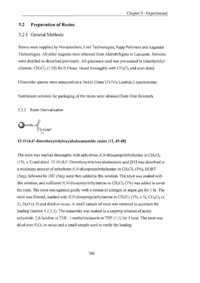
Chapter 5 - Experimental
5.2 Preparation of Resins
5.2.1 General Methods
Resins were supplied by Novabiochem, Link Technologies, Rapp Polymere and Argonaut
Technologies. All other reagents were obtained from Aldrich/Sigma or Lancaster. Solvents
were distilled as described previously. All glassware used was pre-soaked in trimethylsilyl
chloride: CHzCh (1 :20) for 0.5 hour, rinsed thoroughly with CHzCh and oven dried.
Ultraviolet spectra were measured on a Perkin Elmer UV/Vis Lambda 2 spectrometer.
Synthesiser columns for packaging of the resins were obtained from Glen Research.
5.2.2 Resin Derivatisation
HN-I N-ODMT
11
12-0-(4,4' -dimethoxytrityloxy)dodecanamide resins [12, 45-48]
The resin was washed thoroughly with anhydrous N,N-diisopropylethylamine in CHzCh
(1%, x 5) and dried. 12-0-(4,4'-Dimethoxytrityloxy)dodecanoic acid [11] was dissolved in
a minimum amount of anhydrous N,N-diisopropylethylamine in CHzCh (l %), HOBT
(5eq), followed by DIC (5eq) were then added to this solution. The resin was soaked with
this solution, and sufficientN,N-diisopropylethylamine in CHzCh (1%) was added to cover
the resin. The resin was agitated gently with a stream of nitrogen or argon gas for 1 hr. The
resin was filtered, washed with N,N-diisopropylethylamine in CHzCh (1 %, x 5), CHzCh (x
3), EtzO (x 5) and dried in vacuo. A small sample of resin was removed to ascertain the
loading (section 5.2.2.1). The remainder was soaked in a capping solution of acetic
anhydride: 2,6-lutidine in THF: 1-methylimidazole in THF (1:1) for 1 hour. The resin was
dried over PzOs in vacuo and a small sample used to verifY the loading.
166

Chapter 5 - Experimental
o '0
N~O*-H I .b ODMT
N02
UV Linker resin [66]
The LCAA -CPG (250mg) was washed thoroughly with anhydrous N,N
diisopropylethylamine in CHzCh (1 %, x 5) and dried. [65] (25mg, 40.0[.lmol) was
dissolved in a minimum amount of anhydrous N,N-diisopropylethylamine in CHzCh (1 %),
HOBT (1O.1mg, 75.0[.lmol), followed by DIC (9.5mg, 750.0[.lmol) were then added to the
solution. The resin was soaked with this solution, and sufficient N,N-diisopropylethylamine
in CHzCh (1 %) added to cover the resin. The resin was agitated gently with a stream of
nitrogen or argon gas for 2 hrs . The resin was filtered, washed with N,N
diisopropylethylamine in CHzCh (1 %, x 5), CHzCh (x 3), EhO (x 5) and dried in vacuo. A
small sample of resin was removed to ascertain the loading (section 5.2.2.1). The remainder
was soaked a capping solution of acetic anhydride: 2,6-lutidine in THF: I-methylimidazole
in THF (1: 1) for 1 hour. The resin was dried over PZ0 5 in vacuo and a small sample was
used to verify the loading.
5.2.2.1 Determination of res in loading by trityl analysis
A sample of resin was weighed and suspended in hydrochloric acid: EtOH (3:2 v: v, 25mL)
and shaken for 5 minutes. The absorbance of 1mL of the orange solution was measured at
495nm on a Perkin Elmer UV/VIS spectrometer. The amount of 4,4' -dimethoxytrityl cation
present was calculated:
Absorbance for ImL (at 495nm)
For 25mL =a x 25
[The absorbance of 1 flmol of DMT at 495nm in = 71.7]
For c g of resin = b + 71.7
Resin Loading (amount ofDMT+ present) = d + C
167
=a
=b
= dl-lmol
= e /-lmolg-1

Example:
Absorbance for ImL (at 495nm)
For 25mL = 0.8897 x 25
[The absorbance of 1 J1mol of DMr at 495nm in = 71.7J
For c g of resin = 22.2417 + 71.7
Resin Loading (amount ofDMT+ present) = 0.31 + 0.0086
Cpd Batch Resin Type Supplier Resin
No. No. (g)
12 1 LCAA-CPG Link Technologies 2.16
45 1 Polystyrene Link Technologies 0.05
45 2 Polystyrene Link Technologies 0.05
45 3 Polystyrene Link Technologies 0.1
45 4 Polystyrene Link Technologies 0.07
46 1 Tentagel NovaBiochem 1
47 1 Tentagel Rapp Polymere 0.5
48 1 Macropore Argonaut Technologies 0.5
66 LCAA-CPG Link Technologies 0.25
168
Chapter 5 - Experimental
= 0.8897
= 22.2417
= 0.311lmo1
= 36.47 l1ID01g-1
DMTcpd Loading
(/lmol) (Ilmo1g-1)
600 36.47
390 48.89
96 10.02
390 34.00
200 52.00
240 67.00
270 57.60
0.27 46.20
0.04 22.00

5.3 Preparation of Synthetic Oligonu.cleotides
5.3.1 General Methods
All oligonucleotides were synthesised on an Applied Biosystems 394 solid phase
DNA/RNA synthesiser using the standard assembly cycle of acid catalysed detritylation,
coupling, capping and iodine oxidation procedures. Oligonucleotides were assembled on a
0.2Ilmo1, or 1.0llmoi scale, stepwise coupling efficiencies and overall yields were
calculated using automated trityl cation conductivity monitoring. DNA phosphoramidites,
solid supports and additional reagents were purchased from Applied Biosystems, unless
otherwise stated.
Cleavage of the oligonucleotides from the solid support was achieved by exposure to
concentrated ammonia, unless otherwise stated. All oligonucleotides requiring base
deprotection were placed in concentrated ammonia or ammonia: ethanol (3: 1) at 60°C for 4
hours and evaporated to dryness in vacuo
Purification was effected using reversed phase HPLC on a Gilson RPHPLC system,
controlled by Gilson 7.12 software. Elution of oligonucleotides was monitored by UV
detection at desired wavelengths (260-290nm). Buffer systems consisted of buffer A: O.IM
ammonium acetate), buffer B: acetonitrile: buffer A (4: 6 or 3: 7), with a typical gradient
programme as described in figure 5.1 Analytical injections were monitored at 265nm, 0.1
AUFS, preparative injections were monitored at 280-290nm, 1.0 AUFS. The
oligonucleotides were collected manually.
All phosphoramidites synthesised were stored under argon, at -20°C, in bottles suitable for
an ABI 394 DNA synthesiser. The phosphoramidites were dissolved in anhydrous
acetonitrile immediately prior to use.
The resins were packaged into Glen Research columns suitable for an ABI 394 DNA
synthesiser. The amount of resin used was determined by the resin loading and the scale of
the synthesis (O.2llmol or 1.0Ilmol).
169
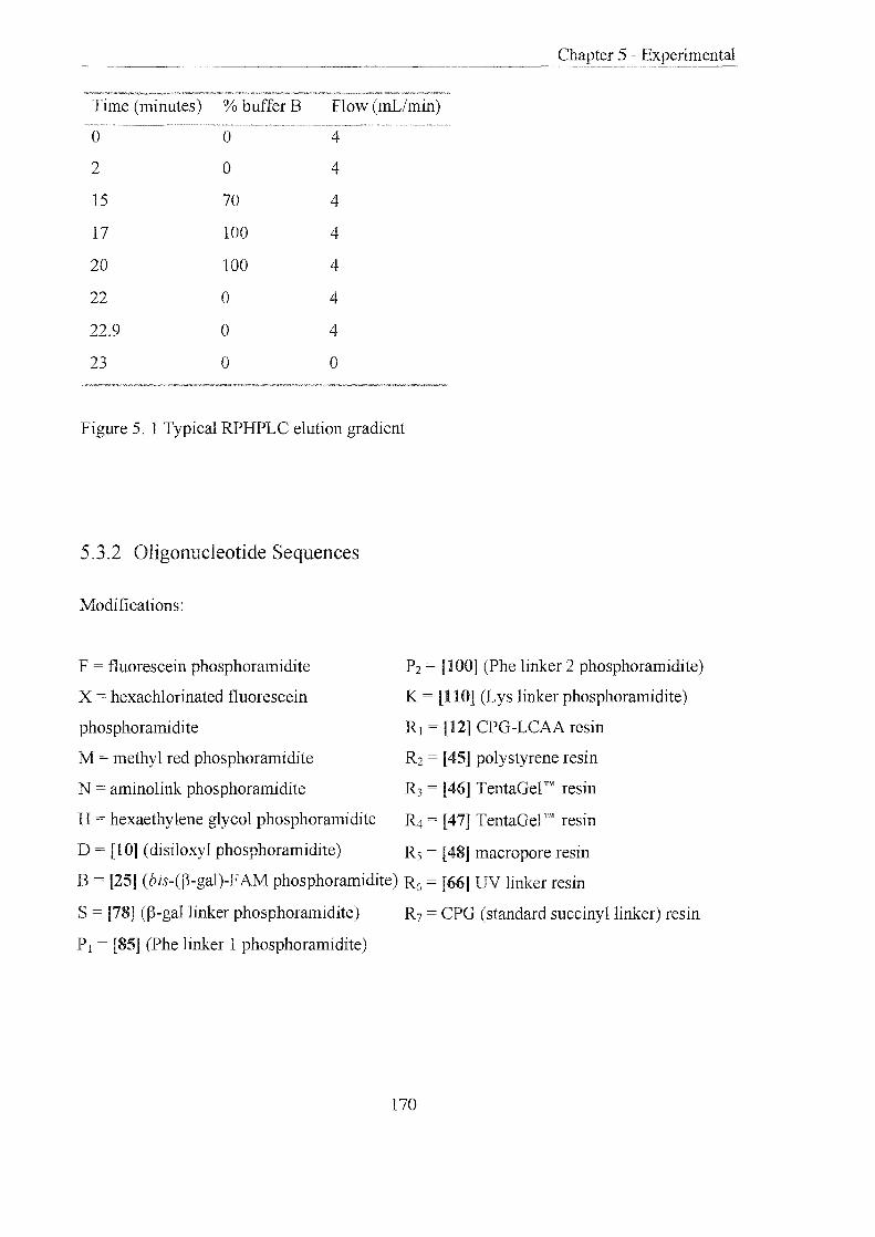
Chapter 5 - Experimental
Time (minutes) % bufferB Flow (mLimin)
0 0 4
2 0 4
15 70 4
17 100 4
20 100 4
22 0 4
22.9 0 4
23 0 0
Figure 5. 1 Typical RPHPLC elution gradient
5.3.2 Oligonucleotide Sequences
Modifications:
F = fluorescein phosphoramidite
X = hexachlorinated fluorescein
phosphoramidite
M = methy I red phosphoramidite
P2 [100] (Phe linker 2 phosphoramidite)
K = [110] (Lys linker phosphoramidite)
RJ = [12] CPG-LCAA resin
R2 [45] polystyrene resin
N = amino link phosphoramidite R3 = [46] TentaGel ™ resin
H hexaethylene glycol phosphoramidite ~ = [47] TentaGel ™ resin
D = [10] (disiloxyl phosphoramidite) R5 = [48] macropore resin
B [25] (bis-(~-gal)-F AM phosphoramidite) R6 = [66] UV linker resin
S = [78] (~-gallinker phosphoramidite)
P j = [85] (Phe linker 1 phosphoramidite)
R7 = CPG (standard succinyllinker) resin
170

Chapter 5 - Experimental
No. Sequence
d
13 TTTTTTTTTTTTDRJ --
17 NTTTTTTTTTTTT 3784.6 3782.9
26 TTTTTTTTTTTTTTT 4500.9 4501.0
27 BTTTTTTTTTTTTTTT 5360.9 536l.0
28 NCATCTGAACTTTGCCAGCTT 6222.2 6223.0
31 GATCTGACTTGGTAGHN 5174.0 5173.9
33 BHACGATGCGCCGATCG 578l.2 5778.8
34 CGATCGGCGCATCGTCTACCAAGTCAGATC 9147.6 9143.9
35 BHCTACTGCGCCGATCG 5732.1 5730.5
36 CGATCGGCGCACAAGTCAGATC 6726.2 6723.6
37 GATCTGACTTGGTAGACTATGCGCCGATCGN 10480.1 10485.4
38 GATCTGACTTGGTAGACTATGCGCCGATCG 9238.0 9242.0
39 CGATCGGCGCATAGTCTACCAAGTCAGATC 9176.0 918l.0
40 CGCACGCGATCGGCGCATAGTCTACCAAGTCGA 13971.4 13975.0
TCCGTGCGM
41 BHHTATTAAGCCTGCAAG 6123.3 6123.1
42 13800.2
CCAAGTCAGATC
43 BHHCGATCGGCCTGCAAG 6126.0
171

44 CTTGCAGGCGCGCATAGTCTACCAAGTCAGATC 10093.7 10095.0
49 FCGCACGCTTAAAGTCACTTCATTTTCGTGCGM
MHHHRJ
50 CGAGATACGGTTTTCACAGCHHHHHHHHR2 --
51 CGAGATACGGTTTTCACAGCR3 --
52 CGAGATACGGTTTTCACAGC~ --
53 AAAATGAAGTGACTTTAAG 5881.1 5881.5
54 GCTGTGAAAACCGTATCTCGM 6213.0 6210.9
55 FCGCACGATGTAGCACATCAGAAGCGTGCGTM --MHHHRJ
56 CTTCTGATGTGCTACAT 5151.4 5151.0
57 FCGCACGCTTAAAGTCACTTCATTTTCGTGCGM --
HHHR2
58 FCGCACGCTTAAAGTCACTTCATTTTCGTGCGM --
HR2
59 FCGCACGCTTAAAGTCACTTCATTTTCGTGCGM --
HHHHHR2
60 FCGCACGCTTAAAGTCACTTCATTTTCGTGCGM --
HHHHHHHHHHHHR2
61 FCGCACGCTTAAAGTCACTTCATTTTCGTGCGM -- --
HR4
62 FCGCACGCTTAAAGTCACTTCATTTTCGTGCGM -- --
HR5
63 TTTTTTTTTTTTTTTTTTT~ -- --
64 CGAGATACGGTTTTCACAGC~
172

67 GCCTCCAGCTTAGTTMMHHR2
68 FAACTAAGCTGGAGGCACGTACGA 7643.1 7640.1
69 TATGATCGTACGTGCCTCCAGCTTAGTTTTCTC 10031.7 10033.4
79 FSTTTTTTTTTTTT 5379.7 5379.0
80 FSSTTTTTTTTTTTTTTT 5722.0 5726.0
86 XPITTTTTTTTTTTTTTT 5771.8 5772.0
87 XHP1 HTTTTTTTTTTTTTTT 6460.3 6459.9
88 CGTGACATCTGCAGTCP1DR1 -- --
89 FCAATGTTP1 TTCATTGM 5488.1 5380.0
101 FTTTTTTP2 TTTTTT 4763.1 4763.0
102 FP2TTTTTT 2940.4 2940.0
103 FP2 TTTTTTR2 --
104 TP2HHTR7 -- --
111 TTTTTTLTTTTTTTTTTTTTTTT 7248.1 7251.1
112 TTCTTCTTLTCCTCTTCTTCTCCTTT 8025.6 8027.0
113 TLHHTR7 --
114 TTGAAAGGCCP2GCTCCAGGGT 6796.8 6795.6
115 TTGAAAGGCCLGCTCCAGGGT 6775.8 6778.0
173

5.4 Molecular Biology
5.4.1 General Methods
All molecular biological grade reagents were obtained from Sigma! Aldrich unless
otherwise stated. Deionsied water (MilliQ) was used in all experiments. NAP 10 Sephadex
columns for desalting oligonucleotides were purchased from Pharmacia.
Fluorescence microscopy was performed using a LEITZ DM IL microscope and
photographed with a ASHAI PENT AX camera using Kodak Ektachrome colour film 160T,
36exp obtained from Sigma! Aldrich Techware. Fluorescent spectra were measured on a
Perkin Elmer LS50B Luminescence Spectrometer using quartz glass 3mL and O.4mL cells.
Irradiation of samples was performed on a transilluminator platform at 260nm. Ultraviolet
spectra and thermal melting experiments were carried out on a Perkin Elmer UV /Vis
Lambda 2 spectrometer with PTP-l temperature programmer.
Agarose gel electrophoresis was performed using a Hoefer 250 Power supply and mini gel
equipment. Polyacrylamide gel electrophoresis on a Hoefer vertical electrophoresis sturdier
SE-410 apparatus and power supply.
Rapid heating and cooling of samples was achieved using a Techne thermocycler PHC-3
Centrifugation was performed in a microcentrifuge at 13,000 rpm for 30 minutes.
Ion exchange and gel filtration chromatography were performed on a Pharmacia LKB
system and elution was monitored at 260/280nm
174

5.4.2 Experimental
Cleavage of oligonucleotides from the solid support using 3HF"Et3N: NMP: EhN201
3HF"Et3N: NMP: Et3N (2: 3: 1.S, 20rnL) was prepared and sealed under argon. This
solution (200 ilL ) was added to the dry resin sample (4mg) in a screw top vial. The resin
solution was heated to 6S a C for I.S hrs and allowed to cool to room temperature. The
solution was then removed from the resin by filtration and the resin was washed with water
(S x 2rnL). The HF solution and aqueous washings were combined and the solvents were
removed in vacuo. The sample was dissolved in water (lrnL) and loaded onto a NAP 10
sephadex column, elution was effected with water (1.SrnL).
Cleavage of oligonucleotides from the solid support using 1.0M TBAFITHF
TBAF in THF (1. OM, 200 ilL) was added to the dry resin in a screw top vial and allowed to
stand, at room temperature, for 4 hrs. The solution was then removed from the resin by
filtration and the resin was washed with water (S x 2rnL). The fluoride solution and
aqueous washings were combined and the solvents were removed in vacuo. The sample
was dissolved in water (lrnL), and loaded onto a NAP 10 sephadex column; elution was
effected with water (1.SmL).
UV thermal melting of oligonucleotides
The two single stranded oligonucleotides to be melted were mixed (1:1, total O.SOD) and
diluted with buffer (sodium phosphate lOmM, EDTA 1mM, NaCI O.IM or 1.0M, pH 7.0).
The solution in an UV cuvette (pathlength lOmm) was heated to 80aC and cooled slowly to
room temperature to effect efficient strand annealing. The sample was melted at a heating
rate of 1 a per minute, monitoring at 260nm on a PC with lOs intervals. The data curves
were smoothed and the first derivatives obtained using the Perkin Elmer PECSS2 software.
175

The time peaks were converted to temperature manually, in order to ascertain Tm values.
The melt process was repeated to acquire consistent results (± 0.25°C).
Preparation of oligonucleotides for electrospray mass spectroscopy
Method 1
The oligonucleotide sample was dissolved in ammonium acetate (5M, 100/lL) and allowed
to stand at room temperature for 1 hr. The oligonucleotide was then precipitated with cold
EtOH (300/lL) and placed in a MeOH/dry ice bath for 2 hrs. The solution was centrifuged
at 14000g for O.5hr, the supernatant removed, washed with cold EtOH (80%), centrifuged
again at 14000g for 15 minutes and the supernatant removed. The sample was dissolved in
a solution of water: isopropanol (1: 1) containing tripropylamine (1 %) for electro spray mass
spectral analysis.
Method 2
The oligonucleotide sample was dissolved in water (1.0mL), loaded on to a NAP 10
sephadex column and eluted with water (1.5mL). The water was removed in vacuo and the
sample was dissolved in a solution of water: isopropanol (1: 1) containing tripropy lamine
(0.2%) for electrospray mass spectral analysis.
(3-D-Galactosidase assay for generation of fluorescence with ((3-gal)2Fam labelled
oligonucleotides
The activity of (3-D-Galactosidase is defined as one unit causes the hydrolysis of one
micromole of o-nitrophenol-(3-D-galactopyranoside per minute at 25°C and pH 7.5.
The oligonucleotide substrate in buffer (1.5 OD, sodium phosphate 0.3M, magnesium
chloride 0.003M, pH 7.5, 500!lL) was placed in a cuvette (lmL, lOmm pathlength) and
allowed to equilibrate at 37°C for 5 minutes. (3-D-galactosidase (500!lL of a O.lmg mL-1
solution in the same buffer) was added to the cuvette and the reaction was monitored
immediately at 3JOC in UV/Vis spectrometer at 490nm, for 1 hr.
176

Preparation of an MRS derivatised oligonucleotide
A 3' amino-derivatised oligonucleotide was prepared by standard DNA synthesis (1. O!lmol
scale) and purified (RPHPLC). The oligonucleotide was desalted twice through a NAP 10
sephadex column (loaded in water (1.0rnL), eluted with water (1.5mL». The water was
removed in vacuo and the oligonucleotide dissolved in sodium carbonate buffer (0. 1M, pH
6.00, 50"",L) and transferred to a vial (3mL). The flask was rinsed with sodimn carbonate
buffer (2 x 25"",L) and added to the vial. A solution ofMBS (5mg) in DMSO (120"",L) was
prepared and an aliquot (l5"",L) was added to the oligonucleotide. The solution was
vortexed until almost clear. The reaction was allowed to stand, at room temperature, for 4
hrs after which a further aliquot of the MBS solution (15"",L) was added. The solution was
left overnight, at room temperature, in the dark. The reaction was diluted with water
(870!lL) and desalted through NAP 10 sephadex eluting with water (1.5mL). The resulting
oligonucleotide was purified by RPHPLC; the MBS derivatised oligonucleotide was
collected and analysed by electrospray mass spectrometry.
Conjugation of j3-D-galactosidase with an MRS derivatised oligonucleotide
An MBS derivatised oligonucleotide was dried in vacuo. A solution of j3-D-galactosidase
(hng) in potassium phosphate buffer (0. 1M, pH 5.6, 500"",L) was added to the
oligonucleotide. The clear solution was allowed to stand, at room temperature, in the dark,
overnight. The reaction was diluted with potassium phosphate buffer (O.lM, pH 5.6,
500IlL) and purified by gel filtration eluting isocratically with potassium phosphate buffer
(0.1 M, pH 5.6). Detection of the peaks was achieved by monitoring the absorbance at
2601280nrn. The major conjugate peak was collected, the solvents were removed in vacuo.
The residue was dissolved in water (1.0mL) and desalted through NAP 10 sephadex eluting
with water (1.5rnL). The conjugate was purified by ion-exchange chromatography eluting
with an increasing gradient of buffer B [buffer A: Tris.HCI (50.0mmol), pH 7.4, buffer B:
buffer A, NaCI (1.0M), pH 7.4)]. Detection of the peaks was achieved by monitoring the
absorbance at 260/280nm. All the major conjugate peaks were collected and assayed for 13-
D-Galactosidase activity and the presence of oligonucleotides (absorbance at 260nm).
177

Chapter 5 - Experimental
Activity assay of j3-D-galactosidase conjugate207
Assay buffer
Substrate solution
Enzyme conjugate
solution
sodium phosphate (O.3M), magnesium chloride (O.003M), pH 7.5
gai;actC)DvnmUVJ''''''-' (hng) in assay buffer (1mL)
from ion exchange chromatography (50ftL)
The assay buffer (900~tL) was placed into a cuvette (pathlength 10mm) and the enzyme
conjugate (50ftL) was added and shaken. The cuvette was placed in a UV/Vis spectrometer
at 3rC and the absorbance measured at 405nm. The cuvette was removed from
spectrometer and the substrate added (50ftL), shaken and quickly placed back in the
spectrometer. The absorbance at 405nm at 3rC was monitored for 5 minutes. The final
colour of the solution and its absorbance at 405nm were recorded.
Testing homogeneous assay (as described section 2.3.7 and 2.3.12)
Signal probe and native (3-galactosidase
The signal probe ([33], [41] or [43], O.187nmol) in buffer (O.3M sodium phosphate,
O.003M MgCh, O.IM, NaCI, pH 7.5, 350ftL) was placed in a fluorimeter cuvette, and
incubated at 37°C for lOmins. The fluorescence intensity was noted and j3-D-galactosidase
(2ftL, 1mg mL-1 solution) was added to the cuvette. The change in fluorescence intensity
was monitored over 1 hour at 3rc.
Signal probe and (3-D-galactosidase conjugate
The signal probe ([33], [41] or [43], O.187nmol) in buffer (O.3M sodium phosphate,
O.003M MgCh, O.lM, NaC!, pH 7.5, 300ftL) was placed in a fluorimeter cuvette, and
incubated at 37°C for lOmins. The fluorescence intensity was noted. The j3-D-galactosidase
conjugate [32] or [37J was evaporated to dryness and dissolved in the same buffer ([32] or
[37], O.187nmol, 50ftL) then added to the cuvette. The change in fluorescence intensity was
monitored over 1 hour at 37°C.
178

Signal probe and f3-D-galactosidase conjugate and target
The signal probe ([33], [41] or [43], 0.187mnol) in buffer (0.3M sodium phosphate,
0.003M MgClz, O.IM, NaCI, pH 7.5, 300"",L) was placed in a fluorimeter cuvette, with the
complementary target ([34], [42] or [44], 0.935nmol). The solution was heated to 70°C and
cooled slowly to room temperature. The solution was incubated at 37°C for 10mins and the
fluorescence intensity noted. The f3-D-galactosidase conjugate [32] or [37] was evaporated
to dryness, then dissolved in the same buffer ([32] or [37], 0.187nmol, 50"",L) and added to
the cuvette. The change in fluorescence intensity was monitored over 1 hour at 37°C.
Dot blot assay
24 x SSC buffer urn chloride (3.6M), trisodium citrate (360.0mmol)
Denhardt's solution bovine serum albumin (0.01 % w:v), Ficoll (0.01 % w:v),
polyvinylpyrrolidone (0.01% w:v)
Prehybridisation Ix Denhardt's solution, SDS (0.1 % w:v), 6x SSC, denatured
solution herring sperm DNA (lO"",gmL -I)
Hybridisation SDS (0.01 %), 6x ssc, Ix Denhardt's solution, f3-D-galactosidase
solution conjugate (50ngmL-1)
Assay Buffer Fdium phosphate (0.3M), magnesium chloride (0.003M), pH 7.5
Serial dilutions, using water, of an oligonucleotide stock solution were perfonned
producing six oligonucleotide solutions: 10pmol, 2pmol, O.4pmol, 0.08pmol, and
0.016pmol. A piece of membrane (Hybond N, Amersham) was cut (5 x 2 cm) and marked
into squares (1 x lcm). Each solution was spotted onto a separate square. The DNA was
crosslinked to the membrane by exposure to ultraviolet irradiation for 5 minutes at 260nm,
on a transilluminator platfonn. Prehybridisation was achieved by sealing the membrane in a
plastic bag with prehybridisation solution at 42°C for 0.5 hr. This solution was replaced
with the hybridisation solution (containing the complementary target oligonucleotide) and
left in the sealed bag at 42°C, in the dark, for a further 0.5 hr. The membrane was washed
thoroughly with 6x SSC buffer, followed by PBS (200mL). The membrane was placed in
179
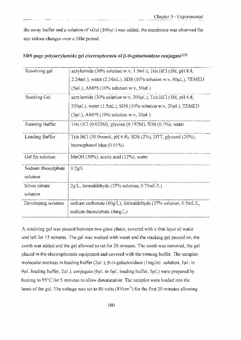
the assay buffer and a solution of xGal (1 OOIAL) was added, the membrane was observed for
any colour changes over a 24hr period.
SDS page polyacrylamide gel electrophoresis of f)-D-galactosidase conjugate208
Resolving gel acrylamide (30% solution w:v, l.SmL), Tris.HCl (1M, pH 8.8,
2.24mL), water (2.24mL), SDS (10% solution w:v, 60IAL), TEMED
(SIAL), AMPS (10% solution w:v, SOIAL)
Stacking Gel acrylamide (30% solution w:v, 200/-lL), Tris.HCI (1M, pH 6.8,
2S0/-lL), water (l.SmL), SDS (10% solution w:v, 20/-lL), TEMED
(3/-lL), AMPS (10% solution w:v, 2OIAL)
Running Buffer Tris.HCI (0.02SM), glycine (0. 192M), SDS (0.1%), water
Loading Buffer Tris.HCI (SO.Olmnol, pH 6.8), SDS (2%), DTT, glycerol (20%),
bromophenol blue (0.01 %)
Gel fix solution n.," \-' I~), ~~etic acid (12%), water
Sodium thiosulphate O.2g/L
solution
Silver nitrate 2g/L, formaldehyde (37% solution, 0.7SmL/L)
solution
Developing solution sodium carbonate (60g/L), fonnaldehyde (37% solution, O.SmL/L,
sodium thiosulphate (4mg/L)
A resolving gel was poured between two glass plates, covered with a thin layer of water
and left for IS minutes. The gel was washed with water and the stacking gel poured on, the
comb was added and the gel allowed to set for 20 minutes. The comb was removed, the gel
placed in the electrophoresis equipment and covered with the running buffer. The samples:
molecular markers in loading buffer (2IAL), f)-D-galactosidase (Img/mL solution, I/-lL in
9/-lL loading buffer, 2IAL), conjugate (6/-lL in 6/-lL loading buffer, SIAL) were prepared by
heating to 9SoC for S minutes to allow denaturation. The samples were loaded into the
lanes of the gel. The voltage was set to 80 volts (8Vcm-1) for the first 20 minutes allowing
180

Chapter 5 - Experimental
the samples to migrate through the stacking gel, and then raised to ISO volts for 2 hours.
The gel was removed from the plates and soaked in gel fix solution (200mL).
Silver staining
The gel was washed twice with EtOH (SO%) and once with EtOH (30%) for 20 minutes
each, and then submerged in sodium thiosulphate solution for exactly 1 minute. Rinsing
with water (x 3) for 20 seconds each was followed by incubation with the silver solution for
20 minutes at room temperature, then the gel was washed with water (x 3) and then placed
in the developing solution, at room temperature, for 10 minutes. The gel was rinsed with
water (x 2) for 2 minutes each, and then placed in gel fix solution for 10 minutes. Finally
the gel was washed with MeOH (SO%), MeOH (30%, 3% glycerol) and glycerol (3%) each
for 20 minutes. The gel was dried overnight.
Annealing molecular beacon to j)-D-galactosidase conjugate
The molecular beacon oligonucleotide (6!lL, 0.04S0D, [40]) was placed in a fluorimeter
cuvette (3mL) in buffer (sodium phosphate lOmM, EDTA ImM, NaCI O.lM or 1.0M, pH
7.0, 3mL). The oligonucleotide was heated and cooled between 20 to 70°C over time and
respective increases and decreases in fluorescence intensity monitored. A solution ofj)-D
galactosidase conjugate in water, was evaporated to dryness and dissolved in the same
buffer (0.1080D, 2.4eq, S!lL) and added to the cuvette at room temperature. The cuvette
was heated to 70°C over time and the fall in fluorescence intensity monitored as the
solution cooled to 20°C over time.
3% Agarose gel electrophoresis (mini gel)
A solution of agarose (1.S0g) in TEE buffer (lx, boric acid (1.lOg), Trizma (2.40g), EDTA
(0.19g) water (200mL), SOmL) was heated to boiling in a microwave oven for 4 minutes to
give a clear colourless viscous solution. The solution was allowed to cool (SO°C), SYBR™
green stain II (4!-AL) was added and the solution was poured slowly into the electrophoresis
apparatus. The gel was left to set (20 minutes). The comb was removed and the gel covered
in TBE buffer (200mL, with SYBR green stain 20!lL). Each sample (S!lL) was diluted with
181

a loading buffer (Tris.HCl (50.0mmol, pH 6.S), SDS (2%), DTT, glycerol (20%),
bromophenol blue (0.01 %), l!-!L) and loaded onto the gel. The gel was exposed to a current
of 100rnA, 100V. When the bands had migrated by the required distance, the current was
removed and the gel was visualised on a transilluminator platform at 260nm.
Chymotrypsin cleavage assay
The activity of chymotrypsin is defined as; one unit causes the hydrolysis of one micromole
ofbenzoyl-L-tyrosine ethyl ester (BTEE) per minute at 25°C at pH 7.5.
Resin cleavage- method I
The resin (l-2mgs) was placed in an UV cuvette in buffer (lmL, Tris.HCl buffer O.OSM,
calcium chloride O.lM, pH 7.S) and shaken. After equilibration at 37°C for 5 minutes,
chymotrypsin (100!-!L ofa 1mg rnL-1 solution) was added and the cuvette shaken and
placed in a UV/Vis spectrometer and the resin allowed to settle. The solution was
monitored at 3rC at either 260nm or 490nm, according to the oligonucleotide released, for
1 hr.
Resin cleavage- method 2
The resin containing a trityl ON oligonucleotide (5.Smgs, [104]) was placed in an
eppendorf in buffer (0.5mL, Tris.HCl buffer O.OSM, calcium chloride 0.1M, pH 7.S) and
shaken. After equilibration at 37°C for 5 minutes, chymotrypsin (1 00 ~tL of a 1 mg mL-1
solution) was added. The solution was incubated at 37°C in for 4 hrs. The resin was
removed, washed thoroughly and dried. The resin was analysed for trityl content (see
section 5.2.2.1)
Oligonucleotide cleavage (substrate limiting)
Into six tubes was placed oligonucleotide substrate (0.11S0D, 5.92!-!g, 1.25nmol) in buffer
(Tris.HCl buffer O.OSM, calcium chloride O.1M, pH 7.8, 100~tL). After equilibration at
37°C for 5 minutes, chymotrypsin (100 !-!L of a 1mg rnL-1 solution in buffer) was added to
each tube. After allotted periods of time (lOmins, 0.5hr, 1hr, 3hrs, 4hrs, 19hrs) each tube
was removed, placed at 95°C for 5 minutes, and the contents diluted with water (400!-!L)
182

Chapter 5 - Experimental
and transferred to a Micron™ filter device (Mol wt cut off= 18,000) and centrifuged at
13,000 rpm for 0.5hrs. The resulting filtrate was diluted with water (0.7mL) and analysed
by analytical RPHPLC.
Oligonucleotide cleavage (1:1 substrate: enzyme)
Into four tubes was placed oligonucleotide substrate (0.0590D, 2.96~tg, 62nmol) in buffer
(Tris.HCl buffer 0.08M, calcium chloride O.IM, pH 7.8, 50~L). After equilibration at 3rC
for 5 minutes, chymotrypsin (0.62 x 10⁻³ units, 1.25 ~L of a O.Img mL-1 solution in buffer)
was added to each tube. After allotted periods of time (Imin, IOmins, Ihr, 4hrs) each tube
was removed placed at 95°C for 5 minutes, diluted with water (400~L), transferred to a
Micron™ filter device (Mol wt cut off = 18,000) and centrifuged at 13,000 rpm for O.5hrs.
The resulting filtrate was diluted with water (0.7mL) and analysed by analytical RPHPLC.
Removal of Dde lysine side-chain protection from resin-bound oligonucleotide
Hydrazine monohydrate in DMF (2%, 200I1L) was added to the dry resin in a screw top
vial and allowed to stand, at room temperature, for 10mins. The solution was then removed
from the resin by filtration and a fresh portion of the hydrazine reagent (2%, 200I1L) added
to the resin and left standing for a further 10mins. The solution was removed from the resin
by filtration and the resin washed with DMF (5 x 2mL) and Et20 (3 x 5mL). The resin was
dried in vacuo.
Trypsin cleavage assay
Resin cleavage
The resin (2.8mgs, [113]) was exposed to a solution ofhydrazine monohydrate: DMF
(1:50) for 15 minutes, washed thoroughly and dried. The resin was placed in an eppendorf
in buffer (HEPES 0.1M, calcium chloride 0.5M, pH 7.30, 5mL) and shaken. After
equilibration at 37°C for 5 minutes, trypsin (100 ~L ofa Img mL-1 solution in same buffer)
was added. The solution was incubated at 3rC in for 4 hrs. The resin was removed,
washed thoroughly and dried. The resin was analysed for trity 1 content (see section 5.2.2.1).
183

Oligonucleotide cleavage (substrate limiting)
Into six tubes was placed oligonucleotide substrate (0.6160D, 25.6!lg, 3.54nmol) in buffer
(REPES O.lM, calcium chloride 0.5M, pH 7.3, lO0!lL). After equilibration at 37°C for 5
minutes, trypsin (100 !lL, ofa Img mL-1 solution in buffer) was added to each tube. After
allotted periods oftime (lOmins, 0.5hr, 1hr, 3hrs, 4hrs, 19hrs) each tube was removed
placed at 95°C for 5 minutes and the contents diluted with water (400!lL), transferred to a
Micron™ filter device (Mol wt cut off = 18,000) and centrifuged at 13,000 rpm for 0.5hrs.
The resulting filtrate was diluted with water (0.7mL) and analysed by analytical RPHPLC.
Oligonucleotide cleavage (1: 1 substrate: enzyme)
Into four tubes was placed oligonucleotide substrate (0.6160D, 25.6!lg, 3.54runol) in buffer
(HEPES 0.1M calcium chloride 0.5M, pH 7.3, 100!lL). After equilibration at 3rC for 5
minutes, trypsin (3.S4 x 10-3 units, 1.2!lL, of a O.OImg mL-1 solution in buffer) was added
to each tube. After allotted periods of time (Imin, 10mins, Ihr, 4hrs) each tube was
removed, placed at 9S0C for 5 minutes, diluted with water (400!lL), transferred to a
Micron™ filter device (Mol wt cut off = 18,000) and centrifuged at 13,000 rpm for O.Shrs.
The resulting filtrate was diluted with water (0.7mL) and analysed by analytical RPHPLC.
Annealing of solid supported molecular beacons with their solution targets
A molecular beacon oligonucleotide was assembled (standard phosphoramidite DNA
synthesis (0.2!lmol scale)) on resin (f12], [45]-[48]) The resin was placed in concentrated
atmnonia: EtOH (4: 1), at 55°C for 4 hours, removed, washed thoroughly with EhO (x 3)
and allowed to dry. A target solution oligonucleotide (lOO!lL, excess) was evaporated to
dryness and dissolved in buffer (Na2HP04/NaH2P04 10.Ommol, EDTA I.Ommol, NaCl
1M, pH 7.0, SO!lL). In three separate wells was placed (i) dry resin (ii) resin and buffer (iii)
resin and target in buffer. The plate was left for one hour, viewed and photographed under a
fluorescence microscope.
184

Chapter 6
Publication

The following published papers were included in the bound thesis. These have not been digitised due to copyright restrictions, but the links are provided.
Brown, Lynda J., Cummins, Jon, Hamilton, Alan and Brown, Tom (2000)
Molecular Beacons Attached to Glass Beads Fluoresce upon Hybridisation to Target DNA
Chemical Communications : Vol. 7, pp.621-622
http://dx.doi.org/10.1039/b000389l

Chapter 7
References

7 - References
7 .. 0 References
1. Watson, J.D., and Crick, F.H.C. Nature (London), 1953,171, 737.
2. Watson, ID., and Crick, F.H.C. Nature (London), 1953,172, 156.
3. Franklin, R, and Gosling, RG. Nature, 1953,172,156.
4. Franklin, R, and Gosling, RG. Nature, 1953, 171, 740.
5. Landridge, R, Wilson, H.R, Hooper, C.W., Wilkins, M.H.F., and Hamilton, L.D. J
Mol. BioI., 1960,3,547.
6. Landridge, R, Wilson, H.R, Hooper, C.W., Wilkins, M.H.F., and Hamilton, L.D. J
Mol. Biol., 1960,2, 19.
7. Avery, O.T, MacLeod, C.M., and McCarty, M. J Exp. Med., 1944,79,137.
8. Chargaff, E. Fed. Proc., 1951, 10, 654.
9. Turner, P.c., McLennan, AG., Bates, AD., and White, M.RH.Instant Notes in
Molecular Biology, BIOS Scientific Publishers Limited, 1998, 237.
10. Blackburn, G.M., and Gait, M.I Nucleic Acids in Chemistry and Biology, Oxford
University Press, 1996, 175.
11. Hayward, G. Applied Genetics, Macmillan Education Limited, 1990, 11.
12. Matthews, lA, and Kricka, L.I Anal. Biochem., 1988,169, 1.
13. Wetmur, lG. Crit. Rev. Biochem. Mol. Bio!., 1991,26,227.
14. Englisch, U, and Guass, D.H. Angew. Chem., Int. Ed. Engl., 1991,30,613.
15. Brown, T, and Grzybowski, l Gene Probes 1, Hames, B.D., and Higgins, S.l,
(eds.), Oxford University Press, 1995, 145.
16. Geever, RF., Wilson, L.B., Nallaseth, F.S., Milner, P.F., Bittner, M., and Wilson,
IT Proc. Natl. Acad. Sci. USA, 1981, 78, 5081
17. DiLella, AG., and Woo, S.L.C. Mol. BioI. Med., 1987,4,183.
18. Reeders, S.T, Breuing, M.H., Davies, K.E., Nicholls, RD., Jarman, A.P., Higgs,
D.R, Pearson, P.L., and Weatherall, DJ. Nature, 1985,317,542.
19. Kostrikis, L.G., Huang, Y, Moore, lP., Wolinsky, S.M., Zhang, L., Guo, Y,
Deutsch, L., Phair, I, Neumann, AU, and Ho, D.D. Nature Medicine, 1998,4,
350.
20. Jeffreys, Al, Wilson, v., and Thein, S.L. Nature, 1985,314,67.
185

7 - References
21. Rigby, P.W.J, Dieckmann, M., Rhodes, c., and Berg, P. J Mol. BioI, 1977,113,
237.
22. Feinburg, AP., and Vogel stein, B. Anal. Biochem., 1983,132,6.
23. Feinburg, AP., and Vogel stein, B. Anal. Biochem., 1984,137,266.
24. Arnheim, N., and Levenson, C.H. C&EN, 1990, October, 36.
25. Lenstra, JA Cell. Mol. BioI., 1995,41,603.
26. Ratliff, RL. The Enzymes, Boyer, P.D., (eds.), Academic Press, 1981, 105.
27. Tu, c.P., and Cohen, S.N. Gene, 1980,10, 177.
28. Southern, E.M. J Mol. BioI, 1975,98,503.
29. Haase, M.L., Walker, D., Stowring, L., Ventura, P., Geballe, A, Blum, H., Brahic,
M., Goldben, R., and O'Brien, K. SCience, 1985,227, 189.
30. Anderson, M.L.M., and Young, B.D. Nucleic Acid Hybridisation: A Practical
Approach, Hames, B.D., and Higgins, S.J, (eds.), IRL press, 1985, 73.
31. \A/ill, D.¥l., Pritchard, C.E., and T., B. Carbohydrate Res., 1991,216,315.
32. Grzybowski, J, Will, D.W., Randall, RE., Smith, C.A, and Brown, T. Nucleic
Acids Res., 1993,21, 1705.
33. Zomer, G., and Stavenuiter, JF.e. Anal. Chimica Acta, 1989,227, 11.
34. Kricka, L.J Clinical Chem., 1991,37, 1472.
35. Theisen, P., McCollwn, c., Upadhya, K., Jacobson, K., Vu, H., and Andrus, A
Tetrahedron Lett., 1992,33,5033.
36. Yamana, K., Ohashi, Y, Nunota, K., and Nakano, H. Tetrahedron, 1997,53,4265.
37. Teigelkamp, S., Ebel, S., Will, D.W., Brown, T., and Beggs, J.D. Nucleic Acids
Res., 1993,21,4651.
38. Smith, L.M., Sanders, Z.S., Kaiser, RJ., Hughes, P., Dodd, c., Connel, C.R,
Heiner, S., Kent, L.E., and Hood, L.E. Nature, 1986,321,674.
39. Reid, T., Baldini, A., Rand, T.e., and \Alard, D.C. Proc. 1\fat!. Acad. Sci. USA, 1992,
89, 1388.
40. Anderson, M.L.M. Gene Probes 2: A Practical Approach, Hames, B.D., and
Higgins, S.J., (eds.), Oxford University Press, 1995,3.
41. Murakami, A, Tada, J, Yamagata, K., and Takano, J. Nucleic Acids Res., 1989,17,
5587.
42. Van V'leeman, B.K., and Schuurs, A.H.W.M. FEBS Lett., 1979,107, 73.
186
7 - References
43. Diamandis, E.P., and Christopoulous, TK. Clinical Chem., 1991,37,629.
44. Zhu, Z., Chao, 1, Yu, H., and Waggoner, A.S. Nucleic Acids Res., 1994,22,3418.
45. Hakeem, M., and Sommer, S.S. Anal. Biochem., 1987,163,433.
46. Maier, M. Nucleosides & Nucleotides, 1995,14,961.
47. Holletz, T, U'.ve, M., Knaf, A., Reinhardt, R, and Cech, D. Liebigs Ann. Chern.,
1993, 1051.
48. Morrison, L.E., Halder, TC., and Stols, L.M. Anal. Biochem., 1989,183,231.
49. Varnosi, G., Gohlke, c., and Clegg, RM. Biophysical . .1, 1996, 71, 972.
50. Agrawal, S. Protocols for Oligonucleotide Conjugates - Synthesis and Analytical
Techniques, Agrawal, S., (eds.), Humana Press, 1994,93.
51. Connolly, B.A Nucleic Acids Res., 1985,16,8722.
52. Foster, AC., McInnes, J.L., Skingle, D.C., and Symons, RH. Nucleic Acids Res.,
1985,13, 745.
53. Keller, G.H., Huang, D.P., and !vianak, M.M. Anal. Biochern., 1989,177,392.
54. Langer, P.R, Waldrop, AA, and Ward, D.C. Proc. Natl. Acad. Sci. USA, 1981, 78,
6633.
55. Wiegant, 1, Verwoerd, N., Mascheretti, S., Bolk, M., Tanke, H.J., and Raap, AX .
.1 Histochem. Cytochem., 1996,44,525.
56. Leary, J.1., Brigati, DJ., and Ward, D.C. Proc. Natl. Acad. Sci. USA, 1983,80,
4045.
57. Reisfeld, A, Rothenburg, M., Bayer, E.A, and Wilchek, M. Biochem. Biophys. Res.
Commun., 1987,142,519.
58. Davidson, A., Duckworth, G., Rao, M.Y., McClean, J., Grzybowski, 1., Potier, P.,
Brown, T, and Cubie, H. Nucleosides & Nucleotides, 1995,14, 1049.
59. McClean, J., Davidson, A, Rao, M.Y., and Brown, T Biomedical Peptides,
Proteins & "A/ucleic ACids, 1996, 7-12, 7.
60. Wiegant, 1., Ried, T, Nederlof, P.M., Vans der Ploeg, M., Tanke, H.T, and Raap,
AK. Nucleic Acids Res., 1991,19,3237.
61. Jablonski, E., Moommv, E.W., Tullis, RH., and Ruth, 1L. Nucleic Acids Res.,
1986,14,6115.
62. Renz, M., and Kurz, C. Nucleic Acids Res., 1984, 12,3435.
63. Nucleic Acids Res., 1992,20,5061.
187
,,-,"'<AI-H''''' 7 - References
64. De Bellis, G., Consani, 1., Caramenti, G., Pergolizzi, R, Debernardi, S., Invernizzi,
L., and Luzzana, M. Biotechniques, 1994,16.
65. Whitcombe, D., Theaker, J., Guy, S.P., Brown, T., and Little, S. Nature Biotech.,
1999, In press.
66. Bergot, B.J, Chakerian, v., COlLTJdl, C.R, Eadie, JS., Fung, S., Hershey, N.D.,
Lee, L.G., Menchen, S.M., and Woo, S.L. Spectrally Resolveable Rhodamine Dyes
for Nucleic Acid Sequence Determination, U.S. Patent, Nov. 22, 1994, 1.
67. Chehab, F.F., and Kan, YW. Proc. l..fat!. Acad. Sci. USA, 1989,86,9178.
68. Diehl, H., and Markuszewski, R Talanta, 1989,36,416.
69. Science, AL. Amersham L?fe Science News, 1995, 17.2.
70. Funatsu, T., Harada, Y, Tokunaga, M., Saito, K., and Yanagida, T. ]'lature, 1995,
374,555.
71. Mujumdar, RB., Ernst, L.A, Mujumdar, RS., and Waggoner, AS. Cytometry,
1989,10, 11.
72. Yu, H., Chao, J, Patek, D., Mujumdar, R, Mujumdar, S., and Waggoner, AS.
Nucleic Acids Res., 1994,22,3226.
73. Zeng, Z., Benson, S.C., and Glazer, A~.N. ~4nal. BiocI1em., 1995,231,256.
74. Haugland, RP. Handbook of Fluorescent Probes and Research Chemicals,
Molecular Probes Inc., 1996, 6th ed., 13.
75. Metzker, M.L., Lu, J, and Gibbs, RA. SCience, 1996,271, 1420.
76. Sammes, P.G., and Yahioglu, G. Natural Products Reports, 1996, l.
77. Diamandis, E.P., and Christopoulos, TK. Anal. Chem., 1990,62, 1149.
78. Selvin, P.R, and Hearst, JH. Proc. Natl. Acad. Sci. USA, 1994,91, 10024.
79. Lakowicz, JR Principles of Fluorescence Spectroscopy, Plenum Press, 1983, 1.
80. Pesce, AJ, Rosen, CG., and Pasby, TL. Fluorescence Spectroscopy - An
Introductionfor Bio!o6'J} and lvfedicine, Pesce, AJ, Rosen, c.G., and Pasby, T.L.,
(eds.), Marcel Dekker, 1971, l.
81. Lakowicz, JR Principles of Fluorescence Spectroscopy, Plenum Press, 1983,
Chapter 9,257.
82. Eftink, M.R Biophysical and Biochemical Aspects of Fluorescence Spectroscopy,
Dewey, TG., (eds.), Plenum Press, 1991, 1.
83. Forster, T. x: Naturforschung, 1949, 4a, 321.
188

'--'U,UIJL'\.'" 7 - References
84. Stryer, L. Ann. Rev. Biochem., 1978,47,819.
85. V'/u, P., and Brand, L. Anal. Biochem., 1994,218, l.
86. Lakowicz, J.R Principles of Fluorescence Spectroscopy, Plenum Press, 1983,
Chapter 10,3.
87. Clegg, RM.1t1ethods Enzymol., 1992,211,353.
88. Clegg, RM., Murchie, A.I.H., Zechel, A., and Lilley, D.M.J Proc. Natl. Acad Sci.
US~, 1993,90,2994.
89. Mergny, lL., Boutorine, S.A., Garestier, T., Belloc, F., Rougee, M., Bulychev,
N.V., Koshkin, AA., Bourson, J., Lebedev, AV., Valeur, B., Thuong, N.T, and
Helene, C. Nucleic Acids Res., 1994,22,920.
90. Sixou, S., Szoka, F.C., Green, G.A., Giusti, B., Zon, G., and Chin, o.J Nucleic
Acids Res., 1994,22,662.
91. Mergny, JL., Garestier, T, Rougee,M., Lebedev, AV., Chassignol, M., Thuong,
N.T, and Helene, C. Biochemistry, 1994,33, 15321.
92. Clegg, RM., Murchie, AL, and Lilley, D.M. Biophysical J., 1994,66,99.
93. Jameson, D.M., and Hazlett, TL. Biophysical and Biochemical Aspects of
Fluorescence Spectroscopy, Dewey, TG., (eds.), Plenum Press, 1991, 105.
94. Gudgin Dickson, E.F., Pollak, A, and Diamandis, E.P. Pharmac. Ther., 1995,66,
207.
95. Hemmila, LA., Dakubu, S., Mukkala, YM., Siitan, H., and Lovgren, T. Anal.
Biochem., 1984, 137,335.
96. Evangelista, RA, Pollak, A, and Gudgin templeton, E.F. Anal. Biochem, 1991,
197,213.
97. Lakowicz, JR Principles o.fFluorescence Spectroscopy, Plenum Press, 1983,
Chapter 2. 19.
98. Perkin-Elmer Introduction to the LS50B Fluorescence Spectrometer: A
Luminescence Tutorial, Unpublished, 1999, 1.
99. Orlando, c., Pinzani, P., and Pazzagli, M. Clin. Chem. Lab. }v1ed., 1998,36,255.
100. Saiki, RK., Gelfand, o.H., Stoffel, S., Scharf, S.l, Higuchi, R, Hom, G.T, Mullis,
K.B., and Erlich, H.A Science, 1988,239,487.
101. Mullis, K. Scientific American, 1990,36.
189

""-u,u.IJV.,,, 7 - References
102. Jayasena, S.D. Advanced Target and Signal Amplification Strategies, IBC
Conference, San Diego, 1997, 1.
103. Swiger, RR, and Tucker, J.D. Environ. Mol. Mutagenesis, 1996,27,245.
104. Heng, h.H.Q., Spyropoulos, B., and Moens, P.B. BioEssays, 1997,19, 75.
105. Pinkel, D., Landegent, J., Collins, c., Fuscoe, 1, Segraves, R, Lucas, 1, and Gray,
1 Proc. Natl. Acad. Sci. USA, 1988,85,9138.
] 06. Lichter, P. TlG, 1997, 13,475.
107. Bischoff, F.Z., Nguyen, D.D., Burt, K.l, and Shaffer, L.G. Cytogenet. Cell. Genet.,
1994,66,237.
108. Kallioniemi, A, Kallioniemi, O.P., Piper, 1, Tanner, M., Stokke, T, Chen, L.,
Smith, H.S., Pinkel, D., Gray, lW., and \Valdermarln, :M. Proc. Nat!. Acad. Sci.
USA, 1994, 91,2156.
109. Livak, K.l, Flood, S.Al, Mannaro, 1, Giusti, W., and Deetz, K. PCR Methods and
Applications, 1995,357.
110. Heid, C.A, Stevens, 1, Livak, K.J, and Williams, P.M. Genome Res., 1996,6,989.
111. Morris, T, Robertson, B., and Gallagher, M. J. Clin. Microbio!', 1996,34,2933.
112. Kimura, B., Kawasaki, S., Fujii, T, Kusunoki, J., Itoh, T, and Flood, S.lA J. Food
Protection, 1999,62,329.
113. Laurendeau, I., Bahuau, M., Vodovar, N., Larramendy, c., Olivi, M., Bieche, I.,
Vidaud, M., and Vidaud, D. Clinical Chem., 1999,45,982.
114. Tyagi, S., and Kramer, F.R Nature Biotech., 1996,14,303.
115. Tyagi, S., Bratu, D.P., and Kramer, F.R Nature Biotech., 1998,16,49.
116. Vet, lA.M., Majithia, A.R, Marras, S.A.E., Tyagi, S., Dube, S., Poiesz, B.l, and
Kramer, Proc. Nat!. Acad. Sci. USA, 1998, September 1998.
117. Giesendorf, B.AJ, Vet, JAM., Mensink, E.IM.G., Trijbels, F.IM., and Bolm,
H.J Clinical Chem., 1998, 44, 482.
118. Kostrikis, L.G., Tyagi, S., Mhlanga, M.M., Ho, D.D., and Kramer, F.R SCience,
1998,279, 1228.
119. Gao, W., Tyagi, T, Kramer, F.R, and Goldman, E. A10lecular A1icrobio!., 1997,25,
707.
120. Matsuo, T Biochim. Biophys. Acta, 1998, 1379, 178.
121. Fang, x., Liu, x., Schuster, S., and Tan, \XI. J. Am. Chem. Soc., 1999,121,2921.
190

,"--,nuuv,"" 7 - References
122. Ortiz, E., Estrada, G., and Lizardi, P.M. Molecular and Cellular Probes, 1998,12,
219.
123. Piatek, A.S., Tyagi, S., Pol, AC., Miller, L.P., Kramer, F.R, and Alland, D. Nature
Biotech., 1998, 16,359.
124. Kramer, F.R, Tyagi, S., Vet, JAM., and Marras, S.AE. http.-!lmolecular-
beacons.org, 1999, 1.
125. De Francesco, L. The Scientist, 1998,12, 16.
126. Lyamichev, V, Brow, M.A.D., and Dahlberg, J.E. Science, 1993,260, 778.
127. Inc., T.W.T. http://www.twt.com/invader/invader.html. 1999, 1.
128. Egholm, M., Buchardt, 0., and Christensen, L. Nature, 1993,365,566.
129. NazarerJco, LA., Bhatnager, S.K., and Hohman, R.J ]\!ucleic Acids Res., 1997,25,
2516.
130. Nazarenko, 1. Advanced Target and Signal Amplification Strategies, IBC
Conference, San Diego, 1997.
131. Winndeen, E. S. Molecular Diagnosis, 1998, 3, 217.
132. Cronin, M.T., Fucini, RV, Kim, S.M., Masion, RS., Wespi, RM., and Miyada,
CG. Human A1utation, 1996, 7, 244.
133. Lemieux, B., Aharoni, A, and Schena, M. Molecular Breeding, 1998,4,277.
134. Southern, E.M. Electrophoresis, 1995,16, 1539.
135. I\1einkoth, J, and Wahl, G. Anal. Biochem., 1984,138,267.
136. Koch, J, Gregson, N., Kolvraa, S., and Bolund, L. Nucleic Acids Res., 1986, 14,
7133.
137. Ruth, JL. Protocols for Oligonucleotide Cop.jugates: Synthesis and Analytical
Techniques, Agrawal, S., (eds.), Humana Press, 1994, 167.
138. Chemicals, O.R Preparation of Alkaline Phosphatase-Oligonucleotide Conjugates,
Unpublished, 1998, 1.
139. Eckstein, F. Oligonucleotides and Analogues: A Practical Approach, IRL Press,
1991, 1.
140. Ravikumar, VT., Krotz, A.H., and Cole, D.L. Tetrahedron Lett., 1995,36,6587.
141. James, LW. Tetrahedron, 1999,55,4855.
142. Oikawa, Y, Tanaka, T., Horita, K., Yoshioka, T., and Yonemitsu, O. Tetrahedron
Lett., 1984,225,5393.
191

7 - References
143. Van Boom, J.H., and Burgers, P.M.J. Tetrahedron Lett., 1976,52,4875.
144. Koeners, H.l, Verhoeven, J., and Van Boom, J.H. J. Royal Netherlands Chem.
Soc., 1981,100,65.
145. Kwiatkowski, M., Nilsson, M., and Landegren, U. Nucleic Acids Res., 1996,24,
4632.
146. Huang, Z., Olson, N.A., You, W., and Haugland, RP. J. Immunol. Methods, 1992,
149,261.
147. Gee, K.R Bioorg. A1ed Chem. Lett., 1999,9, 1395.
148. Rotman, B., Zderic, J.A., and Edelstein, M. Proc. Nat!. Acad Sci. USA, 1963,50, 1.
149. Plovins, A., Alvarez, M., Ibanez, M., Molina, M., and Nombela, C. Appl. Envirn.
It1icrobiol., 1994,60,4638.
150. Craig, D., Arriaga, A., Banks, P., Zhang, Y, renborg, A., Palcic, M.M., and
Dovichi, N.J. Anal. Biochem., 1995,226, 147.
151. Huang, Z. Biochemistry, 1991,30,3535.
152. Huang, Z. Biochemistly, 1991,30,8530.
153. Ralston, E., Hjelmeland, L.M., Klausner, RD., Weinstein, J.N., and Blumenthal, R
Biochim. Biophys. Acta, 1981,649, 133.
154. Rossi, F.M., and Kao, P.Y Bioconjugate Chem., 1997,8,495.
155. Schmidt, RR, Gaden, H., and Jatzke, H. Tetrahedron Lett., 1990,31,327.
156. Rademann, l, and Schmidt, RR Tetrahedron Lett., 1996,37,3989.
157. Paulsen, H., and Lockhoff, O. Chem. Ber., 1981,114,3102.
158. Paulsen, H., Kutschker, W., and Lockhoff, O. Chem. Ber., 1981,114,3233.
159. "'1 orthington-Biochem. http://www.worthington-biochem.comlmanual/GIBG. html,
1999, 1.
160. Wong, S.S. Chemistry o/Protein Conjugation and Crosslinking, CRC Press Inc.,
1993, 1.
161. O'Sullivan, M.J., Gnemmi, E., Morris, D., Chieregatli, G., Simmonds, A.D.,
Simmons, M., Bridges, J.W., and Marks, V. Anal. Biochem., 1979,100, 100.
162. Kitaga\va, T, and i\ikawa, T J. Biochem., 1976, 79,233.
163. Ji, TH. Methods Enzymol., 1993,91,580.
164. Hashida, s., Imagawa, M., Inoue, S., Ruan, K.H., and Ishikawa J. Appl. Biochem.,
1984,6,56.
192

"-'H'"'-'V,"''' 7 - References
165. Young, B.D., and Anderson, M.L.M. Nucleic Acid Hybridisation: A Practical
Approach, Hames, B.D., and Higgins, S.J., (eds.), IRL press, 1985,47.
166. Bolton, E.T., and McCarthy, BJ. Proc. Natl. Acad. Sci. USA, 1962,48, 1390.
167. Kessler, C. Gene Probes 1: A Practical Approach, Hames, B.D., and Higgins, S.J,
(eds.), Oxford University Press, 1995, 126.
168. Wallenfels, K., and Weil, R The Enzymes, 1960, VII. 617.
169. Tool, P. http;llexpasy.hcuge.chlcgi-binlprotparamJ?P00722@nojt@, 1999, 1.
170. Pieles, u., Zurcher, W., Schar, M., and Moser, H. Nucleic Acids Res., 1993,21,
3191.
171.
172.
17'"' ~ . ;).
174.
175.
176.
177.
Potier, N., Dorsselaer, AV., Cordier, Y, roch, 0., and Bischoff, R Nucleic acids
Res., 1994,22,3895.
Calbiochem, and Biochem, N. Catalogue of reagents and resins for peptide
synthesis, Nova Biochem, 1999, 1.
Li, Vv., and Yan, B. 1. Org. Chem, 1998,63,4092.
Sherrington, D.C. Chem. Commun., 1998,2275.
Technologies, A http://www.argotech.coml. 1999, l.
-r..v1cMinn, D.L., and Greenberg, 11.M. Tetrahedron, 1996,52,3827.
Zhu, YF., Taranenko, N.I., Allman, S.L., Taranenko, S.A, Martin, S.A, Haff,
L.A, and c.H., C. Rapid Commun. Mass Spec., 1997,11, 897.
178. Srinivasan, JR, Liu, Y, Venta, P.J., Siemieniak, D., Killeen, AA., Zhu, Y, and
Lubman, D.M. Rapid Commun. Mass Spec., 1997,11, 1144.
179. DelltAquila, c., Imbach, J, and Rayner, B. Tetrahedron Lett., 1997,38,5289-5292.
180. Lyttle, M.H., Hudson, D., and Cook, R.M.l'lucleicAcids Res., 1996,24,2793.
181. Elmore, T.D., Guthrie, D.JS., Wallace, AD., and Bates, S.RE. Chem. Commun.,
1992, 1033.
182. Schuster, M., Wang, P., Paulson, IC., and Wong, C.H. 1. Am. Chem. Soc., 1992,
116, 1135.
183. Bohm, G., Dowden, J, Rice, D.C., Burgess, I., Pilard, J.F., Guilbert, B., Haxton, A,
Hunter, RC., Turner, N.J., and Flitsch, S.L. Tetrahedron Lett., 1998,39,3819.
184. Sauerbrei, B., Jungmann, v., and Waldmann, H. Angew. Chem., Int. Ed. Engl.,
1998,37, 1143.
193
'--'H'U-IJV'-'L 7 - References
185. Garcia-Lopez, M.T, De Las Heras, F.G., and Mendez-Castrillon, P.P. Syn.
Commun., 1985, 1121.
186. Patil, YJ. Tetrahedron Lett., 1996,37, 1481.
187. Han, X.B., Jiang, Z.H., and Schmidt, RR Liebigs Ann. Chem., 1993,853.
188. Greilich, U, Brescello, R, Jung, K.H., and Schmidt, RR. Liebigs Ann., 1996,663.
189. McDougal, P.G., Rico, 1.G., Oh, yr., and Condon, B.D. J. Org. Chem., 1986,51,
3388.
190. Cinget, F., and Schmidt, R.R. Synlett, 1993, 168.
191. Wang, Z.G., Douglas, S.P., and Krepinsk)', J.1. Tetrahedron Lett., 1996,37,6985.
192. Worthington-Biochem. http://www.worthington-biochem.com/manual/C/CHY. html,
1999, 1.
193. Kocian, 0., stransky, K., and Zavada, 1. Collection Czechoslovak Chem. Commun.,
1982,47, 1356.
194. Dreschsler, G.Y, and Smagin, S. J.jilr Praktische Chemie, 1965,28,315.
195. Greene, T Protecting Groups in Organic Synthesis, 1981,59.
196. Lakaye, B., Damblon, c., Jamin, M., Galleni, M., Lepage, S., Joris, B., Marchand-
Brynaert, c., Frydrych, c., and Frere, 1M. J. Biochem, 1994,300, 141.
197. Khanna, P.L., and Ullman, E.F. Anal. Biochem., 1980,108, 156.
198. Haynes, L.l., and Todd, A.R J. Chem. Soc., 1950,1,303.
199. Elmer, o.c. Chem. Abs., 1958,54, 6707b.
200. Pauslen, H., Schleyer, A, Mathieux, N., Meldal, M., and Bock, K. JChem.Soc.,
Perkin Trans., 1997,1,281.
201. \Vincott, F., DiRenzo, A., Shaffer, c., Grimm, S., Tracz, D., \Vorkmann, c., Sweedler, D., Gonalez, c., Scaringe, S., and Usman, N. Nucleic Acid5' Res., 1995,
23,2677.
202. Houille, 0., Schmittberger, T, and Uguen, D. Tetrahedron Lett., 1996,37,625.
203. Plattner, 11., Gless, RD., and Rapoport, H. JAm. Chem. Soc., 1972,94,8613.
204. Kocienski, P.l. Protecting Groups, Thieme, 1994, 1.
205. Curphey, T J Org. Chem., 1979,44,2805.
206. Vogel, A Textbook Of Practical Organic Chemistry, Longmann Group Limited,
1978,334.
207. Craven, G., Steers, and Anfinsen, C. J Bioi. Chem., 1965,240,2468.
194

~U~OJV~' 7 - References
208. Rosenburg, LM. Protein Analysis and Purification, Benchtop Techniques,
Birkhauser, 1996, 1.
195