update on gastric ulcer diseases – diagnosis and treatment
TRANSCRIPT

Update on Gastric Ulcer Diseases – Diagnosis and Treatment Frank M. Andrews, DVM, MS, Diplomate ACVIM
Email: [email protected]
Introduction Equine gastric ulcer syndrome (EGUS) is common in performance horses. Diagnosis of EGUS is
based on history, clinical signs, endoscopic examination, and response to treatment. All ages and breeds of horses are susceptible to EGUS and current therapeutic strategies focus on blocking gastric acid secretion and raising stomach pH. Omeprazole paste formulation (Gastrogard®, Boehringer-Ingelheim Animal Health, Decatur, GA) is currently approved by the FDA for treatment [4.0 mg/kg, orally, Q24h] and prevention of recurrence [2.0 mg/kg, orally, Q24h] of EGUS. Ulcergard® (Boehringer-Ingelheim Animal Health, Decatur, GA) is FDA-approved for prevention (1.0 mg/kg, orally, Q24h) of EGUS. However, a more comprehensive approach to EGUS includes determining and correcting of the underlying cause, environmental management, dietary manipulation and pharmacologic intervention. These proceedings focus on a comprehensive approach to treatment of EGUS and briefly cover basic anatomy and physiology of the equine stomach, multiple etiologies and risk factors for EGUS, and preventative management strategies.
The proximal third of the equine stomach is lined with non-glandular stratified squamous epithelium, an extension of the esophagus. The majority of ulcers are found in this region. The distal two-thirds of the stomach is covered by glandular mucosa, which is responsible for secretion of mucus, hydrochloric acid (HCl) and pepsinogen. Ulcer development in these regions is thought to be an imbalance between protective and aggressive factors. EGUS is an umbrella term used to describe ulcers in the nonglandular and glandular mucosa. Terminology has been expanded to include subgroups of EGUS depending the region affected, as etiology is likely different in these regions (Figure 1). The nonglandular squamous mucosa is predisposed to acid injury because it lacks the protective and buffering capacity provided by the bicarbonate-rich mucus found in the glandular region. The glandular region, on the other hand, has extensive protective mechanisms, including a bicarbonate-rich mucus layer, an extensive capillary network, and rapid restitution of epithelium when injured. Ulcers in this region are less common and heal rapidly. However, recently resistant glandular ulcers have been reported in horses and longer treatment periods may be necessary to heal these ulcers.

Figure 1. EGUS terminology and example of ulcers in the glandular region (photo to the left) and nonglandular region (photo right). Causes of EGUS
Horses are continuous gastric HCl secretors, and acid exposure is thought to be the primary cause of EGUS. Several acids (HCl, volatile fatty acids (VFAs), and bile acids) have been shown to cause damage to the non-glandular region of the equine stomach. In a recent report, HCl alone and in combination with VFAs (acetic, propionic, butyric and valeric acids) caused inhibition of cellular sodium transport, cell swelling, and eventual ulceration, when exposed to the non-glandular squamous mucosa at pH ≤ 4.0. Ulcerogenic effects of the VFAs in combination with HCl were dose dependent and the intensity of damage was correlated to VFA chain length. Bile acids were shown to increase the non-glandular mucosal cell permeability to hydrogen ions, which eventually lead to ulceration. However, the effects of bile acids in EGUS is questionable because they usually come from less acidic duodenal reflux and are non-ulcerogenic at a pH >4. Pepsinogen, which is cleaved to pepsin at a pH<4, might have a role in the development of EGUS. This proteolytic digestive enzyme may act synergistically with HCl to result in damage to the glandular and nonglandular mucosa. While HCl and stomach pH have been incriminated as the main culprits causing EGUS, it is likely that a combination of HCl, organic acids, and pepsin act synergistically to break down the protective barriers in the stomach and cause EGUS. Risk Factors
While acid injury has been implicated in the cause of EGUS, several risk factors have been identified. Recently a risk calculator was introduced for horse owners to see if their horses are at risk for gastric ulcers. Exercise Intensity
Horses involved in training and racing are at high risk to develop EGUS. Current prevalence figures show that 60 to 90% of performance horses have EGUS. It has been shown that abdominal pressures increased in horses running on a high-speed treadmill, which decreased stomach volume. Contraction of the stomach during exercise allowed acid from the dependent portion of the stomach to reflux up into the non-glandular mucosa leading to acid injury. Daily exercise may increase the exposure of the non-glandular mucosa to acids, explaining the increased prevalence of gastric ulcers in horses racing and training. Furthermore, serum gastrin concentration has been shown to increase in exercising horses, which might increase HCl secretion.
Intermittent vs. Continuous Feeding
Horses grazing at pasture have a decreased prevalence of EGUS. During grazing, there is a continuous flow of saliva and ingesta that buffers stomach acid, keeping stomach pH at ≥ 4 for a large

portion of the day. On the other hand, when feed is withheld from horses, before racing or in managed feeding stables, gastric pH drops rapidly and the non-glandular mucosa is exposed to an acid environment. Intermittent feeding has been shown to cause and increase the severity of gastric ulcers in horses and an intermittent feeding model has been used to consistently produce EGUS. The non-glandular mucosa is the most susceptible to ulceration in horses subjected to intermittent feeding due to its lack of mucosal protective factors; however, glandular ulcer number and severity might also increase. Diet Diet has been implicated as a risk factor for EGUS. Serum gastrin concentrations are high in horses fed high concentrate (grain) diets. Also, high concentrate diets are high in digestible carbohydrates (water soluble carbohydrate [WSC]). Water soluble carbohydrates are fermented by resident bacteria, resulting in the production of VFAs, which in the presence of low stomach pH (≤ 4), cause acid damage to the non-glandular squamous mucosa. Furthermore, two studies in horses fed alfalfa hay with and without grain (high protein and calcium) showed higher stomach pH and lower ulcer scores compared to horses fed grass hay (brome grass or Coastal Bermuda). Thus, feeding alfalfa hay has protective effects on the non-glandular mucosa in horses. Transport Stress
Transportation of horses has been associated with dehydration, increased threat of respiratory illness (pleuritis, pleuropneumonia), and immune suppression. When horses are being transported, water and feed consumption is usually decreased, which may cause an increased incidence of EGUS. Transportation has been shown to increase the severity of gastric ulcers in horses. However, a recent endoscopic study in western performance Quarter horses subject to frequent travel and intensive training found a lower prevalence (40%) of gastric ulcers than horses in race training, calling into question the effect of these factors on the development of EGUS in this group of Quarter horses. The authors speculated that demeanor played a role in the lower prevalence of ulcers in these horses. Quarter horses are calm and do not exercise intensively when compared to Thoroughbred racehorse, which may explain the higher prevalence of EGUS (93%) in racing and training Thoroughbreds.
Stall Confinement Horses housed in pastures have a decreased prevalence of gastric ulcers, compared to horses that are housed in stalls. The reason for this may be multifactorial, as horses that are stalled may be fed intermittently and housed without exposure to other horses. Non-Steroidal Anti-Inflammatory Drugs
Non-steroidal anti-inflammatory drugs (NSAIDs), phenylbutazone and flunixin meglumine, have been shown to induce gastric ulcers in horses. However, NSAIDs have not been shown to be a risk factor for EGUS in multiple epidemiologic studies in racehorses. In one study NSAIDs did cause ulcers in the glandular mucosa and increased the severity of ulcers in the non-glandular squamous mucosa. NSAIDs are thought to cause more severe ulcers in the glandular stomach mucosa because of their effect on prostaglandin inhibition. Prostaglandin inhibition results in decreased mucosal blood flow, decreased mucus production, and increased HCl secretion. While prostaglandins are also important in the regulation of acid production and sodium transport, it may be their effect on mucosal blood flow that is the most important. Adequate blood flow is necessary to remove the hydrogen ions that diffuse through the mucus layer covering the glandular mucosa. Gastric mucosal ischemia may lead to a hypoxia-induced cellular acidosis and release of oxygen free radicals, phospholipase, and proteases, which may damage the cell membrane leading to necrosis and ulceration.

Helicobacter spp.
While Helicobacter spp. is an important cause of ulcers in other species, it has not been proven to be a cause of gastric ulcers in horses. Helicobacter-specific DNA was isolated from glandular and non-glandular mucosa of horses and a new species of Helicobacter, H. equorium, was recently isolated from the feces of horses and foals. The importance of this discovery is unknown and the role of Helicobacter spp. in EGUS remains speculative, in light of several reports in which Helicobacter spp. were not seen in necropsy specimens from the stomach of horses with EGUS. However, colonization of gastric ulcers by resident stomach bacteria (E. coli, etc.) may inhibit healing and studies have shown that treatment with probiotics (lactobacillus spp.), antibiotics (SMZ), and antacids may facilitate healing of spontaneously occurring nonglandular gastric ulcers in horses and other species. This might also be true in horses with chronic non-responsive gastric ulcers. Clinical Signs Clinical signs associated with EGUS are numerous, and often vague. Ulcers are more common in horses showing clinical signs (Table 2). In Thoroughbred horses in race training, gastric ulcers were associated with poor performance, poor hair coat, partial anorexia (picky eating), and colic. Of horses with a client complaint of conditions associated with gastric ulcers, or showing subtle signs of poor health, gastric ulcers were identified in 88-92% compared to 37-52% identified in horses’ not showing clinical signs. In addition to an increased prevalence of ulcers in clinically affected horses, the severity of ulceration may be correlated with the severity of the symptoms. Diagnosis While an appreciation of the basic anatomy and physiology of acid production is important, it is equally important to be able to identify horses which would benefit from anti-ulcer therapy. Gastroscopy is the only method for definitive diagnosis of gastric ulcers currently available. The procedure for gastroscopy has been described in detail and information can be found on several websites including http://www.randlab.com.au/gastroscopy-resources.html and https://vimeo.com/219103551 (password: vet). In addition, several instructional videos can be found on YouTube. The procedure requires at least an endoscope of sufficient length to visualize the non-glandular mucosa, margo plicatus, and the pylorus (3 to 3.5 m in length. Once visualized, non-glandular and glandular ulcers should be scored for number and severity. Use of a scoring system allows the clinician to monitor healing and evaluate efficacy of treatment (Figure 2).
Lesion Number - Grade Descriptions 0 1 2 3 4
No lesions 1 - 2 localized lesions 3 – 5 localized lesions 6 – 10 lesions 10 or more lesions or diffuse (very large) lesions
Lesion Severity - Grade Descriptions

0 1 2 3 4
5
No lesions Appears superficial (only mucosa missing) Deeper structures involved (> depth than number 1) Multiple lesions and variable severity (1,2, and/or 4) Deeper structure involved (> depth than number 1) and has active appearance (hyperemic and/or darkened lesion crater) Same as number 4 plus hemorrhage or adherent blood clot
EGUS Scoring Descriptions 0 1 2 3 4
No ulcers Hyperemia or hyperkeratosis (no ulcers, record if score > 1) Small focal or multifocal ulcers Large focal or multifocal ulcers Large coalescing multifocal ulcers
Figure 2. Gastric ulcer scoring system (MacAllister et al. 1996; Sykes et al. JVIM, 2014). Currently there are no hematologic or biochemical markers to diagnose EGUS. However, O’Connor et al. recently evaluated the potential of a sucrose absorption test to diagnose gastric ulcers. Sucrose (table sugar) is a disaccharide broken down in the brush border of the intestine to glucose and fructose. If absorbed by the body, it must be absorbed across damaged gastric mucosa. It is not metabolized by the body, and is excreted in the urine as intact sucrose. Urine sucrose increased in horses with experimentally induced gastric ulcers. Initial evaluation was promising; however, recent studies have proven it to be not reliable to diagnose gastric ulcers. In addition, fecal occult blood tests and SAA testing has proven unreliable as well. Because of the lack of any additional laboratory diagnosis, in situations where ulcers are strongly suspected but gastroscopy is not available, it may be worthwhile to start empirical treatment and observe for resolution of clinical signs. If the horse does not respond to treatment, referral to a facility with a gastroscope is indicated. This is especially recommended if a trial period of treatment does not alleviate clinical signs in a few days. Treatment and Supplements Introduction
The goals of antiulcer therapy are to relieve pain, eliminate clinical signs, promote healing, prevent secondary complications, and prevent recurrence. The mainstay of EGUS treatment is to increase stomach pH and buffer or suppress HCl acid secretion. Because of the high recurrence rate of gastric ulcers in horses, initial effective pharmacologic therapy should be followed by modified management strategies and/or long-term pharmacologic treatment and/or feed supplementation. Pharmacologic Therapy
Once EGUS is diagnosed, therapy should be started to achieve the above outlined goals. Some ulcers heal spontaneously, but the majority of horses require pharmacologic therapy to achieve healing, especially while horses remain in athletic training and competition. There are many approaches to treating EGUS, but acid suppressive therapy remains the gold standard. Increasing gastric juice pH provides a suitable environment to allow ulcer healing. Many pharmacologic agents are available to treat and prevent gastric ulcers in man and other species, but omeprazole is the most effective in
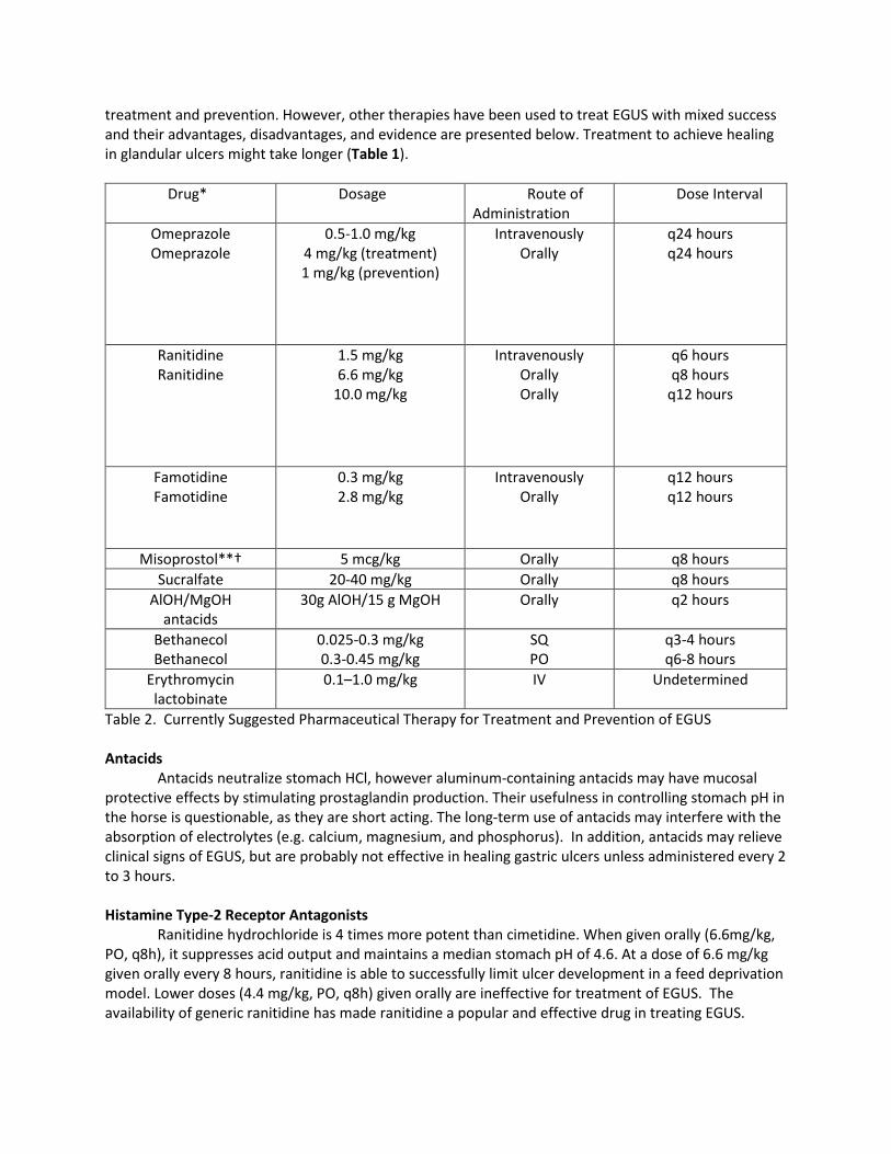
treatment and prevention. However, other therapies have been used to treat EGUS with mixed success and their advantages, disadvantages, and evidence are presented below. Treatment to achieve healing in glandular ulcers might take longer (Table 1).
Drug* Dosage Route of
Administration Dose Interval
Omeprazole Omeprazole
0.5-1.0 mg/kg 4 mg/kg (treatment) 1 mg/kg (prevention)
Intravenously Orally
q24 hours q24 hours
Ranitidine Ranitidine
1.5 mg/kg 6.6 mg/kg
10.0 mg/kg
Intravenously Orally Orally
q6 hours q8 hours
q12 hours
Famotidine Famotidine
0.3 mg/kg 2.8 mg/kg
Intravenously Orally
q12 hours q12 hours
Misoprostol**† 5 mcg/kg Orally q8 hours Sucralfate 20-40 mg/kg Orally q8 hours
AlOH/MgOH antacids
30g AlOH/15 g MgOH Orally q2 hours
Bethanecol Bethanecol
0.025-0.3 mg/kg 0.3-0.45 mg/kg
SQ PO
q3-4 hours q6-8 hours
Erythromycin lactobinate
0.1–1.0 mg/kg IV Undetermined
Table 2. Currently Suggested Pharmaceutical Therapy for Treatment and Prevention of EGUS Antacids
Antacids neutralize stomach HCl, however aluminum-containing antacids may have mucosal protective effects by stimulating prostaglandin production. Their usefulness in controlling stomach pH in the horse is questionable, as they are short acting. The long-term use of antacids may interfere with the absorption of electrolytes (e.g. calcium, magnesium, and phosphorus). In addition, antacids may relieve clinical signs of EGUS, but are probably not effective in healing gastric ulcers unless administered every 2 to 3 hours. Histamine Type-2 Receptor Antagonists
Ranitidine hydrochloride is 4 times more potent than cimetidine. When given orally (6.6mg/kg, PO, q8h), it suppresses acid output and maintains a median stomach pH of 4.6. At a dose of 6.6 mg/kg given orally every 8 hours, ranitidine is able to successfully limit ulcer development in a feed deprivation model. Lower doses (4.4 mg/kg, PO, q8h) given orally are ineffective for treatment of EGUS. The availability of generic ranitidine has made ranitidine a popular and effective drug in treating EGUS.

Ranitidine has efficacy for treatment of EGUS, but owner compliance is difficult as it must be given orally 3 times daily. While ranitidine and cimetidine have been the most studied, other H2 antagonists have been evaluated experimentally and may allow for less frequent dosing and more effective acid suppression. Bioavailability and pharmacodynamic studies with famotidine (2.8 mg/kg, PO, q12h; 0.3 mg/kg, iv, q12h) in horses suggest that it can be used for treatment of EGUS, but may be cost prohibitive. Proton Ion Pump Blockers (PPIs) Omeprazole
Omeprazole, a substituted benzimidazole, is the only FDA-approved drug for the treatment of EGUS. Omeprazole oral paste (GastroGard® Merial LTD, Duluth, GA, 4mg/kg, PO, q24h) inhibits gastric acid secretion for 24 hours in horses. In an acid environment omeprazole is activated to a sulphenamide derivative and binds reversibly to the H+/K+ ATPase in parietal cells, inhibiting transport of hydrogen ions into the stomach. Because of its effect on the cell, omeprazole is often called a proton-pump blocker. The effect on gastric acid secretion is dose and time dependent. Omeprazole has been shown to be an effective treatment for EGUS in the non-glandular stomach and a lower dose, Ulcergard paste® (Merial LTD, Duluth, GA), has been shown to prevent ulcer recurrence. A recent study of 565 horses in race training found that 96% of the 147 horses being administered H2 antagonists had gastric ulcers, with 61% considered to be severely affected. Of the horses not receiving H2 antagonists, 88% had gastric ulcers, with 58% considered severe. All of the horses in the study were put on a 28-day course of omeprazole. There was a statistical improvement in performance, weight gain, attitude, appetite, and appearance after treatment. Endoscopically 65% of the horses with gastric ulcers that were treated were healed and 94% were improved. The primary reason for the failure of treatment with H2 antagonists was owner compliance and incorrect dosing. A second study found that 99% of spontaneous ulcers in adult horses and foals over 4 weeks of age were improved with 86.7% healed with omeprazole treatment. Effectiveness of omeprazole was also shown to increase the rate of healing in horses with ulcers removed from race training. Coating and Binding Agents
Sucralfate and bismuth subsalicylate are two compounds that bind to stomach ulcers and promote healing. Sucralfate is a hydroxyl aluminum salt of sucrose octasulfate and binds to the negatively charged particles in the ulcer bed, buffering HCl by increasing bicarbonate secretion, stimulating prostaglandins production, and adhering to the ulcer bed. In the stomach, sucralfate is converted to a sticky amorphous mass, thought to prevent diffusion of hydrogen into the ulcer. In a clinical trial in horses, sucralfate (22 mg/kg, PO, q8h) did not improve subclinical ulcer healing in 6- and 7-month old foals. In a rat model sucralfate successfully prevented gastric ulceration in a dose dependent manner after an ischemia reperfusion injury. Recently, GastrafateTM (20-30 ml, PO, q12h; MFP, Ltd, Sterling CT), an emulsion (10% Sucralfate, Calcium Carbonate, DASC, Magnesium Hydroxide, Carboxylate, Apple Flavor, Methyl & Propyl Parabens, and Water) has been advertised as a treatment for EGUS. This quantity of Gastrafate would provide 3 g of sucralfate to a patient, which is significantly less than the 12 g currently recommended for treatment of EGUS, but the product might be more potent than sucralfate alone. However, this compound might be more effective when used in horses with glandular ulcers of the stomach.
Bismuth containing compounds may have a coating effect similar to sucralfate. Additionally, they will inhibit the activation of pepsin and increase mucosal secretion. A compound containing 26.25 g of bismuth failed to raise the pH in 5 horses. Bismuth subsalicylate may be converted to sodium subsalicylate in the gastrointestinal tract, which may cause gastric irritation. Also, salicylates, similar to aspirin, decrease prostaglandin secretion and may further compromise an already damaged mucosa.

Thus, compounds containing bismuth are not recommended for treatment of EGUS. However, bismuth is used as part of the therapy in humans with Helicobacter pylori induced gastric ulcers and may be used in horses with chronic recurring gastric ulcers in which Helicobacter is suspected. Antibiotics (Helicobacter spp. and other bacteria)
Treatment of Helicobacter pylori infection in humans greatly accelerates ulcer healing, and eradication reduces the risk of reoccurrence. It has been recommended that oral combination therapy in horses with chronic active, non-healing ulcers could consist of omeprazole, metronidazole and/or trimethoprim/sulfa and bismuth subsalicylate (3.8 mg/kg q6hrs). An initial 14-day treatment period could be prescribed, which should be followed by gastroscopy. Omeprazole therapy should be continued for the full 28 days if ulcers are present at the 14 day endoscopy. Duration of Treatment It is difficult to predict the duration of gastric ulcer therapy, so it must be tailored for the individual horse. In a feed deprivation model of ulcer induction, ulcers were healed or almost healed in the horses after 9 days of pasture turnout. Omeprazole treatment in Thoroughbred horses in training resulted in healing in 57%, 67%, and 77% of horses treated for 14 days, 21 days, and 28 days, respectively. Horses with spontaneous occurring ulcers in a field trial treated with omeprazole showed 86% healing after 28 days of treatment. We recommend endoscopic examination after 14 days of omeprazole therapy to determine if the ulcers are healed. If the gastric ulcers are healed, then the horse can be put on omeprazole (Ulcergard®) to prevent recurrence of ulcers while the horse is in training. If the ulcers are still present, then the full 28-day course of omeprazole should be followed, and the horse further evaluated after that time. When endoscopy is not available, horses should be treated for at least 28 days. It should be noted that clinical signs might resolve before complete healing has taken place. Signs of poor appetite, colic or diarrhea will usually resolve soon after initiating treatment, and the horse is expected to make improvements in body condition and attitude within two to three weeks. H2 antagonist therapy should be continued for at least 14-21 days, but healing has taken over 40 days in some studies.
In general, it may take longer to treat large ulcers, more severe ulcers, and ulcers in the non-glandular mucosa. Likewise, ulcers in the glandular stomach might take up to 60 days to see significant healing. In cases where clinical signs have resolved and the risk factors for ulcer development are low, spontaneous healing of ulcers may occur without further treatment. However, spontaneous healing will not occur in horses that continue intensive training, and ulcers may re-occur in those successfully treated if therapy is discontinued. If clinical signs attributed to EGUS have not resolved after 48 hours of treatment, the diagnosis or therapy should be re-considered. Environmental, Nutritional, and Dietary Management
Pharmacologic therapy may be necessary to heal ulcers in horses, but once pharmacologic therapy is discontinued, the ulcers will likely quickly return if management changes are not instituted. Environmental, nutritional, and dietary management can be initiated during therapy to help facilitate ulcer healing and ultimately prevent ulcer recurrence. Intense or long-duration exercise, stall confinement, and diet are risk factors for EGUS. Managing these risk factors can decrease severity and prevent ulcer recurrence. Modification of Exercise Intensity and Duration
In horses, intense exercise, racing, and race training has been shown to contribute to worsening of squamous ulcers compared with horses kept at pasture or not in training. Also, endurance exercise has been shown to play a role in the cause of EGUS in horses. In one study, 67% of horses competing in

50- and 80-mile endurance rides had gastric ulcers. In addition, repeated oral administration of hypertonic replacement electrolyte solutions to these horses was shown to increase the number and severity of gastric ulcers. Oral hypertonic electrolyte replacement products should be used with caution in exercising horses and should be administered after exercise with a small grain or hay meal. Furthermore, training and exercise intensity should be reduced, when possible, until ulcers have healed. Pasture Turnout
One study found that even giving a horse in a stall ad lib grass hay did not improve ulcers, whereas horses maintained on pasture rarely had gastric ulcers. Pasture turnout is the best dietary method of controlling gastric ulcers. The diet fed to stalled horses can be modified, however, to decrease the risk of ulcers. Current dietary recommendations include providing continuous feeding of alfalfa hay or alfalfa hay mixed with good quality grass. Sweet feed should be kept to a minimum, and grains like barley or oats can be substituted to decrease its fermentation to VFAs. Eliminate Bolus Feeding and Increase Forage and Fiber
Another effective method to decrease EGUS is to feed a high-forage diet continuously. Stabled horses are frequently fed two large meals daily, which results in lower saliva production and increases the rate and extent of intragastric fermentation and the gastric emptying rate. Diet composition, meal size, and feeding frequency have been shown to contribute to the cause of gastric ulcers in horses. In the horse, one study showed that the ingestion of a grain meal resulted in a higher gastrin stimulus than grass hay. Horses fed hay versus withholding feed had similar acid output, but higher gastric pH. It was theorized that the salivary bicarbonate and the buffering effect of the hay were responsible for the higher pH. The composition and frequency of feeding forage is important in preventing EGUS.
In addition to providing constant access to feed to buffer gastric acidity, modifying the diet may help prevent ulcers. Although the gastric juice VFA concentrations were higher in horses fed an alfalfa-grain diet, they had higher gastric juice pH and lower ulcer scores than the same horses fed Brome grass hay. In that study, no gastric hormones were measured, and it was hypothesized that the calcium in the alfalfa hay could have a direct effect on gastric secretions or that the protein was acting as a buffer for the pH. In a rat model, a diet of 2% calcium inhibited basal gastric secretion but not secretions in response to histamine stimulation. Providing constant access to alfalfa hay likely helps to raise gastric pH. Horses fed straw as the only forage available were 4.4 times more likely to have a gastric ulcer severity score greater than or equal to 2 (0–5 scale) compared with hay feeding. Also, horses fed straw were 5.7 times more likely to have a gastric ulcer severity score of greater than or equal to 2, compared with haylage feeding. Also, horses fed greater than 2 g/kg body weight (BW) of starch per day were likely to have a twofold increase in gastric ulcer severity score of greater than or equal to 2, compared with feeding haylage. Although the absolute fiber and grain requirements have not been determined for horses, current recommended levels of long-stem, high-quality forage are at 1 to 1.5 kg/100 kg of BW and 0.5 kg/100-kg BW concentrates. Straw should not be fed as a sole source of forage.
Decrease Size and Increase Frequency of Concentrate Feeding
Serum gastrin concentrations are high in horses fed high-concentrate diets. Gastrin is the only hormone known to stimulate secretion of hydrochloric acid, and rations that contain more readily available nutrients, such as pellets and sweet feed, produce a significant increase in postprandial gastrin concentrations. In particular, grain feeding was shown to delay gastrin secretion, which corresponded to an increase in gastric acid secretion after the stomach had emptied the grain contents. In an empty stomach acid is more likely to be exposed to non-glandular mucosa and cause injury, similar to gastroesophageal reflux disease in people. Also, high-concentrate diets are high in hydrolysable (water soluble) carbohydrates. Hydrolysable carbohydrates are readily fermented by resident stomach bacteria,

resulting in the production of VFAs, which in the presence of a low stomach pH (<4) cause damage to the non-glandular squamous mucosa.
The size of the grain meal may also affect the extent of intragastric fermentation, thereby affecting VFA production. Metayer and colleagues compared the gastric emptying rates in horses fed small (300 g/100 BW) versus large (700 g/100 BW) high-starch concentrate meals. Although the calculated rate of gastric emptying (grams per minute) was higher with the large meal, gastric emptying in terms of percent of the original meal was much slower. When horses are fed large, starch-rich meals, intragastric fermentation and VFA fermentation may be favored because of the large amount of fermentable carbohydrates and the longer retention time within the stomach. In the same study, when comparing the high- and low-starch meals, gastric emptying was significantly faster for horses consuming a meal lower in starch than one high in starch. Larger meal size and higher starch content were associated with gastric emptying in terms of percent of total original meal. A recent study showed that when grain was fed at 0.5 kg/100 kg BW, VFA concentrations were below threshold values for causing damage to nonglandular mucosa. Grain or concentrates should not be fed in excess of 0.5 kg/100 kg BW every 6 hours. Antibiotics vs. Probiotics
Helicobacter pylori and other Helicobacter species have not been shown to cause EGUS, although Helicobacter DNA has been isolated from the glandular and non-glandular stomach mucosa in horses. Instead, other resident, acid-tolerant bacteria (Escherichia coli, Lactobacillus, and Streptococcus) are suspected to cause EGUS, and a large population of these bacteria was isolated from the gastric contents of horses fed various diets in one study. In rats, which have a compound stomach similar to horses, bacteria (E. coli) rapidly colonized acetic acid-induced stomach ulcers and impaired ulcer healing. In this study, oral antibiotic treatment with streptomycin or penicillin suppressed bacterial colonization of the ulcer and markedly accelerated ulcer healing compared with placebo-treated controls. Also, oral administration of lactulose resulted in increased Lactobacillus growth and colonization of the ulcer bed, which may facilitate ulcer healing. In a recent study in horses with spontaneously occurring gastric ulcers, an antibiotic (trimethoprim sulphadimidine) or a probiotic preparation containing Lactobacillus agilis, L. salivarius, L. equi, Streptococcus equinus, and S. bovis administered orally decreased ulcer number and severity compared with untreated controls. These data suggest that resident stomach bacteria are important in maintenance and progression of non-glandular gastric ulcers in horses. Treatment with antibiotic or probiotic preparations may facilitate ulcer healing after 2 weeks of treatment, but a full effect did not occur until after 4 weeks of treatment. Antibiotic treatment may be indicated in horses with chronic nonresponsive gastric ulcers, but more importantly, probiotic preparations containing Lactobacillus and Streptococcus may be helpful in prevention of gastric ulcers or may be used as an adjunct to pharmacologic treatment. Dietary Supplements
A plethora of dietary supplements on the market for horses boast efficacy in treatment and prevention of gastric ulcers. Many of these products have not been tested in the horse, and to date, very little scientific evidence exists on their efficacy. Discussed next are several supplements that have some scientific testing or have ingredients that have been shown to be helpful in ulcer treatment and prevention (Table 5). Sea Buckthorn Berry Extract
There is an increasing interest in the use of herbs and berries that may have therapeutic application in humans and animals. Berries and pulp from the sea buckthorn plant (Hippophae rhamnoides) are high in vitamins, trace minerals, amino acids, antioxidants, and other bioactive

substances and have been used successfully to treat mucosal injury, including decubital ulcers, burns, and glandular and duodenal ulcers in humans. In addition, sea buckthorn berries have been shown successfully to treat and prevent acetic acid-induced gastric ulcers in rats. Two previous studies failed to show failed to show efficacy in treatment or prevention of squamous ulcers in horses fed supplements containing sea buckthorn berry pulp and extract (SeaBuck Complete, Seabuck, LLC, Midvale, Utah), however glandular ulcer scores were significantly improved when compared to untreated controls in the later study. Sea buckthorn berries are high in antioxidant, flavonols, Vitamin C, and Vitamin E, which have been shown in many in vitro and in vivo studies to facilitate healing of glandular ulcers other species.
Calcium Carbonate and Gastroprotectants
Many supplements on the market contain calcium carbonate, sodium bicarbonate and other antacids. These products contain varying concentrations of calcium carbonate and various other herbs and coating agents. The author (FMA) performed a small study with an antacid preparation containing calcium carbonate (Neigh-Lox, Kentucky Performance Products, Versailles, Kentucky, 124.5 g, top-dressed on feed, Q12h, for 3 weeks.) and found no effect on squamous ulcer scores in a small study (published data, 2001); however, gastric juice pH, as measured by an indwelling pH meter was increased (≥ 4) 2 hours after feeding this supplement. In addition, calcium carbonate resulted in recovery of VFA-induced sodium transport in squamous tissue in an in vitro Ussings chamber system. These data suggest that calcium carbonate preparations may have some efficacy in maintaining mucosal integrity, but because of the short duration of effect on gastric juice pH, more frequent feedings may be necessary to prevent squamous ulcers.
Two other commercially available supplements (Egusin® 250; E-250 and Egusin® SLH; E-SLH, Centaur, Inc. Overland Park, KS) containing sodium bicarbonate, calcium carbonate, pectin and lecithin, were evaluated and both supplements showed improvement in squamous ulcer scores, compared to untreated control, after 35 days of treatment in stall-confinement horses. There was no effect on gastric juice pH in this study and blood gases did not show a significant increase in total carbon dioxide.
The other ingredients that might have been effective were pectin and lecithin. In horses, the surface of the NG stomach is coated by dense osmophilic surface-active phospholipids (SAPL), similar in structure to pulmonary surfactant. Because the NG mucosa has only a very thin to nonexistent mucus layer, no bicarbonate secretion compared to the glandular mucosa, and lacks the bicarbonate-mucus layer that is present in the glandular mucosa, these hydrophobic SAPLs may provide the primary defense mechanism against hydrochloric and other organic acids in gastric fluid. Feed additives containing pectin and lecithin have been previously evaluated in horses with gastric ulcers and the results have been mixed. Pectin is a soluble complex polysaccharide derived from the cell wall of fruits and vegetables. Lecithin is a phospholipid derived from soybeans. It is an emulsifier, lubricant, and surfactant. Pectin acts with lecithin to act to form a hydrophobic (water repellent) barrier on the gastric mucosal membranes, protecting them against the corrosive effects of gastric acids. Hydrolyzed Collagen Porcine hydrolyzed collagen (HC; Hydro-P Premium, Sonac, A Darling Ingredient Company, Son, The Netherlands) was recently evaluated in stall-confined horses treated with omeprazole and undergoing intermittent feeding. The HC (45 g) was mixed with sweet feed twice daily for 56 days in a 2-period crossover study. Mean gastric juice pH was higher in the HC-treated horses while the horses were on omeprazole treatment. In addition, HC-treated horses resulted in fewer ulcers at each gastroscopic examination and a significant effect on ulcer scores was seen on day 56 of treatment. Multiple Component Feed Supplement

In a recent study, using a commercially available supplement (SmartGut® Ultra, SmartPak, LLC, Plymouth, MA, USA, 40 g, mixed with grain feed, Q12h) containing a proprietary blend of Sea Buckthorn, Pectin, Lecithin, and other ingredients) showed a significant decrease in the number of squamous ulcers 14 days after omeprazole treatment was discontinued and fewer squamous ulcers after a week of alternating feed deprivation, in the treated group compared to untreated controls. Feeding a supplement with sea buckthorn berries, pectin, lecithin, and antacids, such as SmartGut® Ultra might be an affordable alternative to aid in the protection of the squamous mucosa from acid injury in stall-confined horses undergoing intermittent feeding. Chelated Minerals
A recent study used a chelated mineral supplement (Zinpro Performance Minerals (zinc methionine, copper lysine, manganese methionine, and cobalt glucoheptonate), Zinpro, Inc., Eden Prairie, MI, USA). In that study, ZPM-treatment resulted in lower gastric ulcer scores in horses participating in the later period of this study, when the nutritional plane was higher. ZPM at levels fed in this study may be beneficial in preventing gastric ulcers after omeprazole treatment in horses housed in stalls and fed intermittently, especially when horses are fed a good nutritional diet. Oils (Corn Oil, Rice Bran Oil)
Dietary fats delay gastric emptying time in humans and other species. In contrast to most species, gastric emptying rates are slower in horses fed a high-carbohydrate diet, compared with horses fed a high-fat diet, although these rates were not statistically significant. Gastric relaxation was significantly greater in horses fed the high-carbohydrate diet, however, compared with horses fed the high-fat diet. Supplementation of dietary fat may not have a profound effect on gastric emptying in horses. In another study, ponies fitted with gastric cannulas fed dietary corn oil (45 mL, orally, once daily) by dose syringe had a significantly lower gastric acid output and increased prostaglandin concentration in gastric juice. The authors’ concluded that corn oil supplementation could be an economical approach to the therapeutic and prophylactic management of glandular ulcers in horses, especially those associated with the use of nonsteroidal anti-inflammatory drugs. In contrast to the previous study, results from an evaluation of the antiulcerogenic properties of corn oil, refined rice bran oil, and crude rice bran oil (240 mL, once daily, mixed in grain) showed no statistical differences in non-glandular ulcer scores between the treatment groups. Glandular ulcers were rare, however, in these horses. In this model, dietary oils did not prevent non-glandular gastric ulcers in these horses, suggesting that dietary oils may not be useful in treatment or prevention of non-glandular ulcers, but may be helpful in treatment or prevention of glandular ulcers, since alteration in the protective barrier is the primary cause of glandular ulcers. Concentrated Electrolyte Pastes or Solutions
Repeated oral administration of hypertonic replacement electrolyte solutions, commonly given to endurance horses, has been shown to increase the number and severity of gastric ulcers. These products should be used with caution in horses and may be best given after exercise with feed to minimize their effects on the gastric mucosa. Summary
EGUS is common in horses; stomach acids and environmental, nutritional, and dietary factors are likely important causative factors. On initial diagnosis of EGUS, treatment should be started with effective pharmacologic agents. Prevention of ulcer recurrence depends primarily on environmental, nutritional, and dietary management. When possible, the following summary of important nutritional and dietary recommendations should be followed to lessen severity and prevent EGUS:

1. Keep the horse eating by providing a minimum of 1 to 1.5 kg/100 kg BW of long-stem,
high-quality forage free-choice throughout the day and night. 2. Feed alfalfa hay or a mixture of alfalfa hay to help buffer stomach acid. 3. Feed grain and concentrates sparingly. Give no more than 0.5 kg/100 kg BW of high
starch grain (e.g. sweet feed), and do not feed grain meals less than 6 hours apart. 4. Corn oil or other proven dietary supplements may be helpful to prevent recurrence of
ulcers. 5. Feed hypertonic electrolyte pastes after exercise with a grain and hay meal to avoid
irritation of the gastric mucosa. 6. Consider therapeutic or preventative doses of effective pharmacologic agents
(Gastrogard® or Ranitidine) in horses that are performing high-intensity exercise, traveling, or in a high-stress situation.
7. Use of supplements might benefit horses, especially maintaining stomach health after pharmacologic therapy. Use supplements that have scientific data to back them up!
References Upon Request
Table 1: Physiologic Factors Affecting Ulcer Development AGGRESIVE FACTORS PROTECTIVE FACTORS
NON-GLANDULAR MUCOSA GLANDULAR MUCOSA Hydrochloric acid secretion Epithelial restitution (poor) Epithelial restitution (rapid) Organic acid production (VFAs) Intracellular bicarbonate Bicarbonate-mucus layer Pepsin conversion from Mucosal blood flow, poor Mucosal blood flow, extensive pepsinogen Prostaglandin E production Duodenal reflux of bile acid Table 2: Clinical Signs and Risk Factors of EGUS Table 3: EGUS Risk Calculator for Horse Owners (adapted from Barakat C (2016).
Clinical Signs Adults Foals Risk Factors Acute colic Diarrhea Stress Recurring colic Abdominal pain Transportation Excessive recumbency Restlessness High-grain diet Poor body condition Rolling Stall confinement Partial anorexia Lying in dorsal recumbency Intermittent feeding Poor appetite Excessive salivation Intense exercise Poor performance/training Bruxism Racing Attitude changes Intermittent nursing Illness Stretching often to urinate Poor appetite NSAID use Inadequate energy Management changes Chronic diarrhea.

Table 4: Drug Therapy for Treatment of EGUS
Drug Dosage Dosing Interval Route of Administration Omeprazole 1.0 mg/kg Intravenously Q 24 hrs Omeprazole 4 mg/kg Orally Q 24 hrs Omeprazole (prevention) 1 mg/kg Orally Q 24 hrs Ranitidine 1.5 mg/kg Intravenously Q 6 hrs Ranitidine 6.6 mg/kg Orally Q 8 hrs Famotidine 0.3 mg/kg Intravenously Q 12 hrs Famotidine 2.8 mg/kg Orally Q 12 hrs Pantoprazole 1.5 mg/kg Intravenously Misoprostol 5 mcg/kg Orally Q 8 hrs Sucralfate 12-20 mg/kg Orally Q 8 hrs to Q 6 hrs AlOH/MgOH antacids 30g AlOH/15 g MgOH Orally Q 2hrs Bethanechol 0.025 – 0.30 mg/kg Subcutaneous Q 3-4 hrs Bethanechol 0.3-0.45 mg/kg Orally Q6-8 hrs Erythromycin lactobionate
0.1 – 1.0 mg/kg Intravenously Undetermined
*Cimetidine: Not effective for treatment of EGUS Table 5: Summary of natural products that recently were studied for the treatment of equine gastric (glandular and/or non-glandular) ulcer syndrome.
Name of product Name of Scientists Date of research
Sea buckthorn (Hippophae rhamnoides) Huff et al. 2009
Gastric Ulcer Risk Score1 2 3 4 5 Score
3 or more 2 1
75% 50% 75%
None ≤ 10 days > 10 days
Spend 1 day away at show, clinic or otherTook a trailer ride
Attended competition or clinic at unfamiliar location
Change in herd dynamicsSustained injury or developed illness
None 1 or 2 ≥ 3Total
*RiskScore: 0-5: Low Risk; 6-15 Moderate Risk; 16-25: High RiskBarakat C. What's your horse's ulcer risk. Equus 2016; 464:68-77.
2. How much of the day does your horse spend grazing or eating hay?
3. How many days in a row has your horse been on NSAIDs in the past 3 months?
4. Over the past 3 months, which of the following apply to your horse?
1. How many meals is your horse's grain ration divided into per day?
5. How many days per week is your horse in intense work?

Egusin® Woodward et al. 2014
Apolectol, Saccharomyces cerveisiae, Magnesium hydroxide
Sykes et al. 2014
Pectin-lecitin Sanz et al. 2014 SmartGut® Ultra Andrews et al. 2015
GastroTech Conover et al. 2015 Alfa-Lox-Forage Andrews et al.
Garza, Jr. et al. 2016
Hydrolyzed Collagen Keowen et al. 2016 Succeed paste Kerbyson et al. 2016