urology module…semester 4 lecture name: anatomy of the
TRANSCRIPT

Urology module…Semester 4 Lecture name: Anatomy of the urinary system. By: Dr. Salam Abdulameer Almosawi F.IC.M.S.(URO). [email protected].

Objectives:
1. the gross structure of the urinary system in both the male and female
2. the overall functions of the urinary system
3. the anatomical position of the kidneys ,ureters , the bladder and the prostate and their relationships.
4. the renal blood supply.

Anatomy of the urinary system:
• The urinary system consist of :
1. The kidneys.
2. The ureters.
3. The bladder.
4. The urethra.

The kidneys:
The 2 kidneys are bean shape lies retroperitonealy in the posterior part of the abdomen on each side of the vertebral column.
• They extend from the T12 to the L3.
• Right kidney is usually slightly more caudal.
• Each Kidney is approximately 11 cm to 12 cm in length, 5.0 cm to 7.5 cm in width and 2.5 cm to 3.0 cm in thickness.
• Kidney is surrounded by a tough fibrous capsule, perirenal fat, gerota fascia then Para renal fat.
• The functional unit of the kidneys called nephron.
• Each nephron consist of (glomerulus, renal tubule and collecting duct).
• The filtering occurs at specialized leaky capillaries found in the kidney cortex called glomeruli.
• high pressure in the glomeruli drives water and small molecules out of the plasma at a rate of about 125 ml/min (or 180 L per day).
• This rate of filtration is the glomerular filtration rate (GFR).


Blood supply of the kidneys:
• Each Kidney is supplied normally by a single renal artery originate from the abdominal aorta at the level of L1 , although the presence of one or more accessory renal arteries is not uncommon.
• Renal artery enters the kidney through hilar region and usually divides to form an anterior and a posterior branch.
• the kidneys can receive aberrant arteries from the superior mesenteric, suprarenal, testicular, or ovarian arteries.
Relationships of the kidneys:
• Right kidney:
Sup.: adrenal gl.,liver.
Post.: posterior abd.wall (psoas major m.)
Anteriorly: liver, adrenal gl. ,duodenum and colon.
• Left kidney:
Sup.: adrenal gl., spleen.
Post.: posterior abd.wall (psoas major m.)
Ant.: adrenal gl., spleen, stomach, pancreas, small bowel and colon.


The ureters: Arise from the renal pelvis (puj).
In adult , it is 25-30 cm in length.
In the abdomen , it runs on the transverse processes of lumber vertebrae.
Cross pelvic brim at level of sacro-iliac joint, anterior to bifurcation of common iliac artery to pierce posterior surface of bladder.
Enter posterolateral surface of bladder and run obliquely through bladder wall.
In males: pass under ductus deferens, superior to seminal vesicles
In women: descend posterior to ovary and into base of broad ligament, passing under uterine artery (“water under the bridge”)
Supplied by branches of common and internal iliac arteries and uterine artery (inferior vesicle artery in males) and drained by veins with same names.


The bladder Lies posterior to pubic bones and pubic symphysis.
When empty it lies entirely within true pelvic cavity; spherical when full and may reach as high as umbilicus
When empty has base (posterior surface) and one superior and two inferolateral surfaces
Base (posterior surface) of bladder defined internally by two ureteric openings at superolateral corners and internal urethral opening inferiorly
Triangular area defined by these openings is called the trigone.
Ridge between two urethral openings is interureteric fold.
Neck of bladder is where base and inferolateral sides meet, inferiorly

apex is site of attachment of urachus—fibrous remnant of fetal allantois, which is seen as median umbilical ligament on anterior abdominal wall. Bladder wall is composed of thick layer of interwoven bundles of smooth muscle running transversely, longitudinally, and obliquely—detrusor muscle. In region of neck, detrusor muscle runs circularly as involuntary internal sphincter. Bladder mucosa is thrown into rugae except within trigone, which is smooth.


The urethra The female urethra is about 4-5 cm length and is straight.
The adult male urethra is about 20 cm length and divided into posterior (5cm) part and anterior (15 cm) part.
The post. Part is subdivided into prostatic and membranous urethra.
The ant. Part is divided into bulbus and penile urethra.


Urology module…Semester 4 Lecture name: Radiological anatomy
of the urinary system. By: Dr. Salam Abdulameer Almosawi
F.IC.M.S.(URO). [email protected]

Aims of the lecture At the end of the lecture , the student is able to: 1. Know what are the imaging modalities used for
diagnosis diseases of the urinary system. 2. Understand the roles of imaging study in the urology. 3. Apply the imaging study in the diagnosis and fallow up of urological diseases as: a- congenital anomalies. b- renal cysts. c- renal parenchymal lesions. d- urological tumours.

• MRI: • Radio isotope study: study function of the kidneys


So it study the vascularity of the genitourinary structures mainly: the kidneys and testes.

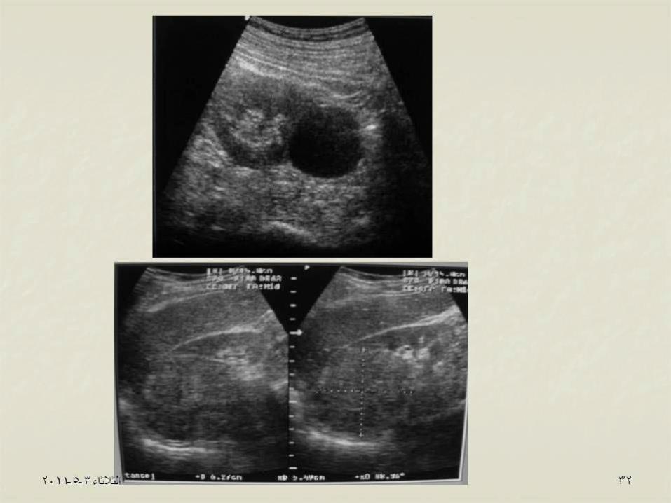
So renal ultrasonography:
• Advantages:
· Easy
· Produces good quality
• Disadvantages:
· Operator dependent
· Does not show the kidney function


With or without contrast
Mainly to:1. dx. Solid tumors. 2. Staging of renal tumors. 3.Staging of urological injuries.