us anatomy of the shoulder: pictorial essay - femede · us anatomy of the shoulder: pictorial essay...
TRANSCRIPT
Journal of Ultrasound (2010) xx, 1e9
+ MODEL
ava i lab le at www.sc iencedi rec t .com
journal homepage: www.e lsev ier .com/ locate / jus
US anatomy of the shoulder: Pictorial essay
M. Precerutti a,*, E. Garioni a, L. Madonia b, F. Draghi b
aDepartment of Radiology, Civil Hospital of Voghera (PV), Italyb IRCCS Foundation, San Matteo Medical Center, Institute of Radiology, University of Pavia, Italy
KEYWORDSShoulder;Ultrasound;Rotator cuff;Acromialclavicularjoint;Sternoclavicular joint.
* Corresponding author. UOC di RadiE-mail address: mprecerutti@liber
Please cite this article in press as:doi:10.1016/j.jus.2010.10.005
1971-3495/$ - see front matter ª 201doi:10.1016/j.jus.2010.10.005
Abstract A thorough knowledge of the anatomy of the shoulder is essential for the assess-ment of its condition. The purpose of this article is to provide a useful tool for the ultrasound(US) study of this joint. The shoulder girdle and upper arm are made up of a number of musclesand tendons: rotator cuff (supraspinatus, infraspinatus, teres minor and subscapularis),humeral biceps, deltoid and pectoral muscles, which can all be evaluated at US examination.Various and complex capsular ligamentous structures contribute to the stability of theshoulder, but only a few can be adequately evaluated by US and will therefore receive partic-ular attention. Numerous serous bursae are situated among muscles, skin, subcutaneoustissues, joint capsule structures and bones to prevent friction and they can be evaluated byUS only in the presence of pathologies. Subacromial-subdeltoid and subcoracoid bursa are mostfrequently involved and will therefore be described in detail. There are furthermore nervesand vessels providing the various components of the shoulder with innervation and vasculari-zation, and they can also be studied by US. The shoulder girdle (humerus, scapula, clavicle andsternal manubrium) is situated in the deep layers; only the cortex of the bone can be seen atUS as a continuous hyperechoic line. For a better understanding of the location and relation-ship between the structures which can be studied by US, magnetic resonance imaging (MRI) canbe carried out as this method provides a wider and more complete view of the structures.
Sommario La conoscenza dell’anatomia della spalla e fondamentale per la valutazione dellasua patologia. Scopo del presente articolo e di fornire un adeguato strumento per la conoscen-za dell’anatomia e dell’anatomia ecografica di quest’articolazione.
Numerosi muscoli e rispettivi tendini partecipano al cingolo scapolo-omerale: i costituenti lacuffia dei rotatori (sovraspinato, sottospinato, piccolo rotondo e sottoscapolare), il bicipiteomerale, il deltoide ed i muscoli grande e piccolo pettorale, tutti valutabili ecograficamente.Varie e complesse strutture capsulo-legamentose contribuiscono alla stabilita della spalla, soloalcune sono ben valutabili ecograficamente e saranno pertanto prese in considerazione in man-iera paticolare. Tra muscoli, cute e sottocute, strutture capsulo-articolari ed ossee si trovanonumerose borse sierose il cui ruolo e quello di evitare l’attrito, sono valutabili ecograficamenteesclusivamente in presenza di patologia; le borse subacromio-deltoidea e sottocoracoidea sonoquelle piu frequentemente interessate da patologia e saranno, pertanto, descritte dettaglia-tamente. Sono inoltre presenti nervi e vasi che forniscono innervazione ed irrorazione ai vari
ologia, Ospedale Civile di Voghera, AO Provincia di Pavia, Italy.o.it (M. Precerutti).
Precerutti M, et al., US anatomy of the shoulder: Pictorial essay, Journal of Ultrasound (2010),
0 Published by Elsevier Srl.
2 M. Precerutti et al.
+ MODEL
Please cite this article in press as:doi:10.1016/j.jus.2010.10.005
componenti della spalla, anch’essi valutabili ecograficamente. In profondita si riconoscono leossa del cingolo (omero, scapola, clavicola e manubrio sternale); dell’osso e valutabile ecogra-ficamente solo la corticale come linea iperecogena continua.
Per una piu semplice comprensione della disposizione spaziale e dei rapporti delle strutturevalutabili ecograficamente si fa riferimento ad immagini di risonanza magnetica, caratteriz-zate da una maggiore panoramicita.ª 2010 Published by Elsevier Srl.
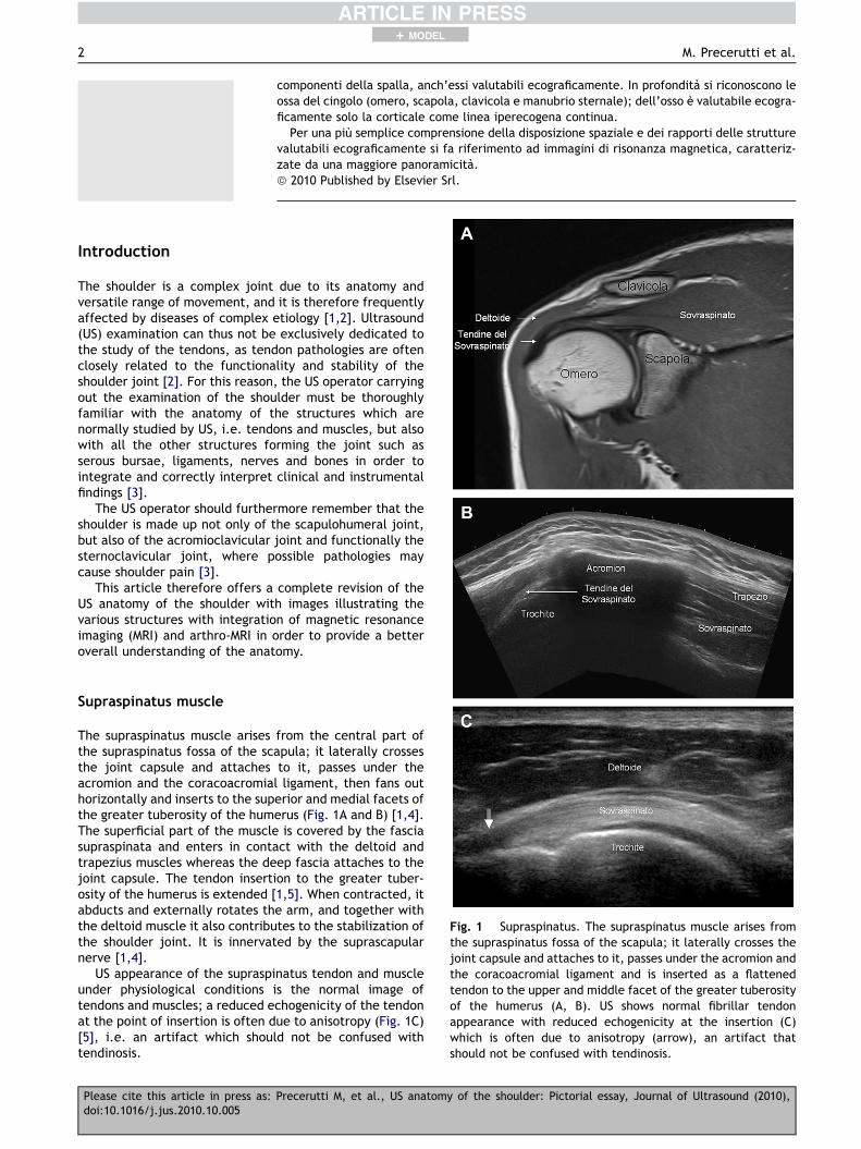
Fig. 1 Supraspinatus. The supraspinatus muscle arises fromthe supraspinatus fossa of the scapula; it laterally crosses thejoint capsule and attaches to it, passes under the acromion andthe coracoacromial ligament and is inserted as a flattenedtendon to the upper and middle facet of the greater tuberosityof the humerus (A, B). US shows normal fibrillar tendonappearance with reduced echogenicity at the insertion (C)which is often due to anisotropy (arrow), an artifact thatshould not be confused with tendinosis.
Introduction
The shoulder is a complex joint due to its anatomy andversatile range of movement, and it is therefore frequentlyaffected by diseases of complex etiology [1,2]. Ultrasound(US) examination can thus not be exclusively dedicated tothe study of the tendons, as tendon pathologies are oftenclosely related to the functionality and stability of theshoulder joint [2]. For this reason, the US operator carryingout the examination of the shoulder must be thoroughlyfamiliar with the anatomy of the structures which arenormally studied by US, i.e. tendons and muscles, but alsowith all the other structures forming the joint such asserous bursae, ligaments, nerves and bones in order tointegrate and correctly interpret clinical and instrumentalfindings [3].
The US operator should furthermore remember that theshoulder is made up not only of the scapulohumeral joint,but also of the acromioclavicular joint and functionally thesternoclavicular joint, where possible pathologies maycause shoulder pain [3].
This article therefore offers a complete revision of theUS anatomy of the shoulder with images illustrating thevarious structures with integration of magnetic resonanceimaging (MRI) and arthro-MRI in order to provide a betteroverall understanding of the anatomy.
Supraspinatus muscle
The supraspinatus muscle arises from the central part ofthe supraspinatus fossa of the scapula; it laterally crossesthe joint capsule and attaches to it, passes under theacromion and the coracoacromial ligament, then fans outhorizontally and inserts to the superior and medial facets ofthe greater tuberosity of the humerus (Fig. 1A and B) [1,4].The superficial part of the muscle is covered by the fasciasupraspinata and enters in contact with the deltoid andtrapezius muscles whereas the deep fascia attaches to thejoint capsule. The tendon insertion to the greater tuber-osity of the humerus is extended [1,5]. When contracted, itabducts and externally rotates the arm, and together withthe deltoid muscle it also contributes to the stabilization ofthe shoulder joint. It is innervated by the suprascapularnerve [1,4].
US appearance of the supraspinatus tendon and muscleunder physiological conditions is the normal image oftendons and muscles; a reduced echogenicity of the tendonat the point of insertion is often due to anisotropy (Fig. 1C)[5], i.e. an artifact which should not be confused withtendinosis.
Precerutti M, et al., US anatomy of the shoulder: Pictorial essay, Journal of Ultrasound (2010),
US anatomy of the shoulder 3
+ MODEL
Infraspinatus muscle
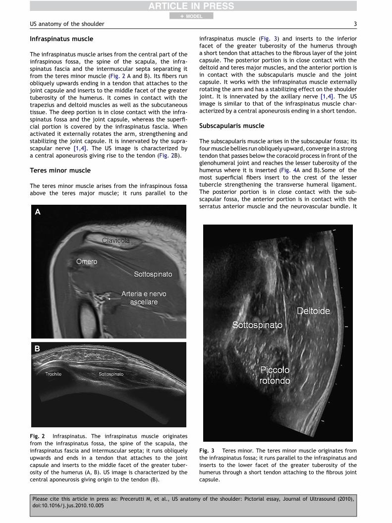
The infraspinatus muscle arises from the central part of theinfraspinous fossa, the spine of the scapula, the infra-spinatus fascia and the intermuscular septa separating itfrom the teres minor muscle (Fig. 2 A and B). Its fibers runobliquely upwards ending in a tendon that attaches to thejoint capsule and inserts to the middle facet of the greatertuberosity of the humerus. It comes in contact with thetrapezius and deltoid muscles as well as the subcutaneoustissue. The deep portion is in close contact with the infra-spinatus fossa and the joint capsule, whereas the superfi-cial portion is covered by the infraspinatus fascia. Whenactivated it externally rotates the arm, strengthening andstabilizing the joint capsule. It is innervated by the supra-scapular nerve [1,4]. The US image is characterized bya central aponeurosis giving rise to the tendon (Fig. 2B).
Teres minor muscle
The teres minor muscle arises from the infraspinous fossaabove the teres major muscle; it runs parallel to the
Fig. 2 Infraspinatus. The infraspinatus muscle originatesfrom the infraspinatus fossa, the spine of the scapula, theinfraspinatus fascia and intermuscular septa; it runs obliquelyupwards and ends in a tendon that attaches to the jointcapsule and inserts to the middle facet of the greater tuber-osity of the humerus (A, B). US image is characterized by thecentral aponeurosis giving origin to the tendon (B).
Please cite this article in press as: Precerutti M, et al., US anatomdoi:10.1016/j.jus.2010.10.005
infraspinatus muscle (Fig. 3) and inserts to the inferiorfacet of the greater tuberosity of the humerus througha short tendon that attaches to the fibrous layer of the jointcapsule. The posterior portion is in close contact with thedeltoid and teres major muscles, and the anterior portion isin contact with the subscapularis muscle and the jointcapsule. It works with the infraspinatus muscle externallyrotating the arm and has a stabilizing effect on the shoulderjoint. It is innervated by the axillary nerve [1,4]. The USimage is similar to that of the infraspinatus muscle char-acterized by a central aponeurosis ending in a short tendon.
Subscapularis muscle
The subscapularis muscle arises in the subscapular fossa; itsfourmuscle bellies runobliquely upward, converge in a strongtendon that passes below the coracoid process in front of theglenohumeral joint and reaches the lesser tuberosity of thehumerus where it is inserted (Fig. 4A and B).Some of themost superficial fibers insert to the crest of the lessertubercle strengthening the transverse humeral ligament.The posterior portion is in close contact with the sub-scapular fossa, the anterior portion is in contact with theserratus anterior muscle and the neurovascular bundle. It
Fig. 3 Teres minor. The teres minor muscle originates fromthe infraspinatus fossa; it runs parallel to the infraspinatus andinserts to the lower facet of the greater tuberosity of thehumerus through a short tendon attaching to the fibrous jointcapsule.
y of the shoulder: Pictorial essay, Journal of Ultrasound (2010),
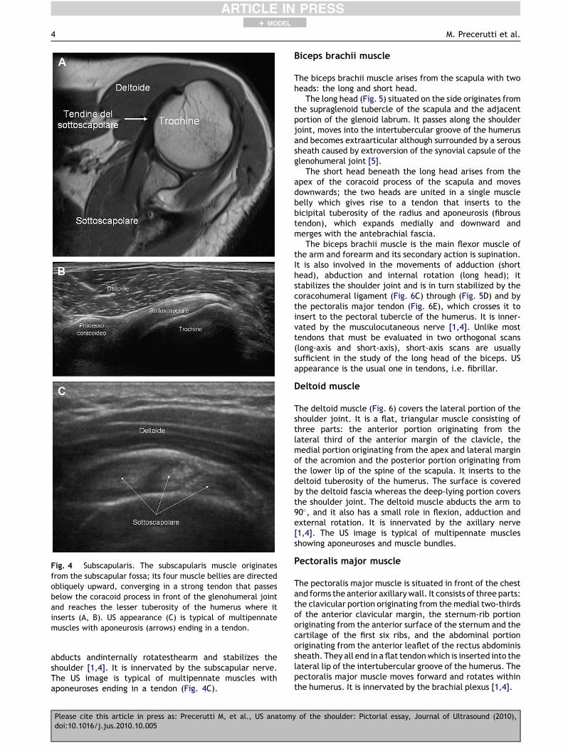
Fig. 4 Subscapularis. The subscapularis muscle originatesfrom the subscapular fossa; its four muscle bellies are directedobliquely upward, converging in a strong tendon that passesbelow the coracoid process in front of the glenohumeral jointand reaches the lesser tuberosity of the humerus where itinserts (A, B). US appearance (C) is typical of multipennatemuscles with aponeurosis (arrows) ending in a tendon.
4 M. Precerutti et al.
+ MODEL
abducts andinternally rotatesthearm and stabilizes theshoulder [1,4]. It is innervated by the subscapular nerve.The US image is typical of multipennate muscles withaponeuroses ending in a tendon (Fig. 4C).
Please cite this article in press as: Precerutti M, et al., US anatomdoi:10.1016/j.jus.2010.10.005
Biceps brachii muscle
The biceps brachii muscle arises from the scapula with twoheads: the long and short head.
The long head (Fig. 5) situated on the side originates fromthe supraglenoid tubercle of the scapula and the adjacentportion of the glenoid labrum. It passes along the shoulderjoint, moves into the intertubercular groove of the humerusand becomes extraarticular although surrounded by a seroussheath caused by extroversion of the synovial capsule of theglenohumeral joint [5].
The short head beneath the long head arises from theapex of the coracoid process of the scapula and movesdownwards; the two heads are united in a single musclebelly which gives rise to a tendon that inserts to thebicipital tuberosity of the radius and aponeurosis (fibroustendon), which expands medially and downward andmerges with the antebrachial fascia.
The biceps brachii muscle is the main flexor muscle ofthe arm and forearm and its secondary action is supination.It is also involved in the movements of adduction (shorthead), abduction and internal rotation (long head); itstabilizes the shoulder joint and is in turn stabilized by thecoracohumeral ligament (Fig. 6C) through (Fig. 5D) and bythe pectoralis major tendon (Fig. 6E), which crosses it toinsert to the pectoral tubercle of the humerus. It is inner-vated by the musculocutaneous nerve [1,4]. Unlike mosttendons that must be evaluated in two orthogonal scans(long-axis and short-axis), short-axis scans are usuallysufficient in the study of the long head of the biceps. USappearance is the usual one in tendons, i.e. fibrillar.
Deltoid muscle
The deltoid muscle (Fig. 6) covers the lateral portion of theshoulder joint. It is a flat, triangular muscle consisting ofthree parts: the anterior portion originating from thelateral third of the anterior margin of the clavicle, themedial portion originating from the apex and lateral marginof the acromion and the posterior portion originating fromthe lower lip of the spine of the scapula. It inserts to thedeltoid tuberosity of the humerus. The surface is coveredby the deltoid fascia whereas the deep-lying portion coversthe shoulder joint. The deltoid muscle abducts the arm to90�, and it also has a small role in flexion, adduction andexternal rotation. It is innervated by the axillary nerve[1,4]. The US image is typical of multipennate musclesshowing aponeuroses and muscle bundles.
Pectoralis major muscle
The pectoralis major muscle is situated in front of the chestand forms the anterior axillarywall. It consists of three parts:the clavicular portion originating from the medial two-thirdsof the anterior clavicular margin, the sternum-rib portionoriginating from the anterior surface of the sternum and thecartilage of the first six ribs, and the abdominal portionoriginating from the anterior leaflet of the rectus abdominissheath. They all end in a flat tendonwhich is inserted into thelateral lip of the intertubercular groove of the humerus. Thepectoralis major muscle moves forward and rotates withinthe humerus. It is innervated by the brachial plexus [1,4].
y of the shoulder: Pictorial essay, Journal of Ultrasound (2010),
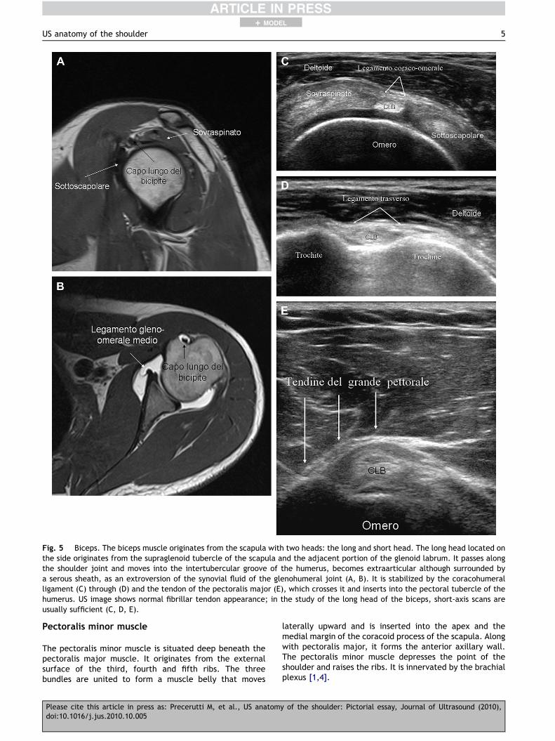
Fig. 5 Biceps. The biceps muscle originates from the scapula with two heads: the long and short head. The long head located onthe side originates from the supraglenoid tubercle of the scapula and the adjacent portion of the glenoid labrum. It passes alongthe shoulder joint and moves into the intertubercular groove of the humerus, becomes extraarticular although surrounded bya serous sheath, as an extroversion of the synovial fluid of the glenohumeral joint (A, B). It is stabilized by the coracohumeralligament (C) through (D) and the tendon of the pectoralis major (E), which crosses it and inserts into the pectoral tubercle of thehumerus. US image shows normal fibrillar tendon appearance; in the study of the long head of the biceps, short-axis scans areusually sufficient (C, D, E).
US anatomy of the shoulder 5
+ MODEL
Pectoralis minor muscle
The pectoralis minor muscle is situated deep beneath thepectoralis major muscle. It originates from the externalsurface of the third, fourth and fifth ribs. The threebundles are united to form a muscle belly that moves
Please cite this article in press as: Precerutti M, et al., US anatomdoi:10.1016/j.jus.2010.10.005
laterally upward and is inserted into the apex and themedial margin of the coracoid process of the scapula. Alongwith pectoralis major, it forms the anterior axillary wall.The pectoralis minor muscle depresses the point of theshoulder and raises the ribs. It is innervated by the brachialplexus [1,4].
y of the shoulder: Pictorial essay, Journal of Ultrasound (2010),

Fig. 6 Deltoid. The deltoid muscle covers the lateral portionof the shoulder. It is a flat, triangular muscle consisting ofthree parts: the anterior portion originating from the lateralthird of the anterior margin of the clavicle, the middle portionoriginating from the apex and the lateral margin of the acro-mion and the posterior portion originating from the lower lip ofthe spine of the scapula; it is inserted at the level of thedeltoid tuberosity of the humerus. US appearance is typical ofmultipennate muscles with aponeurosis and muscle bundles.
6 M. Precerutti et al.
+ MODEL
Subacromial-subdeltoid bursa and subcoracoidbursa
The subacromial-subdeltoid bursa (Fig. 7) is a synovialstructure situated below the clavicle, acromion and cor-acoacromial ligament and above the rotator cuff [3,5,6].Under physiological conditions it does not communicatewith the glenohumeral joint but it may communicate withthe joint cavity after a full-thickness supraspinatus tendontear [1,3,5,6]. The bursa is surrounded by a thin layer ofperibursal fat, and under pathological conditions it is easilydetectable at US examination (Fig. 7) [1,6]; it is situateddeep beneath the deltoid muscle and under the acromionand coracoacromial ligament in front of the supraspinatus
Fig. 7 Subacromial-subdeltoid bursa. The subacromial-sub-deltoid bursa is a synovial structure situated beneath theclavicle, acromion and coracoacromial ligament and over therotator cuff.At US examination it is detectable in pathologicalconditions (arrows).
Please cite this article in press as: Precerutti M, et al., US anatomdoi:10.1016/j.jus.2010.10.005
tendon. It extends forward and downward and covers thebicipital groove, while its rear and side boundaries mayvary. However, it generally reaches a level situated about3 cm over the greater tuberosity of the humerus [6]. It maylie in continuity with the subcoracoid bursa.
The subcoracoid bursa (Fig. 8) is situated below andmedially to the acromion near the joint capsule and thesubscapularis tendon [1]. Under physiological conditions itdoes not communicate with the joint cavity [6]. Bothbursae are there to reduce friction between the tendonsand the surrounding osteofibrous structures. In physiolog-ical circumstances, bursae cannot be seen directly on theUS image, but only the peribursal fat that appears asa hyperechoic line [1,5,7].
Glenohumeral joint: capsular-ligamentousstructures
The glenohumeral joint is an enarthrosis whose articularsurface is the humeral head, which appears as a one-thirdof a sphere, smooth and covered by articular cartilage,which is thicker in the center, and the glenoid cavity, whichis oval and shallow and less extended than the humeralhead (Fig. 1). Also the latter is covered by articular carti-lage, which is thinner at the periphery [1,4,6].
The congruence between the two bone surfaces isincreased by the glenoid labrum applied to the glenoidcircumference. The glenoid labrum is triangular in sectionwith its inner surface facing the joint cavity, and it isattached to the underlying glenoid cartilage [1,4,6]; itsexternal side provides the insertion into the joint capsule.There are anatomical variants of the glenoid labrum,particularly cranially, where a sublabral foramen some-times lies between the glenoid labrum and the underlyingcartilage [8]. The glenoid labrum is poorly visualized at USexamination [1,9,10]. Some authors have described its
Fig. 8 Subcoracoid bursa. The subcoracoid bursa is situatedbelow the acromion near the underlying joint capsule and thesubscapularis tendon. Under physiological conditions it doesnot communicate with the joint cavity. It can be visualized atUS examination when the area is affected by pathology(arrows).
y of the shoulder: Pictorial essay, Journal of Ultrasound (2010),

Fig. 9 Coracoacromial ligament. The coracoacromial liga-ment is a triangular fibrous band stretching between the end ofthe acromion and the external margin of the coracoid process;it completes the osteofibrous surface overlying the gleno-humeral joint.
US anatomy of the shoulder 7
+ MODEL
pathological aspects [10], but currently US cannot beconsidered as a reliable method for studying it [1].
The joint capsule is loose; it inserts medially at thecircumference of the glenoid, and externally it reaches theanatomical neck of the humerus with a number of caudalinsertions which varies from patient to patient. Synoviumlines the entire inside of the joint and also covers theproximal portion of the long head of the biceps tendon. Thejoint is stabilized by the capsule, the thickening, the gle-nohumeral ligaments and a distant ligament, i.e. the cor-acohumeral ligament [1,4]. There are three glenohumeralligaments: the upper, the middle, and the lower gleno-humeral ligament. The upper glenohumeral ligament isnormally thin [1,4,6]; it originates from the top edge of theglenoid labrum and scapular neck and crosses externallybelow the coracohumeral ligament and inserts to the lessertuberosity of the humerus. The middle glenohumeral liga-ment arises from the anterior third of the upper glenoidlabrum and from the neck of the scapula; it runs obliquelyin the inferior external direction until it inserts to the lowerportion of the lesser tuberosity converging with the deepportion of the subscapularis tendon [1,4,6]. The middleglenohumeral ligament may present variants; the bestknown is hypertrophy with or without hypoplasia or absenceof the anterosuperior margin of the glenoid labrum (in caseof absence the anatomy is known as the Buford complex)[8]. Between the lower edge of the upper glenohumeralligament and the upper edge of the middle glenohumeralligament the Weitbrecht foramen is situated, an openingwhich connects the joint cavity with the subscapularisrecess (virtual under physiological conditions) surroundingthe subscapularis tendon below the coracoid [6]. Thisrecess should not be confused with the subcoracoid bursa[1]. The lower glenohumeral ligament is the largest of theglenohumeral ligaments extending from the glenoid labrumand scapular neck for almost the entire circumference tothe lesser tuberosity of the humerus. It has an anterior anda posterior arm which delimit the axillary recess. Betweenthe lower edge of the middle glenohumeral ligament andthe upper edge of the anterior arm of the lower gleno-humeral ligament, the Rouviere foramen is situated whichmay provide communication between the joint capsule andthe subcoracoid bursa [1,4,6].
In addition to the axillary and subscapular recess, thejoint capsule has a further anterior biceps recess, i.e. thetendon sheath of the long head of the biceps. The cor-acohumeral ligament is a distant stabilizer of the scapular-humeral joint; it is a strong fibrous band stretching betweenthe coracoid and the greater tuberosity of the humerus. Itforms the roof of the rotator cuff interval consisting of theintra-articular portion of the long head of the biceps tendonand the proximal humerus [1,3]. US examination does notprovide an adequate study of most of the articular struc-tures and capsular ligaments which must be assessed usingCT arthrography or MR arthrography (Fig. 5B) [1,8].
Coracoacromial ligament
The coracoacromial ligament (Fig. 9) is a triangular fibrousband stretching between the end of the acromion and thecoracoid process; it completes the osteofibrous arch above
Please cite this article in press as: Precerutti M, et al., US anatomdoi:10.1016/j.jus.2010.10.005
the glenohumeral joint. It is covered by the deltoid muscleand it covers the surface of the rotator cuff [1,4,6]. Thisligament can be evaluated at US examination.
Acromioclavicular joint and coracoclavicularligament
The acromioclavicular joint is an arthrodial joint connect-ing the lateral end of the clavicle and the medial end of theacromion (Fig. 10). Both articular bone heads presentsmooth facets covered in hyaline cartilage and interposi-tion of a fibrous disk [1,4,6]. The joint capsule is reinforcedproximally by the acromioclavicular ligament and fibrousbundles of the deltoid and trapezius muscles, and the jointis stabilized by a distant ligament, the coracoclavicularligament [1,4,6].
The coracoclavicular ligament consists of two bundles:the trapezoid and conoid ligaments. The trapezoid liga-ment is a strong quadrilateral fibrous lamina stretchingbetween the upper surface of the coracoid and the cora-coid tuberosity of the clavicle. It lies in front of the conoidligament and is triangular with an apex that attaches to thebase of the coracoid and fibers that diverge in a fan andinsert into the coracoid tuberosity of the clavicle [4,6].
Sternoclavicular joint
The sternoclavicular joint is the only joint that connectsthe shoulder girdle and upper arm to the chest [1]. Thejoint heads involved are the medial end of the clavicle, themanubrium of the sternum and the first rib. The jointsurfaces are flat and incongruous with interposition ofa fibrous disc dividing the joint into two synovial compart-ments which do not communicate: the medial and lateralcompartment. The joint is reinforced by the joint capsule,the strong sternoclavicular ligament situated anteriorcranially, the interclavicular ligaments [1,3,4,6] and, ata distance, the costoclavicular ligament.
Vascular structures
Blood supply to the shoulder girdle and upper arm occursthrough the axillary artery which distally, at the lateral
y of the shoulder: Pictorial essay, Journal of Ultrasound (2010),
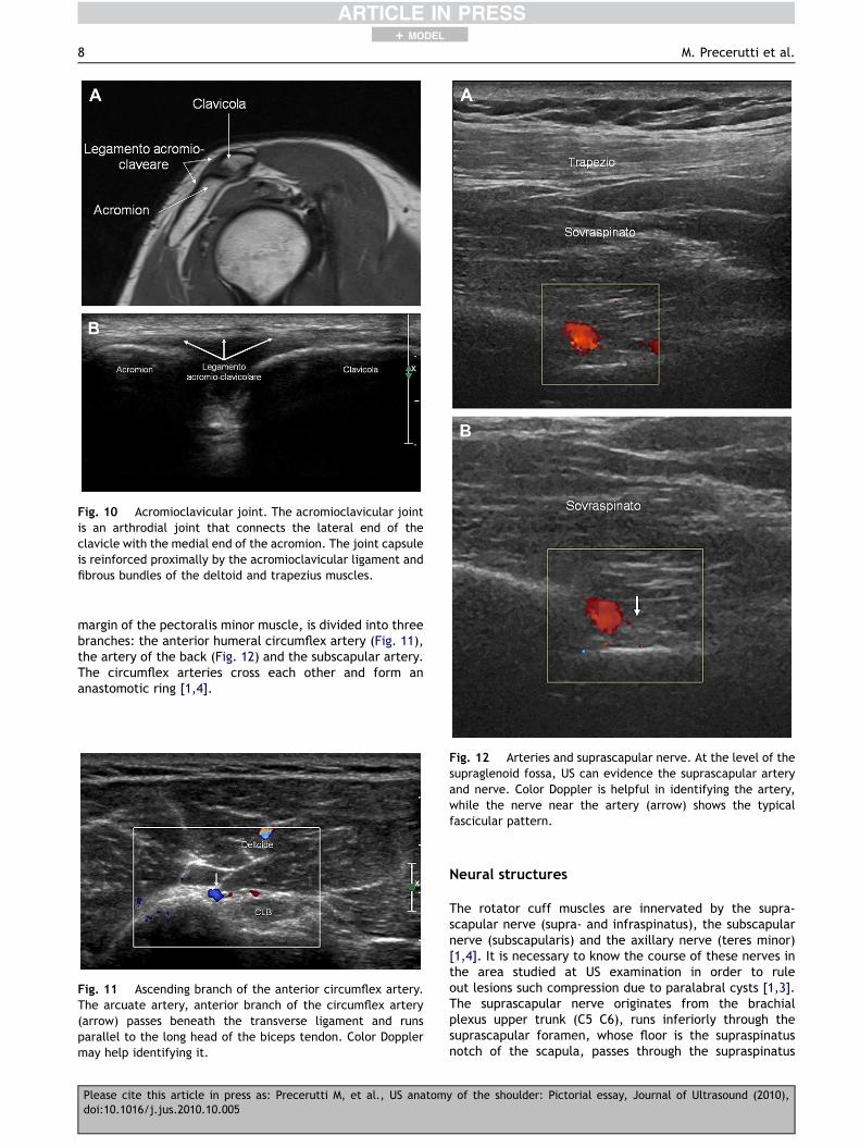
Fig. 10 Acromioclavicular joint. The acromioclavicular jointis an arthrodial joint that connects the lateral end of theclavicle with the medial end of the acromion. The joint capsuleis reinforced proximally by the acromioclavicular ligament andfibrous bundles of the deltoid and trapezius muscles.
8 M. Precerutti et al.
+ MODEL
margin of the pectoralis minor muscle, is divided into threebranches: the anterior humeral circumflex artery (Fig. 11),the artery of the back (Fig. 12) and the subscapular artery.The circumflex arteries cross each other and form ananastomotic ring [1,4].
Fig. 11 Ascending branch of the anterior circumflex artery.The arcuate artery, anterior branch of the circumflex artery(arrow) passes beneath the transverse ligament and runsparallel to the long head of the biceps tendon. Color Dopplermay help identifying it.
Fig. 12 Arteries and suprascapular nerve. At the level of thesupraglenoid fossa, US can evidence the suprascapular arteryand nerve. Color Doppler is helpful in identifying the artery,while the nerve near the artery (arrow) shows the typicalfascicular pattern.
Please cite this article in press as: Precerutti M, et al., US anatomdoi:10.1016/j.jus.2010.10.005
Neural structures
The rotator cuff muscles are innervated by the supra-scapular nerve (supra- and infraspinatus), the subscapularnerve (subscapularis) and the axillary nerve (teres minor)[1,4]. It is necessary to know the course of these nerves inthe area studied at US examination in order to ruleout lesions such compression due to paralabral cysts [1,3].The suprascapular nerve originates from the brachialplexus upper trunk (C5 C6), runs inferiorly through thesuprascapular foramen, whose floor is the supraspinatusnotch of the scapula, passes through the supraspinatus
y of the shoulder: Pictorial essay, Journal of Ultrasound (2010),
US anatomy of the shoulder 9
+ MODEL
fossa through the spinoglenoid notch and finally reaches theinfraspinatus fossa [1,3,4].
The axillary nerve originates from the posterior portionof the brachial plexus upper trunk (C5 C6), descends to theinferior, lateral margin of the subscapularis muscle andreaches the bottom margin of the glenohumeral jointcapsule where it is runs posteriorly. The nerve, togetherwith the posterior circumflex artery, passes through thequadrilateral space delimited medially by the long head oftriceps brachii, laterally by the surgical neck of thehumerus, cranially by teres minor and beneath by teresmajor; its terminal branches innervate teres minor and thedeltoid muscle [1,4].
Conclusions
US has for a long time been considered a highly effectivemeans of studying tendon disorders of the shoulder [1,3,5],and medical doctors who carry out US examinations toevaluate osteoarticular diseases must therefore be thor-oughly familiar with these anatomical structures. However,it should not be forgotten that also the adjacent structuresshould be evaluated; they are generally well displayed, butoften neglected in daily practice, such as the acromiocla-vicular joint, blood vessels and nerves [1,3] in order to fullyexploit the diagnostic possibilities of the method andidentify frequent diseases which are easily diagnosed byUS. It is also important to be familiar with the anatomicalstructures which are poorly evaluated at US, which aremore accurately investigated using other diagnosticimaging methods in order to assess not only part of theproblem, which often happens in these pathologies, but toidentify the disease in a wider context and thereby assistthe clinician in a more meaningful way. A US study shouldnot be exclusively dedicated to tendon pathologies as theyare often closely related to the functionality and stabilityof the shoulder joint [2]. A perfect knowledge of the entire
Please cite this article in press as: Precerutti M, et al., US anatomdoi:10.1016/j.jus.2010.10.005
anatomy is therefore required of those who carry out USexaminations of the shoulder. Familiarity with this anatomyis necessary also to perform US guided interventionalprocedures [11].
Conflict of interest
The authors have no conflict of interest.
References
[1] Bianchi S, Martinoli C. Ultrasound of the musculoskeletalsystem: sholuder. Springer-Verlag; 2007. 190e331.
[2] Yin B, Vella J, Levine WN. Arthroscopic alphabet soup:recognition of normal, normal variants, and pathology. OrthopClin North Am 2010 Jul;41(3):297e308.
[3] Martinoli C, Bianchi S, Prato N, Pugliese F, Zamorani MP,Valle M, et al. US of the shoulder: non-rotator cuff disorders.Radiographics 2003 Mar-Apr;23(2):381e401.
[4] Aa Vv: Anatomia Umana vol 1. Edi Ermes 1990. 203–265.[5] Chevrot A. Artro-TC: spalla. Masson Ed Italia; 1995. 47e56.[6] Rutten MJ, Jager GJ, Blickman JG. US of the rotator cuff:
pitfalls, limitations and artefacts. Radiographics 2006;26:589e604.
[7] Van Holsbeeck M, Strouse PJ. Sonography of the shoulder:evaluation of the subacromial-subdeltoid bursa. AJR Am JRoentgenol 1993;160:561e4.
[8] Beltran J, Bencardino J, Mellado J, Rosenberg ZS, Irish RD. MRArthrography of the shoulder: variants and pitfalls. Radio-graphics 1997;17:1403e12.
[9] Rasmussen OS. Anterior shoulder instability: sonographicevaluation. J Clin Ultrasound 2004;32:430e7.
[10] Hammar MV, Wintzell GB, Astrom KG, Larsson S, Elvin A. Roleof US in the preoperative evaluation of patients with anteriorshoulder instability. Radiology 2001;219:29e34.
[11] Draghi F, Robotti G, Jacob D, Bianchi S. Interventionalmusculoskeletal ultrasonography: precautions and contrain-dications. Journal of Ultrasound. doi:10.1016/j.jus.2010.09.004, in press.
y of the shoulder: Pictorial essay, Journal of Ultrasound (2010),