uterine corpus: two components
TRANSCRIPT

1
Endometrial Cancer
The Old Merges with the New
Lora Hedrick Ellenson, M.D.
Intermediate Level - Endometrial cancer consists of several morphologically and biologically distinct tumors. This lecture will provide an overview of new advances in our understanding of endometrial carcinoma. The lecture will present information ranging from the clinical presentation to gross and microscopic analysis as well as newer molecular studies.
Goals - The learner will understand:Endometrial cancer is more than one disease
Objectives
The major types of endometrial cancerThe clinical relevance in the distinction of different types of endometrial cancer
Objectives - The learned will be able to:Adequately gross hysterectomy specimens for the different types of endometrial cancerKnow the important gross and clinical findings important in an endometrial cancer diagnosisDetermine the important details of the history and gross findings relevant to rendering the correct diagnosis of endometrial cancer
UTERINE CORPUS: TWO COMPONENTS
EndometriumEpithelial (glandular)Stromal
MyometriumSmooth muscle

2
Normal Menstrual Cycle
SecretoryProliferative
Estrogen driven
Proliferation ofepithelium is regulated by GFproduced by thestroma
Progesterone driven
Proliferation is terminated and both componentsdifferentiate

3

4
What is Cancer?
GeneticsEnvironment
Normal Cells
MultipleAlterations Defective
Cancer
Defective Cell EliminatedUncontrolled growth of cells in the
body
Cells circumvent tight controls set in place in tissues
Mutations in tumor suppressor and oncogenes cause cells to overcome all inhibitory signals
Abnormal Cell Growth: Oncogenes
Normal genes (regulate cell growth)
Mutated Protein
1st mutation (leads to accelerated cell division)
Tumor Suppressor Genes
1st mutation (susceptible carrier)
No brakes!
Active oncogene
Normal genes (regulate cell growth)
Tumor suppressor genes
Tumor suppressor genes
Active oncogene
No brakes!
2nd mutation or loss (leads to cancer)
No brakes!

5
Endometrial carcinoma is the most common invasive tumor of the female genital tract
In US it is the fourth most common cancer in women and worldwide it is the fifth most common cancer
Endometrial Carcinoma
Incidence varies widely in the world
In the US incidence is twice as common in whites compared to blacks, but death is higher in blacks due to increase in high-risk cancers
Most cases are sporadic but cases are hereditary forms exist
Classification of Endometrial Carcinoma
Endometrial Tumorigenesis
SH CH Endometrioid CaCAH
Estrogen
(Type I)
Nl epithelium
SH
Atrophy
CH Endometrioid Ca
EIC Serous Ca
CAH
(Type II)
6
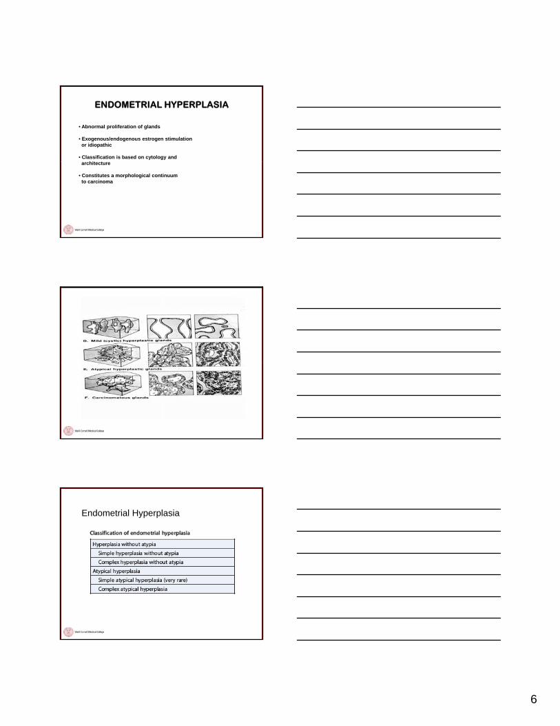
• Abnormal proliferation of glands
• Exogenous/endogenous estrogen stimulationor idiopathic
• Classification is based on cytology and hit t
ENDOMETRIAL HYPERPLASIA
architecture
• Constitutes a morphological continuum to carcinoma
Endometrial Hyperplasia

7
HYPERPLASIA WITHOUT ATYPIA
• Simple Hyperplasia- Minimal glandular complexity- Stroma remains abundant- No cytological atypia- 1% progress to carcinoma
• Complex Hyperplasia- Increased glandular complexity with decreased stroma; glands
can be nearly back-to-back- No cytological atypia- 3% progress to carcinoma
Simple Hyperplasia
Complex Hyperplasia without Atypia

8
HYPERPLASIA WITH ATYPIA
• Simple Hyperplasia- Very rare
• Complex Hyperplasia- Increased glandular complexity with decreased stroma; glands
can be nearly back-to-back- Cytological atypia- 25-40% progress to carcinoma
Complex Atypical Hyperplasia
CONCLUSIONS: The prevalence of endometrial carcinoma in patients who had a community hospital biopsy diagnosis of AEH was high (42.6%).When considering management strategies for women who have a biopsy diagnosis of AEH, clinicians and patients should take into account the considerable rate of concurrent carcinoma. Cancer 2006. © 2006 American Cancer Society.

9
Behavior of hyperplasia
Age is Important
ENDOMETRIOID ADENOCARCINOMA

10
Endometrioid Carcinoma Grade 1
Endometrioid Carcinoma Grade 1
Endometrioid Carcinoma Grade 1
11
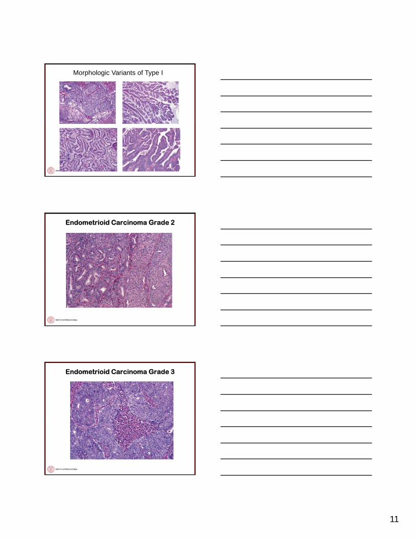
Morphologic Variants of Type I
Endometrioid Carcinoma Grade 2
Endometrioid Carcinoma Grade 3

12
Endometrioid Carcinoma Grade 3
SH CH Endometrioid CaCAH
Estrogen
(Type I)
Endometrial Tumorigenesis
Nl epithelium
SH
Atrophy
CH Endometrioid Ca
EIC Serous Ca
CAH
(Type II)
Endometrial Intraepithelial CarcinomaEIC is characterized by markedly atypical nuclei, identical to those of invasive serouscarcinomas, lining the surfaces, and glands of the atrophic endometrium
The lesion can be very small and focal and is often present on the surface ofa polyp
EIC often has a slightly papillary contour and some cells display hobnail morphology and smudged, hyperchromatic nuclei.
The nuclei are enlarged, and frequently display enlarged eosinophilic nucleoli.
Numerous mitotic figures, including atypical ones, are present. On occasion, the abnormal proliferation involves only a portion of an endometrial gland

13
Endometrial Intraepithelial Carcinoma
Endometrial Intraepithelial Carcinoma
Endometrial Intraepithelial Carcinoma

14
Endometrial Intraepithelial Carcinoma
Minimal Serous Carcinoma
UTERINE SEROUS CARCINOMA
• Histopathological• Often papillary architecture (may be glandular) • Markedly atypical cells• Arises in the setting of atrophy• Resembles ovarian serous carcinoma• All high grade (FIGO Grade 3)
• Clinical• Older women• Not associated with estrogen• Aggressive behavior• Requires thorough staging (even EIC) • Women with “true” stage 1 USCs have a favorable prognosis

15
Serous Carcinoma
Serous Carcinoma
p16
Genetic Alterations Distinguish Endometrioidfrom Serous Endometrial Carcinomas
Endometrioid Carcinoma
• TP53 mutations relatively uncommon (except high grade)
• microsatellite instability present in approximately 20%y p pp y• PTEN mutations common (>50%)• K-RAS and CTNNB1 mutations common
Serous Carcinoma
• TP53 mutations extremely common (nearly 100%)• PTEN, K-RAS, CTNNB1 mutations uncommon• microsatelite instability uncommon

16
Endometrial Tumorigenesis
SH CH Endometrioid CaCAH
Estrogen
PTEN MI KRAS PIK3CA p53
Nl epithelium
Atrophy EIC Serous Ca
p53
Other Type 2 Endometrial Carcinomas
Clear cell carcinoma
Malignant mullerian mixed tumors (MMMT)
Undifferentiated carcinoma
Clear Cell Carcinoma

17
Malignant Mullerian Mixed Tumor
Undifferentiated Carcinoma
INHERITED FORMS OF ENDOMETRIAL CARCINOMA
Lynch SyndromeGermline mutations in DNA mismatch repair genesMost common are MLH1 and MSH2Results in MSIApproximately 1/50 women with EC will have LS25-30% of sporadic EC have MSI25 30% of sporadic EC have MSICurrently routine screening with IHC is not being done
Cowden DiseaseGermline mutations in PTEN1/200,000 individuals5-10% lifetime risk compared to 2.6% Blind endometrial biopsies starting at 35-40 or 5 years prior to earliest diagnosis of EC in the family

18
Gross Examination of a Hysterectomy for Endometrial Cancer
1. Orient the uterus: The round ligaments are most anterior, and the ovaries, if present, are most posterior. The peritoneum extends further inferiorly along the posterior aspect of the uterus than it does anteriorly.
2. Weigh and measure the specimen. Ink the paracervical and parametrial softtissue margins (we also ink the serosa of uterus even though it is not a margin).
3. Place a probe through the endocervical canal into the endometrium. Bivalve theuterus into anterior and posterior halves with a long blade.
4. Longitudinally section the cervix, extending the incision upward through the LUS.serially bread-loaf the uterine corpus with 0.5 cm transverse slices.
5. Describe the size, appearance, and location of the tumor, and the depth of myometrial invasion.
6. Submit sections of the tumor at the deepest point of invasion, anterior and posterior LUS and cervix, uninvolved endometrium. Submit sections of ovaries and the entire fimbriated end of the fallopian tube. If no lesion is visible submit the entire endometrium.

19
Endometrial Carcinoma Staging
Goals - The learner will understand:Endometrial cancer is more than one diseaseThe major types of endometrial cancerThe clinical relevance in the distinction of different types of endometrial cancer
Objectives The learned will be able to:
Summary
Objectives - The learned will be able to:Adequately gross hysterectomy specimens for the different types of endometrial cancerKnow the important gross and clinical findings important in an endometrial cancer diagnosisDetermine the important details of the history and gross findings relevant to rendering the correct diagnosis of endometrial cancer