uterine influences on conceptus development in fertility ... · the corpus luteum), the conceptus...
TRANSCRIPT
Uterine influences on conceptus development infertility-classified animalsJoao G. N. Moraesa, Susanta K. Behuraa, Thomas W. Gearyb, Peter J. Hansenc, Holly L. Neibergsd,e,and Thomas E. Spencera,1
aDivision of Animal Sciences, University of Missouri, Columbia, MO 65211; bFort Keogh Livestock and Range Research Laboratory, United States Departmentof Agriculture Agricultural Research Service, Miles City, MT 59301; cDepartment of Animal Sciences, University of Florida, Gainesville, FL 32611;dDepartment of Animal Sciences, Washington State University, Pullman, WA 99164; and eCenter for Reproductive Biology, Washington StateUniversity, Pullman, WA 99164
Edited by R. Michael Roberts, University of Missouri, Columbia, MO, and approved January 9, 2018 (received for review December 5, 2017)
A major unresolved issue is how the uterus influences infertilityand subfertility in cattle. Serial embryo transfer was previously usedto classify heifers as high-fertile (HF), subfertile (SF), or infertile (IF).To assess pregnancy loss, two in vivo-produced embryoswere trans-ferred into HF, SF, and IF heifers on day 7, and pregnancy outcomewas assessed on day 17. Pregnancy rate was substantially higher inHF (71%) and SF (90%) than IF (20%) heifers. Elongating concep-tuses were about twofold longer in HF than SF heifers. Tran-scriptional profiling detected relatively few differences in theendometrium of nonpregnant HF, SF, and IF heifers. In contrast,there was a substantial difference in the transcriptome responseof the endometrium to pregnancy between HF and SF heifers.Considerable deficiencies in pregnancy-dependent biological path-ways associated with extracellular matrix structure and organiza-tion as well as cell adhesion were found in the endometrium of SFanimals. Distinct gene expression differences were also observed inconceptuses from HF and SF animals, with many of the genes de-creased in SF conceptuses known to be embryonic lethal in mice dueto defects in embryo and/or placental development. Analyses of bi-ological pathways, key players, and ligand–receptor interactions basedon transcriptome data divulged substantial evidence for dysregulationof conceptus–endometrial interactions in SF animals. These resultssupport the ideas that the uterus impacts conceptus survival andprograms conceptus development, and ripple effects of dysregulatedconceptus–endometrial interactions elicit loss of the postelongationconceptus in SF cattle during the implantation period of pregnancy.
endometrium | conceptus | fertility | gene expression | pregnancy
Infertility and subfertility are important and pervasive problemsin agricultural animals and humans (1, 2). In ruminants, em-
bryo mortality is a major factor affecting fertility and thus productionand economic efficiency (3, 4). Pregnancy loss in cattle ranges from30 to 56%, with the majority of losses occurring in the first month ofpregnancy (3, 5, 6). Infertility and subfertility also impact the successof embryo transfer in cattle and humans (7, 8). In cattle, meansurvival rate to calving following transfer of in vivo-derived embryosfrom superovulated donors ranges from 31 to 60%, and in vitro-produced embryo survival rate is lower (5, 9). Failure of the em-bryo to survive and establish pregnancy is due to paternal, ma-ternal, and embryonic factors (10–12). Many of the pregnancylosses observed in natural or assisted pregnancies may be attrib-uted to maternal factors, such as an inability of the uterus tosupport conceptus growth and implantation (13, 14).After fertilization (day 0), the zona pellucida-enclosed bovine
embryo enters the uterus at the morula stage by day 5 and de-velops into a blastocyst. The spherical blastocyst hatches from thezona pellucida on days 7 to 10 and continues to grow, changingfrom spherical to ovoid in shape between days 12 and 14, afterwhich it can be termed a conceptus (embryo and associated ex-traembryonic membranes) (15). The conceptus undergoes elon-gation involving exponential growth from about 2 mm in length onday 13, 60 mm by day 16, and 20 cm or more by day 19 (16). Afterdays 16 to 17, the time of maternal recognition of pregnancy
(when the embryo signals to the mother to prevent regression ofthe corpus luteum), the conceptus begins the processes of im-plantation and placentation that involve apposition, attachment,and adhesion of the trophectoderm to the endometrial luminalepithelium and onset of trophoblast giant binucleate cell differ-entiation (17). Blastocyst growth into an elongated conceptus hasnot been achieved in vitro and requires transfer into the uterus(18), as the endometrium secretes or transports a myriad of factorscritical for conceptus growth and elongation (14, 19). Dynamicchanges in the endometrial transcriptome occur between days7 and 13 that are primarily regulated by progesterone in bothnonpregnant and pregnant cattle (14, 19–21). Those changes inthe endometrium presumably establish a uterine environmentconducive to blastocyst survival and conceptus growth into anelongated, filamentous-type conceptus and subsequently implan-tation and placentation. Conceptus elongation is required forproduction of IFN tau (IFNT) (22), which is the pregnancy rec-ognition signal that acts on the endometrium to sustain continuedproduction of progesterone by the ovary and regulates genes im-plicated in conceptus growth, implantation, and placentation (23,24). Inadequate elongation of the conceptus presumably results inlower production of IFNT, inability to maintain the corpusluteum, and thus pregnancy loss (24–26). Although much in-formation is known about embryo development into a blastocystfrom in vitro systems (27), the essential endometrial genes and
Significance
Successful pregnancy establishment requires synchronous in-teractions of the conceptus with the endometrium of the uterus.This study of pregnancy outcome after assisted reproduction infertility-classified cattle determined how the uterine environ-ment impacts and programs conceptus survival and develop-ment. The study found that ripple effects of dysregulatedconceptus–endometrial interactions elicit postelongation preg-nancy loss in subfertile animals during the implantation period.This research enhances our understanding of the mechanismsthat lead to pregnancy loss in both natural and assisted re-production and has wide implications for improving pregnancysuccess in domestic animals and humans.
Author contributions: J.G.N.M., T.W.G., P.J.H., H.L.N., and T.E.S. designed research;J.G.N.M., T.W.G., P.J.H., and T.E.S. performed research; S.K.B. contributed new reagents/analytic tools; J.G.N.M., S.K.B., and T.E.S. analyzed data; and J.G.N.M. and T.E.S. wrotethe paper.
Conflict of interest statement: Editor R.M.R. has not published any research papers withany of the authors within the last four years and does not have collaborative researchgrants with them.
This article is a PNAS Direct Submission.
Published under the PNAS license.
Data deposition: The data reported in this paper have been deposited in the Gene Ex-pression Omnibus (GEO) database, https://www.ncbi.nlm.nih.gov/geo (accession no.GSE107891).1To whom correspondence should be addressed. Email: [email protected].
This article contains supporting information online at www.pnas.org/lookup/suppl/doi:10.1073/pnas.1721191115/-/DCSupplemental.
www.pnas.org/cgi/doi/10.1073/pnas.1721191115 PNAS | Published online February 5, 2018 | E1749–E1758
AGRICU
LTURA
LSC
IENCE
SPN
ASPL
US
Dow
nloa
ded
by g
uest
on
Apr
il 29
, 202
0
biological pathways important for conceptus survival, growth, andimplantation remain largely unknown (28, 29).One of the major impediments to understanding maternal in-
fluences on pregnancy loss has been a lack of animals with definedhigh and low rates of early pregnancy loss. McMillan and Donnison(30) summarized a unique approach for experimentally identifyinghigh and low fertility in dairy heifers based on serial transfer of invitro-produced embryos. Their approach identified animals withhigh (76%) and low (11%) aggregate pregnancy rates, and a failurein the mechanism involved in conceptus elongation and thus ma-ternal recognition of pregnancy was suggested to be the cause ofearly pregnancy loss in the low-fertility–classified heifers (30, 31).We recently used a similar approach to identify beef heifers withintrinsic differences in pregnancy loss (32). Serial transfer of asingle in vitro-produced embryo was used to classify animals ashigh-fertile (HF), subfertile (SF), or infertile (IF) based on day28 pregnancy rate. In these heifers, no difference in pregnancyrates was observed on day 14 after transfer of a single in vivo-produced embryo on day 7 postestrus (32). Thus, the observeddifference in uterine competence for pregnancy in fertility-classified heifers is hypothesized to manifest during maternalrecognition of pregnancy and implantation. The present studytested this hypothesis and revealed that embryo survival today 17 is compromised in IF heifers, and that asynchronousconceptus–endometrial interactions in SF animals lead topostelongation embryonic loss by day 28 during the implantationperiod of pregnancy.
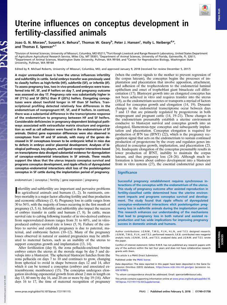
ResultsExperiment Overview. This experiment utilized heifers that werepreviously fertility-classified as HF (100% pregnancy rate), SF(25 to 33% pregnancy rate), or IF (0% pregnancy rate) using serialtransfer (n = 3 to 4 rounds) of a single in vitro-produced embryo(grade 1) on day 7 followed by pregnancy determination on day 28(32). In the present study, those same fertility-classified heifers(HF, n = 21; SF, n = 14; IF, n = 6) were synchronized to estrus(day 0) and received two in vivo-produced embryos on day 7. Allof the embryos were generated from superovulated donor cowsusing a single sire and were cryopreserved for direct thaw transfer.Each heifer received two grade 1 or 2 embryos from the samedonor and developmental stages (compact morula and blastocyst).On day 17 (10 d postembryo transfer), the entire female re-productive tract was obtained, and the uterine lumen wasflushed to recover the conceptus. If a conceptus was not ob-served in the uterine flush, the heifer was considered non-pregnant. As illustrated in Fig. 1A, the proportion of heifers fromwhich one or two conceptuses were recovered was greater for HF(P = 0.02) and SF (P = 0.03) heifers compared with IF heifersbut was not different (P = 0.91) between HF and SF heifers.Accordingly, pregnancy rate was higher (P < 0.05) in HF (71%)and SF (90%) than IF (20%) heifers but not different (P > 0.05)between HF and SF heifers (Fig. 1B). The morphology of theconceptuses recovered from HF and SF heifers was similar andranged from tubular to elongated and filamentous (Fig. 1C). Incontrast, a single small spherical hatched blastocyst (<0.1 cm) was
0102030405060708090
Hei
fers
(%)
HF SF IF
05
101520253035
HF SF IF
Con
cept
us L
engt
h (c
m)
A
None One TwoConceptuses in Uterine Flush
0
20
40
60
80
100
HF SF IF
Preg
nanc
y R
ate
(%)
B
a
a
b
Fila
men
tous
D Tubu
lar
Sphe
rical
Fila
men
tous
C
Fig. 1. Day 17 pregnancy outcome in fertility-classified heifers. HF, SF, and IF heifers were synchronized and received two in vivo-produced embryos on day7 postestrus. The reproductive tract was acquired on day 17 (10 d postembryo transfer) and flushed to recover the conceptus(es). If present, conceptus morphology andlength were determined using a stereomicroscope. (A) Percentage of heifers with an identifiable conceptus in the uterine flush. (B) Column graph showing pregnancyrate (y axis) of the fertility-classified heifers (x axis). Bars with different superscripts (a or b) indicate difference (P < 0.05) in pregnancy rate. (C) Representativeconceptus morphology recovered from heifers. Note a single spherical embryo of less than 1.5 mm was recovered in one IF heifer. (Scale bars, 1 cm.) (D) Conceptuslength was longer (P < 0.05) from HF compared with SF heifers. Individual conceptus length is plotted with the mean length denoted by the dashed red lines.
E1750 | www.pnas.org/cgi/doi/10.1073/pnas.1721191115 Moraes et al.
Dow
nloa
ded
by g
uest
on
Apr
il 29
, 202
0

recovered from the uterus of only one IF heifer. Overall, theconceptus was longer (P < 0.01) in HF (mean 10.6 cm; range1.2 to 32.2 cm) than SF (mean 4.7 cm; range 1.5 to 13.5 cm)heifers (Fig. 1D).There were no differences in circulating progesterone con-
centrations on day 17 between pregnant and nonpregnant heifers(P = 0.55) or among pregnant HF, SF, or IF heifers (P = 0.23)(SI Appendix, Fig. S1 A and B). Similarly, there was no correla-tion of conceptus size and circulating progesterone concentra-tions (R = −0.08; P = 0.80) (SI Appendix, Fig. S1C).
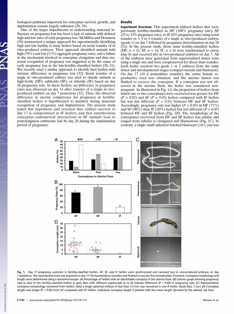
Dysregulation of the Endometrial Transcriptome in Response toPregnancy. Transcriptome analysis was performed using totalRNA isolated from the day 17 endometria of nonpregnant andpregnant fertility-classified heifers. Sequencing of the 25 RNA-seq(sequencing) libraries (n = 5 per group) generated 27.2 to42.8 million quality reads that mapped at an ∼97% rate to the Bostaurus reference genome (assembly UMD3.1). EdgeR robustanalysis was used to identify differentially expressed genes (DEGs)[false discovery rate (FDR), P < 0.05] in the endometria, and agene was defined as expressed if it presented >1 fragment perkilobase of transcript per million mapped reads (FPKM). Similarto our previous study of endometrial biopsies from these fertility-classified animals on day 14 (32), there were relatively few genesthat were different (FDR, P < 0.05) in the endometrium of thenonpregnant HF, SF, and IF animals (Figs. 2 and 3A and DatasetsS1–S3). Functional analysis of those DEGs did not identify anysignificantly enriched gene ontology (GO) terms or biologicalpathways. Several of the DEGs in HF and SF compared with IFendometrium encode proteins involved in immune responses orimmunoglobulins (Fig. 3A). In SF compared with IF endometria,some of the 43 DEGs encode secreted proteins (GRP, IGFBP2,LYZ1, SERPINB7), intracellular enzymes (PLA2G2F), andtransporters for water (AQP5) or amino acids (SLC2A3).Comparison of the endometrial transcriptome of pregnant HF
with pregnant SF animals detected 168 DEGs (Fig. 2 and DatasetS4). Of note, none of the DEGs were classical IFN-stimulatedgenes (ISGs; i.e., IFIT1, IFIT2, ISG15, MX2, RSAD2, OAS2)that are induced or considerably up-regulated by IFNT from theelongating conceptus (24) (Fig. 3B). The 168 DEGs were enrichedfor GO molecular function (extracellular matrix or ECM struc-tural constituent), biological process (ECM organization, cell ad-hesion, collagen catabolic process, locomotion, cellular responseto endogenous stimulus), and cellular component (ECM, extra-cellular space, membrane region) terms (SI Appendix, Table S1).Pathway analysis found enrichment in ECM organization andcollagen formation. Genes increased in pregnant HF compared
with SF endometrium included a water transporter (AQP8), lipidtransporter (FABP3), and secreted protease (MEP1B). Genesdecreased in HF compared with SF endometrium include cellsignaling (CAMK1G, IRS4), ECM constituents, and cell-adhesionmolecules (Dataset S4).Endometrial responses to pregnancy were assessed by com-
paring the transcriptome of nonpregnant with pregnant animals(Figs. 2 and 3B and Datasets S5 and S6). This analysis found3,422 DEGs (1,680 increased, 1,742 decreased) in HF heifersand 1,095 DEGs (833 increased, 262 decreased) in SF animals,with 848 genes commonly responsive to pregnancy in HF and SFanimals (Fig. 3B). Thus, ∼20% of total expressed genes respondto pregnancy in the endometrium of HF animals but only ∼6% ofgenes in the endometrium of SF animals. Of note, the numberof up- and down-regulated genes specific to the endometrium ofHF animals was 6- and 20-fold greater, respectively, than in SFanimals. The lower number of DEGs in SF animals was not dueto higher levels of variation in the endometrial transcriptome,as gene expression was most variable in the endometrium of
High Fertile
3422(1680 1742)
24(19 5)
168(44 124)
Preg
nant
Non
preg
nant
Subfertile
1095(833 262)
51(20 31)
Infertile
1287(558 729)
35(26 9)
Increased
Decreased
Fig. 2. Summary of endometrium and conceptus transcriptome analyses. Dif-ferentially expressed genes (FDR, P < 0.05) were determined by edgeR robustanalysis, and the numbers of increased and decreased transcripts are indicatedin red and blue, respectively, for each comparison. (Scale bars, 1 cm.)
SF vs IF
HF vs SF HF vs IF
1425
43
4
6
4
0
IGJ, IGLL1-2, LOC101906766, LOC104968484, LOC444875,
LOC524810
OXTR
SLC27A6
IGFBP2, SLC2A3, GRP, SERPINB7,
AQP5, LYZ1, 20ALPHA-HSD,
PLA2G2F
MUC13, TPSB2, PTI
MUC16
HF SF
848(664 184)
247(169 78)
ISGsFABP3MEP1B
LGALS3BP
IGFBP1SLC5A11SLC5A4SLC7A9SLC7A1SLC52A3CYP26A1
GJB2ADM2IL27
FADS2SGK1KLF5
MEP1AOXTRMMP2
COL3A1LAMA1
AREGCASP1GRPLYZ1
MUC16PLA2G
SERPINA14SLC5A1
STC1TLR4
S100GSSLP1STMN4
Increased
Decreased
2574(1016 1558)
LIPAPTX3
PTGIRSOCS3S100A2
A Nonpregnant Endometrium
B Pregnant vs Nonpregnant Endometrium
Fig. 3. Transcriptome analysis of endometrium from nonpregnant andpregnant day 17 heifers. Total RNA was extracted from five nonpregnant HF,SF, and IF heifers and five pregnant HF and SF heifers. Normalized and log2-transformed read count data and differentially expressed genes (FDR, P <0.05) were determined by edgeR robust analysis. (A) Venn diagram showingthe number of unique or common transcripts between the endometrium ofnonpregnant HF, SF, and IF heifers. (B) Venn diagram showing the numberof unique or common pregnancy-regulated transcripts in the endometriumof HF and SF heifers. For this analysis, DEGs were determined in the endo-metrium of nonpregnant and pregnant HF or SF heifers and used to de-termine the number of unique or common DEGs.
Moraes et al. PNAS | Published online February 5, 2018 | E1751
AGRICU
LTURA
LSC
IENCE
SPN
ASPL
US
Dow
nloa
ded
by g
uest
on
Apr
il 29
, 202
0

pregnant HF compared with SF animals (SI Appendix, Fig. S2). Animpact analysis of the DEGs was performed based on pathwaytopology (33). This analysis found that specific signaling path-ways were either negatively or positively impacted in response topregnancy in HF and SF heifers (SI Appendix, Table S2). Ofnote, the ECM–receptor interaction pathway was negativelyimpacted by pregnancy only in HF animals. These findingssupport the idea that the endometrial response to pregnancy,namely the conceptus, is considerably altered in SF animals.
Pathways Impacted by Pregnancy. Many of the 664 genes com-monly responsive to pregnancy in HF and SF animals were clas-sical type I ISGs stimulated by IFNT (MX2, ISG15, RSAD2, BST2,IFIT1, IFIT2, IDO1, ISG20, OAS2, IFIT3, etc.) or establishedprogesterone- and conceptus-responsive genes (e.g., DKK1,FABP3, MEP1B, LGALS3BP) (34) (Fig. 3B and Datasets S5and S6). Functional analyses of those 664 up-regulated genesfound enrichment for many GO terms, including molecular func-tion (chemokine receptor binding, chemokine activity, peptidaseactivity), biological process (innate immune response, defense re-sponse, response to cytokine, response to type I IFN, response tovirus), and cellular component (other organism, extracellular space,proteasome core complex) (SI Appendix, Table S3). As expected,pathways associated with the 664 commonly up-regulatedgenes included IFN signaling, IFNA/B signaling, cytokine sig-naling in the immune system, IFNG signaling, ISG15 antiviralmechanism, complement pathway, and Toll-like receptor signaling(SI Appendix, Tables S1 and S3), as they are linked to IFNT actionswithin the endometrium. Analysis of the 184 commonly down-regulated genes found they were enriched for many GO terms, in-cluding molecular function (ECM structural constituent, integrinbinding, cell-adhesion binding, glycosaminoglycan binding), bi-ological process (ECM organization, collage catabolic process, celladhesion), and cellular component (proteinaceous ECM, ECM,collagen, basement membrane, extracellular space) (SI Appendix,Tables S1 and S4). Pathways associated with the 184 commonlydown-regulated genes included ECM organization, collagen bio-synthesis and -modifying enzymes, collagen formation, ECM–receptor interaction, ECM degradation, integrin signaling path-way, and focal adhesion (SI Appendix, Tables S2 and S4).The representation of enriched GO terms and pathways was
substantially different between HF and SF heifers with respect togenes responsive to pregnancy (Fig. 3B, SI Appendix, Tables S1,S2, and S5–S7, and Datasets S5 and S6). The 1,016 uniquely up-regulated genes in the endometrium of pregnant HF comparedwith nonpregnant HF animals were enriched for GO terms in-cluding molecular function (ubiquitin-like protein ligase binding,enzyme binding, RNA binding), biological process (carboxylicacid metabolic process, cellular catabolic process, innate immuneresponse), and cellular component (mitochondrion, proteasomecore complex) (SI Appendix, Tables S1 and S5). The 1,558uniquely down-regulated genes in the endometrium of HFpregnant compared with nonpregnant HF animals were enrichedfor GO terms including molecular function (protein complexbinding, ECM structural constituent, cell-adhesion moleculebinding), biological process (ECM organization, vasculaturedevelopment, biological adhesion, cell adhesion), and cellularcomponent (proteinaceous ECM, ECM, cell junction, cell–celljunction, focal adhesion) (SI Appendix, Tables S1 and S6).Pathways associated with the 1,558 down-regulated genes inpregnant HF endometrium included ECM, ECM organization,collagen formation, structural components of basement mem-branes, focal adhesion, ECM–receptor interaction, and integrinsignaling pathway (SI Appendix, Tables S2 and S6).As provided in SI Appendix, Tables S1 and S7, GO analysis of the
169 genes uniquely up-regulated in the endometrium of SF heifersin response to pregnancy revealed enrichment in GO terms in-cluding molecular functions (amide transmembrane transporteractivity, peptidase regulator activity) and cellular component (cellsurface, extracellular space, plasma membrane). No biologicalpathways were enriched in those genes. The 78 genes uniquely
down-regulated in the endometrium of SF heifers revealed no en-richment in GO terms or pathways. Collectively, these findingsstrongly support the idea that the endometrial response to preg-nancy, the conceptus, is considerably altered in SF animals.
Alterations in the Transcriptome of HF and SF Conceptuses. Tran-scriptional profiling was conducted using individual conceptusesfrom HF (n = 17) and SF heifers (n = 10) (Fig. 2). RNA se-quencing generated 36.5 to 65.5 million quality reads with an∼92% mapping rate to the B. taurus reference genome. EdgeRrobust analysis identified 1,287 DEGs (558 increased, 729 de-creased) in HF compared with SF conceptuses (SI Appendix, Fig.S4 and Dataset S7). To ensure that the observed differences werenot related to the size of the conceptuses (35, 36), conceptusesfrom only HF heifers were categorized as long (range 9.8 to32.2 cm; n = 10) or short (range 1.2 to 6.9 cm; n = 7). SubsequentedgeR robust analysis found only 18 DEGs (7 increased, 11 de-creased) between the long and short conceptuses from HF heifers(Dataset S8). Thus, the differences in the transcriptome betweenHF and SF conceptuses are not due to their length (35, 36) butrather the fertility phenotype of the uterus.Functional annotation of genes between HF and SF con-
ceptuses identified many biological processes enriched in DEGs,including growth and regulation of the actin cytoskeleton in theDEGs increased in HF conceptuses (SI Appendix, Tables S8 andS9). GO analysis of the 558 genes increased in HF conceptusesfound enrichment for molecular function (signal transducer ac-tivity, transcription factor activity, transcriptional activator ac-tivity) and biological process (embryo development, embryonicmorphogenesis, animal organ morphogenesis, epithelium devel-opment, receptor signaling, tissue morphogenesis, morphogen-esis of an epithelium) but no cellular component terms (SIAppendix, Tables S8 and S9). The 558 genes increased in HFconceptuses were enriched for pathways including integrin-mediated cell adhesion, signaling by FGFR, signaling by NGF,focal adhesion, FGF signaling, estrogen signaling, ErbB1 sig-naling, insulin signaling, and MAPK signaling. GO analysisof the 729 genes increased in SF conceptuses found enrichmentfor molecular function (molecular function regulator, receptorbinding, sialyltransferase activity, heparin binding), biologicalprocess (organ development, cellular lipid metabolic process,cell adhesion, lipid metabolic process), and cellular component(extracellular space, ECM, apical part of cell, proteinaceousECM) terms (SI Appendix, Tables S8 and S10). Pathways for the729 up-regulated genes in SF conceptuses were enriched forECM and ECM-associated proteins.To illuminate possible mechanisms impacting the loss of con-
ceptuses in the SF animals, the Mouse Genome Database (37) wasqueried to determine whether genes down-regulated in SF com-pared with HF conceptuses are associated with embryonic lethal-ity. As summarized in SI Appendix, Table S11, many of the genesdecreased in SF conceptuses have knockout database annotationterms corresponding to “embryonic lethality during organogenesis,complete penetrance,” “embryonic lethality during organogene-sis,” “abnormal vascular development,” “abnormal embryonic tis-sue morphology,” “abnormal prenatal growth/weight/body size,”“abnormal vitelline vasculature morphology,” and “lethalitythroughout fetal growth and development, complete penetrance.”
Network Analyses of DEGs in HF and SF Conceptuses. Mutualinformation-based network analyses were conducted by imple-menting a model-based cluster approach. Using the Bayesian in-ference criterion as model selection (38), we identified nine geneexpression clusters in the conceptus transcriptome and predictedgene networks of those clusters from DEGs discovered in con-ceptus of HF compared with SF animals. Using the networkcentrality method, we predicted top key players within each clusterthat may play a central role in gene expression networks in HF andSF conceptuses (SI Appendix, Table S12). This analysis revealedmany key players, including critical enzymes (FADS1, PTGS2),
E1752 | www.pnas.org/cgi/doi/10.1073/pnas.1721191115 Moraes et al.
Dow
nloa
ded
by g
uest
on
Apr
il 29
, 202
0

transcription factors (EFHD2, IRX4, ZBBTB7B), as well as sev-eral secreted proteins (TKDP1, TKDP4, SSLP1).
Conceptus–Endometrial Signaling. Conceptus–endometrial inter-actions during elongation primarily involve secreted factors fromthe trophectoderm and endometrial epithelium (14, 39). The GOextracellular space term (GO:0005615) was used to determinegene products secreted from the endometrium or conceptus intothe uterine lumen based on DEGs in the endometrium (pregnantvs. nonpregnant) and conceptus from HF and SF animals (SIAppendix, Table S13). This analysis revealed substantial differ-ences in genes encoding secreted proteins. For instance, expres-sion of 206 genes was increased and of 296 was decreased in theendometrium by pregnancy in HF animals, but only 36 genes wereincreased and 5 were decreased by pregnancy in SF animals.Further, 79 genes were increased in HF conceptuses and 173 wereincreased in SF conceptuses.A network of ligand receptor-mediated multicellular signaling
from the FANTOM5 database of human cells (40) was used todetermine ligands and receptors expressed by the endometriumand conceptus from HF and SF animals (Datasets S9 and S10)and is represented in Fig. 4. Tanglegram plots of ligand-receptor
expression (SI Appendix, Fig. S3) found that SF heifers have rel-atively higher variation in branch lengths (0 through 300,000) ofreceptor–ligand clusters for conceptuses than those of HF animals(0 through 250,000). Thus, ligand-receptor expression in SF con-ceptuses is more discordant in their correspondence with endo-metrium ligand receptors compared with HF animals, which wasmeasured from the distance measure (Ward’s method) of geneexpression variation of receptors compared with ligands betweenthe endometrium and conceptus in SF and HF groups separately.As illustrated in Fig. 4 and SI Appendix, Fig. S3, some receptorsare expressed with higher expression of ligands in both the con-ceptus and endometrium, but in the majority of cases there is anegative regulation of receptor ligands both within and betweenthe endometrium and conceptus (SI Appendix, Table S14). How-ever, there was a relative lack of receptor–ligand interactions be-tween the conceptus and endometrium in SF compared with HFanimals. Further, ligands and receptors were not as reciprocal inthe SF compared with HF animals, and more ligands in the en-dometrium were uniquely increased or decreased by pregnancy inHF compared with SF animals. The same result was found forreceptors in the endometrium. Differential expression of ligandsand receptors was also observed in the conceptus from HF and SF
Con
cept
usR
ecep
tor
End
omet
rium
Liga
nd
HF SF
HFSF
A
EGFR
AREGBTCEREG
ERBB2ERBB3
ADAM9ADAM15
CSF1
CSF1R
GRP
BRS3
OLR3
CRPADM2 RSP01 CXCL10CXCL11
DKK1
LGALS3BP
CCR3CXCR3
KREMEN1KREMEN2
LRP5LRP6
ITGB1EGFR
FZD8LGR4LGR5LGR6LRP6
CALCRCALCRLRAMP1
CTGF
ITGA5IRGA9ITGAVITGV1ITGB3ITGB5
CXCL16
CXCR6
ERBB4ITGA5ITGAMITGB2LRP1LRP6
NTRK1
SERPINE1
ITGAVITGB5LRP1LRP2
PLAUR
FGF1FGF11
CD44EGFRFGFR1FGFR2FGFR3FGFR4
FGFRL1NRP1
NRG1
ERBB2ERBB3ERBB4GPC1
IL7IL17BIL24IL27
CCL1CCL2CCL4CCL8CCL11
ACKR2ACKR4CCR1CCR2CCR3CCR4CCR5CCR8
CCR10CXCR3
IGF1R
IGF1*IGF2*
ILR
PDGFC
PDGFRBACKR2ACKR4CCR1CCR2CCR3CCR4CCR5CCR8
CCR10CXCR3
CSF1R
CCR3CXCR3
KREMEN1KREMEN2
LRP5LRP6
EGFR
ITGB1 EPHB4 FAS FZD7FZD10
INSR
TIE1
ANGPT1ANPT2
INSN
WNTCALM1FASLG
TNFSF13*
EFNB1EFNB2*EFNB3
EPHB4 FASFZD7
FZD10
INSR
TIE1
MSTN
ACVR2B
CD44EGFRFGFR1FGFR2FGFR3FGFR4
FGFRL1NRP1
Con
cept
usLi
gand
End
omet
rium
Rec
epto
r
HFSF
BAPLN
BMP2BMP5GDF6 EFNB2 FASLG IL6 JAG1
LGALS3BPPDGFB
ACVR1ACVR2AACVR2BBMPR1ABMPR1BBMPR2
ENGHFE2
EPHA3*EPHA4EPHA6EPHB1EPHB2*EPHB3*EPHB4*EPHB6GRM1GRM5
PECAM1
APLNR* FASTNFRSF6B
CSF2RBIL5RA
IL5
F3IL6R
IL6ST
NOTCH1NOTCH2NOTCH3NOTCH4
ITGB1 ART1ITGAVLRP1
PDGFRAPDGFRB*
BMPR1ABMPR1BBMPR2
GDF6
ACVR1BACVR2AACVR2B
GDF11INHBA
BMP7 CCL11 DKK1 FGF21 IL18 LPLNODAL SERPINE1 SPP1
ACKR2ACKR4CCR1CCR2CCR3CCR4CCR5CCR8
CCR10CXCR3
KREMEN1KREMEN2
LRP5LRP6
FGFR2 CD48IL18R1
IL18RAPIL1RAPL1
IL1RL2
IL31RAOSMR
IL31
CD44GPIHBP1
LRP1*LRP2
ITGAVITGB5LRP1*LRP2
PLAUR
CD44ITGA4ITGA5ITGA9ITGAVITGB1
LIFR*LDLR*PTGIR*
OXTR*
Fig. 4. Receptor–ligand interactions between the conceptus and endometrium at day 17 of pregnancy in HF and SF heifers. (A) Diagram of select ligandsfrom the endometrium and receptors in the conceptus. The endometrium ligands (Bottom) are shown as either commonly differentially expressed in both theHF and SF endometrium (overlapping box) or exclusively to pregnant HF (black box) or SF (orange box) endometrium. (B) Diagram of select ligands from theconceptus and receptors in the endometrium. Genes shown in red have higher expression levels than those in black, and those in blue have lower expressionthan those in black.
Moraes et al. PNAS | Published online February 5, 2018 | E1753
AGRICU
LTURA
LSC
IENCE
SPN
ASPL
US
Dow
nloa
ded
by g
uest
on
Apr
il 29
, 202
0

animals. The alterations in ligands and receptors in the endome-trium and conceptus would impact paracrine and autocrine sig-naling for conceptus growth and endometrial receptivity.Based on protein age estimates, protein ligands are generally
evolutionarily younger than the cognate receptors (40). The agesof proteins of B. taurus were assessed from ProteinHistoriananalysis (41) that predicts age from evolutionary conservation ofproteins. Next, a contingency test of up-regulated versus down-regulated genes in day 17 HF and SF endometria was conductedby differentiating them based on the “age” of the encoded pro-tein (SI Appendix, Fig. S5). This analysis revealed that thetranscriptome response of HF and SF pregnant heifer endome-trium is associated with protein age. In HF pregnant heifers, theendometrium transcriptome did not display age differences inencoded proteins, but age of proteins was different (Pearson chisquare 3.37, P = 0.04) in SF animals. The genes up-regulated inthe SF endometrium are biased (P = 0.0064) toward youngerage. Collectively, these analyses of ligands and receptors furthersupport the idea that the endometrium and conceptus areasynchronous in SF animals.
DiscussionOur previous study of the fertility-classified heifers used herefound no difference in pregnancy rate or conceptus developmenton day 14 (7 d postembryo transfer) (32). It has long been pos-tulated that pregnancy loss in heifers and nonlactating cows wasprimarily due to defects in conceptus survival and elongationbetween conception or embryo transfer (day 7) and day 16 (5,42). In the present study, conceptus survival was substantiallycompromised in IF heifers on day 17 (10 d postembryo transfer)but not in SF compared with HF animals. Given that SF animalshave substantially increased embryo mortality by day 28, thesefindings suggest that embryonic loss in heifers after embryotransfer occurs primarily between days 17 and 28 during theimplantation period of pregnancy. True infertility occurs in only5% or so of heifers, and subfertility is more pervasive and costlyin beef and dairy animals (43–45). Although pregnancy rates andembryo survival were not different on day 17 in SF and HF an-imals receiving two embryos on day 7, the conceptus was twofoldlonger in HF than SF animals on day 17 in the present study. Thevariability in recovered conceptus size and morphology wasstriking (Fig. 1), but is consistent with many other studies of beefand dairy heifers and cows (5, 16, 35, 36, 46, 47). In the presentstudy, embryonic loss in the SF heifers had to occur between days17 and 28, which encompasses the implantation period of preg-nancy (48–50). Recent data from dairy heifers and cows as wellas somatic cell nuclear transfer pregnancies support the hy-pothesis that embryonic loss is most prevalent during this period(3, 51–53). However, little is known of the biological pathwaysand gene networks regulating implantation and placentation incattle (49, 52, 54). Progesterone action via the endometrium iscritical for conceptus development and elongation (14, 24), andincreased postovulatory progesterone concentrations are asso-ciated with increased conceptus growth on days 13 and 14 (55,56). In agreement with our recent findings (32, 57), differences inlevels of circulating progesterone concentrations were not asso-ciated with pregnancy outcomes. This reinforces our hypothesisthat mechanisms other than circulating progesterone concentra-tions are associated with pregnancy loss in the fertility-classifiedSF and IF heifers.A prevailing theory is that smaller conceptuses producing less
IFNT are inferior in their ability to establish pregnancy com-pared with longer conceptuses that produce more IFNT (58).This theory has been difficult to test, given the difficulties inretrieving elongating conceptuses and transferring them into arecipient uterus without damage (16). Despite differences in thesize of HF and SF conceptuses, the response of the endometriumto the conceptus was not different with respect to induction ofclassical ISGs by IFNT, such as ISG15, MX2, and so forth, whichis not unexpected given that very little IFNT is needed to max-imally stimulate expression of ISGs in cells and tissues (59). In
fact, the absolute amount of IFNT that must be produced by theconceptus to signal pregnancy recognition has never beenestablished (34). As the pregnancy recognition signal, IFNT actsin a paracrine manner on the endometrial luminal epithelium toregulate expression of the oxytocin receptor (OXTR) gene, asOXTRs are key for endometrial generation of luteolytic pulsesof prostaglandin F2 alpha (34, 60). In cattle, OXTR expressionincreases in the endometrial luminal epithelium after day 14 innonpregnant but not pregnant cattle (61). In the present study,endometrial OXTR mRNA levels were not different amongnonpregnant HF, SF, and IF heifers and, as expected, were lowerin the endometrium of pregnant (mean 2.5 FPKM) comparedwith nonpregnant (mean 19.5 FPKM) HF heifers. In SF heifers,OXTR mRNA levels were not different in the endometrium ofpregnant (mean 3.8 FPKM) and nonpregnant (mean 2.1 FPKM)animals. Although OXTR expression was clearly different in SFheifers on day 17 postestrus, no consistent difference in estrouscycle length was observed in our previous study of these SF andIF heifers (32).Transcriptional profiling of the endometrium was conducted to
begin understanding the biology of pregnancy loss in IF and SFanimals. Minimal differences were observed in the nonpregnantendometrial transcriptome among IF, SF, and HF heifers. Simi-larly, minimal gene expression differences were found in endo-metrial biopsies from these same fertility-classified heifers on day14 (7 d postembryo transfer) (32). Few differences in the endo-metrium were also detected in endometria from fertility-classifiedheifers on day 13 postestrus (57) or day 14 postestrus (62) and alsoendometrial biopsies from lactating dairy cows on day 13 post-estrus (63). The lack of conserved differences among these studiescould be attributed to a myriad of factors, including how fertilitywas classified, breed effects, and endometrial sampling technique.Nonetheless, RNA-seq analysis detects some genes increased inHF or SF compared with IF endometrium that encode factorsinvolved in reproductive tract defense against pathogens and im-mune system modulation (CTLA4, IFI47, IGJ, IGM, MUC13). Inother mammals, the embryo induces expression of molecules inthe endometrium that function to suppress the immune responseand/or promote tolerance to the embryo (52); however, relativelylittle is known about the immunology of pregnancy in cattle (64).In summary, analyses of the nonpregnant endometrium tran-scriptome did not provide significant insights into the biologicalpathways underlying embryo mortality in IF and SF animals.Similar to studies of humans with fertility problems (65), the geneexpression signature in nonpregnant endometrium may not beuseful to predict fertility phenotype and pregnancy outcome.Further studies on the genome (57) or uterine secrotome of IFanimals may provide insight into the failure of conceptus survivaland growth in those animals (66, 67).Of the 44 genes decreased in pregnant SF compared with
HF heifers, several of them encode secreted factors (FABP3,MEP1B) and transporters (SLCO4C1, SLC7A1) implicated inconceptus implantation in ruminants. MEP1B is a zinc metal-loendopeptidase that is expressed by and secreted from epithelialcells. In the bovine uterus, it is induced by progesterone in theendometrial glands and implicated in elongation of the concep-tus (55). FABP3 is involved in the uptake, intracellular metab-olism, and transport of long-chain fatty acids and up-regulated byprogesterone in the endometrial luminal epithelium betweendays 13 and 16 in cattle (21). Long-chain fatty acids are impor-tant for cell growth and production of eicosanoids, which areimportant for conceptus elongation in ruminants (68, 69).SLCO4C1 is an organic ion transporter that is involved in themembrane transport of eicosanoids. Analysis of the conceptustranscriptome data identified PTGS2 as a key player in thepresent study, and PTGS2 is critical for conceptus elongationand pregnancy establishment in sheep (70) and a predictor ofembryo viability and pregnancy success in cattle (71). SLC7A1 isan arginine transporter up-regulated in the luminal epithelium ofthe ovine uterus during early pregnancy, and arginine stimulatestrophectoderm cell proliferation, migration, and IFNT production
E1754 | www.pnas.org/cgi/doi/10.1073/pnas.1721191115 Moraes et al.
Dow
nloa
ded
by g
uest
on
Apr
il 29
, 202
0

in vitro (72). Functional analysis of the 44 genes found enrich-ment for biological processes involved in the inflammatory re-sponse (MEP1B, S100A12, C2, IL17RC, CFI, IDO1, NUPR1,TFRC). Controlled inflammation is important for the estab-lishment of pregnancy in other mammals, although the immu-nological underpinnings of pregnancy are not well-understood inruminants (64, 73).Transcriptome analyses uncovered the remarkable finding that
the endometrial response to the conceptus was extensively alteredin SF animals with an approximate threefold disparity in DEGs.Comparison of the nonpregnant and pregnant endometriumrevealed 2,574 unique DEGs in HF animals and 247 unique DEGsin SF animals. Further, 848 DEGs commonly responded to preg-nancy, including classical ISGs up-regulated by IFNT (CLEC4F,MX2, ISG15, RSAD2, IFIT1, OAS2, etc.) as well as a number ofknown progesterone-induced and conceptus-stimulated genes(FABP3, MEP1B, LGALS3BP, SOCS3, S100A2). Thus, the en-dometrium of SF animals appears to respond appropriately toprogesterone and IFNT, which are the major established regula-tors of endometrial gene expression and function to date in ru-minants (24, 34). Comparisons of the endometrium from pregnantHF and pregnant SF heifers as well as the differential response ofthe endometrium of HF and SF heifers to pregnancy revealedsubstantial alterations in genes regulating ECM structure and or-ganization as well as cell adhesion, cell-matrix adhesion, and cellmovement. Collectively, the results support the idea that ECMremodeling is a normal response of the endometrium to theelongating and implanting conceptus, but this remodeling is sub-stantially diminished in SF animals. Indeed, genes enriched in GOterms for cell adhesion, ECM, basement membrane, and vascula-ture development were down-regulated in pregnant compared withcyclic cows (74, 75). Differences in ECM remodeling were found incomparisons of the transcriptomes of chorionic, endometrial, orplacental tissues from natural compared with embryo transfer orsomatic cell nuclear transfer-derived pregnancies that are pre-dominantly lost during implantation and placentation during thefirst few months (52). Implantation and placentation involveremodeling of the endometrial and chorionic ECM (49, 76, 77).Endometrial and fetal tissues undergo major tissue remodelingduring this period to aid the proliferation, differentiation, andmigration of binucleate cells, which are regulated by locally pro-duced matrix metalloproteinases and tissue inhibitor of matrixmetalloproteinase 1 (78–81). The organization of the ECM is acomplex process that involves interactions between the ECMcomponents and extracellular proteoglycans that undoubtedly im-pact growth and development of the placentomes. The mechanismsof pregnancy loss in SF heifers appear to be associated with theprocess of ECM remodeling and impaired conceptus–endometrialinteractions. ECM and adhesion proteins increased in pregnant SFendometrium are normally decreased in HF endometrium bypregnancy. Trophectoderm attachment and adhesion to theendometrial luminal epithelium is a fundamental event in im-plantation and pregnancy establishment in mammals (82) andthus likely disrupted in SF animals. Excessive ECM in SF ani-mals could possibly inhibit the embryonic adhesion to endo-metrial tissue (83).Many of the 729 genes down-regulated in SF conceptuses are
enriched for GO biological processes that include embryo devel-opment, embryo morphogenesis, animal organ morphogenesis,epithelium development, and morphogenesis of an epithelium,among others, and are associated with embryonic lethality or ab-normal implantation in mice. Several genes down-regulated in theSF conceptus (GCM1, BMP2, FGFR2) are postimplantationembryonic lethal in null mice due to defects in placental devel-opment (SI Appendix, Table S11). Of note, GCM1 is a criticaltranscription factor that regulates placental development andtrophoblast differentiation in mice and humans (84). Further,genes down-regulated in the SF conceptuses were enriched forGO molecular functions involving transcription factor activity andtranscriptional activator activity; however, key transcription factorsregulating conceptus elongation and placental development are
not known in cattle (49). A number of biological processes werealso down-regulated in the SF conceptus, including integrin-mediated cell adhesion, focal adhesion, FGF and FGF signal-ing, and MAPK signaling, which are all important pathwaysfor conceptus growth and implantation (85). Alterations inWNTs and DKK1, a WNT inhibitor, were observed in the SFconceptus, and canonical and noncanonical WNT signalingpathways are conserved regulators of conceptus–endometrialinteractions in mammals and implicated in conceptus elongationand implantation in sheep and cattle (86, 87). FGFR2 was de-creased in the SF conceptus, and FGF2 from the endometriumactivates FGFR2 in the elongating bovine conceptus and in-creases trophectoderm proliferation and IFNT production (88).Factors involved in cell migration and elongation (GJA5,TSPAN1, GJB5, ITGB2, PECAM1) were also decreased inthe SF conceptus. Thus, the developmental program of con-ceptus growth and differentiation is likely compromised in SFanimals and leads to postelongation embryonic mortality dueto defects in extraembryonic tissues (placenta, allantois) and/orthe embryo itself.In addition, 558 genes up-regulated in SF conceptuses are
enriched for GO molecular function (molecular function regu-lator, receptor binding, heparin binding) and cellular component(extracellular space, extracellular matrix). Altered secretion ofproteins that are ligands for receptors expressed on the con-ceptus and/or endometrium could lead to defects in conceptusdevelopment and implantation (52). This supposition is sup-ported by in silico determination of secreted factors and ligandsand their differences in the endometrium and conceptus ofpregnant HF and SF animals. Indeed, the endometrium is anearly biosensor, and distinct endometrial responses are elicitedby embryos produced by artificial means (52, 89, 90). Resultsof the present study suggest that other factors besides IFNTare important mediators of conceptus–endometrial interactionsduring early pregnancy. Bioinformatics analysis identified severalsecreted proteins (TKDP1, TKDP4, SSLP1) as key players inconceptus gene expression networks. The trophoblast Kunitzdomain proteins (TKDPs) are a multigene family that are pre-dominantly expressed in the trophoblast cells of the bovineplacenta during early pregnancy (91). Other potentially impor-tant signaling molecules from the conceptus include BMP2,BMP5, GDF6, and GDF11, which activate signaling pathwaysimportant for endometrial function and implantation in othermammals (92, 93).Collectively, the transcriptional profiling results strongly sup-
port the idea that conceptus–endometrial interactions are dys-regulated in SF animals and underlie the observed pregnancyloss by day 28. This hypothesis is strongly supported by studies incattle, mice, and humans finding that dysregulated interactionsbetween the conceptus and uterus have adverse ripple effectsresulting in pregnancy loss, miscarriage, or preeclampsia (94, 95).Implantation requires carefully orchestrated interactions be-tween the receptive endometrium and elongating conceptus, andstudies of somatic cell nuclear transfer-derived pregnancies incattle clearly support the idea that altered conceptus–endome-trial interactions are a cause of postelongation pregnancy loss(52, 90, 96). Given that only minimal differences were observedin the conceptus transcriptome when short and long HF con-ceptuses were compared, it is likely that the differences in thetranscriptome of HF and SF conceptuses arise initially from in-fluences of the endometrium.Pregnancy loss during the first trimester of gestation of preg-
nancies established by embryo transfer has been hypothesized toinvolve failures or delays in conceptus elongation and/or em-bryonic development resulting in loss of pregnancy by day 30 (3).The present studies were conducted with beef heifers that werefertility-classified by embryo transfer and whose pregnancy rateswere predicated on innate uterine competence to supportpregnancy establishment. The defects in conceptus survival andelongation observed on day 17 in the IF heifers support the ideathat an incompetent uterus is present in 5% or so of cattle, but
Moraes et al. PNAS | Published online February 5, 2018 | E1755
AGRICU
LTURA
LSC
IENCE
SPN
ASPL
US
Dow
nloa
ded
by g
uest
on
Apr
il 29
, 202
0

did not illuminate why the IF uterus fails to support pregnancy.Subfertility is a more prevalent issue in cattle, and studies herefound that conceptus survival was not compromised in SF hei-fers, indicating that pregnancy loss occurs between days 17 and28 in SF animals during the implantation period. Findings fromthe transcriptome analyses clearly support the idea that conceptus–endometrial interactions are dysregulated in the SF animals.Thus, our studies strongly support the adverse ripple effecthypothesis that aberrant communication between the endome-trium and conceptus disrupts normal implantation and placen-tation processes, leading to pregnancy loss and later pregnancycomplications (94). Based on the experimental approach offertility classification here, we propose that the alterations inthe SF conceptus transcriptome result from inappropriate re-sponsiveness of the endometrium to the conceptus in SF ani-mals. The alterations in endometrial and conceptus geneexpression are likely caused, in part, by alterations in DNAmethylation (97–101). Subsequent pregnancy loss occurs due tothe ripple effects of dysregulated conceptus–endometrial in-teractions due to insufficient (i) conceptus attachment andadhesion to the endometrium; (ii) placental development as aconsequence of inadequate endometrial remodeling or defects inallantois growth and development; and/or (iii) embryogenesis. Insummary, the studies here provide an important foundation tounderstand implantation and early placentation-phase pregnancyloss and develop genetic and physiological approaches to im-prove the outcome of natural and assisted pregnancies.
Materials and MethodsAnimals. All animal procedures were conducted in accordance with the Guidefor the Care and Use of Agriculture Animals in Research and Teaching andapproved by the Institutional Animal Care and Use Committees of the USDA-ARS Fort Keogh Livestock and Range Research Laboratory and the Universityof Missouri. In our recent work (32), 269 beef heifers were classified basedon fertility using serial embryo transfer to select animals with intrinsicdifferences in pregnancy loss. In each of the four rounds, a single in vitro-produced high-quality embryo was transferred into heifers on day 7 post-estrus and pregnancy was determined on days 28 and 42 by ultrasound andthen terminated. Heifers were classified based on pregnancy success as HF(100%), SF (25 to 33%), or IF (0%). In the present experiment, 41 heifers thathad been previously (32) classified for fertility (HF, n = 21; SF, n = 14; IF, n = 6)were used.
Embryos. In vivo-produced embryos were generated at the USDA-ARS FortKeogh Livestock and Range Research Station using seven Angus donor cowsusing previously described methods (32) and a total amount of FSH equiv-alent to 320 mg NIH-FSH-P1 (Folltropin-V; Vetoquinol). Donors cows wereinseminated 12 and 24 h after onset of estrus (day 0 and 0.5) with semenfrom a single high-fertile Holstein sire, and were flushed on day 7 afterbreeding for embryo recovery. Recovered embryos were classified by stageof development and graded based on morphological appearance (102). Twoquality grade 1 or 2 embryos (a compact morula and a blastocyst) wereloaded into a 0.25-cc polyvinyl straw containing ViGro Freeze Plus medium(Bioniche Animal Health) and frozen in a programmable embryo freezerusing standard techniques for direct transfer (103).
Embryo Transfer and Sample Collection. Estrous cycles of fertility-classifiedheifers were synchronized with an injection of PGF2α followed in 3 d byan injection of GnRH concurrent with the insertion of a controlled intra-vaginal drug releasing (CIDR) device. Six days later, CIDRs were removed andan injection of PGF2α was administered. Concomitant with CIDR removal,estrus detection patches (Estrotect; Rockway) were applied on the tail headof each heifer to aid in visual detection of estrus. Heifers were observed forsigns of estrus three times a day, beginning at 24 h after CIDR removal. Onday 7 postestrus, each heifer received two in vivo-produced embryos placedin the uterine horn ipsilateral to the ovary containing a corpus luteum usingstandard nonsurgical techniques by a single technician. Ultrasonographywas used to identify the side and presence of the corpus luteum beforeembryo transfer. Only heifers that exhibited estrus signs after CIDR removaland PGF2α injection and that had a corpus luteum on day 7 postestrus re-ceived embryos. For collection of nonpregnant animals, heifers were syn-chronized to estrus (day 0) but did not receive embryos on day 7.
The female reproductive tract was recovered on day 17 postestrus, andtheir uteri were flushed with 20 mL of filtered sterile PBS (pH 7.2). If pre-sent, the state of conceptus development was assessed using a NikonSMZ1000 stereomicroscope (Nikon Instruments) fitted with a Nikon DS-Fi1 digital camera. Conceptus size was determined using a ruler. The volumeof the uterine flush was measured, and the flush was clarified by centrifu-gation (3,000 × g at 4 °C for 15 min). The supernatant was carefully removedwith a pipette, mixed, aliquoted, and stored at −80 °C until analyzed. Theendometrium was physically dissected from the remainder uterine hornusing curved scissors. Endometrial samples as well as conceptuses were fro-zen in liquid nitrogen and stored at −80 °C for subsequent RNA extraction.
Circulating Progesterone Concentrations. For determination of circulatingprogesterone concentrations on the day of slaughter (day 17 postestrus),blood samples were collected from the median coccygeal vein or artery intoevacuated tubes containing K3 EDTA (Becton Dickinson Vacutainer Systems).Blood tubes were then centrifuged at 1,200 × g for 20 min at 4 °C, andplasma was collected and stored at −20 °C until an RIA was performed.Plasma concentrations of progesterone were determined in duplicate 100-μLaliquots of sample using manufacturer (MP Biomedicals) reagents and rec-ommendations for the liquid–liquid phase double-antibody precipitationassay (07-170105; MP Biomedicals). Intraassay coefficient of variationwas 2%.
Statistical Analysis. Statistical analyses were conducted using SAS (SAS In-stitute). Statistical significance was defined as P ≤ 0.05. Binomial data rep-resenting whether or not a conceptus was recovered on the day 17 uterineflush (yes, at least one conceptus was recovered; no, no conceptus was re-covered) were analyzed by logistic regression with Firth’s bias correctionusing the LOGISTIC procedure. The proportion of heifers that had at leastone conceptus recovered on the day 17 flush within each fertility classifi-cation was determined using the FREQ procedure. The effect of fertilityclassification on the number of conceptuses recovered was analyzed by lo-gistic regression using the GLIMMIX procedure with a multinomial distri-bution. Posttest comparisons were conducted using the contrast statement.The effect of fertility classification on conceptus length was estimated in aPoisson regression repeated measurements model using the GENMOD pro-cedure to account for the effect of more than one conceptus being re-covered per heifer. The means of conceptus length were estimated using theMEANS procedure. Continuous variables were assessed for normality usingthe UNIVARIATE procedure. The effect of pregnancy status (pregnant vs.nonpregnant) and fertility classification on plasma progesterone concen-trations was determined by ANOVA using the GLM procedure. Posttestcomparisons were conducted using the LSMEANS statement with the Fisher’sprotected LSD option. Only heifers with only one conceptus recovered wereused in the analysis to investigate the correlation of plasma progesteroneconcentration and conceptus size. Pearson’s correlations were determinedusing the CORR procedure.
RNA Sequencing. Total RNA from day 17 endometrium samples of 25 heifers,HF nonpregnant (n = 5), HF pregnant (n = 5), SF nonpregnant (n = 5), SFpregnant (n = 5), and IF nonpregnant (n = 5), was extracted using Isol-RNALysis Reagent (5 Prime). Briefly, frozen endometrium samples were disruptedand homogenized in Isol-RNA Lysis Reagent with the use of a homogenizer(VDI 25; VWR International), and total RNA was purified following themanufacturer’s instructions. Total RNA from 27 conceptuses (HF, n = 17; SF,n = 10) was extracted using the AllPrep DNA/RNA/Protein Mini Kit (Qiagen).
To eliminate DNA contamination, total RNA from endometrium andconceptuses was treated with DNase I during RNA purification using theRNase-Free DNase Set (Qiagen). RNA concentration was determined byquantitative high-sensitivity RNA analysis on the Fragment Analyzer in-strument (DNF-472; Advanced Analytical Technologies). RNA library prepa-ration and sequencing were conducted by the University of Missouri DNACore Facility. Libraries were constructed following the manufacturer’s pro-tocol with reagents supplied in Illumina’s TruSeq Stranded mRNA SamplePrep Kit. Briefly, the polyadenylated mRNA was purified from total RNA andfragmented. Double-stranded cDNA was generated from the fragmentedRNA, and the index-containing adapters were ligated. The final construct ofeach purified library was evaluated using the Fragment Analyzer in-strument, quantified with the Qubit fluorimeter using the Quant-iT HSdsDNA Reagent Kit (Invitrogen), and diluted according to Illumina’s stan-dard sequencing protocol for sequencing on an Illumina NextSeq 500 se-quencer. All of the raw data and processed data from this study have beensubmitted to the Gene Expression Omnibus for public access (accessionno. GSE107891).
E1756 | www.pnas.org/cgi/doi/10.1073/pnas.1721191115 Moraes et al.
Dow
nloa
ded
by g
uest
on
Apr
il 29
, 202
0

Alignment of Sequences and Analysis of Differential Gene Expression. The rawsequences (fastq) were subjected to adapter removal and quality trimmingusing fqtrim (https://ccb.jhu.edu/software/fqtrim/). The quality reads werethen mapped to the bovine reference genome UMD3.1 using Hisat2 mapper(https://ccb.jhu.edu/software/hisat2/), which is a fast and sensitive alignmentprogram of next-generation sequencing data (104). To produce a globalgene annotation file for comprehensive quantification of gene expressionsacross the samples, the sorted binary alignment maps of sequence readsfrom each sample were assembled to generate transcriptomes that werethen merged, along with the gene annotations from the reference genomeassembly. The transcript assembly step was accomplished using the softwareStringTie (https://ccb.jhu.edu/software/stringtie/), which is considered ahighly efficient assembler of RNA-seq mapping data (105). The mergedglobal transcriptome was then used in StringTie to quantify the transcriptabundance of each sample. Differential expression analysis between samplegroups was performed by robustly fitting the expression data to a weightedgeneralized linear model using edgeR robust (106).
GO, Pathway, and Network Analysis. Additional analyses determined whetherDEGs were significantly enriched in specific pathways and expression networks.First, a model-based cluster analysis of differential gene expressions was per-formed using the Bayesian information criterion to predict whether DEGs areexpressed in clusters and hence may establish expression networks. Afterpredicting clusters, network analysis was performed based on mutual in-formation of expression changes of genes using the R packageminet (107). Keygenes within each network were predicted from centrality scores of DEGs
within the networks using approaches of “key player analysis,” a method usedin analyzing social networks (107). Functional prediction of genes corre-sponding to those expression modules was assessed by mapping the genes toKyoto Encyclopedia of Genes and Genomes pathways and annotating geneontology using DAVID (https://david.ncifcrf.gov). Significant impacts of DEGson signaling pathways were determined using a pathway perturbation algo-rithm called “SPIA” (33). This topology-based pathway analysis was conductedin R using the ToPASeq package (108). Functional annotation analysis wasalso conducted for DEGs using the ToppGene Suite for gene list enrichmentanalysis (109).
Ligand-Receptor Analysis. Determination of ligands and their receptors wasconducted using curated ligand–receptor pairs in the FANTOM5 database forhuman protein-coding genes (40). Tanglegram plots of ligand-receptor ex-pression in the endometrium and conceptus were generated by dendextend(110) based on hierarchical clustering of ligand and corresponding receptorexpression levels separately for the endometrium and conceptus. The clusterbranch and nodes were color-coded, and black lines were used to show thecorrespondence between the same pair of ligand and receptor in the en-dometrium and conceptus. The hierarchical clustering was performed basedon distance measured by Ward’s method from expression data.
ACKNOWLEDGMENTS. This work was supported by NIH Grant 1 R01 HD072898from the Eunice Kennedy Shriver National Institute of Child Health andHuman Development.
1. Roberts RM (2001) The place of farm animal species in the new genomics world ofreproductive biology. Biol Reprod 64:409–417.
2. Hyde KJ, Schust DJ (2015) Genetic considerations in recurrent pregnancy loss. ColdSpring Harb Perspect Med 5:a023119.
3. Wiltbank MC, et al. (2016) Pivotal periods for pregnancy loss during the first tri-mester of gestation in lactating dairy cows. Theriogenology 86:239–253.
4. Santos JE, Thatcher WW, Chebel RC, Cerri RL, Galvão KN (2004) The effect of em-bryonic death rates in cattle on the efficacy of estrus synchronization programs.Anim Reprod Sci 82–83:513–535.
5. Berg DK, van Leeuwen J, Beaumont S, Berg M, Pfeffer PL (2010) Embryo loss in cattlebetween days 7 and 16 of pregnancy. Theriogenology 73:250–260.
6. Diskin MG, Parr MH, Morris DG (2011) Embryo death in cattle: An update. ReprodFertil Dev 24:244–251.
7. Rubio C, et al. (2013) Preimplantation genetic screening using fluorescence in situhybridization in patients with repetitive implantation failure and advanced mater-nal age: Two randomized trials. Fertil Steril 99:1400–1407.
8. Looney CR, Nelson JS, Schneider HJ, Forrest DW (2006) Improving fertility in beefcow recipients. Theriogenology 65:201–209.
9. Ferraz PA, et al. (2016) Factors affecting the success of a large embryo transferprogram in Holstein cattle in a commercial herd in the southeast region of theUnited States. Theriogenology 86:1834–1841.
10. Spencer TE, Forde N, Lonergan P (2016) Insights into conceptus elongation and es-tablishment of pregnancy in ruminants. Reprod Fertil Dev 29:84–100.
11. Hoelker M, Held E, Salilew-Wondim D, Schellander K, Tesfaye D (2013) Molecularsignatures of bovine embryo developmental competence. Reprod Fertil Dev 26:22–36.
12. Miravet-Valenciano JA, Rincon-Bertolin A, Vilella F, Simon C (2015) Understandingand improving endometrial receptivity. Curr Opin Obstet Gynecol 27:187–192.
13. McMillan WH (1997) Expected pregnancy rate in recipient cattle, sheep and goatsderived using a model incorporating embryo and maternal contributions to embryosurvival. Proc N Z Soc Anim Prod 57:218–221.
14. Spencer TE, Forde N, Lonergan P (2016) The role of progesterone and conceptus-derived factors in uterine biology during early pregnancy in ruminants. J Dairy Sci99:5941–5950.
15. Betteridge KJ, Flechon JE (1988) The anatomy and physiology of pre-attachmentbovine embryos. Theriogenology 29:155–187.
16. Betteridge KJ, Eaglesome MD, Randall GC, Mitchell D (1980) Collection, descriptionand transfer of embryos from cattle 10–16 days after oestrus. J Reprod Fertil 59:205–216.
17. Guillomot M (1995) Cellular interactions during implantation in domestic ruminants.J Reprod Fertil Suppl 49:39–51.
18. Fléchon JE, Guillomot M, Charlier M, Fléchon B, Martal J (1986) Experimental studieson the elongation of the ewe blastocyst. Reprod Nutr Dev 26:1017–1024.
19. Forde N, Lonergan P (2012) Transcriptomic analysis of the bovine endometrium:What is required to establish uterine receptivity to implantation in cattle? J ReprodDev 58:189–195.
20. Forde N, et al. (2011) Conceptus-induced changes in the endometrial transcriptome:How soon does the cow know she is pregnant? Biol Reprod 85:144–156.
21. Forde N, et al. (2010) Effect of pregnancy and progesterone concentration on ex-pression of genes encoding for transporters or secreted proteins in the bovine en-dometrium. Physiol Genomics 41:53–62.
22. Farin CE, et al. (1990) Expression of trophoblastic interferon genes in sheep andcattle. Biol Reprod 43:210–218.
23. Shorten PR, et al. (2018) A mathematical model of the interaction between bovineblastocyst developmental stage and progesterone-stimulated uterine factors ondifferential embryonic development observed on day 15 of gestation. J Dairy Sci 101:736–751.
24. Spencer TE, Hansen TR (2015) Implantation and establishment of pregnancy in ru-minants. Adv Anat Embryol Cell Biol 216:105–135.
25. Roberts RM, Chen Y, Ezashi T, Walker AM (2008) Interferons and the maternal-conceptus dialog in mammals. Semin Cell Dev Biol 19:170–177.
26. Thatcher WW, et al. (2001) Uterine-conceptus interactions and reproductive failurein cattle. Theriogenology 56:1435–1450.
27. Lonergan P (2007) State-of-the-art embryo technologies in cattle. Soc Reprod FertilSuppl 64:315–325.
28. Bauersachs S, Mitko K, Ulbrich SE, Blum H, Wolf E (2008) Transcriptome studies ofbovine endometrium reveal molecular profiles characteristic for specific stages ofestrous cycle and early pregnancy. Exp Clin Endocrinol Diabetes 116:371–384.
29. Ulbrich SE, Groebner AE, Bauersachs S (2013) Transcriptional profiling to addressmolecular determinants of endometrial receptivity—Lessons from studies in live-stock species. Methods 59:108–115.
30. McMillan WH, Donnison MJ (1999) Understanding maternal contributions to fertilityin recipient cattle: Development of herds with contrasting pregnancy rates. AnimReprod Sci 57:127–140.
31. Peterson AJ, Lee RS (2003) Improving successful pregnancies after embryo transfer.Theriogenology 59:687–697.
32. Geary TW, et al. (2016) Identification of beef heifers with superior uterine capacityfor pregnancy. Biol Reprod 95:47.
33. Tarca AL, et al. (2009) A novel signaling pathway impact analysis. Bioinformatics 25:75–82.
34. Hansen TR, Sinedino LDP, Spencer TE (2017) Paracrine and endocrine actions of in-terferon tau (IFNT). Reproduction 154:F45–F59.
35. Barnwell CV, et al. (2016) Differences in mRNA populations of short and long bovineconceptuses on day 15 of gestation. Mol Reprod Dev 83:424–441.
36. Ribeiro ES, et al. (2016) Biology of preimplantation conceptus at the onset ofelongation in dairy cows. Biol Reprod 94:97.
37. Bult CJ, Eppig JT, Kadin JA, Richardson JE, Blake JA; Mouse Genome Database Group(2008) The Mouse Genome Database (MGD): Mouse biology and model systems.Nucleic Acids Res 36:D724–D728.
38. Scrucca L, Fop M, Murphy TB, Raftery AE (2016) mclust 5: Clustering, classificationand density estimation using Gaussian finite mixture models. R J 8:289–317.
39. Spencer TE, Sandra O, Wolf E (2008) Genes involved in conceptus-endometrial in-teractions in ruminants: Insights from reductionism and thoughts on holistic ap-proaches. Reproduction 135:165–179.
40. Ramilowski JA, et al. (2015) A draft network of ligand-receptor-mediated multicel-lular signalling in human. Nat Commun 6:7866, and erratum (2016) 7:10706.
41. Capra JA, Williams AG, Pollard KS (2012) ProteinHistorian: Tools for the comparativeanalysis of eukaryote protein origin. PLoS Comput Biol 8:e1002567.
42. Diskin MG, Morris DG (2008) Embryonic and early foetal losses in cattle and otherruminants. Reprod Domest Anim 43(Suppl 2):260–267.
43. Bormann JM, Totir LR, Kachman SD, Fernando RL, Wilson DE (2006) Pregnancy rateand first-service conception rate in Angus heifers. J Anim Sci 84:2022–2025.
44. Azzam SM, Kinder JE, Nielsen MK (1989) Conception rate at first insemination inbeef cattle: Effects of season, age and previous reproductive performance. J Anim Sci67:1405–1410.
Moraes et al. PNAS | Published online February 5, 2018 | E1757
AGRICU
LTURA
LSC
IENCE
SPN
ASPL
US
Dow
nloa
ded
by g
uest
on
Apr
il 29
, 202
0

45. Chebel RC, et al. (2004) Factors affecting conception rate after artificial inseminationand pregnancy loss in lactating dairy cows. Anim Reprod Sci 84:239–255.
46. O’Hara L, Forde N, Kelly AK, Lonergan P (2014) Effect of bovine blastocyst size atembryo transfer on day 7 on conceptus length on day 14: Can supplementary pro-gesterone rescue small embryos? Theriogenology 81:1123–1128.
47. Shorten PR, et al. (2018) A mathematical model of the interaction between bovineblastocyst developmental stage and progesterone-stimulated uterine factors on differ-ential embryonic development observed on day 15 of gestation. J Dairy Sci 101:736–751.
48. King GJ, Atkinson BA, Robertson HA (1982) Implantation and early placentation indomestic ungulates. J Reprod Fertil Suppl 31:17–30.
49. Imakawa K, et al. (2017) Continuous model of conceptus implantation to the ma-ternal endometrium. J Endocrinol 233:R53–R65.
50. Assis Neto AC, et al. (2010) Morpho-physical recording of bovine conceptus (Bos indicus)and placenta from days 20 to 70 of pregnancy. Reprod Domest Anim 45:760–772.
51. Heyman Y, et al. (2002) Frequency and occurrence of late-gestation losses fromcattle cloned embryos. Biol Reprod 66:6–13.
52. Biase FH, et al. (2016) Massive dysregulation of genes involved in cell signaling andplacental development in cloned cattle conceptus and maternal endometrium. ProcNatl Acad Sci USA 113:14492–14501.
53. Wijma R, et al. (2016) Embryo mortality around the period of maintenance of thecorpus luteum causes alterations to the ovarian function of lactating dairy cows. BiolReprod 95:112.
54. Bauersachs S, Wolf E (2015) Uterine responses to the preattachment embryo indomestic ungulates: Recognition of pregnancy and preparation for implantation.Annu Rev Anim Biosci 3:489–511.
55. Forde N, et al. (2012) Effects of low progesterone on the endometrial transcriptomein cattle. Biol Reprod 87:124.
56. Carter F, et al. (2008) Effect of increasing progesterone concentration from day 3 ofpregnancy on subsequent embryo survival and development in beef heifers. ReprodFertil Dev 20:368–375.
57. Minten MA, et al. (2013) Effects of fertility on gene expression and function of thebovine endometrium. PLoS One 8:e69444.
58. Matsuyama S, Kojima T, Kato S, Kimura K (2012) Relationship between quantity ofIFNT estimated by IFN-stimulated gene expression in peripheral blood mononuclearcells and bovine embryonic mortality after AI or ET. Reprod Biol Endocrinol 10:21.
59. Ruéda BR, et al. (1993) Recombinant interferon-tau regulates secretion of two bo-vine endometrial proteins. J Interferon Res 13:303–309.
60. Robinson RS, Mann GE, Lamming GE, Wathes DC (1999) The effect of pregnancy onthe expression of uterine oxytocin, oestrogen and progesterone receptors duringearly pregnancy in the cow. J Endocrinol 160:21–33.
61. Robinson RS, Mann GE, Lamming GE, Wathes DC (2001) Expression of oxytocin,oestrogen and progesterone receptors in uterine biopsy samples throughout theoestrous cycle and early pregnancy in cows. Reproduction 122:965–979.
62. Salilew-Wondim D, et al. (2010) Bovine pretransfer endometrium and embryotranscriptome fingerprints as predictors of pregnancy success after embryo transfer.Physiol Genomics 42:201–218.
63. Moore SG, McCabe MS, Green JC, Newsom EM, Lucy MC (2017) The transcriptome ofthe endometrium and placenta is associated with pregnancy development but notlactation status in dairy cows. Biol Reprod 97:18–31.
64. Vasudevan S, Kamat MM, Walusimbi SS, Pate JL, Ott TL (2017) Effects of earlypregnancy on uterine lymphocytes and endometrial expression of immune-regulatory molecules in dairy heifers. Biol Reprod 97:104–118.
65. Koot YEM, Macklon NS (2013) Embryo implantation: Biology, evaluation, and en-hancement. Curr Opin Obstet Gynecol 25:274–279.
66. Salamonsen LA, et al. (2013) Proteomics of the human endometrium and uterinefluid: A pathway to biomarker discovery. Fertil Steril 99:1086–1092.
67. Evans GE, et al. (2012) Gene and protein expression signature of endometrialglandular and stromal compartments during the window of implantation. FertilSteril 97:1365–1373.e1–2.
68. Dorniak P, Bazer FW, Spencer TE (2013) Physiology and Endocrinology Symposium:Biological role of interferon tau in endometrial function and conceptus elongation.J Anim Sci 91:1627–1638.
69. Ribeiro ES, Santos JE, Thatcher WW (2016) Role of lipids on elongation of the pre-implantation conceptus in ruminants. Reproduction 152:R115–R126.
70. Brooks K, Burns G, Spencer TE (2014) Conceptus elongation in ruminants: Roles ofprogesterone, prostaglandin, interferon tau and cortisol. J Anim Sci Biotechnol 5:53.
71. El-Sayed A, et al. (2006) Large-scale transcriptional analysis of bovine embryo bi-opsies in relation to pregnancy success after transfer to recipients. Physiol Genomics28:84–96.
72. Bazer FW, Johnson GA, Wu G (2015) Amino acids and conceptus developmentduring the peri-implantation period of pregnancy. Adv Exp Med Biol 843:23–52.
73. Lea RG, Sandra O (2007) Immunoendocrine aspects of endometrial function andimplantation. Reproduction 134:389–404.
74. Cerri RL, et al. (2012) Effects of lactation and pregnancy on gene expression of en-dometrium of Holstein cows at day 17 of the estrous cycle or pregnancy. J Dairy Sci95:5657–5675.
75. Klein C, et al. (2006) Monozygotic twin model reveals novel embryo-induced tran-scriptome changes of bovine endometrium in the preattachment period. BiolReprod 74:253–264.
76. Guillomot M (1999) Changes in extracellular matrix components and cytokeratins inthe endometrium during goat implantation. Placenta 20:339–345.
77. Pfarrer C, Hirsch P, Guillomot M, Leiser R (2003) Interaction of integrin receptorswith extracellular matrix is involved in trophoblast giant cell migration in bovineplacentomes. Placenta 24:588–597.
78. Kizaki K, et al. (2008) Gelatinase (MMP-2 and -9) expression profiles during gestationin the bovine endometrium. Reprod Biol Endocrinol 6:66.
79. Mishra B, Kizaki K, Sato T, Ito A, Hashizume K (2012) The role of extracellular matrixmetalloproteinase inducer (EMMPRIN) in the regulation of bovine endometrial cellfunctions. Biol Reprod 87:149.
80. Mishra B, et al. (2012) Expression of extracellular matrix metalloproteinase inducer(EMMPRIN) and its expected roles in the bovine endometrium during gestation.Domest Anim Endocrinol 42:63–73.
81. Ulbrich SE, et al. (2011) Bovine endometrial metallopeptidases MMP14 andMMP2 and the metallopeptidase inhibitor TIMP2 participate in maternal prepara-tion of pregnancy. Mol Cell Endocrinol 332:48–57.
82. Spencer TE, Johnson GA, Bazer FW, Burghardt RC (2004) Implantation mechanisms:Insights from the sheep. Reproduction 128:657–668.
83. Murphy CR (1995) The cytoskeleton of uterine epithelial cells: A new player inuterine receptivity and the plasma membrane transformation. Hum Reprod Update1:567–580.
84. Bainbridge SA, et al. (2012) Effects of reduced Gcm1 expression on trophoblastmorphology, fetoplacental vascularity, and pregnancy outcomes in mice.Hypertension 59:732–739.
85. Bazer FW, Spencer TE, Johnson GA, Burghardt RC, Wu G (2009) Comparative aspectsof implantation. Reproduction 138:195–209.
86. Biase FH, et al. (2013) Changes in WNT signaling-related gene expression associatedwith development and cloning in bovine extra-embryonic and endometrial tissuesduring the peri-implantation period. Mol Reprod Dev 80:977–987.
87. Hayashi K, Burghardt RC, Bazer FW, Spencer TE (2007) WNTs in the ovine uterus: Po-tential regulation of periimplantation ovine conceptus development. Endocrinology148:3496–3506.
88. Michael DD, et al. (2006) Fibroblast growth factor-2 is expressed by the bovine uterusand stimulates interferon-tau production in bovine trophectoderm. Endocrinology147:3571–3579.
89. Sandra O, et al. (2015) Maternal organism and embryo biosensoring: Insights fromruminants. J Reprod Immunol 108:105–113.
90. Mansouri-Attia N, et al. (2009) Endometrium as an early sensor of in vitro embryomanipulation technologies. Proc Natl Acad Sci USA 106:5687–5692.
91. Chakrabarty A, MacLean JA, II, Hughes AL, Roberts RM, Green JA (2006) Rapidevolution of the trophoblast Kunitz domain proteins (TKDPs)—Amultigene family inruminant ungulates. J Mol Evol 63:274–282.
92. Wetendorf M, DeMayo FJ (2012) The progesterone receptor regulates implantation,decidualization, and glandular development via a complex paracrine signalingnetwork. Mol Cell Endocrinol 357:108–118.
93. Tu Z, et al. (2014) Molecular determinants of uterine receptivity. Int J Dev Biol 58:147–154.
94. Cha J, Sun X, Dey SK (2012) Mechanisms of implantation: Strategies for successfulpregnancy. Nat Med 18:1754–1767.
95. Garrido-Gomez T, et al. (2017) Defective decidualization during and after severepreeclampsia reveals a possible maternal contribution to the etiology. Proc NatlAcad Sci USA 114:E8468–E8477.
96. Bauersachs S, et al. (2009) The endometrium responds differently to cloned versusfertilized embryos. Proc Natl Acad Sci USA 106:5681–5686.
97. Salilew-Wondim D, et al. (2015) Genome-wide DNA methylation patterns of bovineblastocysts developed in vivo from embryos completed different stages of devel-opment in vitro. PLoS One 10:e0140467.
98. Dobbs KB, et al. (2014) Sexual dimorphism in developmental programming of the bovinepreimplantation embryo caused by colony-stimulating factor 2. Biol Reprod 91:80.
99. Lucas ES, et al. (2016) Loss of endometrial plasticity in recurrent pregnancy loss. StemCells 34:346–356.
100. Walker CG, Littlejohn MD, Meier S, Roche JR, Mitchell MD (2013) DNA methylation iscorrelated with gene expression during early pregnancy in Bos taurus. PhysiolGenomics 45:276–286.
101. Ponsuksili S, et al. (2012) Gene expression and DNA-methylation of bovine pre-transfer endometrium depending on its receptivity after in vitro-produced embryotransfer. PLoS One 7:e42402.
102. Stringfellow DA, Givens MD; International Embryo Transfer Society (2010)Manual ofthe International Embryo Transfer Society: A Procedural Guide and GeneralInformation for the Use of Embryo Transfer Technology Emphasizing SanitaryProcedures (Int Embryo Transfer Soc, Savory, IL), 4th Ed.
103. Voelkel SA, Hu YX (1992) Use of ethylene glycol as a cryoprotectant for bovineembryos allowing direct transfer of frozen-thawed embryos to recipient females.Theriogenology 37:687–697.
104. Kim D, Langmead B, Salzberg SL (2015) HISAT: A fast spliced aligner with lowmemory requirements. Nat Methods 12:357–360.
105. Pertea M, et al. (2015) StringTie enables improved reconstruction of a transcriptomefrom RNA-seq reads. Nat Biotechnol 33:290–295.
106. Zhou X, Lindsay H, Robinson MD (2014) Robustly detecting differential expression inRNA sequencing data using observation weights. Nucleic Acids Res 42:e91.
107. Meyer PE, Lafitte F, Bontempi G (2008) minet: A R/Bioconductor package for inferringlarge transcriptional networks using mutual information. BMC Bioinformatics 9:461.
108. Ihnatova I, Budinska E (2015) ToPASeq: An R package for topology-based pathwayanalysis of microarray and RNA-seq data. BMC Bioinformatics 16:350.
109. Chen J, Bardes EE, Aronow BJ, Jegga AG (2009) ToppGene Suite for gene list en-richment analysis and candidate gene prioritization. Nucleic Acids Res 37:W305–W311.
110. Galili T (2015) dendextend: An R package for visualizing, adjusting and comparingtrees of hierarchical clustering. Bioinformatics 31:3718–3720.
E1758 | www.pnas.org/cgi/doi/10.1073/pnas.1721191115 Moraes et al.
Dow
nloa
ded
by g
uest
on
Apr
il 29
, 202
0