uva-dare (digital academic repository) vesicle transport ... · chapterchapter 6 introductionn...
TRANSCRIPT
UvA-DARE is a service provided by the library of the University of Amsterdam (http://dare.uva.nl)
UvA-DARE (Digital Academic Repository)
Vesicle transport and neurotransmitter release in central nerve terminals
Leenders, A.G.M.
Link to publication
Citation for published version (APA):Leenders, A. G. M. (2001). Vesicle transport and neurotransmitter release in central nerve terminals
General rightsIt is not permitted to download or to forward/distribute the text or part of it without the consent of the author(s) and/or copyright holder(s),other than for strictly personal, individual use, unless the work is under an open content license (like Creative Commons).
Disclaimer/Complaints regulationsIf you believe that digital publication of certain material infringes any of your rights or (privacy) interests, please let the Library know, statingyour reasons. In case of a legitimate complaint, the Library will make the material inaccessible and/or remove it from the website. Please Askthe Library: http://uba.uva.nl/en/contact, or a letter to: Library of the University of Amsterdam, Secretariat, Singel 425, 1012 WP Amsterdam,The Netherlands. You will be contacted as soon as possible.
Download date: 05 Jun 2018
Chapterr 6
Neurotransmitterr release from tottering mice nerve
terminalss with reduced expression of mutated P-, Q-type
Ca2+-channels s
A.G.Miriamm Leenders, Am M. J. M. van den Maagdenberg*, Fernando H. Lopes da
Silva,, Peter C. Molenaar* and Wim E.J.M. Ghijsen 'Department of Human and
Clinicall Genetics and Neurology, "Department of Physiology, Leiden University Medical Centre.
Submitted Submitted

ChapterChapter 6
114 4
NeurotransmitterNeurotransmitter release from tg/tg nerve terminals
ABSTRACT T
Neurotransmitterr release is triggered by Ca2+-influx through multiple subtypes of high
voltage-activatedd Ca2+-channels. Tottering mice have a mutation in the a1A pore-
formingg subunit of P- and Q-type Ca2+-channels, two prominent subtypes that
regulatee transmitter release from central nerve terminals. Immunoblotting analysis of
purifiedd forebrain terminals from tottering mice revealed an 85 % reduction in the
proteinn expression level of the mutated a1A subunit compared to expression of the
a1AA subunit in wildtype terminals. In contrast, the expression of the a1B subunit of
thee N-type Ca2+-channels was unchanged. Release of the amino acids glutamate and
GABAA and of the neuropeptide cholecystokinin induced by a short (100 ms)
depolarizationn pulse was unchanged in the terminals of tottering mice. Studies using
specificc blockers of Ca2+-channels, however, revealed a reduced contribution of P-
andd Q-type Ca2+-channels to glutamate and cholecystokinin release, whereas a
greaterr reliance on N-type Ca2+-channels for release of these transmitters was
observed.. On the contrary, the contribution of the P-, Q- and N-type Ca2+-channels to
thee release of GABA was not altered in tottering mice. These results indicate that the
expressionn of the a1A subunit was decreased in terminals from tottering mice, and
thatt a decreased contribution of P-, Q- type Ca2+-channels to the release of
glutamatee and cholecystokinin was functionally compensated by an increased
contributionn of N-type Ca2+-channels.
115 5
ChapterChapter 6
INTRODUCTION N
Voltage-gatedd Ca2+-channels play an important role in exocytosis. In central nerve
terminalss P-, Q- and N-type Ca2+-channels control the release of amino acid ("fast")
neurotransmitters,, such as glutamate and GABA (Dunlap et al., 1995). Neuropeptide
("slow")) secretion from LDCVs is regulated by these Ca2+-channel types as well
(Leenders(Leenders et al., 1999). The observation that Ca2+-channels play an essential role in
releasee of both fast and slow neurotransmitters implies that defects in Ca2+-channel
structure,, localization, or modulation may result in abnormal synaptic signaling.
Mutationss in the gene encoding the pore-forming a1A subunit of the P- and Q-type
Ca2+-channelss have been found in tottering and leaner mice (Fletcher et al., 1996),
ass well as in pedigrees with familial hemiplegic migraine (Ophoff et al., 1996). The
totteringg (tg/tg) mutant mouse exhibits seizures resembling human absence epilepsy
associatedd with behavioral arrest and synchronous bilateral cortical polyspike
discharges,, mild ataxia and episodes of dyskinesia (Noebels and Sidman, 1979). The
recessivee mutation in tg/tg mice involves the substitution of a single proline to leucine
aminoo acid in the S5-S6 linker region of repeat domain II of the a1A subunit, at a
positionn close to the pore-forming P-loop (Fletcher et al., 1996).
Inn situ hybridization revealed no difference in the levels of a1A subunit mRNA in tg/tg
micee compared to wildtype (Campbell and Hess, 1998; Fletcher et al., 1996).
However,, the number of functional channels appears to be reduced in the mutants,
sincee whole-cell current density in dissociated Purkinje cell bodies of tg/tg mice is
decreasedd (Wakamori et al., 1998). Moreover, presynaptic P-, Q-type Ca2+-transients
aree reduced in hippocampal CA3-CA1 synapses of tg/tg mice (Qian and Noebels,
2000).. Interestingly, N-type Ca2+-transients were found to be increased in this study,
ensuringg normal excitatory transmission. Evoked transmission is also intact at the
neuromuscularr junction of these tg/tg mice, although run-down of release during
high-frequencyy stimulation is increased slightly (Plomp et al., 2000). In the
ventrobasall thalamic nucleus a decrease in the excitatory, but not inhibitory,
transmissionn was observed (Caddick et al., 1999). Apparently, the decrease in P-, Q-
typee Ca2+-channel function due to a mutation in the a1A subunit leads to a variety of
consequencess for synaptic transmission, dependent on the brain region that was
investigatedd and the method used.
116 6
NeurotransmitterNeurotransmitter release from tg/tQ nerve terminals
Althoughh defects in presynaptic transmitter release have been suggested in tg/tg
micee (Caddick et al., 1999; Qian and Noebels, 2000; Plomp et al., 2000), direct
evidencee is scarce. Recently, a decrease in K+-induced release of glutamate and
GABAA in neocortex was observed (Ayata et al., 2000). However, the microdialysis
methodd used did not allow discrimination between defects in presynaptic release or
inn non-neuronal transporter activity. In order to study effects of mutated P-, Q-type
Ca2+-channelss on release directly, we measured release of diverse neurotransmitters
fromm nerve terminals purified from forebrain of wildtype and tg/tg mice. In addition,
thiss preparation enabled us to relate the expression of presynaptic Ca2+-channel
subtypess to their contribution in the regulation of neurotransmitter release. We
performedd semi-quantitative immunoblotting to compare the protein expression levels
off the oc1A (P, Q) and oc1B (N) Ca2+-channel subunits in forebrain terminals from
wildtypee and tg/tg mice. In addition, the relative contribution of the distinct Ca2+-
channell subtypes to regulation of glutamate, GABA and cholecystokinin (CCK)
releasee from these terminals was determined using specific toxins.
MATERIALSS and METHODS
Materials Materials
Percolll was obtained from Pharmacia Biotech (Uppsala, Sweden). co-Agatoxin IVA
wass obtained from Calbiochem (La Jolla, CA, USA), o>Conotoxin GVIA was obtained
fromm Alomone labs (Jerusalem, Israel). Polyclonal antibodies anti-CNA1 and anti-
CNB11 were from Chemicon (Temecula, CA, USA). Silicone oil (Dow Corning 550)
wass from Mavon B.V. (Alphen a/d Rijn, the Netherlands). Pepstatin A was from
Bachemm (Bubendorf, Switzerland). ECF detection kit was obtained from Amersham
(Buckinghamshire,, UK). All other chemicals were obtained from Sigma (Brunschwig
Amsterdam,, The Netherlands) or Janssen (Beerse, Belgium) and were of the highest
purityy available.
Mice Mice
Breedingg pairs of heterozygous C57BL/6J-tg mice were obtained from the Jackson
Laboratoryy (Bar Harbor, Me., USA). Litters were genotyped after weaning (Plomp et
117 7

ChapterChapter 6
al,, 2000). Experiments with nerve terminals from tg/tg and wildtype mice were
approvedd by the Animal Experimental Committee, University of Amsterdam, 1999.
PreparationPreparation of synaptosomes
Synaptosomess were prepared from forebrains of 4 to 5 months old tg/tg mice and
littermatee wildtype mice (C57BL/6J) and purified by Percoll density gradient
centrifugationn as described by Dunkley et al (1988). The synaptosomal fractions in
thee 10-15 % and the 15-23 % Percoll interfaces were pooled and washed twice in
artificiall cerebrospinal fluid (aCSF) which contained (in mM): NaCI (132), KCI (3),
MgS044 (2), NaH2P04 (1.2), HEPES (10) and D-Glucose (10) + 2 mM CaCl2-
Synaptosomess were kept on ice in aCSF + 2 mM CaCl2 at a protein concentration of
22 mg/ml until use in the release assay, which was within 4 hours after isolation.
Proteinn concentration was determined according to Bradford (1976) with Bovine
Serumm Albumine as a standard.
ImmunoblottingImmunoblotting of calcium channel a1A and cc1B subunits
Synaptosomess (2mg/ml) were lysed in 1% Triton X-100 in the presence of protease
inhibitorr cocktail (Sigma) and 1 mg/ml Pepstatin A. The synaptosome lysates were
boiledd in SDS sample buffer and 100 ug aliquots were separated by SDS-PAGE (7%
gel)) and immunoblotted on Hybond nitrocellulose membranes using semi-dry
transfer.. Blots were incubated overnight at C with polyclonal antibodies against
a1AA (anti-CNA1 (1:150)) or a1B (anti-CNB1 (1:150)) in TBS-Tween with 10% goat
serum.. Signals were quantified on a Molecular Dynamics Fluorlmager (Storm,
Sunnyvale,, CA, USA) using ECF. Signals between different blots were normalized to
thatt of standard samples (synaptosome preparation of normal mice) loaded on each
blot. .
ReleaseRelease assay
Synaptosomess (40 ug protein) were pelleted and resuspended in 20 pi aCSF
supplementedd with 50 uM EGTA and preincubated at C for 5 min, either with or
withoutt Ca2+-channel toxins, oo-Agatoxin IVA or o>Conotoxin GVIA. Subsequently,
synaptosomess were depolarized for 100 ms, by use of a rapid mixing device, as
describedd by Leenders et al. (1999). Depolarization media: aCSF containing 50 mM
118 8
^ _ _ __ __ Neurotransmitter release from tg/tQ nerve terminals
KCII (which iso-osmotically replaced NaCI) in the presence of 2 mM CaCI2 (for total
release)) or 50 |jM EGTA (for the Ca2+-independent release); Stop medium: aCSF
containingg 0.8 mM EGTA. Released amounts of glutamate and GABA were
measuredd by reversed phase HPLC (Verhage et al., 1989). Nonsulfated CCK-8
(CCK)) release was quantified by radioimmunoassay, using the rabbit antiserum C221
(Breukell et al., 1998).
StatisticalStatistical analysis
Thee data were statistically analyzed by the paired or unpaired Student's f-test. The
rejectionn of the null hypothesis was accepted as significant if p<0.05.
RESULTS S
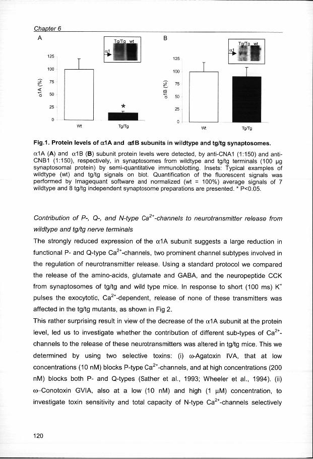
ProteinProtein expression of the cc1A (P, Q) and <x1B (N) Ca2+-channel subunits in
presynapticpresynaptic terminals from wildtype and tg/tg mice
Inn order to study Ca2+-channel expression at the presynaptic level, we purified nerve
terminals,, synaptosomes, from forebrain of tg/tg mice and their wildtype littermates.
Thee effect of the mutation in the a1A pore-forming subunit on protein expression of
thee different Ca2+-channel subtypes was determined by semi-quantitative Western
blotting.. The relative protein levels of the a1A (P, Q) and a1B (N) subunits of the
Ca2+-channelss were quantified in 7 wildtype and 8 tg/tg synaptosome preparations.
Withh the polyclonal antibody CNA1 both the 210 and 190 kDa form of the a1A
subunitt were detected (Westenbroek et al., 1995). Quantification of the 190 kDa
bandd showed a strong decrease of 85 % in the tg/tg synaptosomes as compared to
thee expression level in wildtype synaptosomes (Fig. 1A). The 210 kDa band was
overexposedd in the wildtype samples and therefore not quantified, but was also
clearlyy decreased in tg/tg compared to wildtype. After incubation with the polyclonal
antibodyy CNB1 a faint band corresponding to the low molecular weight and not to the
highh molecular weight form of the a1B subunit (Westenbroek et al., 1992) was
detectedd at around 190 kDa under our conditions. In contrast to the a1A subunit, no
differencee in expression of the low molecular weight form of the a1 B subunit between
tg/tgg and wildtype terminals was detected (Fig. 1B).
119 9
ChapterChapter 6
125 5
100 0
gg 75
< < "aa 50
25 5
0 0
Tg/Tg g
125 5
100 0
CD D
ÜÜ 50
25 5
0 0
Tgfig g
Fig.1 .. Protei n level s of a1A and ocdB subunit s in wildtyp e and tg/t g synaptosomes .
a1AA (A) and a1B (B) subunit protein levels were detected, by anti-CNA1 (1:150) and anti-CNB11 (1:150), respectively, in synaptosomes from wildtype and tg/tg terminals (100 ug synaptosomall protein) by semi-quantitative immunoblotting. Insets: Typical examples of wildtypee (wt) and tg/tg signals on blot. Quantification of the fluorescent signals was performedd by Imagequant software and normalized (wt = 100%) average signals of 7 wildtypee and 8 tg/tg independent synaptosome preparations are presented. * P<0.05.
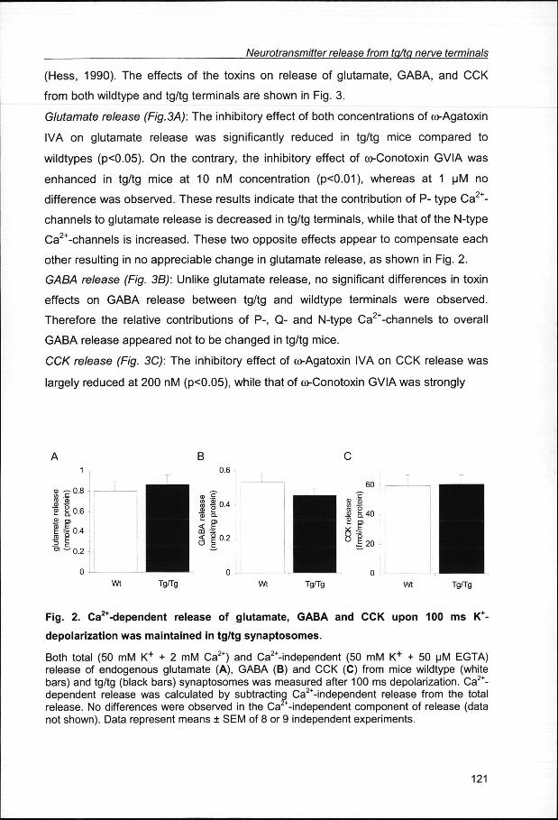
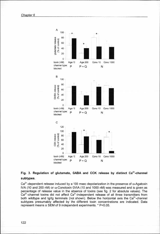
ContributionContribution of P-, Q-, and N-type Ca2+-channels to neurotransmitter release from
wildtypewildtype and tg/tg nerve terminals
Thee strongly reduced expression of the a1A subunit suggests a large reduction in
functionall P- and Q-type Ca2+-channels, two prominent channel subtypes involved in
thee regulation of neurotransmitter release. Using a standard protocol we compared
thee release of the amino-acids, glutamate and GABA, and the neuropeptide CCK
fromm synaptosomes of tg/tg and wild type mice. In response to short (100 ms) K+
pulsess the exocytotic, Ca2+-dependent, release of none of these transmitters was
affectedd in the tg/tg mutants, as shown in Fig 2.
Thiss rather surprising result in view of the decrease of the a1 A subunit at the protein
level,, led us to investigate whether the contribution of different sub-types of Ca2+-
channelss to the release of these neurotransmitters was altered in tg/tg mice. This we
determinedd by using two selective toxins: (i) o>Agatoxin IVA, that at low
concentrationss (10 nM) blocks P-type Ca2+-channels, and at high concentrations (200
nM)) blocks both P- and Q-types (Sather et al., 1993; Wheeler et al., 1994). (ii)
co-Conotoxinn GVIA, also at a low (10 nM) and high (1 u.M) concentration, to
investigatee toxin sensitivity and total capacity of N-type Ca2+-channels selectively
120 0
NeurotransmitterNeurotransmitter release from tg/tg nerve terminals
(Hess,, 1990). The effects of the toxins on release of glutamate, GABA, and CCK
fromm both wildtype and tg/tg terminals are shown in Fig. 3.
GlutamateGlutamate release (Fig.3A): The inhibitory effect of both concentrations of co-Agatoxin
IVAA on glutamate release was significantly reduced in tg/tg mice compared to
wildtypess (p<0.05). On the contrary, the inhibitory effect of co-Conotoxin GVIA was
enhancedd in tg/tg mice at 10 nM concentration (p<0.01), whereas at 1 uM no
differencee was observed. These results indicate that the contribution of P- type Ca2+-
channelss to glutamate release is decreased in tg/tg terminals, while that of the N-type
Ca2+-channelss is increased. These two opposite effects appear to compensate each
otherr resulting in no appreciable change in glutamate release, as shown in Fig. 2.
GABAGABA release (Fig. 3B): Unlike glutamate release, no significant differences in toxin
effectss on GABA release between tg/tg and wildtype terminals were observed.
Thereforee the relative contributions of P-, Q- and N-type Ca2+-channels to overall
GABAA release appeared not to be changed in tg/tg mice.
CCKCCK release (Fig. 3C): The inhibitory effect of co-Agatoxin IVA on CCK release was
largelyy reduced at 200 nM (p<0.05), while that of co-Conotoxin GVIA was strongly
Wtt Tg/Tg Wt Tg/Tg Wt Tg/Tg
Fig.. 2. Ca2+-dependent releas e of glutamate , GABA and CCK upo n 100 ms K+-
depolarizatio nn was maintaine d in tg/t g synaptosomes .
Bothh total (50 mM K+ + 2 mM Ca2+) and Ca2+-independent (50 mM K+ + 50 uM EGTA) releasee of endogenous glutamate (A), GABA (B) and CCK (C) from mice wildtype (white bars)) and tg/tg (black bars) synaptosomes was measured after 100 ms depolarization. Ca2+-dependentt release was calculated by subtracting Ca2+-independent release from the total release.. No differences were observed in the Ca -independent component of release (data nott shown). Data represent means SEM of 8 or 9 independent experiments.
121 1
ChapterChapter 6
100 0
8=.. 80
CDD O
88 £ "5>> c 60 -- o CDD u
II o 40 33 20
0 0 toxinn (nM): Aga10 channell type: p blocked d
II III Aga2000 Cono10 Cono1000
PP + Q N
100 0
80 0
| || 60
aii o 40
3 £ £ 20 0 l .. l l
toxinn (nM): Aga 10 channell type: p blocked d
Agaa 200 Cono 10 Cono 1000
PP + Q N
120 0
100 0
SS t 80 ££ 8 60
II | 40 20 0
0 0 toxinn (nM): Aga 10 channell type: p blocked d
111 1 Agaa 200 Cono 10 Cono 1000
PP + Q N
Fig .. 3. Regulatio n of glutamate , GABA and CCK releas e by distinc t Ca2+-channel
subtypes . .
Ca2+-dependentt release induced by a 100 msec depolarization in the presence of co-Agatoxin IVAA (10 and 200 nM) or co-Conotoxin GVIA (10 and 1000 nM) was measured and is given as percentagee of release value in the absence of toxins (see fig. 2 for absolute values). The Ca2+-channell toxins did not affect Ca2+-independent release of all three transmitters from bothh wildtype and tg/tg terminals (not shown). Below the horizontal axis the Ca2+-channel subtypess presumably affected by the different toxin concentrations are indicated. Data representt means SEM of 9 independent experiments. * P<0.05.
122 2
NeurotransmitterNeurotransmitter release from to/tp nerve terminals
enhanced,, particularly at 1|jM (p<0.01). This indicates that in tg/tg terminals the
contributionn of the P- and Q-type Ca2+-channels to CCK release is decreased, mainly
thatt of the latter considering the larger difference at 200 nM. And, similar to
glutamatee release, the dependence on N-type Ca2+-channels for CCK release was
enhancedd in terminals from tg/tg mice.
DISCUSSION N
Thee most remarkable finding in the present study was the strong selective reduction
off expression of P-, Q- type Ca2+-channels by 85 % in forebrain nerve terminals of
tg/tgg mice, while the overall release of the amino acids glutamate and GABA and the
neuropeptidee CCK was not significantly altered.
ReducedReduced protein expression of the mutated crfA (P, Q) subunit in terminals of tg/tg
mice mice
Thee strong reduction in a1A subunit expression, as indicated by the immunoblots,
evidentlyy is the result of the mutation of this subunit in the tg/tg mice. Since mRNA
levelss of the a1A subunit did not appear to be affected in tg/tg mice (Campbell and
Hess,, 1998), a posttranscriptional defect might lead to mistargeting of P-, Q-type
Ca2+-channelss to the plasma membrane of the nerve terminals. Alternatively,
acceleratedd turnover of the protein might explain its reduced expression. Since the
crtt A subunit forms the ion-conducting pore of P- and Q-type Ca2+-channels (Bourinet
ett al., 1999), the 85% reduction in expression of this subunit implicates that the
numberr of functional P-,Q-type Ca2+-channels is reduced in tg/tg terminals.
Interestingly,, expression of N-type Ca2+-channels appeared not to be changed, since
noo quantitative change in oc1B subunit between tg/tg and wildtype terminals could be
detectedd on the immunoblots.
ContributionContribution of Ca2"-channel subtypes to release in tg/tg and wildtype terminals
Thee strongly reduced expression of P-, Q-type Ca2+-channels in tg/tg terminals would
predictt a reduction in neurotransmitter release as well. However, this prediction was
nott confirmed in our experiments, since overall release of glutamate, GABA and CCK
123 3
ChapterChapter 6
wass maintained in tg/tg mice terminals. This is in accordance with the observation
thatt synaptic transmission in hippocampal CA3-CA1 synapses of tg/tg mice remained
intactt (Qian and Noebels, 2000). In search for an explanation of this apparent
paradox,, we determined the differences in contribution of the P-, Q- and N-type Ca2+-
channelss to the release from wildtype and tg/tg terminals, using specific toxins to
blockk the distinct Ca2+-channel subtypes. This analysis revealed a number of
interestingg features regarding the contribution of Ca2+-channel subtypes to the
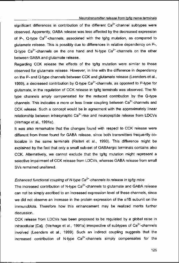
releasee of different neurotransmitters. As indicated in table 1, the reduced
contributionn of mainly P-type Ca2+-channels to glutamate release and of Q-type Ca2+-
channelss to CCK release is compensated by increased contribution of N-type Ca2+-
channelss in the terminals of tg/tg mice.
Ann unexpected finding was that glutamate and GABA release are regulated in
differentt ways. In tg/tg terminals the contribution of mainly P-type Ca2+-channels to
glutamatee release was reduced compared to that in wildtype terminals, which is in
agreementt with a reduced expression of the cc1A subunit. The fact that this did not
resultt in reduced glutamate release can be accounted for by the increased
contributionn of N-type Ca2+-channels. These findings are in line with the observations
off Qian and Noebels (2000) in excitatory CA3-CA1 synapses. For GABA release no
Tabl ee 1. Differences in Ca2+-channel subtypes between tg/tg and wildtype mice
Caz+-channel l subtype e
oc1A A P,, Q
cüAgatxx IVA
a1B B N N
coCtxx GVIA
Protein n expression n
85%% >U
Release e
Glutamate e
26.33 % U (10nM,, P)
33.77 % TT (10nM) )
GABA A
= =
= =
CCK K
377 % U (200nM,, Q)
63.22 % TT O M M ) )
Differencess in effects on protein expression levels of oc1A and a1B Ca2+-channel subunits
andd on the contribution of P,Q and N-type Ca2+-channels to release of glutamate, GABA and
CCKK in tg/tg mice terminals, calculated from the data from fig. 1 and 3. Toxin concentrations
and,, for co-Agatoxin IVA, the channel primarily affected are indicated between parentheses. =
noo effect.
124 4
NeurotransmitterNeurotransmitter release from tg/tg nerve terminals
significantt differences in contribution of the different Ca2+-channel subtypes were
observed.. Apparently, GABA release was less affected by the decreased expression
off P-, Q-type Ca2+-channels, associated with the tg/tg mutation, as compared to
glutamatee release. This is possibly due to differences in relative dependency on P-,
Q-typee Ca2+-channels on the one hand and N-type Ca2+-channels on the other
betweenn GABA and glutamate release.
Regardingg CCK release the effects of the tg/tg mutation were similar to those
observedd for glutamate release. However, in line with the difference in dependency
onn the P- and Q-type channels between CCK and glutamate release (Leenders et al.,
1999),, a decreased contribution by Q-type Ca2+-channels, as opposed to P-type for
glutamate,, in the regulation of CCK release in tg/tg terminals was observed. The re-
typee channels amply compensated for the reduced contribution by the Q-type
channels.. This indicates a more or less linear coupling between Ca2+-channels and
CCKK release. Such a concept would be in agreement with the approximately linear
relationshipp between intrasynaptic Ca2+-rise and neuropeptide release from LDCVs
(Verhageetal.,, 1991a).
Itt was also remarkable that the changes found with respect to CCK release were
differentt from those found for GABA release, since both transmitters frequently co-
localizee in the same terminals (Raiteri et al., 1993). This difference might be
explainedd by the fact that only a small sub-set of GABAergic terminals contains also
CCK.. Alternatively, we cannot exclude that the tg/tg mutation might represent a
selectivee impairment of CCK release from LDCVs, whereas GABA release from small
SVss remained unaltered.
EnhancedEnhanced functional coupling of N-type Ca2* -channels to release in tg/tg mice
Thee increased contribution of N-type Ca2+-channels to glutamate and GABA release
cann not be simply ascribed to an increased expression level of these channels, since
wee did not observe an increase in the protein expression of the a1B subunit on the
immunoblots.. Therefore how this enhancement may be realized merits further
discussion. .
CCKK release from LDCVs has been proposed to be regulated by a global raise in
intracellularr [Ca]i (Verhage et al., 1991a) irrespective of subtypes of Ca2+-channels
involvedd (Leenders et al., 1999). Such an indirect coupling suggests that the
increasedd contribution of N-type Ca2+-channels simply compensates for the
125 5
ChapterChapter 6
decreasedd Ca2+-influx through Q-type Ca2+-channels. This also implies that normally
thee number of Ca2+-channels expressed on the terminals exceeds the minimal
numberr of Ca2+-channels required to reach the threshold for triggering of release
fromm LDCVs.
Ann enhanced association of N-type Ca2+-channels to pre-docked readily releasable
SVss might account for the increased functional coupling of these channels to
glutamatee release in tg/tg terminals. The observation that 10 nM co-Conotoxin GVIA
exertedd already a maximal inhibition of about 50% in tg/tg terminals, whereas in
wildtypee terminals 1 uM was required, would be in agreement with such a functional
compensation.. Alternatively, a difference in the N-type Ca2+-channel, for instance
expressionn of a different splice variant of the a1B subunit or interaction with a
differentt p subunit, could account for the observed difference in inhibition by co-
Conotoxinn GVIA. There is, however, no indication that the affinity for co-Conotoxin
GVIAA is different between splice variants of the oc1B subunit (Lin et al., 1999).
Moreover,, notwithstanding the similar total expression level of the a1B subunit, local
densityy of N-type Ca2+-channels at the release site in the active zone may be
increasedd in tg/tg terminals. In conclusion, the precise molecular and/or spatial
factorss that account for the observed functional compensation by the N-type Ca2+-
channelss needs further investigation.
ImplicationsImplications for the neurological phenotype of tg/tg mice
Thee cellular basis for the neurological deficits, such as absence epilepsy and ataxia,
occurringg in tg/tg mice (Noebels and Sidman, 1979) is still not resolved. The (co-)
expressionn patterns of the different Ca2+-channel subtypes in several brain regions
mayy be of major importance to circumvent severe impairment of synaptic
transmission.. Our results showed a large reduction in the expression level of the
mutatedd tx1 A subunit in forebrain nerve terminals. The fact that we purified terminals
fromm the whole forebrain does not allow to make a differential analysis of separate
brainn regions. It is possible, even likely, that nerve terminals differ in their expression
off Ca2+-channel subtypes, particularly among different brain areas. Nerve terminals
thatt lack N-type Ca2+-channels would not be able to compensate a decreased
contributionn of P-, Q-type Ca2+-channels, which then could result in a decrease in
neurotransmitterr release from these terminals. This might be the case for certain
126 6
NeurotransmitterNeurotransmitter release from tg/tp nerve terminals
GABAergicc terminals in the hippocampus (Poneer et al., 1997). Whether the same
phenomenonn occurs in other systems is not well known. In the thalamus of tg/tg mice
impairedd excitatory, but not inhibitory, neurotransmission was reported (Caddick et
al.,, 1999) but whether this corresponds to a change in the composition of presynaptic
Ca2+-channelss is not known.
Couplingg of depolarization to neurotransmitter release is not the only function of the
P-,, Q-type Ca2+-channels, since these subtypes are also expressed in cell soma and
dendritess (Westenbroek et al., 1995). Reduced expression of the oc1A subunit on
thesee neuronal compartments, similar to what we found in the presynaptic terminals,
couldd lead to a drastically reduced Ca2+-influx, thereby disturbing the intracellular
Ca2++ homeostasis and affecting many down-stream cellular processes, such as for
instancee gene expression and protein translation, which may play an important role in
progressionn of symptoms in tg/tg mice.
ACKNOWLEDGEMENT S S
Thee authors wish to thank Mr J. A. van der Zwet for breeding the tg/tg mice, prof. V. Wiegant
andd Mrs A. Frankhuijzen of the Rudolf Magnus Institute, Utrecht University for assistance
withh the CCK analysis. A.G.M. L. is supported by grant 903-42-016 of the Netherlands
Organizationn of Scientific Research.
127 7

ChapterChapter 6
128 8